problems in staging breast carcinoma · reporting of breast specimens from patients who have...
TRANSCRIPT
Problems in staging breast carcinoma
Primary systemic therapy (PST)Primary systemic therapy (PST)of breast carcinoma – pathologists’ tasks
Dr. Janina Kulka, 2nd Department of Pathology,Semmelweis University Budapest
Austro-Hungarian Pathology Congress 1-3 October 2009

PST or neoadjuvant therapy
• Systemic oncological treatment prior to surgical intervention
• Formerly used only in T4, inflammatory carcinomacarcinoma
• In recent years used in earlier stage disease to achieve– Tumor size reduction, thus the possibility of breast
conserving surgery– Longer disease free survival– Longer overall survival

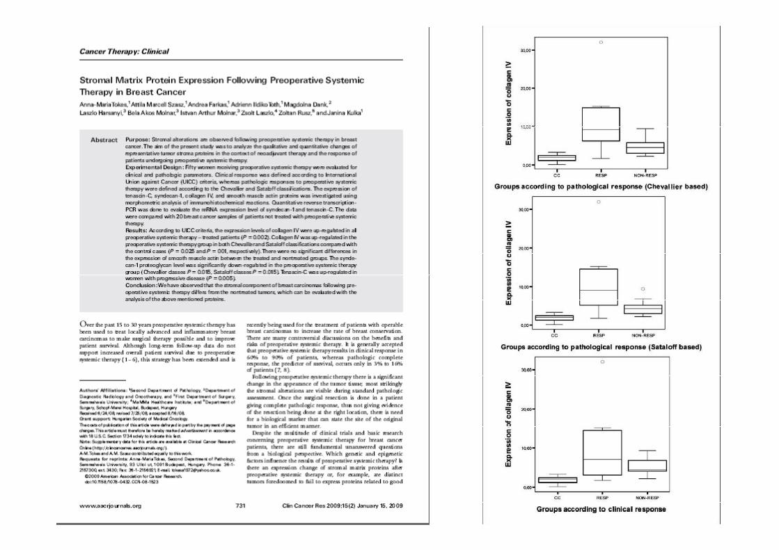
Effect of 6 cycles of FEC100 treatment
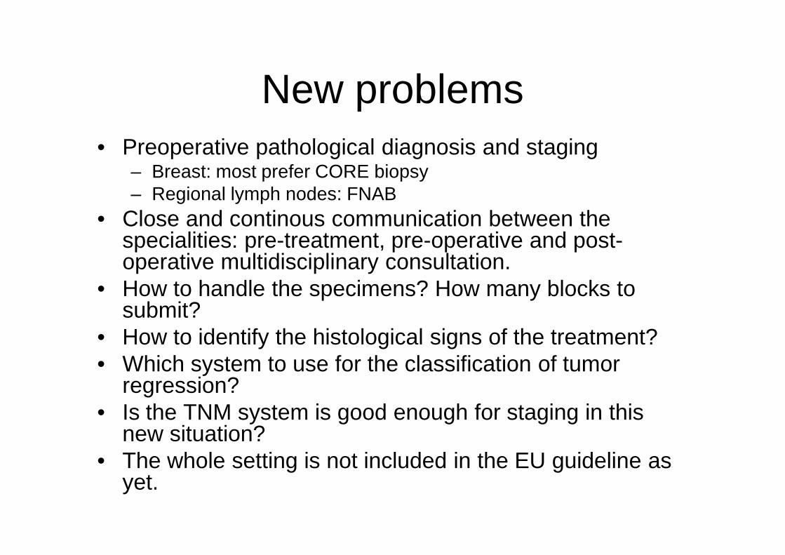
New problems• Preoperative pathological diagnosis and staging
– Breast: most prefer CORE biopsy– Regional lymph nodes: FNAB
• Close and continous communication between the specialities: pre-treatment, pre-operative and post-operative multidisciplinary consultation.
• How to handle the specimens? How many blocks to • How to handle the specimens? How many blocks to submit?
• How to identify the histological signs of the treatment?• Which system to use for the classification of tumor
regression?• Is the TNM system is good enough for staging in this
new situation?• The whole setting is not included in the EU guideline as
yet.

Recent comprehensive literature
• Pinder SE , et al Histopathology 2007;50:409-17– Laboratory handling and histology
reporting of breast specimens from patients who have received neoadjuvant chemotherapy
• Sahoo S , Lester SC. Arch Pathol Lab Med • Sahoo S , Lester SC. Arch Pathol Lab Med 2009;133:633-642– Pathology of breast carcinoma after
neoadjuvant chemotherapy• Pusztai L POR 2008;14:169-171
– Preoperative systemic chemotherapy and pathologic assessment of response

• Alvarado-Cabrero I et al Ann Diagn Pathol 2009;13:151-157– Incidence of pathologic complete response in women
treated with preoperative chemotherapy for locally advanced breast cancer: correlation of histology, hormone receptor status, Her2/neu, and gross pathologic findings
• Jeruss JJ et al. Cancer Res 2008;68:6477– Staging of breast cancer in the neoadjuvant setting
(„Clinical-Pathological Scoring System” CPS)• Kurosumi M Breast Cancer 2006;13:254• Kurosumi M Breast Cancer 2006;13:254
– Significance and problems in evaluations of pathological responses to neoadjuvant therapy for breast cancer
• EU guideline next edition will include a chapter on this subject, the draft by Prof. Angelika Reiner-Concin now available only for the EWBSP members
• http://www3.mdanderson.org/app/medcalc/index.cfm?pagename=jsconvert3 –calculator of residual cancer burden (RCB)
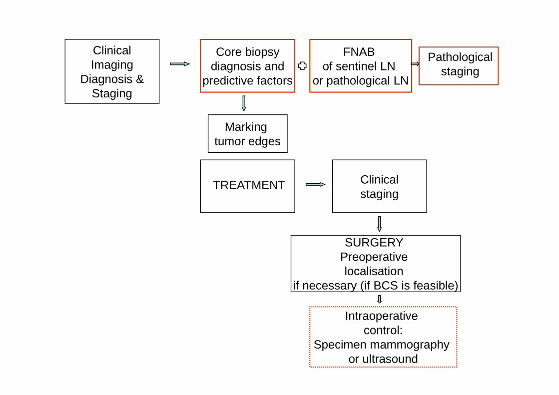
ClinicalImaging
Diagnosis &Staging
Core biopsydiagnosis and
predictive factors
TREATMENT Clinicalstaging
Marking tumor edges
FNAB of sentinel LN
or pathological LN
Pathologicalstaging
SURGERY Preoperative localisation
if necessary (if BCS is feasible)
staging
Intraoperative control:
Specimen mammography or ultrasound
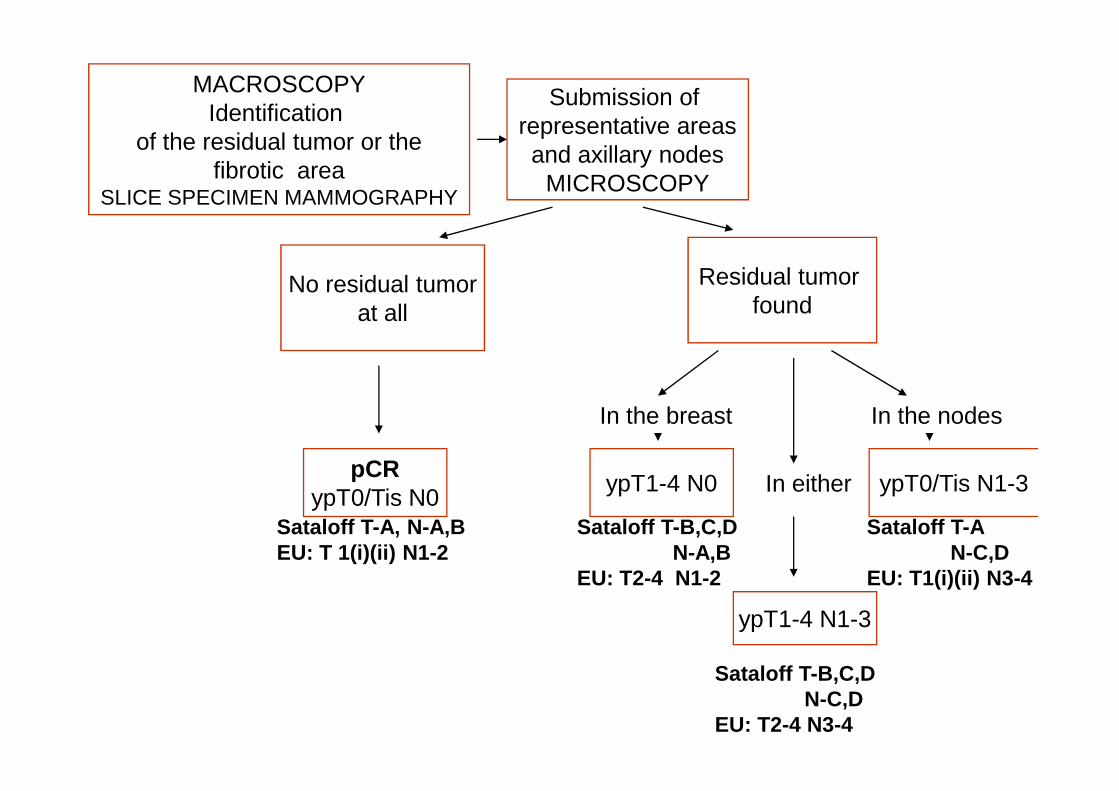
MACROSCOPYIdentification
of the residual tumor or thefibrotic area
SLICE SPECIMEN MAMMOGRAPHY
Submission of representative areasand axillary nodes
MICROSCOPY
No residual tumorat all
Residual tumor found
pCRypT0/Tis N0
In the breast In the nodes
In either ypT0/Tis N1-3ypT1-4 N0
ypT1-4 N1-3
Sataloff T-A, N-A,BEU: T 1(i)(ii) N1-2
Sataloff T-B,C,DN-A,B
EU: T2-4 N1-2
Sataloff T-B,C,DN-C,D
EU: T2-4 N3-4
Sataloff T-AN-C,D
EU: T1(i)(ii) N3-4

Macroscopical examination
• The tumor bed may be– Rubbery fibrous tissue– Fleshy nodule(s)– Bright yellow circumscribed area– May not be recognisable– May not be recognisable
• Sampling– If tumor is visible: 1block/cm– If residual tumor is small: embed the entire area– No tumor found initially: further extensive sampling of
tumor bed may be necessary– Thorough sampling of margins
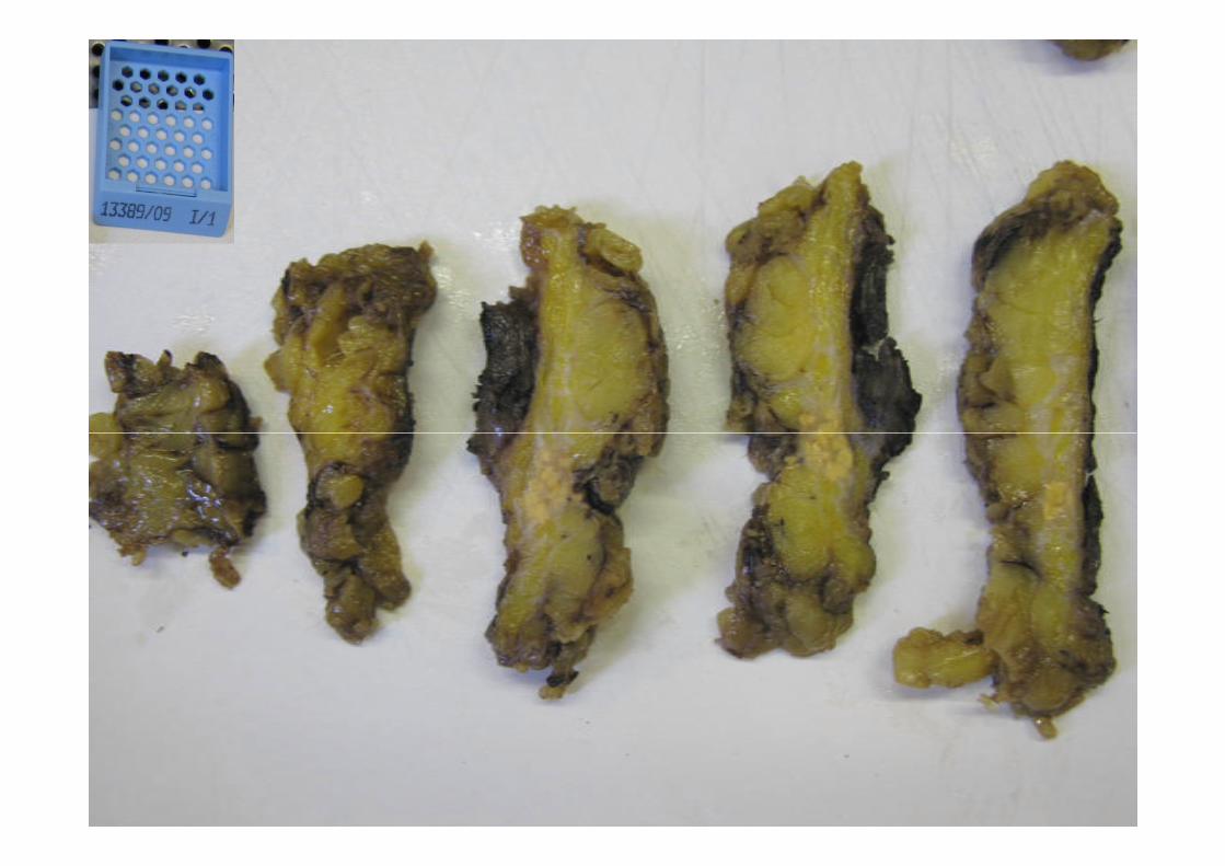

• Lymph nodes– Presence of grossly visible scar may indicate
complete response in the LN

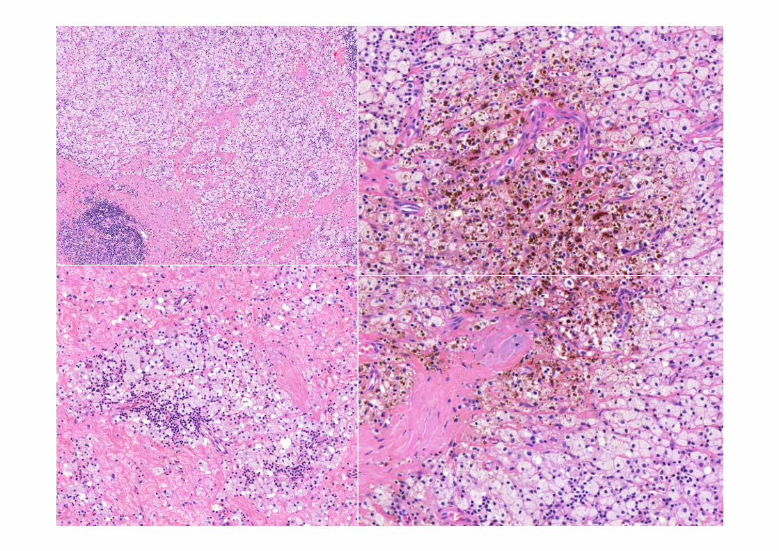
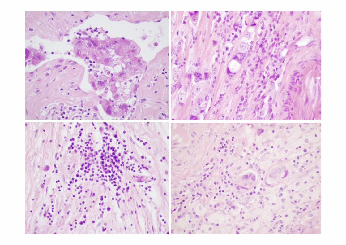
Microscopic examination
• Tumor bed– Hyalinised, fibro-elastotic, vascular stroma– Aggregates of foamy histiocytes and lymphocytes– Haemosiderin pigment– Cholesterol clefts– Cholesterol clefts
• Residual tumor cells– May look identical to that seen in the core biopsy– Enlargement, vacuolisation of the cytoplasm– Pleiomorphic, bizarr nuclei– Decreased mitotic activity– CK immunohistochemistry may be necessary to
identify them (both breast and LNs)


COLLAGENE IV IMMUNOHISTOCHEMISTRY

TENASCIN IMMUNOHISTOCHEMISTRY

• Predictive factors– Must be examined in the pretreatment core-
biopsy– Yet uncertain if re-testing is necessary in the – Yet uncertain if re-testing is necessary in the
excision specimen – contradictory data published

ER/PR Ki67
28 year old woman with no family history of breast or any other cancer
CB11 CK5/61,5 Her2 signal/Chr.17
Assessed as ER/PR/HER2 negative tumor

Assessment of tumor response

Kurosumi M. Breast Cancer Vol. 13 No. 3 July 2006

Sahoo S and Lester CArch Pathol Lab Med—Vol 133, April 2009

Suggested assessment of tumor response
• 1. Complete pathological response– i) no residual carcinoma– ii) DCIS present (no invasive carcinoma)
• 2. Partial response to therapy– i) minimal residual disease: <10% of tumor remained – i) minimal residual disease: <10% of tumor remained – ii) evidence of response, but 10-50% of tumor
remained– iii) >50% of tumor remained, cellularity similar to that
seen in the core biopsy, some features of response present
• 3. No evidence of response
Pinder S et al. Histopathology 2007


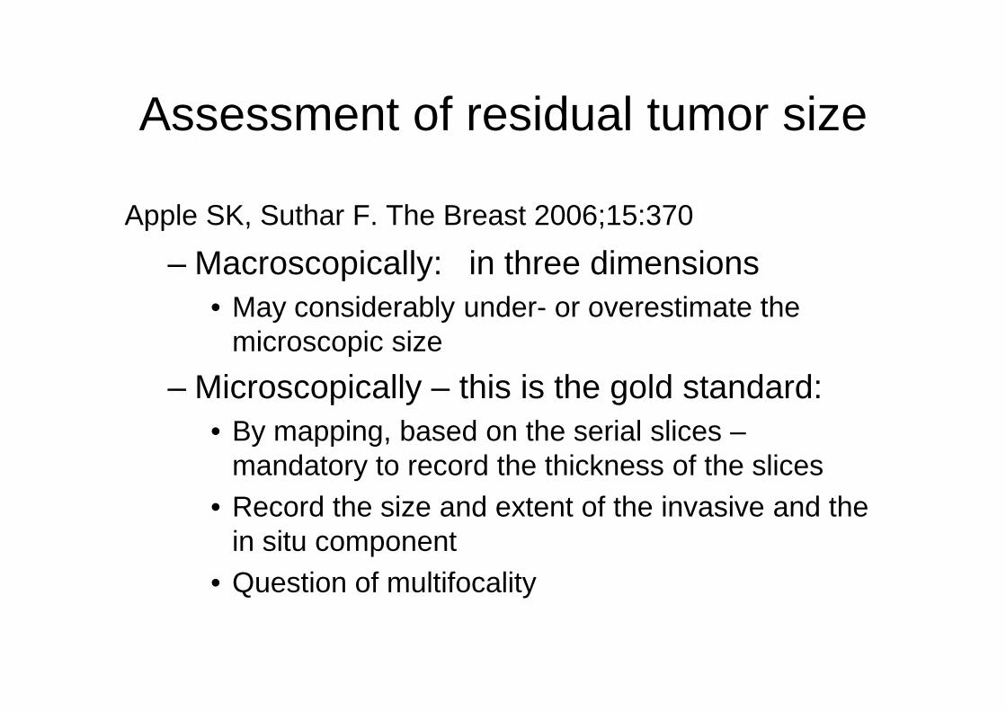
Assessment of residual tumor size
Apple SK, Suthar F. The Breast 2006;15:370
– Macroscopically: in three dimensions• May considerably under- or overestimate the
microscopic sizemicroscopic size
– Microscopically – this is the gold standard: • By mapping, based on the serial slices –
mandatory to record the thickness of the slices• Record the size and extent of the invasive and the
in situ component• Question of multifocality
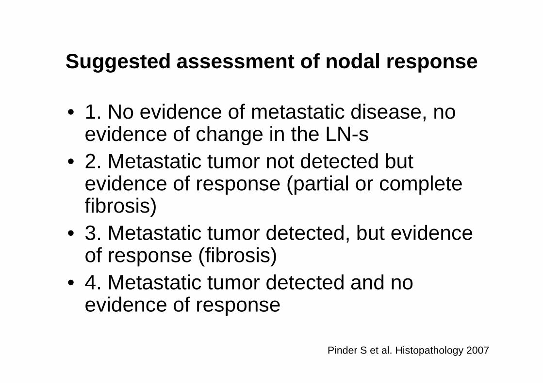
Suggested assessment of nodal response
• 1. No evidence of metastatic disease, no evidence of change in the LN-s
• 2. Metastatic tumor not detected but evidence of response (partial or complete evidence of response (partial or complete fibrosis)
• 3. Metastatic tumor detected, but evidence of response (fibrosis)
• 4. Metastatic tumor detected and no evidence of response
Pinder S et al. Histopathology 2007


Sentinel nodes in this setting• Sentinel lymph node biopsy after preoperative chemo therapy for
breast cancer: findings from the Austrian Sentinel Node Study Group.
Tausch C et al. Ann Surg Oncol 2008 Dec;15(12):3378-83.The results of SLNB after PC are comparable to the results of SLNB without PC.Further investigation in a prospective setting is warranted to confirm these promising results.results.
• Accuracy of sentinel node biopsy after neoadjuvant chemotherapy in breast cancer patients: A systematic review.van Deurzen C et al. Eur J Cancer 2009 Aug 26. [Epub ahead of print]
There is a potential role for SN biopsy following NAC which could be considered on an individual basis. However, there is insufficient evidence to recommend this as a standard procedure. Further research with subgroup analysis using variables reported to be associated with decreased SN accuracy is required in order to clearly define its value in the subgroups of breast cancer patients.
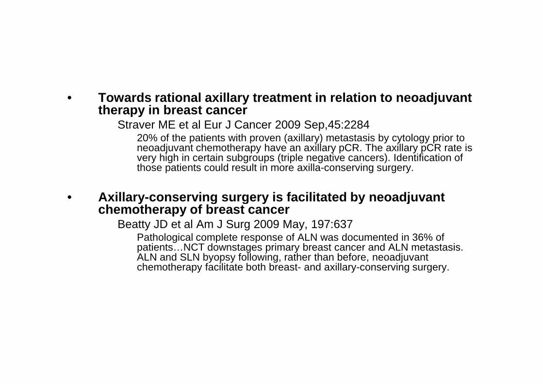
• Towards rational axillary treatment in relation to neoadjuvant therapy in breast cancer
Straver ME et al Eur J Cancer 2009 Sep,45:228420% of the patients with proven (axillary) metastasis by cytology prior to neoadjuvant chemotherapy have an axillary pCR. The axillary pCR rate is very high in certain subgroups (triple negative cancers). Identification of those patients could result in more axilla-conserving surgery.
• Axillary -conserving surgery is facilitated by neoadjuvant • Axillary -conserving surgery is facilitated by neoadjuvant chemotherapy of breast cancer
Beatty JD et al Am J Surg 2009 May, 197:637Pathological complete response of ALN was documented in 36% of patients…NCT downstages primary breast cancer and ALN metastasis. ALN and SLN byopsy following, rather than before, neoadjuvant chemotherapy facilitate both breast- and axillary-conserving surgery.

„Pathologic staging (i.e. AJCC) has not been validated for patients receiving neoadjuvant chemotherapy.”
and
It is unclear, whether the initial clinical stage or the final „It is unclear, whether the initial clinical stage or the final pathological stage is more meaningful in terms of prognosis and… there is no current methodology for incorporating the information on clinical and pathologic response to the chemotherapy into the stage grouping.”
Jeruss JS et al. Cancer Res 2008:68:6477
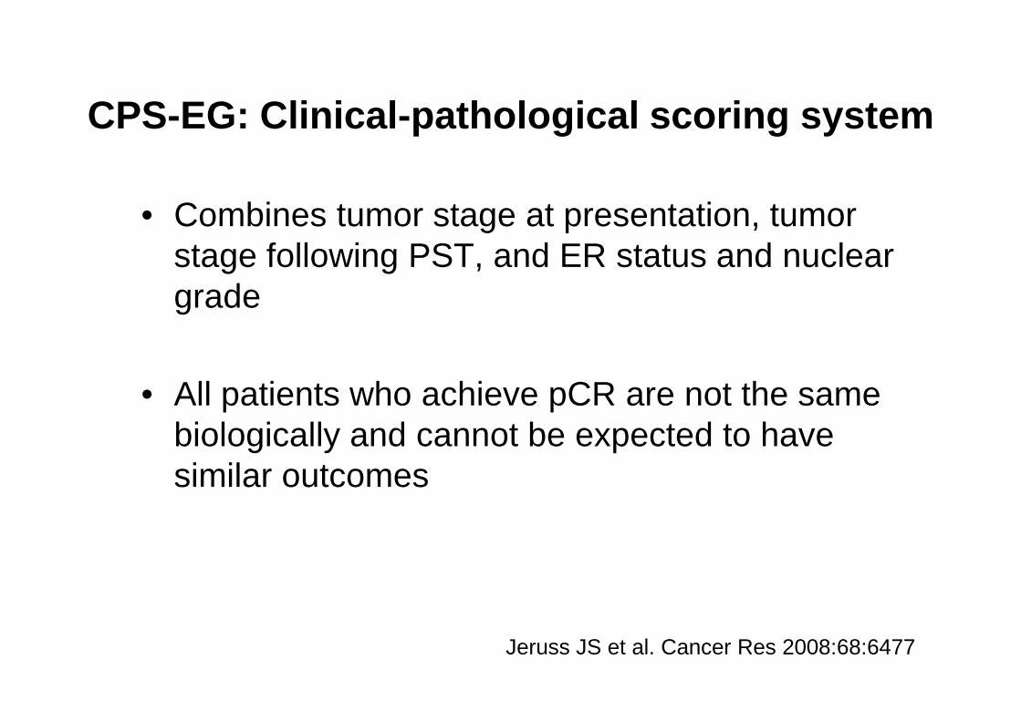
CPS-EG: Clinical-pathological scoring system
• Combines tumor stage at presentation, tumor stage following PST, and ER status and nuclear grade
• All patients who achieve pCR are not the same biologically and cannot be expected to have similar outcomes
Jeruss JS et al. Cancer Res 2008:68:6477


Acknowledgements