procedure intraaortic balloon pump management
TRANSCRIPT
431
Section Seven Circulatory Assist Devices
PROCEDURE
52
Intraaortic Balloon Pump Management John P. Harper
PURPOSE: Intraaortic balloon pump therapy is designed to increase coronary artery perfusion, which increases myocardial oxygen supply, and decrease afterload and myocardial workload, which decreases myocardial oxygen demand.
PREREQUISITE NURSING KNOWLEDGE • Knowledge of the anatomy and physiology of the cardio-
vascular system is needed. • Understanding of the principles of hemodynamic monitor-
ing, electrophysiology, dysrhythmias, and coagulation is necessary.
• Clinical and technical competence related to the use of the intraaortic balloon pump (IABP) is needed.
• Advanced cardiac life support knowledge and skills are necessary.
• Indications for IABP therapy are as follows: ❖ Cardiogenic shock ❖ Refractory unstable angina ❖ Acute myocardial infarction (MI) complicated by left-
ventricular failure 3,13,28,30 ❖ Recurrent ventricular dysrhythmias as a result of
ischemia 18 ❖ Support before, during, and after coronary artery
bypass graft surgery 2,31 ❖ Support before, during, and after coronary artery
angioplasty or additional interventional cardiology procedures for patients at high risk 4,11
❖ Mechanical complications of acute MI, including aortic stenosis, mitral stenosis, mitral valvuloplasty, mitral insuffi ciency, ventricular septal defect, and left-ventricular aneurysm
❖ Intractable ventricular dysrhythmias 14,20 ❖ Bridge to cardiac transplantation, ventricular-assist
devices, or total artifi cial hearts ❖ Cardiac injury, including contusion and coronary artery
tears ❖ Septic shock ❖ Patient at high risk undergoing noncardiac surgery 20,31
• Contraindications to IABP therapy are as follows: ❖ Moderate to severe aortic insuffi ciency ❖ Thoracic and abdominal aortic aneurysms
• The relative value of IABP therapy in the presence of severe aortoiliac disease, major coagulopathies, and ter-minal disease should be evaluated individually.
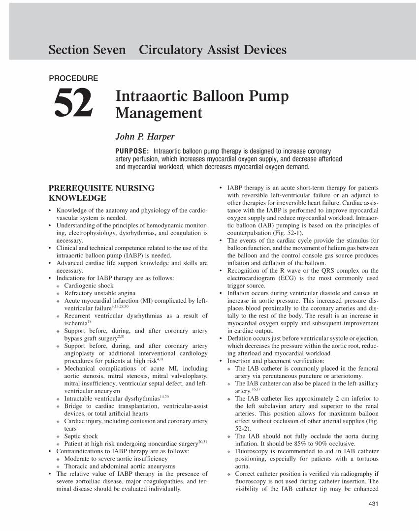
• IABP therapy is an acute short-term therapy for patients with reversible left-ventricular failure or an adjunct to other therapies for irreversible heart failure. Cardiac assis-tance with the IABP is performed to improve myocardial oxygen supply and reduce myocardial workload. Intraaor-tic balloon (IAB) pumping is based on the principles of counterpulsation ( Fig. 52-1 ).
• The events of the cardiac cycle provide the stimulus for balloon function, and the movement of helium gas between the balloon and the control console gas source produces infl ation and defl ation of the balloon.
• Recognition of the R wave or the QRS complex on the electrocardiogram (ECG) is the most commonly used trigger source.
• Infl ation occurs during ventricular diastole and causes an increase in aortic pressure. This increased pressure dis-places blood proximally to the coronary arteries and dis-tally to the rest of the body. The result is an increase in myocardial oxygen supply and subsequent improvement in cardiac output.
• Defl ation occurs just before ventricular systole or ejection, which decreases the pressure within the aortic root, reduc-ing afterload and myocardial workload.
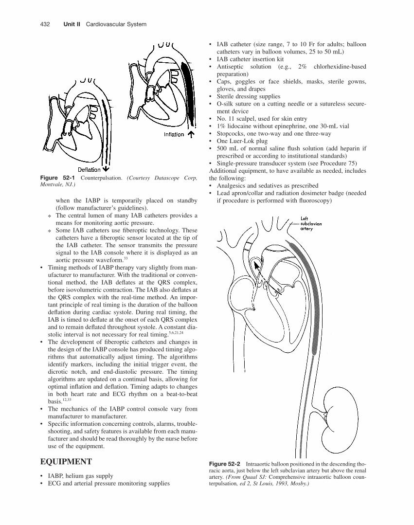
• Insertion and placement verifi cation: ❖ The IAB catheter is commonly placed in the femoral
artery via percutaneous puncture or arteriotomy. ❖ The IAB catheter can also be placed in the left-axillary
artery. 16,17 ❖ The IAB catheter lies approximately 2 cm inferior to
the left subclavian artery and superior to the renal arteries. This position allows for maximum balloon effect without occlusion of other arterial supplies ( Fig. 52-2 ).
❖ The IAB should not fully occlude the aorta during infl ation. It should be 85% to 90% occlusive.
❖ Fluoroscopy is recommended to aid in IAB catheter positioning, especially for patients with a tortuous aorta.
❖ Correct catheter position is verifi ed via radiography if fl uoroscopy is not used during catheter insertion. The visibility of the IAB catheter tip may be enhanced
432 Unit II Cardiovascular System
• IAB catheter (size range, 7 to 10 Fr for adults; balloon catheters vary in balloon volumes, 25 to 50 mL)
• IAB catheter insertion kit • Antiseptic solution (e.g., 2% chlorhexidine-based
preparation) • Caps, goggles or face shields, masks, sterile gowns,
gloves, and drapes • Sterile dressing supplies • O-silk suture on a cutting needle or a sutureless secure-
ment device • No. 11 scalpel, used for skin entry • 1% lidocaine without epinephrine, one 30-mL vial • Stopcocks, one two-way and one three-way • One Luer-Lok plug • 500 mL of normal saline fl ush solution (add heparin if
prescribed or according to institutional standards) • Single-pressure transducer system (see Procedure 75 ) Additional equipment, to have available as needed, includes the following: • Analgesics and sedatives as prescribed • Lead apron/collar and radiation dosimeter badge (needed
if procedure is performed with fl uoroscopy)
Figure 52-1 Counterpulsation. (Courtesy Datascope Corp, Montvale, NJ.)
Figure 52-2 Intraaortic balloon positioned in the descending tho-racic aorta, just below the left subclavian artery but above the renal artery. (From Quaal SJ: Comprehensive intraaortic balloon coun-terpulsation , ed 2, St Louis, 1993, Mosby.)
when the IABP is temporarily placed on standby (follow manufacturer ’ s guidelines).
❖ The central lumen of many IAB catheters provides a means for monitoring aortic pressure.
❖ Some IAB catheters use fi beroptic technology. These catheters have a fi beroptic sensor located at the tip of the IAB catheter. The sensor transmits the pressure signal to the IAB console where it is displayed as an aortic pressure waveform. 33
• Timing methods of IABP therapy vary slightly from man-ufacturer to manufacturer. With the traditional or conven-tional method, the IAB defl ates at the QRS complex, before isovolumetric contraction. The IAB also defl ates at the QRS complex with the real-time method. An impor-tant principle of real timing is the duration of the balloon defl ation during cardiac systole. During real timing, the IAB is timed to defl ate at the onset of each QRS complex and to remain defl ated throughout systole. A constant dia-stolic interval is not necessary for real timing. 5,6,21,24
• The development of fi beroptic catheters and changes in the design of the IABP console has produced timing algo-rithms that automatically adjust timing. The algorithms identify markers, including the initial trigger event, the dicrotic notch, and end-diastolic pressure. The timing algorithms are updated on a continual basis, allowing for optimal infl ation and defl ation. Timing adapts to changes in both heart rate and ECG rhythm on a beat-to-beat basis. 12,33
• The mechanics of the IABP control console vary from manufacturer to manufacturer.
• Specifi c information concerning controls, alarms, trouble-shooting, and safety features is available from each manu-facturer and should be read thoroughly by the nurse before use of the equipment.
EQUIPMENT
• IABP, helium gas supply • ECG and arterial pressure monitoring supplies
52 Intraaortic Balloon Pump Management 433
• Intravenous (IV) solutions as prescribed • Emergency medications and resuscitation equipment • Vasopressors as prescribed • Antibiotics as prescribed • Heparin infusion or dextran if prescribed
PATIENT AND FAMILY EDUCATION
• Assess patient and family understanding of IABP therapy and the reason for its use. Rationale: Clarifi cation or reinforcement of information is an expressed family need.
• Explain the standard care to the patient and family, includ-ing the insertion procedure, IABP sounds, frequency of assessment, alarms, dressings, need for immobility of the affected extremity, expected length of therapy, and param-eters for discontinuation of therapy. Rationale: This explanation encourages the patient and family to ask ques-tions and prepares the patient and family for what to expect.
• After catheter removal, instruct the patient to report any warm or wet feeling on the leg and any dizziness or light-headedness. Rationale: These feelings may be indicative of bleeding at the insertion site.
PATIENT ASSESSMENT AND PREPARATION Patient Assessment • Assess the patient ’ s medical history, specifi cally related to
competency of the aortic valve, aortic disease, or periph-eral vascular disease. Rationale: This assessment pro-vides baseline data regarding cardiac functioning and identifi es contraindications to IABP therapy.
• Assess the patient ’ s cardiovascular, hemodynamic, periph-eral vascular, and neurovascular status. Rationale: This assessment provides baseline data.
• Assess the extremity for the intended IAB catheter place-ment for the quality and strength of the femoral, popliteal, dorsalis pedal, and posterior tibial pulses. 7,29
• Assess the ankle/arm index as follows. Rationale: The IAB catheter is inserted into the vasculature of the extrem-ity that exhibits the best perfusion. Also, this assessment provides baseline data related to peripheral blood fl ow, which may be compromised by the IAB. ❖ Record the brachial systolic pressure with a Doppler
scan signal. ❖ Locate the posterior tibial or dorsalis pedal pulse with
a Doppler scan signal. ❖ Apply the blood pressure cuff around the ankle, above
the malleolus. ❖ Infl ate the cuff to 20 mm Hg above the brachial systolic
pressure. ❖ Note the reappearance of the Doppler scan signal as
the cuff defl ates.
❖ Divide the ankle systolic pressure by the brachial sys-tolic pressure to determine the ankle/arm index (normal range, 0.8 to 1.2).
• Assess the patient ’ s current laboratory profi le, including complete blood count, platelet count, prothrombin time, international normalized ratio, partial thromboplastin time, and bleeding time. Rationale: Provides baseline data. Baseline coagulation studies are helpful in determin-ing the risk for bleeding. Platelet function may be affected by the mechanical trauma from balloon infl ation and defl ation.
• Assess for signs and symptoms of heart failure that neces-sitate IABP therapy, including the following. Rationale: Physical signs and symptoms result from the heart ’ s inability to adequately contract and from inadequate coro-nary or systemic perfusion. ❖ Unstable angina 4 ❖ Altered mental status ❖ Heart rate greater than 110 beats/min ❖ Dysrhythmias ❖ Systolic blood pressure less than 90 mm Hg ❖ Mean arterial pressure (MAP) less than 70 mm Hg with
vasopressor support ❖ Cardiac index less than 2.4 3 ❖ Pulmonary artery occlusion pressure (pulmonary capil-
lary wedge pressure) greater than 18 mm Hg ❖ Decreased mixed venous oxygen saturation (S vo 2 ) ❖ Inadequate peripheral perfusion ❖ Urine output less than 0.5 mL/kg/hr
Patient Preparation • Verify that the patient is the correct patient using two
identifi ers. Rationale: Before performing a procedure, the nurse should ensure the correct identifi cation of the patient for the intended intervention.
• Ensure that the patient and family understand prepro-cedural teaching. Answer questions as they arise, and reinforce information as needed. Rationale: Understand-ing of previously taught information can be evaluated and reinforced.
• Validate that the informed consent form has been signed. Rationale: Informed consent protects the rights of the patient and makes a competent decision possible for the patient; however, in emergency circumstances, time may not allow the form to be signed.
• Perform a preprocedure verifi cation and fi nal time out. Rationale: Ensures patient safety.
• Validate the patency of central and peripheral intravenous access. Rationale: Central access is needed for vasopres-sor administration; peripheral access is needed for fl uid administration.
• Assist the patient to a supine position. Rationale: Posi-tions the patient for IAB insertion.
434 Unit II Cardiovascular System
Steps Rationale Special Considerations
1. HH 2. PE 3. Turn on the IABP console and
the helium gas. Provides power source and
activates the gas that drives the IABP.
Follow the manufacturer ’ s recommendations.
4. Sedate the patient as prescribed and as needed; the affected extremity may need to be restrained.
Movement of the lower extremity may inhibit insertion of the catheter or contribute to catheter kinking once the IAB is in place.
A knee immobilizer or a sheet placed over the affected leg and tucked in may minimize movement of the affected leg.
5. Establish ECG input to the IABP console and obtain an ECG confi guration with optimal R wave amplitude and absence of artifact. Indirect ECG input can be obtained via a “slave” of the bedside ECG to the IABP console.
The R wave is the preferred trigger signal from which the IABP can reference systole and diastole and therefore establish infl ation and defl ation points.
Usually, one set of ECG electrodes connects to the bedside monitoring system and the second set of ECG electrodes connects to the IABP console.
With use of a slave signal, refer to the bedside monitor manufacturer instructions for optimizing the ECG and pacemaker recognition.
6. Assist with placement of hemodynamic monitoring catheters if they are not already present (refer to Procedure 72 ).
Hemodynamic monitoring aids in the assessment and management of the patient who needs IABP therapy.
A radial arterial catheter is commonly inserted. 27
7. Complete the IABP console preparation. Refer to the instruction manual. (Level M * )
Ensures adequate functioning of the IABP device.
Models of the pump console vary. Review of manufacturer instructions is recommended.
8. All personnel performing and assisting with the procedure should apply personal and protective sterile equipment (e.g., masks, head covers, goggles or face shields, sterile gowns, and gloves).
Minimizes the risk of infection and maintains standard and sterile precautions.
9. Wear lead apron/collar and radiation dosimeter badge if inserted under fl uoroscopy.
Lead apron/collar minimizes radiation exposure. Radiation dosimeter badges track radiation exposure.
10. Assist if needed with prepping and draping the intended insertion site with the sterile drapes.
Provides a sterile fi eld and reduces the transmission of microorganisms.
11. Assist as needed with removing the IAB catheter from the sterile packing and place the catheter and insertion tray on the sterile fi eld.
Makes supplies available and maintains sterility.
Catheters vary in balloon volumes. An adequate volume is necessary to
achieve optimal hemodynamic effects from IABP therapy.
Patient height may be used as a guideline for selection of balloon volume.
Clinical judgment and patient factors, such as patient torso length, are considered. 11
Procedure for Assisting with IAB Catheter Insertion
* Level M: Manufacturer ’ s recommendations only.
52 Intraaortic Balloon Pump Management 435
Steps Rationale Special Considerations
12. Administer a heparin bolus before arterial puncture, if clinically indicated and prescribed.
Anticoagulation therapy may decrease the incidence of thromboemboli related to the indwelling IAB catheter.
Systemic anticoagulation therapy may not be used in all patients. 32
13. Attach the supplied one-way valve to the Luer-tip of the distal end of the balloon helium lumen.
Creates a device for removing air from the balloon catheter.
14. Pull back slowly on the syringe until all the air is aspirated.
Removes air from the balloon, creating a vacuum.
Maintains the wrap of the balloon for insertion.
15. Disconnect the syringe only, leaving the one-way valve in place.
Prevents air entry back into the balloon.
16. Follow the manufacturer recommendations for lubricating the catheter before insertion. (Level M * )
May decrease the drag on the catheter during insertion.
Not all IAB catheters need lubrication. Review manufacturer instructions.
17. Flush the inner lumen of the IAB catheter before insertion.
Removes air from the central lumen.
If the catheter is not fl ushed before insertion, allow the backfl ow of arterial blood before connection to the fl ush system. 25
Follow institutional policy or physician or advanced practice nurse prescription regarding the use of heparinized normal saline solution.
18. Assist as needed with the introducer sheath or dilator assembly and insertion.
Prepares for balloon catheter entry. Some IABs are inserted without a sheath. If the IAB is inserted via the sheathless
method, only the vessel dilator is used. 9,10
19. Assist with balloon catheter insertion.
Catheter placement is a necessary part of IAB setup.
Some fi beroptic IABP catheters need to be calibrated before insertion. Follow manufacturer ’ s guidelines.
20. Assist with removal of the one-way valve according to the manufacturer ’ s recommendations.
Releases the vacuum and readies the balloon for counterpulsation.
21. If the inner lumen of a double-lumen catheter is used to monitor arterial pressure, attach a three-way stopcock with a single-pressure transducer system (see Procedure 75 ) connected to the monitor and set the alarms.
Monitors the arterial pressure. Follow institutional policy or physician or advanced practice nurse prescription regarding the use of heparinized normal saline solution.
The inner lumen, if used, must be attached to an alarm system because undetected disconnection could result in life-threatening hemorrhage.
The proximal tip of the inner lumen used for arterial pressure monitoring is at the level of the left subclavian artery, not at the aortic arch; therefore, this location is not the same as a central line placed at the aortic root. 25,26
Procedure for Assisting with IAB Catheter Insertion—Continued
*Level M: Manufacturer’s recommendations only.
Procedure continues on following page

436 Unit II Cardiovascular System
Steps Rationale Special Considerations
22. Avoid fast fl ush and blood sampling from the central aortic lumen.
Air may enter the system during fast fl ush and also during blood sampling, resulting in air emboli.
Some manufacturers and institutions recommend hourly fast fl ush of central lumen lines. If fast fl ush is required and prescribed, ensure that the IABP is on standby (not pumping) during the fl ush. However, the risk of air embolus entry or dislodging a thrombus at the lumen tip is a major concern. Refer to institutional policy in regard to fast fl ush of central lumen catheters.
23. Attach the helium tubing to the balloon helium lumen and connect the helium tubing to the IABP console.
Attachment is necessary to initiate therapy.
The helium tubing is packaged with the IAB.
24. Follow the steps for timing, troubleshooting, and patient monitoring.
Provides for appropriate operation of counterpulsation.
Many IABP consoles have features for automatic timing. Refer to specifi c manufacturer ’ s instructions.
25. Conventional IAB: A. Level the air-fl uid interface
of the stopcock. Ensures accurate arterial pressure
measurement. B. Zero the hemodynamic
monitoring system. Negates the effects of atmospheric
pressure. 26. Fiberoptic IAB:
A. Calibrate the system. Prepares the equipment. B. Follow the manufacturer ’ s
instructions for calibration. 23,27
Prepares the equipment. Refer to specifi c manufacturer ’ s instructions for fi beroptic IAB catheters.
Some fi beroptic IAB catheters perform automatic in vivo calibration.
27. Ensure that a portable chest radiograph is obtained.
Note: Temporarily place the IABP on standby while obtaining the chest radiograph.
Correct IAB catheter position must be confi rmed to prevent complications associated with the interference of the arterial blood supply.
Placing the IABP on standby enhances the visibility of the balloon on the radiograph.
Some IAB catheters have radiopaque markers at the tip and base of the IAB membrane to identify the position of the catheter.
If fl uoroscopy is used for insertion of the catheter, a radiograph immediately after placement is not necessary.
Some patients may have hemodynamic instability when the IABP is on standby for more than a few seconds; assess each patient ’ s hemodynamic response to IABP therapy.
28. Ensure that the IAB is secured to the patient ’ s skin.
Maintains optimal position and reduces the risk of IAB catheter migration.
The IAB catheter may be sutured or a sutureless securement device may be used to secure the catheter.
29. Assist as needed with applying a sterile dressing to the catheter-insertion site.
Minimizes the risk of infection. The IAB catheter may have a sleeve to allow for repositioning of the IAB catheter under aseptic conditions.
30. Ensure sharps are discarded in a sharp container; remove PE and sterile equipment and discard used supplies in appropriate receptacles.
Reduces the risk of injury and the transmission of microorganisms; Standard Precautions.
31. HH
Procedure for Assisting with IAB Catheter Insertion—Continued
52 Intraaortic Balloon Pump Management 437
Steps Rationale Special Considerations
1. Select an ECG lead that optimizes the R wave. (Level M * )
The R wave of the ECG is the preferred trigger source for identifying the cardiac cycle.
Refer to manufacturer instructions for trigger options.
2. Assess the timing of the IABP with the arterial waveform.
The arterial waveform assists in identifying accurate IAB infl ation and defl ation. 23,25,26
Refer to specifi c manufacturer ’ s instructions for automatic timing.
3. Set the IABP to auto mode. The IABP console automatically adjusts timing of infl ation and defl ation.
Some IABP consoles have this feature. Fiberoptic catheters have a sensor at
the tip of the IAB catheter that transmits the pressure signal back to the IABP console, producing an aortic pressure waveform.
Timing algorithms adjust infl ation and defl ation automatically. 33
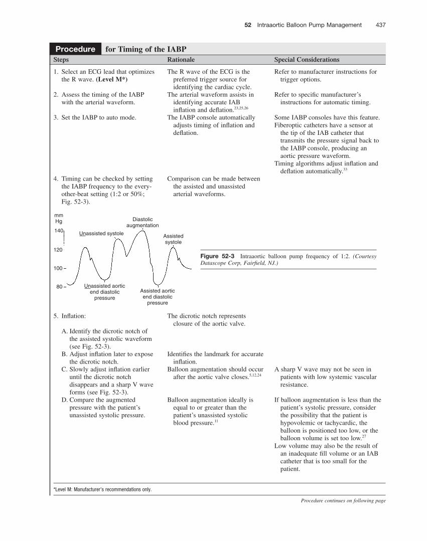
4. Timing can be checked by setting the IABP frequency to the every-other-beat setting (1:2 or 50%; Fig. 52-3 ).
Comparison can be made between the assisted and unassisted arterial waveforms.
5. Infl ation: The dicrotic notch represents closure of the aortic valve.
A. Identify the dicrotic notch of the assisted systolic waveform (see Fig. 52-3 ).
B. Adjust infl ation later to expose the dicrotic notch.
Identifi es the landmark for accurate infl ation.
C. Slowly adjust infl ation earlier until the dicrotic notch disappears and a sharp V wave forms (see Fig. 52-3 ).
Balloon augmentation should occur after the aortic valve closes. 5,12,24
A sharp V wave may not be seen in patients with low systemic vascular resistance.
D. Compare the augmented pressure with the patient ’ s unassisted systolic pressure.
Balloon augmentation ideally is equal to or greater than the patient ’ s unassisted systolic blood pressure. 11
If balloon augmentation is less than the patient ’ s systolic pressure, consider the possibility that the patient is hypovolemic or tachycardic, the balloon is positioned too low, or the balloon volume is set too low. 27
Low volume may also be the result of an inadequate fi ll volume or an IAB catheter that is too small for the patient.
Procedure for Timing of the IABP
Figure 52-3 Intraaortic balloon pump frequency of 1:2. (Courtesy Datascope Corp, Fairfi eld, NJ.)
120
100
80
Unassisted systole Assistedsystole
Diastolicaugmentation
Unassisted aorticend diastolic
pressureAssisted aorticend diastolic
pressure
mmHg
140
* Level M: Manufacturer ’ s recommendations only.
Procedure continues on following page
438 Unit II Cardiovascular System
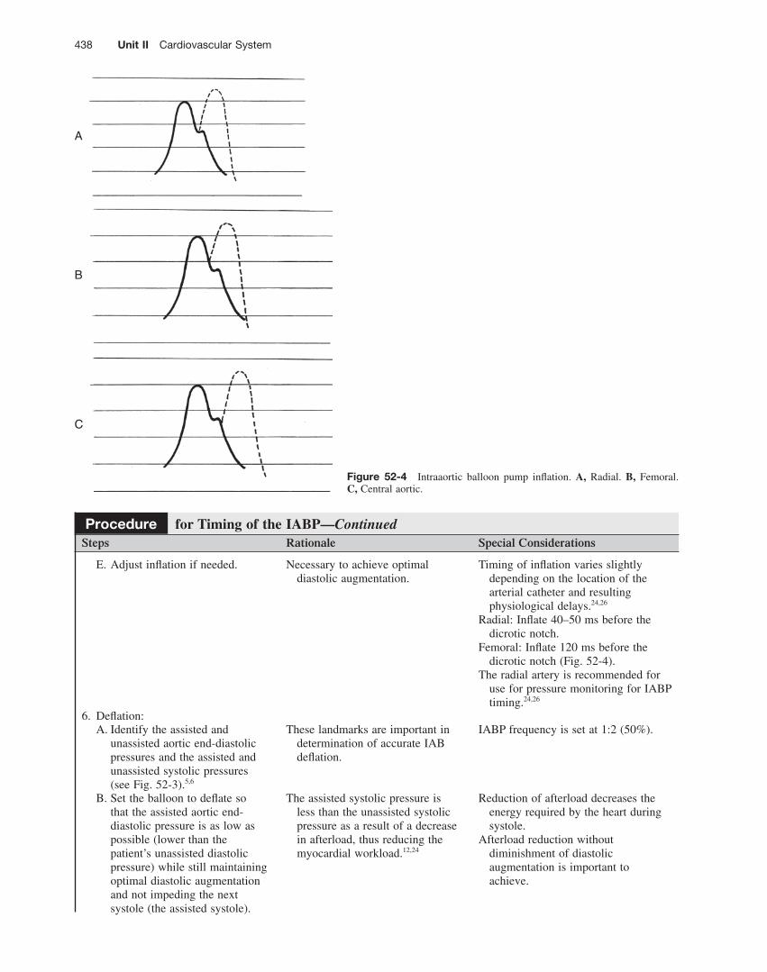
Figure 52-4 Intraaortic balloon pump infl ation. A, Radial. B, Femoral. C, Central aortic.
A
B
C
Steps Rationale Special Considerations
E. Adjust infl ation if needed. Necessary to achieve optimal diastolic augmentation.
Timing of infl ation varies slightly depending on the location of the arterial catheter and resulting physiological delays. 24,26
Radial: Infl ate 40–50 ms before the dicrotic notch.
Femoral: Infl ate 120 ms before the dicrotic notch ( Fig. 52-4 ).
The radial artery is recommended for use for pressure monitoring for IABP timing. 24,26
6. Defl ation: A. Identify the assisted and
unassisted aortic end-diastolic pressures and the assisted and unassisted systolic pressures (see Fig. 52-3 ). 5,6
These landmarks are important in determination of accurate IAB defl ation.
IABP frequency is set at 1:2 (50%).
B. Set the balloon to defl ate so that the assisted aortic end-diastolic pressure is as low as possible (lower than the patient ’ s unassisted diastolic pressure) while still maintaining optimal diastolic augmentation and not impeding the next systole (the assisted systole).
The assisted systolic pressure is less than the unassisted systolic pressure as a result of a decrease in afterload, thus reducing the myocardial workload. 12,24
Reduction of afterload decreases the energy required by the heart during systole.
Afterload reduction without diminishment of diastolic augmentation is important to achieve.
Procedure for Timing of the IABP—Continued

52 Intraaortic Balloon Pump Management 439
Figure 52-5 Correct intraaortic balloon pump timing (1:1). (Courtesy Data-scope Corp, Fairfi eld, NJ.)
120
100
80
Assistedsystole Assisted
systole
Diastolicaugmentation
Assistedaortic end-diastolic
pressure
1:1 IABP Frequency
mmHg
140
Steps Rationale Special Considerations
7. Set the IABP frequency to 1:1 (100%; Fig. 52-5 ).
Ensures that each heartbeat is assisted.
8. Assess timing every hour, whenever the heart rate changes by more than 10 beats/min, and when the rhythm changes.
Inappropriate timing prevents effective IABP therapy.
Many IABP models use algorithms to automatically adjust timing for changes in heart rate and rhythm.
Refer to the specifi c manufacturer guidelines for a description of automatic timing modes and their specifi c features.
9. Assess and intervene to correct inappropriate timing.
Ensures accurate timing and optimal functioning of the IABP.
A. Problem: early infl ation ( Fig. 52-6 ). Intervention: move infl ation later.
Infl ation occurs before closure of the aortic valve, leading to premature aortic valve closure, increased left-ventricular volume, and decreased stroke volume. 12
Early infl ation is the worst timing error, reducing left-ventricular performance and IABP effi ciency. 12
B. Problem: late infl ation ( Fig. 52-7 ). Intervention: adjust infl ation earlier.
A delay in infl ation leads to a decrease in coronary artery perfusion.
C. Problem: early defl ation ( Fig. 52-8 ). Intervention: adjust defl ation later.
Defl ation occurs before the aortic valve opens, leading to decreased balloon augmentation and less or no afterload reduction; coronary artery perfusion may also be decreased.
Note the sharp diastolic wave after augmentation and the increase in the assisted systolic pressure.
D. Problem: late defl ation ( Fig. 52-9 ). Intervention: adjust defl ation earlier.
Defl ation occurs after the aortic valve has opened, leading to an increase in the aortic end-diastolic pressure and an increase in afterload.
Note the delayed diastolic wave after augmentation and the diminished assisted systole.
Late defl ation is identifi ed by a diminished assisted systolic pressure, an increase in heart rate, an increase in fi lling pressures, a decrease in cardiac output and cardiac index, and an increased afterload. Maintaining a reliable trigger minimizes the risk of late defl ation. 5,6,11,23
Procedure for Timing of the IABP—Continued
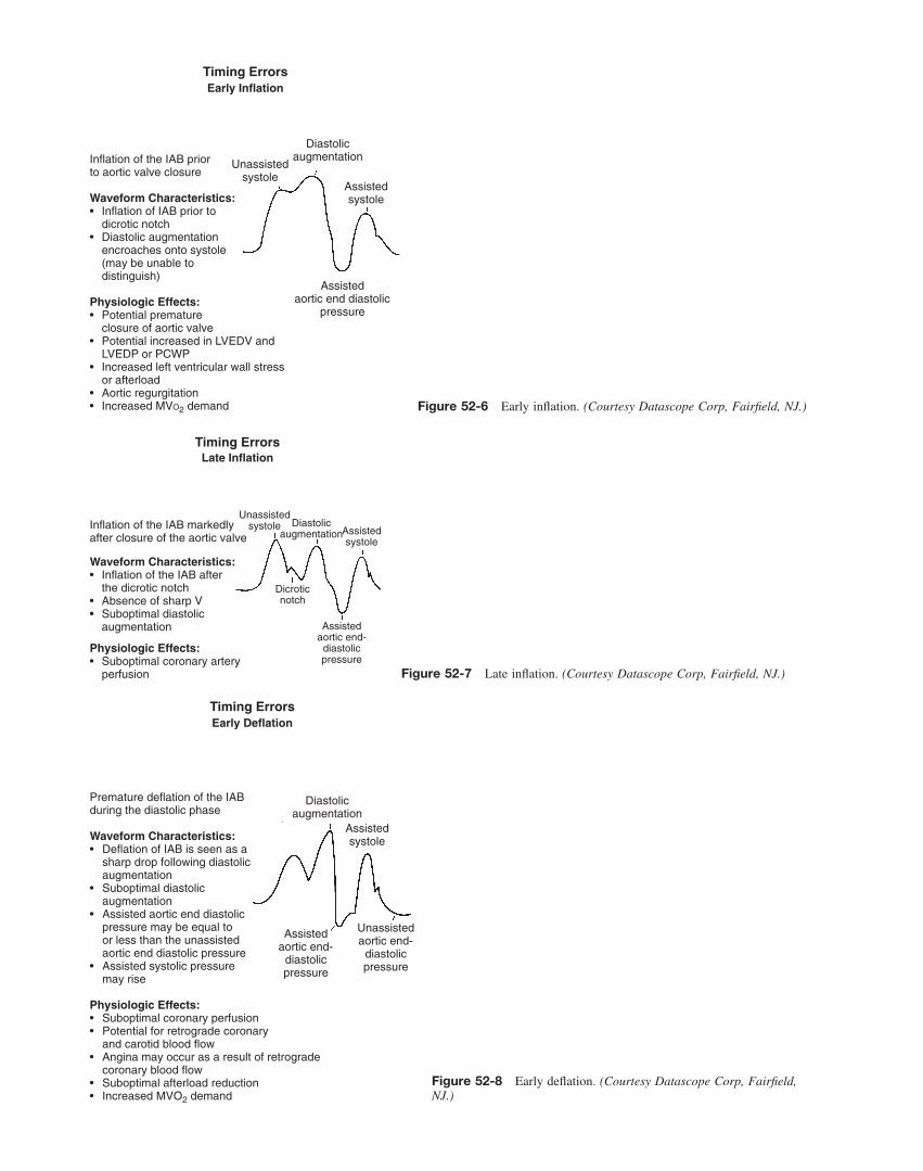
Figure 52-7 Late infl ation. (Courtesy Datascope Corp, Fairfi eld, NJ.)
Inflation of the IAB markedlyafter closure of the aortic valve
Waveform Characteristics:• Inflation of the IAB after the dicrotic notch• Absence of sharp V• Suboptimal diastolic augmentation
Physiologic Effects:• Suboptimal coronary artery perfusion
Unassistedsystole Assisted
systole
Dicroticnotch
Diastolicaugmentation
Assistedaortic end-
diastolicpressure
Timing ErrorsLate Inflation
Figure 52-8 Early defl ation. (Courtesy Datascope Corp, Fairfi eld, NJ.)
Assistedsystole
Diastolicaugmentation
Assistedaortic end-
diastolicpressure
Unassistedaortic end-
diastolicpressure
Premature deflation of the IABduring the diastolic phase
Waveform Characteristics:• Deflation of IAB is seen as a sharp drop following diastolic augmentation• Suboptimal diastolic augmentation• Assisted aortic end diastolic pressure may be equal to or less than the unassisted aortic end diastolic pressure• Assisted systolic pressure may rise Physiologic Effects:• Suboptimal coronary perfusion• Potential for retrograde coronary and carotid blood flow• Angina may occur as a result of retrograde coronary blood flow• Suboptimal afterload reduction• Increased MVO2 demand
Timing ErrorsEarly Deflation
Figure 52-6 Early infl ation. (Courtesy Datascope Corp, Fairfi eld, NJ.)
Unassistedsystole
Assistedsystole
Diastolicaugmentation
Assistedaortic end diastolic
pressure
Inflation of the IAB prior to aortic valve closure
Waveform Characteristics:• Inflation of IAB prior to dicrotic notch• Diastolic augmentation encroaches onto systole (may be unable to distinguish)
Physiologic Effects:• Potential premature closure of aortic valve• Potential increased in LVEDV and LVEDP or PCWP• Increased left ventricular wall stress or afterload• Aortic regurgitation• Increased MVO2 demand
Timing ErrorsEarly Inflation
52 Intraaortic Balloon Pump Management 441
Steps Rationale Special Considerations
1. Determine whether the IABP console has a balloon-pressure waveform.
Helium is shuttled in and out of the IAB catheter, and the balloon-pressure waveform represents this movement.
Refer to the specifi c manufacturer ’ s instructions regarding the balloon-pressure waveform.
2. Assess the balloon-pressure waveform.
Refl ects pressure that is in the IAB.
3. Determine whether the balloon pressure waveform is normal ( Fig. 52-10 ). A normal balloon pressure waveform:
A normal balloon-pressure waveform refl ects that the IAB is infl ating and defl ating properly. 22
A. Has a fi ll pressure (baseline pressure) slightly above zero.
Refl ects pressure in the tubing between the IAB and the IABP driving mechanism.
B. Has a sharp upstroke. Occurs as helium infl ates the IAB catheter.
C. Has peak infl ation artifact. This overshoot pressure artifact is caused by helium gas pressure in the pneumatic line. 1
D. Has a pressure plateau. This plateau is created as the IAB remains infl ated during diastole.
The plateau indicates the length of time of infl ation and whether full infl ation (volume) has been delivered to the IAB. If no plateau pressure is found, the IAB may not be fully infl ated.
E. Has a rapid defl ation. Helium is quickly shuttled from the IAB.
Procedure for Balloon-Pressure Waveform
Figure 52-9 Late defl ation. (Courtesy Datascope Corp, Fairfi eld, NJ.)
Unassistedsystole
Widenedappearance
Diastolicaugmentation
Assistedaortic end-
diastolicpressure
Deflation of the IAB late indiastolic phase as aortic valveis beginning to open
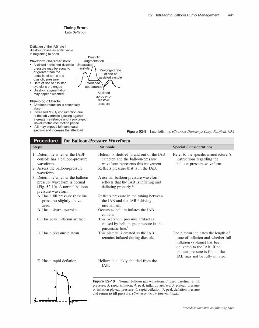
Waveform Characteristics:• Assisted aortic end-diastolic pressure may be equal to or greater than the unassisted aortic end diastolic pressure• Rate of rise of assisted systole is prolonged• Diastolic augmentation may appear widened Physiologic Effects:• Afterload reduction is essentially absent• Increased MVO2 consumption due to the left ventricle ejecting against a greater resistance and a prolonged isovolumetric contraction phase• IAB may impede left ventricular ejection and increase the afterload
Prolonged rateof rise of
assisted systole
Timing ErrorsLate Deflation
Figure 52-10 Normal balloon gas waveform. 1, zero baseline; 2, fi ll pressure; 3, rapid infl ation; 4, peak infl ation artifact; 5, plateau pressure or infl ation plateau pressure; 6, rapid defl ation; 7, peak defl ation pressure and return to fi ll pressure. (Courtesy Arrow International.)
Procedure continues on following page
442 Unit II Cardiovascular System
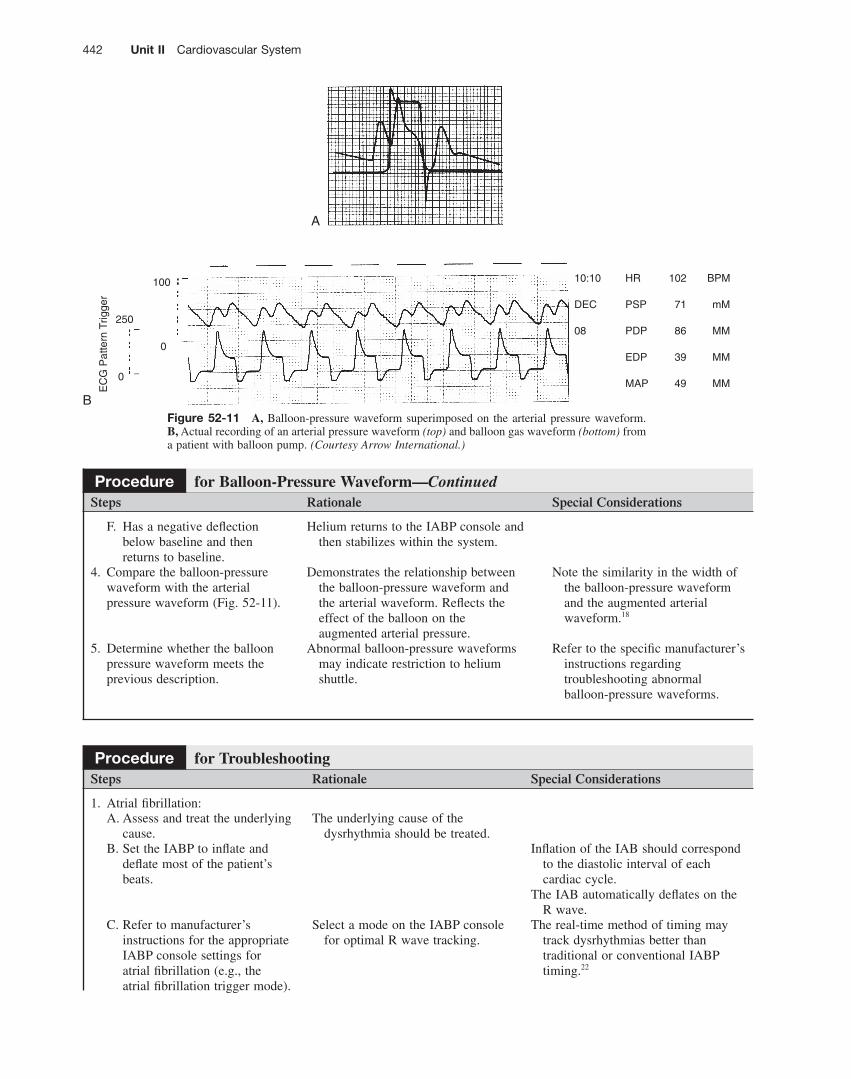
Figure 52-11 A, Balloon-pressure waveform superimposed on the arterial pressure waveform. B, Actual recording of an arterial pressure waveform (top) and balloon gas waveform (bottom) from a patient with balloon pump. (Courtesy Arrow International.)
10:10 HR 102 BPM
DEC PSP 71 mM
08 PDP 86 MM
EDP 39 MM
MAP 49 MM
100
0
0
250
EC
G P
atte
rn T
rigge
r
A
B
Steps Rationale Special Considerations
F. Has a negative defl ection below baseline and then returns to baseline.
Helium returns to the IABP console and then stabilizes within the system.
4. Compare the balloon-pressure waveform with the arterial pressure waveform ( Fig. 52-11 ).
Demonstrates the relationship between the balloon-pressure waveform and the arterial waveform. Refl ects the effect of the balloon on the augmented arterial pressure.
Note the similarity in the width of the balloon-pressure waveform and the augmented arterial waveform. 18
5. Determine whether the balloon pressure waveform meets the previous description.
Abnormal balloon-pressure waveforms may indicate restriction to helium shuttle.
Refer to the specifi c manufacturer ’ s instructions regarding troubleshooting abnormal balloon-pressure waveforms.
Procedure for Balloon-Pressure Waveform—Continued
Steps Rationale Special Considerations
1. Atrial fi brillation: A. Assess and treat the underlying
cause. The underlying cause of the
dysrhythmia should be treated. B. Set the IABP to infl ate and
defl ate most of the patient ’ s beats.
Infl ation of the IAB should correspond to the diastolic interval of each cardiac cycle.
The IAB automatically defl ates on the R wave.
C. Refer to manufacturer ’ s instructions for the appropriate IABP console settings for atrial fi brillation (e.g., the atrial fi brillation trigger mode).
Select a mode on the IABP console for optimal R wave tracking.
The real-time method of timing may track dysrhythmias better than traditional or conventional IABP timing. 22
Procedure for Troubleshooting
52 Intraaortic Balloon Pump Management 443
Procedure continues on following page
Steps Rationale Special Considerations
2. Tachycardia: Because diastole is shortened during tachycardia, the IAB infl ation time also is shortened.
The IABP may need to be changed to a 1:2 frequency.
Pumping every other beat may improve the patient ’ s hemodynamic status.
Some IABPs with automatic timing can track rates as high as 220 beats/min.
A. Assess and treat the underlying cause.
B. Set the timing and the frequency of the IABP to optimize hemodynamic response.
The underlying cause of the tachycardia should be treated.
IAB timing and frequency should be set to optimize coronary perfusion and afterload reduction.
3. Asystole: A. Switch the trigger to arterial
pressure. This trigger can be used if an
arterial pressure is generated from chest compressions.
Follow advanced cardiac life support (ACLS) standards for emergency care.
B. If the IABP console is not in the auto-operation mode: i. Set infl ation to provide
diastolic augmentation. ii. Set defl ation to occur
before the upstroke of the next systole.
Sets the IABP timing. Refer to the manufacturer ’ s manual. Preliminary research suggests that
when used during cardiopulmonary resuscitation, IAB counterpulsation increases cerebral and coronary perfusion. 2,11
C. If chest compressions do not provide an adequate trigger:
Refer to manufacturer ’ s guidelines for recommendations for minimal balloon volume. i. Turn or push the control to
internal trigger. The internal trigger keeps the IAB
catheter moving so that clot formation is minimized. 22
ii. Set the rate at 60–80 beats/min.
Maintains consistent movement of IAB catheter.
iii. Set the IABP frequency to 1:2.
A 1:2 frequency is adequate to prevent thrombus formation on the IAB catheter.
iv. Turn the balloon augmentation down to 50%.
Slight infl ation and defl ation of the IAB catheter prevents clot formation.
D. If the IABP console is in the auto-timing mode, the console automatically attempts to self-time if an arterial pressure is generated, or switches to an internal trigger.
Sets the IABP timing and maintains consistent movement of the IAB catheter.
4. Ventricular tachycardia or ventricular fi brillation: A. Assess and treat the underlying
cause. The underlying cause of the
tachycardia should be treated. B. Cardiovert or defi brillate as
necessary (see Procedures 35 and 36).
Attempts to convert the dysrhythmia.
Follow ACLS standards for emergency care.
Ensure that personnel are cleared from the patient and equipment before cardioversion or defi brillation.
The IABP console is electrically isolated.
5. Loss of vacuum or IABP failure: A. Check and tighten the
connections on the pneumatic tubing.
A loose connection may contribute to a loss of vacuum.
Procedure for Troubleshooting—Continued

444 Unit II Cardiovascular System
Steps Rationale Special Considerations
B. Check the compressor power source.
Ensures that power is available to drive the helium.
C. Hand infl ate and defl ate the balloon every 5 minutes if necessary. (Level M * )
Prevents clot formation along the dormant balloon.
Refer to specifi c manufacturer ’ s guidelines for manually infl ating and defl ating the IAB.
Ensure that the correct syringe is kept with the IABP console for this emergency; check manufacturer ’ s guidelines for frequency of hand infl ation.
D. Change the IAB console. (Level M * )
Establishes a power source and effective IABP therapy.
6. Suspected balloon perforation: A. Observe for loss of
augmentation. Helium may be gradually leaking
from the balloon catheter.Set the alarm limits so the alarms
sound with a decrease of 10 mm Hg in diastolic augmentation.
B. Check for blood in the balloon lumen tubing.
Blood or any discoloration in the helium tubing indicates that the balloon has perforated and that arterial blood is present.
It is possible for a balloon leak to be self-sealing as a result of the surface tension between the inside and the outside of the IAB membrane. This may be evidenced by the presence of dried blood in the balloon lumen tubing. The dried blood may appear as a brownish, coffee-ground–like substance.
C. Assess for changes or lack of a normal balloon-pressure waveform.
The balloon-pressure waveform may be absent if the balloon is unable to retain helium, or the pressure plateau may gradually decrease if the IAB is leaking helium.
7. Balloon perforation: A. Place the IABP on standby. Prevents further IAB pumping and
continued helium exchange.Some IABP consoles automatically
shut off if a leak is detected. The IAB catheter should be removed within 15–30 minutes. 8
B. Clamp the IAB catheter. Prevents arterial blood backup. C. Disconnect the IAB catheter
from the IABP console. Prevents blood from backing up into
the IABP console.
D. Notify the physician or advanced practice nurse.
The IAB catheter needs to be removed or replaced immediately.
If the IAB leak has sealed itself off, this may result in entrapment of the IAB in the vasculature. Surgical removal may be necessary.
E. Prepare for IAB catheter removal or replacement.
The IAB catheter should not lie dormant for longer than 30 minutes.
Do not manually infl ate and defl ate the IAB if balloon perforation is suspected. Perforation of a balloon membrane may indicate that the patient ’ s vascular condition may induce abrasion or perforation in subsequent balloon membranes.
F. Discontinue anticoagulation therapy as prescribed.
Clotting occurs more readily if anticoagulation therapy is stopped (necessary if removing the catheter).
Procedure for Troubleshooting—Continued
* Level M: Manufacturer ’ s recommendations only.
52 Intraaortic Balloon Pump Management 445
Steps Rationale Special Considerations
1. HH 2. PE 3. Assess clinical readiness for
weaning. Optimal clinical and
hemodynamic parameters validate readiness for weaning.
Patient hemodynamic status should be optimal before weaning from IABP therapy.
Signs of clinical readiness include the following: no angina, heart rate < 110 beats/min, absence of unstable dysrhythmias, MAP > 70 mm Hg with minimal or no vasopressor support, pulmonary artery occlusion pressure < 18 mm Hg, cardiac index > 2.4 mixed venous oxygen saturation between 60% and 80%, capillary refi ll < 2 seconds, and urine output > 0.5 mL/kg/hr.
4. Change the assist ratio to 1:2 (50%), and monitor the patient ’ s response for 1–6 hours, as prescribed, or per the institution ’ s protocol.
The length of time required to wean from IABP therapy depends on the hemodynamic response of the patient and the length of time the patient has received IABP therapy. 13,29
Follow physician or advanced practice nurse prescription or institutional policy on IABP weaning.
5. If hemodynamic parameters remain stable, further change the ratio (depending on the patient and the balloon-console assist frequencies, or as prescribed).
IABP consoles vary in assist ratios.
Follow physician or advanced practice nurse prescription or institutional policy on IABP weaning.
6. Discontinue heparin or dextran 4–6 hours before IAB catheter removal, or reverse heparin with protamine (as prescribed) just before catheter removal.
Decreases the likelihood of bleeding after balloon removal.
7. Turn the IABP to standby or off and disconnect the IAB from the console.
Ensures defl ation of the IAB catheter.
The patient ’ s arterial pressure collapses the balloon membrane in preparation for withdrawal.
8. Assist with removing sutures or the sutureless securement device.
Prepares for IAB removal.
9. Assist the physician or advanced practice nurse with removal of the percutaneous catheter.
Facilitates removal. The IAB catheter is not withdrawn into the sheath but removed as an entire unit to avoid shearing the balloon.
10. Ensure that pressure is held on the insertion site for 30–45 minutes after the IAB catheter is withdrawn.
Ensures that hemostasis is obtained and decreases the incidence of bleeding and hematoma formation.
A femoral compression system can be used to achieve hemostasis (see Procedure 76 ).
Pressure may be needed for a longer period of time if the patient has been receiving anticoagulant therapy or if coagulation study results are abnormal.
11. Assess the insertion site for signs of bleeding or hematoma formation before application of a sterile pressure dressing.
Assists in the detection of bleeding.
12. Apply a pressure dressing to the insertion site for 2–4 hours or as prescribed.
Minimizes bleeding from the insertion site.
Procedure for Weaning and IAB Catheter Removal
Procedure continues on following page
446 Unit II Cardiovascular System
Steps Rationale Special Considerations
13. Obtain vital signs and hemodynamic parameters every 15 minutes × 4, every 30 minutes × 2, then every hour as the patient ’ s condition warrants, or as prescribed.
Determines patient stability or instability.
14. Assess the quality of perfusion to the decannulated extremity immediately after removal and every 1 hour × 2, then every 2 hours, or as prescribed.
Removal of the IAB catheter may dislodge thrombi on the catheter and lead to arterial occlusion.
15. Maintain immobility of the decannulated extremity and maintain bed rest with the head of the bed no greater than 30 degrees for 8 hours, as prescribed or according to institutional protocol.
Promotes healing and decreases stress at the insertion site.
16. Remove PE and discard used supplies in appropriate receptacle.
Reduces the transmission of microorganisms and body secretions; Standard Precautions.
17. HH
Procedure for Weaning and IAB Catheter Removal—Continued
Expected Outcomes • Increased myocardial oxygen supply • Decreased myocardial oxygen demand • Increased cardiac output • Increased tissue perfusion, including cerebral, renal,
and peripheral circulation
Unexpected Outcomes • Impaired perfusion to the extremity with the IAB
catheter in place • Balloon perforation • Inappropriate IAB placement • Pain • Bleeding or coagulation disorders • Aortic dissection • Infection
Patient Monitoring and Care Steps Rationale Reportable Conditions
These conditions should be reported if they persist despite nursing interventions.
1. Perform systematic cardiovascular, peripheral vascular, and hemodynamic assessments every 15–60 minutes as patient status requires or as prescribed. A. Level of consciousness Assesses for adequate cerebral
perfusion; thrombi may develop and dislodge during IABP therapy; the IAB may migrate, decreasing blood fl ow to the carotid arteries.
• Change in level of consciousness
B. Vital signs and pulmonary artery pressures
Demonstrates effectiveness of IABP therapy.
• Unstable vital signs • Signifi cant changes in
hemodynamic pressures • Lack of response to IABP therapy
52 Intraaortic Balloon Pump Management 447
Procedure continues on following page
Patient Monitoring and Care Steps Rationale Reportable Conditions
C. Arterial and balloon pressures Ensures effectiveness of IABP timing and therapy.
• Diffi culty achieving effective IABP therapy
D. Cardiac output, cardiac index, and systemic vascular resistance values
Demonstrates effectiveness of IABP therapy.
• Abnormal cardiac output, cardiac index, and systemic vascular-resistance values
E. Circulation to extremities Determines peripheral perfusion. If reportable conditions are found, they may indicate catheter or embolus obstruction of perfusion to the extremity. Specifi cally, decreased perfusion to the left arm may indicate misplacement of the IAB catheter. 2,8,15,29
• Capillary refi ll > 2 seconds • Diminished or absent pulses (e.g.,
antecubital, radial, femoral, popliteal, tibial, pedal)
• Color pale, mottled, or cyanotic • Diminished or absent sensation • Pain • Diminished or absent movement • Cool or cold to the touch
F. Urine output Determines perfusion to the kidneys. • Urine output < 0.5 mL/kg/hr 2. Assess heart and lung sounds
every 4 hours and as needed. Abnormal heart and lung sounds may
indicate the need for additional treatment.
Special note: When the patient ’ s condition permits, place the IABP on standby to accurately auscultate heart and lung sounds because IABP therapy creates extraneous sounds and impairs heart and lung sound assessment.
• Abnormal heart and lung sounds
3. Maintain the head of the bed at less than 45 degrees.
Prevents kinking of the IAB catheter and migration of the catheter.
4. Monitor for signs of balloon perforation by assessing the balloon tubing on a regular basis for evidence of discoloration or blood in the tubing.
In the event of balloon perforation, a very small amount of helium could be released into the aorta, potentially causing an embolic event. Because of pressure gradients in the aorta, blood is more likely to enter the balloon membrane and be dehydrated by the helium.
• Blood or brown fl ecks in the tubing • Loss of IABP augmentation • Control-console alarm activation
(e.g., gas loss)
5. Maintain accurate IABP timing. If timing is not accurate, cardiac output may decrease rather than increase.
• Signs and symptoms of hemodynamic instability
6. Log-roll the patient every 2 hours. Prop up pillows to support the patient and to maintain alignment. Consider use of pressure-relief devices. (Level E*)
Promotes comfort and skin integrity and prevents kinking of the IAB catheter. Special note: log-rolling may not be tolerated in patients with severe hemodynamic compromise; low-pressure beds are necessary for these patients. Low-pressure beds can decrease the occurrence of pressure ulcers in patients who need IABP therapy. 4,25
—Continued
*Level E: Multiple case reports, theory-based evidence from expert opinions, or peer-reviewed professional orga nizational standards without clinical studies to support recommendations.
448 Unit II Cardiovascular System
Patient Monitoring and Care Steps Rationale Reportable Conditions 7. Immobilize the cannulated
extremity with a draw sheet tucked under the mattress or with a soft ankle restraint or a knee immobilizer as prescribed.
Prevents dislodgment and migration of the IAB catheter.
Special note: assess skin integrity and perfusion distal to the restraint every hour.
• Alteration in skin integrity • Alteration in peripheral perfusion
8. Initiate passive and active range-of-motion exercises every 2 hours to extremities that can be mobilized.
Prevents venous stasis and muscle atrophy.
9. Assess the area around the IAB catheter insertion site every 2 hours and as needed for evidence of hematoma or bleeding.
IAB catheter infl ation and defl ation traumatizes red blood cells and platelets.
• Bleeding at insertion site • Hematoma at insertion site
10. Maintain anticoagulation therapy as prescribed; monitor coagulation studies.
Prophylactic anticoagulation therapy may be used to prevent thrombi and emboli development.
Anticoagulation therapy may alter hemoglobin, hematocrit, and coagulation values. 32
• Abnormal coagulation study results • Abnormal hemoglobin and
hematocrit study results
11. Monitor patient for systemic evidence of bleeding or coagulation disorders.
Hematologic and coagulation profi les may be altered as a result of blood loss during balloon insertion, anticoagulation, and platelet dysfunction as a result of mechanical trauma by balloon infl ation and defl ation. 32
• Bleeding from IAB insertion site • Bleeding from incisions or mucous
membranes • Petechiae or ecchymosis • Guaiac-positive nasogastric aspirate
or stool • Hematuria • Decreased hemoglobin or
hematocrit • Decreased fi lling pressures • Increased heart rate • Retroperitoneal hematoma • Pain in the lower abdomen, fl ank,
thigh, or lower extremity 12. Follow institutional standards for
assessing pain. Administer analgesia as prescribed.
Promotes comfort. • Continued pain despite pain interventions
13. Replace gauze dressings at the IAB catheter site every 2 days and transparent dressings at least every 7 days. Cleanse the site with an antiseptic solution (e.g., 2% chlorhexidine solution). (Level D*)
Decreases the incidence of infection and allows an opportunity for site assessment. Although guidelines do not exist specifi cally for IAB site dressings, the Centers for Disease Control and Prevention (CDC) 19 recommend replacing invasive line dressings when the dressing becomes damp, loosened, or soiled, or when inspection of the site is necessary.
• Signs or symptoms of infection
—Continued
*Level D: Peer-reviewed professional and organizational standards with the support of clinical study recommen dations.

52 Intraaortic Balloon Pump Management 449
Patient Monitoring and Care Steps Rationale Reportable Conditions 14. Assess for balloon migration. The IAB should be positioned 2 cm
below the left subclavian artery and just above the renal arteries. If the IAB migrates proximally, it may occlude the subclavian or carotid arteries. If the IAB migrates too low, it could occlude the renal or mesenteric arteries.
• Signs of possible subclavian artery occlusion: unequal or absent radial pulse and dampening or loss of the arterial pressure waveform in the ipsilateral radial artery (radial artery on the same side as the IAB catheter)
• Signs of possible carotid artery occlusion include change in level of consciousness and orientation or unilateral neurological defi cit
• Signs of renal artery occlusion: oliguria or anuria, back or fl ank pain, nausea, and anorexia
• Signs of mesenteric artery occlusion: abdominal pain, diarrhea, nausea, and decreased bowel sounds
15. Identify parameters that demonstrate clinical readiness to wean from IABP therapy.
Close observation of the patient ’ s tolerance to weaning procedures is necessary to ensure that the body ’ s oxygen demands can be met. The presence of these reportable conditions indicates that consideration should be given to weaning the patient from the IABP.
• No angina • Heart rate < 110 beats/min • Absence of unstable dysrhythmias • MAP > 70 mm Hg with little or no
vasopressor support • Pulmonary artery occlusion
pressure < 18 mm Hg • Cardiac index > 2.4 • S vo 2 between 60% and 80% • Capillary refi ll < 2 seconds • Urine output > 0.5 mL/kg/hr
—Continued
Documentation Documentation should include the following: • Patient and family education • Informed consent • Universal protocol requirements • Insertion of the IAB catheter (including size of
catheter used and balloon volume) • Peripheral pulses and neurovascular assessment of the
affected extremity • Any diffi culties with insertion • IABP frequency • Patient response to the procedure and to IABP
therapy
• Assessment of pain, interventions, and response to interventions
• Confi rmation of placement (e.g., chest radiograph) • Insertion site assessment • Hemodynamic status • IABP pressures (unassisted end-diastolic pressure,
unassisted systolic pressure, balloon-augmented pressure, assisted systolic pressure, assisted end-diastolic pressure, and MAP)
• Occurrence of unexpected outcomes • Additional nursing interventions taken
References and Additional Readings For a complete list of references and additional readings for this procedure, scan this QR code with any freely available smartphone code reader app, or visit http://booksite.elsevier.com/9780323376624 .