processing in vitro of an abasic site reacted with methoxyamine: a
TRANSCRIPT

Nucleic Acids Research, Vol. 19, No. 20 5569-5574
Processing in vitro of an abasic site reacted withmethoxyamine: a new assay for the detection of abasicsites formed in vivo
S.Rosa, P.Fortini, P.Karran1, M.Bignami and E.Dogliotti*Laboratory of Comparative Toxicology and Ecotoxicology, Istituto Superiore di Sanita', Viale ReginaElena 299, 00161 Rome, Italy and imperial Cancer Research Fund, Clare Hall Laboratories, SouthMimms, Herts EN6 3LD, UK
Received July 24, 1991; Revised and Accepted September 24, 1991
ABSTRACT
In this study we demonstrate that the differentsubstrate recognition properties of bacterial and humanAP endonucleases might be used to quantify andlocalize apurinic (AP) sites formed in DNA in vivo. Byusing a model oligonucleotide containing a single APsite modified with methoxyamine (MX), we show thatendonuclease III and IV of E. coli are able to cleave thealkoxyamine-adducted site whereas a partially purifiedHeLa AP endonuclease and crude cell-free extractsfrom HeLa cells are inhibited by this modification. Inaddition MX-modified AP sites in a DNA template retaintheir ability to block DNA synthesis in vitro. Since MXcan efficiently react with AP sites formed in mammaliancells in vivo we propose that the MX modified abasicsites thus formed can be quantitated and localized atthe level of the individual gene by subsequent sitespecific cleavage by either E. coli endonuclease III orIV in vitro.
INTRODUCTION
Apurinic/apyrimidinic (AP) sites are common DNA lesions thatare produced both spontaneously and after exposure to a varietyof DNA-damaging agents. The hydrolysis of the glycosyl bondbetween deoxyribose and purines or pyrimidines occurs at asignificant rate at neutral pH and is accelerated by certain typesof DNA damage, such as alkylation of the base. Furthermore,DNA glycosylases excise a variety of damaged DNA bases andform AP sites as intermediates in DNA repair (1). AP sites are'non-informational' DNA lesions (2,3) with potential cytotoxicand mutagenic effects (4,5). Excision repair processes removeAP sites very efficiently in vivo. A single-strand break (ssb) isfirst introduced on the 5' side of a base-free deoxyribose-phosphate residue which is excised, most likely by a DNAdeoxyribophosphodiesterase (6), to generate a single nucleotidegap. The replacement of the missing nucleotide by DNA
polymerase followed by chain joining by DNA ligase restoresthe integrity of the DNA. As a consequence of this efficient repairsystem, the AP site is a short-lived lesion in vivo.
Analysis of mutational spectra, both spontaneous as well asDNA damage induced, strongly implicates AP sites as commonintermediates in mutagenesis. Many physical and chemical agents(i.e. ionizing radiation, bulky chemicals, alkylating agents) inducethe same types of mutations as those induced by heat and acid-generated AP sites, namely GC-TA and AT-TA transversions(as in the case of depurination) and GC-AT transitions (as in thecase of depyrimidination) (7). Prokaryotic polymerases areblocked at AP sites in template DNA in vitro and the SOSresponse is required for mutagenesis in vivo (8). In contrast,eukaryotic enzymes appear to be able to replicate past AP sitesvia preferential incorporation of dAMP (9,10,11).
It is of interest to develop assays to quantify and localize APsites formed in vivo at the level of the gene and to correlate theiroccurrence with specific types of mutational events. Manyattempts have been made in the past to assay AP site formation,but detection of AP sites is still limited to indirect assays, suchas alkaline sucrose gradients and alkaline elution (12). Theseassays exploit the alkali lability of AP sites to quantitate themas DNA ssb produced at a given time after DNA damage. Adirect assay of AP sites based on the specific reaction ofmethoxyamine (MX) with the aldehyde group of deoxyriboseresidues has been successfully applied to the quantitation of APsites in DNA in vitro (13,14). We have shown that AP sitesproduced in vivo after alkylation damage react specifically withMX and become resistant to alkaline cleavage (15). AP sites arealso a component of the mutagenic and cytotoxic response toalkylation damage in mammalian cells (P. Fortini et al.,manuscript submitted).
In this study a synthetic oligonucleotide containing a single APsite is used as a model to explore the biochemical characteristicsof the adduct formed upon reaction with MX. Using this modelsubstrate in conjunction with purified bacterial or human APendonucleases and with human cell-free extracts, we have
* To whom correspondence should be addressed
Downloaded from https://academic.oup.com/nar/article-abstract/19/20/5569/2387645by gueston 08 April 2018
5570 Nucleic Acids Research, Vol. 19, No. 20
demonstrated that the different substrate specificities of these APendonucleases might be exploited to quantify and localize sitesof base loss in DNA in vivo. A new assay for the direct detectionof AP sites formed in vivo at the level of the gene is proposed.
MATERIALS AND METHODSEnzymesE. coli endonuclease IV and uracil-DNA glycosylase werepurified by published procedures (16,17). E. coli endonucleaseIII was kindly supplied by Dr. C. Jones, ICRF, UK. HeLa APendonuclease was a generous gift of Dr. A.M. Pedrini, Instituteof Biochemical and Evolutionary Genetics, Pavia, Italy. T4polynucleotide kinase was purchased from Bio-Rad andSequenase from Pharmacia.
ChemicalsMethoxyamine hydrochloride was purchased from Sigma. Stocksolutions were prepared in water and stored at —20°C.
Cell extractsCell extracts were prepared from human HeLa cells following theprocedure of Li and Kelly (18). Briefly, spinner cultures of HeLacells were grown in Eagle's minimal essential mediumsupplemented with 10% foetal calf serum. 1 x 10¹⁰ cells wereharvested in mid-log phase by centrifugation at 1000xg for 5 min.The cell pellet was rapidly washed in 500 ml of ice-cold hypotonicbuffer with 250mM sucrose followed by 500 ml of ice-coldhypotonic buffer alone (20mM Hepes-KOH, 5mM KC1, 1.5mMMgCl2, 0.5mM dithiothreitol, pH 7.5). The washed cell pelletwas suspended in hypotonic buffer and the cells allowed to swellon ice for 30 min. Cells were then lysed with 10 strokes of a tightlyfitting pestel in a Dounce homogenizer. After 30 min incubationon ice, the lysate was centrifuged for 20 min at 10,000 g, at 0°C.The supernatant was collected and NaCl was added to a finalconcentration of lOOmM. The extracts were centrifuged at 100,000g for lh, at 0°C. The supernatant was removed, aliquoted, quicklyfrozen in ethanol/dry ice and stored at -70°C. The proteinconcentration of the extracts is approximately 10
Preparation of oligonucleotide substratesOligonucleotides containing a single AP site. 30-meroligonucleotides were prepared containing either cytosine oruracil at position 13 (5'-TACGGATCGCAG C/U TGGGTTA-GGGAAGTTGG-3')- These oligonucleotides were 5' end-labeledas already described (19) and annealed to their complementarystrands. The single AP site was produced in the duplexoligonucleotide by enzymatic removal of the uracil residue. 5'(32P)-end-labeled oligonucleotide (270 fmoles) was incubatedwith 2x lO" 6 U (17) uracil-DNA glycosylase for 30 min at37°C in 70mM Hepes buffer (pH 7.8), lOmM dithiothreitol,lmM EDTA (final volume 20/xl).
Oligonucleotides containing a single MX-modified AP site.Modification of the AP site with MX was carried out essentiallyby the method of Talpaert-Borle' and Liuzzi (13). Oligonucleo-tides containing a single AP site were incubated with 30mM MXfor 30 min at 37 °C in the same buffer used for digestion withuracil-DNA glycosylase. The oligonucleotides were ethanolprecipitated and resuspended in lOmM Tris-HCl (pH 7.5) andlmM EDTA.
Oligonucleotide assaysDetermination of AP-endonuclease activity. Endonuclease activitywas monitored on denaturing polyacrylamide gels by the extentof formation of the 12-mer cleavage product from theoligonucleotides containing either the AP site or the MX-modifiedAP site. Incubation with E. coli endonuclease III was performedin lOmM Tris-HCl, (pH 7.5), lmM EDTA, 0. lmM KC1, lmMmercaptoethanol. The incubation buffer for E. coli endonucleaseIV was 0.2M NaCl, 50mM Hepes-KOH, (pH 7.8), lmM EDTA,O.lmM dithiothreitol, 50/ig/ml BSA. Both enzymatic digestionswere carried out at 30°C for 30 min. HeLa AP endonucleasedigestion was performed in lOOmM Hepes-NaOH, (pH 8.25),3mM MgCl2 for 30 min at 37°C. The amounts of APendonucleases used in this study were comparable by a factorof two. When cell extracts were used, HeLa cell extract (10^g)was incubated with the labelled oligonucleotides for lh at 37°C.When necessary, MX was added to the incubation mixture.
The endonuclease-treated samples were then deproteinized bytreatment with proteinase K, phenol extracted and ethanolprecipitated. The oligonucleotides were resuspended and resolvedon 12% denaturing polyacrylamide gels (0.75 mm thick).Electrophoresis was carried out in TBE buffer at roomtemperature at 350 V for lh. The gels were dried and DNAvisualized by autoradiography.
Termination of DNA synthesisPreparation of single-stranded template containing AP sites andMX-modified AP sites. Uracil containing single-stranded DNAwas prepared according to published procedures (20) with somemodifications. E. Coli CJ 236 dut~, ung~ cells were infectedat a cell density corresponding to ODeooof 0.3 with M13mpl8bacteriophage at a multiplicity of infection of <0.2. After 5h,the supernatant was collected and digested with RNAse (150/tg)for 30 min at 20°C and then precipitated with 1/4 volume of3.5M ammonium acetate, 20% PEG 8000 for 30 min on ice.The pellet of phage particles was resuspended in 200/tl high saltbuffer (300mM NaCl, lOOmM Tris-HCl, pH 8.0, lmM EDTA)and DNA was purified by several extractions with phenol,phenol/chloroform and chloroform/isoamyl alcohol. DNA wasprecipitated with 7.8M ammonium acetate and 2.5 volumes ofethanol. A 10/ig sample of uracil-containing M13 DNA wasincubated with 10~5 U (17) of uracil-DNA glycosylase in a 20/*lreaction mixture containing 70mM Hepes-KOH (pH 8), lOmMdithiothreitol, lmM EDTA. AP sites were modified with MXby 30 min incubation with 30mM MX at 37°C. The presenceof AP sites after uracil-DNA glycosylase digestion as well astheir modification with MX was monitored on alkaline gels.Alkaline gels were prepared by dissolving 1 % agarose in 50mMNaCl, 4mM EDTA. The running buffer was 30mM NaOH,2mM EDTA. DNA samples were resuspended before loadingin 0.5M NaOH, 25% glycerol, 0.025% bromocresol green. Thegels were run for 2h at 60 V after a pre-run of 30 min at 50 V.
DNA synthesis on AP and AP-MX DNA templates. DNApolymerase reactions on AP and AP-MX DNA templates werecarried as described by Sagher and Strauss (10). Briefly, 1/igDNA was annealed to a 17bp DNA primer (3ng). Primed M13DNA was used to synthesize up to the lesion with 0.4U/^lSequenase in a buffer containing 20mM Hepes-KOH (pH 7.8),5mM DTT, lOmM MgCl2, 12/iM 35S dATP and the other threedeoxynucleoside-5'-triphosphates (300/tM). After 15 min at20°C, dATP (267/tM) was added to the reaction mixture to
Downloaded from https://academic.oup.com/nar/article-abstract/19/20/5569/2387645by gueston 08 April 2018

Nucleic Acids Research, Vol. 19, No. 20 5571
complete DNA synthesis and the incubation was prolonged fora further 15 min at 20°C. The reaction was stopped by adding95% formamide, 20mM EDTA, 0.05% bromophenol blue,0.05% xylene cyanol. Aliquots of the DNA samples were thenrun on a 8% denaturing polyacrylamide gel in TBE buffer.Electrophoresis was carried out at 2000 V for 2h.
RESULTSReaction of MX with AP sites produced in vitro
Compounds containing an -ONH2 group such as MX are knownto react with the aldehyde group on C'i of an AP site withoutdegrading DNA (21). M13 bacteriophage was grown on anung~ (uracil glycosylase deficient) dut "(deoxyuridinetriphosphatase deficient) E. coli mutant. As a consequence of thegrowth in this strain, the M13 genome contains uracil insteadof thymine residues. Uracil-containing DNA was treated withuracil-DNA glycosylase to produce AP sites. This substrate isalkali-sensitive, since AP sites are labile at alkaline pH. As shownin Fig. 1, a smear of DNA molecules of different length isproduced on alkaline agarose gels (Fig. 1, lane B). In contrast,the uracil-DNA glycosylase digested substrate treated with 30mMMX (lane C) migrates as the untreated, uracil-containing M13DNA (lane A). The presence of some linear single-stranded DNAin the MX-reacted sample indicates that a minor percentage ofAP sites did not react with MX before the alkaline treatment.Therefore, the addition of MX on the aldehyde group at an APsite in vitro inhibits the alkaline rupture of the adjacentphosphodiester bond.
A B C
Fig. 1. Reaction of MX with AP sites produced in vitro. Alkaline gelelectrophoresis of : (A) uracil-containing M13 DNA; (B) after incubation withuracil-DNA glycosylase (2 x 10"5 U) for 30 min at 37°C; (C) as in (B) followedby 30 min treatment with 30 mM MX, at 37°C. The form which migrates fasterthan the single-stranded DNA is single-stranded linear DNA.
Substrate recognition of oligonucleotides containing AP sitesor MX-modified AP sites by bacterial AP-endonucleasesTo further explore the structure of the abasic site modified bythe formation of an alkoxyamine adduct at the C\ aldehyde inDNA, we utilized a synthetic DNA substrate containing a singleAP site. The oligonucleotide containing the uracil residue atnucleotide position 13 was 5' end labeled and annealed to thecomplementary strand. AP sites were produced by incubationwith uracil-DNA glycosylase, and were modified by exposureto MX. The extent of cleavage by different AP-endonucleaseswas monitored on denaturing polyacrylamide gels by theformation of the expected 12-mer cleavage product. As shownin Fig. 2, both E. coli endonuclease III and endonuclease IV areable to cleave the AP site containing oligonucleotide leading tothe formation of a 12-mer cleavage product (lanes 3 and 5,respectively). Endonuclease III has both a DNA N-glycosylaseactivity and an AP lyase activity, i.e. cleaves 3' to the AP site,while endonuclease IV is a class II AP endonuclease and cleaves5' to the AP site. The cleavage product of endonuclease HI (lanes3,4) migrates slightly slower than the cleavage product ofendonuclease IV (lanes 5, 6), because the latter is shorter by adeoxyribose 5'-phosphate residue. The nicking of AP sites bythese bacterial endonucleases is only slightly inhibited by thereaction of MX with the aldehyde group of the AP site (lanes4 and 6). Therefore, the presence of the alkoxyamine adduct doesnot significantly affect either the ^-elimination reaction catalysedby endonuclease III or the hydrolytic cleavage of thephosphodiester bond by endonuclease IV.
Substrate recognition of oligonucleotides containing an APsite or MX-modified AP site by HeLa AP-endonucleaseThe substrate recognition by a partially purified HeLa AP-endonuclease was also tested. This enzyme, which has been
Endo Endo IV
8 8 8 8 83 S'S1 2 3 4 5 6
Fig. 2. Substrate recognition of oligonucleotides containing AP sites or MX-modified AP sites by bacterial AP-endonucleases. Lane I: duplex dU:dG; lane2: duplex dX*:dG (X*= AP site modified with MX) ; lane 3: duplex dX:dG(X = AP site) after 30 min incubation with endonuclease III of E. coli; lane4: duplex dX*:dG after 30 min incubation with endonuclease III; lane 5; duplexdX:dG after 30 min incubation with endonuclease IV of E. coli; lane 6: duplexdX*:dG after 30 min incubation with endonuclease IV.
Downloaded from https://academic.oup.com/nar/article-abstract/19/20/5569/2387645by gueston 08 April 2018
5572 Nucleic Acids Research, Vol. 19, No. 20
HeLaAPendo
•oO O
Z> >< «>< « X•O -O 'O T>
1 2 3 4
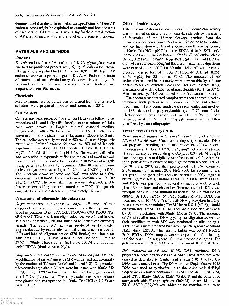
Fig. 3. Substrate recognition of oligonucleotides containing AP sites or MX-modified AP sites by HeLa AP-endonuclease. Lane 1: duplex dU:dG; lane 2:duplex dX:dG after 30 min incubation with partially purified HeLa AP-endonuclease; lane 3: duplex dX*:dG; lane 4: duplex dX*:dG after 30 minincubation with partially purified HeLa AP-endonuclease.
previously described and purified to apparent homogeneity (22),is Mg2+-dependent and hydrolyzes DNA 5' to the AP site. Asshown in Fig. 3, cleavage of the AP site by the HeLa AP-endonuclease produces the expected 12-mer oligonucleotide (lane2), while almost complete inhibition of cleavage is observed whenthe AP site is modified with MX (lane 4). The MX-modifiedAP site containing oligonucleotide is shown in lane 3. Therefore,the reaction of MX with AP sites renders them no longersubstrates for cleavage by the HeLa AP-endonuclease.
The substrate recognition characteristics of both bacterial andthe mammalian AP endonucleases were also confirmed by usingas substrate plasmid DNA containing multiple AP sites (data notshown).
Substrate recognition of oligonucleotides containing an APsite or MX-modified AP site by HeLa cell extractsTwo types of AP endonucleases have been found in mammaliancells: those hydrolyzing DNA 5' to the AP site, which are themajor mammalian AP endonucleases, and those cleaving DNA3' to the AP site via a ̂ -elimination mechanism. In order to assesswhether the inhibition by MX of the endonucleolytic activity ofthe 5' HeLa AP endonuclease could be extended to all the AP-endonucleases present in human cells, the uracil-containing DNAsubstrate was incubated with HeLa cell extracts in the presenceof MX.
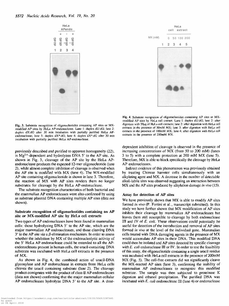
As shown in Fig. 4, the combined action of uracil-DNAglycosylase and AP endonuclease in extracts from HeLa cellscleaves the uracil containing substrate (lane 2). The cleavageproduct comigrates with the product of class II AP-endonucleases(data not shown) confirming that the major mammalian cellularAP endonucleases hydrolyze DNA 5' to the AP site. A dose-
HeLacell extract
MX(mM) o 50 100 200
1 2 3 4 5
Fig. 4. Substrate recognition of oligonucleotides containing AP sites or MX-modified AP sites by HeLa cell extract. Lane I: duplex dU:dG; lane 2: afterdigestion with 20/ig of HeLa cell extracts; lane 2: after digestion with HeLa cellextracts in the presence of 50mM MX; lane 3: after digestion with HeLa cellextracts in the presence of lOOmM MX; lane 4; after digestion with HeLa cellextracts in the presence of 200mM MX.
dependent inhibition of cleavage is observed in the presence ofincreasing concentrations of MX (from 50 to 200 mM) (lanes3 to 5) with a complete protection at 200 mM MX (lane 5).Therefore, MX is able to block specifically the cleavage by HeLaAP endonucleases.
Indirect evidence of this phenomenon was previously obtainedby treating Chinese hamster cells simultaneously with analkylating agent and MX. A decrease in the number of detectablealkali-labile sites was observed suggesting an interaction betweenMX and the AP sites produced by alkylation damage in vivo (15).
Assay for detection of AP sitesWe have previously shown that MX is able to modify AP sitesformed in vivo (P. Fortini et al., manuscript submitted). In thisstudy we have further shown that MX modification of AP sitesinhibits their cleavage by mammalian AP endonucleases butleaves them still susceptible to cleavage by both endonucleaseIII and IV of E. coli. These observations could potentially beuseful for detection of the introduction and removal of AP sitesformed in vivo at the level of the individual gene. Mammaliancells treated with DNA damaging agents in the presence of MXwould accumulate AP sites in their DNA. This modified DNAcould then be isolated and AP sites detected by specific cleavagewith E. coli endonuclease HI or IV. In order to test the feasibilityof this assay, the oligonucleotide containing a single uracil residuewas incubated with HeLa cell extracts in the presence of 200mMMX (Fig. 5). The cell-free extracts did not significantly cleavethe MX-reacted AP sites (lane 3), confirming the inability ofmammalian AP endonucleases to recognize this modifiedsubstrate. The sample was then subjected to proteinase Kdigestion and ethanol precipitation. The purified DNA wasincubated with E. coli endonuclease III (lane 4) or endonuclease
Downloaded from https://academic.oup.com/nar/article-abstract/19/20/5569/2387645by gueston 08 April 2018
Nucleic Acids Research, Vol. 19, No. 20 5573
= >o•oc111
HeLa extract -
MX (200 mM) .
1 2 3 4 5
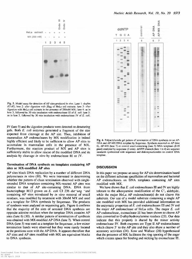
Fig. 5. Model assay for detection of AP sites produced in vivo. Lane 1: duplexdU:dG; lane 2: after digestion with 20/ig of HeLa cell extracts; lane 3: afterdigestion with HeLa cell extracts in the presence of 200mM MX; lane 4: as inlane 3, followed by 30 min incubation with endonuclease III of E. coli; lane 5:as in lane 3, followed by 30 min incubation with endonuclease IV of E. coli.
IV (lane 5) and the digestion products were detected on denaturinggels. Both E. coli enzymes generated a fragment of the sizeexpected from cleavage at the AP site. Thus, inhibition ofmammalian AP endonucleases by MX modification is indeedhighly efficient and likely to be sufficient to allow AP sites toaccumulate in mammalian cells in the presence of MX.Furthermore, the reaction product of MX and AP sites issufficiently stable to allow rescue of the modified DNA and itsanalysis by cleavage in vitro by endonuclease III or IV.
ddNTP
T G C A
1 2 3 4
oo
z x
Q. Q.
6 7
\
8
Fig. 6. Polyacrylamide gel pattern of termination of DNA synthesis on an AP-DNA and AP-MX DNA template by Sequenase. Synthesis occurred on AP (lane6), AP-MX (lane 7) or control uracil-containing (lane 5) DNA templates (0.05pmol) catalyzed by sequenase (3 units). ddNTP channels (lane 1 to 4) are sequencestandards synthesized with sequenase and dideoxynucleotides on control DNAtemplate.
Termination of DNA synthesis on templates containing APsites or MX-modified AP sitesAP sites block DNA replication by a number of different DNApolymerases in vitro (10). We were interested in determiningwhether the pattern of chain termination observed with single-stranded DNA templates containing MX-reacted AP sites wassimilar to that of AP site-containing DNA. DNA frombacteriophage M13 grown on E. coli CJ 236 durung "andcontaining AP sites introduced by in vitro removal of uracilresidues, was modified by treatment with 30mM MX and usedas a template for DNA synthesis by Sequenase. The productsof synthesis were analysed on sequencing gels. Figure 6 confirmsthat the majority of the sites of arrested DNA synthesis areopposite adenine residues when the template DNA contains APsites (lane 6) (10). A similar pattern of termination of synthesiswas obtained with MX-modified AP DNA (lane 7). When controlDNA (lane 5) was used as a template for Sequenase, a few minortermination bands were observed but they were rarely locatedat the positions seen with the AP DNA. It appears therefore thatAP sites and AP sites modified with MX are equivalent blocksto DNA synthesis.
DISCUSSION
In this paper we propose an assay for AP site determination basedon the different substrate specificities of mammalian and bacterialAP endonucleases on DNA templates containing AP sitesmodified with MX.
We have shown that E. coli endonucleases IH and IV are highlytolerant to the alkoxyamine modification of the C'i aldehyde,while the major HeLa AP endonuclease(s) do not cleave thissubstrate. Our use of a model substrate containing a single APsite modified with MX has provided additional information onthe enzymatic properties of E. coli endonucleases HI and IV andthe major AP endonuclease of HeLa cells. The major E. coliAP endonuclease, exonuclease III has been shown to cleave APsites converted to O-alkylhydroxylamine residues (23). Our dataindicate that this property is shared by the minor activityendonuclease IV. Both enzymes are Type II AP endonucleaseswhich cleave 5' to the AP site and they also share a number ofaccessory activities (24). Kow and Wallace (26) hypothesizedthat the presence of MX facilitates the ring opening of the sugarwhich creates space for binding and nicking by exonuclease III.
Downloaded from https://academic.oup.com/nar/article-abstract/19/20/5569/2387645by gueston 08 April 2018

5574 Nucleic Acids Research, Vol. 19, No. 20
A similar mechanism might be envisaged for the cleavage byE. coli endonuclease IV of MX-modified AP sites.
Our data obtained with E. coli endonuclease III show that thephosphodiester bond 3' to a MX residue can also be cleaved.In contrast to the hydrolytic mechanism of AP site cleavage ofexonuclease HI and endonuclease IV, endonuclease III catalysesa ^-elimination of the 3' phosphate (25,26). It has been proposed(26) that endonuclease III catalysis proceeds via the sequentialaction of its DNA glycosylase and AP lyase activities.Interestingly, the DNA glycosylase function recognizes assubstrate DNA containing urea residues which share structuralfeatures with MX, in particular a secondary amine group. Kowand Wallace (26) have suggested that the presence of thissecondary amine might facilitate the ring opening of the sugarleading to a transimination reaction in which a Schiff base wouldbe formed between the enzyme amino group and the C'|aldehyde of the sugar. The consequent release of MX would befollowed by a concerted chain cleavage.
A second AP lyase with associated DNA glycosylase activity,T4 endonuclease V, does not cleave AP sites modified with MX(27; our unpublished results). T4 endonuclease V specifically cleavesthe glycosyl bond of a thymine residue in a cyclobutane pyrimidinedimer (28). Its catalysis is not therefore facilitated by a secondaryamine of the type produced by MX modification of AP sites. Theseobservations indicate that two different reaction mechanisms existfor AP lyases (24). In this regard it has been proposed that T4endonuclease V cleaves by a j3, S-elimination (29).
A partially purified HeLa AP endonuclease does not recognizeas substrate AP sites adducted with MX. Similar results wereobtained by Sanderson and coworkers with a bovine APendonuclease (30). This endonuclease was unable to cleave theabasic site when a urea residue or an alkoxyamine group wasattached to the C'i position of the abasic site. Moreover,inhibition of cleavage at AP sites by MX is also observed withHeLa cell extracts suggesting that other forms of AP endonucleasein HeLa cells are either present in very low levels or are alsoinhibited by MX modification of AP sites.
The interest in quantitation of DNA damage at gene level hasbeen heightened by the accumulation of evidence ofheterogeneous distribution and repair of DNA damage inmammalian genome (31). Quantitation of pyrimidine dimers indefined sequences has been achieved using T4 endonuclease V(32). Data from these assays have led to the suggestion that UVphotodimers are repaired preferentially in active genes in bothbacterial and mammalian cells (33,34). Attempts have been madeto develop a quantitative method to assay the introduction of APsites into DNA. These lesions have been detected in specificsequences by their conversion to alkali-labile sites (35) and bya 32P end-labelling assay (36). The first method has beenproposed as a general tool to quantify abasic sites at the genelevel, but the detection limit of this assay is defined by the rateof resealing of the AP sites in vivo, which is a fast process. Wepropose the use of MX to convert AP sites formed in vivo intolesions which are resistant to cleavage by the endogenousmammalian AP endonuclease(s). The abasic sites reacted withMX can then be quantitated in Southern blots of specificrestriction fragments of isolated DNA following specific cleavagewith E. coli endonuclease HI or IV. This approach should allowthe quantitative detection of AP sites formed within cells in vivo.
In addition, our data indicate that MX-modified AP sites areeffective blocks to DNA replication in vitro and are equivalentto unmodified AP sites in this regard. Such modified AP sites
might be localized at the nucleotide level in episomally maintainedDNA by assaying the ability of purified molecules to block DNAsynthesis in vitro or in endogenous genes by a variation of theligase-mediated PCR (37). Our proposed methods should be ofgeneral value for studies of any kind of damage which producesabasic sites in DNA. These sites are common intermediates incytotoxicity and mutagenesis by a variety of chemical and physicalcarcinogens.
ACKNOWLEDGMENTS
We thank I.Goldsmith for oligonucleotide synthesis and C.Jonesand A.M. Pedrini for preparations of AP endonucleases. Thiswork has been partially supported by the E.E.C grantEV4V-044-I(A).
REFERENCES
1. Sakumi, K. and Sekiguchi, M. (1990) Mutat. Res., 236, 161-172.2. Kunkel, T.A., Shearman, C.W. and Loeb, L.A. (1981) Nature, 291,
349-351.3. Strauss, B., Rabkin, S., Sagher, D. and Moore, P. (1982) Biochimie, 64,
829-838.4. Schaaper, R.M., Glickman, B.W. and Loeb, L.A. (1982) Mutat. Res., 106,
1-9.5. Gentil, A., Margot, A. and Sarasin, A. (1984) Mutat. Res., 129, 141 - 147.6. Franklin, W.A. and Lindahl, T. (1988) EMBOJ., 7, 3617-3622.7. Loeb, L.A. and Preston, B.D. (1986) Ann. Rev. Genet., 20, 201-230.8. Schaaper, R.M., Kunkel, T.A. and Loeb, L.A. (1983) Proc. Nat!. Acad.
Sci. USA, 80, 487-491.9. Boiteux, S. and Laval, J. (1982) Biochemistry, 21, 6746-6751.
10. Sagher, D. and Strauss, B. (1983) Biochemistry, 22, 4518-4526.11. Loeb, L.A. (1985) Cell, 40, 483-484.12. Kohn, K.W. (1979) Methods Cancer Res., 16, 291-345.13. Talpaert-Borle', M. and Liuzzi, M. (1983) Biochim. Biophys. Acta, 740,
410-416.14. Liuzzi, M. and Talpaert-Borle1, M. (1985)7. Biol. Chem., 260, 5252-5258.15. Fortini, P., Bignami, M. and Dogliotti, E. (1990)Mutat. Res., 236, 129-137.16. Ljungquist, S. (1977) J. Biol. Chem., 252, 2808-2814.17. Lindahl, T., Ljungquist, S., Siegert, W., Nyberg, B. and Sperens, B. (1977)
J. Biol. Chem., 252, 3286-3294.18. Li, J.J. and Kelly, T.J. (1985) Mol. Cell. Biol., 5, 1238-1246.19. Bignami, M. and Lane, D.P. (1990) Nucl. Acid Res., 18, 3785-3793.20. Sambrook, J., Fritsch, E.F. and Maniatis, T. (1989), Molecular cloning.
A laboratory manual. Cold Spring Harbor University Press, Cold SpringHarbour.
21. Coombs, M.M. and Livingston, D.C. (1969) Biochim. Biophys. Acta, 174,161-173.
22. Kane, CM. and Linn, S. (1981) J. Biol. Chem., 256, 3405-3414.23. Kow, Y.W. (1987) Biochemistry, 28, 3280-3287.24. Doetsch, P.W. and Cunningham, R.P. (1990) Mutat. Res., 236, 173-201.25. Bailly, V. and Verly, W.G. (1987) Biochem. J., 242, 565-572.26. Kow, Y.K. and Wallace, S.S. (1987) Biochemistry, 26, 8200-8206.27. Liuzzi, M., Weinfeld, M. and Paterson, M.C. (1987) Biochemistry, 26,
3315-3321.28. Gordon, L.K. and Haseltine, W.A. (1980) J. Biol. Chem., 255,
12047-12050.29. Bailly, V., Seme, B. and Verly, W.G. (1989) Biochem. J., 259, 751 -759.30. Sanderson, B.J.S., Chang, C , Grollman, A.P. and Henner, W.D. (1989)
Biochemistry, 28, 3894-3901.31. Bohr, V.A., Phillips, D.H. and Hanawalt, P.C. (1987) Cancer Res., 47,
6426-6436.32. Bohr, V.A., Smith, C.A., Okumoto, D.S. and Hanawalt, P.C. (1984) Cell.
40, 359-369.33. Mellon, I., Bohr, V.A., Smith, C.A. and Hanawalt, P.C. (1986) Proc. Nail.
Acad. Sci. U.S.A., 83, 8878-8882.34. Mellon, I. and Hanawalt, P.C. (1989) Nature, 342, 95-98.35. Scicchitano, D.A. and Hanawalt. P.C. (1989) Proc. Nail. Acad. Sci. U.S.A..
86, 3050-3054.36. LeDoux, S.P., Patton, N.J., Nelson, J.W., Bohr. V.A. and Wilson, G.L.
(1990)7. Biol. Chem., 265. 14875-14880.37. Pfeifer. G.P., Drouin. R.. Riggs, A.D. and Holmquist, G.P. (1991) Proc.
Nail. Acad. Sci. USA, 88, 1374-1378.
Downloaded from https://academic.oup.com/nar/article-abstract/19/20/5569/2387645by gueston 08 April 2018