program book a5 for print - fornerislab.unipv.itfornerislab.unipv.it/aics2017/program.pdf ·...
TRANSCRIPT

School Program
and
Book of Abstracts
Welcome to AIC School 2017
We are delighted to welcoming you to Pavia for the
International school “Bridging the gap between Cryo-EM and
Crystallography”. This booklet contains the final version of the
school program, the abstracts of all talks, and information you
may find useful for your stay in Pavia during the following days.
Please do not hesitate to get in touch with the local staff in case
of special needs.
We wish you a scientifically profitable and pleasant stay in
Pavia during the next days.
The AICS2017 Organizers

AICS2017 Program Sunday, Sept 3rd, 2017 (University of Pavia, Aula del ‘400) 16:00-17:30 - Participant registration and accommodation check-in 17:45-18:00 - Welcome by Organizing Committee 18:00-19:00 - OPENING LECTURE (Chair: A. Mattevi): Werner Kuhlbrandt (MPI Frankfurt) Membrane protein complexes and the resolution revolution in cryoEM 19:00-20:30 - Welcome Party Monday, Sept 4th, 2017 (IUSS, Sala del Camino) "SAMPLES AND INSTRUMENTS" (Chair: B. Vallone) 09:00-09:40 - Tillmann Pape (Imperial, UK) THEORY: Introduction to cryo-electron microscopy: from biological sample to 3D structure the beauty of cryo-EM 09:40-10:25 - Paolo Swuec (University of Milano, IT) REAL LIFE: Overview of Sample Preparation for Single-particle EM 10:25-10:55 - Frederic Leroux (Leica Microsystems) Frozen in milliseconds, but what's next? 10:55-11:20 - Coffee Break 11:20-12:00 - Marta Carroni (SciLife Lab, SE) THEORY: Electron microscope architecture and image formation 12:00-12:30 - Sacha De Carlo (DECTRIS) Detector technologies for high-resolution TEMs 12:30-14:00 - Lunch (University of Pavia, Mensa Centrale)

Monday, Sept 4th, 2017 (IUSS, Sala del Camino) "THE SINGLE-PARTICLE EXPERIMENT" (Chair: S. Tarantino) 14:00-14:40 – Fabrizio Martino (CIB Madrid, ES) REAL LIFE: Theory to practice: grid preparation 14:40-15:30 - Arjen J. Jakobi (EMBL Heidelberg, DE) THEORY: Fundamentals of cryo-EM image processing 15:30-16:00 - Andy Jarwood (JEOL) Introduction to a New Cryo-TEM generation and associated techniques 16:00-16:30 - Coffee Break 16:30-17:10 - Fabrizio Martino (CIB Madrid, ES) REAL LIFE: Low cost cryo-EM... the Madrid example 16:30-17:10 - Marta Carroni (SciLife Lab, SE) REAL LIFE: Different mechanisms but conserved functions: AAA+ protein activity control studied by cryo-EM at various resolutions 17:10-18:00 - Discussion, Q&A Tuesday, Sept 5th, 2017 (IUSS, Sala del Camino) "BEYOND SINGLE PARTICLE" (Chair: M. Gemmi) 09:00-09:40 - Christoph A. Diebolder (NeCEN, NL) THEORY AND REAL LIFE: Cryo electron tomography part I: Concepts and practical considerations for sample preparation and data collection 09:40-10:20 - Mauro Gemmi (IIT Pisa, IT) THEORY: Electron Diffraction Tomography of Protein Nanocrystals 10:20-10:50 - Max Maletta (FEI) Single particle screening and data acquisition made easy 10:50-11:20 - Coffee Break 11:20-12:05 - Selected young speaker: Jérôme Bürgi (EMBL Hamburg) Towards the structural discovery of the peroxisomal translocation of functional matrix proteins
12:05-12:30 - Selected young speaker: Digant Nayak (NUS Singapore) Structural basis for the indispensable role of a unique zinc finger motif in LNX2 ubiquitination 12:30-14:00 - Lunch (University of Pavia, Mensa Centrale) Tuesday, Sept 5th, 2017 (IUSS, Sala del Camino) "SOFTWARE PACKAGES AND DATA PROCESSING" (Chair: F. Forneris) 14:00-14:40 - B. Tom Burnley (CCP-EM, STFC, UK) THEORY: Atomic model building and refinement in CCP-EM 14:40-15:20 - Christoph A. Diebolder (NeCEN, NL) THEORY: Cryo electron tomography part II: Software packages for data acquisition, reconstruction and subvolume averaging 15:20-16:00 - Pablo Conesa (I2PC Madrid, ES) THEORY & REAL LIFE: A software framework toward integration, reproducibility and validation in 3D electron microscopy 16:00-16:30 - Coffee Break 16:30-17:10 - Arjen J. Jakobi (EMBL Heidelberg, DE) THEORY & REAL LIFE: Building, refining and validating atomic models in cryo-EM density maps 17:10-17:40 - Fabrizio Martino (CIB Madrid, ES) REAL LIFE: Processing toolbox... one problem, many solutions 19:00-22:00 - Social Dinner (Granai Certosa) Wednedsay, Sept 6th, 2017 (IUSS, Sala del Camino) "BIOLOGY WITH CRYO-EM" (Chair:G. Zanotti) 09:00-09:40 - Filippo Mancia (Columbia University, US) The Molecular Mechanisms Underlying Cellular Uptake of Vitamin A 09:40-10:20 - Alessandro Vannini (ICR London, UK) Structural basis of RNA Polymerase III transcription initiation

10:20-11:00 - Arjen J. Jakobi (EMBL Heidelberg, DE) Structural basis of selective autophagy receptor assemblies 11:00-11:30 - Coffee Break 11:30-11:55 - Selected young speaker: Andrew Hedger (University of Queensland) Introduction to protein filament characterisation 11:55-12:20 - Selected young speaker: Ilaria Peschiera (CNB Madrid) Deciphering the oligomerization role of the bacterial lipid rafts scaffold protein by Cryo-EM 12:30-14:00 - Lunch (University of Pavia, Mensa Centrale) Wednedsay, Sept 6th, 2017 (IUSS, Sala del Camino) "CRYO-EM FACILITIES" (Chair: M. Bolognesi) 14:00-14:25 - Martin Walsh (Diamond eBIC, UK) Integrating cryo-EM at Diamond... Challenges, benefits and opportunities 14:25-14:50 - Guy Schoehn (IBS Grenoble, FR) Current and future state of the EPN cryoEM facility 14:50-15:15 - Marta Carroni (SciLife La, SE) A Swedish facility for cryo-EM 15:15-15:40 - Paolo Swuec (University of Milano, IT) Introducing the Cryo-Electron Microscopy Lab in Milan (CEMiL) 15:40-16:05 - Ludo Renault (NeCEN, NL) Operations at NeCEN 16:05-16:30 - Coffee Break 16:30-18:30 - Social Program 18:30-19:30 - CLOSING LECTURE (Chair: F. Mancia): Wayne Hendrickson (Columbia University, USA) Electron Microscopy and X-ray Crystallography of Ion Channels that Control Muscle Excitation 19:30-20:00 - Concluding Remarks

AICS2017 Organizers:
Scientific Organizing Committee: - Federico Forneris (University of Pavia)
- Andrea Mattevi (University of Pavia)
- Filippo Mancia (Columbia University, USA)
- Martino Bolognesi (University of Milan)
- Mauro Gemmi (Italian Institute of Technology)
- Beatrice Vallone (Sapienza University of Rome)
Local Organizing Committee (@ University of Pavia): - Federico Forneris
- Andrea Mattevi
- Claudia Binda
- Serena Chiara Tarantino
Treasurer: - Simona Galli (Insubria University)
AIC Teaching Committee - Anna Grazia Moliterni (CNR Bari)
- Serena Chiara Tarantino (University of Pavia)
- Chiara Massera (University of Parma)
- Emanuela Schingaro (University of Bari)
Membrane protein complexes and the resolution revolution
in cryoEM
Werner Kühlbrandt
Max Planck Institute of Biophysics, Department of Structural Biology, Max-von-Laue Str. 3, 60438 Frankfurt am Main, Germany.
e-mail:[email protected] With the arrival of a new generation of electron detectors and image
processing software, cryoEM of biological macromolecules has entered a
new era (Kühlbrandt, 2014). We have used high-resolution cryoEM to
determine the structures of mitochondrial membrane protein complexes,
including ATP synthase dimers from Polytomella (Allegretti et al, 2015)
and the yeast Yarrowia lipolytica (Hahn et al, 2016), and of the bovine
respirasome (Sousa et al, 2016). Very recently, we unravelled the
membrane insertion mechanism of pneumolysin, the pore-forming toxin of
the human pathogen Streptococcus pneumoniae, by a combination of cryo-
EM and x-ray crystallography (van Pee et al, 2017). Applying the new
detector technology to electron cryo-tomography, we obtained maps of
ATP synthase dimers in the inner mitochondrial membranes from a range
of organisms (Mühleip et al, 2016; 2017). Our results provide new insights
into how membrane protein complexes work in the membrane.
References Allegretti, M., Klusch, N., Mills, D.J., Vonck, J., Kühlbrandt, W. & Davies, K.M.
(2015). Horizontal membrane-intrinsic α-helices in the stator a-subunit of an F-type ATP synthase. Nature 521: 237-240. doi: 10.1038/nature14185
Hahn A et al, (2016). Structure of a Complete ATP Synthase Dimer Reveals the Molecular Basis of Inner Mitochondrial Membrane Morphology. Mol Cell 63:445-456. doi: 10.1016/j.molcel.2016.05.037

Kühlbrandt W (2014). The resolution revolution. Science 343: 1443-1444. doi:
10.1126/science.1251652 Mühleip, A. W., Joos, F., Wigge, C., Frangakis, A. S., Kühlbrandt, W., & Davies, K.
M. (2016). Helical arrays of U-shaped ATP synthase dimers form tubular cristae in ciliate mitochondria. PNAS, 113: 8442–8447. doi:10.1073/pnas.1525430113
Mühleip, A. W., Dewar, C. E., Schnaufer, A., Kühlbrandt, W., & Davies, K. M. (2017). In situ structure of trypanosomal ATP synthase dimer reveals a unique arrangement of catalytic subunits. PNAS 114: 992-997. doi: 10.1073/pnas.1612386114
Sousa, J. S., Mills, D. J., Vonck, J., & Kühlbrandt, W. (2016). Functional asymmetry and electron flow in the bovine respirasome. eLife, 5, e21290. doi:10.7554/eLife.21290
van Pee, K., Neuhaus, A., D’Imprima, E., Mills, D., Kühlbrandt, W., Yildiz, Ö. (2017). CryoEM structures of membrane pore and prepore complex reveal cytolytic mechanism of Pneumolysin. eLife 6: e23644. doi:10.7554/eLife.23644

THEORY: Introduction to cryo-electron microscopy: from biological sample to 3D structure
the beauty of cryo-EM
Tillmann Pape
Department of Life Sciences, Imperial College London, London SW7 2AZ, UK. e-mail: [email protected]
Cryo-electron microscopy (cryo-EM), the structural analysis of samples
embedded in vitreous ice, is a powerful method for determining three-
dimensional (3D) structures of biological specimens. Over the past two
decades, all technical aspects of cryo-EM have been considerably
improved, including hardware (microscopes, detectors, computers) and
software (image processing, automation). All these exciting new advances
have led to a more widespread use of the technique, as it is now on pace to
determine atomic resolution 3D structures. I will give a short overview of
the basic approaches but also attempt to illustrate that the beauty of cryo-
EM is in generating insights into the architecture of challenging biological
samples that cannot be visualised otherwise.

REAL LIFE: Overview of Sample Preparation for Single-particle EM
Paolo Swuec
Cryo-Electron Microscopy Lab, Pediatric Clinical Research Center Romeo and Enrica Invernizzi, Department of BioSciences,
University of Milano, Via Celoria 26 - 20133 (Italy). e-mail: [email protected]
Single-particle electron microscopy (EM) has the ability to unravel the
three-dimensional structure of biological molecules and assemblies by
imaging non-crystalline specimens. This technique mainly relies on
collecting hundreds of images of the specimen, each comprising of many
individual and randomly oriented particles. Imaging a material with
electrons at near-atomic resolution requires a thin specimen that is stable in
the vacuum of the transmission electron microscope. For the biological
specimen to be imaged it has to be embedded either in a layer of heavy
metal salt solution (negative staining EM) or a thin layer of vitreous ice
(Cryo-EM). In this talk, practical aspects of both sample preparation
techniques will be addressed as well as different strategies to overcome
problematic samples.
Frozen in milliseconds, but what’s next?
Frederic Leroux
Leica Microsystems – e-mail: [email protected]
Plunge freezing and cryo imaging of proteins and complexes has revealed
new details for understanding the machinery of the cell and how molecules
are involved in cellular processes. However, most eukaryotic cells and
tissue samples cannot be plunge frozen due to the rapid decay of the
cooling rate within the sample. High pressure freezing is currently the main
approach to vitrify larger samples (up to 200 µm) and to capture the
intrinsic changes in fine structure or cellular dynamics. In an effort to
further improve its cryo solutions, Leica developed a new cryo platform:
the EM ICE. This new generation cryo platform combines speed, reliability
and at the same time flexibility in order to facilitate research in various
scientific fields. It allows users to freeze samples within milliseconds and
even allows combining high pressure freezing with light or electrical
stimulation. It gives researchers the possibility to visualize highly dynamic
processes or structural changes of samples at a nanometer scale and with
millisecond precision. During this lecture a brief overview on high pressure
freezing and follow-up techniques will be given.

THEORY: Electron microscope architecture and image formation
Marta Carroni
Swedish Cryo-EM Facility, Science for Life Laboratory (SciLifeLab) Stockholm University, Box 1031, SE-171 21 Solna, Sweden. e-mail:
This lecture will quickly introduce you to the main components of the
electron microscope (electron sources, lenses, apertures, energy filter),
describe how the TEM image is formed and how it is affected by the
contrast transfer function (CTF).
Detector technologies for high-resolution TEMs
Sacha De Carlo
Dectris Ltd., Baden, Switzerland. e-mail: [email protected]
Better performing detectors are considered a key to further improvements
in the resolution of cryo-electron microscopy (Kühlbrandt, 2014). In our
talk, we will briefly cover the history of detection in its broadest meaning
in transmission electron microscopy. We will briefly discuss the
fluorescence screen, argentic film, CCD, CMOS and hybrid photon
counting technology. Concepts like Edge Spread Function, Modulation
Transfer Function and Detection Quantum Efficiency, and their importance
in assessing the quality of a TEM detector, will be discussed (Faruqi, 2007;
Meyer, 1998). DECTRIS revolutionized macromolecular crystallography
with its PILATUS detector, since its introduction in 2007. Ten years after
that, DECTRIS is looking into new markets, and we will discuss the
potential of DECTRIS hybrid photon counting detection technology for
high-resolution TEM.
References Kühlbrandt W (2014). The resolution revolution. Science 343: 1443-1444. doi:
10.1126/science.1251652 Faruqi A and Henderson R (2007). Electronic detectors for electron microscopy. Curr
Opin Struct Biol 17: 549-55. doi: 10.1016/j.sbi.2007.08.014 Meyer R and Kirkland A (1998). The effects of electron and photon scattering on
signal and noise transfer properties of scintillators in CCD cameras used for electron detection. Ultramicroscopy 75: 23-33. doi: 10.1016/S0304-3991(98)00051-5

THEORY: Fundamentals of cryo-EM image processing
Arjen J. Jakobi
European Molecular Biology Laboratory (EMBL), Heidelberg, Germany. e-mail: [email protected]
This lecture covers the fundamental principles underlying electron cryo-
microscopy (cryo-EM), starting with an introduction to Fourier transforms
and the principles of image formation. Fourier transforms are a
fundamental mathematical tool for the analysis of periodic signals in which
arbitrary functions are expanded into a combination of simple waves. We
will introduce the basic principles of Fourier transforms and reciprocal
space, discuss their properties and illustrate their use in diffraction
experiments and cryo-EM image processing. We will address sampling and
Nyquist limit, treat the convolution theorem and its relationship to
correlation and look at fundamental filtering operations. We further explore
the process of image formation in the electron microscope, starting at the
interaction of electrons with matter and different forms of image contrast,
the effect of electromagnetic lenses, contrast transfer function (CTF) and
defocus, and briefly introduce the central-section (or projection-slice)
theorem.
Selection of recommended reading Harburn G, Taylor CA and Welberry TR (1975) Atlas of Optical Transforms. (Bell
Publishing). Stewart, M (1988) Introduction to the computer image processing of electron
micrographs of two-dimensionally ordered biological structures. J. Elec. Microsc. Tech. 9: 301–324. doi: 10.1002/jemt.1060090403
Glaeser RM, Downing KH, DeRosier D, Chiu W and Frank J (2007) Electron Crystallography of Biological Macromolecules. (Oxford University Press).
Steward EG (2004) Fourier Optics: An Introduction. (Halsted Press)

Introduction to a New Cryo-TEM generation and associated techniques
Andy Jarwood
JEOL (UK) Ltd
Recent advances in cryo-TEM design and the introduction of direct detector cameras have both contributed greatly to the resolution revolution in protein structure imaging. JEOL has recently announced its new CRYO ARM™ instruments which are designed to take the resolution of protein structures to a new level. This presentation will explain the features of the JEOL Cryo-TEM range which is designed to improve the performance specifically when observing low contrast and beam sensitive speciments. Examples of Cryo-TEM data from protein samples will be used to exemplify how the Omega filter and the Cold FEG electron gun, as well as the Hole Free Phase Plate, can be used to greatly improve the data quality for single particle analysis and cryo-TEM tomography. JEOL's latest conventional cryo-TEM will also be presented to show how our 200kV CFEG TEM can be used to screen speciments very quickly at a high structural resolution prior to acquiring high resolution data in the fully automatic CRYO ARM™ systems.

REAL LIFE: Low cost cryo-EM... the Madrid example
Fabrizio Martino
Centro de Investigaciones Biológicas (CIB), C/ Ramiro de Maeztu 9, 28040 ∙ Madrid, Spain. e-mail: [email protected]
Yes! You can still do high resolution cryo-EM even if you do not own a
Krios, a Vitrobot and a cluster! We will discuss together affordable ways to
prepare and screen grids, to collect data and process them.

REAL LIFE: Different mechanisms but conserved functions: AAA+ protein activity control
studied by cryo-EM at various resolutions
Marta Carroni
Swedish Cryo-EM Facility, Science for Life Laboratory (SciLifeLab) Stockholm University, Box 1031, SE-171 21 Solna, Sweden. e-mail:
Hsp100 AAA+ unfoldases play a crucial role in protein quality control.
They are involved in protein disaggregation and degradation and for full
activation they require cooperating adaptor proteins that target substrates
and concurrently stimulate the AAA+ ATPase activity. In the absence of
substrate and protein cofactors, the ATPases are kept in a repressed state by
regulatory coiled-coil domains. Using cryo-EM and single-particle
analysis, we dissected the molecular basis of this activity control and we
identify molecular switches that act in a conserved manner among various
AAA+ proteins. This research example is aimed to show what type of
biological information can be extrapolated from cryo-EM maps at different
resolutions.

THEORY AND REAL LIFE: Cryo electron tomography part I: Concepts and practical considerations for sample
preparation and data collection
Christoph A. Diebolder
Netherlands Centre for Electron Nanoscopy (NeCEN), Gorlaeus Laboratory Einsteinweg 55, 2333 CC Leiden, The Netherlands.
e-mail: [email protected]
This first of two talks about electron tomography will cover two major
topics. First, the theoretical concepts of tomography will be explained by
describing similarities and differences between this technique and the
earlier covered single particle analysis (SPA). Understanding these
differences and knowing about shortcomings and benefits of either of these
techniques will help the students to decide which of both is to be applied
for answering their individual research question.
The second part of this talk will deal with practicalities. Different methods
of sample preparation will be explained, followed a description of the data
acquisition workflow. This section will include considerations about hard-
and software setup and all parameters relevant for successful acquisition of
raw tilt series.
THEORY: Electron diffraction tomography of proteins nanocrystals
Mauro Gemmi
Italian Institute of Tecnology (IIT), Center for Nanotechnology @NEST, Piazza S.
Silvestro 12, 56127 Pisa, Italy. e-mail: [email protected]
The development of the electron diffraction tomography method allows
nowadays to collect three dimensional electron diffraction data on nano
crystals obtaining at the same time a large reciprocal space coverage and
quasi kinematical intensities suitable for structure solution. The data
collection is performed by recording a sequence of electron diffraction
patterns while the crystal is rotating around the goniometric axis. The
illuminated area of the crystal can be as small as few hundreds nanometers.
Thanks to the appearance on the market of very sensitive detector for
electron diffraction this technique has been successfully applied to proteins
crystals. By recording patterns under electron doses of less than 0.1 e-/(Å2s)
it is possible to cover 80° or more of reciprocal space with a resolution of
2.0 Å without damaging seriously the crystal. The method can be
alternative to cryoEM when the proteins are very small or when they can
be crystallized in crystals too small to be investigated with the usual x-ray
diffraction methods. During the lecture the theoretical and the experimental
basis of the method will be treated in details together with some successful
examples.
Single particle screening and data acquisition made easy
Max Maletta
FEI - Thermo Fisher Scientific, Achtseweg Noord 5, Building AAE, 5651 GG Eindhoven, The Netherlands. e-mail: [email protected]
During the last few years cryo electron microscopy (cryo-EM) and Single Particle Analysis (SPA) have grown from techniques able to produce low-resolution structures of protein complexes (aka blobology) to tools capable of achieving atomic and quasi-atomic resolution for complexes that nobody could ever solve with any other technique. This incredible leap forward has made possible by introduction and adoption of new tools, in particular direct electron detectors (DED), ultra-stable cryo-microscope, such as Titan Krios, and the adoption of new SW for automatic data collection and processing. Cryo-EM benefit of specific advantages, respect to other structural biology techniques such as NMR and X-ray diffraction: Crystallization or isotopic labelling is not needed. Amount of sample required is two orders of magnitude lower. With cryo-EM different functional conformation of a complex may be sorted
out. Those advantages make of cryo-EM a very useful technique to be integrated with X-ray and NMR for structure-based drug design. So it is no surprise that many structural biology groups all over the world are seeking access to this technology in order to find answers to their most relevant biological questions. Nevertheless most new comers to the field are struggling to overcome the adoption barrier that this technique may pose in term of: sample preparation and screening, automatic data acquisition and progressive users training.
In this presentation we will introduce the novelties of our SPA workflow that are aimed to lower the adoption barrier for researchers that are about to get started with cryo-EM. In particular we will describe the Glacios™ Cryo-TEM: A 200kV X-FEG autoloader-provided system capable
of automatic screening of multiple grids. Provided of smaller footprint and lower price.
The new KriosTM G3i: The latest Krios version with improved automation, increased cryo-performance and higher throughput.
An outcome-based support that consists of: remote and On-site application support, remote monitoring, workflow validation, spa app and much more.

THEORY: Atomic model building and refinement in CCP-EM
B. Tom Burnley
Science and Technology Facilities Council (STFC), Harwell Research Campus, Oxfordshire, UK. e-mail: [email protected]
The CCP-EM (Collaborative Computational Project for Electron cryo-
Microscopy) software suite contains a number of programs for the
building, refining and assessing of atomistic models in 3D volumes derived
from EM. The lecture will review the CCP-EM framework and suggest
optimal program usage based on the resolution of the experimental data. It
will cover the following programs: Buccaneer / Nautilus for automated
model building; Molrep for rapid docking in high-resolution datasets,
Refmac for high-resolution refinement; Dock-EM for medium resolution
exhaustive docking and Flex-EM for medium resolution refinement. Tools
for validation will also be covered.
Selection of recommended reading Burnley T et al (2017) Recent developments in the CCP-EM software suite. Acta
Crystallogr D Biol Crystallogr 73: 469–477. doi: 10.1107/S2059798317007859 Brown A et al (2015) Tools for macromolecular model building and refinement into
electron cryo-microscopy reconstructions. Acta Crystallogr D Biol Crystallogr 71: 136–153. doi: 10.1107/S1399004714021683
Joseph AP et al (2016) Refinement of atomic models in high resolution EM reconstructions using Flex-EM and local assessment. Methods 100: 42-49. doi: 10.1016/j.ymeth.2016.03.007
Joseph AP et al (2017) Improved metrics for comparing structures of macromolecular assemblies determined by 3D electron-microscopy. J. Struct. Biol (in press). doi: 10.1016/j.jsb.2017.05.007
Murshudov GN (2016) Refinement of Atomic Structures Against cryo-EM Maps. Meth Enzymol 579: 277–305. doi: 10.1016/bs.mie.2016.05.033

THEORY: Cryo electron tomography part II: Software packages for data acquisition, reconstruction and subvolume
averaging
Christoph A. Diebolder
Netherlands Centre for Electron Nanoscopy (NeCEN), Gorlaeus Laboratory Einsteinweg 55, 2333 CC Leiden, The Netherlands.
e-mail: [email protected]
This talk will start with a detailed description of the data processing
workflow in (cryo) electron tomography which can be divided in
preprocessing of the raw tilt series, tilt series alignment, tomogram
reconstruction, tomogram post processing, subvolume averaging and
quality assessment.
The last part of this lecture will try to give an overview over the landscape
of available software packages in electron tomography. This overview will
largely be based on the recommendable “Wikibook: Software tools for
molecular microscopy” (Smith, 2008).
References: Smith, Ross, and Bridget Carragher (2008) Software tools for molecular microscopy.
Journal of structural biology 163.3 (2008): 224-228.

THEORY AND REAL LIFE: A software framework toward integration, reproducibility and validation in 3D electron
microscopy
Pablo Conesa
Centro Nacional de Biotecnología, Consejo Superior de Investigaciones Cientificas, Madrid, Spain. e-mail: [email protected]
In the past few years, 3D electron microscopy (3DEM) has undergone a
revolution in instrumentation and methodology. One of the central players
in this wide-reaching change is the continuous development of image
processing software. Here we present Scipion, a software framework for
integrating several 3DEM software packages through a workflow-based
approach. Scipion allows the execution of reusable, standardized, traceable
and reproducible image-processing protocols. These protocols incorporate
tools from different programs while providing full interoperability among
them. Scipion is an open-source project that can be downloaded from
http://scipion.cnb.csic.es.

THEORY AND REAL LIFE: Building, refining and validating atomic models in cryo-EM density maps
Arjen J. Jakobi
European Molecular Biology Laboratory (EMBL), Heidelberg, Germany. e-mail: [email protected]
The interpretation of the cryo-EM density by atomic models is typically the last step of the structure determination process. Atomic models are worth much more than making for a pretty picture. Frequently, important mechanistic insight is based on those models and producing reliable and accurate atomic models is therefore of great importance. This lecture provides an introduction to strategies and tools developed for the building, refinement and validation of atomic models in cryo-EM density maps. We start with a discussion of resolution of three-dimensional cryo-EM reconstructions and will treat post-processing procedures for map representations such as sharpening, blurring and filtering. We will consider the fundamental challenges for model building in cryo-EM maps, introduce available methods and tools that help address them, work out the similarities and differences to related procedures in X-ray crystallography and discuss some strategies and ‘best practices’ with challenging examples. We will further explore basic aspects of coordinate refinement strategies in real and reciprocal space and briefly touch upon more advanced issues such as posed e.g. by local variations in effective resolution of cryo-EM maps. Equivalently important are methods for validation of the built atomic models. We will discuss various metrics that assess agreement of the models with the experimental data or rank and validate model geometries based on prior knowledge and expectations from high-resolution structures deposited in community databases.
Selection of recommended reading Brown A et al (2015) Tools for macromolecular model building and refinement into
electron cryo-microscopy reconstructions. Acta Cryst D 71: 136–153. doi: 10.1107/S1399004714021683
Murshudov GN (2016) Refinement of Atomic Structures Against cryo-EM Maps. Meth. Enzymol. 579: 277–305. doi: 10.1016/bs.mie.2016.05.033
DiMaio F, Zhang J, Chiu W and Baker D (2013) Cryo-EM model validation using independent map reconstructions. Protein Sci 22: 865–868. doi: 10.1002/pro.2267
Falkner B and Schröder GF (2013) Cross-validation in cryo-EM-based structural modeling. PNAS 110, 8930–8935. doi: 10.1073/pnas.1119041110
REAL LIFE: Processing toolbox... one problem, many solutions
Fabrizio Martino
Centro de Investigaciones Biológicas (CIB), C/ Ramiro de Maeztu 9, 28040 ∙ Madrid, Spain. e-mail: [email protected]
In this presentation, we will compare examples of motion correction, CTF
estimation, beam damage compensation, 2D and 3D classification and
atomic modeling.

The Molecular Mechanisms Underlying Cellular Uptake of Vitamin A
Yunting Chen1, Oliver B. Clarke2, Brianna Costabile1, Jonathan Kim1, Paul T. Wilder3, David Weber3, Wayne A. Hendrickson2, Filippo Mancia1
Departments of 1Physiology & Cellular Biophysics, and 2Biochemistry & Molecular Biophysics, Columbia University. 3University of Maryland School of
Medicine. USA. e-mail: [email protected]
Many biological processes, including the visual cycle and embryonic development are crucially dependent on an adequate supply of Vitamin A (Goodman, 1984). A cell receives Vitamin A either directly from food intake, or from the liver, released as retinol (vitamin A alcohol; ROH) bound to its carrier retinol-binding protein (RBP, also termed RBP4), which allows the highly hydrophobic retinol to circulate in plasma (Soprano, 1994). Once inside the cell, retinol binds specific intracellular carriers, specifically cellular retinol-binding proteins (CRBPs; Li, 2014). How retinol is released from RBP and internalized by target cells has been the subject of intense debate. In a landmark study in 2007, the RBP receptor was cloned (Kawaguchi, 2007) and found to be a protein encoded by a gene previously identified and classified as stimulated by retinoic acid gene 6 (STRA6). STRA6 is a 75 kDa protein with 9 predicted TM segments, showing no sequence similarity to any known transporter, channel or receptor. However, since then progress in understanding how this system works at a molecular level has been hampered by the absence of an atomic model of STRA6. We present the structure of zebrafish STRA6 determined to 3.9Å resolution by single-particle cryoelectron microscopy (Chen, 2016). STRA6 displays one intramembrane and nine transmembrane helices in an intricate dimeric assembly (Fig. 1). Unexpectedly, calmodulin is bound tightly to STRA6 in a non-canonical arrangement. Residues identified with RBP binding map to an arch-like structure that covers a deep lipophilic cleft. This cleft is open

to the membrane, suggesting a possible mode for internalization of retinol via direct diffusion into the lipid bilayer. We also discuss functional, biophysical and structural data aimed at understanding how STRA6-mediated retinol uptake may occur, and how the process may be regulated by Ca2+. In particular, we present recent structural data on STRA6 reconstituted in lipid-filled nanodiscs, in different CaMbound states (apo-CaM and Ca2+-CaM), showing how STRA6 and the surrounding lipid bilayer interact to facilitate the transfer of retinol.
References: Goodman, D.S. (1984) Vitamin A and retinoids in health and disease. N Engl J Med
310: 1023-31. doi: 10.1056/NEJM198404193101605 Soprano DR and Blaner WS (1994) Plasma Retinol-binding protein. in The Retinoids,
Biology, Chemistry and Medicine (Raven, New York). Li Y et al (2014) The multifaceted nature of retinoid transport and metabolism.
Hepatobiliary Surg Nutr 3: 126–139. doi: 10.3978/j.issn.2304-3881.2014.05.04 Kawaguchi R et al (2007) A membrane receptor for retinol binding protein mediates
cellular uptake of vitamin A. Science 315: 820-825. doi: 10.1126/science.1136244 Chen Y et al (2016) Structure of the STRA6 receptor for retinol uptake. Science 353:
aad8266. doi: 10.1126/science.aad8266
Figure 1. The cryo-EM structure of STRA6 in complex with calmodulin. STRA6 is the receptor for retinol (vitamin A) bound to its carrier retinol-binding protein. The protein is a dimer (in red), and each protomer binds one molecule of calmodulin (in gold). Two putative cholesterol molecules are shown in green. The approximate location of the membrane is displayed, in cream color.

Structural basis of RNA Polymerase III transcription initiation
Alessandro Vannini
The Institute of Cancer Research (ICR), 123 Old Brompton Road, London SW7 3RP, UK. e-mail: [email protected]
RNA Polymerase (Pol) III is the eukaryotic nuclear enzyme devoted to the transcription of essential non-coding RNAs, including the entire pool of tRNAs, the 5S ribosomal RNA and the U6 spliceosomal RNA. Initiation of gene transcription by RNA Pol III requires the activity of TFIIIB, a complex formed by Brf1, TBP and Bdp1. TFIIIB is required for recruitment of Pol III and to promote the transition from a closed to an open Pol III pre-initiation complex (PIC), a process stimulated by the activity of the Bdp1 subunit. Here we present two distinct cryo-EM reconstructions of an open RNA Pol III PIC at 3.8 Å and 3.3 Å, and a reconstruction of apo RNA Pol III at better than 3.0 Å. The DNA in the RNA Pol III PIC is almost fully enclosed by the TFIIIB complex. Bdp1 stabilize the assembly of the RNA Pol III PIC and induce an allosteric change that positions the winged helix domains of the C34 subunit at the upstream edge of the transcription bubble, destabilizing the DNA double-strand, analogously to the transcription factor TFIIF in the Pol II system. The structures reveal that the initiating event for DNA melting is the flipping of a specific base of the non-template strand, which is stabilized by an invariably conserved pocket in the clamp domain of RNA Pol III, a process so far only observed in bacterial polymerases. The subunit C82 further stabilizes the single stranded non-template strand, similarly to TFIIE. Furthermore, the zinc-ribbon domain of Brf1 freezes a mobile region of RNA Pol III in a conformation that allows engagement of the template strand in the active site. The structures presented unravel the molecular mechanisms underlying RNA Pol III transcription initiation, highlighting the specific features of this highly efficient essential machinery but also the general conserved mechanisms of gene transcription initiation.

Structural basis of selective autophagy receptor assemblies
Arjen J. Jakobi
European Molecular Biology Laboratory (EMBL), Heidelberg, Germany. e-mail: [email protected]
We illustrate the general concept and practical implementation of (single particle-based) helical reconstruction using recent cryo-EM structures of filamentous autophagy receptor assemblies determined in our laboratory. Macroautophagy is a catabolic pathway that targets large, long-lived structures such as protein aggregates, damaged organelles and pathogens for cellular degradation via lysosomes. Adaptor proteins, termed autophagy receptors, support the selective sequestration and transport of specifically marked cargo to nascent double-membrane vesicles referred to as autophagosomes. We show that the PB1 domains of the selective autophagy receptors P62 and Nbr1 assemble into long and regular helical polymers that allowed us to dissect the mechanism of self-polymerization. We present atomic models of these PB1 filaments using 3.9 Å density maps obtained by single particle-based helical reconstruction from electron micrographs. This model, supported by biochemical data, demonstrates that the helical structure is formed and stabilized via conserved charge cluster motifs. Mutation of critical residues in these interfaces abolishes polymerization and biological function. Structural polymorphism observed in the reconstructed assemblies illustrates the presence of distinct interconvertible symmetries. We suggest that PB1 domain-mediated polymers of autophagy receptors function as conserved helical scaffolds for cargo recruitment in selective autophagy. Comparison with related interaction motifs of other signalling proteins in plants and mammals suggests that molecular organization into helical scaffolds by self-polymerization may be a more general theme.

Integrating cryo-EM at Diamond... Challenges, benefits and opportunities
Martin Walsh
Diamond Light Source Ltd, Diamond House, Harwell Science & Innovation Campus Didcot Oxfordshire, OX11 0DE, UK.
e-mail: [email protected]
The Electron Bio-imaging Centre (eBIC) is the UK’s national research
centre for Cryo-electron microscopy. eBIC has spearheaded the
introduction of a synchrotron beamline based peer-review access model for
the scientific user community which is now being mirrored at other
synchrotron sites world-wide. eBIC is co-located with Diamond Light
Source at the Rutherford Appleton Laboratory on the Harwell Campus in
the UK and, welcomed first users in July of 2015. eBIC offers rapid and
frequent peer-reviewed access to high-end microscopes for cryo-EM single
particle and electron cryotomography experiments and provides a focus for
developments in software, hardware and experimental workflows for
CryoEM. Moreover, co-location with Diamond provides exciting
opportunities to develop correlative microscopy and exploit general
synergies with similar challenges faced by the synchrotron science
community, especially in the areas of sample delivery, data analysis and
management. This talk will provide an overview of eBIC, its current status,
achievements and future plans; highlighting the challenges and
opportunities presented by exploiting, in an integrated way light, X-rays
and electrons for biological imaging.

Current and future state of the EPN cryoEM facility
Guy Schoehn
Institut de Biologie Structurale (IBS), 71 avenue des Martyrs, CS 10090, 38044 Grenoble Cedex 9, France. e-mail: [email protected]
The Grenoble EPN (European Photon and Neutron) campus is developing a
cryo-EM facility. At the moment this facility is slipped into two parts but in
the future all the EPN EMs will form an integrated PSB (Partnership for
Structural Biology) cryoEM platform. Today the IBS part is composed of
three electron microscopes (T12, F20 and Polara) whereas the ESRF part
will be equipped with a Titan Krios (installation has started). The Polara
electron microscope is equipped with a bottom mounted K2 summit direct
electron detector and the Titan Krios will be equipped with phase plate,
quantum energy filter and a K2 summit direct electron detector. Current
and futures modes of access and possibilities offered by the facility will be
described as well as some recent results obtained using the IBS facility.

A Swedish facility for cryo-EM
Marta Carroni
Swedish Cryo-EM Facility, Science for Life Laboratory (SciLifeLab) Stockholm University, Box 1031, SE-171 21 Solna, Sweden. e-mail:
With the explosion of cryo-EM to get high-to-medium resolution structures
of proteins and protein complexes, request for data collected on high-end
microscopes is rising. The costs of the equipment as well as the need for
expertise on both microscope operation and image processing, is pushing
institutions to gather forces for the creation of national centres for cryo-
EM. In Sweden, funding from the Knut and Alice Wallenberg, Family
Erling Persson, and Kempe Foundations, SciLifeLab, Stockholm
University and Umeå University allowed the creation of the Cryo-EM
Swedish National Facility, which provides the scientific community with
state-of-the-art equipment and expertise in single particle cryo-EM and
cryo-electron tomography (cryo-ET). The talk will present the building of
the Facility, the equipment available and the way projects are handled in
both screening and data acquisition.

Introducing the Cryo-Electron Microscopy Lab in Milan (CEMiL)
Paolo Swuec
Cryo-Electron Microscopy Lab, Pediatric Clinical Research Center Romeo and Enrica Invernizzi, Department of BioSciences,
University of Milano, Via Celoria 26 - 20133 (Italy). e-mail: [email protected]
Over the last decade, cryo-electron microscopy (cryo-EM) has stepped up as the mainstream technology for studying the structure of cells, viruses and protein complexes at molecular resolution. This “resolution revolution” was made possible by recent advances in microscope design and imaging hardware, paired with enhanced image processing and automation capabilities. Despite the relevance and experimental potential of this technique, a research centre specifically designed and equipped to perform high-resolution single-particle EM was still missing in Italy. In this talk, I introduce the newly established Cryo-Electron Microscopy Laboratory (C-EMiL), based in Milan at Dept. of BioSciences. The installation, the first of this kind in Italy, is supported by the University of Milano and by Fondazione Invernizzi. The facility owns a FEI Talos Arctica 200 keV Transmission and Scanning Electron Microscope (STEM) equipped with a FEI Falcon 3 EC, a state-of-the art direct electron detector, and Volta Phase-plate. CEMiL also benefits from a dedicated sample preparation room to ensure the highest specimen quality and a GPU-based computing cluster for efficient and hustle-free data analysis. C-EMiL is set up as a powerful, versatile laboratory for high-resolution structural characterization of biological samples. The goal of C-EMiL is twofold - to integrate the expertise of other research groups, and to open the way to new biologically relevant questions that could not be solved by other structural approaches.
Operations at NeCEN
Ludo Renault
Netherlands Centre for Electron Nanoscopy (NeCEN), Gorlaeus Laboratory Einsteinweg 55, 2333 CC Leiden, The Netherlands.
e-mail: [email protected]
The Netherlands Centre for Electron Nanoscopy (NeCEN) is the open access research facility for high-resolution cryo electron microscopy in The Netherlands. NeCEN offers research institutes and companies, both Dutch and international, access to advanced cryo electron microscopy and expertise. The cryo electron microscopes at NeCEN are specifically designed to explore complex biological structures. Between 2007 and 2011 experts from eleven organizations in the Netherlands worked closely together in a consortium to realize the NeCEN. In 2012, it opened its doors. Today, after several changes in its organization, NeCEN is embedded within the Leiden Institute of Biology (IBL) as part of the Faculty of Science. NeCEN offers a variety of services for its various customers from academia and industry. The core service provided by NeCEN to our users is high throughput data collection on two state of the art Titan Krios transmission electron microscopes. NeCEN is equipped with a FEI Vitrobot for plunge freezing of biological samples. For challenging samples, we offer assistance during buffer optimization and sample screening. For this a basic equipped wet-lab and dedicated entry level screening TEM are available on site. Our two Titan Krios microscopes give unique possibilities to perform a very broad range of experiments; Cs corrected atomic resolution single particle acquisition, phase plate tilt series acquisition, STEM and diffraction applications. NeCEN's experienced microscope operators in combination with the advanced and highly automated hardware allow the user to get most out of their sample and microscope time. In addition to assistance in sample preparation, microscopic imaging and data processing, NeCEN is also meant as a training facility for structural biologists. Therefore, an academic team at NeCEN and at CNB-CSIC, together with Thermo Fischer (formerly FEI) started an initiative to organize an in-depth cryo-EM school. An extensive 9-week pilot was held at NeCEN at the beginning of 2017. The new version of the course will be held at NeCEN in April 2018 for a duration of 4-weeks. For more information, please contact Susanne Roodhuyzen ([email protected]).

Electron Microscopy and X-ray Crystallography of Ion Channels that Control Muscle Excitation
Wayne A. Hendrickson
Department of Biochemistry and Molecular Biophysics,
Columbia University, New York, USA. e-mail: [email protected]
Intracellular calcium-release channels known as ryanodine receptors (RyRs) are vital for excitation-contraction coupling in muscle. Neural excitation of muscle cells triggers voltage-gated calcium (Ca2+) channels to activate RyR for channel opening, the opened RyR channels release a transient flood of Ca2+ ions from stores in the sarcoplasmic reticulum (SR), Ca2+ ions bathe the myofilaments where binding to troponin-tropomyosin complexes exposes actomyosin contacts, and muscle contraction ensues. RyRs are stimulated by Ca2+ and ATP as activating ligands, and the RyR1 of skeletal muscle is also activated directly by mechanical interaction with calcium channel CaV1.1. Caffeine is a biochemical co-activator, and the eponymous plant alkaloid ryanodine pharmacologically locks RyR channels partially open at moderate concentrations and blocks them entirely at higher concentrations. RyR activity is modulated by various protein ligands, including calstabins (FKBP12s) and calmodulin. Vertebrate RyRs are colossal, being tetramers 5000-residue chains for a total mass of ~2.3 MDa. We extracted RyR1 from rabbit skeletal muscle and crystallized the RyR1-calstabin complex. These crystals diffracted rather poorly however, and we turned to cryogenic electron microscopy (cryo-EM). Using the very same preparations that yielded crystals, we obtained an initial RyR1 structure at 5.0 Å resolution for the Ca2+ free state (Zalk, 2015). We then used this cryo-EM image to solve the crystal structure at 7Å resolution. Layers in the crystal proved to replicate RyR1 arrays that are seen on SR membranes, and this gives insight into coordinated Ca2+ release. We subsequently extended our cryo-EM studies to RyR1 complexes with activating ligands (Ca2+, ATP and caffeine) as well as with ryanodine (des Georges, 2016). We fitted an atomic model for most of RyR1 (4170 of 5037 residues) to the best of the reconstructions (that from the Ca2+ complex at 3.8 Å resolution) and then refined this model into maps for the other states (resolutions

ranging from 4.3 to 4.6 Å overall). A number of other RyR structures have also been described (Clarke, 2016). We more recently also analyzed the structure of RyR1 stabilized with Ca2+-calmodulin (CaM), which inhibits RyR1 activity at micromolar [Ca2+]. CaM binds in an unusual, extended conformation at a heretofore disordered RyR1 linker that connects shell to core domains. CaM binding also affects interprotomer interfaces implicated by mutation in disease. The RyR1-CaM structure is at 3.6 Å resolution overall as analyzed with imposed C4 symmetry; however, an asymmetric refinement procedure has improved the resolution substantially, so far to 2.9 and 3.0 Å within two locally defined masks that encompass 76% of the RyR1-CaM-FKBP complex. The interpretability of more flexible portions is dramatically improved, which has allowed us to add 1130 residues of newly assigned sequence into the atomic model. Coordinated Ca2+ release through RyR receptors generates transient luminal negative charge in the SR, which would impede further Ca2+ release. Counter-ion currents through TRIC cation channels are thought to act in rapid neutralization of this impediment. Our structures of prokaryotic TRIC channels provide insight into this activity (Su, 2017).
References Zalk R, Clarke OB, des Georges A, Grassucci RA, Reiken S, Mancia F, Hendrickson
WA, Frank J and Marks AR (2015) Structure of a mammalian ryanodine receptor. Nature 517: 44-49. doi: 10.1038/nature13950
des Georges A, Clarke OB, Zalk R, Yuan Q, Condon KJ, Grassucci RA, Hendrickson WA, Marks AR and Frank J (2016) Structural basis for gating and activation of RyR1. Cell 167, 145-157 (2016). doi: 10.1016/j.cell.2016.08.075
Clarke OB and Hendrickson WA (2016) Structures of the colossal RyR1 calcium release channel. Curr. Opin. Struct. Biol. 39, 144-152 (2016). doi: 10.1016/j.sbi.2016.09.002
Su M, Gao F, Yuan Q, Mao Y, Li D, Guo Y, Yang C, Wang X, Bruni R, Kloss B, Zhao H, Zeng Y, Zhang F, Marks AR, Hendrickson WA and Chen Y (2017) Structural Basis for Conductance through TRIC Cation Channels. Nat. Commun. 8, 15103 (2017). doi: 10.1038/ncomms15103

AICS2017 Speakers Werner Kühlbrandt studied chemistry and crystallography at the Free University Berlin, and biochemistry and biophysics at King’s College London. He did his PhD with Nigel Unwin at the MRC Laboratory of Molecular Biology in Cambridge, UK, investigating the structure of two-dimensional ribosome crystals by electron microscopy. As a postdoc, he turned to structural studies of membrane proteins, first at the ETH Zürich, and then at Imperial College London, to determine the high-resolution structure of the plant light-harvesting complex, LHC-II. After a short stay at UC Berkeley, CA, he became a group leader at EMBL Heidelberg in 1988, where he solved the cryoEM structure of LHC-II at 3.4 Å resolution. Since 1997 he is a director at the Max Planck Institute of Biophysics in Frankfurt, Germany. His department of Structural Biology investigates the structure and function of membrane transport proteins by X-ray or electron crystallography, and the structure of large membrane protein complexes, such as the mitochondrial ATP synthase, by single-particle cryoEM and electron tomography. Paolo Swuec is facility manager of the newly established Cryo-Electron Microscopy Lab, a joint research centre between the Paediatric Clinical Research Centre Romeo and Enrica Invernizzi and University of Milan. He joined Prof. Martino Bolgnesi’s group in March 2017 as PostDoc after receiving a Ph.D. in Structural Biology at The Francis Crick Institute, UK. During his Ph.D. he focused on the architectural characterization of macromolecular machines involved in DNA repair by a combination of protein biochemistry, biophysics and electron microscopy. He also made contributions to the understanding of protein machineries involved in DNA replication and viral DNA integration. He has been involved in tutoring, teaching and outreach activities to promote molecular biology and electron microscopy.

Tillmann Pape first became fascinated with cryo-electron microscopy and single particle analysis during his PhD more than 20 years ago. After 8 years of PhD work on different molecular machines, he decided take the opportunity to become more involved with the technology itself, the training of its varied users and the management of a cryo-EM facility. To this day, he enjoys tremendously the interaction with people from different scientific backgrounds and introducing, supervising and consulting them in this beautiful structural technique in the effort to explain biological function. Marta Carroni is Head of the Cryo-EM National Facility Stockholm node at SciLife Laboratory, Sweden. She joined as a staff member of the Department of Biochemistry and Biophysics at Stockholm University, after receiving a PhD in Structural Biology at Imperial College London and a postdoc in electron microscopy at Birkbeck College London, UK. During her PhD she worked on the structural characterization of transcription and replication factors, by using different biophysical and biochemical assays as well as electron microscopy. For her postdoc she joined Helen Saibil’s group where she worked on cryo-EM structures of AAA+ molecular chaperones. This is still her main research interest besides the running of the National Facility. She has been involved as a tutor in various editions of the EMBO course on EM image processing and she organized a pilot course for single particle EM at Stockholm University. She enjoys the possibility that the Facility offers of working with large number of scientists and on a variety of projects. Fabrizio Martino obtained his master degree in biology at the University of Pavia in the laboratory of Elena Raimondi where he applied methods of cytogenetics to characterize fragile sites on human chromosome 9. Fabrizio then moved to Geneva where he did his PhD in the laboratory of Susan Gasser. He set up an in vitro reconstitution system to study the biochemical properties of yeast heterochromatin. In 2008 Fabrizio joined the group of Daniela Rhodes at the LMB in Cambridge, UK. At the LMB Fabrizio solved the X-ray crystal structure of the BAH domain of the heterochromatin protein Sir3 bound to the nucleosome core particle. Fabrizio is now an independent scientist at the CIB in Madrid where he applies techniques of cell biology, X-ray crystallography and electron microscopy to study how multi-subunit protein complexes recognize and modify chromatin.

Arjen J. Jakobi studied Molecular Sciences at Leiden University (NL) and the University of Erlangen (DE), where he majored in computational chemistry and structural biophysics. After developing quantum-based methods for rational drug design at F. Hoffmann – La Roche in Basel (CH), he trained in X-ray crystallography during his PhD with Piet Gros and Eric Huizinga at Utrecht University where he determined the structural basis for force-sensitive activity regulation of the giant shear sensor protein von Willebrand Factor. In 2012 he moved to EMBL Heidelberg as an EIPOD postdoctoral fellow. At EMBL he focuses on elucidating the structure of large macromolecular assemblies in selective autophagy using cryo-EM and on method development in cryo-EM and X-ray crystallography. He has a particular interest in image processing algorithms for the reconstruction of helical specimen and the improvement of methods for atomic model refinement against high-resolution cryo-EM density maps. He will soon be setting up a cryo-EM research group at the Kavli Institute for Nanoscience and Department of Bionanoscience at Delft University of Technology (NL). Christoph A. Diebolder is Scientific Operator at the Netherlands Center for Electron Nanoscopy (NeCEN) with 9 years hands-on experience in cryoEM. He studied Technical Biology at Stuttgart University, Germany. After there being exposed to electron microscopy during a research rotation with Mike Schweikert he decided to learn about cryoEM and moved to the Netherlands where he got trained by Roman Koning at the Leiden University Medical Center and wrote his master thesis about the genome structure of bacteriophage MS2. The decided to stay in the Netherlands and did his PhD in structural biology in the groups of Piet Gros (Utrecht University) and Bram Koster (LUMC), investigating several protein complexes relevant for initiation pathways of the innate immune system. After a short Postdoc period in Bram Kosters lab he joined NeCEN in 2015. As scientific operator he maintains and operates the cryolab and Titan Krios TEMs of the facility and trains new users in the art of cryoEM.
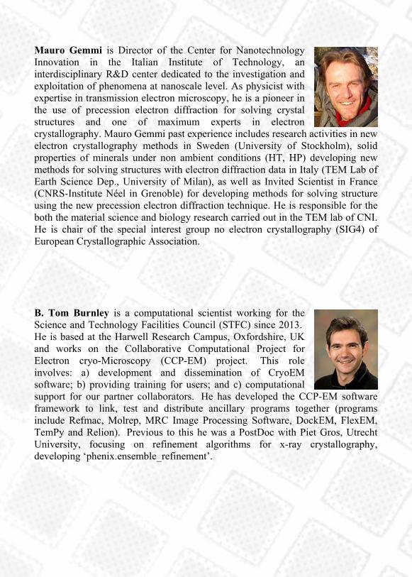
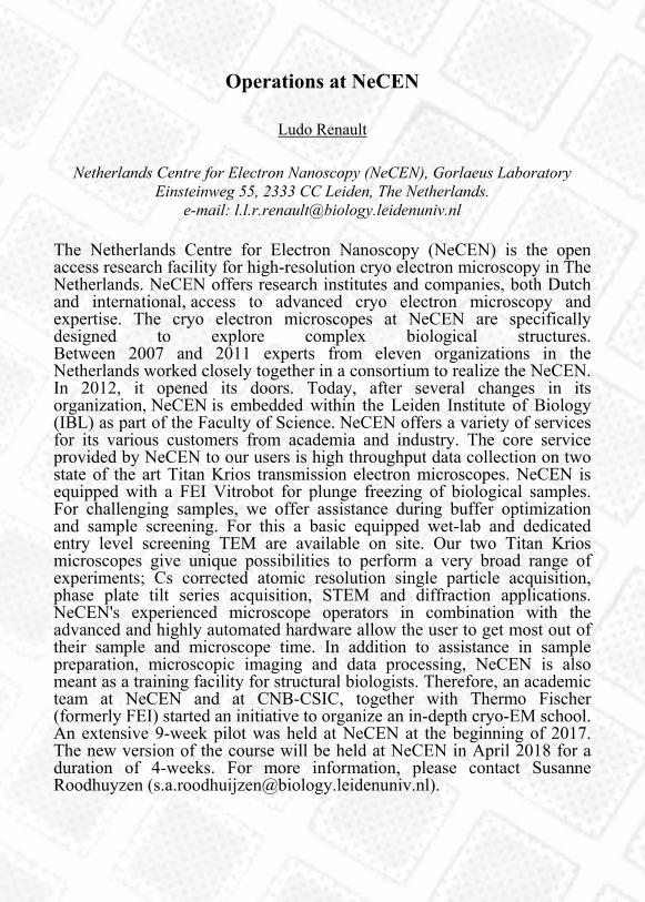
Mauro Gemmi is Director of the Center for Nanotechnology Innovation in the Italian Institute of Technology, an interdisciplinary R&D center dedicated to the investigation and exploitation of phenomena at nanoscale level. As physicist with expertise in transmission electron microscopy, he is a pioneer in the use of precession electron diffraction for solving crystal structures and one of maximum experts in electron crystallography. Mauro Gemmi past experience includes research activities in new electron crystallography methods in Sweden (University of Stockholm), solid properties of minerals under non ambient conditions (HT, HP) developing new methods for solving structures with electron diffraction data in Italy (TEM Lab of Earth Science Dep., University of Milan), as well as Invited Scientist in France (CNRS-Institute Néel in Grenoble) for developing methods for solving structure using the new precession electron diffraction technique. He is responsible for the both the material science and biology research carried out in the TEM lab of CNI. He is chair of the special interest group no electron crystallography (SIG4) of European Crystallographic Association. B. Tom Burnley is a computational scientist working for the Science and Technology Facilities Council (STFC) since 2013. He is based at the Harwell Research Campus, Oxfordshire, UK and works on the Collaborative Computational Project for Electron cryo-Microscopy (CCP-EM) project. This role involves: a) development and dissemination of CryoEM software; b) providing training for users; and c) computational support for our partner collaborators. He has developed the CCP-EM software framework to link, test and distribute ancillary programs together (programs include Refmac, Molrep, MRC Image Processing Software, DockEM, FlexEM, TemPy and Relion). Previous to this he was a PostDoc with Piet Gros, Utrecht University, focusing on refinement algorithms for x-ray crystallography, developing ‘phenix.ensemble_refinement’.
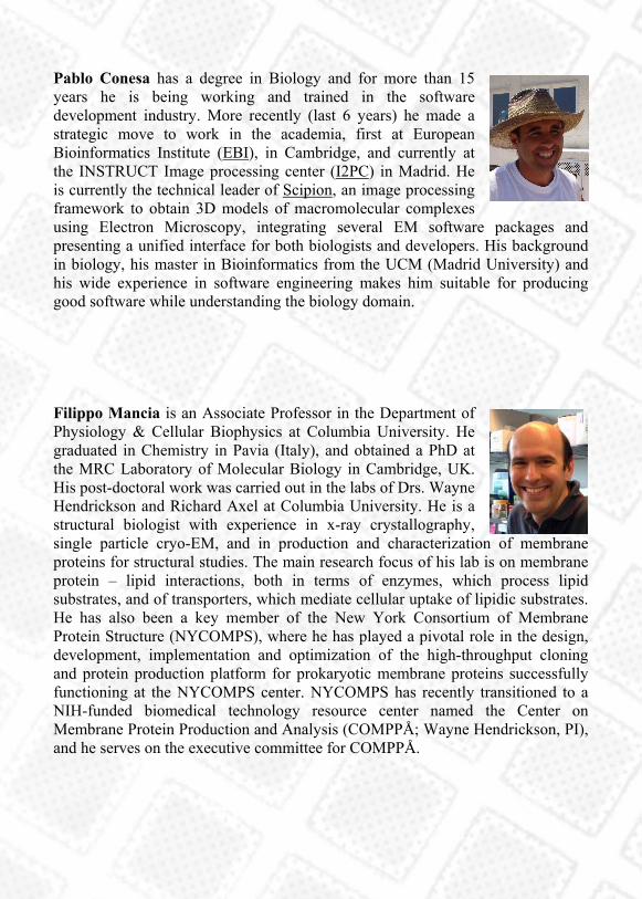
Pablo Conesa has a degree in Biology and for more than 15 years he is being working and trained in the software development industry. More recently (last 6 years) he made a strategic move to work in the academia, first at European Bioinformatics Institute (EBI), in Cambridge, and currently at the INSTRUCT Image processing center (I2PC) in Madrid. He is currently the technical leader of Scipion, an image processing framework to obtain 3D models of macromolecular complexes using Electron Microscopy, integrating several EM software packages and presenting a unified interface for both biologists and developers. His background in biology, his master in Bioinformatics from the UCM (Madrid University) and his wide experience in software engineering makes him suitable for producing good software while understanding the biology domain. Filippo Mancia is an Associate Professor in the Department of Physiology & Cellular Biophysics at Columbia University. He graduated in Chemistry in Pavia (Italy), and obtained a PhD at the MRC Laboratory of Molecular Biology in Cambridge, UK. His post-doctoral work was carried out in the labs of Drs. Wayne Hendrickson and Richard Axel at Columbia University. He is a structural biologist with experience in x-ray crystallography, single particle cryo-EM, and in production and characterization of membrane proteins for structural studies. The main research focus of his lab is on membrane protein – lipid interactions, both in terms of enzymes, which process lipid substrates, and of transporters, which mediate cellular uptake of lipidic substrates. He has also been a key member of the New York Consortium of Membrane Protein Structure (NYCOMPS), where he has played a pivotal role in the design, development, implementation and optimization of the high-throughput cloning and protein production platform for prokaryotic membrane proteins successfully functioning at the NYCOMPS center. NYCOMPS has recently transitioned to a NIH-funded biomedical technology resource center named the Center on Membrane Protein Production and Analysis (COMPPÅ; Wayne Hendrickson, PI), and he serves on the executive committee for COMPPÅ.

Alessandro Vannini studied Biology at the University of Rome "Roma Tre" and undertook his Ph.D. research at IRBM "P. Angeletti" (Merck Research Lab), Rome, focussing on the structural characterisation of quorum-sensing proteins in pathogenic bacteria and of human histone-deacetylases (HDACs). For his post-doctoral research, supported by Marie-Curie and EMBO long-term fellowships, he joined Professor Patrick Cramer's laboratory in Munich, focussing on the architecture and regulation of yeast RNA polymerase III, the largest among the three eukaryotic RNA polymerases, by combining X-ray crystallography and cryo-electron microscopy. In 2012, Dr Alessandro Vannini joined the Institute of Cancer Research in London, UK, as Team Leader in the Division of Structural Biology. Using an Integrative Structural Biology approach, he and his team are focusing on the structural and functional characterisation of large macromolecular complexes that assemble at RNA polymerase III binding sites across the eukaryotic genome, in order to understand their role in tumorigenesis as well as in the 3D spatial organization of the genome. In 2016, Dr. Vannini was elected EMBO Young Investigator and Wellcome Trust Investigator. Martin Walsh’s research is focused primarily on understanding the mechanisms used by respiratory bacterial pathogens to adhere to host cell surfaces, regulation of biofilms and carbohydrate/nutrient uptake (ABC transporters). He is deputy to the director of life sciences at Diamond Light Source (DLS) and has been a key player in the instigation and implementation of high-throughput methods for structural biology, in particular automation of sample handling, standardisation of workflows and development of ISPyB for sample management. He has been intimately involved in the establishment of the UK’s X-FEL hub at Diamond and the associated SFX/SPB instrument at the European X-FEL in Hamburg as well as playing a key role in the establishment of eBIC the UK’s national CryoEM centre. He has over 25 years’ experience in macromolecular X-ray crystallography (MX) and has deposited >90 structures with the Protein Data Bank.
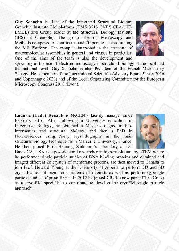
Guy Schoehn is Head of the Integrated Structural Biology Grenoble Institute EM platform (UMS 3518 CNRS-CEA-UJF-EMBL) and Group leader at the Structural Biology Institute (IBS) in Grenoble). The group Electron Microscopy and Methods composed of four teams and 20 people is also running the ME Platform. The group is interested in the structure of macromolecular assemblies in general and viruses in particular. One of the aims of the team is also the development and spreading of the use of electron microscopy in structural biology at the local and the national level. Guy Schoehn is also President of the French Microscopy Society. He is member of the International Scientific Advisory Board 5Lyon 2016 and Copenhague 2020) and of the Local Organizing Committee for the European Microscopy Congress 2016 (Lyon). Ludovic (Ludo) Renault is NeCEN’s facility manager since February 2016. After following a University education in Integrative Biology, he obtained a Master’s degree in bio-informatics and structural biology, and then a PhD in Neurosciences using X-ray crystallography as the main structural biology technique from Marseille University, France. He then joined Prof. Henning Stahlberg’s laboratory at UC Davis CA, USA as a post-doctoral researcher in high-resolution cryo-TEM where he performed single particle studies of DNA-binding proteins and obtained and imaged different 2d crystals of membrane proteins. He then moved to Canada to join Prof. Howard Young at the University of Alberta to perform 2D and 3D crystallization of membrane proteins of interests as well as performing single particle studies of prion fibrils. In 2012 he joined CRUK (now part of The Crisk) as a cryo-EM specialist to contribute to develop the cryoEM single particle approach.

Wayne A. Hendrickson is a University Professor at Columbia University. He has a B.A. in physics and biology from the University of Wisconsin at River Falls and a Ph.D. in biophysics from Johns Hopkins University, which was based on work with Warner Love. His postdoctoral research was with Jerome Karle at the Naval Research Laboratory (NRL). He remained at NRL as a Research Biophysicist until 1984 when he joined the Department of Biochemistry and Molecular Biophysics at Columbia. Hendrickson was also an HHMI Investigator from 1986 to 2012; in 2008, he was named the Violin Family Professor of Physiology and Cellular Biophysics at Columbia; and in 2010 he became the Scientific Director of the New York Structural Biology Center. Hendrickson and his colleagues use x-ray crystallography, cryogenic electron microscopy (cryo-EM), and biochemical analyses to study biological macromolecules in atomic detail. Their advances in diffraction methods (notably, stereochemically restrained refinement, phase evaluation from anomalous diffraction, selenomethionyl proteins, and synchrotron instrumentation) have been instrumental in the emergence of structural biology as a major force in modern biology and molecular medicine. Their biological applications include investigations on membrane receptors and cellular signaling, on viral proteins and HIV infection, on molecular chaperones and protein folding, and in structural genomics of membrane proteins. Hendrickson has published numerous scientific articles. He serves on advisory bodies for various scientific organizations. He is a founding editor of Current Opinion in Structural Biology and of Structure, and he was a founder of SGX Pharmaceuticals. His honors include the Aminoff Prize of the Royal Swedish Academy of Sciences, the Canada Gairdner International Award, and the Harvey Prize of the Technion – Israel Institute of Technology. He is a fellow of the American Academy of Arts and Sciences and a member of the National Academy of Sciences.
Oral presentations by Young Participants
Towards the structural discovery of the peroxisomal
translocation of functional matrix proteins
Jérôme Bürgi
European Molecular Biology Laboratory - Department Structural biology c/o DESY, Building 25A, Notkestraße 85 22607 Hamburg (Germany). E-mail:
Peroxisomes are intracellular organelles found in virtually all eukaryotic
cells and involved in the beta-oxidation of fatty acids and inactivation of
reactive oxygen species. Importantly, peroxisome activity requires the
import of fully folded and functional matrix proteins across their
membrane using a hypothesized pore. Our goal is to visualize this pore
using a wide range of biophysical and biochemical methods, such as
electron microscopy, cross-linking coupled to mass-spectrometry and
small angle x-ray scattering.
Structural basis for the indispensable role of a unique zinc
finger motif in LNX2 ubiquitination
Digant Nayak
National University of Singapore - Department Department of Biological Sciences Department of Biological Sciences S3, Level 3, SBL 5, 117543 Singapore
(Singapore). E-mail: [email protected]
LNX (Ligand of Numb Protein-X) proteins, LNX1 and LNX2, are RING-
and PDZ-based E3-ubiquitin ligases known to interact with Numb.
Silencing of LNX2 has been reported to down-regulate WNT and NOTCH,
two key signaling pathways in tumorigenesis. Here we report the
identification of the domain boundary of LNX2 to confer its ubiquitination
activity, its crystal structure along with functional studies. We show that
the RING domain in LNX2 is flanked by two Zinc-binding motifs (Zn-
RING-Zn), in which the N-terminal Zinc-binding motif adopts novel
conformation. Although this motif follows the typical Cys2His2-type zinc
finger configuration, it is devoid of any secondary structure and forms an
open circle conformation, which has not been reported yet. This unique N-
terminal Zn-finger motif is indispensable for the activity and stability of
LNX2, as verified using mutational studies. The Zn-RING-Zn domain of
LNX2 is a dimer and assumes a rigid elongated structure that undergoes
autoubiquitination and undergoes N-terminal polyubiquitination. The
ubiquitin chains consist of all seven possible isopeptide linkages.
Moreover we have demonstrated the ubiquitination of cell fate determinant
protein, Numb by LNX2. We have further extended our study to full-
length LNX2.

Introduction to protein filament characterisation
Andrew Hedger
University of Queensland - Department School of Chemistry and Molecular Biosciences 4072 Brisbane (Australia). E-mail: [email protected]
Many proteins have the potential to assemble into higher-order structures
such as filament or micro crystals. This is a common theme present in
proteins of the innate immune system. My work focuses on characterizing
the Toll-like receptors and their adaptors from the TRIF-dependent
(MyD88-independent) pathway. TRIF signaling can be either protective or
pathological depending on the nature of infection. The TRIF-dependent
pathway is particularly important for recognition of dsRNA viruses and
gram-negative bacteria. Pathologically TRIF signaling is responsible for
several chronic inflammatory diseases such as chronic hepatitis B and
hepatitis B-related hepatocellular carcinoma. Using electron microscopy
we have shown that TRAM is able to undergo assembly formation into an
f-actin like filament which is similar to MAL filament strand found in the
adjacent MyDD88-dependent pathway.

Deciphering the oligomerization role of the bacterial lipid
rafts scaffold protein by Cryo-EM
Ilaria Peschiera
CNB-CSIC - Department of Microbiology Madrid (Spain). e-mail: [email protected]
Bacteria organize a number of cellular processes in functional membrane
microdomains that in certain structural and functional aspects resemble
lipid rafts of eukaryotic cells. These microdomains assemble by
aggregation of unusual membrane lipids and colocalization with the
membrane scaffold protein flotillin, which provides structural consistency
to the rafts. These membrane platforms accumulate specific membrane
proteins, whose localization in the rafts is essential for its functionality.
We are using the human pathogen S.aureus as as model system to
investigate the structure of lipid rafts in bacteria. One of the aims of this
project is to structurally characterized flotillin, as is a key protein on rafts
organization in both eukaryotes and prokaryotes. Different constructs of
flotilin were expressed, purified and used for a biochemical and
preliminary structure characterization confirming the oligomeric nature of
the protein.

AICS2017 Sponsors
With more than 60 years of innovation and leadership, FEI part of Thermo Fisher Scientific, enables customers to find meaningful answers to questions that accelerate breakthrough discoveries, increase productivity, and ultimately change the world. FEI designs, manufactures, and supports the broadest range of high-performance microscopy workflows that provide images and answers in the micro-, nano-, and picometer scales. JEOL is a leading global supplier of scientific instruments used for research and development in the fields of nanotechnology, life sciences, optical communication, forensics, and biotechnology. Utilizing its unique technologies, products, services, and knowledge, JEOL helps its customers make significant breakthroughs in product development and scientific research. JEOL products range from scientific instrumentation to industrial equipment including Scanning electron microscopes (SEM), Transmission electron microscopes (TEM), Auger micro probe analyzers (AES), Electron probe micro analyzers (EPMA), Photoelectron spectrometers (XPS), Mass spectrometers, NMR spectrometers, Electron spin resonance, and semiconductor tools. JEOL (ITALIA) S.p.A. ensure both commercial and service assistance of JEOL instruments installed on the Italian territory thanks to highly organized and specialized structure.

DECTRIS Ltd. is the leading company in Hybrid Photon Counting (HPC) X-ray detection. DECTRIS’ pioneering technology has transformed basic research at synchrotron light sources, as well as X-ray applications in laboratory diffractometers. The broad portfolio of DECTRIS’ detectors is carefully scaled to meet the needs of various applications. With an aim to continuously improve the measurement quality, DECTRIS also provides solutions for customer developments in scientific and industrial x-ray detection, thereby pushing the state of the art and enabling new scientific findings.
Leica Microsystems develops and manufactures microscopes and scientific instruments for the analysis of microstructures and nanostructures. Widely recognized for optical precision and innovative technology, the company is one of the market leaders in compound and stereo microscopy, digital microscopy, confocal laser scanning and super-resolution microscopy with related imaging systems, electron microscopy sample preparation, and surgical microscopy.
Quantifoil Micro Tools GmbH is a world leading manufacturer of Holey Carbon and Gold Support Films for electron microscopy. Founded in 1999 to provide Universities and Research Institutes with TEM supports we are focused on improving and customising our products to specific applications.

Campoverde is the Italian distributor of products for research in the bioscience area: reagents, consumables, software and instruments . Campoverde represents many international companies who develop and manufacture antibodies, reagents, culture media — including a line dedicated to insect cells — labware, technical-scientific software and instruments such as fluorescent microscopes, cell sorters, and much more.
Nanovision S.r.l. is responsible for the technical assistance of the entire line of Hitachi Electron Microscopes. We sell directly the tabletop SEM and the compact SEM. We support Hitachi in the sales of conventional Electron Microscopes. Our catalog includes main accessories related to the world of Electron Microscopy, instrumentation for samples preparation and a wide range of consumables. Because our customers’ research is important, we are focused on being the leading supplier in crystallography. We provide innovative tools and solutions that measurably improve the ease, reproducibility, and quality of experiments. MiTeGen engineers, manufactures and distributes a full range of the leading products for crystallography, Cryo-EM, and other structural methods.


For additional info about AICS venues,
please check the school website
https://tinyurl.com/aics2017venue
or scan the QR code on this page.

You can download the PDF version
of this program book by scanning
this QR code, or by reaching the
URL:
https://tinyurl.com/aics2017program
