program cancer research symposium - ontario … · university of guelph hospitality services and...
TRANSCRIPT

Program
Cancer Research Symposium
Thursday, May 3, 2011 Room 1714 LLC, Ontario Veterinary College
University of Guelph

Introductory Remarks Welcome to the 4th Annual Guelph ICCI Cancer Symposium. This meeting is intended to bring together individuals interested in the study of any aspect of cancer in any species, from the most basic biological elements, to clinical therapies and on to social, emotional and ethical aspects of this often-devastating disease. Through interactions facilitated by this meeting, it is hoped that new insights and collaborations will develop that will enhance the research and scholarship in the area of cancer research at the University of Guelph and collaborating institutions. We would like to thank the OVC Dean’s Office and the Arthur Willis Visiting Professorship for financial support of the meeting, and for sponsoring the visit of Dr. Cheryl London, who is this year’s Arthur Willis Distinguished Speaker. We hope you find this symposium interesting and informative, and that it leads to fruitful research collaborations for all our attendees.
Co-Organizers Brenda Coomber and Jon LaMarre. Biomedical Sciences, University of Guelph
Additional thanks to Ms Barb Gaudette, OVC Office of the Dean, for her administrative expertise and invaluable assistance in organizing this event, to University of Guelph Hospitality Services and Physical Resources, and to Adrian Hollingbury and his crew at the OVC Cafeteria for help with set up and refreshments.
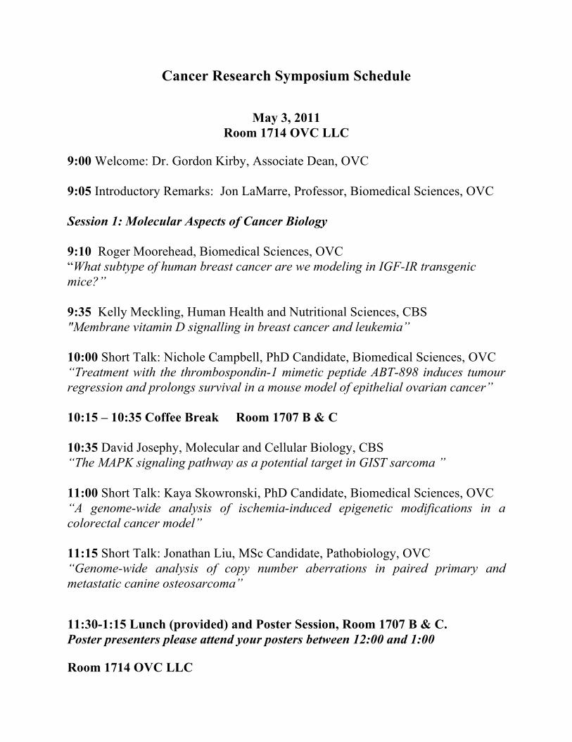
Cancer Research Symposium Schedule
May 3, 2011 Room 1714 OVC LLC
9:00 Welcome: Dr. Gordon Kirby, Associate Dean, OVC 9:05 Introductory Remarks: Jon LaMarre, Professor, Biomedical Sciences, OVC Session 1: Molecular Aspects of Cancer Biology 9:10 Roger Moorehead, Biomedical Sciences, OVC “What subtype of human breast cancer are we modeling in IGF-IR transgenic mice?” 9:35 Kelly Meckling, Human Health and Nutritional Sciences, CBS "Membrane vitamin D signalling in breast cancer and leukemia” 10:00 Short Talk: Nichole Campbell, PhD Candidate, Biomedical Sciences, OVC “Treatment with the thrombospondin-1 mimetic peptide ABT-898 induces tumour regression and prolongs survival in a mouse model of epithelial ovarian cancer” 10:15 – 10:35 Coffee Break Room 1707 B & C 10:35 David Josephy, Molecular and Cellular Biology, CBS “The MAPK signaling pathway as a potential target in GIST sarcoma ” 11:00 Short Talk: Kaya Skowronski, PhD Candidate, Biomedical Sciences, OVC “A genome-wide analysis of ischemia-induced epigenetic modifications in a colorectal cancer model” 11:15 Short Talk: Jonathan Liu, MSc Candidate, Pathobiology, OVC “Genome-wide analysis of copy number aberrations in paired primary and metastatic canine osteosarcoma” 11:30-1:15 Lunch (provided) and Poster Session, Room 1707 B & C. Poster presenters please attend your posters between 12:00 and 1:00 Room 1714 OVC LLC

Session II: The Patient and Their Disease 1:15 Introductory Remarks: Dr. Brenda Coomber, Biomedical Sciences, OVC 1:20 Sarah Boston, Clinical Studies, OVC “Retrospective study of scapulectomy in dogs” 1:45 Robert Cruz, Clincial Studies, OVC “Evaluation of a MRI hepatobiliary contrast agent (gadoxetic acid/Primovist) in dogs with splenic hemangiosarcoma for detection of metastasis” 2:10 Short Talk: Michelle Oblack, DVSc Candidate, Clinical Studies, OVC “Comparison of different concurrent imaging modalities in staging of dogs with appendicular osteosarcoma” 2:25 Lee Niel, Population Medicine, OVC “How much is too much? Assessing quality of life in animal cancer patients” 2:50 Gillian Marit, Family Relations & Human Development, CSAHS “Examining differences in perceptions of adjustment between mothers and daughters when a mother has breast cancer” 3:15 Break 3:30 Keynote Address Room 1714 OVC LLC Dr. Cheryl London DVM, PhD, Diplomate ACVIM Associate Professor of Medical Oncology The Ohio State University College of Veterinary Medicine “Targeted Therapy in Veterinary Medicine: The emerging role of tyrosine kinase inhibitors” 4:30 Closing Reception Room 1707 B & C Cash Bar
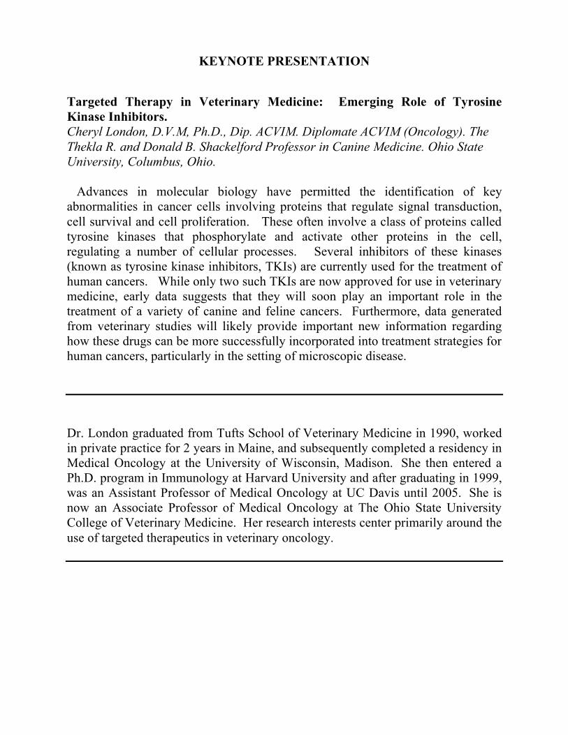
KEYNOTE PRESENTATION Targeted Therapy in Veterinary Medicine: Emerging Role of Tyrosine Kinase Inhibitors. Cheryl London, D.V.M, Ph.D., Dip. ACVIM. Diplomate ACVIM (Oncology). The Thekla R. and Donald B. Shackelford Professor in Canine Medicine. Ohio State University, Columbus, Ohio. Advances in molecular biology have permitted the identification of key abnormalities in cancer cells involving proteins that regulate signal transduction, cell survival and cell proliferation. These often involve a class of proteins called tyrosine kinases that phosphorylate and activate other proteins in the cell, regulating a number of cellular processes. Several inhibitors of these kinases (known as tyrosine kinase inhibitors, TKIs) are currently used for the treatment of human cancers. While only two such TKIs are now approved for use in veterinary medicine, early data suggests that they will soon play an important role in the treatment of a variety of canine and feline cancers. Furthermore, data generated from veterinary studies will likely provide important new information regarding how these drugs can be more successfully incorporated into treatment strategies for human cancers, particularly in the setting of microscopic disease. Dr. London graduated from Tufts School of Veterinary Medicine in 1990, worked in private practice for 2 years in Maine, and subsequently completed a residency in Medical Oncology at the University of Wisconsin, Madison. She then entered a Ph.D. program in Immunology at Harvard University and after graduating in 1999, was an Assistant Professor of Medical Oncology at UC Davis until 2005. She is now an Associate Professor of Medical Oncology at The Ohio State University College of Veterinary Medicine. Her research interests center primarily around the use of targeted therapeutics in veterinary oncology.
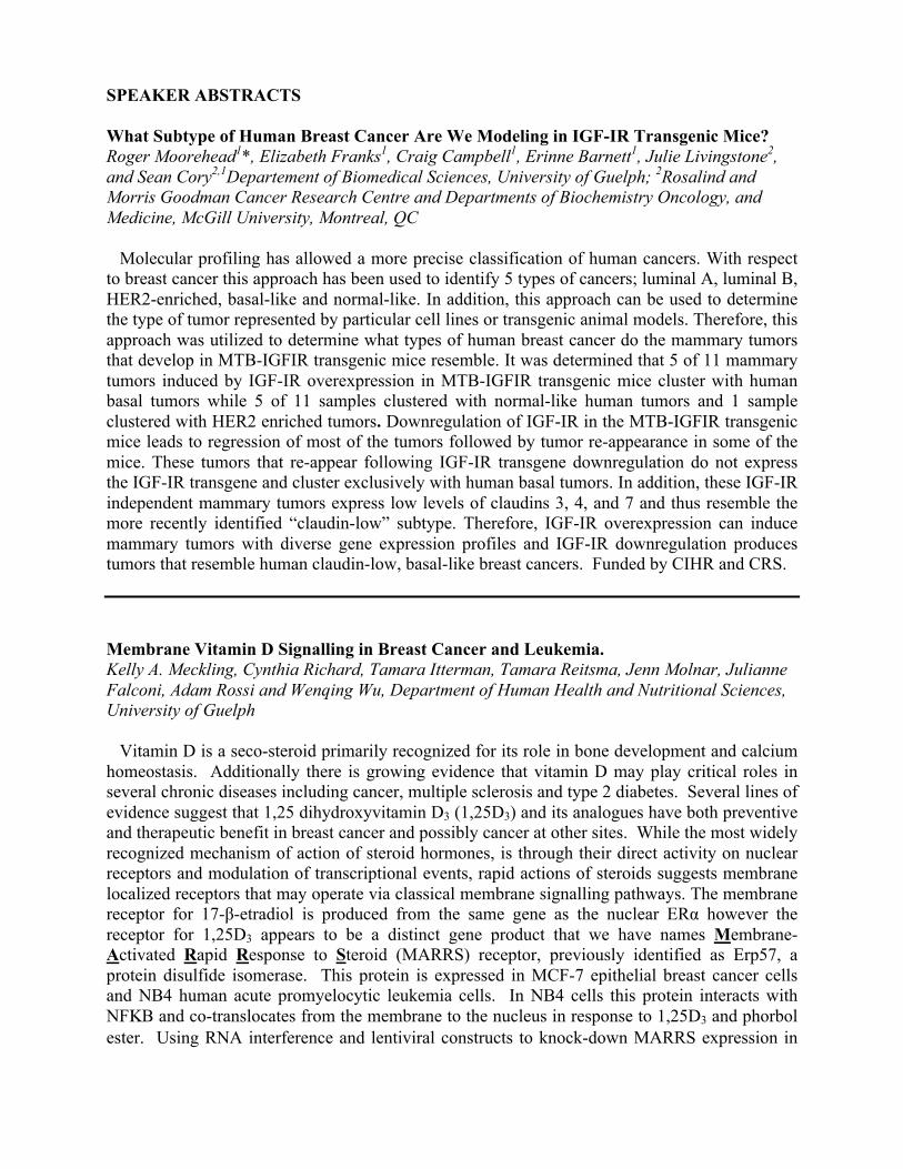
SPEAKER ABSTRACTS What Subtype of Human Breast Cancer Are We Modeling in IGF-IR Transgenic Mice? Roger Moorehead1*, Elizabeth Franks1, Craig Campbell1, Erinne Barnett1, Julie Livingstone2, and Sean Cory2,1Departement of Biomedical Sciences, University of Guelph; 2Rosalind and Morris Goodman Cancer Research Centre and Departments of Biochemistry Oncology, and Medicine, McGill University, Montreal, QC Molecular profiling has allowed a more precise classification of human cancers. With respect to breast cancer this approach has been used to identify 5 types of cancers; luminal A, luminal B, HER2-enriched, basal-like and normal-like. In addition, this approach can be used to determine the type of tumor represented by particular cell lines or transgenic animal models. Therefore, this approach was utilized to determine what types of human breast cancer do the mammary tumors that develop in MTB-IGFIR transgenic mice resemble. It was determined that 5 of 11 mammary tumors induced by IGF-IR overexpression in MTB-IGFIR transgenic mice cluster with human basal tumors while 5 of 11 samples clustered with normal-like human tumors and 1 sample clustered with HER2 enriched tumors. Downregulation of IGF-IR in the MTB-IGFIR transgenic mice leads to regression of most of the tumors followed by tumor re-appearance in some of the mice. These tumors that re-appear following IGF-IR transgene downregulation do not express the IGF-IR transgene and cluster exclusively with human basal tumors. In addition, these IGF-IR independent mammary tumors express low levels of claudins 3, 4, and 7 and thus resemble the more recently identified “claudin-low” subtype. Therefore, IGF-IR overexpression can induce mammary tumors with diverse gene expression profiles and IGF-IR downregulation produces tumors that resemble human claudin-low, basal-like breast cancers. Funded by CIHR and CRS. Membrane Vitamin D Signalling in Breast Cancer and Leukemia. Kelly A. Meckling, Cynthia Richard, Tamara Itterman, Tamara Reitsma, Jenn Molnar, Julianne Falconi, Adam Rossi and Wenqing Wu, Department of Human Health and Nutritional Sciences, University of Guelph Vitamin D is a seco-steroid primarily recognized for its role in bone development and calcium homeostasis. Additionally there is growing evidence that vitamin D may play critical roles in several chronic diseases including cancer, multiple sclerosis and type 2 diabetes. Several lines of evidence suggest that 1,25 dihydroxyvitamin D3 (1,25D3) and its analogues have both preventive and therapeutic benefit in breast cancer and possibly cancer at other sites. While the most widely recognized mechanism of action of steroid hormones, is through their direct activity on nuclear receptors and modulation of transcriptional events, rapid actions of steroids suggests membrane localized receptors that may operate via classical membrane signalling pathways. The membrane receptor for 17-β-etradiol is produced from the same gene as the nuclear ERα however the receptor for 1,25D3 appears to be a distinct gene product that we have names Membrane-Activated Rapid Response to Steroid (MARRS) receptor, previously identified as Erp57, a protein disulfide isomerase. This protein is expressed in MCF-7 epithelial breast cancer cells and NB4 human acute promyelocytic leukemia cells. In NB4 cells this protein interacts with NFΚB and co-translocates from the membrane to the nucleus in response to 1,25D3 and phorbol ester. Using RNA interference and lentiviral constructs to knock-down MARRS expression in

MCF-7 cells lead to increased sensitivity of these cells to the growth inhibitory effects of 1,25D3 in vitro and in vivo. In NB4 leukemia cells 50-80% knock-down and knock-out were achieved with lentiviral constructs. Preliminary experiments suggest that the leukemia cells, like the breast cancer cells are more sensitive to the differentiative effects of 1,25D3. Experiments are underway to generate tissue specific MARRS knockout mice. The MAPK Signaling Pathway as a Potential Target in GIST Sarcoma. P. David Josephy*, Adrián Mariño-Enríquez, Jonathan A. Fletcher, Department of Molecular and Cellular Biology, University of Guelph and Department of Pathology, Brigham & Women’s Hospital and Harvard Medical School, Boston, MA GIST (Gastrointestinal Stromal Tumor) is a relatively rare cancer, but it is, nevertheless, the most common sarcoma of the human gastrointestinal tract. The cell of origin of GIST is the interstitial cell of Cajal (the pacemaker cell of g.i. tract peristalsis) or a closely-related cell. GIST cells express the KIT gene product, a transmembrane receptor tyrosine kinase (RTK), and most GISTs carry an activating mutation in this oncogene or the closely related PDGFRA gene. Targeted therapy directed against KIT, with oral drugs such as imatinib and sunitinib, has hugely improved the prognosis for GIST patients. However, even after dramatic and sustained clinical responses to these KIT inhibitors, drug resistance and disease progression often develop. New targets and new drugs are urgently needed. We have been studying the role of the RAS/RAF/MEK/ERK signaling pathway in the survival and proliferation of the cultured human GIST cell line GIST430. Compound PD0325901, a potent and specific inhibitor of MEK, is used to block the pathway. A genome-wide shRNA pooled screen has been conducted, with the goal of identifying genes whose suppression has a synthetic-lethal activity in combination with PD0325901. Also, a drug-resistant sub-line has been developed by chronic treatment of GIST430 cells with the inhibitor. The cells are cross-resistant to KIT inhibitors such as nilotinib. Immunoblotting studies have shown that the mechanism of this resistance involves AKT activation, and it is hoped that further characterization of these cells will provide new insights into GIST cell signaling pathways. Retrospective Study of Scapulectomy in Dogs. Vincenzo Montinaro, Sarah Boston* Department of Clinical Studies, University of Guelph Scapulectomy is an alternative to amputation for tumors arising from the scapula that is not commonly performed. The purpose of this retrospective study was to identify dogs with primary scapular tumors treated with scapulectomy. Inclusion criteria were dogs that had a scapulectomy to treat a primary tumor of the scapula. Cases were submitted by VSSO members. Forty-two dogs met the inclusion criteria. The median age was 8.3 years and median weight was 34.8 kg. In eighteen dogs (42.9%) a subtotal scapulectomy (removal of ≥ 75% of the scapula) was performed. Osteosarcoma was diagnosed in 27 dogs (64.3%). Limb use was evaluated immediately post surgery in 41/42 dog and it was poor (15), fair (17), good (7) and excellent (2). Information on limb use at other time post (1, 2 3 and > 3months) postoperatively was also available in some cases and was good to excellent overall. For the parameters assessed for their effect on survival time (ST) and disease free interval (DFI), only the use of adjunctive chemotherapy had a significant effect on ST (p=.0003) and DFI. (p=.00011). Scapulectomy can

be performed to remove primary tumors of the scapula and preserve limb function. The most common primary scapular tumor in this study was osteosarcoma. Primarily older, large breed dogs were affected. Although long-term follow-up of limb use was not available in all dogs, limb use was fair to excellent for the majority of dogs in this study. The addition of adjunctive chemotherapy prolonged the DFI and MST. Evaluation of a MRI Hepatobiliary Contrast Agent (gadoxetic acid/Primovist) Dogs with Splenic Hemangiosarcoma for Detection of Metastasis. Robert Cruz, Department of Clinical Studies, University of Guelph In many dogs examined by the Oncology Service at the OVC-VTH for the evaluation of splenic hemangiosarcoma, liver nodules are commonly noted on routine abdominal ultrasound. Differentiating between benign nodular hyperplasia and malignant liver nodules frequently not possible. The ability to accurately and efficiently (i.e. not wait for a biopsy) determine if liver nodules are malignant is particularly important in dogs with splenic hemangiosarcoma as the presence of metastatic disease may impact the owner’s decision to proceed with treatment. Our group has recently shown that a new liver specific MRI contrast agent (gadoxetic acid/Primovist) is safe and efficacious in dogs. This study will evaluate the ability of this contrast agent to differentiate benign hyperplastic liver nodules from metastasis in dogs presenting with cavitated splenic masses. How Much Is Too Much? Assessing Quality of Life in Animal Cancer Patients. Lee Niel, Department of Population Medicine, University of Guelph When canine and feline companions are affected by cancer there are many potential impacts on their quality of life. In addition to the direct effects of their disease, animal patients may experience fear and anxiety during their veterinary visits, and pain and illness during the course of treatments and as a result of unexpected complications. Prior to beginning treatment, owners and veterinarians need to make important decisions about what is best for the individual animal. Survival time is one potential measure of treatment success, but ultimately it is the quality of life of the individual animal during and after treatment that should guide the decision making process. Initially, we can attempt to predict which patients will benefit from further treatment by considering their clinical prognosis, as well as the influence of their individual ‘personality’ on their ability to tolerate and respond appropriately to the recommended treatment. During and after treatment, we can also monitor potential indicators of quality of life to ensure that the patient has the capacity to experience positive affective states and is not significantly affected by physical discomfort. Recent studies have developed and tested various schemes for quality of life assessment in companion dogs and cats. However, only one published study has attempted to tailor an assessment scheme for cancer-related concerns, which is surprising considering the potential impact of this group of diseases in terms of both animal numbers and severity of symptoms. The development of novel treatments for cancer has been progressing rapidly, and research on companion animal quality of life needs to keep pace to ensure decisions for individuals are based on valid and reliable indicators of animal welfare.
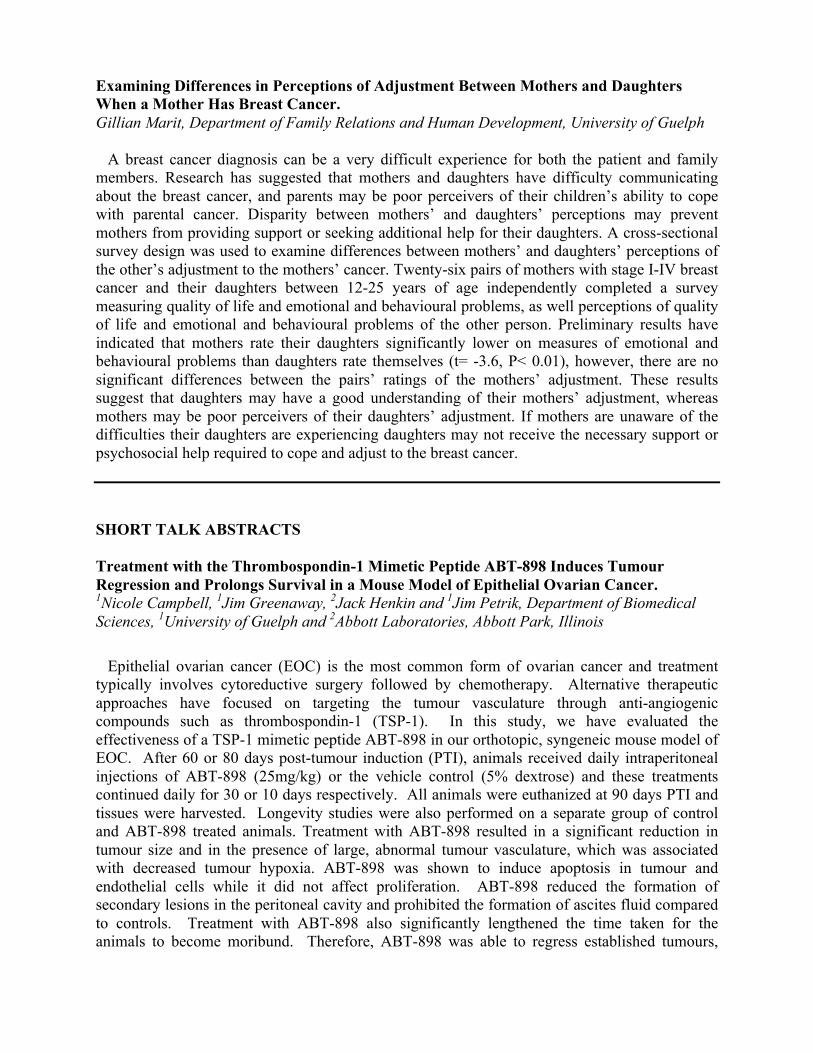
Examining Differences in Perceptions of Adjustment Between Mothers and Daughters When a Mother Has Breast Cancer. Gillian Marit, Department of Family Relations and Human Development, University of Guelph
A breast cancer diagnosis can be a very difficult experience for both the patient and family members. Research has suggested that mothers and daughters have difficulty communicating about the breast cancer, and parents may be poor perceivers of their children’s ability to cope with parental cancer. Disparity between mothers’ and daughters’ perceptions may prevent mothers from providing support or seeking additional help for their daughters. A cross-sectional survey design was used to examine differences between mothers’ and daughters’ perceptions of the other’s adjustment to the mothers’ cancer. Twenty-six pairs of mothers with stage I-IV breast cancer and their daughters between 12-25 years of age independently completed a survey measuring quality of life and emotional and behavioural problems, as well perceptions of quality of life and emotional and behavioural problems of the other person. Preliminary results have indicated that mothers rate their daughters significantly lower on measures of emotional and behavioural problems than daughters rate themselves (t= -3.6, P< 0.01), however, there are no significant differences between the pairs’ ratings of the mothers’ adjustment. These results suggest that daughters may have a good understanding of their mothers’ adjustment, whereas mothers may be poor perceivers of their daughters’ adjustment. If mothers are unaware of the difficulties their daughters are experiencing daughters may not receive the necessary support or psychosocial help required to cope and adjust to the breast cancer. SHORT TALK ABSTRACTS Treatment with the Thrombospondin-1 Mimetic Peptide ABT-898 Induces Tumour Regression and Prolongs Survival in a Mouse Model of Epithelial Ovarian Cancer. 1Nicole Campbell, 1Jim Greenaway, 2Jack Henkin and 1Jim Petrik, Department of Biomedical Sciences, 1University of Guelph and 2Abbott Laboratories, Abbott Park, Illinois Epithelial ovarian cancer (EOC) is the most common form of ovarian cancer and treatment typically involves cytoreductive surgery followed by chemotherapy. Alternative therapeutic approaches have focused on targeting the tumour vasculature through anti-angiogenic compounds such as thrombospondin-1 (TSP-1). In this study, we have evaluated the effectiveness of a TSP-1 mimetic peptide ABT-898 in our orthotopic, syngeneic mouse model of EOC. After 60 or 80 days post-tumour induction (PTI), animals received daily intraperitoneal injections of ABT-898 (25mg/kg) or the vehicle control (5% dextrose) and these treatments continued daily for 30 or 10 days respectively. All animals were euthanized at 90 days PTI and tissues were harvested. Longevity studies were also performed on a separate group of control and ABT-898 treated animals. Treatment with ABT-898 resulted in a significant reduction in tumour size and in the presence of large, abnormal tumour vasculature, which was associated with decreased tumour hypoxia. ABT-898 was shown to induce apoptosis in tumour and endothelial cells while it did not affect proliferation. ABT-898 reduced the formation of secondary lesions in the peritoneal cavity and prohibited the formation of ascites fluid compared to controls. Treatment with ABT-898 also significantly lengthened the time taken for the animals to become moribund. Therefore, ABT-898 was able to regress established tumours,

which is significant as most women are detected with advanced disease. These results demonstrate that ABT-898 has potent effects on tumour cells and tumour vasculature and highlights its potential as an important therapeutic agent in the treatment of EOC. Funded by CIHR CGS Doctoral Award. A Genome-Wide Analysis of Ischemia-Induced Epigenetic Modifications in a Colorectal Cancer Model. Karolina Skowronski1, Joseph Andrews2, David Rodenhiser3, Brenda Coomber1* Department of Biomedical Sciences, University of Guelph, London Regional Cancer Centre and Children’s Health Research Institute, and 3. Departments of Biochemistry, Oncology and Paediatrics; University of Western Ontario; London Regional Cancer Centre and Children’s Health Research Institute Ischemia is a common occurrence in solid tumors due to their irregular vasculature, and has many known effects on gene expression which influence tumor progression. However, the impact of ischemia on epigenetics is not well characterized. Since epigenetic modifications are involved in the initiation and progression of cancer, we examined the link between the tumor microenvironment and DNA methylation. Solid tumors exhibit areas of hypoxia and hypoglycaemia, and we previously demonstrated that both these conditions reduced expression and activity of DNA methyltransferases in human colorectal carcinoma cells (HCT116). In this study we hypothesized that ischemia-mediated dysregulation of DNMTs will significantly alter the epigenome, thereby causing changes in gene expression. HCT116 cells were subjected to hypoxia and hypoglycaemia for 48 hours, and DNA (enriched for 5-methylcytosine) and RNA were collected in triplicate. Through the use of high resolution, whole-genome expression (Affymetrix HG-U133 Plus 2.0) and promoter tiling (1.0R) arrays, we examined whether DNA methylation patterns at specific genes were modified by ischemia, and if these epigenetic mutations correlated with expression changes. Preliminary results show hypoglycaemia had the most profound impact on both gene expression and promoter methylation. The cross-platform analysis revealed that there were 18 and 96 genes hypomethylated and upregulated by hypoxia and hypoglycaemia, respectively. Functional analysis indicated that ischemic conditions significantly affected genes involved in cellular motility, which is not surprising since it is well known that hypoxic cells are more metastatic then cells in normoxia. The novel aspect of this research is that we are linking the upregulation of genes involved in metastasis to ischemia-induced DNA hypomethylation. Since DNMT inhibitors may prove to be inefficient in an ischemic environment, and anti-angiogenic therapy may increase tumor ischemia and influence epigenetic patterning, these findings may play a crucial role in refining our future strategies for cancer control via epigenetic regulation. Funded by CCSRI. Genome-wide analysis of copy number aberrations in paired primary and metastatic canine osteosarcoma Jonathan Liu, Geoffrey Wood* Department of Pathobiology, University of Guelph Canine osteosarcoma (OSA) is the most common primary bone tumour in dogs, and is particularly malignant and difficult to treat. Though often undetectable, lung metastases are usually present at the time of initial diagnosis, leading to a median survival rate of only 8-12

months after amputation and chemotherapy. Thus, metastasis is a major clinical problem, but is relatively poorly understood despite being the ultimate therapeutic target. At the DNA level, OSA is characterized by massive genomic instability, leading to abnormal gene copy numbers in various regions throughout the genome. Our goal was to identify key genes that differ between primary tumours and metastases in order to determine the driving factors behind metastasis. We conducted genome-wide comparisons of primary tumours and metastasis within the same patients by array comparative genomic hybridization, a technique used to detect gene copy number aberrations. It has been hypothesized that since metastatic cells acquire properties that allow them to leave the primary site and grow in distant organs, they must harbour new mutations that are not common in the primary tumour. Although we found increased copy numbers of known OSA oncogenes (MYC, MET) and tumour suppressor genes (p53, PTEN), we found that metastases and primary tumours have similar levels of genomic instability. Furthermore, primary tumours were more genomically similar to their matched metastasis than they were to each other, and we could not identify genes common to metastases but not primaries. Interestingly, we found several chromosomal regions that were amplified or deleted in multiple cases, but do not contain known cancer genes. In conclusion, our preliminary data suggest that primary OSA tumours and their corresponding metastases are genomically very similar. Since primary OSA cells do not appear to require significant additional mutations to become metastatic, this may explain the early formation of metastases in canine OSA. Funded by Pet Trust. Comparison of Different Concurrent Imaging Modalities in Staging of Dogs With Appendicular Osteosarcoma. Abbreviated Title: Multiple Staging Methods in Dogs with OSA. Michelle Oblak, Stephanie Nykamp, Galina Hayes, Sarah Boston*. Clinical Studies, University of Guelph. Osteosarcoma (OSA) is the most common canine primary bone tumor. Staging includes assessment for thoracic and bone metastasis. The study objectives were to compare different imaging modalities for the detection of thoracic and bone metastasis in dogs with appendicular OSA. Study design was a prospective cross-sectional observational study (n=15). Thoracic radiographs negative for metastatic lesions were compared against thoracic CT. Test modalities (survey radiographs, whole body CT scan, nuclear scintigraphy) were assessed against a gold standard for detection of bone metastasis. Discriminatory capacity of the test modalities was assessed using receiver operator characteristic (ROC) analysis. Twenty percent (3/15) of dogs had pulmonary lesions on CT. Three bone lesions (3/15) were identified on scintigraphy and no lesions were identified on survey radiographs or CT during blinded assessment. Evaluation for bone metastasis showed that the AUROC characteristics of the different test modalities ranged between 0.76 and 0.86. The AUROC values of radiographs and CT scan was 0.76 and of scintigraphy was 0.86. Differences between the discriminating capacity of the test modalities failed to reach significance. Based on this study, CT identified more lesions than radiographs for thoracic staging. While not statistically significant, this study supports previous findings that scintigraphy is an important part of staging for dogs with OSA. Whole body survey radiographs and CT have not been previously reported as modalities for general staging of dogs with OSA and although these tests do not appear to be useful as alternatives to scintigraphy, they may have some utility as adjunctive diagnostic modalities.
POSTER ABSTRACTS 1. Potential correlation between PDK expression and apoptosis induced by DCA in human colorectal cancer cells. Nelson Ho, Siranoush Shahrzad, Brenda L. Coomber* Biomedical Sciences, University of Guelph
While glycolysis is a suboptimal metabolic strategy used by cells under hypoxic conditions, many cancer cells preferentially utilize glycolysis for energy production, even in the presence of abundant oxygen, a phenomenon known as the “Warburg Effect”. Dichloroacetate (DCA) is a small molecule which inhibits mitochondrial function by suppressing pyruvate dehydrogenase kinase (PDK) activity, thereby shifting cellular metabolism from glycolysis to glucose oxidation. Resultant alterations in mitochondrial membrane potential and permeability will lead to the release of sequestered cytochrome C and downstream effects, including increased generation of reactive oxygen species (ROS) and activation of the apoptosome. DCA has been shown to induce apoptosis in cancer cells but not in normal cells, presumably due to their paradoxical reliance on glycolysis and inability to adapt to the mitochondrial remodeling likely associated with its action. DCA has been proposed as an effective and benign adjuvant to other anti-cancer therapies, and is currently used by cancer patients in an unsupervised and often unreported fashion. Previous work in our laboratory has shown that some cancer cells are protected from hypoxia-induced apoptosis upon exposure to DCA. It is hypothesized that differential expression of PDK will correlate with the ability of different cancer cells to withstand the apoptotic effects of DCA in hypoxia, and that these effects are due to cytoprotective events related to mitochondrial remodeling. We are currently investigating the expression of different isoforms of PDK upon exposure to various concentrations of DCA in human colorectal cancer cell lines by qRT-PCR and in the future, western blotting. These studies coincide with experiments looking at the effects of DCA on mitochondrial remodeling through the use of fluorescent mitochondrial dyes and flow cytometry. Based on the results of these studies, we hope to determine a potential cause by which DCA asserts differential effects on cancer cells. Funded by NSERC 2. The Regulation of Integrin Expression/Signaling in Mammary Epithelial Cells by TGFβ – A Role in Apoptosis. Geordon Avery-Cooper, Patricia Huether, and Alicia Viloria-Petit*, Department of Biomedical Sciences, University of Guelph. The TGFβ-Par6 polarity pathway mediates epithelial-to-mesenchymal transition (EMT), a process implicated in breast cancer metastasis. We have evidence to suggest that this pathway also regulates TGFβ induced apoptosis in normal mammary cells. Integrins are adhesion molecules which regulate cell polarity, cell survival, and have been linked to TGFβ signaling. We investigated the regulation of integrin expression and signaling by the TGFβ-Par6 polarity pathway to identify its role in TGFβ induced apoptosis in normal murine mammary gland (NMuMG) cells. Parental NMuMG cells or NMuMG with a constitutive or blocked Par6 pathway were cultured either as monolayers or as 3D acini-like structures on reconstituted basement membrane. Cells were treated with TGFβ, the Smad pathway inhibitor SB431542, or both. Levels of integrin expression and localization were assessed using western blotting and immunoflourescence (IF). Activation status of integrin signaling molecules FAK and NFκB were examined by western blotting. We found the expression of β1 and β4 integrins, as well as the activation status of FAK to be regulated by the Smad pathway. While an active Par6 pathway
reduced a phospho-active form of NFκB and regulates the localization of β1 and α6 integrins. These results suggest that regulation of integrin localization and the activation status of NFκB by the Par6 pathway could mediate the apoptotic response of mammary cells following TGFβ stimulation. Establishing the mechanisms by which signaling through Par6 leads to cell death will further enhance our understanding of the TGFβ-Par6 polarity pathway and will aid in determining the feasibility of using it as a target for the treatment of breast cancer. Funded by Banting Research Foundation. _______________________________________________________________________ 3. The Oncogenic Envelope Proteins from Jaagsiekte Sheep Retrovirus and Enzootic Nasal Tumour Virus are Internalized from the Cell Surface. de Jong, Jondavid & Wootton, Sarah*, Department of Pathobiology, University of Guelph Jaagsiekte sheep retrovirus (JSRV) is the causative agent of ovine pulmonary adenocarcinoma (OPA), a naturally occurring lung cancer of sheep. The closely related Enzootic nasal tumour virus (ENTV) is thought to be the causative agent of nasal tumours in sheeps and goats. Unlike most oncogenic retroviruses, the virally encoded JSRV and ENTV envelope (Env) proteins are potent oncogenes. Expression of Env alone is sufficient to induce transformation in a variety of cell types. In vitro Env expression results in the activation of the PI-3K/Akt and Ras-MEK-MAPK signal transduction pathways. The cytoplasmic tails of both JSRV and ENTV Env encode putative binding sites for the p85 subunit of PI-3K (YXXM) and the tyrosines have been implicated in transformation, however, the tyrosine residues do not appear to be phosphorylated during transformation as would be expected. Furthermore mutations to residues surrounding the YXXM motifs either leads to abolishment of transformation or a "super-transformer" phenotype. An alternate role of the YXXM motifs may be to coordinate the internalization of Env from the cell surface. Here we demonstrate that both JSRV and ENTV Env are internalized from the cell surface, and hypothesize that the super-transforming mutants demonstrate altered or greater rates of internalization compared to the wild-type Envs. Funded by NSERC, CFI. ________________________________________________________________________ 4. The Role of Folate Availability in the Regulation of DNMT Expression by Colorectal Cancer Cells. Amanda Barber, Brenda Coomber*, Department of Biomedical Sciences, University of Guelph Genomic instability is associated with a variety of diseases, most notably cancer. It is influenced by two factors: mutation rate, and the cell’s ability to respond to and repair these mutations. The availability of folate (folic acid/vitamin B9) has been shown to modulate tumor formation, potentially by regulating expression of DNMTs, the enzymes responsible for methylating DNA. Misregulation of DNMTs may lead to epigenetic modifications affecting expression patterns, and may increase the point mutation rate, leading to a cancerous phenotype. One of the key processes involved in DNA repair is mismatch repair (MMR), the mechanisms used to sense and correct DNA mismatches and small insertions/deletions. Previous work in our laboratory has demonstrated that human colorectal cancer cells with impaired MMR are prone to mutation and altered DNA methylation when exposed to an ischemic environment. Here, we explore how folate deficiency or hypersupplementation could further modulate mutagenesis,

potentially through changes in DNMT expression. Human colorectal cancer cells were cultured in folate-free media supplemented with 0, 0.1, 1, 4, 8 and 16 mg/L of folic acid over a course of 14 days. Protein samples were collected every 3-4 days and were analyzed using western blot analysis for expression of DNMT1. Cells in folate-free conditions initially saw a decrease in DNMT1 expression, but these levels recovered after 10 days. Cells which were hypersupplemented with folic acid (8 and 16 mg/L) showed increased expression of DNMT1 relative to cells at a physiologically relevant dose (4 mg/L). This may result in reduced expression of tumor suppressor genes, or increased point mutation rates. Future work will include assaying for DNMT activity directly and determining if cells with impaired MMR capabilities have similar responses to folate hypersupplementation. Recent studies have demonstrated that folate over-supplementation can readily occur in the Canadian population, thus our findings may have relevance for understanding dietary impacts on carcinogenesis. Funded by CCSRI. 5. The Role of MicroRNAs in Mammary Tumorigenesis. Erinne Barnett, Dr. Roger Moorehead*, Department of Biomedical Sciences, University of Guelph. MicroRNAs (miRNAs) are small non-coding regulatory RNA molecules that act post-transcriptionally on mRNA to regulate the expression of most cellular genes. Many miRNAs are aberrantly expressed in tumour tissue, acting either as tumour suppressors or oncogenes, and their expression has been implicated in the development of breast cancer. Using a transgenic mouse model of mammary tumorigenesis (MTB-IGFIR), a miRNA array was performed to study the expression level of various miRNAs in mammary tumours compared to wild-type mammary tissue. Next, the expression of a number of the differentially expressed miRNAs was confirmed and manipulated in a tumour cell line (RM11A) that was generated from a MTB-IGFIR mammary tumour. We then used synthetic miRNA precursors and inhibitors to overexpress and knockdown, respectively, the levels of five miRNAs: miR-31, miR-183, miR-200c, miR-210, and miR-378. These miRNAs have all been shown to be involved in breast cancer metastasis and tumour progression. Their overexpression with synthetic precursors was optimized using real-time PCR to increase expression levels between 30 and 100 fold as this magnitude of change was observed in the miRNA array. To determine the impact of miRNAs on mammary tumorigenesis, assays for proliferation (Ki67 immunofluorescence), invasion (Boyden chamber) and cell survival (JC-1 fluorescence) were performed. We have observed that when compared to negative controls, overexpression of miR-210 significantly increased proliferation by approximately 1.5- fold while the overexpression or inhibition of the other miRNAs did not have any significant effect. Furthermore, inhibition of miR-183 was found to significantly increase apoptosis by 2.2-fold, and the overexpression of all five miRNAs showed a significant decrease in cell invasion when compared to negative controls. Discovering the differential expression of miRNAs and their effects on proliferation, invasion, and apoptosis in the RM11A cells will enhance our understanding of the function of miRNAs in human breast cancer. Funded by CIHR.

6. Pituitary and Ovarian Reponses After An Application of A Low Therapeutic Dose of the LHRH-Lytic Peptide Conjugate During the Pre-Ovulatory Period in Ewes. Pawel Bartlewski1*, William Hansel2, Bret McLeod1, Genje Buenviaje1, 1Department of Biomedical Sciences, University of Guelph; 2Pennington Biomedical Research Center, Louisiana State University System. The cationic lytic peptide (CLYP) technology is used to deliver multiple membrane disrupting peptides and antibody drug conjugates to selectively kill cancer cells without harming normal somatic cells. Potential indications include reproductive tissue tumors (i.e., ovarian, endometrial, prostate and breast cancers) as well as hematologic malignancies. The aim of the present experiment was to determine the effects of a low therapeutic dose (0.2 mg/kg body weight i.v.) of a lytic peptide Phor21, conjugated with luteotropin-releasing hormone (LHRH), on the periovulatory ovarian and endocrine events, ensuing luteal function and fertility in an ovine experimental model. We hypothesized that the dense expression of LHRH receptors on the anterior pituitary and ovarian structures would make them highly susceptible to the membrane disrupting ability of LHRH-Phor21. Six sexually mature Rideau Arcott ewes were used to test the effects of the conjugate; seven ewes served as controls. Prior to drug administration, estrus was synchronized with intravaginal progestogen-releasing sponges (medroxyprogesterone acetate, 60 mg) that were left in place for 12 days and a single i.m. injection of 750 IU of equine chorionic gonadotropin (eCG) given at sponge removal. LHRH-Phor21 or saline were administered 36 h after sponge withdrawal/eCG injection, around the expected onset of the endogenous discharge of gonadotropins. Treatment resulted in greater (P<0.05) peak LH concentrations and an earlier rise (P<0.05) in estradiol production in LHRH-Phor21 treated than control ewes; however, there were no differences (P>0.05) in the mean number of ultrasonographically detected luteal structures and serum progesterone concentrations during the luteal phase post-treatment. There were no differences (P>0.05) in the number of ewes that lambed or lamb characteristics between the two groups at lambing 9 months post-treatment. Overall, there was no significant effect of LHRH-Phor21 on the ovulatory process, luteal function and lamb productivity post-treatment in the ewes of the present study. With a lack of adverse effects of the LHRH-Phor21 on pituitary and ovarian function and fertility, our results provide further support towards the suitability and safety of LHRH-Phor21 as a cancer pharmaceutical in reproductive-aged patients. Funded by NSERC. 7. Investigation of Axin2 in Zebrafish (Danio rerio) Development and Its Role in Canonical Wnt Signaling. Whitney Lum and Terry Van Raay, Department of Molecular and Cellular Biology, University of Guelph Canonical Wnt signaling is highly conserved in eukaryotes and plays an important role in both embryonic development and homeostasis of adult stem cells. Mutations in, and thus dysregulation of, canonical Wnt signaling has been implicated in disease, including the initiation of cancer. In the absence of a Wnt signal the scaffolding protein Axin1, in association with other proteins, forms a destruction complex that targets cytosolic β-catenin for ubiquitin-mediated degradation, inhibiting canonical Wnt-mediated cell signaling. Axin2 is speculated to have a similar regulatory role within the cell. However, while mutations in axin1 result in embryonic lethality, mutations in axin2 display only cranial abnormalities. Gene replacement studies have also demonstrated that axin2 can rescue axin1-/- embryonic lethality, suggesting that axin1 and axin2 are functionally redundant. Other studies demonstrate that Axin1 and Axin2 interact with
different core components of the Wnt pathway, suggesting different molecular functions, which is supported by differences in their expression patterns. Therefore, identifying the differences between Axin1 and Axin2 will provide insight into the regulation of Wnt signaling. Towards this end, in this study we investigate the expression of axin2 and its role as a potential inducible negative regulator of Canonical Wnt signaling during zebrafish development. In this analysis, we have established axin2 as a bona fide target of the canonical Wnt pathway and demonstrate that Axin2 is both necessary and sufficient to antagonize Wnt signaling during early zebrafish development. Further investigations into the cellular distribution and molecular function of Axin2 in comparison to Axin1 are underway. 8. Platelet-Derived Growth Factor Receptors Mediate Cell Motility and Suppress Survival in IGF-IR-Independent Recurrent Mammary Tumors. Craig I Campbell, Roger A Moorehead*, Department of Biomedical Sciences, University of Guelph. Targeted therapeutics are becoming an essential part of breast cancer treatment; however, resistance to such drugs remains one of the most challenging obstacles faced. The type I insulin-like growth factor receptor (IGF-IR) is becoming recognized as a potential target and as such clinical trials are currently underway to validate this contention. Subverting intrinsic and acquired resistance will ultimately improve their efficacy. Here, MTB-IGFIR transgenic mice, with inducible overexpression of the IGF-IR were used to model resistance to IGF-IR targeting agents. IGF-IR independent mammary tumor growth, previously shown to be accompanied by characteristics associated with EMT, was observed to be associated with increases in platelet-derived growth factor recptor (PDGFR) α and β. Furthermore, these receptors were shown to be inversely regulated with the IGF-IR in this model. Using cell lines derived from IGF-IR independent recurrent tumors (from MTB-IGFIR mice), it was demonstrated that downregulation of PDGFRα and to a lesser extent PDGFRβ resulted in increased survival through increasing proliferation and decreasing apoptosis. In addition, these receptors were also shown to be important for cell migration and invasion in these cells. Therefore, during IGF-IR independence, PDGFRs function to restrict cell growth and enhance motility and invasion. Funded by CIHR and CBCF. 9. Effect of the Lipid Environment on Interaction of P-Glycoprotein with Drugs. Adam T. Clay and Frances J. Sharom, Molecular and Cellular Biology, University of Guelph. P-glycoprotein (Pgp), a member of the ATP-binding cassette (ABC) superfamily, is a ~170 kDa integral plasma membrane protein. Pgp is known to be over-expressed in ~50 % of human cancers, and it contributes to multidrug resistance (MDR). Its ability to confer MDR arises because the protein restricts the entry of hundreds of different amphipathic compounds into the cell, by actively pumping them out of the membrane, using the energy of ATP hydrolysis. Previous studies suggested that membrane lipid phase, composition and fluidity are important factors in Pgp’s ability to bind and transport drugs. However, the complex interplay of these factors as a result of drug-membrane, Pgp-membrane and drug-Pgp interactions is not well understood. To further explore the relationship between drug binding and membrane phase state, Pgp was purified and reconstituted into bilayers composed of either 1-palmitoyl-2-
dimyristoylphosphatidylcholine or dimyristoylphosphatidylcholine. The gel-to-liquid crystalline phase transition temperature, Tm, of the proteoliposomes was verified using differential scanning calorimetry. Intrinsic Trp fluorescence quenching was used to determine the binding affinity of Pgp for various drugs at several temperatures spanning the phase transition. Pgp was found to have a higher binding affinity for all drugs in the gel phase state. Furthermore, the enthalpies of binding to Pgp for some drugs were substantially higher in the gel phase compared to the liquid crystalline phase. Enthalpies of drug binding to Pgp do not appear to be dependent on phospholipid acyl chain length, but rather on membrane phase state. Drug partitioning into the membrane of synthetic liposomes was investigated using a dialysis method. These studies showed that drug partitioning was also affected by membrane phase state, which inherently affects drug interaction with Pgp. These data support earlier work which showed that Pgp transports drugs at a higher rate in the rigid membrane environment of the gel phase. Funded by the Canadian Cancer Society. ________________________________________________________________________ 10. Role of Insulin-Like Growth Factor Receptor in Lung Tumorigenesis. S. Elizabeth Franks, Roger Moorehead*, Department of Biomedical Sciences, University of Guelph. Lung cancer is the leading cause of cancer related mortalities worldwide. The type I insulin-like growth factor receptor (IGF-IR) is frequently expressed at high levels in lung cancer. This receptor is involved in cell proliferation, apoptosis, migration and differentiation and is emerging as a potential target for molecular therapies. Furthermore, IGF-IR downregulation has been reported to sensitize cancer cells to agents that promote apoptosis. Our lab has determined IGF-IR overexpression is sufficient to induce lung tumor development using a transgenic mouse model. To further investigate the role of IGF-IR in lung tumorigenesis, the levels and function of IGF-IR were evaluated in two human lung tumor cell lines (A549 and NCI-H358) and one murine lung tumor cell line (LA4) as well as in human and murine normal bronchial epithelial cells. Western blotting revealed increased levels of IGF-IR the lung cancer cell lines compared to the normal lung epithelial cells lines. The efficacy of the IGF-IR small-molecule inhibitor, BMS-754807, which is a drug currently in clinical trials was also evaluated in the lung tumor cells. BMS-754807 inhibited A549, NCI-H358 and LA4 cell survival in a dose-dependent manner with IC50 concentrations of 1.06uM for A549, 0.65uM for NCI-H358 and 0.26uM for LA4 cells. Treatment of cells with BMS-754807 decreased proliferation (Ki67 immunofluorescence), and decreased cell survival (Caspase-3 immunofluorescence). To evaluate whether BMS-754807 could sensitize lung tumor cells to the cytotoxic effects of chemotherapeutic agents commonly employed in the treatment of human lung cancer, BMS-754807 was combined with cisplatin and with carboplatin. Neither the BMS-754807/cisplatin nor the BMS-754807/carboplatin combinations appeared to have a synergistic effect on cell survival. Initial results support the importance of IGF-IR signaling in lung cancer. Further understanding of the function of IGF-IR in lung cancer will enable the development of more effective targeted therapies and improve existing therapeutic strategies. Funded by the Canadian Cancer Society.

11. Single-Codon Modification of the C-Terminal Helix of Human Glutathione Transferase T1-1. Michael Ianni, P. David Josephy, University of Guelph. Glutathione (GSH) participates in numerous biochemical processes, primarily the maintenance of redox homeostasis and the trapping of electrophiles. Glutathione transferases (GST) catalyze the glutathione conjugation of electrophiles, including quinines, epoxides, halogenated compounds, etc. (Josephy et. al, 2006). GST play a pivotal role in cellular defence as they are the main contributors to the inactivation of genotoxic compounds of exogenous and endogenous origins. Soluble GST proteins are dimers of 25 kDa subunits. Each subunit folds into two domains: the first (N-terminal) domain harbours the GSH-binding site (‘‘G-site”) and the second, a-helical domain contributes most of the ‘‘H-site” interactions responsible for binding of the electrophilic substrate. A primary determinant for divergent substrate acceptance among GSTs is the H-site. Studies have shown that the tryptophan 234 residue is close to the H-site and restricts access to the active site. Shokeer and colleagues reported that replacement of methionine 232 residue by alanine in mouse GST T1-1 greatly altered the enzyme’s catalytic activities . The corresponding residue in human GST T1-1 is also methionine, but in other mammalian GST T1-1 orthologues, leucine or arginine are found at this position. The goal of this research project is to test systematically the effects of amino acid substitutions at position 232 on the catalytic activity and substrate specificity of human GST T1-1, with the hope of obtaining insight into the structure-function relationships for this enzyme. Funded by NSERC Canada. 12. The Influence of Low-Grade, Chronic, Systemic Inflammation on the Progression of Epithelial Ovarian Cancer (EOC). Amanda Kerr, Lisa Kellenberger, James Greenaway, Jim Petrik*, Department of Biomedical Sciences, University of Guelph Inflammation can enhance tumorigenesis, although the mechanisms are not well understood. Many sources of chronic inflammation, including viral and bacterial, are associated with accelerated tumor growth. Epidemiological data have described a link between chronic inflammatory diseases and ovarian cancer suggesting that systemic inflammation can increase the risk of EOC potentially by synergizing with the local ovarian inflammation that follows ovulation. The purpose of this study is to identify the impact of prolonged exposure to chronic low-grade inflammation on EOC cell viability in vitro and EOC tumor progression in vivo. We hypothesize that exposure to chronic inflammation will enhance the growth of epithelial ovarian tumors by increasing cell survival, angiogenesis and metastatic capability. In an in vitro setting, normal ovarian surface epithelium (NOSE) and tumorigenic human ovarian epithelial cell lines OVCAR-3 and SKOV-3 were exposed to a proinflammatory environment created using cytokines interleukin (IL)-1β, IL-6 and tumor necrosis factor (TNF)-α at pathophysiologically relevant concentrations. Preliminary results suggest these cytokines caused an increase in viability and proliferation of NOSE cells only. We propose to evaluate the role of chronic inflammation in the progression of EOC in an established mouse model using bacterial endotoxin lipopolysaccharide (LPS) to induce a low-grade level of chronic inflammation. Analysis of tumor growth, tumor cell survival, angiogenesis, and local and systemic inflammation will be evaluated. Evaluation of the relationship between chronic, systemic inflammation and the progression of EOC may be useful in developing anti-inflammatory treatment approaches for EOC and could provide insight into the role of inflammation in the progression of other human cancers. Additionally, as the level of inflammation used in this

study mimics that seen in common metabolic syndromes such as obesity and diabetes, the results from this work may increase our understanding of the relationship between these disorders and EOC to facilitate complimentary therapeutic interventions. Funded by CIHR. 13. Molecular Determinants of Malignancy in Canine Osteosarcoma. Meegan Larsen, Paul Venditti, Rama Khoka*, Geoff Wood*, Deparatment of Pathobiology, University of Guelph (Meegan, Paul, Geoff) and Division of Cell Signaling, Ontario Cancer Institute (Rama Khoka). Osteosarcoma is the most common primary bone tumor in dogs and is characterized by aggressive growth and early metastasis. Median survival is approximately one year with amputation and chemotherapy and pulmonary metastases are detectable in over ten percent of cases at the time of diagnosis. Recent work in comparative oncogenomics using a mouse model of osteosarcoma has implicated the type 1 alpha regulatory subunit of cAMP-dependent protein kinase (PRKAR1A) in osteosarcoma tumourigenesis. Low PRKAR1A expression is associated with a favorable response to chemotherapy in human osteosarcoma but accelerated tumor formation in the mouse model. A polymorphism that alters insulin-like growth factor (IGF) signaling is a major genetic determinant of body size in dogs, and large size is a risk factor for appendicular, but not axial osteosarcoma development. The objective of this project was to use immunohistochemistry to assess PRKAR1A and IGF2 expression in canine osteosarcoma tissue obtained from Veterinary Teaching Hospital cases over the past ten years. Post-chemotherapy overall survival was significantly longer in patients with tumours expressing low levels of PRKAR1A, and PRKAR1A expression did not correlate with mitotic index. IGF2 expression did not differ between axial and appendicular sites or correlate with survival. Low PRKAR1A expression appears to confer a survival advantage to patients that is independent of cell proliferation rate. Elucidating the mechanism underlying this survival advantage may enable the development of novel therapeutic modalities. Immunohistochemical staining for PRKAR1A may prove to be a useful prognostic indicator in canine osteosarcoma. Funded by Pet Trust. 14. Reconstitution of the Jaagsiekte Sheep Retrovirus Envelope Protein with a Dual Adeno-Associated Virus Vector Platform. Ray Socha*, Scott Walsh, and Sarah Wootton, Department of Pathobiology, University of Guelph. The envelope protein of Jaagsiekte sheep retrovirus (JSRV Jenv) has been shown to induce lung tumours in mice that bear resemblance to human non-small cell lung cancer in both morphology and the cellular pathways that are activated. Researchers have hoped to use Jenv to model this disease; however, for this model to be practical, it must be adapted to mice. This requires Jenv to be delivered by adeno-associated virus (AAV) vectors, but the use of AAV vectors introduces safety concerns, as they can infect humans. Recently, a novel method for overcoming the low coding capacity of AAV vectors by splitting the gene between two viruses, with a slight overlap between each sequence, has been developed. Through cellular recombination events, the split gene is reconstituted and the protein expressed. This technique was applied to Jenv to obtain a safe and effective lung cancer model, as neither virus would be oncogenic until recombination. It was found that the 208F cells did not adopt a transformed phenotype and no Jenv expression was detected. Additionally, no viral DNA was detected in any of the cells, suggesting transduction efficiency of 208F by AAV was very low. This may have
resulted in the absence of Env. In the future, a cell line that is more susceptible to AAV transduction should be adopted for further in vitro tests. It is hoped that by applying the dual overlap virus method to Jenv, a safe and effective human non-small cell lung cancer murine model will be obtained. Funded by NSERC. 15. Establishment and Characterization of Two Cell Lines Derived From Canine Cutaneous Mast Cell Tumours. JJ Thompson, SE Boston, JA Morrison, RA Foster and BL Coomber*, Department of Pathobiology (JT, RF), Department of Clinical Studies (SB) and Department of Biomedical Sciences (BC), University of Guelph Canine cutaneous and subcutaneous mast cell tumours (MCT) represent about 21% of skin tumours in this species. Some are aggressive with high rates of reoccurrence and metastasis. The behaviour, prognosis and response to therapy cannot be reliably predicted for many, and no additional clinical studies can help without a molecular breakthrough. Many in vitro studies of neoplastic canine mast cells using the limited number of cell lines available have restricted our understanding of the biological diversity of these tumours. Also, many of these cell lines were passaged in mice, potentially causing unforeseen phenotypic alterations which limit their value. We recently established two cell lines derived from cutaneous MCT using a fibroblast co-culture technique. Both cell lines are important breakthroughs because they possess mutations in c-KIT and exhibit independent cell growth without the addition of exogenous stem cell factor (SCF). The cells also have phosphorylated KIT and vascular endothelial growth factor (VEGFR2) receptors without the addition of recombinant canine SCF or VEGF. Both receptors demonstrate a dose-dependent increase in phosphorylation with supplementation of their respective ligand. Phosphorylation of KIT and VEGFR2 in both cell lines is downregulated by the tyrosine kinase inhibitor, toceranib. The cells also produce and secrete high levels of VEGF as determined by ELISA analysis of conditioned media, suggesting an autocrine loop. These new cell lines are invaluable for our understanding of pathogenic mechanisms of canine MCT, tyrosine kinase receptor signaling, and have wider implications for human and animal cancer intervention. Funded by Pet Trust, Rouse Memorial Scholarship in Veterinary and Comparative Cancer Studies. 16. Mapping Drug Interactions in the Substrate-Binding Pocket of the P-Glycoprotein Multidrug Efflux Pump. David Ward and Frances J. Sharom*, Department of Molecular and Cellular Biology, University of Guelph P-Glycoprotein (Pgp, ABCB1 in humans), is a 170 kDa polyspecific multidrug transporter which interacts with hundreds of structurally and functionally unrelated compounds. Pgp has been implicated in multidrug resistance (MDR) in many human cancers, where it transports chemotherapeutic drugs out of tumour cells, limiting their cytotoxicity. The Pgp drug-binding pocket appears to be large and flexible, with several overlapping sub-sites, and both the X-ray crystal structure and biochemical studies indicate that two drug molecules can bind simultaneously. Bound drug molecules interact with each other in a complex way to either inhibit or stimulate each other’s transport. Our long term aim is to map functional interactions inside the Pgp binding pocket by covalently linking fluorescent substrates to two previously identified interacting transport sites, the R-site and the H-site. The R-site drug azido-

tetramethylrosamine (AzTMR) was crosslinked to Pgp with a drug:protein stoichiometry of 1, and the binding affinity of other substrates to the Pgp-drug adduct was estimated by quenching of the intrinsic Trp fluorescence. Results have revealed that a second drug molecule can bind with lower affinity when compared to native Pgp, suggesting a partial overlap of the sub-site where the drug binds with that of TMR binding. In contrast, some drugs, and large (>750 Da) drug dimers joined by an aliphatic linker, bind to Pgp-AzTMR with higher affinity, providing evidence of binding cooperativity. It also appears that both native and Pgp-AzTMR prefer to bind drug dimers with a specific separation between the monomers. The local environment around AzTMR appears to have a polarity similar to that of methylene chloride. This is consistent with the location of the lower sub-site in the X-ray crystal structure of Pgp. Understanding the biochemical basis of drug binding to Pgp will facilitate the design of selective inhibitors to reverse MDR in clinical oncology. Funded by the Canadian Cancer Society. _______________________________________________________________________ 17. Effects of Tumour Microenvironment on Nanoparticle Uptake and Distribution in Vitro. Zak J1, Gu F2, Coomber BL1*, 1: Department of Biomedical Sciences, University of Guelph, 2: Department of Chemical Engineering, University of Waterloo The effectiveness of cytotoxic chemotherapy is often limited by factors influencing drug delivery. In this study, the qualitative effects of tumour microenvironment, cell adhesion characteristics, and doxorubicin formulation on uptake and distribution of doxorubicin in human colorectal cancer cells was studied using immunofluorescent microscopy. Two doxorubicin formulations were used: NP-DOX contained doxorubicin at a concentration of 3.5µM encapsulated in ferrous nanoparticles coated with dextran; DOX contained unencapsulated doxorubicin, also at a concentration of 3.5µM. NP control contained the equivalent concentration of nanoparticle polymer without doxorubicin. Experiments were done at 4oC to limit non-specific drug uptake. Briefly, cells were cultured until confluent, exposed to treatments for 15 to 120 min followed by fixation and DAPI counterstain. Uptake and cellular distribution were determined using fluorescent microscopy to visualize doxorubicin red autofluorescence. In all cases, uptake was significantly greater using NP-DOX relative to DOX; no significant autofluorescence was seen with NP control. Doxorubicin uptake and distribution varied between the two cell-lines. Distribution in PC7 cells, which are tightly adhesive and polarized, was primarily perimembranous and showed little fluorescence within the nucleus. LIIa cells, which lack alpha-catenin showed an asymmetric doxorubicin distribution pattern with majority of doxorubicin localized to the nucleus as well as one pole of the cell. Treatment with unencapsulated doxorubicin showed similar cell type dependent patterns of distribution. To study the effects of hypoxia, cells were incubated in 0.01% O2, 5% CO2, balance N2 for 24 hours, followed by drug exposure. These low oxygen levels increased uptake in both cell-lines, with a greater effect seen in PC7 cells. Potential mechanisms of this effect are related to increased drug uptake, increased drug retention, or a combination. We conclude that the conditions examined in this study do affect doxorubicin uptake and distribution, which may be relevant for clinical use of this agent. Funded by CCSRI.

18. Characterization of Endothelial-Mesenchymal-Transition (EnMT) in Response to Transforming Growth Factor-Beta (TGFβ) and Its Potential Role in Angiogenesis. Sonja Zours, Patricia Huether, Dr. Alicia Viloria-Petit*, Department of Biomedical Sciences, University of Guelph. Angiogenesis is the process of formation of new blood vessels by sprouting from pre-existing ones. It is essential for embryonic development, and is also required for tumor growth and metastasis. Transforming growth factor-beta (TGFβ) is a growth factor that promotes angiogenesis but how this occurs is not completely understood. Since TGFβ is known to promote changes in cellular morphology that lead to the acquisition of migratory capacity, a phenomenon known as epithelial-to-mesenchymal transition (EMT), we hypothesized that TGFβ promotes endothelial-to-mesenchymal transition (EnMT) in endothelium. This enables endothelial cells initially embedded in the cell layer that lines the blood vessel to detach from this layer and migrate to form the sprout that will give origin to a new vessel during the process of angiogenesis. The purpose of this work is to identify the various time points and concentrations of TGFβ, which have the highest EnMT inducing capacity. For this purpose, bovine aortic endothelial cells (BAEC) grown as monolayer on reconstituted basement membrane were stimulated with varying concentrations of TGFβ or TGFβ plus the potent pro-angiogenic factor VEGF. EnMT response was evaluated at various time points over a period of 96 hours. Immunofluorescence and confocal microscopy imaging were used to examine various parameters of EnMT. Loss of ZO-1, a tight junction marker, and induction of Snail, an EnMT inducing transcription factor, were found to be the most pronounced at the highest treatment concentrations of TGFβ, with the addition of VEGF enhancing these effects. The longest period of exposure of 96 hours yielded the strongest EnMT-like response. Immunoblotting techniques also demonstrated that the induction of Snail increased with increasing concentrations of TGFβ. Findings from this project will be important to define TGFb’s role in angiogenesis, as well as the possibility of using TGFβ as a target for anti-angiogenic therapy in cancer. Funded by NSERC. 19. Construction of an Infectious Molecular Clone of Enzootic Nasal Tumor Virus (ENTV) and Experimental infection of Newborn Lambs. Scott R. Walsh, Darrick L. Yu, Andrés Diaz-Mendez, and Sarah K. Wootton* Pathobiology, University of Guelph
Enzootic nasal tumor virus (ENTV) is a betaretrovirus of sheep (ENTV-1) and goats (ENTV-2) associated with neoplastic transformation of epithelial cells of the ethmoid turbinate. Confirmation of the role of ENTV in the pathogenesis of enzootic nasal adenocarcinoma (ENA) has yet to be resolved due to our inability to propagate the virus in cell culture and the lack of an infectious molecular clone. To this end, the ENTV-1 proviral sequence of a North American isolate was cloned into a low copy number plasmid and the U3 region of the 5' LTR replaced with the cytomegalovirus immediate early (CMV) promoter to construct a molecular clone of the virus that could generate viral transcripts in HEK 293 transfected cells. The molecular properties and infectious nature of the virions generated were analyzed using a variety of molecular techniques including immunoblotting, RT activity assay and PCR. In order to address the role of ENTV-1 infection in the development of ENA, groups of newborn lambs were infected via nebulization with either recombinant ENTV-1 virus derived from the infectious molecular clone or with clarified nasal tumor homogenate from ENA affected sheep. Lambs are being monitored weekly for evidence of exogenous ENTV replication and/or dissemination using our highly
sensitive hemi-nested PCR assay. Our molecular clone is the first of its kind and represents a vital step towards the confirmation of ENTV as the etiologic agent of ENA and will enable further studies of the pathogenic and oncogenic mechanisms of ENTV. Funded by NSERC 20. Continuous Low-Dose (Metronomic) Oral Chemotherapy Therapy of Appendicular Osteosarcoma in Dogs. J. Paul Woods1*, Sarah Boston1, Brenda Coomber2, Anthony J Mutsaers1. 1Clinical Studies, 2Biomedical Sciences, University of Guelph Osteosarcoma is a highly metastatic disease and the most common primary bone tumour in dogs. Most patients ultimately die of metastatic disease usually to lungs or bone. Standard treatment for appendicular osteosarcoma in dogs involves amputation of the painful cancer bearing leg followed by chemotherapy to fight the metastatic disease. In veterinary oncology, continuous low-dose schedules of chemotherapy have become more popular for patient management. Therefore, the purpose of this study was to retrospectively investigate the safety and effectiveness of low-dose chemotherapy in dogs with appendicular osteosarcoma. Material and Methods: Dogs presented to the Ontario Veterinary College with a diagnosis of appendicular osteosarcoma that were treated with standard of care amputation followed by adjuvant carboplatinum at maximum tolerated dose (MTD) were enrolled. Dogs nonrandomly received either (a) no further chemotherapy, or (b) metronomic chemotherapy. The metronomic chemotherapy protocol consisted of cyclophosphamide; nonsteroidal anti-inflammatory (NSAID; and doxycycline. Results: Sixty dogs were entered into the study with (Group A) 47 dogs receiving amputation and carboplatinum and no further chemotherapy, and (Group B) 13 dogs receiving amputation, carboplatinum followed by metronomic treatment. In group A the median survival was 273 days (47 dogs), in group B the median survival was 247 days (9 dogs) with 4 dogs still alive (over 200 days). Conclusions: Low-dose chemotherapy had few adverse effects; however, efficacy was difficult to determine due to other previous or concomitant treatments. Future studies are needed to determine selection of metronomic agents (timing & dosing schedules); biomarkers for efficacy and toxicity; and combinations with standard drug dosing, newer targeted drugs, or immunotherapy.