program in physical therapy the rutgers ankle system · 2010-01-12 · pairs of exhaust/intake...
TRANSCRIPT

Judith E. DeutschProgram in Physical TherapyUMDNJ-SHRPNewark, New Jersey
Jason LatonioGrigore C. [email protected]
Rares BoianCAIP CenterRutgers UniversityPiscataway, New Jersey 08854http://www.caip.rutgers.edu/vrlab/ankle.html
Presence, Vol. 10, No. 4, August 2001, 416–430
© 2001 by the Massachusetts Institute of Technology
Post-Stroke Rehabilitation withthe Rutgers Ankle System:A Case Study
Abstract
The “Rutgers Ankle” is a Stewart platform-type haptic interface designed for use in
rehabilitation. The system supplies six-degree-of-freedom (DOF) resistive forces on
the patient’s foot, in response to virtual reality-based exercises. The Rutgers Ankle
controller contains an embedded Pentium board, pneumatic solenoid valves, valve
controllers, and associated signal conditioning electronics. The rehabilitation exercise
used in our case study consists of piloting a virtual airplane through loops. The ex-
ercise difficulty can be selected based on the number and placement of loops, the
airplane speed in the virtual environment, and the degree of resistance provided by
the haptic interface. Exercise data is stored transparently, in real time, in an Oracle
database. These data consist of ankle position, forces, and mechanical work during
an exercise, and over subsequent rehabilitation sessions. The number of loops com-
pleted and the time it took to do that are also stored online. A case study is pre-
sented of a patient nine months post-stroke using this system. Results showed that,
over six rehabilitation sessions, the patient improved on clinical measures of
strength and endurance, which corresponded well with torque and power output
increases measured by the Rutgers Ankle. There were also substantial improve-
ments in task accuracy and coordination during the simulation and the patient’s
walking and stair-climbing ability.
1 Introduction
Impairments of strength, flexibility, sensory processing, coordination,and balance are consequences of stroke and will affect walking ability. Rehabil-itation of stroke patients includes the reduction of impairments and the re-training of function (Schenkman, Bliss, Day, Kemppainen, & Pratt, 1999). Inthe case of patients who have locomotion problems following stroke, the aimis to reduce the impairments of the affected leg and to restore functional mo-bility. Such treatment has an intensive stage at the hospital, followed by outpa-tient clinic regimens, and/or home-based therapeutic intervention.
Impaired walking function is a prevalent deficit post-stroke. Immediatelypost-stroke, only 37% of stroke survivors are able to walk (Jorgensen, Na-kayama, Raaschou, & Olsen, 1995). Of those patients with initial paralysispost-stroke, only 10% regain functional independence (Wandel, Jorgensen,Nakayama, Raaschou, & Olsen, 2000). Of stroke survivors who are not ini-tially paralyzed, 75% do regain their ability to use their affected leg and walk
416 PRESENCE: VOLUME 10, NUMBER 4

independently (Jorgensen et al., 1995). These walkingoutcomes post-stroke, however, may overestimate re-covery because they do not relate walking ability tofunctional indicators of recovery (Sullivan & Duncan,2000). For example, a patient who may be independenton the Barthel Index measure used in walking outcomestudies (Mahoney & Barthel, 1965) may not be able tocross a street in time for the light to change, or clear acurb when there are distractions in the environment.
There is evidence to support intensive training to de-crease walking disability for stroke survivors even a yearpost-stroke (Wade, Collen, Robb, & Warlow, 1992).Researchers of rehabilitation of the upper extremitypost-stroke have shown that the efficacy training appearsto require a high intensity (Werner & Kessler, 1996,Kwakkel, Wagenaar, Twisk, Lankhorst, & Koeser,1999) and repetition (Butefisch, Hummelsheim,Denzler, & Mauritz, 1995). Animal and human modelsof training post-stroke have been used to demonstratethat recovery at the neural (Nudo, Wise, Sifuentes, &Milliken, 1996; Nudo, 1998), as well as the behaviorallevel (Dean & Shepherd, 1997) is possible. There is alsoevidence that training at the level of the impairmentmay effect function (Duncan et al., 1998). Finally, ani-mal work has shown that brain plasticity in the form ofsynaptogenesis is greater when the training of a task re-quires some problem solving (Kleim, Lussnig, Schwarz,Comery, & Greenough, 1996). Therefore, post-stroketraining has to be intense, repetitive, and requiringproblem solving to produce recovery.
Virtual environments appear to be well suited for re-habilitation of impairments as well as function (Burdea,1996). By engaging patients, they permit the repetitionrequired for their neural and behavioral recovery. Use ofvirtual environments that simulate upper-extremity taskshave provided preliminary evidence of task improve-ments and transfer to real-life environments (Holden,Todorow, Callahan, & Bizzi, 1999, Holden, Dyar, Cal-lahan, Schwamm & Bizzi, 2000). The novelty, interac-tiveness, and real-time characteristics of virtual environ-ments make them an ideal patient motivational tool.Therapists are also interested in the ability to build andcustomize VR exercises, and to transparently gatherreal-time, online, objective clinical data. Therefore, it is
not surprising that many researchers have been workingon VR-based rehabilitation, for patients with orthopedic(Burdea, Popescu, Hentz, & Colbert, 2000), cognitive,and behavior deficits (Rizzo et al., 2000). Pilot studiesdone recently for post-stroke patients seem to indicatethat hand function improved following VR-enhancedrehabilitation of patients who had had no therapy foryears (Jack et al., 2001; Merians et al., 2001).
This paper describes a rehabilitation system designedto provide lower-extremity training using VR for pa-tients with lower-extremity dysfunction. Section 2 pre-sents an overview of the experimental system hardwareand software. Section 3 is a case study about the use ofthe system for rehabilitation of a post-stroke patient.Section 4 concludes the paper.
2 Experimental System
2.1 Hardware
Figure 1 illustrates the components of the “Rut-gers Ankle” rehabilitation system used in the presentstudy (Girone & Burdea, 1998; Girone, Burdea, &Bouzit, 1999; Girone, Burdea, Bouzit, Popescu, &Deutsch, 2000). The system’s main component is aStewart platform haptic interface that reads foot posi-tion and orientation and applies resistive forces (Stewart,1966). The Stewart platform design allows the controlof forces and torques in six DOF and movementthroughout the ankle’s full range of motion (ROM).The interface actuators are six commercial glass/graph-ite, double-acting, pneumatic cylinders produced byAirpot Corporation. The low friction of these actuators(1% of the load) allows control of the very small forcesrequired for low-impact exercises. Their high outputforce permits high-force exercises as well (133 N at 690kPa air pressure). Linear potentiometers are attached inparallel with each cylinder and serve as position sensors.A six-DOF force sensor sandwiched between the mobileplatform and foot restraint measures the forces andtorques at the patient’s foot. The assembly’s overall di-mensions are a cylinder of approximately 22 cm radiusand 34 cm height.
The electropneumatic controller shown in figure 1regulates the air pressure in the platform actuators using
Deutsch et al. 417

pairs of exhaust/intake solenoid valves. The valves werechosen for their low response time of 2 ms (500Hz)and high airflow of 200 Nl/min (Patounakis, Bouzit, &Burdea, 1998). In addition to the solenoid valves andtheir electronic control boards, the controller box con-tains amplifier boards, A/D/A boards, and an embed-ded 233MHz Pentium board running Windows 95.The embedded computer handles the actuator servocontrol, offloading the corresponding computationsfrom a host computer, which is also part of the system.
2.2 Exercise Software
The system software includes the low-level servocontrol of the platform and the high-level software usedfor rehabilitation. Other software components are thedatabase necessary to store patient files and the graphi-cal user interface (GUI) that allows the therapist to setexercise parameters. The overall software block diagramis illustrated in figure 2 (Girone, Burdea, & Bouzit,1999).
2.2.1 Low-level Control Software The servo-control software performs the position and force controlof the platform. Both types of control use inverse andforward kinematics algorithms to map the lengths of thecylinders to the position/orientation of the mobile plat-form. The inverse-kinematics algorithm input is a de-sired position/orientation of the mobile platform withrespect to the fixed (global) coordinate system. Its out-puts are the six cylinder lengths necessary to reach thatposition. The forward kinematics algorithm has manysolutions and thus requires the use of an iterative ap-proach. Its inputs are the six cylinder lengths, and itsoutput are the position and orientation of the mobileplatform (Dieudonne, Parrish, & Bardusch, 1972;Nguyen & Pooran, 1989).
The low-level control software consists of two func-tions: an untimed loop and a timed function. If theservo loop receives a position/orientation from the hostPC, it transforms this data into six desired cylinderlengths using the inverse-kinematics algorithm. If thesoftware receives force and torque targets from the host
Figure 1. The Rutgers Ankle rehabilitation system (Girone, Burdea, & Bouzit, 1999). Copyright ASME. Reprinted by permission.
418 PRESENCE: VOLUME 10, NUMBER 4

PC, it transforms these values into desired forces foreach cylinder. It also transforms the measured forcesand torques from the force sensor into forces for eachcylinder. Regardless of the command received, the con-trol software uses forward kinematics to transform themeasured cylinder lengths into platform position/orien-tation for transmission to the host PC. This loop typi-cally operates at 115Hz. The roundtrip delay is the timebetween the host computer sending a desired position/orientation and the interface controller reporting thatthe motion has begun. It is approximately 50 ms.
The position/orientation measurement in Cartesianspace has an error margin of 3.5% for translation and6.7% for rotation due to approximations made in thekinematics model. This position-sensing resolution lim-its the ankle motion that can be accurately measured bythe device. Certain patients may have excursions that areon the order of magnitude of the Rutgers Ankle posi-tion resolution. For them, accurate measurement usingour system is difficult.
2.2.2 High-level Software The host PC high-level software components are the VR exercise library,the patient database, and the GUI. The PC runs a VRrehabilitation simulation written in WorldToolKit(WTK). The VR exercise consists of three main compo-nents: the baseline procedure, the Difficulty Level Selec-tion GUI, and the main ankle exercise.
Figure 3a illustrates a baseline screen used to recordthe patient’s performance before and after an actual rou-tine. The parameters displayed are ankle range of mo-tion (plantarflexion, dorsiflexion, inversion, and ever-sion), as well as maximum force/torque exertion. Thetherapist guides the patient through the baseline proce-dure while the system stores data transparently in a data-base. The therapist can set the resistance level for theankle baseline motion at four levels ((1) smallest to larg-est (4)). The baseline values are then stored and repli-cated in the difficulty level screen, shown in figure 3b.There are three options for placement of the hoopsthrough which the airplane has to pass: vertical, hori-
Figure 2. Software components (Girone, Burdea, & Bouzit, 1999). Copyright ASME. Reprinted by permission.
Deutsch et al. 419

zontal, and combination. For each hoop placement set-ting, there are three levels of difficulty: easy, medium,and hard. The levels of difficulty adjust the excursion ofthe motion. The easy setting requires that patients movethe ankle through only part of their available range ofmotion, the medium setting is at their maximum range,and the hard setting is greater than the maximum range.The level of difficulty and the type of motions are se-lected by the therapist, based on a patient’s individual
needs and characteristics, and from the therapy progres-sion.
Figure 4 shows a typical exercise screen with the VRsimulation routine, requiring the patient to steer theairplane through loops. The target loop is colored yel-low, but once successfully passed it turns green. If thepatient clips the loop frame, that portion of the targetchanges color to red, and a specific auditory cue is pro-vided (see also the back cover of this issue).
Figure 3. The GUI screen used in ankle rehab: (a) baseline; (b) level of difficulty. Copyright Rutgers University. Reprinted by permission.
420 PRESENCE: VOLUME 10, NUMBER 4

A score, the number of loops missed, number ofloops entered, mode, time, and current loop numberare displayed throughout the exercise. The bars on theright side of the exercise screen display the patient’s cur-rent ankle angles and forces with respect to the patient’sbaseline values. When the baseline value, represented asa horizontal line, is exceeded, the graph bar turns yel-low, indicating that the patient has surpassed the ROMrecorded in the baseline procedure.
The therapist can set a multitude of options in theuser interface. As in the baseline procedure, the thera-pist can record data transparently during the exercise, aswell as adjust the amount of force applied to the ankle.To adjust the difficulty of the simulation, a variety ofparameters can be changed. The airplane movement canbe limited to strictly plantarflexion and dorsiflexion, oreversion and inversion, by pressing the respective but-tons in the user interface (“normal,” “pitch,” or“yaw”). This would help a patient that has difficultycontrolling one range of movement. The speed of theairplane, as well as the view of the simulation, can also
be adjusted. If the therapist sees the patient having diffi-culty maneuvering the airplane, the therapist can slowdown the speed to give the patient more time to get toeach loop. Conversely, the therapist can increase thespeed to make the patient exercise at a higher level ofdifficulty. If the patient has difficulty seeing target loops,the therapist can adjust the view of the virtual scene ac-cordingly.
At the end of an exercise, the time history of the an-kle motion and forces, as well as mechanical work, issent to the Oracle database and recorded for future ref-erence. A second layer of data logs the variation of thestored variables between sessions, providing the thera-pist with objective measures of patient performance.Reports are generated to provide high-level informationto the therapist based on the measured raw data. Toassess a patient’s ROM and force-output capabilities,therapists can observe the extreme values of the jointangle graphs and torque graphs, respectively. The thera-pist can observe performance improvement by lookingat graphs depicting, for example, the number of loops
Figure 4. Exercise screen showing the airplane piloted by the patient’s ankle. Copyright Rutgers University. Reprinted by permission.
Deutsch et al. 421

missed for a given difficulty level, or whether the patientis exercising close to his maximum capability.
3 Case Study
3.1 Patient
The patient was a 69-year-old male who sustaineda left cerebral vascular accident (CVA) nine months be-fore enrolling in this study. At the time of the stroke, hepresented with slurred speech and left-sided paresis. Af-ter his stroke, he received three months of inpatienttherapy followed by five weeks of physical therapy in hishome. At the conclusion of home physical therapy, hewas walking short distances with a walker, and on occa-sion he used a cane.
At the time the patient participated in the virtual real-ity training, he was attending outpatient physical ther-apy two times a week. His therapy had focused on im-proving the use of his right hand, the strength andcoordination of his right leg, and his balance and coor-dination in standing. The patient described that he waswalking short distances, approximately a block, using asmall base-quad cane, and he was able to negotiate foursteps in his house. He occasionally caught or trippedover his left foot during walking. The toe area of hisshoe was scuffed.
The purpose of the case study was to determine if VRtraining (i) produced changes in the patient’s ankle andfoot mobility, force generation, and coordination, and(ii) whether the VR training transferred to his ability towalk and climb stairs.
3.2 Protocol
The patient agreed to participate in a two-weekpilot study using virtual reality to complement his cur-rent therapy and signed a consent form. A baseline clini-cal exam testing impairments and abilities was per-formed prior to initiating the VR training. The patientpresented with active isolated movement in both lowerextremities. There was no resistance to speed-dependentmovements of the affected lower extremity. His leftupper-extremity motor control was impaired, and hewas able to isolate movement against gravity, but had
difficulty with fine motor coordination. The patient ex-hibited speed-dependent resistance to movement in theelbow, wrist, and finger flexors. During gait, he pre-sented with some associated reactions of the right upperextremity. The same clinical tests were administered atthe end of the two-week training program.
The Stroke Impact Scale (SIS) (Duncan et al., 1999)was administered at the beginning of the study. His self-report on the SIS section related to mobility and homecommunity indicated that he could not walk one block,walk fast, climb one flight of stairs or several flights ofstairs. He reported that it was very difficult to get out ofa chair without the use of his hands. Standing and walk-ing without losing his balance and moving from bed tochair were only a little difficult. Sitting and getting inand out of the car were not difficult at all. With the ex-ception of cutting his food and going shopping, he re-ported that activities that were executed in a typical daywere not difficult at all. On a scale of 0–100, he re-ported that he was 55% recovered from his stroke.
Figure 5. Stroke patient exercising on the Rutgers Ankle system.
Copyright Rutgers University. Reprinted by permission.
422 PRESENCE: VOLUME 10, NUMBER 4

Baseline measures were collected for both the affectedand the unaffected lower extremities, using the RutgersAnkle. Motions were executed first with the unaffectedside and then with the affected side. The baseline con-sisted of having the patient move as far as possible intoplantarflexion/dorsiflexion (corresponding to pitch) andinversion/eversion (a combination of roll and yaw) forfive repetitions. These movements were performed atfour different resistance levels of the Rutgers Ankle plat-form. A second baseline, which was used to gauge theappropriate level of difficulty of the VR routine, wasestablished by working at the second level of resistanceand asking the patient to move as far and as fast as pos-sible. A subjective evaluation was administered on thefirst, second, and sixth sessions of training, in which thepatient was asked to rate aspects of the device andtraining.
After the baseline testing on the first day, the patientwas instructed in the use of the virtual exercise. Theplane simulation and its relationship to foot movementswere explained briefly to the patient who then practicedpiloting the plane. On the second through sixth days ofthe study, the patient practiced on the device, as illus-trated in figure 5.
To ensure reliability of the patient’s position withrespect to the Rutgers Ankle and the angle at which hislower extremity worked on the device, several measureswere taken. First, the distance of the chair to the devicewas standardized for each session. Second, the knee an-gle was measured with a goniometer to ensure that thepatient worked within 65 deg. between sessions. Finally,each time the patient used the Rutgers Ankle, the devicewas manually set so that the interface read the movementstarting from the zero position in the x, y, and z planes.
On the third day of training, changes were made tothe inversion/eversion (roll and yaw) motion to makethe movements better match the foot. Throughout thetraining, the degree of difficulty was adjusted by increas-ing the speed of the plane or the excursion of the foot.As the number of errors (missed loops) decreased, thesimulation was made more challenging. The time thatthe patient practiced was increased, as he was able totolerate longer durations of exercise. Table 1 summa-rizes the patient’s training schedule. At the sixth session,post-testing was performed.
3.3 Experimental Results
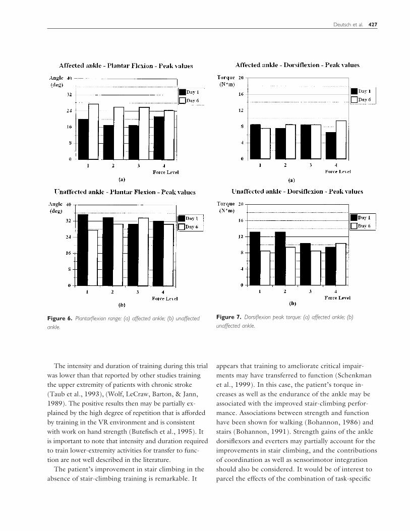
The results from the patient’s clinical exam arepresented in table 2. The results of the patient’s subjec-tive questionnaire are presented in table 3. Post-testresults comparing the torque and excursions of the af-fected and unaffected ankles during performance base-lines are presented in table 4. Results comparing theperformance accuracy of the affected and unaffected sideduring the VR simulations are presented in table 5. Re-sults for plantar flexion range of motion changes areshown in figure 6. Results for changes in torque fordorsiflexion and eversion, measured with the RutgersAnkle, are plotted in figures 7 and 8. Changes in poweroutput of the affected and unaffected ankles are pre-sented in figure 9. The variation in the number of loopsentered/minute is shown in figure 10.
3.4 Discussion
Upon the completion of a six-session, VR trainingprogram of the ankle, improvements were noted in theforce generation, endurance, and coordination of theaffected ankle, as well as in the functional mobility ofthe patient. Improvements in the clinical measures cor-related well with the variables collected by the RutgersAnkle interface.
Clinical measures of force generation (using manualmuscle test scores, see table 2) corresponded withchanges in torque measured with the Rutgers Ankleduring the uniplanar movements for dorsiflexion torque(see figure 7) and everter torque (see figure 8) as well asthe performance baseline peak torque values (see table4). The changes in the clinical muscle test scores may beattributed to neural, rather than skeletal, changes in themuscle. Use of a dynamometer would aid in increasingthe sensitivity of the muscle test scores.
Clinically endurance was quantified by the length oftime the patient was able to execute individual simula-tions and the total training time for each session. (Seetable 1.) These values increased steadily and corre-sponded well with the measures of power output, col-lected using the Rutgers Ankle system. The power out-put of the affected ankle, namely the sum of ankle
Deutsch et al. 423

Table 1. Exercise Progression
Exercise
Day Two Day Three Day Four Day Five Day Six
Speed Time Speed Time Speed Time Speed Time Speed Time
Pitch 40* 3:50 40* 5:00 40 5:00 35** 2:30 40* 2:00--- --- 50* 1:30
50* 2:00 302 2:0040* 5:15 40**
Roll 30* 5:00 30* 3:00 20* 5:00 202 1:15 30* 2:00--- --- 152 3:00 30** 2:15 20** 2:00
20* 5:00 20** 20** 2:00 20** 2:0040*
Combined 102 5:00 20* 3:00 15* 5:00 40* 3:00 20* 2:0020* --- --- 15** 5:00 25** 2:20 35* 2:00
10* 4:00 40* 1:00 25** 2:00
Total Time
All Exercises 13:50 11:00 23:00 14:20 17:30---
17:00
Speed is in framesTime is in minutesDifficulty setting in degree of excursion, related to the performance baseline: it was rated as * easy (part of theexcursion) ** medium (the complete excursion)---line on day three indicates work before the modification in the simulation (above the line) and after the modificationof the simulation (below the line)
Table 2. Clinical Exam
Before Virtual Reality Training After Virtual Reality Training
Affected Side Unaffected Side Affected Side Unaffected Side
Strength (MMT)*Dorsiflexion 5 5 5 5Inversion 4 4 5 5Eversion 5 4 5 5Pain/discomfort To palpation along
the posteriormalleolus andwith eversion
Soreness alongthe posteriortibialis
Soreness alongthe evertersurface
Stairs Negotiated four steps rail on left in 1:30sec. (descended with a step-to-steppattern)
Negotiated four steps rail on left 20sec. (descended with a reciprocalpattern)
Negotiated eleven steps which hedeclined to do pre-training
MMT manual muscle test: strength measured on a scale of 0-5, with 5 being the strongest.
424 PRESENCE: VOLUME 10, NUMBER 4
mechanical effort over time (see figure 9), had robustincreases over the six sessions.
Coordination, which was measured as an accuracyscore reflecting the number of loops the airplane suc-cessfully passed, also improved. This was especially true
for the Rutgers Ankle configurations that required mov-ing the ankle into all its available motions, switchingfrom agonist to antagonist. (See table 5.) Accuracyscores improved for the affected ankle, and, at the com-pletion of training, they even surpassed the scores of the
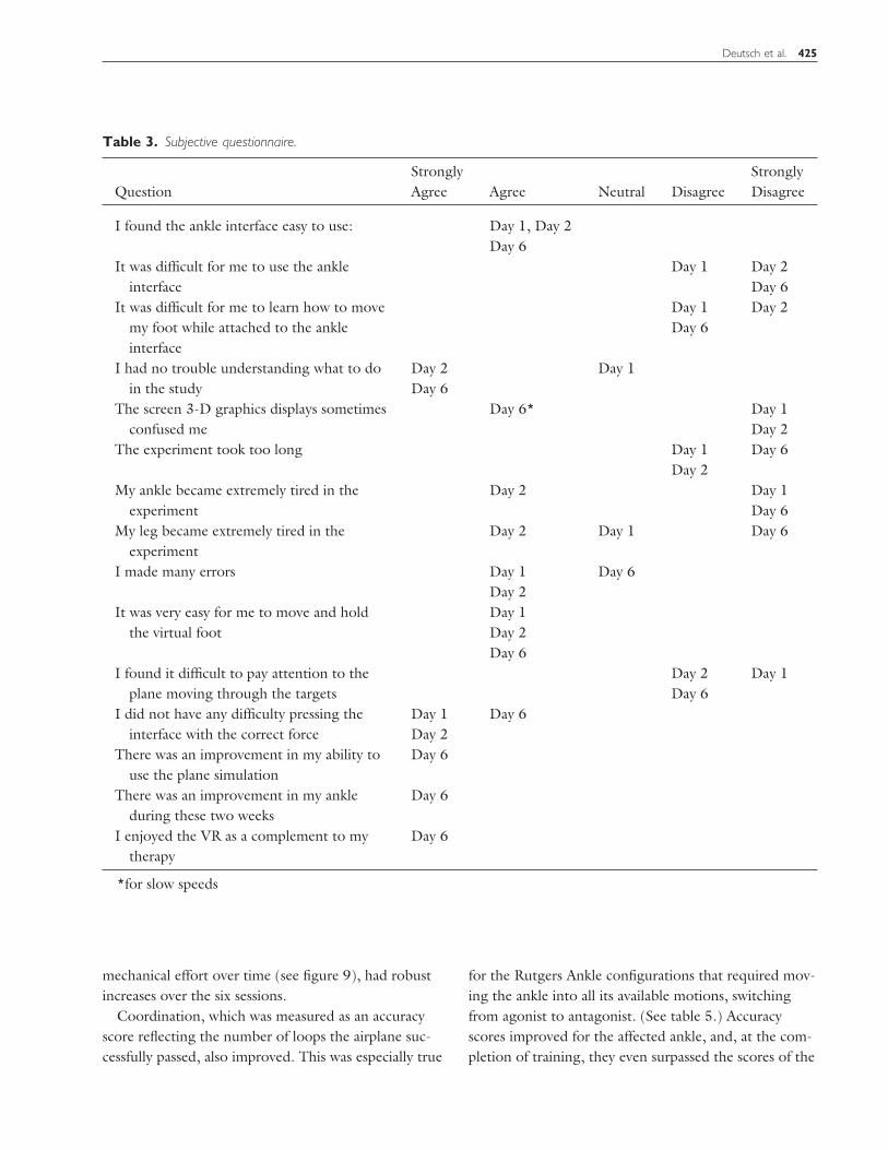
Table 3. Subjective questionnaire.
QuestionStronglyAgree Agree Neutral Disagree
StronglyDisagree
I found the ankle interface easy to use: Day 1, Day 2Day 6
It was difficult for me to use the ankleinterface
Day 1 Day 2Day 6
It was difficult for me to learn how to movemy foot while attached to the ankleinterface
Day 1Day 6
Day 2
I had no trouble understanding what to doin the study
Day 2Day 6
Day 1
The screen 3-D graphics displays sometimesconfused me
Day 6* Day 1Day 2
The experiment took too long Day 1Day 2
Day 6
My ankle became extremely tired in theexperiment
Day 2 Day 1Day 6
My leg became extremely tired in theexperiment
Day 2 Day 1 Day 6
I made many errors Day 1Day 2
Day 6
It was very easy for me to move and holdthe virtual foot
Day 1Day 2Day 6
I found it difficult to pay attention to theplane moving through the targets
Day 2Day 6
Day 1
I did not have any difficulty pressing theinterface with the correct force
Day 1Day 2
Day 6
There was an improvement in my ability touse the plane simulation
Day 6
There was an improvement in my ankleduring these two weeks
Day 6
I enjoyed the VR as a complement to mytherapy
Day 6
*for slow speeds
Deutsch et al. 425

unaffected ankle. Furthermore, the rate at which loopswere entered improved by 45% over the six rehabilitationsessions, as illustrated in figure 10. This is further indica-tion of increased coordination on the affected ankle.
Several outcomes of the VR training were measured(strength, range, coordination, and flexibility), but sev-eral variables that may have been affected by the VRtraining were not specifically measured. For example,the contribution of the sensory input from the RutgersAnkle provided by the force feedback may have stimu-lated the patient’s proprioceptive and kinesthetic pro-cessing. The visual perceptual effects of looking at the3-D simulation and coordinating it with the foot move-
ments were also not quantified in this case study. Expla-nations for the positive outcome of the training will beenhanced with the measurement of these factors.
The patient was able to learn the VR simulation andconcentrate on the therapy in an active rehabilitationclinic, with activity and distractions. His subjective eval-uation of the experience (detailed in table 3) suggeststhe patient was engaged in the simulation and found itto be a useful complement to his rehabilitation. His re-ports of most challenging motions for him—horizontalmovements at first (especially eversion) and then thecombination movements—are consistent with his per-formance (using accuracy scores) during the trial.
Table 4. Post-Test Results Comparing the Affected and Unaffected Sides During Performance Baselines
Day Two Day Six
Affected Side Unaffected Side Affected Side Unaffected Side
Excursion*Dorsiflexion 42 50 35 40Plantarflexion 12 18 26 35Inversion 37 42 42 38Eversion 2 18 12 20
(20 on day 5)Torque**
Plantarflexion (y) 6 5 8 9Dorsiflexion (y) 6 4 8 4Inversion (x) 5 8 8 8Eversion (x) 2 5 4 5
*Excursion: Range during performance baseline comparison is between day 2 and day 6**Torque during performance baseline (on day 2 and day 6).
Table 5. Accuracy during VR simulations
Day Three Day 6
Affected Unaffected Affected Unaffected
Accuracy*PF/DF vertical 32% (50) NT 95% (50) 60% (50)Inv/Ev horizontal 84% (20) NT 86% (30) 90% (30)Combination 58% NT 88% (30) 62% (30)
*Accuracy Targets Hit/Targets Entered * 100 during comparable simulations, speed of plane in parenthesis
426 PRESENCE: VOLUME 10, NUMBER 4
The intensity and duration of training during this trialwas lower than that reported by other studies trainingthe upper extremity of patients with chronic stroke(Taub et al., 1993), (Wolf, LeCraw, Barton, & Jann,1989). The positive results then may be partially ex-plained by the high degree of repetition that is affordedby training in the VR environment and is consistentwith work on hand strength (Butefisch et al., 1995). Itis important to note that intensity and duration requiredto train lower-extremity activities for transfer to func-tion are not well described in the literature.
The patient’s improvement in stair climbing in theabsence of stair-climbing training is remarkable. It
appears that training to ameliorate critical impair-ments may have transferred to function (Schenkmanet al., 1999). In this case, the patient’s torque in-creases as well as the endurance of the ankle may beassociated with the improved stair-climbing perfor-mance. Associations between strength and functionhave been shown for walking (Bohannon, 1986) andstairs (Bohannon, 1991). Strength gains of the ankledorsiflexors and everters may partially account for theimprovements in stair climbing, and the contributionsof coordination as well as sensorimotor integrationshould also be considered. It would be of interest toparcel the effects of the combination of task-specific
Figure 6. Plantarflexion range: (a) affected ankle; (b) unaffected
ankle.
Figure 7. Dorsiflexion peak torque: (a) affected ankle; (b)
unaffected ankle.
Deutsch et al. 427

training complemented with this VE simulation aswell as the use of VR simulations that include task-specific training.
4 Conclusions and Future Work
To our knowledge, this is the first time that a vir-tual environment with force feedback has been reportedfor the use of lower-extremity rehabilitation for a pa-tient post-stroke. The Rutgers Ankle is a novel approachto ankle rehabilitation for stroke patients. Patients inter-act with a Stewart platform robot, exercising their an-kles’ three degrees of freedom. The high-level control of
positions and forces is handled by a host PC running aninteractive virtual environment (VE) simulation. Thesystem with its use of VEs is intended to make rehabili-tation more accessible, effective, fun, and motivating.
This case study provides preliminary and promisingresults about the efficacy of using the Rutgers Ankle forlower-extremity rehabilitation of an individual post-stroke. Further study using controlled research designsis required to define the intensity and duration of thetraining, as well as how it should complement func-tional training. The sensitivity of the strength measurescould be increased by using a dynamometer instead ofthe ordinal manual muscle testing scores. The mecha-
Figure 8. Eversion peak torque: (a) affected ankle; (b) unaffected
ankle.
Figure 9. Power output of the affected versus unaffected ankle.
Figure 10. Loops per minute entered using the Rutgers Ankle on
the affected ankle.
428 PRESENCE: VOLUME 10, NUMBER 4

nism by which the VE promotes rehabilitation will alsohave to be elucidated.
Through the proof-of-concept patient trial, we wereable to receive feedback from patients and physical ther-apists. Their suggestions for improvement will be takeninto account as the system matures. Suggestions to im-prove the comfort of the device include modifying thefoot-attachment straps, using an adjustable chair, andstabilizing the knee with a strap.
Further improvements will be done on the technol-ogy side as well. In the future, the electronic controllermay be integrated into the base of the Stewart platformto increase the compactness and portability of the sys-tem. Furthermore, a calibration routine will be added,such that forces due to the foot’s passive weight will be“zeroed out” at the start of the exercise routine. Simi-larly, position measurements will be zeroed out whenthe platform supporting the patient’s foot is in its initialposition parallel to the floor. This calibration will allowa more accurate and reliable measure of foot excursionand mechanical exertion during the VR rehabilitationexercise.
The system will be extended using the Internet as acommunication link with the patient’s home. Data willthen be uploaded from the host PC by a therapist at aremote site for evaluation. As the patient improves, thetherapist should be able to remotely modify exerciseparameters such as required duration, maximum-opposing forces, allowed ROM, and VE complexity.Finally, the simulations will include standing and walk-ing activities. This will allow the patient to train thelimb in the loaded position, which is consistent withmany of the functions of the lower extremity.
Acknowledgments
The research reported here was supported by grants from theNational Science Foundation (BES-9708020 and REU) andfrom Rutgers University (SROA and CAIP grants). We wishto acknowledge the Suburban Physical Therapy Center for theuse of their facility to conduct the clinical trial. We also thankJeff Fitzgerald, M.S. P.T., for invaluable assistance with pa-tient recruitment.
References
Bohannon, R. W. (1986). Strength of lower limb muscle re-lated to gait velocity and cadence in stroke patients. Physio-therapy Canada, 38(4), 204–206.
———. (1991). Association of paretic lower extremitystrength and balance with stair climbing ability in patientswith stroke. Journal of Stroke and Cerebrovascular Disease,1, 65–69.
Burdea, G. (1996). Force and touch feedback for virtual reality.New York: John Wiley & Sons. Inc., New York.
Burdea, G., Popescu, V., Hentz, V., & Colbert, K. (2000).Virtual reality-based orthopedic tele-rehabilitation. IEEETransactions on Rehabilitation Engineering, 8(3),429–432.
Butefisch, C., Hummelsheim, H., Denzler, P., & Mauritz,K. H. (1995). Repetitive training of isolated movementsimproves the outcome of motor rehabilitation of the cen-trally paretic hand. Journal of the Neurological Sciences,130(1), 59–68.
Dieudonne, J. E., Parrish, R. V., & Bardusch, R. E. (1972).An actuators extension transformation for a motion simula-tor and an inverse transformation applying Newton-Raphson’s method. (Langley Research Center, NASA TND-7067).
Dean, C. M., & Shepherd, R. B. (1997). Task-related trainingimproves performance of seated reaching tasks followingstroke: A randomized clinical trial. Stroke, 28, 722– 728.
Duncan, P. W., Richards, L, Wallace, D., Stoker-Yates, J.,Pohl, P., Luchies, C., Ogle, A., & Studenski, S. (1998). Arandomized controlled pilot study of a home-based exerciseprogram for individuals with mild and moderate stroke.Stroke, 29, 2055–2060.
Duncan, P. W., Wallace, D., Min Lai, S., Johnson, D., Em-bretson, S., & Jacobs Laster, L. (1999). The Stroke ImpactScale Version 2.0-Evaluation of reliability, validity and sensi-tivity to change. Stroke, 30, 2131–2140.
Girone, M. J., & Burdea, G. C. (1998). Ankle rehabilitationin virtual reality. Report to the National Science Foundationon grant BES-970802.
Girone, M. J., Burdea, G. C., & Bouzit, M. (1999). The Rut-gers Ankle orthopedic rehabilitation interface. Proc. of theASME, Dynamic Systems and Control Division, 67, 305–312.
Girone, M. J., Burdea, G. C., Bouzit, M., Popescu, V. &Deutsch, J. E. (2000). Orthopedic Rehabilitation Using the“Rutgers Ankle” Interface. Proc. of Medicine Meets VirtualReality 2000, 89–95.
Deutsch et al. 429

Holden, M., Todorow, E., Callahan, J., & Bizzi, E. (1999).Virtual environment training improves motor performancein two patients with stroke: Case report. Neurology Report,23 (2), 57–67.
Holden, M. K., Dyar T., Callahan, J., Schwamm, L., & Bizzi,E. (2000). Motor learning and generalization followingvirtual environment training in a patient with stroke. Neu-rology Report, 24(5), 170.
Jack, D., Boian, R., Merians, A., Tremaine, M., Burdea, G.,Adamovich, S., Recce, M., & Poizner, H. (2001). Virtualreality-enhanced stroke rehabilitation. IEEE Transactions onRehabilitation Engineering (forthcoming).
Jorgensen, H. S., Nakayama, H., Raaschou, H. O., & Olsen,T. S. (1995). Recovery of walking function in stroke pa-tients: The Copenhagen Stroke Study. Archives of PhysicalMedicine and Rehabilitation, 76 (1), 27–32.
Kleim, J. A., Lussnig, E., Schwarz, E. R., Comery, T. A, &Greenough, W. T. (1996). Synaptogenesis and Fos Expres-sion in the motor cortex of the adult rat after motor skilllearning. J. Neuroscience, 16, 4529–4535.
Kwakkel, G., Wagenaar, R. C., Twisk, J. W. R., Lankhorst,G. J., & Koesier, J. C. (1999). Intensity of leg and armtraining after primary middle cerebral artery stroke: A ran-domised trial. Lancet 354, 191–196.
Mahoney, F. I., & Barthel, D. W. (1965). Functional evalua-tion: The Barthel Index. Maryland State Medical Journal,14, 61–65.
Merians, A., Jack, D., Bolan, R., Tremaine, M., Burdea, G.,Ademovich, S., Recce, M., & Poizner, H. (2001). VirtualReality—Augmented Rehabilitation for Patients PostStroke: Three Case Studies. Physical Therapy (submitted).
NASA Langley Research Center. (1999). Simulation SystemsBranch. Visual Motion Simulator. Available at: http://bigben.larc.nasa.gov/facility/vms.html.
Nguyen, C. C., & Pooran, F. J. (1989). Kinematic analysisand workspace determination of a 6DOF CKCM robotend-effector. Journal of Mechanical Working Technology, 20,283–294.
Nudo, R. J., Wise, B. M., Sifuentes, F., & Milliken, G. W.(1996). Neural substrates for the effects of rehabilitativetraining on motor recovery after ischemic infarct. Science,272, 1791–1794.
Nudo, R. J. (1998). Role of cortical plasticity in motor recov-ery after stroke. Neurology Report, 22 (2), 61–67.
Patounakis, G., Bouzit, M., & Burdea, G. (1998). Study of theelectromechanical bandwidth of the Rutgers Master (TechnicalReport CAIP-TR-225) Piscataway, NJ: Rutgers University.
Rizzo, A., Buckwalter, J., van der Zaag, C., Neumann U.,Tiebaux, M., Chua, C., van Rooyen, A., Humphrey, L., &Larson, P. (2000). Virtual environment applications in clini-cal neuropsychology. Proc. of IEEE Virtual Reality 2000,63–70.
Schenkman, M., Bliss, S. T., Day, D. L, Kemppainen, S., &Pratt, J. (1999). Multisystem model for management ofneurologically impaired adults: An update and illustrativecase. Neurology Report, 24 (4), 145–157.
Stewart, D. (1966). A platform with 6 degrees of freedom.Proc. of the Institution of Mechanical Engineers, 1965–1966.
Sullivan, K. J., & Duncan, P. W. (2000). New perspectives forlocomotor training after stroke: Emerging evidence frombasic science and clinical research. Neurology Report, 25(2),55–59.
Taub, E., Miller, N. E., Novack, T. A., Cook, E. W., Fleming,W. C., Nepomuceno, C. S., Connell, J. S., & Crago, J. E.(1993). A technique for improving chronic motor deficitafter stroke. Archives of Physical Medicine and Rehabilita-tion, 74, 347–354.
Wade, D. T., Collen, F. M., Robb, G. F., & Warlow, C. P.(1992). Physiotherapy intervention late after stroke andmobility. British Medical Journal, 304, 609–613.
Wandel, A., Jorgensen, H. S., Nakayama, H., Raaschou,H. O., & Olsen, T. S. (2000). Prediction of walking func-tion in stroke patients with initial lower extremity paralysis:The Copenhagen Stroke. Archives of Physical Medicine &Rehabilitation, 81(6), 736– 738.
Werner, R. A., & Kessler, S. (1996). Effectiveness of an inten-sive outpatient rehabilitation program for postacute strokepatients. Am. J. of Physical Medicine, 75(2), 114–120.
Wolf, S. L., LeCraw, D. E., Barton, L. A., & Jann, B. B.(1989). Forced used of hemiplegic upper extremities to re-verse the effect of learned nonuse among chronic stroke andhead injured patients. Experimental Neurology, 104, 125–132.
430 PRESENCE: VOLUME 10, NUMBER 4