programmed necrosis in the cross talk of cell death and ... · 2centro de pesquisas gonc¸alo...
TRANSCRIPT

IY33CH04-Chan ARI 20 March 2015 19:30
Programmed Necrosis in theCross Talk of Cell Deathand InflammationFrancis Ka-Ming Chan,1 Nivea Farias Luz,1,2
and Kenta Moriwaki11Department of Pathology, Immunology and Microbiology Program, University ofMassachusetts Medical School, Worcester, Massachusetts 01605;email: [email protected] de Pesquisas Goncalo Moniz/Fundacao Oswaldo Cruz, Salvador 40295-001, Brazil;Universidade Federal da Bahia, Salvador 40110-060, Brazil
Annu. Rev. Immunol. 2015. 33:79–106
First published online as a Review in Advance onDecember 10, 2014
The Annual Review of Immunology is online atimmunol.annualreviews.org
This article’s doi:10.1146/annurev-immunol-032414-112248
Copyright c© 2015 by Annual Reviews.All rights reserved
Keywords
necroptosis, RIPK1, RIPK3, MLKL, TNF, inflammation, TRIF, DAI,MCMV, vaccinia virus
Abstract
Cell proliferation and cell death are integral elements in maintaining home-ostatic balance in metazoans. Disease pathologies ensue when these pro-cesses are disturbed. A plethora of evidence indicates that malfunction ofcell death can lead to inflammation, autoimmunity, or immunodeficiency.Programmed necrosis or necroptosis is a form of nonapoptotic cell deathdriven by the receptor interacting protein kinase 3 (RIPK3) and its sub-strate, mixed lineage kinase domain-like (MLKL). RIPK3 partners with itsupstream adaptors RIPK1, TRIF, or DAI to signal for necroptosis in re-sponse to death receptor or Toll-like receptor stimulation, pathogen in-fection, or sterile cell injury. Necroptosis promotes inflammation throughleakage of cellular contents from damaged plasma membranes. Intriguingly,many of the signal adaptors of necroptosis have dual functions in innate im-mune signaling. This unique signature illustrates the cooperative nature ofnecroptosis and innate inflammatory signaling pathways in managing celland organismal stresses from pathogen infection and sterile tissue injury.
79
Ann
u. R
ev. I
mm
unol
. 201
5.33
:79-
106.
Dow
nloa
ded
from
ww
w.a
nnua
lrev
iew
s.or
g A
cces
s pr
ovid
ed b
y U
nive
rsity
of
Mas
sach
uset
ts M
edic
al S
choo
l - W
orce
ster
on
01/2
2/16
. For
per
sona
l use
onl
y.

IY33CH04-Chan ARI 20 March 2015 19:30
INTRODUCTION
Cell death is an important biological process that sculpts the development of multicellular or-ganisms. In the immune system, cell death plays critical roles in immune cell development andpathogen defense. Apoptosis is an orderly form of cell death marked by chromatin condensation,DNA fragmentation, and membrane blebbing into apoptotic bodies. Apoptosis can be triggeredby receptors in the tumor necrosis factor (TNF) superfamily (extrinsic pathway) or through di-rect activation of mitochondrial effectors (intrinsic pathway). Caspases are cysteine proteases thatdrive apoptosis. Effector caspases cleave and inactivate the flippase adenosine triphosphatase type11C and scramblase X Kell blood group precursor related family member 8 (Xkr8) (1, 2). Thisresults in exposure of phosphatidyl serine (PS) on the cell surface, which flags the dying cell foruptake and clearance by professional phagocytes such as macrophages (3). The rapid clearance ofapoptotic cells ensures minimal risk of detrimental inflammation. This explains why apoptosis isthe preferred and dominant pathway by which multicellular organisms eliminate unwanted cellsduring development. In contrast, necrosis is marked by rapid loss of plasma membrane integrity.Plasma membrane leakage in necrosis is widely thought to occur prior to or concomitant withexposure of PS and other eat-me signals. This early rupture of the plasma membrane releasesendogenous danger signals or damage-associated molecular patterns (DAMPs), which are potentstimulants of inflammation (4). As such, necrosis is often detected in infections and inflammatorydiseases. This association has led to the popular view that necrosis represents pathological celldeath, whereas apoptosis is more central for development.
Pathologists have historically relied on morphology to distinguish between apoptosis andnecrosis. Apoptosis is marked by cell shrinking, the appearance of membrane blebs called ap-optotic bodies, and condensation of chromatin. In contrast, necrosis is associated with cell andorganelle swelling and limited chromatin condensation. Biologists have long considered necrosisas the consequence of trauma or accidental injury. This view has now been revised with recentadvances showing the existence of dedicated molecular pathways controlling necrotic cell death.Notably, the classical markers that define apoptosis can sometimes be detected in necrosis. Forexample, Annexin V staining is a commonly used method to detect exposure of PS on the outerleaflet of the plasma membrane in early apoptotic cells. In some necrotic cells, PS exposure canbe detected without significant plasma membrane leakage (5). PS exposure is supposed to markapoptotic cells for clearance by phagocytes. However, researchers have also described scavengerreceptors that recognize necrotic cells (6, 7). Moreover, terminal deoxynucleotidyl transferasedUTP nick end labeling (TUNEL) staining, which detects DNA strand breaks in apoptosis, alsodetects such breaks in necrotic cells (8). These observations suggest that the morphological defini-tion of apoptosis and necrosis is insufficient to distinguish between these two cell death modules.Instead, we favor a molecular definition based on genetic pathways. Terms such as programmednecrosis, regulated necrosis, and necroptosis are now used to describe necrosis induced by thereceptor interacting protein kinases (RIPKs) (9). In addition, certain forms of regulated necrosiscan occur without the RIPKs (10, 11).
Here, we focus our discussion on RIPK-driven necrosis. The term necroptosis is used through-out to distinguish RIPK-dependent necrosis from other forms of regulated necrosis. As is discussedbelow, pathways that control necroptosis and inflammation often use overlapping signaling adap-tors. The sharing of common signal adaptors establishes an intimate link between inflammationand necroptosis that goes beyond their association in disease pathologies. Hence, necroptosis andinflammation can be mutually reinforcing processes that govern not only inflammatory diseasesbut also immune and organismal homeostasis.
80 Chan · Luz · Moriwaki
Ann
u. R
ev. I
mm
unol
. 201
5.33
:79-
106.
Dow
nloa
ded
from
ww
w.a
nnua
lrev
iew
s.or
g A
cces
s pr
ovid
ed b
y U
nive
rsity
of
Mas
sach
uset
ts M
edic
al S
choo
l - W
orce
ster
on
01/2
2/16
. For
per
sona
l use
onl
y.

IY33CH04-Chan ARI 20 March 2015 19:30
THE MOLECULAR MACHINERY OF PROGRAMMED NECROSIS
Necroptosis can be activated by death receptors in the TNF superfamily, Toll-like receptor 3(TLR3) and TLR4, and the interferon receptors (12). The signaling pathway for necroptosis isbest characterized for TNF receptor 1 (TNFR1). TNFR1 is the prototypic member of a sub-family within the TNF receptor superfamily that contains an essential protein interaction domaincalled the death domain (DD). DD-containing death receptors include CD95/FAS/APO-1, TNF-related apoptosis-inducing ligand (TRAIL) receptor 1 and 2, death receptor 3 (DR3), DR6, andectodysplasin A receptor. However, cell death is not the only signaling outcome for the deathreceptors. In fact, nuclear factor-κB (NF-κB) activation is often the dominant response emanat-ing from these receptors. TNFR1 is a prime example of such a receptor. Ligation of the pre-assembled TNFR1 trimer (13) with TNF causes a conformation change that promotes formationof a short-lived membrane-signaling complex termed Complex I by Micheau & Tschopp (14).This membrane complex is composed of the adaptors TNF receptor–associated death domain(TRADD), TNF receptor–associated factor 2 (TRAF2), RIPK1, cellular inhibitor of apoptosis 1(cIAP1), cIAP2, and the linear ubiquitin chain assembly complex (LUBAC), which is made up ofthe subunits RanBP-type and C3HC4-type zinc finger containing 1 (aka HOIL-1), ring fingerprotein 31 (aka HOIP), and Shank-associated RH domain interacting protein (SHARPIN) (15).The E3 ligases cIAP1, cIAP2, and HOIL-1 within LUBAC critically control ubiquitination ofmany of the adaptors within Complex I. Ubiquitin linkages of different types have been found withvarious adaptors in Complex I. This ubiquitin network within Complex I is essential for recruit-ment and activation of the inhibitor of κB kinase (IKK) complex. The activated IKK phosphory-lates IκBα, leading to IκBα degradation by the proteasome and nuclear translocation of NF-κBdimers.
NF-κB is a key transcription factor for many proinflammatory and survival genes. A properNF-κB response is crucial for cell survival and to counteract the cytotoxic effects of TNF. Theprosurvival effect of NF-κB is mediated in part by its transcriptional targets cIAP1, cIAP2, and thelong form of cellular FLICE-like inhibitor protein (cFLIPL) (16, 17). As we discuss below, cFLIPL
and the cIAPs critically regulate cellular sensitivity to apoptosis and necroptosis. Hence, Complex Iis a critical checkpoint for cell death versus cell survival signaling (Figure 1). In addition, ComplexI adaptors appear to have NF-κB-independent survival functions. For example, mice lackingboth cIAP1 and cIAP2 die at an earlier stage in embryonic development than RelA/p65-deficientmice (18). The adaptor TRAF2 stabilizes cIAP1 expression by preventing its autoubiquitinationand proteasomal degradation (19). Consistent with this association, Traf2−/− mice also exhibitembryonic lethality (20). In contrast, cpdm mice that lack the LUBAC subunit SHARPIN showdefective NF-κB activation but are nonetheless viable (21–23). These results demonstrate that thecytoprotective effects of TRAF2, cIAP1, cIAP2, and X-linked IAP (XIAP) are mediated throughNF-κB-dependent and -independent functions.
Unlike conventional death receptors such as Fas or TRAIL receptors, Fas-associated via deathdomain (FADD) and caspase-8 are not recruited to the TNFR1-associated Complex I (14, 24).Instead, rapid receptor internalization is important for docking of the adaptor FADD and the ini-tiator caspase, caspase-8, to the complex. Although evidence suggests that FADD and caspase-8can be recruited to the TNFR1 complex (25), standard biochemical pull-down supports a modelin which TNFR1 dissociation occurs prior to docking of FADD and caspase-8 (14). The cytosoliccomplex that contains FADD and caspase-8 is often referred to as Complex IIa (Figure 1). Nor-mally, apoptosis is prevented by dimerization between caspase-8 and cFLIPL, an enzyme-inactivehomolog of caspase-8. The caspase-8/cFLIPL heterodimer inhibits full activation of caspase-8and apoptosis but retains cleavage of essential necrosis regulators such as RIPK1, RIPK3, and
www.annualreviews.org • Programmed Necrosis in Cross Talk 81
Ann
u. R
ev. I
mm
unol
. 201
5.33
:79-
106.
Dow
nloa
ded
from
ww
w.a
nnua
lrev
iew
s.or
g A
cces
s pr
ovid
ed b
y U
nive
rsity
of
Mas
sach
uset
ts M
edic
al S
choo
l - W
orce
ster
on
01/2
2/16
. For
per
sona
l use
onl
y.

IY33CH04-Chan ARI 20 March 2015 19:30
cylindromatosis (CYLD) (26–30). Hence, NF-κB-dependent induction of cFLIPL inhibits apo-ptosis as well as necroptosis.
Active caspase-8 in Complex IIa not only initiates the caspase cascade and the apoptoticprogram but also cleaves and inactivates essential necroptosis mediators such as RIPK1, RIPK3,and CYLD. Hence, inhibition of caspase-8 or its upstream adaptor FADD primes cells fornecroptosis by preserving the integrity of RIPK1 and RIPK3. Stabilization of RIPK1 andrecruitment of RIPK3 convert Complex IIa to Complex IIb or the necrosome (Figure 1). The
TR
AD
D
RIP
K1
TRA
F2
cIA
P1/2
TNF
TNFR1
NF-кB
NEMOIKKα/β
TAK1
TAB1/2
HO
IP
HO
IL-1
SH
AR
PIN
LUBAC
Apoptosis inductionNecrosis inhibition
cFLIPL
Complex IComplex IComplex I
Complex IIaComplex IIa
Inhibition of apoptosisand necrosis
cIAP1/2CYLD
RIP
K1
RIP
K3
RIP
K1
RIP
K3
RIP
K1
RIP
K3
RIP
K1
RIP
K3
MLKL
MLKL
MLKLMLKL
MLKLMLKL
Complex IIb(necrosome)Complex IIb(necrosome)
?
Membranerupture
Releaseof DAMPs
Ub
Ub
Cleaved RIPK1
FADDFADD
Casp-8Casp-8
cFLIPL
Cleaved RIPK3
Cleaved RIPK1Cleaved RIPK3
FAD
D
Ca
sp-8
RIP
K1
RIP
K3
Ca
sp-8
FAD
D
Ca
sp-8
RIP
K1
RIP
K3
Ca
sp-8
FAD
D
Ca
sp-8
RIP
K1
RIP
K3
Ca
sp-8
FAD
DC
asp
-8
RIP
K1
RIP
K3
cFLI
PL
FAD
DC
asp
-8
RIP
K1
RIP
K3
cFLI
PL
OR
P
P
P
P
P
P P P P
P P P P
FAD
DC
asp
-8
RIP
K1
RIP
K3
cFLI
PL
Inhibition ofcaspase-8
Casp-8
82 Chan · Luz · Moriwaki
Ann
u. R
ev. I
mm
unol
. 201
5.33
:79-
106.
Dow
nloa
ded
from
ww
w.a
nnua
lrev
iew
s.or
g A
cces
s pr
ovid
ed b
y U
nive
rsity
of
Mas
sach
uset
ts M
edic
al S
choo
l - W
orce
ster
on
01/2
2/16
. For
per
sona
l use
onl
y.

IY33CH04-Chan ARI 20 March 2015 19:30
cytosolic complexes that contain the RIPKs have also been referred to as the ripoptosome (31, 32),although this term does not make the distinction between apoptosis and necroptosis. RIPK1 andRIPK3 interact via the RIP homotypic interaction motif (RHIM) to form an amyloid-like complexthat is essential for recruitment and activation of the downstream RIPK3 substrate mixed lineagekinase domain-like (MLKL) (33–35). RIPK3 phosphorylates MLKL at Thr357 and Ser358 tostimulate its oligomerization and translocation to intracellular and plasma membranes (34, 36–39)(Figure 1). The precise mechanism by which MLKL induces membrane rupture is controversial,with some reports implicating disruption of calcium or sodium ion channels (36, 37) and othersshowing direct binding to membrane phospholipids and disruption of membrane integrity(38, 39). In contrast to MLKL, another reported substrate of RIPK3, the mitochondrialphosphatase phosphoglycerate mutase family member 5 (PGAM5) (40), may not be crucial, assmall hairpin RNA–mediated knockdown of Pgam5 did not consistently confer protection againstTNF-induced necroptosis (41, 42). In agreement with the notion that PGAM5 is not a corecomponent of the necroptosis machinery, widespread depletion of mitochondria did not impairnecroptosis (43). The differential requirement for mitochondrial signaling further distinguishesnecroptosis from apoptosis.
Because excessive necrosis in FADD- or caspase-8-deficient mice was rescued by inactivationof RIPK1 or RIPK3 (28, 44, 45), FADD and caspase-8 are paradoxically prosurvival factors duringdevelopment. This yin-yang relationship between the RIPKs and FADD/caspase-8 also plays outin the skin keratinocytes, intestinal epithelium, and T cells (46–49). Genetic evidence also pro-vides a mechanistic explanation for the biochemical interaction between FADD, caspase-8, andthe RIPKs. Intriguingly, although caspase-8-deficient Jurkat T cells are sensitized to necroptosisinduced by TNF, Fas ligand (FasL), and TRAIL, FADD-deficient Jurkat cells are only sensitizedto TNF-induced necroptosis (50). The molecular basis for the resistance of these cells to FasL- andTRAIL-induced necrosis is unknown. One possibility is that because FADD is the apical adaptorrecruited to Fas and TRAIL receptors, downstream signaling will be completely blunted in its ab-sence. Although this is certainly the case for FADD-mediated apoptosis, it is insufficient to explainRIPK3 signaling in T cells. Fadd−/− T cells undergo RIPK1- and RIPK3-dependent necroptosisin response to T cell receptor (TCR) stimulation (45, 48, 49). Because Fadd−/−Ripk3−/− micedevelop lymphoproliferation resembling that caused by the Fas mutant in lpr mice (51), one canargue that Fas triggers necroptosis of Fadd−/− T cells through RIPK3. Alternatively, necropto-sis of Fadd−/− T cells could be the consequence of direct TCR signaling. As we explore furtherbelow, RIPK3 is also capable of signaling for necroptosis in the absence of RIPK1 under certainconditions. These perplexing results highlight the fact that the traditional model of Complex I toComplex II transition may not be adequate to account for signaling in necroptosis.
←−−−−−−−−−−−−−−−−−−−−−−−−−−−−−−−−−−−−−−−−−−−−−−−−−−−−−−−−−−−−−−−−−−−−−−−−Figure 1TNF-induced signaling complexes. The membrane-associated Complex I is chiefly responsible for NF-κBactivation. The ubiquitin chains are represented by red hexagons. Induction of cFLIPL expression by NF-κBinhibits apoptosis and necroptosis. Active caspase-8 in Complex IIa promotes apoptosis and inhibitsnecroptosis by cleavage of RIPK1, RIPK3, and CYLD (scissors). When caspase-8 is inactive (circled X), RIPK1and RIPK3 initiate Complex IIb assembly, amyloid conversion, and recruitment of MLKL. Both ComplexIIa and Complex IIb are also regulated by protein ubiquitination. CYLD acts as the deubiquitinase thatpromotes Complex II activity by removing ubiquitin chains on RIPK1 and RIPK3. (Abbreviations: cIAP,cellular inhibitor of apoptosis 1; CYLD, cylindromatosis; DAMP, damage-associated molecular pattern;FADD, Fas-associated via death domain; IKK, inhibitor of κB kinase; LUBAC, linear ubiquitin chainassembly complex; MLKL, mixed lineage kinase domain-like; NF, nuclear factor; P, phosphorylation; RIPK,receptor interacting protein kinase; TNF, tumor necrosis factor; TNFR1, TNF receptor 1; Ub, ubiquitin.)
www.annualreviews.org • Programmed Necrosis in Cross Talk 83
Ann
u. R
ev. I
mm
unol
. 201
5.33
:79-
106.
Dow
nloa
ded
from
ww
w.a
nnua
lrev
iew
s.or
g A
cces
s pr
ovid
ed b
y U
nive
rsity
of
Mas
sach
uset
ts M
edic
al S
choo
l - W
orce
ster
on
01/2
2/16
. For
per
sona
l use
onl
y.

IY33CH04-Chan ARI 20 March 2015 19:30
UBIQUITINATION: A CRITICAL CHECKPOINT FOR NECROPTOSIS
RIPK1 and other Complex I adaptors are key substrates of the E3 ubiquitin ligases cIAP1 andcIAP2. As discussed above, the ubiquitin network within Complex I functions to recruit the IKKcomplex and to promote survival through NF-κB-dependent and -independent mechanisms (52–56). Bivalent IAP antagonists or Smac mimetics (SMs) are often used to deplete cIAP1, cIAP2,and XIAP. SMs are small-peptide mimetics of second mitochondrial-derived activator of caspases(Smac) that trigger autoubiquitination and degradation of the IAPs. Because RIPK1 ubiquitinationdoes not occur in the absence of the IAPs, SM tips the balance of TNF signaling toward cell death.Moreover, because the NF-κB-inducing kinase (NIK) is constitutively targeted for ubiquitinationand degradation by the IAPs (57–60), SM can additionally stabilize NIK, leading to noncanonicalNF-κB activation and autocrine TNF production. Thus, SM primes cells for cell death throughtwo mutually reinforcing mechanisms: elimination of a cytoprotective ubiquitin network andinduction of TNF. Given that many tumors overexpress cIAPs and are resistant to traditionalchemotherapies, SM can provide a powerful one-two punch to trigger cancer cell death througheither apoptosis or necroptosis (61, 62). Physiologically, IAP depletion occurs in response tostimulation of TNFR2, the TNF receptor whose expression is highly inducible (63). AlthoughTNFR2 does not contain a cytoplasmic DD, it recruits TRAF2 and the cIAPs and triggers theirproteasomal degradation. Hence, similar to the action of SM, TNFR2 also skews TNF signalingtoward cell death (64–67). Hence, the IAPs and ubiquitination play important roles in fending offthe cytotoxic effects of TNF.
The importance of cIAPs and the ubiquitin machinery in regulating RIPK activities and necrop-tosis is illustrated by the partial rescue of embryonic lethality of ciap1−/−xiap−/− or ciap1−/−ciap2−/−
embryos by loss of Ripk3 or a single Ripk1 allele (18). Systemic autoinflammatory disease of micewith myeloid-specific deletion of cIAP1, cIAP2, and XIAP was also corrected by inactivation ofRIPK1 or RIPK3 (68). These results provide strong evidence that the IAPs are crucial guardiansthat keep RIPK1 and RIPK3 in check to prevent deleterious cell injury and inflammation. Furtherevidence that the ubiquitin network within Complex I serves critical functions in limiting cell deathand inflammation comes from mice lacking the LUBAC components SHARPIN or HOIL-1. Cellslacking SHARPIN or HOIL-1 are sensitized to apoptosis as well as necroptosis (21, 69), and micelacking these components develop systemic autoinflammatory diseases (21–23, 70). Interestingly,the severe skin and multiorgan inflammation in SHARPIN-deficient cpdm mice was corrected bycrosses to knock-in mice expressing kinase-inactive RIPK1 (Ripk1-K45A) (71). Fibroblasts andmacrophages from Ripk1-K45A knock-in mice exhibit normal mitogen-activated protein kinaseand NF-κB responses but are resistant to TNF-induced necroptosis (71, 72). Hence, excessive celldeath appears to be the major driver for RIPK1-dependent inflammation in cpdm mice. However,because the kinase activity of RIPK1 is required for apoptosis under certain conditions (73), itremains to be determined if RIPK1-induced inflammation in cpdm mice is driven by apoptosis ornecroptosis.
In addition to mouse models, human patients with mutations in the E3 ligase subunit ofLUBAC HOIL-1 exhibit chronic inflammation, increased cytokine expression in response to IL-1β, cardiomyopathy, and susceptibility to pyogenic bacteria due to impaired NF-κB activation(74, 75). Consistent with the key role of the LUBAC complex for recruitment of the IKK com-plex, mutations in the IKK regulatory subunit NF-κB essential modulator (NEMO)/IKKγ causeincontinentia pigmenti (IP), a disease marked by skin lesions and multiorgan inflammation. Be-cause Nemo is an X-linked gene, male mice lacking NEMO are embryonic lethal (76–78), andmale patients of IP are rarely found. In the few rare cases of male patients harboring mild mu-tations in NEMO, patients develop a variant form of the disease called hypohidrotic ectodermal
84 Chan · Luz · Moriwaki
Ann
u. R
ev. I
mm
unol
. 201
5.33
:79-
106.
Dow
nloa
ded
from
ww
w.a
nnua
lrev
iew
s.or
g A
cces
s pr
ovid
ed b
y U
nive
rsity
of
Mas
sach
uset
ts M
edic
al S
choo
l - W
orce
ster
on
01/2
2/16
. For
per
sona
l use
onl
y.

IY33CH04-Chan ARI 20 March 2015 19:30
dysplasia, which is marked by abnormalities in the teeth, hair, and eccrine sweat glands (79). Thechronic inflammatory phenotypes caused by mutations in the cIAPs, LUBAC, and NEMO seemto contradict the fact that cells lacking these adaptors are impaired in cytokine-induced NF-κBresponses. Because cells lacking these components in the ubiquitin network are also highly sensi-tive to death signals, heightened cell death is likely the driver of the chronic inflammation. It willbe interesting to determine if the kinase function of RIPK1 is similarly responsible for driving thelethal inflammatory disease of Nemo−/− mice as in cpdm mice (71).
The NF-κB transcriptional target A20 and the tumor suppressor CYLD are believed to fa-cilitate Complex I transition to Complex II by promoting deubiquitination of RIPK1. Althoughboth A20 and CYLD are recruited to Complex I, small interfering RNA (siRNA) knockdownof CYLD, but not A20, protects cells against TNF-induced necroptosis (69, 80, 81). Surpris-ingly, RIPK1 ubiquitination in Complex I was not altered in Cyld−/− cells. Rather, RIPK1 andRIPK3 ubiquitination within the necrosome was greatly elevated in Cyld−/− cells (81). Hence,rather than regulating RIPK1 ubiquitination in Complex I, CYLD acts within the necrosome todeubiquitinate RIPK1 and RIPK3. Moreover, these results suggest that in addition to regulatingNF-κB activation within Complex I, the E3 ligases cIAP1 and cIAP2 may also control necrosomeactivation through ubiquitination of RIPK1 and RIPK3.
THE JANUS NATURE OF RIPK1
Besides acting as the upstream activator of TNF-induced, RIPK3-mediated necroptosis, RIPK1 isalso required for SM-primed, TNF-induced apoptosis (73). Paradoxically, RIPK1 also functions asan inhibitor of RIPK3- and caspase-8-mediated cell injury and inflammation. Ripk1−/− mice sufferfrom perinatal lethality that was originally believed to be caused by defective NF-κB-mediatedinduction of survival genes (82). However, Ripk1−/− mice are born alive, whereas RelA−/− mice diein utero at E15.5. In addition, a recent report argues that RIPK1 does not play a significant role inNF-κB activation (83). These results suggest that defective NF-κB activity may not fully accountfor the perinatal lethality of Ripk1−/− mice. Mice that lack multiple cIAPs, such as ciap1−/−Xiap−/−
mice, suffer from embryonic lethality at E10.5. This lethality is eerily similar to that in mice lackingFadd, Casp8, or cFlip. Strikingly, hemizygous Ripk1 deficiency significantly prolonged survival ofciap1−/−Xiap−/− mice until weaning age (18). Thus, researchers have proposed an alternative modelin which RIPK1 ubiquitination sterically hinders recruitment of downstream cell death effectors(55, 56).
Recently, the molecular basis that underlies the perinatal lethality of Ripk1−/− mice was exam-ined in further detail. Ripk1−/− mice exhibit extensive cleaved caspase-3 in multiple tissues and asystemic increase in inflammatory cytokines. The increase in caspase-3 activation and apoptosisappears to be partly due to a failure to upregulate cFLIPL expression (84). Cleaved caspase-3and apoptosis were significantly reduced in Ripk1−/−Casp8−/− mice. However, these mice stillsuccumbed to perinatal lethality (85), indicating that apoptosis is not the only driver for thelethal phenotype. Deletion of Ripk3 also had minimal effect on the survival of Ripk1−/− mice (84).However, Ripk1−/−Ripk3−/−Casp8−/− mice survived until adulthood and developed an lpr-like au-toimmune disease that is also observed in Ripk3−/−Casp8−/− and Ripk3−/−Fadd−/− mice (84–86).Tissue-specific deletion of Ripk1 and bone marrow reconstitution experiments show that RIPK1is essential for the survival of hematopoietic stem cells, skin keratinocytes, and intestinal epithelialcells (85, 87, 88). Inactivation of TNFR1 and the IFN receptor significantly increased survivalof Ripk1−/− mice (84), suggesting that RIPK1 inhibits TNFR1- and IFN-induced cell death inmultiple cell types (Figure 2). Surprisingly, knock-in mice expressing kinase-inactive RIPK1 areviable and do not exhibit the abnormalities found in Ripk1−/− mice (71, 72). Hence, although
www.annualreviews.org • Programmed Necrosis in Cross Talk 85
Ann
u. R
ev. I
mm
unol
. 201
5.33
:79-
106.
Dow
nloa
ded
from
ww
w.a
nnua
lrev
iew
s.or
g A
cces
s pr
ovid
ed b
y U
nive
rsity
of
Mas
sach
uset
ts M
edic
al S
choo
l - W
orce
ster
on
01/2
2/16
. For
per
sona
l use
onl
y.

IY33CH04-Chan ARI 20 March 2015 19:30
TRA
F2
cIA
P1/2
TAK1
TAB1/2 RIPK1RIPK3
HO
IP
HO
IL-1
SH
AR
PIN
TR
AD
D
RIP
K1
NEMO
IKKα/βFADD
Casp-8
RIPK1
MLKL
RIPK1 RIPK1
FADD
Casp-8
RIPK3
MLKL
RIPK1
IL-1β Necroptosis
IFN TNF?
Apoptosis
+
TNF
+
NecroptosisApoptosis
Smacmimetics
Caspaseinhibition
c Survival signaling (scaffold-dependent)b Death signaling (kinase-dependent)
LUBAC
Ub
Ub
a NF-кB (scaffold-dependent)
P
P P
P
Figure 2RIPK1 mediates cell survival and cell death through distinct mechanisms. (a) RIPK1 facilitates assembly of the ubiquitin scaffold thatstimulates NF-κB activation. This function does not require the kinase activity of RIPK1. (b) The kinase activity of RIPK1 promotesapoptosis and necroptosis. (c) The scaffolding function of RIPK1 promotes survival and suppresses inflammation by inhibitingFADD–caspase-8 and RIPK3-MLKL activation. This RIPK1 function is required to neutralize deleterious signals from the interferonreceptor, the TNF receptor, and other yet-to-be-identified receptors. The kinase activity of RIPK1 is dispensable for this survivalfunction. [Abbreviations: cIAP, cellular inhibitor of apoptosis; FADD, Fas-associated via death domain; HOIL-1, heme-oxidized IRP2ubiquitin ligase-1; HOIP, HOIL-1-interacting protein; IFN, interferon; IKK, inhibitor of κB kinase; LUBAC, linear ubiquitin chainassembly complex; MLKL, mixed lineage kinase domain-like; NEMO, NF-κB essential modulator; NF, nuclear factor; P,phosphorylation; RIPK, receptor interacting protein kinase; SHARPIN, SHANK-associated RH domain interacting protein(SHARPIN); Smac, second mitochondrial-derived activator of caspases; TAB, TAK1-binding protein; TAK, TGF-β-activated kinase;TNF, tumor necrosis factor; TRADD, TNF receptor–associated death domain; TRAF, TNF receptor–associated factor 2; Ub,ubiquitin.]
its kinase activity promotes cell death through apoptosis and necroptosis, RIPK1 has a separatescaffolding function that curbs the death signals emanating from multiple innate immune anddeath receptors (Figure 2).
RHIM-MEDIATED AMYLOID CONVERSION IN NECROPTOSIS
In addition to needing to avoid caspase-8-mediated cleavage and deubiquitination by CYLD,induction of necroptosis also requires a RHIM-mediated interaction between RIPK1 and RIPK3(89, 90). The RHIM is defined by a highly conserved tetrapeptide core flanked by hydrophobicresidues that predominantly form β-sheets (Figure 3). RHIM-like adaptors are found in virusesand in Drosophila, arguing for a critical role for RHIM-mediated interaction immunity throughoutevolution (see below). Strikingly, RHIM-containing adaptors exhibit a strong propensity to adoptan amyloid-like conformation either alone or in complex with another RHIM-containing adaptor.This unique structural scaffold is important for signaling. In the case of RIPK1 and RIPK3,disruption of this amyloid scaffold severely impairs autophosphorylation and activation of RIPK1and RIPK3, as well as downstream execution of necroptosis (33).
Although RHIM-mediated interaction is essential for RIPK1- and RIPK3-dependent necrop-tosis downstream of TNFR1, not all RHIM-mediated interactions lead to cell death. For example,Toll/Interleukin-1 receptor (TIR) domain–containing adaptor-inducing interferon-β (TRIF) andRIPK1 interact via their respective RHIMs to mediate NF-κB activation downstream of TLR3or TLR4 (91, 92). By contrast, RIPK3 inhibits this response, apparently through disruption ofRHIM-RHIM interaction between RIPK1 and TRIF. Similarly, the murine cytomegalovirus(MCMV) necrosis inhibitor M45/viral inhibitor of RIPK activation (vIRA) inhibits premature
86 Chan · Luz · Moriwaki
Ann
u. R
ev. I
mm
unol
. 201
5.33
:79-
106.
Dow
nloa
ded
from
ww
w.a
nnua
lrev
iew
s.or
g A
cces
s pr
ovid
ed b
y U
nive
rsity
of
Mas
sach
uset
ts M
edic
al S
choo
l - W
orce
ster
on
01/2
2/16
. For
per
sona
l use
onl
y.

IY33CH04-Chan ARI 20 March 2015 19:30
T
N
L
S
R
K
S
R
R
A
N
N
N
I
I
I
I
I
L
L
I
I
L
I
F
L
Y
Y
I
A
M
S
Q
S
S
T
S
S
I
N
N
H
N
N
G
N
D
D
N
N
N
N
-
-
H
-
G
-
-
-
-
-
-
-
-
S
C
A
S
V
V
V
N
S
S
S
A
S
T
S
Q
E
S
S
S
N
S
T
T
N
T
G
G
M
A
G
G
G
F
F
D
N
N
G
I
V
V
I
I
I
L
V
V
V
V
L
V
Q
Q
Q
Q
Q
Q
Q
Q
Q
T
H
H
S
I
V
L
I
I
I
I
C
C
F
I
F
F
G
G
G
G
G
G
G
G
G
G
G
G
G
A
D
L
H
N
N
N
S
S
D
N
S
V
Y
N
N
G
H
S
W
N
N
K
V
V
A
N
N
N
N
N
N
N
C
C
H
T
Y
N
hRIPK1
hRIPK3
hTRIF
hDAI
vIRA/M45
R45
E45
ICP6
ICP10
PGRP-LC
PGRP-LE
IMD
Relish
546
465
694
213
67
78
48
70
79
223
109
125
107
ApoptosisProgrammed
necrosis NF-кB
+
+
+
+
inhibits
?
?
inhibits
inhibits
?
?
+
?
+
+
+
+
-
?
?
?
?
?
?
?
?
+
+
+
+
-
?
?
?
-
+
+
+
+
a.a
Mammalian
Drosophila
Viral
Figure 3The RHIM is a conserved signaling motif in innate immune and death signaling adaptors. Sequence alignment of mammalian (human),viral, and Drosophila RHIM-containing adaptors is shown. R45 and E45 are the M45 homologs in the Maastricht and English isolates ofrat CMV, respectively. ICP6 and ICP10 are the M45 homologs of HSV-1 and HSV-2, respectively. Although all the viral RHIMadaptors encode a ribonucleotide reductase domain, not all are active enzymes (149). The Drosophila RHIM-like adaptors and receptorsare included for comparison, although there is currently no evidence to indicate that they function like the mammalian RHIMs. Inaddition to the PGRPs, the Drosophila IMD and Relish also contain RHIM-like motifs (A. Kleino & N. Silverman, personalcommunication). The red box indicates the tetrapeptide core of the RHIM. The black and gray shading represents highly andmoderately conserved residues, respectively, as defined by functional side chains. (Abbreviations: +, positive inducer of the indicatedresponse; –, unable to induce the indicated response; ?, undefined function for the indicated response; a.a., amino acid position of thelast residue shown in the sequence alignment; CMV, cytomegalovirus; HSV, herpes simplex virus; IMD, immune deficiency; NF,nuclear factor; PGRP, peptidoglycan recognition protein; RHIM, receptor interacting protein homotypic interaction motif.)
necroptosis in infected cells by binding to RIPK3 and preventing it from interacting with its part-ner, DNA activator of interferon (DAI) (93, 94) (see below). Because all these RHIM-containingadaptors have the propensity to form amyloid fibrils in vitro (33), it will be vital to determinewhether amyloid conversion also occurs in situations that do not result in cell death.
Amyloid complexes are widely perceived to be the etiological agents for age-related dementiassuch as Alzheimer’s disease (AD) and Parkinson’s disease. The neurological pathologies in thesediseases are often marked by necrosis and inflammation. Recent evidence indicates that signalingthrough the nucleotide-binding oligomerization domain-like receptor, pyrin domain containing3 (NLRP3) inflammasome drives the disease pathology in AD (95, 96). Interestingly, the NLRP3inflammasome is activated by agonists that are aggregate in nature, including silica, uric acidcrystals, and amyloid β (Aβ) peptides. This raises the tantalizing possibility that the RIPK1-RIPK3 amyloid fibrils may stimulate neuronal injury and inflammation by activating the NLRP3inflammasome. Alternatively, released RIPK1-RIPK3 amyloid fibrils from dying neurons mayseed the amyloid conversion of pathogenic amyloid proteins such as Aβ, α-synuclein, or tau.RIPK1 has been implicated in several models of ischemia-reperfusion-induced brain injury (97,98). It will be interesting to determine if there is any interaction between the RIPK1-RIPK3complex and any of the neurotoxic, amyloid-like peptides and whether the RIPKs contribute toage-related neurodegeneration.
NONCANONICAL NECROSOMES
As we alluded to in the above section, the RHIM is also found in the TLR3/4 adaptors TRIF andDAI. A common link for the mammalian RHIM-containing adaptors is that they share functionsin innate immune signaling, cell death signaling, or both (Figure 3).
www.annualreviews.org • Programmed Necrosis in Cross Talk 87
Ann
u. R
ev. I
mm
unol
. 201
5.33
:79-
106.
Dow
nloa
ded
from
ww
w.a
nnua
lrev
iew
s.or
g A
cces
s pr
ovid
ed b
y U
nive
rsity
of
Mas
sach
uset
ts M
edic
al S
choo
l - W
orce
ster
on
01/2
2/16
. For
per
sona
l use
onl
y.

IY33CH04-Chan ARI 20 March 2015 19:30
In the presence of caspase inhibition, TLR3 and TLR4 stimulation causes necroptosis mediatedby TRIF and RIPK3 (99, 100). Similar to RIPK1 and RIPK3, TRIF is a cleavage substrate ofcaspase-8. TRIF cleavage by caspase-8 inhibits its ability to stimulate NF-κB-dependent cytokinegene expression (101). However, researchers do not know whether caspase-8 cleavage of TRIF alsoinhibits necroptosis. TRIF-dependent necroptosis requires binding to RIPK3 via the RHIM. Incontrast to that of TRIF and RIPK3, the role of RIPK1 in TLR3- and TLR4-induced necroptosisis enigmatic. The RIPK1 inhibitor necrostatin-1 (Nec-1) inhibited TLR3- and TLR4-inducednecroptosis in primary bone marrow–derived macrophages, the macrophage cell line J774, and,to a lesser extent, the endothelial cell line SVEC4-10 (99, 100). However, Ripk1−/− fibroblasts orsiRNA knockdown of RIPK1 in 3T3 fibroblasts and SVEC4-10 did not rescue TLR3-inducednecroptosis. Because Nec-1 was able to enhance survival of TLR3- and TLR4-induced necroptosisin J774 macrophages with silenced expression of RIPK1 (100), the protection conferred by Nec-1might be due to off-target effects (102, 103).
Unlike RIPK1, TRIF does not possess kinase activity. This implies that the mechanism bywhich TRIF activates RIPK3 is different from that used by RIPK1. We therefore propose theterm noncanonical necrosome to distinguish pronecrotic RIPK3 complexes that do not containRIPK1. In addition to TRIF, RIPK3 can also partner with DAI to induce necroptosis duringMCMV infection (see below). Canonical necrosome activation requires RIPK1-dependent phos-phorylation of RIPK3 at specific sites including Ser199, Ser357, and Ser358 (34, 104). Will thesemodifications also be required for noncanonical necrosome activation? If they are, what kinasesmediate these events in the absence of RIPK1? These and other questions will need to be addressedin the future.
NECROPTOSIS IS CONTROLLED BY PHOSPHORYLATION
Both RIPK1 and RIPK3 are heavily phosphorylated in the necrosome. Mass spectrometry analyseshave identified multiple phosphorylation sites on RIPK1 and RIPK3 (34, 105), with the majorityof these phosphorylation sites localized within the N-terminal kinase domains. Interestingly,expression of truncated RIPK1 or RIPK3 lacking the kinase domain, but not full-length proteins,results in spontaneous formation of amyloid fibrils. Because alanine substitutions of individualserine or threonine residues on RIPK1 have little effect on RIPK1 kinase activity and TNF-induced necroptosis (104), these results are most consistent with a model in which the kinasedomain inhibits RIPK activation by masking the RHIM. In this model, phosphorylation of RIPK1in the kinase domain alters the conformation of the kinase, perhaps through charge repulsion, toallow RHIM-mediated interaction with downstream signal adaptors (Figure 4a).
Although it is widely accepted that RIPK1 is the upstream kinase that activates RIPK3, evidencesuggests that RIPK3 can signal for necroptosis independently of RIPK1. For example, inducibledimerization of RIPK3 drives RHIM- and MLKL-dependent necroptosis independently of RIPK1(106–108). These results argue that the major function of RIPK1 is to initiate the nucleation eventfor RIPK3 oligomerization. Indeed, the phosphomimetic mouse RIPK3 mutant S204D (S199Din human RIPK3) restored TNF-induced necroptosis in Ripk3−/− fibroblasts that was no longersensitive to inhibition by the RIPK1 kinase inhibitor Nec-1 or siRNA knockdown of RIPK1 (104).Moreover, overexpression of RIPK3 also leads to TNF-induced necroptosis that is independent ofRIPK1 (109). Because RIPK3 expression is highly inducible by different activation signals (9, 90),the result from RIPK3 overexpression suggests that TNF-induced necroptosis can indeed proceedwithout RIPK1 under certain physiological conditions. The current model predicates that RIPK1is essential for recruitment and activation of RIPK3; thus, these results do raise questions about
88 Chan · Luz · Moriwaki
Ann
u. R
ev. I
mm
unol
. 201
5.33
:79-
106.
Dow
nloa
ded
from
ww
w.a
nnua
lrev
iew
s.or
g A
cces
s pr
ovid
ed b
y U
nive
rsity
of
Mas
sach
uset
ts M
edic
al S
choo
l - W
orce
ster
on
01/2
2/16
. For
per
sona
l use
onl
y.

IY33CH04-Chan ARI 20 March 2015 19:30
Death domain
Death effector domain
RHIM
TIR domain
Z-DNA binding
Cell death–independent functions
Caspase-dependentapoptosis
MLKL-dependentnecroptosis
cba
RIPK1
DD
KD
KD
RIPK3
+ Phosphorylation
RHIM becomesaccessible
FADD
Caspase-8 homodimer
RIPK3i
RHIM
KD
RIPK3
KD
DD
KD
KD
RIPK1
RHIM
KD
D161N mutant
RHIM becomesaccessible
KD
RHIM
KD
RIPK3
LPS
FADDDD
KD
KD
RIPK1
Caspase-8/cFLIPL heterodimer
RelB-p50activation
?
TIRZ
Z
TRIF DAI
P
P
P
P
P
PP
P
P
P
Figure 4RIPK3 signals for cell death and inflammation through diverse mechanisms. (a) RIPK3 mediates necroptosis by binding to RIPK1 orother RHIM-containing adaptors. This causes amyloid conversion of RIPK3, which serves as a platform for docking and recruitment ofthe RIPK3 substrate MLKL. (b) Binding of the RIPK3 kinase inhibitor or introduction of the D161N mutation causes aconformational change that promotes a different form of RHIM-mediated interaction between RIPK1 and RIPK3 that leads to FADDand caspase-8 binding and apoptosis. (c) Although the mechanisms have yet to be defined, RIPK3 can also induce pro-IL-1β processingthrough caspase-1 and caspase-8. In overexpression studies, RIPK3 has also been shown to either enhance or inhibit NF-κB signaling.(Abbreviations: DAI, DNA activator of interferon; DD, death domain; FADD, Fas-associated death domain protein; KD, kinasedomain; LPS, lipopolysaccharide; MLKL, mixed lineage kinase domain-like; NF, nuclear factor; P, phosphorylation; RHIM, RIPhomotypic interaction motif; RIPK, receptor interacting protein kinase; RIPK3i, RIPK3 inhibitor; TIR, Toll/interleukin-1 receptor;TRIF, TIR domain–containing adaptor-inducing interferon-β.)
the veracity of the traditional Complex I to Complex II transition model downstream of TNFR1signaling.
In addition to Ser204, phosphorylation of RIPK3 at Ser227 was reported to mediate MLKLbinding (34). Although alanine substitution at this residue abrogated RIPK3 function (34), aphosphomimetic mutant was unable to restore TNF-induced necroptosis (104). Therefore, thenegative charge that results from phosphorylation of RIPK3 at Ser227 is not crucial for MLKLrecruitment. Rather, Ser227 phosphorylation may convert RIPK3 into a permissive conformationto interact with MLKL. This type of conformation-sensitive interaction involving RIPK3 is alsofound in the kinase-inactive RIPK3 mutant D161N. Mice and cells that express RIPK3-D161Nundergo apoptosis following assembly of an alternative caspase-8-activating complex that containsRIPK1, RIPK3-D161N, FADD, and caspase-8 (72). However, not all kinase-inactive mutants ofRIPK3 drive assembly of this apoptosis-inducing complex. For example, expression of RIPK3-K51A and RIPK3-D143N is not toxic to cells. Surprisingly, high doses of RIPK3-specific kinaseinhibitors can drive assembly of this apoptosis complex in cells that express wild-type RIPK3 orthe kinase-inactive RIPK3 mutants K51A or D143N (110) (Figure 4b). Because an intact RHIM isalso required to drive assembly of this caspase-8-activating complex, RHIM-mediated interactionalone is not sufficient to determine the cell death mode. Additional factors such as differences
www.annualreviews.org • Programmed Necrosis in Cross Talk 89
Ann
u. R
ev. I
mm
unol
. 201
5.33
:79-
106.
Dow
nloa
ded
from
ww
w.a
nnua
lrev
iew
s.or
g A
cces
s pr
ovid
ed b
y U
nive
rsity
of
Mas
sach
uset
ts M
edic
al S
choo
l - W
orce
ster
on
01/2
2/16
. For
per
sona
l use
onl
y.
IY33CH04-Chan ARI 20 March 2015 19:30
in conformation or recruitment of distinct adaptors are likely important in determining the celldeath module being activated.
RIPKs, NF-κB ACTIVATION, AND IL-1β
The receptors that induce necroptosis are also potent inducers of the proinflammatory transcrip-tion factor NF-κB. NF-κB induces expression of prosurvival factors such as cFLIP and the cIAPgenes and hence is generally considered to be a mutually exclusive signaling outcome from apopto-sis or necroptosis. However, this is not always the case. For example, activated T cells upregulateexpression of TNFR2 and are highly sensitive to cell death signals (111, 112). The sensitizationto cell death can be recapitulated in Jurkat T cell leukemia by expression of TNFR2. Under theseconditions, enhanced TNF-induced apoptosis or necroptosis is accompanied by strong NF-κBactivation (64). In addition, SM, which sensitizes cells to death cytokines, also causes noncanonicalNF-κB activation (57, 58, 113). Hence, NF-κB and necroptosis can synergize with each other tomaximize the inflammatory response to stress signals.
To further highlight the cross talk between necroptosis and inflammation signaling, bothRIPK1 and RIPK3 can promote NF-κB activation. As discussed above, RIPK1 facilitates NF-κBdownstream of TNFR1 and other innate immune receptors such as TLR3 and TLR4 (114, 115).Because of its homology to RIPK1, early studies on RIPK3 also focused on its ability to modulateNF-κB signaling. Overexpression of RIPK3 either stimulates or inhibits NF-κB activation in acontext-dependent manner (92, 116–119). However, embryonic fibroblasts and macrophages fromRipk3−/− mice were normal for TNF- and TLR4-induced IκBα phosphorylation and degradation,and cytokine expression was unaffected (90, 120). Although these results suggest that RIPK3 isnot a core component of the NF-κB pathway, we found that RIPK3 can indeed modulate NF-κBsignaling, especially that of RelB and p50, in certain dendritic cell subsets (121) (Figure 4c). Takentogether, these results indicate that RIPK1 and RIPK3 can promote inflammation in vivo throughnecrosis-dependent and -independent mechanisms.
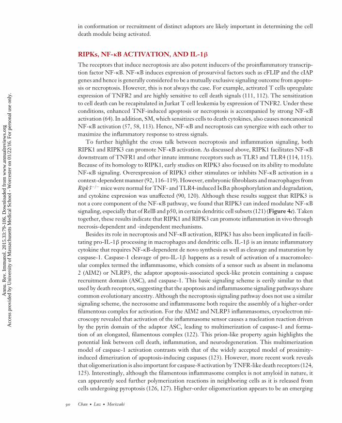
Besides its role in necroptosis and NF-κB activation, RIPK3 has also been implicated in facili-tating pro-IL-1β processing in macrophages and dendritic cells. IL-1β is an innate inflammatorycytokine that requires NF-κB-dependent de novo synthesis as well as cleavage and maturation bycaspase-1. Caspase-1 cleavage of pro-IL-1β happens as a result of activation of a macromolec-ular complex termed the inflammasome, which consists of a sensor such as absent in melanoma2 (AIM2) or NLRP3, the adaptor apoptosis-associated speck-like protein containing a caspaserecruitment domain (ASC), and caspase-1. This basic signaling scheme is eerily similar to thatused by death receptors, suggesting that the apoptosis and inflammasome signaling pathways sharecommon evolutionary ancestry. Although the necroptosis signaling pathway does not use a similarsignaling scheme, the necrosome and inflammasome both require the assembly of a higher-orderfilamentous complex for activation. For the AIM2 and NLRP3 inflammasomes, cryoelectron mi-croscopy revealed that activation of the inflammasome sensor causes a nucleation reaction drivenby the pyrin domain of the adaptor ASC, leading to multimerization of caspase-1 and forma-tion of an elongated, filamentous complex (122). This prion-like property again highlights thepotential link between cell death, inflammation, and neurodegeneration. This multimerizationmodel of caspase-1 activation contrasts with that of the widely accepted model of proximity-induced dimerization of apoptosis-inducing caspases (123). However, more recent work revealsthat oligomerization is also important for caspase-8 activation by TNFR-like death receptors (124,125). Interestingly, although the filamentous inflammasome complex is not amyloid in nature, itcan apparently seed further polymerization reactions in neighboring cells as it is released fromcells undergoing pyroptosis (126, 127). Higher-order oligomerization appears to be an emerging
90 Chan · Luz · Moriwaki
Ann
u. R
ev. I
mm
unol
. 201
5.33
:79-
106.
Dow
nloa
ded
from
ww
w.a
nnua
lrev
iew
s.or
g A
cces
s pr
ovid
ed b
y U
nive
rsity
of
Mas
sach
uset
ts M
edic
al S
choo
l - W
orce
ster
on
01/2
2/16
. For
per
sona
l use
onl
y.

IY33CH04-Chan ARI 20 March 2015 19:30
theme in innate and cell death signaling, as other intracellular pattern-recognition receptors andsensors including retinoic acid inducible gene-I and mitochondrial antiviral signaling protein arealso activated by similar polymerization mechanisms (128–130).
In addition to processing by the caspase-1-associated inflammasome, pro-IL-1β can alsobe processed by caspase-8 in certain situations (131–136). For example, the chemotherapeuticagent doxorubicin exclusively induces caspase-8-mediated pro-IL-1β processing in bone marrow–derived dendritic cells (133). Moreover, in lipopolysaccharide (LPS)-primed macrophages that lackcIAP1, cIAP2, and XIAP, pro-IL-1β processing is mediated through caspase-1 and caspase-8 ina RIPK3-dependent manner (137). The mechanism by which RIPK3 promotes IL-1β processingis unclear at present. As we discussed above, RIPK3 inhibitors and the kinase-inactive RIPK3 mu-tant D161N can drive formation of an alternative caspase-8-activating complex. Could a similarcomplex be involved in caspase-8-mediated pro-IL-1β processing? In the case of the D161N mu-tant, this complex promotes apoptosis. However, if caspase-8 is paired with its inhibitor cFLIPL,this complex may no longer promote apoptosis but instead facilitate pro-IL-1β processing. Thismodel is consistent with published reports that the caspase-8/cFLIPL heterodimer exhibits al-tered substrate specificity compared with the caspase-8 homodimer (29). Because these effects aremanifested when the cIAPs are depleted, the IAPs are crucial gatekeepers of RIPK3 activity in celldeath and inflammation.
In addition to promoting caspase-8, RIPK3 can also promote caspase-1-mediated pro-IL-1β processing. Fadd−/− and Casp8−/− macrophages and dendritic cells produced greatly elevatedlevels of IL-1β that was reversed by deletion of Ripk3 (138, 139). However, researchers disagreeon whether the enhanced IL-1β production was due to increased necrosis-associated releaseof DAMPs or direct effects of RIPK3 on caspase-1 activation. Regardless of the mechanism,RIPK3 can clearly promote caspase-1- and caspase-8-mediated pro-IL-1β processing via distinctmechanisms.
The necrosis-independent effects of RIPK1 and RIPK3 on NF-κB and pro-IL-1β processingillustrate an important principle: The RIPKs facilitate inflammation through multiple means. Theyalso reinforce the notion that death-signaling adaptors often have important functions beyondcell death. The multifaceted nature of death-inducing adaptors is not a novel concept. FADD, forexample, has been implicated in regulating cell cycle entry (140–142), and caspases have importantfunctions in cell differentiation, wound repair, and pruning of neuronal dendrites (143–145). Thediverse functions of RIPK3 remind us that inhibition of necroptosis is not the only possibleexplanation for why Ripk3−/− mice often show protection in many inflammatory disease models.
NECROPTOSIS IN VIRAL INFECTIONS
Because the release of DAMPs can stimulate pattern-recognition receptors such as TLRs, necrop-tosis is widely recognized to be beneficial in innate immune responses against pathogens. However,studies also show that necrosis-dependent inflammation can lead to detrimental pathology in ster-ile injury–induced diseases (Table 1). Given the fact that caspase inhibition is a priming signal fornecroptosis, perhaps it is not surprising that viruses that encode caspase inhibitors are susceptibleto host cell necroptosis (146). Poxviruses are master evaders of the host cell death machinery.In the case of vaccinia virus, the viral serpin Spi2/B13R is a potent inhibitor of caspase-1 andcaspase-8. As in the case of most pathogens, vaccinia virus infection causes an early wave of TNFexpression, which triggers RIPK1/RIPK3-dependent necroptosis in different infected tissues (65,90). In vitro experiments confirmed that wild-type cells infected with vaccinia virus were sensi-tized to TNF-induced cytotoxicity, but infected Ripk1−/− and Ripk3−/− cells were highly resistantto TNF-induced necroptosis (65, 90). Ripk3−/− mice had reduced necrosis and inflammation in
www.annualreviews.org • Programmed Necrosis in Cross Talk 91
Ann
u. R
ev. I
mm
unol
. 201
5.33
:79-
106.
Dow
nloa
ded
from
ww
w.a
nnua
lrev
iew
s.or
g A
cces
s pr
ovid
ed b
y U
nive
rsity
of
Mas
sach
uset
ts M
edic
al S
choo
l - W
orce
ster
on
01/2
2/16
. For
per
sona
l use
onl
y.

IY33CH04-Chan ARI 20 March 2015 19:30
Table 1 Necroptosis-related diseases
Cause of cell injury Disease Model Reference(s)Viral infection Vaccinia virus Ripk3−/− and Ripk1-D138N mice 65, 90, 147
Murine cytomegalovirus Ripk3−/− and Dai−/− mice 93, 94Bacterial infection Mycobacterium tuberculosis Zebrafish 158
Salmonella enterica serovarTyphimurium
Ripk3−/− mice and macrophages;necrostatin-1
157
Sterile injury–inducedinflammation
Psoriasis Keratinocyte-specific deletion of Fadd orCasp8; cpdm mice
47, 71, 187
Inflammatory bowel disease Intestinal epithelium–specific deletion ofFadd or Casp8
8, 46
Myocardial infarction Necrostatin-1 170
Hypoxia-ischemia-induced braininjury
Necrostatin-1 97
Ischemia-reperfusion kidneyinjury
Necrostatin-1; Ripk3−/− mice 161, 188
Retinal degeneration Retinal detachment; interphotoreceptorretinoid-binding protein–deficient mice;poly(I:C)-induced retinal injury; rd10mice; pde6c mutant zebrafish
175–178
Pancreatitis Cerulein-induced pancreatitis inRipk3−/−mice
153, 168, 189
Atherosclerosis Ripk3 deletion in apolipoprotein E orlow-density lipoprotein cholesterolreceptor–deficient mice
174
Gaucher’s disease Conduritol B epoxide inhibition ofglucocerebrosidase in Ripk3−/− mice
169
infected tissues and ultimately succumbed to the infection because of failure to control viral repli-cation. In agreement with these results, mice expressing a kinase-inactive RIPK1 (D138N) werealso partially impaired in clearance of vaccinia virus (147). These results established necroptosisas an important antiviral response against certain viral pathogens. This innate immune defensemechanism may be important to tamp down viral replication before robust, virus-specific T cellresponses are mobilized (Figure 5a).
The results from vaccinia virus are surprising because they seem to indicate that by blockingcaspase activation, viruses set themselves up for destruction by the host. Because ablation ofnecroptosis leads to rapid death of the host, blocking necroptosis may actually deprive the virus ofthe opportunity to disseminate and infect another host. From this perspective, one can argue thatnecroptosis is beneficial not only to the host but also to the invading virus (Figure 5a). Moreover,when compared to other naturally occurring poxviruses, vaccinia virus contains large deleted genesegments (148). Thus, a tantalizing possibility is that the gene that inhibits necroptosis is lost invaccinia virus because of these gene deletions. In this scenario, triggering of necroptosis may bean exception rather than a rule for vaccinia virus. To this end, it will be important to determine ifother poxviruses also cause infected cells to undergo necroptosis.
Viral inhibition of apoptosis is widely perceived to give the virus an edge in its struggle withthe host. Because of its antiviral effects, it stands to reason that some viruses may have alsodeveloped strategies to counteract necroptosis. Herpesviruses are highly adept at countering the
92 Chan · Luz · Moriwaki
Ann
u. R
ev. I
mm
unol
. 201
5.33
:79-
106.
Dow
nloa
ded
from
ww
w.a
nnua
lrev
iew
s.or
g A
cces
s pr
ovid
ed b
y U
nive
rsity
of
Mas
sach
uset
ts M
edic
al S
choo
l - W
orce
ster
on
01/2
2/16
. For
per
sona
l use
onl
y.

IY33CH04-Chan ARI 20 March 2015 19:30
DAI
RIPK3
Infectedmacrophage
vIRAvICA
Antiviralnecroptosis
Necroptosisinhibition
Caspase-8inhibition
B13R/Spi2
Host survival Viral dissemination
Promote viral replication
Mycobacteriumtuberculosis
Vaccinia virus
MCMV
Enhanced extracellularbacterial growth
Macrophagenecroptosis
Infection of other hoststhrough sputum/cough
TNF
TNF
Caspase-8inhibition
c
b
a
Figure 5Necroptosis in host-pathogen interactions. (a) Vaccinia virus inhibits caspase-8 via the viral inhibitorB13R/Spi2. This primes the cells toward necroptosis. Although necroptosis and the ensuing inflammationhave antiviral effects, they may in fact promote viral dissemination to another host by avoiding prematuredeath of the infected host. (b) MCMV inhibits caspase-8 and necroptosis via vICA and vIRA. Geneticexperiments show that vIRA is essential to prevent premature death of the infected cells. Hence,vIRA-mediated necroptosis inhibition is important for the virus to complete its replication cycle and togenerate more viral progeny. (c) Mycobacterium tuberculosis uses RIPK3-dependent necroptosis to release thebacteria into a growth-permissive environment, which in turn enhances spread of the pathogen to uninfectedhosts via the sputum. (Abbreviations: DAI, DNA activator of interferon; MCMV, murine cytomegalovirus;RIPK, receptor interacting protein kinase; TNF, tumor necrosis factor; vICA, viral inhibitor of caspase-8-induced apoptosis; vIRA, viral inhibitor of RIPK activation.)
host cell death machinery. MCMV encodes several viral cell death inhibitors, one of which is theviral inhibitor of caspase-8-induced apoptosis (vICA). Because inhibition of caspase-8 is a primingsignal for necroptosis, one would expect that cells infected with MCMV would become susceptibleto necroptosis. However, MCMV-infected cells are spared from necroptosis because the virusalso encodes vIRA, the product of the M45 gene. M45/vIRA is a RHIM-containing viral celldeath inhibitor that binds to RIPK3 to prevent virus-induced necroptosis (94, 146). RecombinantMCMV expressing a tetra-alanine-substitution RHIM mutant of vIRA fails to inhibit RIPK3 andsuccumbs to rapid necrosis. Because of this premature cell death, the mutant virus fails to establisha productive infection in cells and mice. Productive infection was reestablished with the mutantvirus in Ripk3−/− cells and Ripk3−/− mice (93).
In contrast to RIPK3, TNF signaling and RIPK1 are both dispensable for mutant MCMV-induced necroptosis. Instead, RIPK3 interacts with DAI, another RHIM-containing adaptor, toform a noncanonical necrosome that drives virus-induced necrosis (Figure 5b). M45 encodes aribonucleotide reductase (RNR) with no enzymatic activity. Interestingly, many RNRs from otherherpesviruses also encode a RHIM (Figure 3) (149). This suggests that viral inhibitors that targetthe RIPKs via the RHIM represent a common viral immune evasion strategy for herpesviruses.The results from vaccinia virus and MCMV highlight the importance of necroptosis in acute viralinfections. Yet questions still remain on whether necroptosis can influence the quality and magni-tude of adaptive immune responses, the generation of immunological memory, and viral latency.
www.annualreviews.org • Programmed Necrosis in Cross Talk 93
Ann
u. R
ev. I
mm
unol
. 201
5.33
:79-
106.
Dow
nloa
ded
from
ww
w.a
nnua
lrev
iew
s.or
g A
cces
s pr
ovid
ed b
y U
nive
rsity
of
Mas
sach
uset
ts M
edic
al S
choo
l - W
orce
ster
on
01/2
2/16
. For
per
sona
l use
onl
y.

IY33CH04-Chan ARI 20 March 2015 19:30
BACTERIAL AND PARASITIC INFECTIONS
TNF is a major driver of bacterial sepsis, a life-threatening condition marked by systemic cy-tokine storm and multiorgan failure. In agreement with the idea that RIPK-dependent necroptosispromotes damaging inflammation, Ripk3−/− mice are resistant to TNF-induced systemic inflam-matory syndrome (SIRS) (150, 151). In contrast to RIPK3, the role of RIPK1 in TNF-inducedSIRS is more controversial. Although several reports show that mice expressing kinase-inactiveRIPK1 and wild-type mice treated with RIPK1 kinase inhibitors are protected from TNF-inducedSIRS (71, 151, 152), another study found that Nec-1 exacerbates the disease (150). Furthermore,the response of Ripk3−/− and Mlkl−/− mice against cecal ligation and puncture–induced sepsisis also variable (151, 153). Because Ripk3−/− mice and Ripk3−/− macrophages exhibit a nor-mal response to LPS (120, 121), RIPK1 and RIPK3 likely play minor roles in acute bacterialsepsis.
Although the role of the RIPKs in LPS-induced responses is ambiguous, they are nonethelesscrucial in controlling certain bacterial pathogens. Yersinia pestis, the etiological agent of the blackdeath pandemic, causes rapid RIPK1- and caspase-8-dependent macrophage apoptosis. As inTNF- and SM-induced apoptosis, Y. pestis–induced macrophage apoptosis requires intact RIPK1kinase activity. In addition, RIPK1 is required for inflammatory cytokine production in responseto Y. pestis infection (154, 155). However, RIPK3 appears to play a minimal role in Y. pestisinfection. Salmonella enterica, a flagellated, gram-negative bacterium, is also a potent inducerof macrophage cell death. Although it is widely accepted that macrophage cell death inducedby Salmonella is caused by inflammasome activation and caspase-1-mediated pyroptosis (156), arecent report argues that RIPK3-dependent necroptosis is also involved (157). These discrepantconclusions could be reconciled by the fact that RIPK3 can also modulate inflammasome andcaspase-1 activation (137, 139).
Host control of Mycobacterium tuberculosis (Mtb) critically requires TNF. One of the majorprotective functions of TNF is to promote granuloma formation, which is thought to be crucialin containment of the bacteria. Using zebrafish as a model, Roca & Ramakrishnan (158) showthat RIPK1 and RIPK3 are both required to trigger TNF-induced reactive oxygen species (ROS)production and necroptosis in response to tuberculosis infection. Although necroptosis of infectedmacrophages initially inhibits bacterial growth, it later enhances growth as bacteria are releasedinto the growth-permissive extracellular environment. Hence, unlike the situation with vacciniavirus and MCMV, one can view Mtb as a pathogen that hijacks the host necroptosis machineryto promote its own growth and dissemination (Figure 5c). Consistent with this thesis, necrosis isoften associated with severe Mtb infection (159).
Mechanistically, TNF-induced necroptosis in Mtb-infected macrophages requires mitochon-drial cyclophilin D (CypD) and acid sphingomyelinase–induced ceramide production. Ceramidehas long been implicated in death receptor–induced apoptosis. However, its role in mammaliancell necroptosis has yet to be thoroughly tested. CypD is an inner mitochondrial protein and animportant component of the mitochondrial permeability transition pore. It is required for cer-tain forms of necrosis, such as that induced by calcium and ROS (160). CypD and RIPK3 act insynergy to mediate acute kidney injury in an ischemia-reperfusion model (161). However, CypDdeficiency did not rescue excessive necroptosis of Casp8−/− T cells (48). Moreover, widespreadelimination of mitochondria through induced mitophagy did not alter the cellular response toTNF-induced necroptosis (43). Hence, rather than being a core component of the necroptosismachinery, the CypD pathway appears to be uniquely involved in Mtb-induced necroptosis. Itwill be interesting to determine whether similar mechanisms involving RIPK1, RIPK3, CypD,and ceramide are involved in immune defense against Mtb infections in mammals.
94 Chan · Luz · Moriwaki
Ann
u. R
ev. I
mm
unol
. 201
5.33
:79-
106.
Dow
nloa
ded
from
ww
w.a
nnua
lrev
iew
s.or
g A
cces
s pr
ovid
ed b
y U
nive
rsity
of
Mas
sach
uset
ts M
edic
al S
choo
l - W
orce
ster
on
01/2
2/16
. For
per
sona
l use
onl
y.

IY33CH04-Chan ARI 20 March 2015 19:30
Parasitic diseases such as malaria and leishmaniasis target red blood cells, leading to anemia,hemolysis, and bleeding in some cases. These symptoms are caused by red blood cell lysis, whichreleases cell-free hemoglobin into the circulation. Oxidation of hemoglobin releases heme totrigger the Fenton reaction and generation of highly reactive oxygen radicals. The oxidative stressdrives lipid and protein peroxidation, DNA damage, and other insults to the cell (162). Free hemegreatly sensitizes hepatocytes to TNF-induced apoptosis in response to infection with Plasmodium,the etiological agent for malaria (163). In addition to hepatocytes, macrophages are also highlysusceptible to heme-induced cytotoxicity. Through a poorly defined mechanism, heme directlyactivates TLR4, leading to autocrine TNF and ROS production, which synergize with each otherto induce RIPK1- and RIPK3-dependent necroptosis (164). By inducing macrophage necroptosis,RIPK1 and RIPK3 may restrict the niche within which parasites can replicate. In vivo infectionswill be required to validate the biological role of necroptosis in parasitic infections.
NECROPTOSIS IN STERILE INFLAMMATION
Besides its role in pathogen infections, necrosis is also a hallmark of acute and chronic sterileinflammation. In agreement with induced expression of RIPK3 in response to acute and chronicexposure to alcohol, Ripk3−/− mice were protected from alcoholic liver disease (165). More-over, Ripk3−/− mice were protected from acetaminophen-induced liver injury (166), and elevatedphospho-MLKL signals were detected in drug-induced liver diseases (38). Repeated doses ofcerulein led to a biphasic cell death reaction in the acinar cells that was partially dependent onTNF, RIPK3, and MLKL (167). As such, Ripk3−/− and Mlkl−/− mice are partially protected fromcerulein-induced acute pancreatitis (153, 168). RIPK3 deficiency also improves the neurologicalmanifestation of Gaucher’s disease, a lysosomal storage disease caused by mutations in glucocere-brosidase (169).
Necroptosis appears to be an important mechanism of cell injury in ischemia-reperfusion-induced tissue injury. As discussed above, RIPK3-dependent necroptosis is partially responsiblefor ischemia-reperfusion-induced kidney injury (161). The RIPK1 kinase inhibitor Nec-1 is effec-tive in alleviating hypoxia-ischemia-induced oxidative brain injury and inflammation in neonatalmice (97). Nec-1 also reduced mouse and rat models of ischemia-reperfusion-induced myocardialcell death and infarct formation (170). However, in a model of permanent left anterior descend-ing coronary artery ligation, the resulting inflammation and tissue remodeling were impaired inRipk3−/− mice (171). Because Nec-1 has been shown to exhibit off-target effects (102, 172), theseresults need to be interpreted with caution. As mice expressing kinase-inactive RIPK1 have recentlybeen generated (71, 72), they will be useful in further dissecting the kinase-dependent necroptoticsignaling versus scaffold-dependent non-necroptotic signaling in these disease models.
In addition to drug- and trauma-induced tissue injury and inflammation, RIPK3-dependentnecroptosis also contributes to chronic inflammatory diseases such as atherosclerosis. Macrophagenecrosis is widely viewed as a key factor in atherosclerotic plaque formation (173). Mice deficientin apolipoprotein E or low-density lipoprotein receptor (LDL-R) that are fed a high-fat diet de-veloped atherosclerosis marked by macrophage necrosis in the atherosclerotic plaques. Strikingly,RIPK3 deletion ameliorates macrophage necrosis in the plaques and atherosclerosis in ApoE−/−
and Ldl-r−/− mice (174). Given that Ripk3−/− macrophages are resistant to oxidized LDL–inducednecroptosis, these results strongly suggest that RIPK3-dependent macrophage necroptosis is adirect driver of atherosclerotic plaque formation. Finally, investigators have also shown RIPK3-dependent necroptosis to be causative of mouse models of retinal injury (175–178). These examplespoint to the emerging role of the RIPKs in diverse inflammatory diseases. However, researchers
www.annualreviews.org • Programmed Necrosis in Cross Talk 95
Ann
u. R
ev. I
mm
unol
. 201
5.33
:79-
106.
Dow
nloa
ded
from
ww
w.a
nnua
lrev
iew
s.or
g A
cces
s pr
ovid
ed b
y U
nive
rsity
of
Mas
sach
uset
ts M
edic
al S
choo
l - W
orce
ster
on
01/2
2/16
. For
per
sona
l use
onl
y.

IY33CH04-Chan ARI 20 March 2015 19:30
need to consider both necroptosis-dependent and -independent effects of the RIPKs when inter-preting these results.
EVOLUTIONARY PERSPECTIVES
Studies in Caenorhabditis elegans and Drosophila melanogaster have contributed greatly to our knowl-edge of apoptosis signaling mechanisms. The conservation of apoptosis machinery through evo-lution illustrates its importance in the maintenance of organismal homeostasis. Is the mammaliannecroptosis pathway also conserved in C. elegans and Drosophila? Interestingly, RHIM-like adap-tors are found in Drosophila (Figure 3). In response to gram-negative bacteria, the innate immunereceptors peptidoglycan recognition protein (PGRP)-LC and PGRP-LE stimulate antimicro-bial peptide expression through immune deficiency (IMD), a RIPK1-like adaptor, and Relish, aDrosophila NF-κB (179). The tetrapeptide core sequences of Drosophila RHIM-containing adap-tors differ from those in the mammalian RHIMs (Figure 3). Mutations of the RHIM-like motifin PGRP-LC and PGRP-LE compromise antimicrobial peptide expression in response to pep-tidoglycan stimulation (180), indicating that these variant RHIMs are functional. The structuralsimilarity between mammalian and Drosophila RHIM adaptors argues that they may have evolvedfrom a common primordial pathway. Although the PGRP-IMD-Relish pathway is generally notknown to promote cell death, overexpression of IMD has been shown to result in cell death. In-terestingly, IMD-induced cell death was only partially rescued by the caspase inhibitor p35 (181),suggesting the possibility that nonapoptotic cell death may be involved.
In addition to the IMD pathway, transgenic overexpression of the Drosophila TNF orthologEiger in the developing eye primordium leads to JNK-dependent necrosis-like cell death (182).Interestingly, suppression of genes involved in glycolysis and mitochondrial respiration inhibitsEiger-induced cell death (182). The apoptosis protease activating factor 1 (Apaf1) interacts withcaspase-9 and cytochrome c to form the apoptosome, a macromolecular structure essential formitochondria-mediated apoptosis. Surprisingly, an Apaf1 hypomorph mutant also exhibits pro-gressive wing cell necrosis, which triggers a systemic inflammatory response, wasting, and expres-sion of antimicrobial peptides (183). In both Eiger- and Apaf1-mediated necrosis, the cell deathphenotype is associated with changes in energy metabolism. This is in contrast to mammaliannecroptosis, which does not require JNK or mitochondria (43, 102). Other pathways of non-apoptotic cell death have recently been described in Drosophila nurse cells (184) and developingneuroblasts (185). It will be interesting to determine if similar principles that govern mammaliannecroptosis are conserved in these situations.
CLOSING THOUGHTS
In considering the molecular machinery that controls necroptosis and its roles in different diseases,perhaps it will be helpful to step back and ponder why evolution has preserved this unique celldeath module. Phylogenetic analysis indicates that modern-day RIPKs evolved through a seriesof gene duplication events. The relatively short branch between RIPK1 and the RIPK progenitorsuggests that RIPK1 is probably the most ancient RIPK (Figure 6). This evolutionary model isappealing, as RIPK1 is the only RIPK that is crucial for embryonic survival and beyond. Althoughthe Drosophila IMD was once thought to be a RIPK1 ortholog (181), it shares homology only inthe DD and lacks the essential kinase domain. The absence of RIP-like kinases in lower organismssuch as Drosophila or C. elegans argues that these kinases are relatively novel products of evolution.The earliest example of RIPK1-like kinases is found in bony fish. How are we supposed to makesense of this? One possible explanation is that necroptosis is the product of coevolution with certain
96 Chan · Luz · Moriwaki
Ann
u. R
ev. I
mm
unol
. 201
5.33
:79-
106.
Dow
nloa
ded
from
ww
w.a
nnua
lrev
iew
s.or
g A
cces
s pr
ovid
ed b
y U
nive
rsity
of
Mas
sach
uset
ts M
edic
al S
choo
l - W
orce
ster
on
01/2
2/16
. For
per
sona
l use
onl
y.

IY33CH04-Chan ARI 20 March 2015 19:30
RIPK1: 81 chordate homologs
RIPK3: 51 chordate homologs
RIPK2: 74 chordate homologs
RIPK4: 63 chordate homologs
ANKK1: 48 chordate homologsSpeciation node
Duplication node
RIPKprogenitor
Figure 6The RIPK gene family evolved through a series of gene duplication events. This reconstructed phylogeny was generated by theEnsembl genome browser (186). Internal nodes correspond to key speciation ( purple) and gene duplication (red ) events. Branch lengthscorrespond to rates of evolutionary change. ANKK1 does not function as a RIPK but is closely related to RIPK4 and other RIPKs.(Abbreviations: ANKK1, ankyrin repeat and protein kinase domain–containing protein 1; RIPK, receptor interacting protein kinase.)
viruses that target vertebrates. The strongest support for this argument comes from the examplesof vaccinia virus and MCMV. According to this model, sterile injury–induced necroptosis is theprice we pay in this evolutionary struggle with viruses. This is appealing because necroptosis tendsto associate with detrimental pathologies in sterile inflammation. Studies of more viruses will becrucial to validating this hypothesis.
SUMMARY POINTS
1. The receptors that stimulate programmed necrosis or necroptosis are also apoptosisinducers.
2. Necroptosis is regulated by RIPK3 and MLKL.
3. Protein phosphorylation, ubiquitination, and FADD/caspase-8-mediated proteolyticprocessing are the three major post-translational mechanisms that control the induc-tion of necroptosis.
4. RIPK3 promotes inflammation through necroptosis, NF-κB activation, and caspase-1-and caspase-8-mediated pro-IL-1β maturation.
5. RIPK-dependent necroptosis contributes to pathogen-induced and sterile inflammation.
6. Necroptosis can promote or suppress antiviral immune responses in a pathogen-specificmanner.
7. The existence of viral inhibitors of necroptosis argues for an important role for necrop-tosis in host-pathogen interactions.
DISCLOSURE STATEMENT
The authors are not aware of any affiliations, memberships, funding, or financial holdings thatmight be perceived as affecting the objectivity of this review.
ACKNOWLEDGMENTS
We thank N. Silverman, D. Caffrey, and R. Siegel for discussion and critical reading of themanuscript and many other colleagues for discussion and ideas. We apologize to our colleagues
www.annualreviews.org • Programmed Necrosis in Cross Talk 97
Ann
u. R
ev. I
mm
unol
. 201
5.33
:79-
106.
Dow
nloa
ded
from
ww
w.a
nnua
lrev
iew
s.or
g A
cces
s pr
ovid
ed b
y U
nive
rsity
of
Mas
sach
uset
ts M
edic
al S
choo
l - W
orce
ster
on
01/2
2/16
. For
per
sona
l use
onl
y.

IY33CH04-Chan ARI 20 March 2015 19:30
whose work we could not cite because of space limitations. This work is supported by US NationalInstitutes of Health grant AI083497 (to F.K.-M.C.). N.F.L. receives a fellowship from NationalCouncil for Scientific and Technological Development (Brazil).
LITERATURE CITED
1. Segawa K, Kurata S, Yanagihashi Y, Brummelkamp TR, Matsuda F, Nagata S. 2014. Caspase-mediatedcleavage of phospholipid flippase for apoptotic phosphatidylserine exposure. Science 344:1164–68
2. Suzuki J, Imanishi E, Nagata S. 2013. Exposure of phosphatidylserine by Xk-related protein familymembers during apoptosis. J. Biol. Chem. 289:30257–67
3. Hochreiter-Hufford A, Ravichandran KS. 2013. Clearing the dead: apoptotic cell sensing, recognition,engulfment, and digestion. Cold Spring Harb. Perspect. Biol. 5:a008748
4. Kono H, Rock KL. 2008. How dying cells alert the immune system to danger. Nat. Rev. Immunol.8:279–89
5. Sawai H, Domae N. 2011. Discrimination between primary necrosis and apoptosis by necrostatin-1 inAnnexin V-positive/propidium iodide-negative cells. Biochem. Biophys. Res. Commun. 411:569–73
6. Yamasaki S, Ishikawa E, Sakuma M, Hara H, Ogata K, Saito T. 2008. Mincle is an ITAM-coupledactivating receptor that senses damaged cells. Nat. Immunol. 9:1179–88
7. Sancho D, Joffre OP, Keller AM, Rogers NC, Martinez D, et al. 2009. Identification of a dendritic cellreceptor that couples sensing of necrosis to immunity. Nature 458:899–903
8. Gunther C, Martini E, Wittkopf N, Amann K, Weigmann B, et al. 2011. Caspase-8 regulates TNF-α-induced epithelial necroptosis and terminal ileitis. Nature 477:335–39
9. Moriwaki K, Chan FKM. 2013. RIP3: a molecular switch for necrosis and inflammation. Genes Dev.27:1640–49
10. Sosna J, Voigt S, Mathieu S, Lange A, Thon L, et al. 2013. TNF-induced necroptosis and PARP-1-mediated necrosis represent distinct routes to programmed necrotic cell death. Cell. Mol. Life Sci.71:331–48
11. Fu D, Jordan JJ, Samson LD. 2013. Human ALKBH7 is required for alkylation and oxidation-inducedprogrammed necrosis. Genes Dev. 27:1089–100
12. Thapa RJ, Nogusa S, Chen P, Maki JL, Lerro A, et al. 2013. Interferon-induced RIP1/RIP3-mediatednecrosis requires PKR and is licensed by FADD and caspases. PNAS 110:E3109–18
13. Chan FKM, Chun HJ, Zheng L, Siegel RM, Bui KL, Lenardo MJ. 2000. A domain in TNF receptorsthat mediates ligand-independent receptor assembly and signaling. Science 288:2351–54
14. Micheau O, Tschopp J. 2003. Induction of TNF receptor I-mediated apoptosis via two sequentialsignaling complexes. Cell 114:181–90
15. Rieser E, Cordier SM, Walczak H. 2013. Linear ubiquitination: a newly discovered regulator of cellsignalling. Trends Biochem. Sci. 38:94–102
16. Papa S, Zazzeroni F, Bubici C, Jayawardena S, Alvarez K, et al. 2004. Gadd45β mediates the NF-κBsuppression of JNK signalling by targeting MKK7/JNKK2. Nat. Cell Biol. 6:146–53
17. Kreuz S, Siegmund D, Scheurich P, Wajant H. 2001. NF-κB inducers upregulate cFLIP, acycloheximide-sensitive inhibitor of death receptor signaling. Mol. Cell. Biol. 21:3964–73
18. Moulin M, Anderton H, Voss AK, Thomas T, Wong WW, et al. 2012. IAPs limit activation of RIPkinases by TNF receptor 1 during development. EMBO J. 31:1679–91
19. Csomos RA, Brady GF, Duckett CS. 2009. Enhanced cytoprotective effects of the inhibitor of apoptosisprotein cellular IAP1 through stabilization with TRAF2. J. Biol. Chem. 284:20531–39
20. Yeh WC, Shahinian A, Speiser D, Kraunus J, Billia F, et al. 1997. Early lethality, functional NF-κBactivation, and increased sensitivity to TNF-induced cell death in TRAF2-deficient mice. Immunity7:715–25
21. Ikeda F, Deribe YL, Skanland SS, Stieglitz B, Grabbe C, et al. 2011. SHARPIN forms a linear ubiquitinligase complex regulating NF-κB activity and apoptosis. Nature 471:637–41
22. Tokunaga F, Nakagawa T, Nakahara M, Saeki Y, Taniguchi M, et al. 2011. SHARPIN is a componentof the NF-κB-activating linear ubiquitin chain assembly complex. Nature 471:633–36
98 Chan · Luz · Moriwaki
Ann
u. R
ev. I
mm
unol
. 201
5.33
:79-
106.
Dow
nloa
ded
from
ww
w.a
nnua
lrev
iew
s.or
g A
cces
s pr
ovid
ed b
y U
nive
rsity
of
Mas
sach
uset
ts M
edic
al S
choo
l - W
orce
ster
on
01/2
2/16
. For
per
sona
l use
onl
y.

IY33CH04-Chan ARI 20 March 2015 19:30
23. Gerlach B, Cordier SM, Schmukle AC, Emmerich CH, Rieser E, et al. 2011. Linear ubiquitinationprevents inflammation and regulates immune signalling. Nature 471:591–96
24. Harper N, Hughes M, MacFarlane M, Cohen GM. 2003. Fas-associated death domain protein andcaspase-8 are not recruited to the tumor necrosis factor receptor 1 signaling complex during tumornecrosis factor-induced apoptosis. J. Biol. Chem. 278:25534–41
25. Schneider-Brachert W, Tchikov V, Neumeyer J, Jakob M, Winoto-Morbach S, et al. 2004. Compart-mentalization of TNF receptor 1 signaling: internalized TNF receptosomes as death signaling vesicles.Immunity 21:415–28
26. Lin Y, Devin A, Rodriguez Y, Liu ZG. 1999. Cleavage of the death domain kinase RIP by caspase-8prompts TNF-induced apoptosis. Genes Dev. 13:2514–26
27. Feng S, Yang Y, Mei Y, Ma L, Zhu DE, et al. 2007. Cleavage of RIP3 inactivates its caspase-independentapoptosis pathway by removal of kinase domain. Cell. Signal. 19:2056–67
28. Oberst A, Dillon CP, Weinlich R, McCormick LL, Fitzgerald P, et al. 2011. Catalytic activity of thecaspase-8-FLIPL complex inhibits RIPK3-dependent necrosis. Nature 471:363–67
29. Pop C, Oberst A, Drag M, Van Raam BJ, Riedl SJ, et al. 2011. FLIPL induces caspase-8 activity in theabsence of interdomain caspase-8 cleavage and alters substrate specificity. Biochem. J. 433:447–57
30. O’Donnell MA, Perez-Jimenez E, Oberst A, Ng A, Massoumi R, et al. 2011. Caspase-8 inhibits pro-grammed necrosis by processing CYLD. Nat. Cell Biol. 13:1437–42
31. Tenev T, Bianchi K, Darding M, Broemer M, Langlais C, et al. 2011. The Ripoptosome, a signalingplatform that assembles in response to genotoxic stress and loss of IAPs. Mol. Cell 43:432–48
32. Feoktistova M, Geserick P, Kellert B, Dimitrova DP, Langlais C, et al. 2011. cIAPs block Ripoptosomeformation, a RIP1/caspase-8 containing intracellular cell death complex differentially regulated by cFLIPisoforms. Mol. Cell 43:449–63
33. Li J, McQuade T, Siemer AB, Napetschnig J, Moriwaki K, et al. 2012. The RIP1/RIP3 necrosome formsa functional amyloid signaling complex required for programmed necrosis. Cell 150:339–50
34. Sun L, Wang H, Wang Z, He S, Chen S, et al. 2012. Mixed lineage kinase domain-like protein mediatesnecrosis signaling downstream of RIP3 kinase. Cell 148:213–27
35. Zhao J, Jitkaew S, Cai Z, Choksi S, Li Q, et al. 2012. Mixed lineage kinase domain-like is a key receptorinteracting protein 3 downstream component of TNF-induced necrosis. PNAS 109:5322–27
36. Cai Z, Jitkaew S, Zhao J, Chiang HC, Choksi S, et al. 2014. Plasma membrane translocation of trimerizedMLKL protein is required for TNF-induced necroptosis. Nat. Cell Biol. 16:55–65
37. Chen X, Li W, Ren J, Huang D, He WT, et al. 2014. Translocation of mixed lineage kinase domain-likeprotein to plasma membrane leads to necrotic cell death. Cell Res. 24:105–21
38. Wang H, Sun L, Su L, Rizo J, Liu L, et al. 2014. Mixed lineage kinase domain-like protein MLKLcauses necrotic membrane disruption upon phosphorylation by RIP3. Mol. Cell 54:133–46
39. Dondelinger Y, Declercq W, Montessuit S, Roelandt R, Goncalves A, et al. 2014. MLKL compromisesplasma membrane integrity by binding to phosphatidylinositol phosphates. Cell Rep. 7:971–81
40. Wang Z, Jiang H, Chen S, Du F, Wang X. 2012. The mitochondrial phosphatase PGAM5 functions atthe convergence point of multiple necrotic death pathways. Cell 148:228–43
41. Murphy JM, Czabotar PE, Hildebrand JM, Lucet IS, Zhang JG, et al. 2013. The pseudokinase MLKLmediates necroptosis via a molecular switch mechanism. Immunity 39:443–53
42. Remijsen Q, Goossens V, Grootjans S, Van den Haute C, Vanlangenakker N, et al. 2014. Depletionof RIPK3 or MLKL blocks TNF-driven necroptosis and switches towards a delayed RIPK1 kinase-dependent apoptosis. Cell Death Dis. 5:e1004
43. Tait SW, Oberst A, Quarato G, Milasta S, Haller M, et al. 2013. Widespread mitochondrial depletionvia mitophagy does not compromise necroptosis. Cell Rep. 5:878–85
44. Kaiser WJ, Upton JW, Long AB, Livingston-Rosanoff D, Daley-Bauer LP, et al. 2011. RIP3 mediatesthe embryonic lethality of caspase-8-deficient mice. Nature 471:368–72
45. Zhang H, Zhou X, McQuade T, Li J, Chan FKM, Zhang J. 2011. Functional complementation betweenFADD and RIP1 in embryos and lymphocytes. Nature 471:373–76
46. Welz PS, Wullaert A, Vlantis K, Kondylis V, Fernandez-Majada V, et al. 2011. FADD prevents RIP3-mediated epithelial cell necrosis and chronic intestinal inflammation. Nature 477:330–34
www.annualreviews.org • Programmed Necrosis in Cross Talk 99
Ann
u. R
ev. I
mm
unol
. 201
5.33
:79-
106.
Dow
nloa
ded
from
ww
w.a
nnua
lrev
iew
s.or
g A
cces
s pr
ovid
ed b
y U
nive
rsity
of
Mas
sach
uset
ts M
edic
al S
choo
l - W
orce
ster
on
01/2
2/16
. For
per
sona
l use
onl
y.

IY33CH04-Chan ARI 20 March 2015 19:30
47. Bonnet MC, Preukschat D, Welz PS, van Loo G, Ermolaeva MA, et al. 2011. The adaptor protein FADDprotects epidermal keratinocytes from necroptosis in vivo and prevents skin inflammation. Immunity35:572–82
48. Ch’en IL, Tsau JS, Molkentin JD, Komatsu M, Hedrick SM. 2011. Mechanisms of necroptosis in Tcells. J. Exp. Med. 208:633–41
49. Lu JV, Weist BM, van Raam BJ, Marro BS, Nguyen LV, et al. 2011. Complementary roles of Fas-associated death domain (FADD) and receptor interacting protein kinase-3 (RIPK3) in T-cell homeo-stasis and antiviral immunity. PNAS 108:15312–17
50. Holler N, Zaru R, Micheau O, Thome M, Attinger A, et al. 2000. Fas triggers an alternative, caspase-8-independent cell death pathway using the kinase RIP as effector molecule. Nat. Immunol. 1:489–95
51. Dillon CP, Oberst A, Weinlich R, Janke LJ, Kang TB, et al. 2012. Survival function of the FADD-CASPASE-8-cFLIPL complex. Cell Rep. 1:401–7
52. Zhang SQ, Kovalenko A, Cantarella G, Wallach D. 2000. Recruitment of the IKK signalosome to thep55 TNF receptor: RIP and A20 bind to NEMO (IKKγ) upon receptor stimulation. Immunity 12:301–11
53. Ea CK, Deng L, Xia ZP, Pineda G, Chen ZJ. 2006. Activation of IKK by TNFα requires site-specificubiquitination of RIP1 and polyubiquitin binding by NEMO. Mol. Cell 22:245–57
54. Wu CJ, Conze DB, Li T, Srinivasula SM, Ashwell JD. 2006. Sensing of Lys 63-linked polyubiquitinationby NEMO is a key event in NF-κB activation. Nat. Cell Biol. 8:398–406
55. O’Donnell MA, Hase H, Legarda D, Ting AT. 2012. NEMO inhibits programmed necrosis in anNFκB-independent manner by restraining RIP1. PLOS ONE 7:e41238
56. O’Donnell MA, Legarda-Addison D, Skountzos P, Yeh WC, Ting AT. 2007. Ubiquitination of RIP1regulates an NF-κB-independent cell-death switch in TNF signaling. Curr. Biol. 17:418–24
57. Varfolomeev E, Blankenship JW, Wayson SM, Fedorova AV, Kayagaki N, et al. 2007. IAP antagonistsinduce autoubiquitination of c-IAPs, NF-κB activation, and TNFα-dependent apoptosis. Cell 131:669–81
58. Vince JE, Wong WW, Khan N, Feltham R, Chau D, et al. 2007. IAP antagonists target cIAP1 to induceTNFα-dependent apoptosis. Cell 131:682–93
59. Bertrand MJ, Milutinovic S, Dickson KM, Ho WC, Boudreault A, et al. 2008. cIAP1 and cIAP2 facilitatecancer cell survival by functioning as E3 ligases that promote RIP1 ubiquitination. Mol. Cell 30:689–700
60. Dueber EC, Schoeffler AJ, Lingel A, Elliott JM, Fedorova AV, et al. 2011. Antagonists induce a confor-mational change in cIAP1 that promotes autoubiquitination. Science 334:376–80
61. Petersen SL, Peyton M, Minna JD, Wang X. 2010. Overcoming cancer cell resistance to Smac mimeticinduced apoptosis by modulating cIAP-2 expression. PNAS 107:11936–41
62. Chromik J, Safferthal C, Serve H, Fulda S. 2014. Smac mimetic primes apoptosis-resistant acute myeloidleukaemia cells for cytarabine-induced cell death by triggering necroptosis. Cancer Lett. 344:101–9
63. Kim EY, Teh HS. 2004. Critical role of TNF receptor type-2 (p75) as a costimulator for IL-2 inductionand T cell survival: a functional link to CD28. J. Immunol. 173:4500–9
64. Chan FKM, Lenardo MJ. 2000. A crucial role for p80 TNF-R2 in amplifying p60 TNF-R1 apoptosissignals in T lymphocytes. Eur. J. Immunol. 30:652–60
65. Chan FKM, Shisler J, Bixby JG, Felices M, Zheng L, et al. 2003. A role for tumor necrosis factorreceptor-2 and receptor-interacting protein in programmed necrosis and antiviral responses. J. Biol.Chem. 278:51613–21
66. Li X, Yang Y, Ashwell JD. 2002. TNF-RII and c-IAP1 mediate ubiquitination and degradation ofTRAF2. Nature 416:345–47
67. Fotin-Mleczek M, Henkler F, Samel D, Reichwein M, Hausser A, et al. 2002. Apoptotic crosstalk of TNFreceptors: TNF-R2-induces depletion of TRAF2 and IAP proteins and accelerates TNF-R1-dependentactivation of caspase-8. J. Cell Sci. 115:2757–70
68. Wong WW, Vince JE, Lalaoui N, Lawlor KE, Chau D, et al. 2014. cIAPs and XIAP regulate myelopoiesisthrough cytokine production in an RIPK1- and RIPK3-dependent manner. Blood 123:2562–72
69. Vanlangenakker N, Vanden Berghe T, Bogaert P, Laukens B, Zobel K, et al. 2011. cIAP1 and TAK1protect cells from TNF-induced necrosis by preventing RIP1/RIP3-dependent reactive oxygen speciesproduction. Cell Death Differ. 18:656–65
100 Chan · Luz · Moriwaki
Ann
u. R
ev. I
mm
unol
. 201
5.33
:79-
106.
Dow
nloa
ded
from
ww
w.a
nnua
lrev
iew
s.or
g A
cces
s pr
ovid
ed b
y U
nive
rsity
of
Mas
sach
uset
ts M
edic
al S
choo
l - W
orce
ster
on
01/2
2/16
. For
per
sona
l use
onl
y.

IY33CH04-Chan ARI 20 March 2015 19:30
70. Tokunaga F, Sakata S, Saeki Y, Satomi Y, Kirisako T, et al. 2009. Involvement of linear polyubiquitylationof NEMO in NF-κB activation. Nat. Cell Biol. 11:123–32
71. Berger S, Kasparcova V, Hoffman S, Swift B, Dare L, et al. 2014. Cutting edge: RIP1 kinase activity isdispensable for normal development but is a key regulator of inflammation in SHARPIN-deficient mice.J. Immunol. 192:5476–80
72. Newton K, Dugger D, Wickliffe KE, Kapoor N, de Almagro C, et al. 2014. Activity of protein kinaseRIPK3 determines whether cells die by necroptosis or apoptosis. Science 343:1357–60
73. Wang L, Du F, Wang X. 2008. TNF-α induces two distinct caspase-8 activation pathways. Cell 133:693–703
74. Boisson B, Laplantine E, Prando C, Giliani S, Israelsson E, et al. 2012. Immunodeficiency, autoinflam-mation and amylopectinosis in humans with inherited HOIL-1 and LUBAC deficiency. Nat. Immunol.13:1178–86
75. Nilsson J, Schoser B, Laforet P, Kalev O, Lindberg C, et al. 2013. Polyglucosan body myopathy causedby defective ubiquitin ligase RBCK1. Ann. Neurol. 74:914–19
76. Makris C, Godfrey VL, Krahn-Senftleben G, Takahashi T, Roberts JL, et al. 2000. Female mice het-erozygous for IKKγ/NEMO deficiencies develop a dermatopathy similar to the human X-linked disorderincontinentia pigmenti. Mol. Cell 5:969–79
77. Rudolph D, Yeh WC, Wakeham A, Rudolph B, Nallainathan D, et al. 2000. Severe liver degenerationand lack of NF-κB activation in NEMO/IKKγ -deficient mice. Genes Dev. 14:854–62
78. Schmidt-Supprian M, Bloch W, Courtois G, Addicks K, Israel A, et al. 2000. NEMO/IKKγ-deficientmice model incontinentia pigmenti. Mol. Cell 5:981–92
79. Doffinger R, Smahi A, Bessia C, Geissmann F, Feinberg J, et al. 2001. X-linked anhidrotic ectodermaldysplasia with immunodeficiency is caused by impaired NF-κB signaling. Nat. Genet. 27:277–85
80. Hitomi J, Christofferson DE, Ng A, Yao J, Degterev A, et al. 2008. Identification of a molecular signalingnetwork that regulates a cellular necrotic cell death pathway. Cell 135:1311–23
81. Moquin DM, McQuade T, Chan FKM. 2013. CYLD deubiquitinates RIP1 in the TNFα-inducednecrosome to facilitate kinase activation and programmed necrosis. PLOS ONE 8:e76841
82. Kelliher MA, Grimm S, Ishida Y, Kuo F, Stanger BZ, Leder P. 1998. The death domain kinase RIPmediates the TNF-induced NF-κB signal. Immunity 8:297–303
83. Wong WWL, Gentle IE, Nachbur U, Anderton H, Vaux DL, Silke J. 2010. RIPK1 is not essential forTNFR1-induced activation of NF-κB. Cell Death Differ. 17:482–87
84. Dillon CP, Weinlich R, Rodriguez DA, Cripps JG, Quarato G, et al. 2014. RIPK1 blocks early postnatallethality mediated by caspase-8 and RIPK3. Cell 157:1189–202
85. Rickard JA, O’Donnell JA, Evans JM, Lalaoui N, Poh AR, et al. 2014. RIPK1 regulates RIPK3-MLKL-driven systemic inflammation and emergency hematopoiesis. Cell 157:1175–88
86. Kaiser WJ, Daley-Bauer LP, Thapa RJ, Mandal P, Berger SB, et al. 2014. RIP1 suppresses innate immunenecrotic as well as apoptotic cell death during mammalian parturition. PNAS 111:7753–58
87. Takahashi N, Vereecke L, Bertrand MJM, Duprez L, Berger SB, et al. 2014. RIPK1 ensures intestinalhomeostasis by protecting the epithelium against apoptosis. Nature 513:95–99
88. Dannappel M, Vlantis K, Kumari S, Polykratis A, Kim C, et al. 2014. RIPK1 maintains epithelialhomeostasis by inhibiting apoptosis and necroptosis. Nature 513:90–94
89. Cho Y, Challa S, Chan FKM. 2011. A RNA interference screen identifies RIP3 as an essential inducerof TNF-induced programmed necrosis. Adv. Exp. Med. Biol. 691:589–93
90. Cho YS, Challa S, Moquin D, Genga R, Ray TD, et al. 2009. Phosphorylation-driven assembly of theRIP1-RIP3 complex regulates programmed necrosis and virus-induced inflammation. Cell 137:1112–23
91. Kaiser WJ, Offermann MK. 2005. Apoptosis induced by the Toll-like receptor adaptor TRIF is depen-dent on its receptor interacting protein homotypic interaction motif. J. Immunol. 174:4942–52
92. Meylan E, Burns K, Hofmann K, Blancheteau V, Martinon F, et al. 2004. RIP1 is an essential mediatorof Toll-like receptor 3–induced NF-κB activation. Nat. Immunol. 5:503–7
93. Upton JW, Kaiser WJ, Mocarski ES. 2010. Virus inhibition of RIP3-dependent necrosis. Cell HostMicrobe 7:302–13
www.annualreviews.org • Programmed Necrosis in Cross Talk 101
Ann
u. R
ev. I
mm
unol
. 201
5.33
:79-
106.
Dow
nloa
ded
from
ww
w.a
nnua
lrev
iew
s.or
g A
cces
s pr
ovid
ed b
y U
nive
rsity
of
Mas
sach
uset
ts M
edic
al S
choo
l - W
orce
ster
on
01/2
2/16
. For
per
sona
l use
onl
y.

IY33CH04-Chan ARI 20 March 2015 19:30
94. Upton JW, Kaiser WJ, Mocarski ES. 2012. DAI/ZBP1/DLM-1 complexes with RIP3 to mediate virus-induced programmed necrosis that is targeted by murine cytomegalovirus vIRA. Cell Host Microbe 11:290–97
95. Halle A, Hornung V, Petzold GC, Stewart CR, Monks BG, et al. 2008. The NALP3 inflammasome isinvolved in the innate immune response to amyloid-β. Nat. Immunol. 9:857–65
96. Heneka MT, Kummer MP, Stutz A, Delekate A, Schwartz S, et al. 2013. NLRP3 is activated inAlzheimer’s disease and contributes to pathology in APP/PS1 mice. Nature 493:674–78
97. Northington FJ, Chavez-Valdez R, Graham EM, Razdan S, Gauda EB, Martin LJ. 2011. Necrostatindecreases oxidative damage, inflammation, and injury after neonatal HI. J. Cereb. Blood Flow Metab.31:178–89
98. Chavez-Valdez R, Martin LJ, Flock DL, Northington FJ. 2012. Necrostatin-1 attenuates mitochondrialdysfunction in neurons and astrocytes following neonatal hypoxia-ischemia. Neuroscience 219:192–203
99. He S, Liang Y, Shao F, Wang X. 2011. Toll-like receptors activate programmed necrosis in macrophagesthrough a receptor-interacting kinase-3–mediated pathway. PNAS 108:20054–59
100. Kaiser WJ, Sridharan H, Huang C, Mandal P, Upton JW, et al. 2013. Toll-like receptor 3-mediatednecrosis via TRIF, RIP3, and MLKL. J. Biol. Chem. 288:31268–79
101. Rebsamen M, Meylan E, Curran J, Tschopp J. 2008. The antiviral adaptor proteins Cardif and Trif areprocessed and inactivated by caspases. Cell Death Differ. 15:1804–11
102. Cho Y, McQuade T, Zhang HB, Zhang JK, Chan FKM. 2011. RIP1-dependent and independent effectsof necrostatin-1 in necrosis and T cell activation. PLOS ONE 6:e23209
103. Takahashi N, Duprez L, Grootjans S, Cauwels A, Nerinckx W, et al. 2012. Necrostatin-1 analogues:critical issues on the specificity, activity and in vivo use in experimental disease models. Cell Death Dis.3:e437
104. McQuade T, Cho Y, Chan FKM. 2013. Positive and negative phosphorylation regulates RIP1- andRIP3-induced programmed necrosis. Biochem. J. 456:409–15
105. Degterev A, Hitomi J, Germscheid M, Ch’en IL, Korkina O, et al. 2008. Identification of RIP1 kinaseas a specific cellular target of necrostatins. Nat. Chem. Biol. 4:313–21
106. Wu XN, Yang ZH, Wang XK, Zhang Y, Wan H, et al. 2014. Distinct roles of RIP1–RIP3 hetero- andRIP3-RIP3 homo-interaction in mediating necroptosis. Cell Death Differ. 21:1709–20
107. Orozco S, Yatim N, Werner MR, Tran H, Gunja SY, et al. 2014. RIPK1 both positively and negativelyregulates RIPK3 oligomerization and necroptosis. Cell Death Differ. 21:1511–21
108. Cook WD, Moujalled DM, Ralph TJ, Lock P, Young SN, et al. 2014. RIPK1- and RIPK3-induced celldeath mode is determined by target availability. Cell Death Differ. 21:1600–12
109. Moujalled DM, Cook WD, Okamoto T, Murphy J, Lawlor KE, et al. 2013. TNF can activate RIPK3and cause programmed necrosis in the absence of RIPK1. Cell Death Dis. 4:e465
110. Mandal P, Berger SB, Pillay S, Moriwaki K, Huang C, et al. 2014. RIP3 induces apoptosis independentof pronecrotic kinase activity. Mol. Cell 56:481–95
111. Zheng L, Fisher G, Miller RE, Peschon J, Lynch DH, Lenardo MJ. 1995. Induction of apoptosis inmature T cells by tumour necrosis factor. Nature 377:348–51
112. Lenardo M, Chan FKM, Hornung F, McFarland H, Siegel R, et al. 1999. Mature T lymphocyteapoptosis—immune regulation in a dynamic and unpredictable antigenic environment. Annu. Rev. Im-munol. 17:221–53
113. Petersen SL, Wang L, Yalcin-Chin A, Li L, Peyton M, et al. 2007. Autocrine TNFα signaling rendershuman cancer cells susceptible to Smac-mimetic-induced apoptosis. Cancer Cell 12:445–56
114. Vivarelli MS, McDonald D, Miller M, Cusson N, Kelliher M, Geha RS. 2004. RIP links TLR4 to Aktand is essential for cell survival in response to LPS stimulation. J. Exp. Med. 200:399–404
115. Cusson-Hermance N, Khurana S, Lee TH, Fitzgerald KA, Kelliher MA. 2005. Rip1 mediates the Trif-dependent Toll-like receptor 3- and 4-induced NF-κB activation but does not contribute to interferonregulatory factor 3 activation. J. Biol. Chem. 280:36560–66
116. Kasof GM, Prosser JC, Liu D, Lorenzi MV, Gomes BC. 2000. The RIP-like kinase, RIP3, inducesapoptosis and NF-κB nuclear translocation and localizes to mitochondria. FEBS Lett. 473:285–91
117. Yu PW, Huang BC, Shen M, Quast J, Chan E, et al. 1999. Identification of RIP3, a RIP-like kinase thatactivates apoptosis and NFκB. Curr. Biol. 9:539–42
102 Chan · Luz · Moriwaki
Ann
u. R
ev. I
mm
unol
. 201
5.33
:79-
106.
Dow
nloa
ded
from
ww
w.a
nnua
lrev
iew
s.or
g A
cces
s pr
ovid
ed b
y U
nive
rsity
of
Mas
sach
uset
ts M
edic
al S
choo
l - W
orce
ster
on
01/2
2/16
. For
per
sona
l use
onl
y.

IY33CH04-Chan ARI 20 March 2015 19:30
118. Pazdernik NJ, Donner DB, Goebl MG, Harrington MA. 1999. Mouse receptor interacting protein 3does not contain a caspase-recruiting or a death domain but induces apoptosis and activates NF-κB. Mol.Cell. Biol. 19:6500–8
119. Sun X, Lee J, Navas T, Baldwin DT, Stewart TA, Dixit VM. 1999. RIP3, a novel apoptosis-inducingkinase. J. Biol. Chem. 274:16871–75
120. Newton K, Sun X, Dixit VM. 2004. Kinase RIP3 is dispensable for normal NF-κBs, signaling by theB-cell and T-cell receptors, tumor necrosis factor receptor 1, and Toll-like receptors 2 and 4. Mol. Cell.Biol. 24:1464–69
121. Moriwaki K, Balaji S, McQuade T, Malhotra N, Kang J, Chan FKM. 2014. The necroptosis adaptorRIPK3 promotes injury-induced cytokine expression and tissue repair. Immunity 41:567–78
122. Lu A, Magupalli VG, Ruan J, Yin Q, Atianand MK, et al. 2014. Unified polymerization mechanism forthe assembly of ASC-dependent inflammasomes. Cell 156:1193–206
123. Pop C, Salvesen GS. 2009. Human caspases: activation, specificity, and regulation. J. Biol. Chem.284:21777–81
124. Schleich K, Warnken U, Fricker N, Ozturk S, Richter P, et al. 2012. Stoichiometry of the CD95 death-inducing signaling complex: experimental and modeling evidence for a death effector domain chainmodel. Mol. Cell 47:306–19
125. Dickens LS, Boyd RS, Jukes-Jones R, Hughes MA, Robinson GL, et al. 2012. A death effector domainchain DISC model reveals a crucial role for caspase-8 chain assembly in mediating apoptotic cell death.Mol. Cell 47:291–305
126. Franklin BS, Bossaller L, De Nardo D, Ratter JM, Stutz A, et al. 2014. The adaptor ASC has extracellularand ‘prionoid’ activities that propagate inflammation. Nat. Immunol. 15:727–37
127. Baroja-Mazo A, Martin-Sanchez F, Gomez AI, Martinez CM, Amores-Iniesta J, et al. 2014. The NLRP3inflammasome is released as a particulate danger signal that amplifies the inflammatory response. Nat.Immunol. 15:738–48
128. Cai X, Chen J, Xu H, Liu S, Jiang QX, et al. 2014. Prion-like polymerization underlies signal transductionin antiviral immune defense and inflammasome activation. Cell 156:1207–22
129. Peisley A, Wu B, Yao H, Walz T, Hur S. 2013. RIG-I forms signaling-competent filaments in anATP-dependent, ubiquitin-independent manner. Mol. Cell 51:573–83
130. Wu B, Peisley A, Richards C, Yao H, Zeng X, et al. 2013. Structural basis for dsRNA recognition,filament formation, and antiviral signal activation by MDA5. Cell 152:276–89
131. Maelfait J, Vercammen E, Janssens S, Schotte P, Haegman M, et al. 2008. Stimulation of Toll-likereceptor 3 and 4 induces interleukin-1β maturation by caspase-8. J. Exp. Med. 205:1967–73
132. Gringhuis SI, Kaptein TM, Wevers BA, Theelen B, van der Vlist M, et al. 2012. Dectin-1 is an ex-tracellular pathogen sensor for the induction and processing of IL-1β via a noncanonical caspase-8inflammasome. Nat. Immunol. 13:246–54
133. Antonopoulos C, El Sanadi C, Kaiser WJ, Mocarski ES, Dubyak GR. 2013. Proapoptotic chemother-apeutic drugs induce noncanonical processing and release of IL-1β via caspase-8 in dendritic cells.J. Immunol. 191:4789–803
134. Shenderov K, Riteau N, Yip R, Mayer-Barber KD, Oland S, et al. 2014. Cutting edge: Endoplasmicreticulum stress licenses macrophages to produce mature IL-1βin response to TLR4 stimulation througha caspase-8– and TRIF-dependent pathway. J. Immunol. 192:2029–33
135. Man SM, Tourlomousis P, Hopkins L, Monie TP, Fitzgerald KA, Bryant CE. 2013. Salmonella infectioninduces recruitment of caspase-8 to the inflammasome to modulate IL-1β production. J. Immunol.191:5239–46
136. Gurung P, Anand PK, Malireddi RKS, Vande Walle L, Van Opdenbosch N, et al. 2014. FADD andcaspase-8 mediate priming and activation of the canonical and noncanonical Nlrp3 inflammasomes.J. Immunol. 192:1835–46
137. Vince JE, Wong WW, Gentle I, Lawlor KE, Allam R, et al. 2012. Inhibitor of apoptosis proteins limitRIP3 kinase-dependent interleukin-1 activation. Immunity 36:215–27
138. Young JA, He TH, Reizis B, Winoto A. 2013. Commensal microbiota are required for systemic inflam-mation triggered by necrotic dendritic cells. Cell Rep. 27:1932–44
www.annualreviews.org • Programmed Necrosis in Cross Talk 103
Ann
u. R
ev. I
mm
unol
. 201
5.33
:79-
106.
Dow
nloa
ded
from
ww
w.a
nnua
lrev
iew
s.or
g A
cces
s pr
ovid
ed b
y U
nive
rsity
of
Mas
sach
uset
ts M
edic
al S
choo
l - W
orce
ster
on
01/2
2/16
. For
per
sona
l use
onl
y.
IY33CH04-Chan ARI 20 March 2015 19:30
139. Kang TB, Yang SH, Toth B, Kovalenko A, Wallach D. 2013. Caspase-8 blocks kinase RIPK3-mediatedactivation of the NLRP3 inflammasome. Immunity 38:27–40
140. Alappat EC, Feig C, Boyerinas B, Volkland J, Samuels M, et al. 2005. Phosphorylation of FADD atserine 194 by CKIα regulates its nonapoptotic activities. Mol. Cell 19:321–32
141. Hua ZC, Sohn SJ, Kang C, Cado D, Winoto A. 2003. A function of Fas-associated death domain proteinin cell cycle progression localized to a single amino acid at its C-terminal region. Immunity 18:513–21
142. Osborn SL, Sohn SJ, Winoto A. 2007. Constitutive phosphorylation mutation in Fas-associated deathdomain (FADD) results in early cell cycle defects. J. Biol. Chem. 282:22786–92
143. Zermati Y, Garrido C, Amsellem S, Fishelson S, Bouscary D, et al. 2001. Caspase activation is requiredfor terminal erythroid differentiation. J. Exp. Med. 193:247–54
144. Lee P, Lee DJ, Chan C, Chen SW, Ch’en I, Jamora C. 2009. Dynamic expression of epidermal caspase-8simulates a wound healing response. Nature 458:519–23
145. Hyman BT, Yuan J. 2012. Apoptotic and non-apoptotic roles of caspases in neuronal physiology andpathophysiology. Nat. Rev. Neurosci. 13:395–406
146. Upton JW, Chan FKM. 2014. Staying alive: cell death in antiviral immunity. Mol. Cell 54:273–80147. Polykratis A, Hermance N, Zelic M, Roderick J, Kim C, et al. 2014. Cutting edge: RIPK1 kinase inactive
mice are viable and protected from TNF-induced necroptosis in vivo. J. Immunol. 93:1539–43148. Kotwal GJ, Moss B. 1988. Analysis of a large cluster of nonessential genes deleted from a vaccinia virus
terminal transposition mutant. Virology 167:524–37149. Lembo D, Brune W. 2009. Tinkering with a viral ribonucleotide reductase. Trends Biochem. Sci. 34:25–32150. Linkermann A, Brasen JH, De Zen F, Weinlich R, Schwendener RA, et al. 2012. Dichotomy between
RIP1- and RIP3-mediated necroptosis in tumor necrosis factor-α-induced shock. Mol. Med. 18:577–86151. Duprez L, Takahashi N, Van Hauwermeiren F, Vandendriessche B, Goossens V, et al. 2011. RIP kinase-
dependent necrosis drives lethal systemic inflammatory response syndrome. Immunity 35:908–18152. Harris PA, Bandyopadhyay D, Berger SB, Campobasso N, Capriotti CA, et al. 2013. Discovery of small
molecule RIP1 kinase inhibitors for the treatment of pathologies associated with necroptosis. ACS Med.Chem. Lett. 4:1238–43
153. Wu J, Huang Z, Ren J, Zhang Z, He P, et al. 2013. Mlkl knockout mice demonstrate the indispensablerole of Mlkl in necroptosis. Cell Res. 23:994–1006
154. Philip NH, Dillon CP, Snyder AG, Fitzgerald P, Wynosky-Dolfi MA, et al. 2014. Caspase-8 mediatescaspase-1 processing and innate immune defense in response to bacterial blockade of NF-κB and MAPKsignaling. PNAS 111:7385–90
155. Weng D, Marty-Roix R, Ganesan S, Proulx MK, Vladimer GI, et al. 2014. Caspase-8 and RIP kinasesregulate bacteria-induced innate immune responses and cell death. PNAS 111:7391–96
156. Broz P, Ruby T, Belhocine K, Bouley DM, Kayagaki N, et al. 2012. Caspase-11 increases susceptibilityto Salmonella infection in the absence of caspase-1. Nature 490:288–91
157. Robinson N, McComb S, Mulligan R, Dudani R, Krishnan L, Sad S. 2012. Type I interferon inducesnecroptosis in macrophages during infection with Salmonella enterica serovar Typhimurium. Nat. Im-munol. 13:954–62
158. Roca FJ, Ramakrishnan L. 2013. TNF dually mediates resistance and susceptibility to mycobacteria viamitochondrial reactive oxygen species. Cell 153:521–34
159. Divangahi M, Behar SM, Remold H. 2013. Dying to live: how the death modality of the infectedmacrophage affects immunity to tuberculosis. Adv. Exp. Med. Biol. 783:103–20
160. Nakagawa T, Shimizu S, Watanabe T, Yamaguchi O, Otsu K, et al. 2005. Cyclophilin D-dependent mito-chondrial permeability transition regulates some necrotic but not apoptotic cell death. Nature 434:652–58
161. Linkermann A, Brasen JH, Darding M, Jin MK, Sanz AB, et al. 2013. Two independent pathways ofregulated necrosis mediate ischemia-reperfusion injury. PNAS 110:12024–29
162. Chiabrando D, Vinchi F, Fiorito V, Mercurio S, Tolosano E. 2014. Heme in pathophysiology: a matterof scavenging, metabolism and trafficking across cell membranes. Front. Pharmacol. 5:61
163. Seixas E, Gozzelino R, Chora A, Ferreira A, Silva G, et al. 2009. Heme oxygenase-1 affords protectionagainst noncerebral forms of severe malaria. PNAS 106:15837–42
164. Fortes GB, Alves LS, de Oliveira R, Dutra FF, Rodrigues D, et al. 2012. Heme induces programmednecrosis on macrophages through autocrine TNF and ROS production. Blood 119:2368–75
104 Chan · Luz · Moriwaki
Ann
u. R
ev. I
mm
unol
. 201
5.33
:79-
106.
Dow
nloa
ded
from
ww
w.a
nnua
lrev
iew
s.or
g A
cces
s pr
ovid
ed b
y U
nive
rsity
of
Mas
sach
uset
ts M
edic
al S
choo
l - W
orce
ster
on
01/2
2/16
. For
per
sona
l use
onl
y.

IY33CH04-Chan ARI 20 March 2015 19:30
165. Roychowdhury S, McMullen MR, Pisano SG, Liu X, Nagy LE. 2013. Absence of receptor interactingprotein kinase 3 prevents ethanol-induced liver injury. Hepatology 57:1773–83
166. Ramachandran A, McGill MR, Xie Y, Ni HM, Ding WX, Jaeschke H. 2013. Receptor interacting proteinkinase 3 is a critical early mediator of acetaminophen-induced hepatocyte necrosis in mice. Hepatology58:2099–108
167. Malleo G, Mazzon E, Genovese T, Di Paola R, Muia C, et al. 2007. Etanercept attenuates the develop-ment of cerulein-induced acute pancreatitis in mice: a comparison with TNF-α genetic deletion. Shock27:542–51
168. He S, Wang L, Miao L, Du F, Zhao L, Wang X. 2009. Receptor interacting protein kinase-3 determinescellular necrotic response to TNF-α. Cell 137:1100–11
169. Vitner EB, Salomon R, Farfel-Becker T, Meshcheriakova A, Ali M, et al. 2014. RIPK3 as a potentialtherapeutic target for Gaucher’s disease. Nat. Med. 20:204–8
170. Smith CC, Davidson SM, Lim SY, Simpkin JC, Hothersall JS, Yellon DM. 2007. Necrostatin: a poten-tially novel cardioprotective agent? Cardiovasc. Drugs Ther. 21:227–33
171. Luedde M, Lutz M, Carter N, Sosna J, Jacoby C, et al. 2014. RIP3, a kinase promoting necroptotic celldeath, mediates adverse remodelling after myocardial infarction. Cardiovasc. Res. 103:206–16
172. Vandenabeele P, Grootjans S, Callewaert N, Takahashi N. 2013. Necrostatin-1 blocks both RIPK1and IDO: consequences for the study of cell death in experimental disease models. Cell Death Differ.20:185–87
173. Tabas I. 2010. Macrophage death and defective inflammation resolution in atherosclerosis. Nat. Rev.Immunol. 10:36–46
174. Lin J, Li H, Yang M, Ren J, Huang Z, et al. 2013. A role of RIP3-mediated macrophage necrosis inatherosclerosis development. Cell Rep. 3:200–10
175. Trichonas G, Murakami Y, Thanos A, Morizane Y, Kayama M, et al. 2010. Receptor interacting proteinkinases mediate retinal detachment-induced photoreceptor necrosis and compensate for inhibition ofapoptosis. PNAS 107:21695–700
176. Murakami Y, Matsumoto H, Roh M, Suzuki J, Hisatomi T, et al. 2012. Receptor interacting proteinkinase mediates necrotic cone but not rod cell death in a mouse model of inherited degeneration. PNAS109:14598–603
177. Murakami Y, Matsumoto H, Roh M, Giani A, Kataoka K, et al. 2014. Programmed necrosis, not ap-optosis, is a key mediator of cell loss and DAMP-mediated inflammation in dsRNA-induced retinaldegeneration. Cell Death Differ. 21:270–77
178. Viringipurampeer IA, Shan X, Gregory-Evans K, Zhang JP, Mohammadi Z, Gregory-Evans CY. 2014.Rip3 knockdown rescues photoreceptor cell death in blind pde6c zebrafish. Cell Death Differ. 21:665–75
179. Kleino A, Silverman N. 2014. The Drosophila IMD pathway in the activation of the humoral immuneresponse. Dev. Comp. Immunol. 42:25–35
180. Kaneko T, Yano T, Aggarwal K, Lim JH, Ueda K, et al. 2006. PGRP-LC and PGRP-LE have essentialyet distinct functions in the drosophila immune response to monomeric DAP-type peptidoglycan. Nat.Immunol. 7:715–23
181. Georgel P, Naitza S, Kappler C, Ferrandon D, Zachary D, et al. 2001. Drosophila immune deficiency(IMD) is a death domain protein that activates antibacterial defense and can promote apoptosis. Dev.Cell 1:503–14
182. Kanda H, Igaki T, Okano H, Miura M. 2011. Conserved metabolic energy production pathways governEiger/TNF-induced nonapoptotic cell death. PNAS 108:18977–82
183. Obata F, Kuranaga E, Tomioka K, Ming M, Takeishi A, et al. 2014. Necrosis-driven systemic immuneresponse alters SAM metabolism through the FOXO-GNMT axis. Cell Rep. 7:821–33
184. Jenkins VK, Timmons AK, McCall K. 2013. Diversity of cell death pathways: insight from the fly ovary.Trends Cell Biol. 23:567–74
185. Kuang C, Golden KL, Simon CR, Damrath J, Buttitta L, et al. 2014. A novel Fizzy/Cdc20-dependentmechanism suppresses necrosis in neural stem cells. Development 141:1453–64
186. Vilella AJ, Severin J, Ureta-Vidal A, Heng L, Durbin R, Birney E. 2009. EnsemblCompara GeneTrees:complete, duplication-aware phylogenetic trees in vertebrates. Genome Res. 19:327–35
www.annualreviews.org • Programmed Necrosis in Cross Talk 105
Ann
u. R
ev. I
mm
unol
. 201
5.33
:79-
106.
Dow
nloa
ded
from
ww
w.a
nnua
lrev
iew
s.or
g A
cces
s pr
ovid
ed b
y U
nive
rsity
of
Mas
sach
uset
ts M
edic
al S
choo
l - W
orce
ster
on
01/2
2/16
. For
per
sona
l use
onl
y.

IY33CH04-Chan ARI 20 March 2015 19:30
187. Kovalenko A, Kim JC, Kang TB, Rajput A, Bogdanov K, et al. 2009. Caspase-8 deficiency in epidermalkeratinocytes triggers an inflammatory skin disease. J. Exp. Med. 206:2161–77
188. Linkermann A, Brasen JH, Himmerkus N, Liu S, Huber TB, et al. 2012. Rip1 (receptor-interactingprotein kinase 1) mediates necroptosis and contributes to renal ischemia/reperfusion injury. Kidney Int.81:751–61
189. Zhang DW, Shao J, Lin J, Zhang N, Lu BJ, et al. 2009. RIP3, an energy metabolism regulator thatswitches TNF-induced cell death from apoptosis to necrosis. Science 325:332–36
106 Chan · Luz · Moriwaki
Ann
u. R
ev. I
mm
unol
. 201
5.33
:79-
106.
Dow
nloa
ded
from
ww
w.a
nnua
lrev
iew
s.or
g A
cces
s pr
ovid
ed b
y U
nive
rsity
of
Mas
sach
uset
ts M
edic
al S
choo
l - W
orce
ster
on
01/2
2/16
. For
per
sona
l use
onl
y.

IY33-Frontmatter ARI 23 March 2015 20:35
Annual Review ofImmunology
Volume 33, 2015ContentsRemembrance of Immunology Past: Conversations with Herman Eisen
Herman N. Eisen and Sondra Schlesinger � � � � � � � � � � � � � � � � � � � � � � � � � � � � � � � � � � � � � � � � � � � � � � � � � � � � 1
HLA-B27Paul Bowness � � � � � � � � � � � � � � � � � � � � � � � � � � � � � � � � � � � � � � � � � � � � � � � � � � � � � � � � � � � � � � � � � � � � � � � � � � � � � � � � � � �29
Inflammasome-Independent Regulation of IL-1-Family CytokinesMihai G. Netea, Frank L. van de Veerdonk, Jos W.M. van der Meer,
Charles A. Dinarello, and Leo A.B. Joosten � � � � � � � � � � � � � � � � � � � � � � � � � � � � � � � � � � � � � � � � � � � � � � �49
Programmed Necrosis in the Cross Talk of Cell Death and InflammationFrancis Ka-Ming Chan, Nivea Farias Luz, and Kenta Moriwaki � � � � � � � � � � � � � � � � � � � � � � � � �79
Endoplasmic Reticulum Stress in ImmunitySarah E. Bettigole and Laurie H. Glimcher � � � � � � � � � � � � � � � � � � � � � � � � � � � � � � � � � � � � � � � � � � � � � � � 107
Insights into Cytokine–Receptor Interactions from Cytokine EngineeringJamie B. Spangler, Ignacio Moraga, Juan L. Mendoza, and K. Christopher Garcia � � � 139
T Cell Antigen Receptor Recognition of Antigen-Presenting MoleculesJamie Rossjohn, Stephanie Gras, John J. Miles, Stephen J. Turner,
Dale I. Godfrey, and James McCluskey � � � � � � � � � � � � � � � � � � � � � � � � � � � � � � � � � � � � � � � � � � � � � � � � � 169
Immunity to Helminths: Resistance, Regulation, and Susceptibility toGastrointestinal NematodesRichard K. Grencis � � � � � � � � � � � � � � � � � � � � � � � � � � � � � � � � � � � � � � � � � � � � � � � � � � � � � � � � � � � � � � � � � � � � � � � � � � 201
Microbiota-Mediated Inflammation and Antimicrobial Defense in theIntestineSilvia Caballero and Eric G. Pamer � � � � � � � � � � � � � � � � � � � � � � � � � � � � � � � � � � � � � � � � � � � � � � � � � � � � � � � � 227
Innate Immune Pattern Recognition: A Cell Biological PerspectiveSky W. Brubaker, Kevin S. Bonham, Ivan Zanoni, and Jonathan C. Kagan � � � � � � � � � � � 257
Ion Channels in Innate and Adaptive ImmunityStefan Feske, Heike Wulff, and Edward Y. Skolnik � � � � � � � � � � � � � � � � � � � � � � � � � � � � � � � � � � � � � � � 291
TAM Receptor Signaling in Immune HomeostasisCarla V. Rothlin, Eugenio A. Carrera-Silva, Lidia Bosurgi, and Sourav Ghosh � � � � � � � 355
Structural Biology of Innate ImmunityQian Yin, Tian-Min Fu, Jixi Li, and Hao Wu � � � � � � � � � � � � � � � � � � � � � � � � � � � � � � � � � � � � � � � � � � � 393
v
Ann
u. R
ev. I
mm
unol
. 201
5.33
:79-
106.
Dow
nloa
ded
from
ww
w.a
nnua
lrev
iew
s.or
g A
cces
s pr
ovid
ed b
y U
nive
rsity
of
Mas
sach
uset
ts M
edic
al S
choo
l - W
orce
ster
on
01/2
2/16
. For
per
sona
l use
onl
y.

IY33-Frontmatter ARI 23 March 2015 20:35
The Immunobiology of Interleukin-27Hiroki Yoshida and Christopher A. Hunter � � � � � � � � � � � � � � � � � � � � � � � � � � � � � � � � � � � � � � � � � � � � � � � � 417
Innate Immune Recognition of CancerSeng-Ryong Woo, Leticia Corrales, and Thomas F. Gajewski � � � � � � � � � � � � � � � � � � � � � � � � � � � � 445
Natural Antibody Repertoires: Development and Functional Role inInhibiting Allergic Airway DiseaseJohn F. Kearney, Preeyam Patel, Emily K. Stefanov, and R. Glenn King � � � � � � � � � � � � � � � 475
Transcription Factor Networks Directing the Development, Function,and Evolution of Innate Lymphoid EffectorsJoonsoo Kang and Nidhi Malhotra � � � � � � � � � � � � � � � � � � � � � � � � � � � � � � � � � � � � � � � � � � � � � � � � � � � � � � � � � 505
Early T Cell Activation: Integrating Biochemical, Structural,and Biophysical CuesBernard Malissen and Pierre Bongrand � � � � � � � � � � � � � � � � � � � � � � � � � � � � � � � � � � � � � � � � � � � � � � � � � � � � 539
The Varieties of Immunological Experience: Of Pathogens,Stress, and Dendritic CellsBali Pulendran � � � � � � � � � � � � � � � � � � � � � � � � � � � � � � � � � � � � � � � � � � � � � � � � � � � � � � � � � � � � � � � � � � � � � � � � � � � � � � � 563
Transcriptional Regulation of Innate and Adaptive Lymphocyte LineagesMaria Elena De Obaldia and Avinash Bhandoola � � � � � � � � � � � � � � � � � � � � � � � � � � � � � � � � � � � � � � � � 607
Macrophages: Development and Tissue SpecializationChen Varol, Alexander Mildner, and Steffen Jung � � � � � � � � � � � � � � � � � � � � � � � � � � � � � � � � � � � � � � � 643
Dynamic Tuning of Lymphocytes: Physiological Basis, Mechanisms,and FunctionZvi Grossman and William E. Paul � � � � � � � � � � � � � � � � � � � � � � � � � � � � � � � � � � � � � � � � � � � � � � � � � � � � � � � 677
Stromal Cells in Chronic Inflammation and Tertiary LymphoidOrgan FormationChristopher D. Buckley, Francesca Barone, Saba Nayar, Cecile Benezech,
and Jorge Caamano � � � � � � � � � � � � � � � � � � � � � � � � � � � � � � � � � � � � � � � � � � � � � � � � � � � � � � � � � � � � � � � � � � � � � � 715
Interleukin-22: Immunobiology and PathologyJarrod A. Dudakov, Alan M. Hanash, and Marcel R.M. van den Brink � � � � � � � � � � � � � � � 747
The Immunology of Epstein-Barr Virus–Induced DiseaseGraham S. Taylor, Heather M. Long, Jill M. Brooks, Alan B. Rickinson,
and Andrew D. Hislop � � � � � � � � � � � � � � � � � � � � � � � � � � � � � � � � � � � � � � � � � � � � � � � � � � � � � � � � � � � � � � � � � � � 787
Molecular Mechanisms in Genetically Defined Autoinflammatory Diseases:Disorders of Amplified Danger SignalingAdriana Almeida de Jesus, Scott W. Canna, Yin Liu,
and Raphaela Goldbach-Mansky � � � � � � � � � � � � � � � � � � � � � � � � � � � � � � � � � � � � � � � � � � � � � � � � � � � � � � � � � 823
vi Contents
Ann
u. R
ev. I
mm
unol
. 201
5.33
:79-
106.
Dow
nloa
ded
from
ww
w.a
nnua
lrev
iew
s.or
g A
cces
s pr
ovid
ed b
y U
nive
rsity
of
Mas
sach
uset
ts M
edic
al S
choo
l - W
orce
ster
on
01/2
2/16
. For
per
sona
l use
onl
y.