project title: cucumber – improving control of gummy stem ... · ahdb, operating through its hdc...
TRANSCRIPT

1 © Agriculture and Horticulture Development Board 2012. All rights reserved
Project title: Cucumber – Improving Control of Gummy
Stem Blight caused by Mycosphaerella melonis (Didymella bryoniae)
Project number: PE 001a Project leader: Dr G M McPherson Report: Annual report, September 2012 Previous report: Annual report, May 2011 Key staff: Dr T O’Neill (ADAS) Prof. R Kennedy (NPARU)
Alison Wakeham (NPARU) Kirsty Wright (STC) Matthew Goodson (STC)
Cathryn Lambourne (STC)* Location of project: Stockbridge Technology Centre,
ADAS, Boxworth Worcester University
Industry Representative: Derek Hargreaves, 111 Copandale Road,
Beverley Date project commenced: 1 February 2010 Date project completed (or expected completion date):
Phase 2 due to complete April 2013
* Left STC December 2011

2 © Agriculture and Horticulture Development Board 2012. All rights reserved
DISCLAIMER AHDB, operating through its HDC division seeks to ensure that the information contained
within this document is accurate at the time of printing. No warranty is given in respect
thereof and, to the maximum extent permitted by law the Agriculture and Horticulture
Development Board accepts no liability for loss, damage or injury howsoever caused
(including that caused by negligence) or suffered directly or indirectly in relation to
information and opinions contained in or omitted from this document.
Copyright, Agriculture and Horticulture Development Board 2012. All rights reserved.
No part of this publication may be reproduced in any material form (including by photocopy
or storage in any medium by electronic means) or any copy or adaptation stored, published
or distributed (by physical, electronic or other means) without the prior permission in writing
of the Agriculture and Horticulture Development Board, other than by reproduction in an
unmodified form for the sole purpose of use as an information resource when the
Agriculture and Horticulture Development Board or HDC is clearly acknowledged as the
source, or in accordance with the provisions of the Copyright, Designs and Patents Act
1988. All rights reserved.
AHDB (logo) is a registered trademark of the Agriculture and Horticulture Development
Board.
HDC is a registered trademark of the Agriculture and Horticulture Development Board, for
use by its HDC division.
All other trademarks, logos and brand names contained in this publication are the
trademarks of their respective holders. No rights are granted without the prior written
permission of the relevant owners.
The results and conclusions in this report are based on an investigation conducted over a
one year period. The conditions under which the experiments were carried out and the
results have been reported in detail and with accuracy. However, because of the biological
nature of the work it must be borne in mind that different circumstances and conditions
could produce different results. Therefore, care must be taken with interpretation of the
results, especially if they are used as the basis for commercial product recommendations.

3 © Agriculture and Horticulture Development Board 2012. All rights reserved
AUTHENTICATION We declare that this work was done under our supervision according to the procedures described herein and that the report represents a true and accurate record of the results obtained. Dr T O’Neill Principal Research Scientist ADAS Boxworth Signature ............................................................ Date ............................................ Prof. R Kennedy Director University of Worcester Signature ............................................................ Date ............................................ Kirsty Wright Project Manager Stockbridge Technology Centre Ltd. Signature ............................................................ Date ............................................ Report authorised by: Dr G M McPherson Science Director Stockbridge Technology Centre Ltd. Signature ............................................................ Date ............................................

4 © Agriculture and Horticulture Development Board 2012. All rights reserved
CONTENTS Grower Summary ..................................................................................................... 1
Headlines ...................................................................................................... 1
Background and expected deliverables ........................................................ 1
Summary of the project and main conclusions ............................................. 3
Financial Benefits .......................................................................................... 7
Action Points for growers……………………………………………………….7 Science Section ....................................................................................................... 9
Introduction ................................................................................................... 9
Disinfectant Performace………………………………………………………11 Immunoassay Spore Detection ................................................................ 26
Seed testing……………………………………………………………………..32
Secondary Fungicide Screening…………………………………………….34
Conclusions ............................................................................................... 48
Knowledge and Technology Transfer ..................................................... 49
References .................................................................................................. 49
Appendix 1 Crop diaries for disinfectant work ............................................. 43
Appendix 2 In vitro product screening results ............................................ 45

1 © Agriculture and Horticulture Development Board 2012. All rights reserved
GROWER SUMMARY
Headlines
• Several disinfectants were shown to have good activity at killing spores and mycelium of
M. melonis in a range of different tests.
• A broad range of novel fungicides and bio-control products have been screened in in
vitro and in planta tests and a number of novel products have been demonstrated to
have good activity against M. melonis.
Background and expected deliverables
Black stem rot, gummy stem blight or ‘Myco’ as growers prefer to call it, is caused by the
ascomycete fungus Mycosphaerella melonis (syn. Didymella bryoniae). It is an
economically damaging pathogen of cucumber and other cucurbits. It causes extensive
stem and leaf infections which when severe can debilitate or even kill plants. Air-borne
infection of flowers and developing fruit leads to fruit rot. Such infections may become
visible in the crop but at other times, probably under specific environmental conditions, this
type of infection remains latent (hidden) only developing visually once the fruit has been
marketed. These internally infected fruit can sometimes be identified by a tapering to the tip
of the fruit though this does not always occur and these latent infections continue to have an
economic impact in the industry. They lead to rejection and reduced retailer and consumer
confidence in the product. Effective control of the disease is difficult in intensive production
systems and likely to be made worse by recent changes to EU pesticide legislation which
have effectively prohibited some of the more effective approved fungicides.
An extensive literature review was carried out during Phase 1 of the study. It discussed in
detail the pathogen, the disease it causes in cucumbers and the various factors that
influence its occurrence, survival, infection and control. The review helped to identify
various areas for work on this host/pathogen combination with the work being split into two
phases. The expected deliverables from phase 2 of this project were:
• To validate the developed immunoassay system in a semi-commercial crop.
• To carry out in vitro screening of experimental products for disease control.
• To further test short-listed products from above under semi-commercial conditions.

2 © Agriculture and Horticulture Development Board 2012. All rights reserved
• To investigate the efficacy of disinfectants against Mycosphaerella to limit secondary
spread of infection.
• To investigate the potential for systemic infection under UK conditions.
• To devise an integrated strategy for Mycosphaerella control and validate its use in a
commercial cropping situation.

3 © Agriculture and Horticulture Development Board 2012. All rights reserved
Summary of the project and main conclusions
Seed-borne infection
Although the pathogen was suspected at a very low level from work in Phase 1, further
extensive testing in 2011 did not find any conclusive evidence of a seed-borne infection
route. It therefore seems likely that this route of infection is either absent or very low in
current commercial seed stocks. However, as seed-borne infection has been documented
previously (Lee et al, 1984) growers need to keep alert to the risk, especially when they are
trialling small areas of new experimental (numbered) varieties.
Immunoassay spore trap
Work to develop and validate an immunoassay spore trapping system for use on-site by
growers and consultants has continued with some promising results. A monoclonal
antibody (MAb) to M. melonis has been produced following the inoculation of mice with
ascospores of the fungus. It has proved insufficiently sensitive and additional work is now
being conducted to improve sensitivity. Spore trapping was carried out using two types of
samplers in a cucumber crop in Yorkshire over a five month period during 2011. Spores
were trapped either on microtitre wells, or on melinex tape, depending on the type of air
sampler. Results indicate that spore load is higher low down in the crop and that spore
release significantly greater between 17.30 and 03.00 hrs than at other times. This
coincides with optimum conditions for infection in the crop when the vents are shut and RH
levels are high.
The spore traps are currently being processed using bright field microscopy, which is very
time consuming. Once a MAb which is both sensitive and specific has been produced, this
can be used to speed up the checking of spore traps. The MAb will also be used to develop
a lateral flow test for on-site use to help growers and consultants identify high disease risk
periods during cropping. If this alerts them to put control mechanisms into place this should
help to reduce severe outbreaks of M. melonis arising from ascospore infection.
Novel fungicides and biocontrol products
In Phase 1, some initial laboratory-based studies, using a broad range (29) of isolates of M.
melonis collected from nurseries in the north and south of England, was carried out. This
work checked the current efficacy of approved fungicides (in terms of mycelial inhibition on
agar). The work showed that in general mycelial growth of M. melonis was inhibited when
grown on agar amended with some of the fungicides tested e.g. Teldor (fenhexamid) or by
either of the active ingredient components of Switch (cyprodinil & fludioxonil). However,
4 © Agriculture and Horticulture Development Board 2012. All rights reserved
isolates grown on agar amended with Amistar (azoxystrobin), Bravo 500 (chlorothalonil) or
Nimrod (bupirimate) were generally less inhibited. This work was extended substantially in
Phase 2 of the study to screen a broad range of novel fungicides (and some bio-control
products) for their potential efficacy against M. melonis. An initial agar plate screen was
conducted and then a second screen was done on young plants using a detached leaf
bioassay. A broad range of experimental products (conventional chemicals and bio-control
products) were included, listed as coded compounds at the request of the manufacturers
and HDC. The identity of the coded compounds will be available when the products become
available commercially on the crop.
In the agar plate tests various commercially available and experimental products including
Prestop (Gliocladium catenulatum), Serenade ASO (Bacillus subtilis), HDC F84, HDC F86,
HDC F88, HDC F89, HDC F90, HDC F91, HDC F92, HDC F93 and HDC F104 showed
potentially good activity against M. melonis.
Subsequent tests were carried out on young cucumber plants with a similar range of
experimental products (27) and using 2 separate detached leaf bioassays. The tests were
carried out following inoculation with two isolates of M. melonis (isolated from a northern
and southern crop in 2010). In these tests Switch (cyprodinil+fludioxonil), HDC F86, HDC
F88, HDC F90, HDC F96 and HDC F98 showed good activity. A short-list of products
which showed promise in these bioassays is being taken forward into a large replicated
glasshouse study at STC during 2012 (Table 1.)
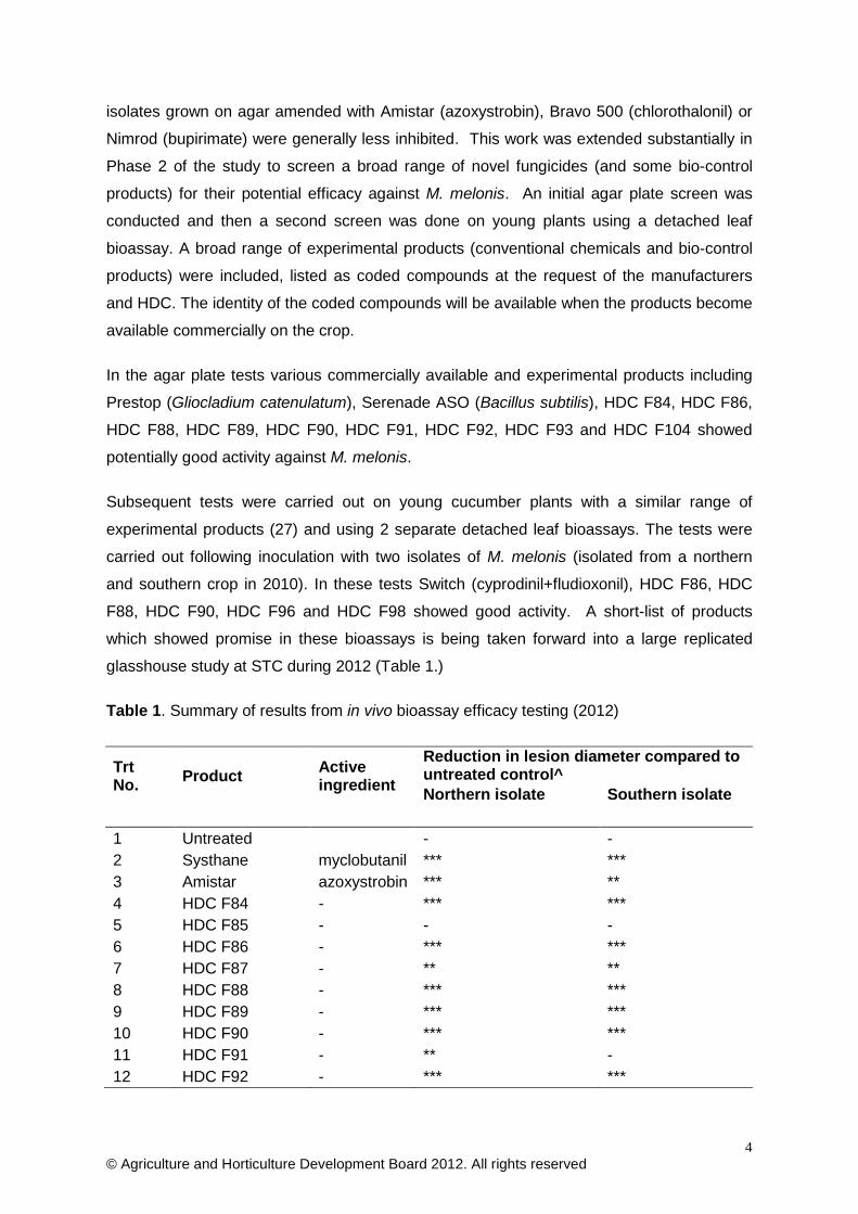
Table 1. Summary of results from in vivo bioassay efficacy testing (2012)
Trt No. Product Active
ingredient
Reduction in lesion diameter compared to untreated control^ Northern isolate Southern isolate
1 Untreated - - 2 Systhane myclobutanil *** *** 3 Amistar azoxystrobin *** ** 4 HDC F84 - *** *** 5 HDC F85 - - - 6 HDC F86 - *** *** 7 HDC F87 - ** ** 8 HDC F88 - *** *** 9 HDC F89 - *** *** 10 HDC F90 - *** *** 11 HDC F91 - ** - 12 HDC F92 - *** ***

5 © Agriculture and Horticulture Development Board 2012. All rights reserved
Trt No. Product Active
ingredient
Reduction in lesion diameter compared to untreated control^ Northern isolate Southern isolate
13 HDC F93 - *** *** 14 HDC F94 - *** *** 15 HDC F95 - *** *** 16 HDC F96 - *** *** 17 HDC F97 - * *** 18 HDC F98 - *** ***
19 Switch cyprodinil + fludioxonil *** ***
20 Teldor fenhexamid ** *** 21 Nimrod bupirimate ** *** 22 HDC F99 - ** *** 23 HDC F100 - * ** 24 HDC F101 - ** **
25 Prestop Gliocladium catenulatum ** ***
26 Serenade ASO B. subtilis * **
27 - Potassium bicarbonate * **
^ based on data from undamaged leaves 5DAT - No reduction in lesion development compared to the inoculated control. * represents a slight reduction in lesion development (1-20%) ** represents a moderate reduction (21-60%) *** represents a good reduction in lesion development (61-100%)
Disinfection
A series of experiments was undertaken to identify disinfectants with good activity against
M. melonis. Six disinfectant products containing active ingredients from different chemical
classes were tested for activity against conidia and mycelium of the fungus. Products were
tested at their full recommended rate and at half-rate after exposure for 5 mins and 30 mins.
Jet 5 (hydrogen peroxide/peracetic acid) and Fam 30 (iodophor) were most effective.
These products, together with bleach (sodium hypochlorite) and Unifect G (glutaraldehyde +
Quaternary Ammonium Compound, QAC) were fully effective after just 5 mins and at half
their recommended rates. Menno Florades (benzoic acid) was effective after 5 mins at full
rate and after 30 mins at half rate; Vitafect (QAC + biquanidine salt) was effective at full rate
but ineffective at half rate even after 30 mins. The most effective products against
mycelium in filter paper discs were Jet 5, bleach, Unifect G and Vitafect.
An experiment was designed and undertaken to examine the influence of different surfaces
on the activity of disinfectants against M. melonis. Overall, perhaps not surprisingly, it was
more difficult to disinfect concrete than aluminium, glass or plastic. Jet 5, bleach and
Unifect G used at their recommended rates were fully effective on all four surfaces.

6 © Agriculture and Horticulture Development Board 2012. All rights reserved
However, Fam 30 on concrete, Menno Florades on aluminium and concrete, and Vitafect on
glass all showed reduced activity.
An experiment was done to determine how effective various disinfectant soak treatments
were at reducing disease transmission of M. melonis on knives contaminated with the
fungus by cutting through infected cucumber leaves and stems. Disease transmission was
relatively low. Soaking contaminated knives in water, Jet 5, Menno Florades, bleach or
Vitafect for 1 hour reduced the development of gummy stem blight in cucumber fruit slices
compared with transmission from untreated knives. Results of all the disinfection tests
described above are summarised in Table 2.
Two experiments were carried out to compare different treatments for cleansing hands
contaminated with M. melonis following handling of cucumber fruit affected by M. melonis,
and through contamination of hands with a paste of the fungus in cucumber sap. A finger
from a washed hand was placed on a culture plate to check for pathogen viability. Washing
hands in soap and water, with an alcohol gel, or with alcohol foam, all greatly reduced
transmission of M. melonis from hands. Soap and water alone was less effective at
reducing transmission of M. melonis than soap and water followed by alcohol gel or foam,
or the alcohol foam or gel used directly on contaminated hands. Rinsing hands in water
alone gave no reduction in transmission of M. melonis.
Table 2. Summary of disinfectant activity against M. melonis in various tests – 2011
Disinfectant Rate
used
Growth of M. melonis recorded after treatmenta of
Spores* in water
Mycelium on filter paper in water
Spores*/mycelium dried on: Dirty knifeb Alu Con Gla Pla
Water (control) N/A + + + + + + (+)
Fam 30 1:125 - (+) - + - - NT
Jet 5 1:125 - - - - - - (+)
Menno Florades 10 ml/L - + (+) + - - -
Sodium hypochlorite
(10-14%) 1 in 10 (+) - - - - - (+)
Unifect G 4% - - - - - - NT
Vitafect 1% - - - - (+) - (+)
a Results shown after exposure to disinfectant for 5 mins (spores or filter paper in water) or 30 mins (all other tests). b Disease transmission test. N/A – not applicable; NT – not tested. - no growth; (+) occasional growth; + growth common. Alu – aluminium; Con – concrete; Gla – glass; Pla – plastic

7 © Agriculture and Horticulture Development Board 2012. All rights reserved
* The spore type evaluated was not differentiated though considered to comprise largely of conidia rather than ascospores
Financial Benefits
The results from the disinfectant study carried out during 2011 will have immediate benefits
for growers both during the growing season and during the clean-down between crops.
Effective use of disinfectants should help to reduce disease spread and the survival of
inoculum between crops and hence improve crop yield, marketable quality and hence the
economic value of the crop. However, due to the sporadic nature of such pathogen
infections it is difficult to put a precise value on this.
Although several fungicide and bio-control products have been shown to provide effective
control of M. melonis in small-scale laboratory studies, many of these products are not yet
approved for use in cucumbers and therefore cannot yet be used commercially. However,
the preliminary results help the design of an effective larger glasshouse study conducted
during 2012. The results from this work could then be used to recommend additional
effective products which may be put forward for approval via SOLA.
If one or more fungicides or bio-control products can be identified and subsequently
approved for use on cucumber (with a 1-2 day harvest interval ideally) then significant
economic loss could be avoided each year due to premature plant death (from girdling stem
lesions) and from symptomatic or latent fruit infections. It is estimated that between 1-10%
plants and fruit may be lost as a result of infection by Mycosphaerella each year.
It is also worth noting that if a product or products could be found with activity against
powdery mildew and Mycosphaerella then the financial benefit could be even greater.
It is a little too early to judge the potential financial benefits from the immunoassay work that
is in progress but, if the pathogen could be successfully monitored as proposed, then it will
help to better time intervention treatments including spray applications and this could
provide significant economic benefits in the longer-term through improved disease
prediction.
Action points for growers
• Consider using effective disinfectants identified in this project to limit secondary
spread of infection during crop work and between crops.
• Ensure the use of good quality seed from reputable suppliers, and be aware of the
potential for a seed borne risk on new cultivars.

8 © Agriculture and Horticulture Development Board 2012. All rights reserved
• Prestop, Serenade and Switch showed potential efficacy for the control of
Mycosphaerella in cucumbers in agar plate and small plant tests and should be
considered as part of an effective control regime in commercial crops. A number of
experimental or unapproved products also showed promise and may be available for
use in the future.

© Agriculture and Horticulture Development Board 2012. All rights reserved 9
SCIENCE SECTION
Introduction
Gummy stem blight caused by Mycosphaerella melonis (Didymella bryoniae) has been a
persistent leaf, stem & fruit disease in glasshouse cucumber for many years (Fig. 1). It has
been generally suppressed, rather than controlled, over the years using a combination of
rigorous hygiene precautions (to remove debris that might otherwise allow the pathogen to
carry-over from crop to crop in the glasshouse), environmental manipulation (to avoid
conditions conducive to infection), use of fungicides (to prevent infection and spread of the
pathogen) and more recently through the use of better cultivars (to reduce the rate of
disease progression in the host crop). However, more recently, a number of factors have
impacted on the disease and it is becoming more prevalent and damaging economically
with fewer opportunities for effective control. This is of considerable concern for growers
due to the potential economic damage this pathogen can cause either through direct loss of
plants (stem girdling) or yield reduction (as a result of symptomatic or latent (internal) fruit
infection). Increased energy costs are a significant factor leading to increased infection as
the higher cost discourages the use of pipe heat early in the morning to dry the foliage and
avoid conditions conductive to infection. Similarly, the loss of key active substances as a
result of the EU pesticide review programme has meant that growers have fewer useful
products with good activity against the pathogen to prevent infection. This is further
influenced by the increased shift in consumer (retailer) perception regarding pesticide
residues. An indirect impact of all this is the increased use of cultivars with tolerance to
powdery mildew (where most fungicides are usually used for control). This means that
growers are applying fewer fungicide sprays which otherwise would have provided
incidental control, or at least suppression, of Mycosphaerella infections. There is also some
evidence to suggest that such mildew tolerant cultivars are actually more susceptible to
Mycosphaerella.

© Agriculture and Horticulture Development Board 2012. All rights reserved 10
Figure 1. Mycosphaerella melonis stem and fruit infection Picture courtesy of Dr G M McPherson
No recent studies have been undertaken in the UK to determine the sensitivity of existing
and/or new fungicides and bio-control products against Mycosphaerella and growers have
to rely on an ever diminishing armoury of products. There is a direct parallel here with the
use of antibiotics for disease control in human & animal populations and likewise in
horticulture we are facing an increased risk of fungicide resistance in phytopathogen
populations. Unless we can find alternative approaches to the control of such endemic
pathogens we could potentially expect a continued increase in disease, potentially reaching
epidemic proportions.
The purpose of this project is firstly to establish ‘state of the art’ with respect to our
knowledge on this important pathogen and to establish the sensitivity of the current
population to widely used fungicides (Phase 1). Guided by this knowledge, the aim is then
to seek alternative control strategies (Phase 2). This includes the evaluation of novel
fungicides & alternative bio-control products and the use of novel immunosassay or
serological techniques to predict disease risk by monitoring the pathogen spore population
in the glasshouse in order to take action before infection is allowed to occur; thereby
improving application timing to prevent economic loss due to the disease.

© Agriculture and Horticulture Development Board 2012. All rights reserved 11
Evaluation of disinfectants for activity against M. melonis
Introduction
M. melonis can persist between crops in infected plant debris and possibly as spores
contaminating glasshouse surfaces. Within a crop there is potential for dispersal of the
fungus by contact transmission on equipment and on the hands of crop workers. Use of
disinfectants that reduce inoculum of M. melonis should reduce the risk of early infection in
a newly planted crop and the rate at which gummy stem blight spreads through a crop. The
aim of this work was to identify disinfectants and hand cleansers with good activity against
M. melonis that could be used as part of a strategy to control the disease. The specific
objectives were: to establish the efficacy of a range of chemical disinfectants, from different
active ingredient groups, against spores and mycelium of M. melonis; to determine the
efficacy of some hand cleansers in preventing transmission of M. melonis; to determine the
efficacy of selected disinfectants for reduction of M. melonis on four surfaces (aluminium,
concrete, glass, plastic), and to establish the efficacy of some knife-dip disinfection
treatment in preventing transmission of M. melonis at levels sufficient to cause disease.
Materials and methods
Experiments were carried out in 2011 and 2012 at ADAS Boxworth.
Experiment 1: Efficacy of disinfectants on spores of M. melonis
A culture of M. melonis isolated from cucumber in 2010 was incubated at 20°C, on plates of
potato dextrose agar + streptomycin sulphate (PDA+S) under alternating periods of 12 h UV
lights and 12 h dark until spore-producing pycnidia formed. Plates were flooded with 5 to
10 ml of acidified (pH 3.5 - 4.5), sterile-distilled water (SDW) and a loop was used to scrape
the agar surface. A few drops of Tween-80 (wetter) were added to the acidified water in
order to increase spore discharge from pycnidia and mitigate spore agglutination. The
mixture was filtered through four layers of sterile muslin to remove mycelia, pycnidia, and
dislodged agar. A haemocytometer was used to produce a concentration of approximately
1 x 10⁶ spores/ml. The spore suspension was stored at 5°C until use.
Products were tested at the label recommended rate and at half that rate with exposure
times of 5 and 30 minutes. For each disinfectant product (Table 1), 10 ml of the spore
suspension was pipetted into each of six Universal tubes. For each disinfectant rate
(untreated, full rate and ½ rate), requisite volumes were added to the spore suspension in
each of two Universal tubes, to give the required concentrations. Five minutes after adding
the disinfectant, the two tubes containing the full rate and ½ rate were centrifuged at 2000

© Agriculture and Horticulture Development Board 2012. All rights reserved 12
rpm for 2 mins. The supernatant fluid was removed and the pellet re-suspended in 5 ml
SDW. This was repeated on other tubes of spores after 30 mins disinfectant exposure time,
again testing products at the full rate and half rate.
Three replicate 25-well plates of PDA+S were used per treatment, each containing five
columns: an untreated control (water), the disinfectant tested at full rate for 5 and 30 mins,
and at half rate for 5 and 30 mins; there were five replicate wells for each rate x time
combination column. A droplet of the appropriate spore suspension was placed into the
centre of each well. The Petri plates were incubated at 20oC and the proportion of wells
with visible growth of M. melonis was determined after 7 days.
Table 1. Details of disinfectant products used in Experiments 1, 2, 4 and 5.
Product Active ingredient(s) Recommended product rate
1. Untreated control - -
2. Jet 5 Hydrogen peroxide + PAA 1:125
3. Fam 30 Iodophor 1:125
4. Menno Florades Benzoic acid 10 ml/L
5. Sodium hypochlorite (10-14%) Sodium hypochlorite 1:10 a
6. Unifect-G QAC +glutaraldehyde 4%
7. Vitafect QAC + biguanidine salts 1% a Equates to 10,000 ppm hypochlorite.
Experiment 2: Efficacy of disinfectants on mycelium of M. melonis in filter paper
The same treatments were tested as in Experiment 1. Squares of sterile filter paper
(approximately 0.5 cm2) were cut, and placed on the surface of an actively growing culture
of M. melonis. The filter paper was left on the cultures for 7 days to allow the fungal
mycelium to grow into the paper. The filter paper was then immersed in the disinfectant
products at the recommended rate and ½ rate for 5 mins and 30 mins. The filter paper
pieces (infested with M. melonis) were immersed in SDW as the control treatment. Treated
pieces of paper were rinsed three times in SDW, left to dry in the air flow from a laminar
flow hood, then plated on to PDA+S in 25-well plates with treatments arranged as described
previously for the spore test. The plates were incubated at 20oC and scored on the
proportion of wells with growth of M. melonis after 7 days.

© Agriculture and Horticulture Development Board 2012. All rights reserved 13
Experiment 3: Efficacy of hand cleansers against M. melonis
Fingers were contaminated by crushing cucumber fruits naturally infected with M. melonis
between thumb tip and forefinger tip 10 times. The contaminated thumb tip was applied to
a PDA+S plate for 10 successive contacts in paired tests: a) directly and b) after using the
hand cleansing treatments (Table 2). Contact was done sequentially from top left to bottom
right of the agar plate so that any reduction in transmission with successive contacts would
be visible. Hand cleansing was standardized by applying the treatment for 1 minute
followed by rinsing under a tap for 10 seconds (direct contact treatments), or allowing the
foam sanitiser and hand gel to evaporate for 1 minute. The same thumb and forefinger tip
was re-contaminated between treatments by using a fresh piece of cucumber tissue
naturally infected with M. melonis.
The number of agar plate contacts that developed M. melonis after 27 days incubation at
20ºC was recorded. A record was made of the number of transfers to agar that resulted in
growth (out of 10).
Table 2. Details of hand cleansing products used in Experiment 3.
Treatment Active ingredients
1. Untreated control -
2. Warm water and soap (bar) Sodium palmate & other salts
3. Cutan Foam Hand Sanitiser Alcohol
4. Antibacterial Hand Gel Alcohol
This experiment was repeated using a modified procedure due to a high occurrence of
bacteria and yeasts developing at finger contact sites on agar plates in the original
experiment which may have affected growth of M. melonis.
Mycelium from a 21 day old culture of M. melonis on PDA was mixed with internal tissue
from a healthy cucumber to form a paste. The paste was rubbed between thumb and
forefinger 50 times in order to contaminate fingers in a standard manner. A contaminated
forefinger was then applied to a PDA+S plate as described above both immediately and
after allowing the paste to air dry. The thumb and forefinger were re-contaminated, allowed
to dry and then hands were washed with soap and water (or other hand cleansing test
treatment) for 30 seconds; hands were rinsed in tap water, dried on a paper towel and then
the contaminated finger was applied to a PDA+S plate. Hands were then washed with soap
and water, followed by alcohol gel, before re-contaminating finger and thumb with the M.
melonis in cucumber paste, and testing another hand-cleansing treatment (Table 3). The

© Agriculture and Horticulture Development Board 2012. All rights reserved 14
same soap, hand sanitizer and hand gel were used as in the original experiment. Each of
the eight treatments was tested three times. Plates were incubated at 20ºC.
The number of agar plate contacts that developed M. melonis, and the density of growth of
the fungus (0 – nil, 1 – slight, 2 – moderate, 3 – dense), were recorded after 6 and 9 days.
Results were examined by regression analysis and analysis of variance.

© Agriculture and Horticulture Development Board 2012. All rights reserved 15
Table 3. Detail of hand cleansing treatments examined (Experiment 3 revised repeat)
Hand cleansing treatment Post-cleansing action before touching agar plate
1. None – wet paste None
2. None – dry paste Allowed to dry in air
3. Soap and water on dried paste Water rinse, paper towel dry
4. Foam on dried paste Allow to evaporate
5. Gel on dried paste Allow to evaporate
6. Soap and water on dried paste, rinse then foam Allow to evaporate
7. Soap and water on dried paste, rinse then gel Allow to evaporate
8. Water rinse only Paper towel dry
Experiment 4: Effect of surfaces on efficacy of disinfectants against M. melonis
Surfaces of aluminium (glasshouse bench), concrete (pathway), glass (glasshouse wall)
and rigid plastic (tray) were initially cleaned by washing in warm water and rinsing with fresh
water. They were then contaminated by spaying marked areas (10 x 10 cm) with a
suspension of spores and mycelium of M. melonis in SDW and allowed to dry for 30 mins.
The contaminated surface was then spray-treated with disinfectant and again allowed to dry
for 30 minutes. Each disinfectant in Table 1 was tested at its full recommended rate, and
an untreated was included. The treated surface was tested for viable M. melonis by
swabbing with a new cotton bud moistened in SDW and streaking it over a PDA+S agar
plates. Ten swabs were done for each of the 28 disinfectant x surface combinations. The
number of swabs that resulted in growth of M. melonis after incubation of agar plates for 7
and 14 days at 20ºC was recorded. Results were examined by Generalised linear modeling
to determine the effect of surface and disinfectant on transmission of M. melonis.
Experiment 5: Practical test – knife treatment to reduce disease transmission
Knife blades were contaminated with M. melonis by using them to cut (5 cuts per blade)
through cucumber stem and leaf tissue naturally infected with the fungus. The
contaminated knife blades were placed in a small container of the test disinfectant at its full
rate for 1 hour. The knife blades were then allowed to dry for 15 minutes. Using each knife,
three cuts were made across 2-cm thick cucumber slices, to around half the depth of the
slice; the same knife was used to cut 10 cucumber slices arranged on damp paper towels in
a plastic container. The cucumber slices were incubated at 20ºC in the plastic container

© Agriculture and Horticulture Development Board 2012. All rights reserved 16
and the proportion of cut slices that developed gummy stem blight was assessed after 5, 7
and 13 days. There were seven treatments (Table 4) with four replicate knife blades for
each treatment. Results were examined by generalized linear modeling.
Table 4. Details of treatments used for a practical test on transmission of gummy stem
blight (Experiment 5)
Treatment Rate
1. Tap water -
2. Jet 5 1:125
3. Menno Florades 10 ml/L
4. Sodium hypochlorite 1:10
5. Vitafect 1%
6. No dip (positive control) -
7. New knife (negative control) -
Results and discussion
Efficacy of disinfectants against spores (Experiment 1) and mycelium (Experiment 2) of M. melonis
Results are summarized in Table 5 and illustrated in Figures 1- 2. Differences between
treatments were clear and no statistical tests were done. For Experiment 1, all treatment
combinations reduced the viability of M. melonis spores compared with the untreated
control. The Vitafect treatment at half its recommended rate was least effective with 14/15
wells showing growth after 5 and 30 minutes. The Jet 5 and the Fam 30 treatments had
greatest efficacy against M. melonis spores compared with other treatments, with no wells
showing growth in any of the treatment combinations; Sodium hypochlorite and Unifect G
were almost as effective.
For Experiment 2, all treatment combinations except those of Menno Florades reduced the
viability of M. melonis mycelium in filter paper compared with the untreated control. In
contrast, M. melonis established in most if not all of the wells for all treatment combinations
of Menno Florades except when it was used at full rate for 30 mins.
In both experiments Menno Florades showed a fall-off in activity when used at half rate,
suggesting that the full rate we used is only just sufficient to kill M. melonis. The contrast in
the efficacy of Vitafect on spores and mycelium on filter paper at half rate is striking and
counterintuitive, as one would expect the filter paper to reduce disinfectant activity. Further
work is needed to determine if this is a true difference. When used at full rate however,
Vitafect worked well in both tests.

© Agriculture and Horticulture Development Board 2012. All rights reserved 17
Table 5. Effect of some disinfectant treatments on viability of spores and mycelium of M.
melonis
Product Number of wells (of 15) with growth of M. melonis
Full rate (5 mins)
Full rate (30 mins)
Half rate (5 mins)
Half rate (30 mins)
Spores
Untreated 15 15 15 15
Fam 30 0 0 0 0
Jet 5 0 0 0 0
Menno Florades 0 0 5 0
Sodium hypochlorite 1 0 0 0
Unifect G 0 1 0 0
Vitafect 1 0 14 14
Mycelium
Untreated 15 15 15 15
Fam 30 3 0 1 1
Jet 5 0 0 0 0
Menno Florades 13 0 15 14
Sodium hypochlorite 0 0 0 0
Unifect G 0 0 0 0
Vitafect 0 0 0 0

© Agriculture and Horticulture Development Board 2012. All rights reserved 18
Fig 2.1 Jet 5 Fig 2.2. Fam. 30
Fig 2.3. Sodium hypochlorite Fig 2.4. Menno Florades
Fig 2.5. Unifect G Fig 2.6. Vitafect
Figure 2. Effect of six disinfectants on viability of M. melonis spores. Treatment columns,
from left to right: Untreated; full rate for 5 minutes; full rate for 30 minutes; half rate for 5
minutes; half rate for 30 minutes. Each treatment was tested 5 times (rows within a column)
on each of three replicate plates.

© Agriculture and Horticulture Development Board 2012. All rights reserved 19
Fig 3.1 Jet 5 Fig 3.2. Fam. 30
Fig 3.3. Menno Florades Fig 3.4. Sodium hypochlorite
Fig 3.5. Unifect G Fig 3.6. Vitafect
Figure 3. Effect of six disinfectants on viability of M. melonis mycelium in filter paper.
Treatment columns, from left to right: Untreated; full rate for 5 minutes; full rate for 30
minutes; half rate for 5 minutes; half rate for 30 minutes. Each treatment was tested 5 times
(rows within a column) on each of three replicate plates.

© Agriculture and Horticulture Development Board 2012. All rights reserved 20
Experiment 3: Efficacy of hand cleansers
After handling fruit affected by gummy stem blight, growth on agar showed that fingers were
contaminated with M. melonis, Penicillium sp. and bacteria (Table 6). The alcohol foam,
alcohol gel and the combined treatment were all effective in reducing levels of M. melonis
and Penicillium recovered from fingers, with the combined treatment resulting in the
cleanest plates. There was no evidence of fall-off in transmission with 10 successive
contacts on an agar plate.
Table 6. Effect of hand-cleansing treatments on transmission of M. melonis and other
microorganisms following contamination of fingers with cucumber fruit affected by gummy
stem blight – Experiment 3
Treatment Number of agar plate contacts (of 10) with growth of:
M. melonis Penicillium sp. Bacteria/Yeast
1. Untreated* 8 10 10
2. Soap 0 10 10
3. Foam 0 2 10
4. Gel 1 0 10
5. Foam and gel 0 0 3
* Mean of 4 replicates.

© Agriculture and Horticulture Development Board 2012. All rights reserved 21
Figure 4. Effect of four hand-cleansing treatments on transmission of M. melonis after
contamination of fingers with infected fruit. M. melonis appears as dark-green to black
colonies (arrowed).
In the revised repeat experiment, growth of M. melonis on the agar plates from untreated
hands was good with considerably less overgrowth from bacteria and Penicillium sp. than in
the original experiment. The results of assessments at 6 and 9 days were similar;
assessment of growth after 9 days is shown in Table 7. Both wet and dry contaminated
fingers resulted in growth of M. melonis at all contact sites. There was no evidence of fall-
off in transmission of M. melonis with successive contacts on an agar plate. Transmission
of M. melonis was significantly reduced (p <0.001) by all of the hand-cleansing treatments
that used soap and water and/or alcohol foam or gel; it was not reduced at all simply by
rinsing hands in water. The soap and water treatment was significantly (p <0.001) less
effective (growth at 27% of contact sites) than treatments that included alcohol foam or gel
(growth at 0-7% of contact sites). The mean density of M. melonis growth after 9 days was
reduced by treatments which reduced transmission of the fungus.

© Agriculture and Horticulture Development Board 2012. All rights reserved 22
Hand-cleansing treatment also affected transmission of Penicillium sp. and bacteria. All
treatments were fully effective against Penicillium sp. except for rinsing in water. The
alcohol foam treatment was most effective at preventing growth of bacteria (Table 7).
Table 7. Effect of hand-cleansing treatments on transmission of M. melonis following
contamination of fingers with a paste of the fungus in cucumber tissue – Experiment 3
revised repeat, growth after 9 days.
Treatment Mean % contact sites with growth of: Density of M. melonis (0-3) M. melonis Penicillium sp. Bacteria
1. None – wet paste 100 (-) 26 (6.1) 73 (8.4) 3.0
2. None – dry paste 100 (-) 23 (5.8) 46 (9.4) 3.0
3. Soap and water 27 (6.8) 0 63 (9.1) 0.5
4. Alcohol foam 0 (-) 0 0 0
5. Alcohol gel 7 (3.8) 0 30 (8.6) 0.1
6. Soap and water; foam 3 (2.7) 0 43 (9.4) 0.1
7. Soap and water; gel 0 (-) 0 13 (6.4) 0
8. Water rinse only 100 (-) 3 (2.5) 10 (5.7) 3.0
Significance (23 df) <0.001 <0.001 <0.001 <0.001
LSD - - - 0.29
( ) – standard error.
Experiment 4: Effect of four surfaces on disinfectant efficacy
At the rates tested, three disinfectants (Jet 5, sodium hypochlorite and Unifect G), were fully
effective against M. melonis on all surfaces (Tables 8 and 9). Fam 30 was fully effective on
all surfaces except concrete; Menno Florades was fully effective only on glass and rigid
plastic. Overall, concrete was significantly more difficult to disinfect of M. melonis than
glass, plastic and aluminium (Table 10). This may be due to the porous nature of concrete,
possibly resulting in entrapment of organic matter that reduced disinfectant activity; or
surface tension preventing good contact with contaminated surfaces within pores.

© Agriculture and Horticulture Development Board 2012. All rights reserved 23
Table 8. Effect of four surfaces on efficacy of various disinfectants against M. melonis
Disinfectant Mean number of swabs (of 10) resulting in growth of
M. melonis after 7 days on:
Aluminium Concrete Glass Plastic
1. Untreated 4 (0.9) 7 (0.9) 6 (0.9) 9 (0.6)
2. Fam 30 0 6 (0.9) 0 0
3. Jet 5 0 0 0 0
4. Menno Florades 1 (0.6) 4 (0.9) 0 0
5. Sodium hypochlorite 0 0 0 0
6. Unifect G 0 0 0 0
7. Vitafect 0 0 2 (0.7) 0
( ) – standard error.
Table 9. Mean effect of various disinfectants against M. melonis across four surface types
Disinfectant Mean number of swabs (of 10) resulting in growth of
M. melonis after:
7 days 14 days
1. Untreated 6.5 (0.4) 6.3 (0.4)
2. Fam 30 1.5 (0.2) 1.5 (0.2)
3. Jet 5 0 0
4. Menno Florades 1.3 (0.3) 1.3 (0.3)
5. Sodium hypochlorite 0 0
6. Unifect G 0 0
7. Vitafect 0.1 (0.02) 0.1 (0.02)
( ) – standard error
Table 10. Mean effect of four surfaces recovery or M. melonis across all treatments
(including untreated)
Surface Mean number of swabs (of 10) resulting in growth
of M. melonis after 7 days
Aluminium 0.7 (1.5)
Concrete 2.3 (0.2)
Glass 1.1 (0.2)
Plastic 1.2 (0.1)
( ) – standard error.
© Agriculture and Horticulture Development Board 2012. All rights reserved 24
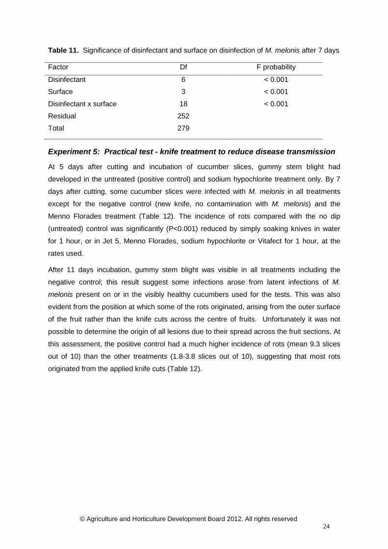
Table 11. Significance of disinfectant and surface on disinfection of M. melonis after 7 days
Factor Df F probability
Disinfectant 6 < 0.001
Surface 3 < 0.001
Disinfectant x surface 18 < 0.001
Residual 252
Total 279
Experiment 5: Practical test - knife treatment to reduce disease transmission
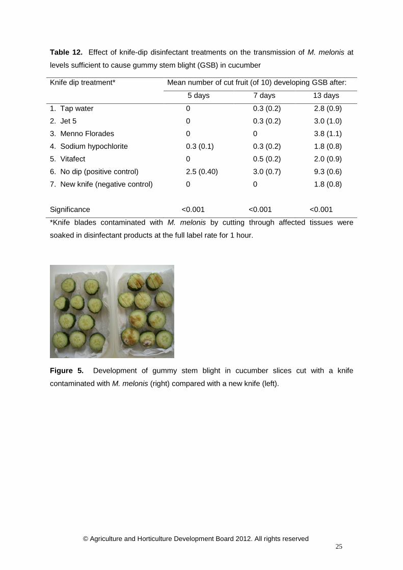
At 5 days after cutting and incubation of cucumber slices, gummy stem blight had
developed in the untreated (positive control) and sodium hypochlorite treatment only. By 7
days after cutting, some cucumber slices were infected with M. melonis in all treatments
except for the negative control (new knife, no contamination with M. melonis) and the
Menno Florades treatment (Table 12). The incidence of rots compared with the no dip
(untreated) control was significantly (P<0.001) reduced by simply soaking knives in water
for 1 hour, or in Jet 5, Menno Florades, sodium hypochlorite or Vitafect for 1 hour, at the
rates used.
After 11 days incubation, gummy stem blight was visible in all treatments including the
negative control; this result suggest some infections arose from latent infections of M.
melonis present on or in the visibly healthy cucumbers used for the tests. This was also
evident from the position at which some of the rots originated, arising from the outer surface
of the fruit rather than the knife cuts across the centre of fruits. Unfortunately it was not
possible to determine the origin of all lesions due to their spread across the fruit sections. At
this assessment, the positive control had a much higher incidence of rots (mean 9.3 slices
out of 10) than the other treatments (1.8-3.8 slices out of 10), suggesting that most rots
originated from the applied knife cuts (Table 12).
© Agriculture and Horticulture Development Board 2012. All rights reserved 25
Table 12. Effect of knife-dip disinfectant treatments on the transmission of M. melonis at
levels sufficient to cause gummy stem blight (GSB) in cucumber
Knife dip treatment* Mean number of cut fruit (of 10) developing GSB after:
5 days 7 days 13 days
1. Tap water 0 0.3 (0.2) 2.8 (0.9)
2. Jet 5 0 0.3 (0.2) 3.0 (1.0)
3. Menno Florades 0 0 3.8 (1.1)
4. Sodium hypochlorite 0.3 (0.1) 0.3 (0.2) 1.8 (0.8)
5. Vitafect 0 0.5 (0.2) 2.0 (0.9)
6. No dip (positive control) 2.5 (0.40) 3.0 (0.7) 9.3 (0.6)
7. New knife (negative control) 0 0 1.8 (0.8)
Significance <0.001 <0.001 <0.001
*Knife blades contaminated with M. melonis by cutting through affected tissues were
soaked in disinfectant products at the full label rate for 1 hour.
Figure 5. Development of gummy stem blight in cucumber slices cut with a knife
contaminated with M. melonis (right) compared with a new knife (left).

© Agriculture and Horticulture Development Board 2012. All rights reserved 26
Development of monoclonal antibody cell lines to ascospore inoculum of Mycosphaerella melonis
Introduction
Spore trapping techniques have previously been shown to indicate when pre-symptomatic
control of air-borne diseases in the field may be possible e.g. for Botrytis blight of onion.
However, as different pathogens with differing spore types have varying environmental
requirements, accurate differentiated spore counts are necessary. Determining the spore
threshold at which infection occurs is also important.
Within glasshouse crop production, monitoring air-borne inoculum concentrations has
shown when there is increased risk of disease and where disease control can be targeted.
Spore trapping techniques could be used to improve control of gummy stem blight by
highlighting periods when there is an infection risk. To achieve this will require the
development and validation of an immuno-monitoring system for spores of M. melonis.
Knowledge gained from previous work carried out by Dr Roy Kennedy and his colleagues
on Mycosphaerella brassicicola in field-grown brassica crops has been used as a starting
point for this aspect of the project, although work carried out in Phase 1 of this study
showed that the monoclonal antibodies developed previously, for M. brassicicola, were not
sensitive enough for use with M. melonis spores and therefore new antibodies have had to
be produced.
Materials and Methods
The work was carried out by NAPRU – University of Worcester.
Antibody production
Six mice were immunised with a range of Mycosphaerella melonis ascospore fractions:
• Whole ascospore
• Disrupted ascospore fraction > 30 Kda
• Disrupted ascospore fraction< 30 Kda
Immunisations took place on the 28/6/11, 27/7/11 and 23/08/11. Mouse tail bleeds taken on
1/9/11 showed a variable immune response (Figure 6a, b). Immunising with an ascospore
fraction of < 30 Kda failed to elicit a good immune response. Two mice have been selected
and fusions are underway to identify and select hybridoma cell lines with sensitivity and
specificity to ascospores of M. melonis.

© Agriculture and Horticulture Development Board 2012. All rights reserved 27
0
0.2
0.4
0.6
0.8
1
1.2
1.4
1.6
1.8
1 in250
1 in500
1 in1000
1 in2000
1 in4000
1 in8000
1 in16000
1 in32000
1 in64000
Abso
rban
ce a
t 45
0nm
Tail bleed dilutions
mouse 1
mouse 2
mouse 3
mouse 4
mouse 5
mouse 6
6a
Figure 6. Mouse tail bleeds - Immune response as recorded by PTA-ELISA to M. melonis ‘whole’ (a) and soluble ascosporic material (b)
6b
Figure 6. Mouse tail bleeds - Immune response as recorded by PTA-ELISA to M. melonis ‘whole’ (a) and soluble ascosporic material (b)
Monitoring glasshouse aerosols for M. melonis
MTIST air sampler.
Two microtitre immunospore traps (MTIST air samplers) were set at variable heights within
a commercial cucumber crop in Yorkshire and were operated continuously over a five
month period (July to December 2011). The MTIST air samplers operated at a sampling
volume of 57 L min-1 and air particulates were impacted directly on to the base of 4 x 8 well
microtitre strips. To inhibit germination of trapped spora the microtitre wells were pre-
coated with 0.05 mg m-1l NaN3. Following each seven day exposure period the microtitre
strips were removed and stored at -20°C prior to analysis by PTA ELISA.
As the weekly Melinex tapes are processed (see Burkard sampler below) the spore counts
can be compared to those observed on the weekly MTIST counts. The correlation of these
results to-date is quite promising. Spore load was significantly reduced when monitored at
‘high canopy’ compared to the numbers of spores observed from the air samplers situated
low in the canopy.

© Agriculture and Horticulture Development Board 2012. All rights reserved 28
Commercial crop diary
Aviance crop in situ at time of initiation of monitoring
4th July 2011 Bravo application to crop
14th July Bravo application to crop
25th July Systhane application to crop
9th August Crop removal
10th August Crop replant
11th August Amistar application
15th August Bravo application
31st August Systhane application
15th Sept Systhane application
4th Oct Switch/chalk stem spray
5th Oct Rocket application
18th Oct Crop removed
2nd Dec Polythene removed
5-10th Dec Pressure washed glasshouse
10/11th Dec Horticide spray space treatment
14th Dec New polythene laid
22nd Dec Last despatch of air-sampling tapes and wells.

© Agriculture and Horticulture Development Board 2012. All rights reserved 29
Figure 7. Burkard (large light green unit at base of crop) and MTIST (two small cylindrical units in upper & lower parts of the crop canopy) air samplers in position at Anchor Nurseries
The PTA ELISA of the glasshouse exposed MTIST wells will be completed on selection of a
suitable monoclonal antibody cell lines i.e. immune-quantification of any trapped air-borne
ascospores of M. melonis in the MTIST wells and this can then be compared alongside the
more conventional spore trap results using a Burkard volumetric spore trap (see below).
Burkard volumetric air sampler.
A volumetric air sampler was placed at ground level within a commercial cucumber cropping
system and adjacent to an MTIST air sampler (Figure 7). The sampler operated at an air
flow rate of 10 L minute-1 throughout the five month sampling period. A Melinex tape fixed
to a rotating drum and positioned inside the volumetric spore trap, operated continuously for
seven day periods, where air particulates in the air were impacted directly onto the tape. At
seven day intervals the Melinex tape was removed and sectioned into 24 hr periods. Under
bright field microscopy at a magnification of x400, each of the tape sections was examined
for the presence of ascospores of M. melonis (Figure 8). After which, and on selection of a

© Agriculture and Horticulture Development Board 2012. All rights reserved 30
suitable MAb cell line the Melinex slides will be processed by immunofluorescence for the
presence of M. melonis ascospores.
Results
MAb selection
Work is currently on-going to screen and assess the Monoclonal antibodies (MAb). One
MAb which has shown good specificity but low sensitivity to M. melonis has been identified.
This MAb has been used to assess single strips of the weekly MTIST wells (from high and
low in the canopy). An additional set of mice have now been immunised and the search for
a cell line that will provide both good specificity and sensitivity continues. This will help to
make the test transferable to a lateral flow (on-site) test to allow growers/consultants to
monitor M. melonis spore loads in the cropping area.
© Agriculture and Horticulture Development Board 2012. All rights reserved 31
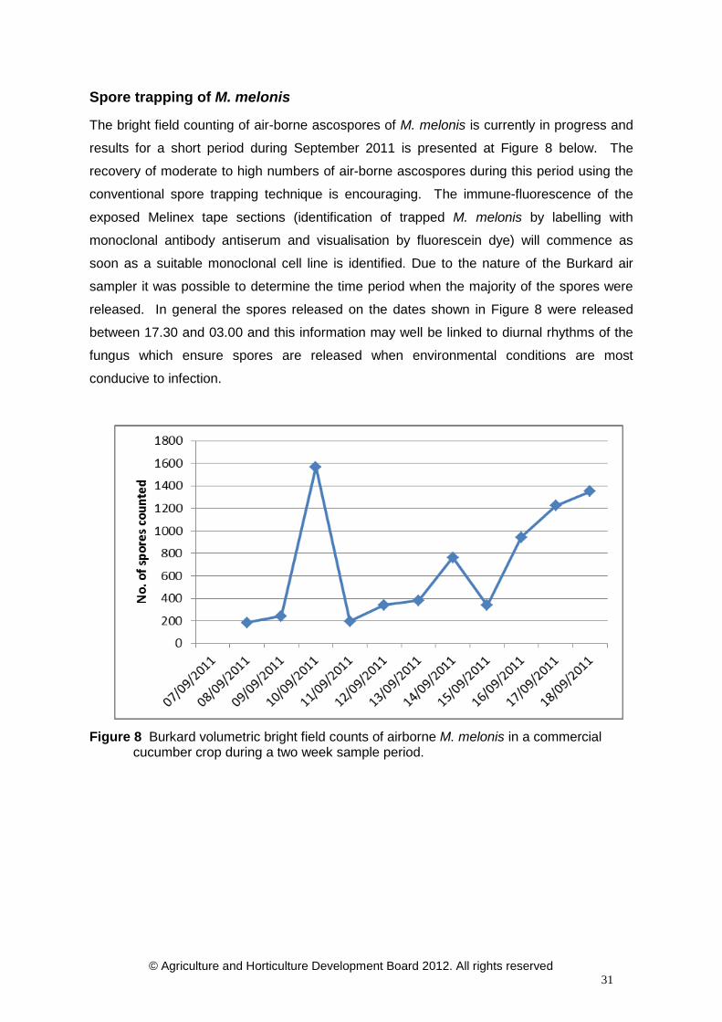
Spore trapping of M. melonis
The bright field counting of air-borne ascospores of M. melonis is currently in progress and
results for a short period during September 2011 is presented at Figure 8 below. The
recovery of moderate to high numbers of air-borne ascospores during this period using the
conventional spore trapping technique is encouraging. The immune-fluorescence of the
exposed Melinex tape sections (identification of trapped M. melonis by labelling with
monoclonal antibody antiserum and visualisation by fluorescein dye) will commence as
soon as a suitable monoclonal cell line is identified. Due to the nature of the Burkard air
sampler it was possible to determine the time period when the majority of the spores were
released. In general the spores released on the dates shown in Figure 8 were released
between 17.30 and 03.00 and this information may well be linked to diurnal rhythms of the
fungus which ensure spores are released when environmental conditions are most
conducive to infection.
Figure 8 Burkard volumetric bright field counts of airborne M. melonis in a commercial cucumber crop during a two week sample period.
© Agriculture and Horticulture Development Board 2012. All rights reserved 32
Seed testing
Introduction
The extensive literature review carried out in Phase 1 of this study suggested that although
there is some compelling evidence of M. melonis being seed-borne in some cucurbit crops,
extensive research has found the pathogen only sporadically in cucumber seed. Lee et al
(1984) reported that of over 90 cucurbit seed (cucumber & pumpkin) samples tested from
thirteen countries, nine from four countries were found to be infected with Didymella
bryoniae (M. melonis). The pathogen was reported to be located on and in the seed coat
including the perisperm and in the tissue of the cotyledons. Primary seedling infection
occurred on the radicle, hypocotyl and cotyledons. Infection of the radicle generally caused
a pre-emergence rot while infection on the hypocotyl and cotyledons developed further
inoculum for infection of the first true leaves and the stem.
A small-scale investigation into the potential for M. melonis to be seed-borne was carried
out during phase 1 of this study. At that time five seed batches were tested and M. melonis
was detected on two of these, due to the significance of these findings it was agreed that
these batches would be retested along with additional batches of different cultivars from a
range of suppliers.
Methods & materials
A total of 16 cucumber seed batches were obtained for additional testing at STC during
2011. Re-testing of a retained sample of the cultivar which had given a positive result for M.
melonis when testing in 2010 was also carried out. Each batch was visually examined
under a low power microscope to check for fungal growth on the seeds or debris mixed in
with the seed which might prove to be particles of infectious material such as pycnidia. Any
suspicious material found was removed and cultured on a suitable artificial growth media to
determine its identity. A batch of 100 seed/cv were then plated aseptically onto multiple
agar plates (square 25 well plates were used to avoid the potential for contamination to
grow across Petri-dishes) which were incubated at 23°C for 7-10 days in order to be able to
identify any fungal contaminants on or in the seed. A further batch of 25 seeds was sown in
fresh compost (Levington F2+S) in half seed trays and allowed to germinate and grow on
for 2-3 weeks. The seedlings were then examined for any visual signs of infection before
being excised to allow a microscopic examination of the vascular tissues to take place.
Stem slices (2mm) were then cut from each stem and plated onto artificial growth media for

© Agriculture and Horticulture Development Board 2012. All rights reserved 33
incubation. The stem slices were then examined for the development of any fungal growth
which was identified and recorded.
Results
Seed testing was carried out on a total of 17 batches of seed during 2011. The results of
the tests are summarised in Table 13 below. One batch (E528/55) that tested positive for
M. melonis in 2010 was found to be free of M. melonis in 2011. It is unclear whether the
2010 result was a false positive (mis-identification or contamination) or, if true, whether the
level and viability of the fungus had declined to zero by the re-test. None of the seedlings
grown for 3 weeks from these seed batches developed symptoms of gummy stem blight.
Microscopic examination of stem vascular tissue plated out onto an agar medium for the
presence of Mycosphaerella and other potential cucumber pathogens was negative and no
plant pathogens were detected from the vascular tisues.
Table 13 . Results of the seed-testing carried out during 2010 and 2011
Seed batch code
When tested
M. melonis detected from
seed plating (no. of infected seed)
Systemic M. melonis detected (stem sections)
E528/S1a Sept 2010 0 Not tested E528/S1b April 2011 0 0 E528/S2a Sept 2010 0 Not tested E528/S2b May 2011 0 0 E528/S3a Sept 2010 0 Not tested E528/S3b April 2011 0 0 E528/S4a^ Sept 2010 1 Not tested E528/S4b April 2011 0 0
E528/S5* Sept 2010
& May 2011
3 0
Not tested 0
E528/S6 May 2011 0 0 E528/S7 April 2011 0 0 E528/S8 May 2011 0 0 E528/S9 May 2011 0 0 E528/S10 April 2011 0 0 E528/S11 May 2011 0 0 E528/S12 May 2011 0 0 E528/S13 April 2011 0 0 E528/S14 May 2011 0 0 E528/S15 April 2011 0 0 E528/S16 May 2011 0 0 E528/S17 May 2011 0 0 * re-tested due to significant result ^ not enough seed left for re-testing S#a and S#b signify different batches of the same cultivar No evidence of Mycosphaerella was detected on the surface or internal tissues of the additional
batches of seed tested using the agar plate test during 2011, or on the re-test of batch E528/S5.
None was detected in the growing-on tests in any batches tested.

© Agriculture and Horticulture Development Board 2012. All rights reserved 34
Secondary screen of novel fungicide and bio-control products for efficacy against M. melonis
Introduction
This aspect of the work aims to identify potential products following the loss of key
fungicides which has resulted in growers experiencing increasing problems with control of
gummy stem blight in cucumbers. Pressure is mounting to reduce the use of conventional
chemical products in edible crops and it is therefore important to also evaluate alternative
bio-control strategies that could help maintain effective control without recourse to frequent
chemical application. It should be remembered that pathogens such as Mycosphaerella are
one of the reasons for the industry repeatedly re-plant crops each season, and this has cost
implications which effective disease control mechanisms may help to reduce.
Materials & Methods This aspect of the work was carried out in two stages at STC in North Yorkshire.
A primary screen of 20 conventional fungicides and 3 bio-control products was carried out in
the laboratory using an in-vitro agar plant assay. The details of these products are shown in
Table 14 below. The potential efficacy of the products was tested using an amended agar
plate test which provides quantitative data on the inhibition of fungal growth (mycelium only)
when grown on agar plates amended with each of the products and compared to growth of
the fungus on un-amended agar plates. Each product was added to a standard fungal agar
medium – Potato Dextrose Agar (PDA) at 2, 20 and 100ppm of the active ingredient in the
case of the conventional fungicide products and at the label dilution rate for the bio-control
products (Prestop & Serenade). Two previously collected isolates of M. melonis were used
in the tests, one which had been collected from infected crops in the north of England
(Humberside) and the other from a southern crop (Lea Valley) during the 1st year of the
study and which had been retained in the STC culture collection. Each isolate was grown
on PDA to ensure purity of the culture and tests were set up using 5-7 day old cultures. The
sterile PDA was amended with the product under test and poured into Petri-dishes before
being allowed to set. Once set, a 5mm plug of the actively growing isolate was positioned
centrally on the agar plates. Each isolate was tested against each product, at each
concentration, in triplicate.

© Agriculture and Horticulture Development Board 2012. All rights reserved 35
Table 14. Details of products used in in vitro fungicide and bio-control screen - 2011
Trt No. Product Active ingredient Rate/ha rate/L
1 Untreated - - - 2 Systhane 20EW myclobutanil 0.375L 0.37ml 3 Amistar azoxystrobin 1L 1ml 4 HDC F84 - 1L 1ml 5 HDC F85 - 1L 1ml 6 HDC F86 - 0.5kg 0.5g 7 HDC F87 - 0.3kg 0.3g 8 HDC F88 - 0.25L 0.25ml 9 HDC F89 - 1.5L 1.5ml 10 HDC F90 - 0.4L 0.4ml 11 HDC F91 - 1.5L 1.5ml 12 HDC F92 - 0.9L 0.9ml 13 HDC F93 - 0.3L 0.3ml 14 HDC F94 - 1 L 1ml 15 HDC F103 - 0.71kg 0.71g 16 Teldor fenhexamid 0.1kg/100L 1g 17 Nimrod bupirimate 0.2L/100L 2g 18 HDC F99 - 0.25L 0.25ml 19 HDC F101 - 0.25L 0.25ml 20 Unix* cyprodinil - 1.35g 21 Bravo 500+ chlorothalonil - 2.0ml 22 Prestop Gliocladium catenulatum 3.5% v/v 35g 23 Serenade ASO B. subtilis 10L 10ml 24 - Potassium bicarbonate - I molar
* Rate chosen to match ai content in Switch
The test plates were incubated at 23°C for 3 days before the diameter of the fungal growth
was recorded. The mean colony diameter for each product/concentration/isolate was
calculated and compared to the growth of each respective isolate on un-amended PDA.
The percentage inhibition of growth was calculated using the following formula.
© Agriculture and Horticulture Development Board 2012. All rights reserved 36
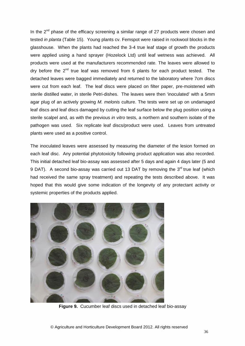
In the 2nd phase of the efficacy screening a similar range of 27 products were chosen and
tested in planta (Table 15). Young plants cv. Femspot were raised in rockwool blocks in the
glasshouse. When the plants had reached the 3-4 true leaf stage of growth the products
were applied using a hand sprayer (Hozelock Ltd) until leaf wetness was achieved. All
products were used at the manufacturers recommended rate. The leaves were allowed to
dry before the 2nd true leaf was removed from 6 plants for each product tested. The
detached leaves were bagged immediately and returned to the laboratory where 7cm discs
were cut from each leaf. The leaf discs were placed on filter paper, pre-moistened with
sterile distilled water, in sterile Petri-dishes. The leaves were then ‘inoculated’ with a 5mm
agar plug of an actively growing M. melonis culture. The tests were set up on undamaged
leaf discs and leaf discs damaged by cutting the leaf surface below the plug position using a
sterile scalpel and, as with the previous in vitro tests, a northern and southern isolate of the
pathogen was used. Six replicate leaf discs/product were used. Leaves from untreated
plants were used as a positive control.
The inoculated leaves were assessed by measuring the diameter of the lesion formed on
each leaf disc. Any potential phytotoxicity following product application was also recorded.
This initial detached leaf bio-assay was assessed after 5 days and again 4 days later (5 and
9 DAT). A second bio-assay was carried out 13 DAT by removing the 3rd true leaf (which
had received the same spray treatment) and repeating the tests described above. It was
hoped that this would give some indication of the longevity of any protectant activity or
systemic properties of the products applied.
Figure 9. Cucumber leaf discs used in detached leaf bio-assay

© Agriculture and Horticulture Development Board 2012. All rights reserved 37
Table 15. Details of products and rates used in detached-leaf bio-assay - 2011
Trt No. Product Active ingredient Rate/ha Rate/L 1 Untreated 2 Systhane myclobutanil 0.375L 0.37ml 3 Amistar azoxystrobin 1L 1ml 4 HDC F84 - 1L 1ml 5 HDC F85 - 1L 1ml 6 HDC F86 - 0.5kg 0.5g 7 HDC F87 - 0.3kg 0.3g 8 HDC F88 - 0.25L 0.25ml 9 HDC F89 - 1.5L 1.5ml 10 HDC F90 - 0.4L 0.4ml 11 HDC F91 - 1.5L 1.5ml 12 HDC F92 - 0.9L 0.9ml 13 HDC F93 - 0.3L 0.3ml 14 HDC F94 - 1 L 1ml 15 HDC F95 - 0.02kg/10L 2g 16 HDC F96 - 0.8L 0.8ml 17 HDC F97 - 0.71kg 0.71g 18 HDC F98 - 0.9L 0.9ml 19 Switch cyprodinil + fludioxonil 0.8kg 0.8g 20 Teldor fenhexamid 0.1kg/100L 1g 21 Nimrod bupirimate 0.2L/100L 2g 22 HDC F99 - 0.25L 0.25ml 23 HDC F100 - 0.125L 0.125ml 24 HDC F101 - 0.25L 0.25ml 25 Prestop Gliocladium catenulatum 3.5% v/v 35g 26 Serenade ASO B. subtilis 10L 10ml 27 - Potassium bicarbonate 1 molar

© Agriculture and Horticulture Development Board 2012. All rights reserved 38
Results
In vitro product efficacy screen
The results for each isolate are shown in Appendix 2, however here the two values have
been averaged to give an overall indication of the potential efficacy of the products to inhibit
mycelial growth.
At the lowest concentration of active ingredient (2ppm) several of the products did not inhibit
mycelial growth to any great degree (Figure 10). However, several of the products,
including all the experimental products, Serenade ASO and Prestop amongst others did
result in appreciable levels of inhibition in this test.
Figure 10. Percentage inhibition of mycelial growth of each product compared to the growth of the fungus on un-amended agar (negative control) for products tested at 2ppm (mean of north & south isolate results).
More of the products under test achieved a higher level of inhibition of growth when used at
20ppm of active ingredient. Nimrod, Teldor, Systhane, HDC F85 and HDC F94 all showed
an increase in inhibition at this concentration (figure 11). Products such as Potassium
bicarbonate, HDC F87, HDC F99, HDC F101, Bravo 500 and Amistar were less effective
resulting in <60% inhibition of growth in these tests.

© Agriculture and Horticulture Development Board 2012. All rights reserved 39
Figure 11. Percentage inhibition of mycelial growth of each product compared to the growth of the fungus on un-amended agar (negative control) for products tested at 20 ppm (mean of north & south isolate results). When the isolates were grown on agar amended with the products at 100ppm of active
ingredient all but 5 of the products resulted in >60% inhibition of growth of the fungus (figure
12). Potassium bicarbonate, HDC F87, HDC F99, HDC F101 and Amistar were still the
poorest of the products tested, Bravo 500 and HDC F103 increased their inhibition activity
slightly, whilst 50% (11) of the products resulted in 100% inhibition of growth.

© Agriculture and Horticulture Development Board 2012. All rights reserved 40
Figure 12. Percentage inhibition of mycelial growth of each product compared to the growth
of the fungus on un-amended agar (negative control) for products tested at 100 ppm (mean
of north & south isolate results).
It should be borne in mind that due to the nature of this in vitro test it only provides a
preliminary impression of the potential efficacy of the products to control the fungus. It is
not definitive as some products are likely to have a different mode of action other than
inhibition of mycelial growth e.g. disruption of spore production, and this would not be
measurable in this type of test. Therefore there is a risk that such products would appear to
perform poorly and therefore be excluded prematurely and this needs to be considered
carefully when drawing any conclusions from such work. However, as a stand-alone test
the initial results are encouraging and suggest that several experimental products have
potential activity against Mycosphaerella.
In planta product efficacy screen
Data was recorded separately for each isolate, and any evidence of a potential phytotoxicity
effect from the treatments was recorded at each assessment date. The mean values
(across replicates) are shown in tables 16 & 17 and figures 14 & 15 show the overall
(averaged) picture of the north and south isolate response.

© Agriculture and Horticulture Development Board 2012. All rights reserved 41
Fig 13a. Mycosphaerella lesions developing on leaf discs T5 (HDC F85). Fig 13b. Leaf discs treated with T8 (Exp.2) showing no lesion.

© Agriculture and Horticulture Development Board 2012. All rights reserved 42
Table 16. Detached leaf bio-assay results for the northern isolate. Results shown as the % inhibition of lesion development compared to the untreated control (T1)
Treatment UNDAMAGED DAMAGED Possible phytotoxicity effects observed
Bio-assay 1 Bio-assay 2 Bio-assay 1 Bio-assay 2 5 DAT 9 DAT 13 DAT 5 DAT 9 DAT 13 DAT
2. Systhane
No data – no growth on untreated leaves
13.27 73.91 46.75 28.26 3. Amistar 24.02 81.52 85.70 23.55 4. HDC F84 15.64 78.80 64.99 21.94 5. HDC F85 35.48 -5.98 -219.53 34.05 6. HDC F86 100.00 97.28 68.93 97.04 7. HDC F87 51.54 48.37 -76.04 25.71 8. HDC F88 98.88 97.83 83.23 97.58 9. HDC F89 25.42 95.11 57.59 37.42 10. HDC F90 100.00 100.00 96.55 99.60 11. HDC F91 39.53 29.89 -24.26 39.43 12. HDC F92 81.98 94.57 85.70 51.82 13. HDC F93 83.67 78.02 18.75 78.44 83.31 15.92 14. HDC F94 85.71 74.83 42.42 99.54 97.03 2.92 Leaf edges chlorotic 15. HDC F95 95.41 43.62 78.41 89.91 68.74 -31.03 16. HDC F96 100.00 96.81 99.24 99.54 98.30 90.98 Leaf edges chlorotic 17. HDC F97 -2.04 -3.36 -0.76 13.30 23.76 -61.27 18. HDC F98 100.00 94.30 99.43 100.00 95.05 72.15 Stunting and rolling
of leaf edges 19. Switch 90.31 79.03 71.78 100.00 89.82 85.68 20. Teldor -13.78 -7.21 -6.44 54.59 47.38 -47.75 21. Nimrod 65.31 10.07 -7.20 50.92 17.68 -30.50 22. HDC F99 18.88 21.81 NO DATA 32.11 47.24 NO DATA Severe leaf chlorosis 23. HDC F100 -5.61 0.17 15.72 16.51 8.35 -49.60 24. HDC F101 18.37 -7.92 -5.49 41.28 8.06 -68.17 25. Prestop 17.35 3.86 -6.63 54.59 36.78 -63.66 26. Serenade 22.45 -8.39 11.36 6.88 -1.84 -66.84 27. Pot bicarbonate 0.51 -8.56 -8.14 9.63 6.79 -62.33

© Agriculture and Horticulture Development Board 2012. All rights reserved 43
Table 17. Detached leaf bio-assay results for the southern isolate. Results shown as the % inhibition of lesion development compared to the untreated control (T1)
Treatment UNDAMAGED DAMAGED Possible phytotoxicity effects observed
Bio-assay 1 Bio-assay 2 Bio-assay 1 Bio-assay 2 5 DAT 9 DAT 13 DAT 5 DAT 9 DAT 13 DAT
2. Systhane 100.00 100.00 -36.58 70.37 65.57 49.45 3. Amistar 35.62 84.03 -58.35 46.30 77.05 96.15 4. HDC F84 96.42 100.00 64.37 83.33 93.44 41.76 5. HDC F85 -168.24 -88.19 80.21 -51.85 -127.05 -9.34 6. HDC F86 100.00 100.00 98.02 100.00 100.00 100.00 7. HDC F87 100.00 100.00 93.07 35.19 77.87 98.90 8. HDC F88 100.00 100.00 100.00 98.15 97.54 100.00 9. HDC F89 100.00 100.00 45.57 100.00 72.95 98.90 10. HDC F90 100.00 100.00 100.00 100.00 95.08 100.00 11. HDC F91 -57.37 70.14 100.00 -33.33 10.66 36.26 12. HDC F92 71.39 100.00 40.62 85.19 94.26 82.42 13. HDC F93 97.26 97.17 78.69 100.00 98.92
No data – no growth on
untreated leaves
14. HDC F94 94.52 85.84 97.54 100.00 90.32 Leaf edges chlorotic 15. HDC F95 100.00 100.00 79.91 97.17 50.54 16. HDC F96 100.00 87.73 99.59 98.11 97.85 Leaf edges chlorotic 17. HDC F97 82.18 85.84 38.92 91.51 84.95 18. HDC F98 100.00 100.00 100.00 100.00 100.00 Stunting and rolling
of leaf edges 19. Switch 100.00 100.00 100.00 100.00 98.92 20. Teldor 73.96 72.63 78.28 93.40 91.40 21. Nimrod 94.52 74.52 29.09 90.57 46.24 22. HDC F99 61.62 23.56 NO DATA 93.40 -35.48 Severe leaf chlorosis 23. HDC F100 41.06 15.06 -26.66 40.56 7.53 24. HDC F101 20.50 -33.07 84.83 58.49 40.86 25. Prestop 54.77 61.31 66.39 73.58 46.24 26. Serenade 58.88 51.87 90.16 55.66 18.28 27. Pot bicarbonate 43.80 59.42 93.44 51.88 37.63

© Agriculture and Horticulture Development Board 2012. All rights reserved 44
A comparison of the data shown in Tables 16 &17 shows that some of the products resulted
in a consistent and comparable response between the two isolates, yet the results for other
products are very variable between the isolates, with a moderate/good response with one
isolate and a complete lack of efficacy in the other. It is difficult to explain the discrepancy
in the results here and therefore the data should be treated with caution.
Damaged leaves did not appear to be more susceptible to infection with the fungal isolates
than undamaged leaves in general and, as the virulence of these isolates was already
proven, this was not unexpected. Products can be characterised by being effective as
contact products e.g. showing inhibition of lesion development very quickly following
application, how persistent they are, and also whether they have any systemic activity e.g.
the product is translocated around the treated plant. It may be expected that most products,
where effective, would show some control of lesion development 5 and 9 days after
treatment application and this was determined during the 1st bioassay. To determine
whether any of the products under investigation also demonstrated longevity of activity, or
possibly had systemic activity a 2nd bioassay was carried out 13 days after treatment
application. Table 18 shows the product responses compared in this way, and also helps to
highlight the different responses observed between the isolates, and therefore where
potentially unreliable data may have been gathered. Several of the products used showed
good efficacy with both isolates and also as contact and systemic products e.g. HDC F86,
HDC F88, HDC F90, HDC F96, HDC F98 and Switch (cyprodinil+fludioxonil). Products
such as HDC F85 and HDC F91 were perhaps more effective as systemic products,
although the results for these products were less consistent across the two isolates.
The tables of results show that possible phytotoxicity symptoms were observed on the
plants following application of HDC F94, HDC F96, HDC F98 and HDC F99. This
information is important and if any of these products are taken forward into a larger study
this aspect of their performance will require careful monitoring.
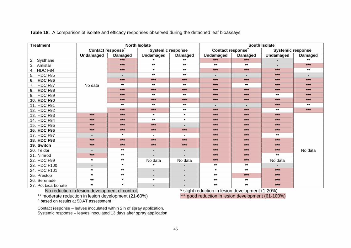
45
Table 18. A comparison of isolate and efficacy responses observed during the detached leaf bioassays
Treatment North Isolate South Isolate Contact response^ Systemic response Contact response^ Systemic response
Undamaged Damaged Undamaged Damaged Undamaged Damaged Undamaged Damaged 2. Systhane
No data
*** * ** *** *** - ** 3. Amistar *** ** ** ** ** - *** 4. HDC F84 *** * ** *** *** *** ** 5. HDC F85 - ** ** - - *** - 6. HDC F86 *** *** *** *** *** *** *** 7. HDC F87 ** ** ** *** ** *** *** 8. HDC F88 *** *** *** *** *** *** *** 9. HDC F89 *** ** ** *** *** ** *** 10. HDC F90 *** *** *** *** *** *** *** 11. HDC F91 ** ** ** - - *** ** 12. HDC F92 *** *** ** *** *** ** *** 13. HDC F93 *** *** * * *** *** ***
No data
14. HDC F94 *** *** ** * *** *** *** 15. HDC F95 *** *** *** - *** *** *** 16. HDC F96 *** *** *** *** *** *** *** 17. HDC F97 - * - - *** *** ** 18. HDC F98 *** *** *** *** *** *** *** 19. Switch *** *** *** *** *** *** *** 20. Teldor - ** - - *** *** *** 21. Nimrod *** ** - - *** *** ** 22. HDC F99 * ** No data No data *** *** No data 23. HDC F100 - * * - ** ** - 24. HDC F101 * ** - - * ** *** 25. Prestop * ** - - ** *** *** 26. Serenade ** * * - ** ** *** 27. Pot bicarbonate * * - - ** ** ***
- No reduction in lesion development cf control, * slight reduction in lesion development (1-20%) ** moderate reduction in lesion development (21-60%) *** good reduction in lesion development (61-100%) ^ based on results at 5DAT assessment
Contact response – leaves inoculated within 2 h of spray application. Systemic response – leaves inoculated 13 days after spray application

46
Figure 14. Percentage reduction of lesion size observed in detached leaf bioassay on undamaged leaves (mean of north & south isolate
results).
NB – Mean negative values capped at -100% in this chart

47
Figure 15. Percentage reduction of lesion size observed in detached leaf bioassay on damaged leaves (mean of north & south isolate results). NB – Mean negative values capped at -100% for graphing purposes

48
Conclusions
Disinfectants
1. Six disinfectants tested at their full rate; (Fam 30 at 1:125; Jet 5 at 1:125; Menno
Florades at 10 ml/L; 10-14% sodium hypochlorite at 1:10; Unifect G at 4% and Vitafect at
1%) were fully effective against spores of M. melonis in water. When tested at half-rate,
all products were fully effective except for Menno Florades (effective at 30 mins but not
after 5 mins) and Vitafect (not effective after 5 or 30 mins).
2. Four disinfectants tested at their full rate (Jet 5, sodium hypochlorite, Unifect G and
Vitafect) were fully effective against mycelium of M. melonis in filter paper after 5 mins.
Fam 30 and Menno Florades were effective after 30 mins.
3. On surfaces contaminated with a suspension of M. melonis spores and mycelium,
concrete was more difficult to disinfect than aluminium, glass or plastic. Jet 5, sodium
hypochlorite and Unifect G were fully effective on all four surfaces; Fam 30 on concrete,
Menno Florades on aluminium and concrete and Vitafect on glass all showed reduced
activity.
4. In a practical disease transmission test, soaking knives contaminated with cucumber sap
and M. melonis for 1 hour in water, Jet 5, Menno Florades, sodium hypochlorite or
Vitafect appeared to reduce development of gummy stem blight in cucumber fruit.
5. Dry hands contaminated with a paste of M. melonis in cucumber sap resulted in
transmission of the fungus when placed on an agar culture plate.
6. Washing hands with an alcohol foam or gel was more effective than soap and water in
reducing transmission of M. melonis from hands onto a culture medium. Rinsing hands
in water did not reduce transmission of the fungus.
Immunoassay spore detection
1. Spore trapping carried out at a commercial nursery during 2012 has provided information
on the diurnal periodicity (daily cycle or rhythm) and positional effects of spore release.
Significantly more spores were found to be released lower in the crop and peak times for
release were between 17.30 and 03.00.
2. A monoclonal antibody cell line to ascospores of M. melonis was produced. However, it
is not as specific or sensitive as would be desired and further work to select a better
antibody is in progress.

49
Seed testing
1. No conclusive evidence of seed infected with M. melonis was observed during this study.
However, previous research has proven this to be a potential source of infection and
therefore both seed providers and growers should remain alert to the possibility of this
risk especially on new or experimental cultivars.
Fungicide screening 1. The fungicide screening work identified a number of potential new products with efficacy
against Mycosphaerella. The results from the agar plate tests suggest that products
such as HDC F84, HDC F86, HDC F88, HDC F89, HDC F90, HDC F91, HDC F92, HDC
F93, HDC F104, Prestop, and Serenade may all be worth further investigation in larger
glasshouse studies.
2. The detached leaf bio-assays confirmed some of these findings (for products such as
HDC F86 and HDC F88 & HDC F90), but also identified other products which performed
better in planta than in vitro e.g. HDC F96, HDC F98 and Switch. Other products gave
promising, but inconsistent results.
3. These tests have clearly identified some good potential products for further work and
consideration, and have also identified some products that have no efficacy against this
pathogen and can therefore be dropped from further studies e.g. Potassium bicarbonate.
Knowledge and Technology Transfer The results from Phase 1 of the work were discussed at an HDC Project Review Meeting at
Stoneleigh on the 21st January 2011. Dr Martin McPherson also presented the results to the
Cucumber Growers Association meeting on the 1st February 2011. Dr McPherson gave a
detailed update on the project to the industry at the Annual Cucumber Growers Association
Conference on the 5th October 2011.
References
Lee D H; S B Mathur & P Neergaard (1984). Detection and location of seed-borne inoculum of Didymella bryoniae and its transmission in seedlings of cucumber and pumpkin. The Danish Government Institute of Seed Pathology for Developing Countries, Copenhagen, Denmark Phytopath. Z. 109, 301-308.
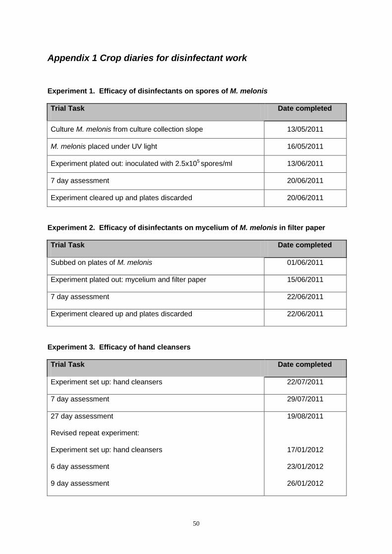
50
Appendix 1 Crop diaries for disinfectant work
Experiment 1. Efficacy of disinfectants on spores of M. melonis
Trial Task Date completed
Culture M. melonis from culture collection slope 13/05/2011
M. melonis placed under UV light 16/05/2011
Experiment plated out: inoculated with 2.5x10⁵ spores/ml 13/06/2011
7 day assessment 20/06/2011
Experiment cleared up and plates discarded 20/06/2011
Experiment 2. Efficacy of disinfectants on mycelium of M. melonis in filter paper
Trial Task Date completed
Subbed on plates of M. melonis 01/06/2011
Experiment plated out: mycelium and filter paper 15/06/2011
7 day assessment 22/06/2011
Experiment cleared up and plates discarded 22/06/2011
Experiment 3. Efficacy of hand cleansers
Trial Task Date completed
Experiment set up: hand cleansers 22/07/2011
7 day assessment 29/07/2011
27 day assessment 19/08/2011
Revised repeat experiment:
Experiment set up: hand cleansers 17/01/2012
6 day assessment 23/01/2012
9 day assessment 26/01/2012

51
Experiment 4. Effect of four surfaces on disinfectant efficacy
Trial Task Date completed
Surfaces cleaned and then sprayed with M. melonis spore
suspension 25/08/2011
Surfaces sprayed with relevant disinfectant after 30 minutes 25/08/2011
Surfaces swabbed after 30 minutes and streaked onto agar 25/08/2011
7 day assessment of agar plates 01/09/2011
14 day assessment of agar plates 15/09/2011
Experiment 5. Practical test- knife dip treatment to reduce disease transmission
Trial Task Date completed
Preliminary experiment 1 set up (to check decay of slices) 16/05/2011
3 day assessment of cucumbers in preliminary experiment 19/05/2011
7 day assessment of cucumbers in preliminary experiment 23/05/2011
10 day assessment of cucumbers in preliminary experiment 26/05/2011
14 day assessment of cucumbers in preliminary experiment 30/05/2011
Preliminary experiment 2 set up (to check decay of slices cut with
a contaminated knife) 22/07/2011
3 day assessment of cucumbers in preliminary experiment 25/07/2011
7 day assessment of cucumbers in preliminary experiment 29/07/2011
Experiment set up 19/08/2011
5 day assessment 24/08/2011
7 day assessment 26/08/2011
14 day assessment 02/08/2011

52
Appendix 2. In vitro product screening results Charts showing results for the isolates of M. melonis collected from the north and south
nurseries during the in vitro (agar plate) product screen.

53