proportional estimation of finger movements from high ...3a10... · background the dexterity of...
TRANSCRIPT

RESEARCH Open Access
Proportional estimation of fingermovements from high-density surfaceelectromyographyNicolò Celadon1, Strahinja Došen2, Iris Binder3, Paolo Ariano1 and Dario Farina2*
Abstract
Background: The importance to restore the hand function following an injury/disease of the nervous system led tothe development of novel rehabilitation interventions. Surface electromyography can be used to create a user-drivencontrol of a rehabilitation robot, in which the subject needs to engage actively, by using spared voluntary activation totrigger the assistance of the robot.
Methods: The study investigated methods for the selective estimation of individual finger movements fromhigh-density surface electromyographic signals (HD-sEMG) with minimal interference between movements ofother fingers. Regression was evaluated in online and offline control tests with nine healthy subjects (per test)using a linear discriminant analysis classifier (LDA), a common spatial patterns proportional estimator (CSP-PE),and a thresholding (THR) algorithm. In all tests, the subjects performed an isometric force tracking task guided by amoving visual marker indicating the contraction type (flexion/extension), desired activation level and the finger thatshould be moved. The outcome measures were mean square error (nMSE) between the reference and generatedtrajectories normalized to the peak-to-peak value of the reference, the classification accuracy (CA), the mean amplitudeof the false activations (MAFA) and, in the offline tests only, the Pearson correlation coefficient (PCORR).
Results: The offline tests demonstrated that, for the reduced number of electrodes (≤24), the CSP-PE outperformedthe LDA with higher precision of proportional estimation and less crosstalk between the movement classes (e.g., 8electrodes, median MAFA ~ 0.6 vs. 1.1 %, median nMSE ~ 4.3 vs. 5.5 %). The LDA and the CSP-PE performed similarlyin the online tests (median nMSE < 3.6 %, median MAFA < 0.7 %), but the CSP-PE provided a more stable performanceacross the tested conditions (less improvement between different sessions). Furthermore, THR, exploiting topographicalinformation about the single finger activity from HD-sEMG, provided in many cases a regression accuracy similar tothat of the pattern recognition techniques, but the performance was not consistent across subjects and fingers.
Conclusions: The CSP-PE is a method of choice for selective individual finger control with the limited numberof electrodes (<24), whereas for the higher resolution of the recording, either method (CPS-PA or LDA) can be usedwith a similar performance. Despite the abundance of detection points, the simple THR showed to be significantlyworse compared to both pattern recognition/regression methods. Nevertheless, THR is a simple method to apply (notraining), and it could still give satisfactory performance in some subjects and/or simpler scenarios (e.g., control ofselected fingers). These conclusions are important for guiding future developments towards the clinical application ofthe methods for individual finger control in rehabilitation robotics.
Keywords: Surface electromyography, High-Density electrodes, Machine learning, Human–machine interfaces,Rehabilitation robotics, Finger control, Hand rehabilitation
* Correspondence: [email protected] for Neurorehabilitation Systems, University Medical CenterGöttingen, Göttingen, GermanyFull list of author information is available at the end of the article
© 2016 The Author(s). Open Access This article is distributed under the terms of the Creative Commons Attribution 4.0International License (http://creativecommons.org/licenses/by/4.0/), which permits unrestricted use, distribution, andreproduction in any medium, provided you give appropriate credit to the original author(s) and the source, provide a link tothe Creative Commons license, and indicate if changes were made. The Creative Commons Public Domain Dedication waiver(http://creativecommons.org/publicdomain/zero/1.0/) applies to the data made available in this article, unless otherwise stated.
Celadon et al. Journal of NeuroEngineering and Rehabilitation (2016) 13:73 DOI 10.1186/s12984-016-0172-3

BackgroundThe dexterity of human hand is the result of complexmotor patterns that generate a coordinated response ofmultiple muscles placed intrinsically in the hand and inthe forearm. The control signals to move each finger ofthe hand are generated in separate regions of the pri-mary motor cortex (M1) [1], and are delivered to themuscles via the efferent pathways of the spinal cord andperipheral nervous system [2]. The neural commandselicit muscle electrical activity and a mechanical re-sponse. In recent years, it has been demonstrated thatthe intention to move the hand can be decoded usingpattern recognition applied to recorded and processedelectromyography (EMG) signals [3, 4]. This researchwas motivated by the importance to restore the handfunction following amputation or an injury/disease ofthe nervous system, such as stroke. Most often, the aimwas to detect less dexterous arm movements, such asthe wrist rotations (e.g., pronation/supination) [5–7]and/or overall grasping patterns (e.g., palmar, lateralgrip) [8, 9], whereas the classification and regression offinger movements has been less explored. Only recently,motivated by the development of modern dexterous
hand prostheses [10] and hand exoskeletons [11–13],researchers started exploring the classification and re-gression of finger movements with the aim of establish-ing methods for intuitive control of these sophisticatedsystems, mimicking the dexterity of the human hand.Most studies addressed the classification of individual
finger movements (see Table 1). In this context, the aimwas to predict the finger that moved but without pro-portional information (e.g., exerted force or position).Despite a good level of classification accuracy, generallyhigher than 90 % [14, 15], the discrete output of thesepattern recognition algorithms led to a limited clinicalapplicability. In addition to discrete classification, con-tinuous variables such as forces or positions can also beestimated from the EMG signals using regression.Regression algorithms have been applied under the mainassumption that the EMG signal is related to the forcegenerated by the muscle [16]. Since the force producedby muscles acting on a joint determines the position ofthe joint, the algorithms were trained to learn themapping from EMG to force and/or position. Previousstudies demonstrated that the hand kinematics can beestimated from surface EMG [6, 17–20]. For example, in
Table 1 Journal papers on classification and regression of finger movements using electromyography
Ref. Year Classifier Features Finger moves Subjects Window (ms) Electrodes Accuracy
[38] 2002 kNN DFT, AR F (T,I,M-R-L) ND (4) - 3 98 %
[39] 2009 ANN TD F-E (T,I,M,R,L) TR (1)ND (5)
200 32 90 % Classification
[18] 2010 kNN MAV F (T,I,M-R-L) TR (1) 250 16 86 %
[68] 2010 EPM TD F (T,I,M,L,R) ND (2) - 4 >97 %
[69] 2011 kNN MAV F (T,I,M,L,R) TR (5)ND (5)
250 8 79 % (TR)89 % (ND)
[70] 2012 SVM, kNN TD, AR F (T,I,M,L,R) ND (8) 250 2 90 %
[14] 2012 LDA, SVM, GMM TD, AR F-E (T,I,M-R-L) PS (12) 256 89 95 %
[15] 2013 LDA, SVM TD, AR F-E (T,I,M-R-L) ND (10)TR (6)
200 ms 1211
98 % (ND)90 % (TR)
[41] 2014 KRLS TD F (T,I,M,R,L) ND(40) 100–400 12 90 %
[42] 2015 LDA TD, AR F (T,I,M) ND(7) 250 5 (iEMG) 85 %
[71] 2006 ANN ENV F (T,I,M,L,R) TR(2) - 8 (JA) Norm RMS error 8–20 % Regression
[17] 2009 ANN RMS F-E (I) ND (15) 100 1 (JA) RMS error 0.085 rad −0.163 rad
[19] 2012 ANN WL F-E (T,I,M-R-L) ND (5) 32 4 Norm RMS error 7–14 %
[20] 2014 ANN, GP EMD F-E (T,I,M-R-L) ND (10) - 8 (JA) Mean CORR0.85 ± 0.07 (MCP)0.78 ± 0.06 (PIP)0.73 ± 0.04 (DIP)
[37] 2014 ANN ENV F-E (I,M,L,R) ND (8) - 14 – 16 (JA) R2 = 0.8
[22] 2014 RR RMS F (T,I,M,R,L) ND (10) 200 10 (FF) Norm RMS error 16 %
[23] 2014 RR ENV F-E (I,M-R-L) ND (10) - 10 (FF) Norm RMS error 10–20 %
ANN artificial neural network, AR autoregressive, CORR coefficient of correlation, DFT discrete Fourier transform, E extension, EMD electromechanical delay, ENVEnvelope, EPM entropy probabilistic model, F flexion, FF fingertip forces, GMM gaussian mixture model, GP nonparametric gaussian process, I index finger, JA jointangles, KRLS kernel regularized least squares, kNN K-nearest neighbors, L little finger, M middle finger, MAV mean absolute value, ND nondisabled, PS post-stroke,RA regression accuracy, R ring finger, RMS root mean square, RR ridge regression, SVM support vector machine, T thumb, TD time domain, TR transradial amputee,WL waveform length
Celadon et al. Journal of NeuroEngineering and Rehabilitation (2016) 13:73 Page 2 of 19

[20], the authors proposed an innovative control strategyusing a muscle activation model that parameterized theelectro-mechanical delays (EMD). The study demonstratedgood accuracy, estimating metacarpophalangeal (MCP),proximal interphalangeal (PIP) and the distal interphalan-geal (DIP) finger joint with the mean correlation coefficientof 0.85 ± 0.07, 0.78 ± 0.06 and 0.73 ± 0.04, respectively. Aspointed out in [21], the position control is effective only inthe absence of interaction with objects. Since the func-tional applications include direct contact through graspingand manipulation, a force control is likely a morerelevant solution. Recently, proportional control wasinvestigated in the context of prediction of individualfinger forces [22, 23], demonstrating that a non-linearincremental learning method could predict fingertipforces during flexion and extension with a correlationof ~0.9 between the estimated and measured forces.Recently, considerable attention has been devoted to
investigating rehabilitation interventions which canfacilitate the recovery of the sensory-motor functionsimpaired due to an injury/disease of the nervous system[24]. Numerous studies [25–28] demonstrated that themotor ability could be regained through a task-specificintensive practice. In this context, robotic rehabilitationis a promising method for the restoration and relearningof motor functions, since it can provide mass practice inwell-controlled conditions [29]. Moreover, sEMG can beused to estimate the intention of the subject and operatethe robot accordingly [12, 30–32]. This would create auser-driven control of a rehabilitation robot, in whichthe patient needs to provide a minimal activation totrigger and maintain the assistance. The benefit of thisapproach is that the subject is motivated to actively en-gage in therapy by recruiting his/her spared voluntarymotor control, instead of passively relying on the robotto guide the movement [33]. Furthermore, the EMGcontrol allows highly disabled patients who cannot pro-duce detectable forces and/or motions, but can generateresidual EMG, to participate early in the user-responsivetherapy. More specifically, the context for the presentwork is the rehabilitation of selective finger movementsusing a specialized hand rehabilitation robot (Amadeo,Tyromotion GmbH, AT). Rather than aiming at the simul-taneous control of multiple fingers to achieve functionalmovements (e.g., grasps) as required in prosthetics, theemphasis here is on the selective activation of individualfingers (i.e., one finger at a time) while reducing the simul-taneous false co-activations. The motivation for thisapproach is to promote relearning of the selective motorcontrol skills, which are heavily impaired in neurologicalpatients (e.g., stroke [34]).The present study advances the state of the art of
individual finger control by investigating proportionalestimation of fingertip forces during tasks that combined
different force profiles, force levels and rates of changeof force. Three different methods based on commonspatial filtering (CSP-PE), linear discriminant analysis(LDA) and simple thresholding (THR) were applied tolearn the mapping from High-Density sEMG (HD-sEMG) to finger activation; their performance werecompared in offline/online tests and across differentnumbers of electrodes. To the best of our knowledge,such a comprehensive set of conditions was not consid-ered before in the context of single finger classificationand regression (see Table 1).The focus of the present study was on the estimation of
the finger forces using HD-sEMG to record the electricalactivity of the extrinsic hand muscles during isometricfinger flexion and extension. The thumb, however, has aspecific anatomy and a functional behavior, with anadditional degree-of-freedom (opposition) fully controlledthrough the intrinsic muscles. Consequently, the presentstudy considered only the four long fingers. Nevertheless,the thumb activation could be estimated as well by placingadditional electrodes over the intrinsic muscles. This couldbe accomplished using conventional bipolar electrodes,and therefore, this was not relevant for the present study.HD-sEMG was selected since it provides a high
resolution of sensing points, capturing the high-fidelityspatial and temporal patterns of muscle activity and reveal-ing a topographical map of focal activation areas corre-sponding to individual muscles. The muscle heads movingindividual fingers are located close to each other, withinthe relatively small volume of the forearm [35]. Therefore,HD-sEMG was chosen to selectively capture the individualmuscle activity despite the significant spatial and temporaloverlap. HD-sEMG has been used before to characterizethe activity of the forearm muscles [36] [37]. However, thepresent study represents the first application where anHD-sEMG interface has been applied for individual fingermovement classification and regression, investigating acomprehensive set of conditions that were not consideredbefore. The high resolution of the recording (192 channels)was exploited to assess the robustness of the testedmethods with respect to the reduction in the number ofelectrodes, providing important perspectives regarding thepotential practical applications. Moreover, to the best ofour knowledge, there are no studies presenting an on-line protocol of finger control based on HD-sEMG,evaluating three control methods: one direct (THR),and two based on pattern recognition (LDA andCSP-PE). Furthermore, the two methods, CSP-PEand THR, have not been considered before for thecontrol of individual fingers. All the experimentswere conducted using a commercial rehabilitationrobot, mimicking closely a real clinical context. Inconclusion, this study presents some important in-sights for guiding future developments towards the
Celadon et al. Journal of NeuroEngineering and Rehabilitation (2016) 13:73 Page 3 of 19

clinical application of the methods for individualfinger control in rehabilitation robotics.
MethodsThe aim of the study was to test different methods, for adexterous finger control, estimating the intended level ofactivation of individual fingers (index, middle, ring andlittle) while minimizing the simultaneous unintended co-activations of other fingers during flexion and extensionmovements. Therefore, the task was to classify among 9classes (four fingers x two movements and rest, seeFig. 1), with the simultaneous regression of the fingeractivation level within the selected class. For all threemethods, the inputs were processed sEMG signals (featurevector), from the full set or subsets of electrodes, whilethe outputs were the estimated finger activation levelsproportional to the exerted force. The regression wasevaluated in the context of a linear discriminant analysis
(LDA) classifier [38], a multi-class proportional estimatorbased on common spatial pattern (CSP-PE) [8] and a non-pattern recognition method based on a thresholdscrossing (THR) [3], where the THR was applied only inthe online experiment. The LDA was selected as a widelyused method for movement classification and regression[39] (common benchmark). The CSP-PE was selectedunder the hypothesis that its mathematical propertieswould make the method especially effective in the contextof selective finger activation, reducing the crosstalkbetween the estimated movements. The THR was chosenbecause it is a simple method, easy to understand, imple-ment (no training) and apply even by a non-technicalpersonal, and thereby convenient for prospective practicalapplication in clinical settings. The hypothesis was thatthe THR could still perform well when used with the HD-sEMG interface due to its high resolution and ability toreveal focal areas of muscle activations. Summarizing, the
Fig. 1 Outline of the experiments. HD-sEMG recordings were processed (root mean square, data windowing with overlap) and used as inputsfor classification/regression to estimate the level of activation of individual fingers during flexion (F) and extension (E) movements. Two machine-learningapproaches for myoelectric control, a standard benchmark (LDA) and a recently presented novel method (CSP-PE), as well as direct control via simplethresholding (THR) were assessed in the context of selective finger control. Both offline and online tests were performed. In offline tests, isometric forcesof individual fingers were measured and predicted by applying the above-mentioned methods. During the online tests, the task for the subjects was totrack the reference trajectories specifying the desired individual finger activation levels assessed using EMG normalized to maximum voluntarycontraction. To this aim, the subjects controlled a visual marker, which was moving according to the finger activation levels predicted onlineusing the selected estimation method
Celadon et al. Journal of NeuroEngineering and Rehabilitation (2016) 13:73 Page 4 of 19

methods were selected to compare: i) machine learning(CSP-PE and LDA) vs. direct (THR) control; and ii) anovel multiclass algorithm (CSP-PE) vs. the golden stand-ard for the myoelectric control using pattern recognition(LDA). Their performance was compared in offline/onlinetests and across different subsets of electrodes, in order toassess how the nature of control (open vs. closed loop)and the resolution of the recording affect the perform-ance, respectively. In the offline tests, the full experimentalsession could be devoted to data collection, leading to acomprehensive dataset enabling a thorough assessment ofthe methods across many conditions. The online testsincluded both training and assessment within a singlesession, and therefore only the selected conditions couldbe evaluated. As pointed in [40], in online control the usercan exploit the visual feedback to adapt to the error map-ping provided by the algorithms (closed-loop control),resulting in different performance when compared tooffline estimation (open-loop control). Therefore, for anobjective assessment, it is recommended to test bothconditions. In offline tests, isometric forces of individualfingers were measured and offline predicted using theindicated estimation methods. During the online tests, thesubjects activated the fingers to accomplish an onlinecontrol task, while the selected method estimated the levelof activation for each individual finger in real-time. Thesubjects received online visual feedback about the desired
and estimated level of activation expressed as a percent ofmaximum voluntary contraction (MVC). In the onlinetests, the aim was to produce a control signal proportionalto the exerted force, but the fingertip forces were notmeasured and directly estimated. Instead, the referenceand estimated activation levels were calibrated accordingto the MVC of each subject, as measured by EMG (seesection Online Experiment). To collect the training data,the subjects were asked to perform isometric trackingtasks, as in the previous studies [21, 23, 41, 42]. In offlinetests, the reference trajectory was a predefined force pro-file expressed in Newtons, whereas in the online test, thereference profile was expressed as a percent of MVC.
Experimental setupThe four long fingers of the dominant arm were attachedto the finger slides of a robot specifically designed for thehand rehabilitation in stroke patients (Amadeo, Tyromo-tion GmbH, AT) as indicated in Fig. 2-b. Magnetic pieceswere embedded in the ergonomic finger pads that weresecured to each finger tip using medical tape. The padswere then positined on the respective magnetic connec-tion point of each finger slide. Magnetic force was enoughto keep the fingers in position during all experimentalconditions in healthy subjects (see Fig. 2-b). The slideswere driven individually using dedicated linear motors in-strumented with position and force sensors. The position
Fig. 2 Experimental setup. a The subject’s arm positioned in the finger -hand rehabilitation robot (Amadeo, Tyromotion GmbH, AT), the multichannelEMG amplifier on the desk next to the subject, HD-sEMG electrode placed on the forearm and flat cables connecting the electrode to the amplifier.b Hand connection by means of magnetic pieces embedded in the finger pads. c-d Approximated position of the High-Density 192-channelelectrode grid
Celadon et al. Journal of NeuroEngineering and Rehabilitation (2016) 13:73 Page 5 of 19

of the subject wrist and the slides was adjusted for eachsubject individually so that the PIP joints were flexed atapproximately 90° while the DIP joints of all fingers werefully extended (180°). Such an “orthogonal” setup allowedfor an optimal transmission of forces between the fingersand the robot guides and sensors. After setting up thehand configuration, the slides were kept in stationary posi-tions during the rest of the experimental session, measur-ing individual finger forces during isometric contractions.The sensor range for finger extension and flexion was ±20 N. The forces were recorded only during the offline ex-periment. The signals were sampled at 10 Hz, internallyby the robot controller, sent to the host computer viaTCP/IP and shown on a computer screen as feedback tothe subject.The EMG signals were recorded using a High-Density
192-channel electrode grid (ELSCH064NM 3–3, OTBioelettronica, 8x24 channels, 10 mm inter-electrode-distance, 8 × 24 cm) in a monopolar configurationplaced on the dominant arm. The forearm length wasmeasured in each subject using a measuring tape. Theelectrode array was positioned 6.4 ± 0.4 cm (25 % of theforearm length) from the elbow crease (Fig. 2c and d),covering 8 cm of the forearm longitudinally and 24 cmcircumferentially. The electrode configuration allowedacquiring the sEMG activity of distal and proximalmuscles such as the flexor digitorum superficialis andextrensor digitorium. The EMG signals were recordedusing a multichannel electrophysiological amplifier(EMG-USB2, OT Bioelettronica, IT) connected to thehost computer via a USB port. The gain was set to 500,the signals were band-pass filtered (eight order analogBessel filter, bandwidth 10-750 Hz), sampled at 2048 Hzand digitally converted (12 bit A/D converter, 5 Vdynamic range) with a resolution per least significant bitof 2.44 μV. The reference electrode was a ground stripplaced at the distal end of the forearm, just next to thewrist joint.
Regression methodsThe present study aimed at comparing three differentproportional controllers (LDA, CSP-PE and THR) asthey are applied to decode the activation of individualfingers from HD-sEMG patterns. The inputs were theprocessed EMG signals, and the regression methodsoutputted the estimated levels of finger activation pro-portional to the exerted force. The raw EMG datawere segmented into a series of overlapping data ana-lysis windows. The window length of 200 ms with50 % overlap was selected since it represents a goodtrade-off between classification accuracy (CA) andcontroller delay [43]. The Root Mean Square (RMS)was computed over the data window and used as aninput for the classification/regression, since it is a time
domain feature related to the force exerted by themuscle [43]. Therefore, a class decision was producedfor each data analysis window (every 100 ms), wherethe input for the regression was a vector of RMSvalues (one per electrode) computed over the 200-msdata window [44].The LDA classification represents one of the most
popular pattern recognition methods for myoelectriccontrol. In summary, it models the distribution of thedata within each class using a Gaussian distribution,where the means are estimated for each class individu-ally and the covariance matrix is computed over thepooled data (shared covariance). The classification isperformed by computing the class posterior probability(Bayes rule), which in the case of a shared covariance re-duces to evaluating the linear discriminant functionsseparating the classes [45]. As demonstrated by severalstudies, this simple and fast method performed similarlyto or even better than the other, more complex non-linear pattern recognition methods for time-domainfeatures [46, 47]. Since the LDA classifier has becomethe golden standard for the pattern recognition of EMGsignals, it was selected as common benchmark. In thisstudy once a class decision was taken, a proportionalcontrol value was extracted in a different way dependingon the experiment. In the offline test, this value wasextracted as an approximation of the recorded fingertipforces obtained by linear regression. In the online test,as the mean of the RMS values from a subset of chan-nels related to the specific class, scaled to a percentageof MVC for the detected movement class.Common Spatial Patterns (CSP) is a semi-supervised
algorithm to determine a filter whose output has max-imal and minimal variance when the multichannel inputdata come from the first and second class, respectively.Therefore, the filter maximizes the separation of the twoclasses based on the variance of the filter output signal.Commonly, CSP is used as a spatial filter for raw signalsof two distinct classes, but there are several options forthe extension to multi-class problems [48]. The methodhas been originally used in brain-computer interfacingas a spatial filter for data preprocessing [49–51].However, it has been recently adapted and tested forclassification and regression in myoelectric control ofprostheses, with promising results [8]. In this study, weapplied CSP as a proportional estimator (CSP-PE) in onevs. one configuration between all possible class pairs, aspresented in [8]. The CSP-PE has been selected sincethis is a novel method for myoelectric control withunique mathematical properties. Namely, the methodaims at maximizing the contrast between classes andthereby minimizing the false co-activations. This indi-cates that it could be especially effective in the contextof selective finger activation, which is the aim of the
Celadon et al. Journal of NeuroEngineering and Rehabilitation (2016) 13:73 Page 6 of 19

present work. In summary, the first step in the applicationof the CSP-PE was to determine the CSP filters for eachpair of classes. The output of each filter was thereforetuned to maximize the response for the input data (featurevector) coming from one class, and respond minimally tothe data from the other class. In the second step, the out-puts of all the pairwise-class filters were fused in post-processing to estimate the class corresponding to the inputdata. Therefore, the activation of the class was determinedby taking into account its relative contrast with respect toall the other classes. The selected class is the one that winsthe competition, and the estimated activation reflects theuncertainty of this decision. For example, if the class losesat least one competition, the activation will be penalizedleading to a small value. The raw outputs (activation levels)were scaled to yield a maximum value for 100 % of MVC.The method, including the original CSP formulation aswell as the novel steps for CSP-based classification andregression, are given in detail in [8].The THR is a simple method that involves direct con-
trol of each individual finger by identifying the focalareas of activity within the HD-sEMG interface. TheTHR was chosen because it is a simple method, easy tounderstand, implement (no training) and apply even bya non-technical personal, and thereby convenient forprospective practical application in clinical settings. Thefinger movement was detected if the activity at the selectedsubset of channels was above the predetermined threshold.THR was applied only during the online tests and a set ofrelevant channels was selected for each class (finger ×movement) by the experimenter based on the visual obser-vation of the EMG activity generated during the respectivemovements. The calibration trials were used to adjust thethresholds for each set of channels maximizing the correctclassification. A class was recognized when the mean of theRMS values over the associated channel set crossed thethreshold, and the RMS was higher than for the other clas-ses. The proportional control value was the mean of theRMS values for the set of channels, scaled to a percentageof the MVC of each movement.
Offline experimentNine healthy subjects (age between 26 and 41 years)participated in the experiments, which were approved bythe Ethical Committee of the University Medical CenterGöttingen (UMG). Before starting the tests, the subjectssigned an informed consent form. The experimentalsession lasted approximately 1.5 h.
Experimental protocolSubjects performed cyclical isometric contractionsactivating individual fingers in the direction of flexionand extension, as specified by the reference force profilepresented on the screen. The marker indicating the
currently generated force was displayed on the computerscreen as the feedback for the subject. The subjects wereinstructed to activate the fingers selectively by minimiz-ing simultaneous activation of other fingers. Before thebeginning of the training session, the subjects wereallowed to familiarize with the experimental setup andtasks. Twelve different tasks were carried out in randomorder with regard to the finger and each task wasrepeated 10 times in succession. The tasks combinedforce profiles (square with 50 % duty cycle and triangu-lar), force levels (33 and 66 % MVC) and two executionspeeds for each force profile (see Table 2), evaluatingthereby how well the methods estimated the forceduring gradual increase/decrease (triangles) and levelholding (squares) for different rates of change and peakforces. There were trials comprising only flexions, onlyextensions or both contraction types.
Signal processingForce signals were analyzed in order to extract segmentsof sEMG activity associated with specific muscularcontraction (flexion or extension), level of force and dur-ation. The processed EMG signals (windowing, RMS)had an effective sampling frequency of 10 Hz (one valueper 100 ms) synchronized with the force recordings,performed at the same sampling rate. For each task, theforce signal measured from the finger involved in thetask was used to identify the flexion/extension forcecycles, in order to leave out from the analysis the signalportion without the link to specific movements. A forcecycle was defined by detecting when the generated fingerforce crossed the predefined threshold, which was set to10 % of the maximum force exerted with that finger
Table 2 Tasks included in the offline assessment. The taskscombined square (S) and triangular (T) force profile, force levels(33 and 66 % MVC) of flexion (F) and extension (E), and twoexecution speed for each force profile
N Movement Profile % MVC Cycle Length (sec)
1 F S 33 6 Training
2 F S 66 12
3 F T 66 8
4 F T 66 4
5 E S 33 6
6 E S 66 12
7 E T 66 8
8 E T 66 4
9 F-E S 33 6 Testing
10 F-E S 66 12
11 F-E T 66 8
12 F-E T 66 4
Celadon et al. Journal of NeuroEngineering and Rehabilitation (2016) 13:73 Page 7 of 19
across all tasks, indicating the start of the contraction.When the force returned to a subthreshold level(<10 %), this denoted the end of the contraction.Occasional atypical contractions (outliers) were identi-fied on the basis of the force cycle length and the forcerange. A regression line was estimated from the mid-points of the plateaus in the case of square profile andfrom the vertices of the triangle profiles. The mean andthe standard deviation of the cycle length, and the MeanSquared Error (MSE) between the generated force dur-ing plateau and the regression line were calculated.Force cycles that were longer or shorter (Toutlier) than2.58 times the standard deviation from the meanduration (Tmean) (see Eq. 1) or for which the force error(eoutlier) was higher than 1.96 times the MSE from theregression line were considered as outliers (see Eq. 2)and excluded from the analysis. The confidence levels(1.96 × std and 2.58 × std) were chosen in order toenforce conservative outlier detection. The interval of±1.96 × std corresponds to the standard 95 % confidenceinterval. The detected outliers were also confirmed by avisual check.
Toutlier < 2:58 � stdð Þ−Tmean ∨ Toutlier
> 2:58 � stdð Þ þ Tmean ð1Þeoutlierj j > 1:96 �MSE ð2Þ
Data analysisWe investigated the performance of the algorithms withfull (192) and reduced number of channels (96, 48, 24,16, 12, 10, 8, 6 and 4) where the channels were selectedin the form of regular grids (see Fig. 6-e). The latter waschosen having in mind the future practical application ofthe methods, which should ideally, for the sake ofsimplicity, rely on the regular placement independent ofthe anatomy or the activity hot spots. The recorded datafor each subject was split into training and testing sets(approximately 70 and 30 % of the whole datasetrespectively, see Table 2): the tasks with only flexion orextension contractions were assigned to the training set(Table 2, Task 1–8), while the tasks with both flexionand extension were selected for the testing (Table 2,Task 9–12). The performance measures were commonfor online and offline tests and are described below (seesection Performance measures).
Online experimentNine healthy male subjects (age between 23 and 38 years)were recruited for the online algorithm evaluation. Eachsubject signed an informed consent before commencingthe experiment, which was approved by the EthicalCommittee of the University Medical Center Göttingen(UMG). The same test was performed twice on
consecutive days in order to evaluate the effect oftraining. The experimental session lasted maximum 2 h.
Training data collectionThe subjects were asked to perform sustained isometriccontractions activating selectively one of the four fingersin the direction of flexion or extension. For each move-ment, the experimenter selected groups of electrodes(from 4 to 6) in which the maximum activity wasobserved (Fig. 3-a) and measured the MVC as the max-imum of processed sEMG (RMS, 200 ms window, 50 %overlap) over the specific electrode subsets. After theelectrode subsets were chosen, the subjects were askedto reproduce the reference activation profile after receiv-ing a visual cue indicating the finger and the contractiontype (flexion or extension). Auditory “icons” (soundbeeps) were used in addition to the visual cues. Thereference profiles were trapezoidal (i.e., gradual increase,plateau, gradual decrease) with the plateaus of 30, 60and 90 % MVC of the respective finger (Fig. 3-b). Thecurrent muscle activation level generated by the subjectwas indicated by a cursor moving with the constantvelocity in the horizontal direction. The vertical coordin-ate of the cursor was equal to the mean RMS of theelectrode subset corresponding to the current class(finger × contraction type + rest). Each contraction lastedfor 7 s (2 s rise time, 3 s plateau, 2 s fall time) with 5-srest intervals in-between and it was repeated 3 times(30, 60 and 90 % MVC), resulting in 27 contractions in asingle run. If necessary, specific contractions could berepeated. For the training of both machine learningmethods the same data set was used and dynamicmovement phases were not excluded [52].
Online testAfter the training, a test with online control was per-formed. The subject controlled 4 visual markers (bluecircle in Fig. 3-c), each associated with the activationof one finger. The subjects now actively and propor-tionally controlled the vertical position of the controlmarker by increasing or decreasing the finger force inthe direction of flexion (marker moving downward)or extension (marker moving upward). The verticalposition of the marker (blue circle in Fig. 3-c) wasdetermined by a normalized output of the tested con-trol method (CSP-PE, LDA and THR), as explained insection Regression Methods. To quantify the controlperformance, the subjects performed online targettracking tasks, in which the subject tracked a movingreference marker (red circle in Fig. 3-c) by activatingappropriate finger at the appropriate level. The aimwas to maintain the smaller blue marker (Fig, 3-c),indicating the estimated activation level, within thelarger red circle (Fig. 3-c), representing the desired
Celadon et al. Journal of NeuroEngineering and Rehabilitation (2016) 13:73 Page 8 of 19

activation. Whenever the controlled marker was withinthe reference circle, the color of the reference would turninto green. The task comprised 16 individual finger activa-tions (4 for each finger) in which the reference marker
moved vertically from 0 % MCV to 80 % MVC position,rested on the 80 % MVC level, and then returned back tothe 0 % MVC value. The upward and downward move-ments of the reference marker lasted for 4 (slow ramp) or
Fig. 3 Three phases of the online experiment. a sEMG Root Mean Square (RMS) maps calculated over a 200-ms data window of (1 RMS sample).The RMS for each channel (white circle) was color coded as indicated by the color legend (μV), and the pixels between the channels were obtained byinterpolation. The figure shows one representative subject performing isometric sustained contractions of individual fingers in the direction of flexion orextension. The fingers produce characteristic and spatially localized, but partly overlapping, EMG responses. b Training data collection. The subjects wereasked to reproduce trapezoidal reference activation profiles (i.e., gradual increase, plateau, gradual decrease) with plateau at 30, 60 and 90 % of MVC ofthe respective finger. The red and blue lines depict the generated and reference activation levels from an example tracking trial at 60 %,respectively. c Online test where the subjects controlled 4 visual markers (blue circles), each associated with the activation of one finger, as indicatedby the horizontal axis. The vertical position of the marker was set by the output of the tested control method (LDA, CSP-PE and THR) computing theestimated finger activation level. The subjects therefore proportionally controlled the vertical position of the marker by increasing or decreasing thefinger force in the direction of flexion (marker moving downward) or extension (marker moving upward). The task for the subject was to activate thefingers, one at a time, tracking online the reference marker (red sphere) moving along vertical direction and representing the desired finger activation level
Celadon et al. Journal of NeuroEngineering and Rehabilitation (2016) 13:73 Page 9 of 19

2 (fast ramp) seconds, respectively, and the marker stayedat the plateau level for 3 s. Each finger was thereforeactivated two times in flexion and two times in extensionand for both contractions one cycle was slow and theother fast. The fingers (reference markers) were activatedin a random order. The test was repeated for each controlmethod, also in a random order to avoid that the learningacross methods influences the performance. The subjectswere blinded as to which algorithm was under test in eachsession.
Performance measuresThe estimated fingertip forces in the offline experimentand muscle activation levels in the online experimentwere low-pass filtered using a moving average filterapplied to 5 successive samples starting at each sampleof the original signal. The Pearson correlation coefficient(PCORR) between the estimated and reference forceswas computed to quantify the similarity in the signalshapes, and the mean square error normalized (nMSE)by the peak-to-peak value of reference force profile wascalculated to assess the difference in signal amplitudes[22]. As shown in Results section, the trends revealed bythe two outcome measures, nMSE and PCORR, wereequivalent over a comprehensive dataset collected andanalyzed in the offline experiments. Therefore, the qual-ity of the online tracking was evaluated by computingthe nMSE only. In both the experiments, this analysiswas performed for each finger during the segments ofthe reference trajectory in which that specific finger wassupposed to be active (target finger in the task). Thecorresponding segments were named active phases. Inorder to evaluate the amount of false finger activations(i.e., finger estimated to be active when it should havebeen relaxed), the mean amplitude of the false activa-tions (MAFA) outside of the respective active phaseswas calculated. The segments of the reference trajectoryin which the finger was not supposed to be activatedwere named silent phases. The CA was evaluated calcu-lating the overall success rate as the trace of the confu-sion matrix, divided by the total number of classifiedinstances. Finally, the selectivity and specificity of theclassifiers were calculated in one vs all configuration,where the median success rate of all the classes was usedto compare the performance of the two methods fordifferent electrode subsets.
Statistical evaluationThe Kolmogorov-Smirnov test determined that the datawere not normally distributed. Therefore, the data werestatistically analyzed using non-parametric tests. To assessthe statistically significant difference at the group level, theFriedman test was applied. If the Friedman test determinedthe difference, the conditions were compared pairwise
using the Wilcoxon signed-rank tests with Bonferroni cor-rection. A level of p < 0.05 was selected as the threshold forthe statistical significance. In the offline experiment, thefactors were number of channels (96, 48, 24, 16, 12, 10, 8,6 and 4), and method (CSP-PE and LDA). In the onlineexperiment, the factors were finger movements (IF, IE, MF,ME, RF, RE, LF, LE), and method (CSP-PE, LDA andTHR). Bartlett multiple-sample test for equal variances wasapplied to determine statistically significant difference indispersions within the conditions overall, followed byAnsari-Bradley two-sample test with Bonferroni correctionfor pairwise comparisons of the force variability betweenthe conditions.
ResultsOffline finger force predictionFigure 4 illustrates the finger force estimation in onerepresentative subject, when applying the LDA (Fig. 4-a)and the CSP-PE (Fig. 4-b) to a subset of 10 sEMGchannels selected as a regular grid (see Fig. 6-e). Duringactive phases (red line), both regression methods suc-cessfully tracked the force trajectories of different shapesand rates of change, i.e., triangles with faster/slowerslopes and squares with longer/shorter plateaus. In thisspecific configuration, with only 10 electrodes, the forceprofile for the index finger was estimated with the lowestaccuracy, and the estimation was better with the CSP-PE(nMSE = 6 %) than the LDA (nMSE = 7 %). During thesilent phases, the LDA generated false activations thatwere more frequent and with the higher amplitudescompared to the CSP-PE. For example, the LDA falselyestimated that the little finger was activated substantiallyand consistently throughout the active phase of theindex finger (Fig. 4-a).The recorded forces (gray lines) during the silent
phases showed that the subject exerted a small pressureon the force sensors also outside the active periods. Thiswas because the subject could not generate perfectly iso-lated activations of individual fingers, due to the naturalpassive coupling [53]. Group data are represented inFig. 5, depicting the fingertip forces (mean ± standarddeviation) recorded from all subjects across differentfinger tasks. The coupling between the fingers is evident,and the amount of force decreases for the fingers furtheraway from the activated one. Nevertheless, the force inthe active finger was several times higher from all theothers, and the difference was statistically significant,demonstrating the selective activation.Figure 6 shows the summary results for the quality of
estimation (median and interquartile range - IQR 25–75 %) using different number of electrodes. For bothmethods, the performance initially increased with moreelectrodes, i.e., the regression and classification becomemore accurate and the false activations less pronounced.
Celadon et al. Journal of NeuroEngineering and Rehabilitation (2016) 13:73 Page 10 of 19

Fig. 4 Representative results from one subject during offline experiment. Figure shows the individual finger force estimation when applying theLDA (a) and the CSP-PE (b) using a subset of 10 sEMG channels selected as a regular grid as input for the regression. The task for the subject wasto activate the fingers individually and selectively, one at a time. The dashed gray line is the recorded finger force. The continuous red line is theestimated force for the finger that was supposed to be active in the task (active phase), while the continuous black line is the estimated force forthe fingers that should have been relaxed (silent phase, false activation). The vertical lines delineate the active phases for different fingers. Thequality of tracking is similar for the two methods, in this specific example, but the CSP-PE generated less false activations. For example, comparethe false activation of the little finger in LDA vs. CSP-PE during the active phase of the index finger
Fig. 5 Fingertip forces (mean ± standard deviation) recorded during the offline experiment across different finger tasks. The subjects could notgenerate totally isolated movements of individual fingers, due to the natural passive coupling of the fingers
Celadon et al. Journal of NeuroEngineering and Rehabilitation (2016) 13:73 Page 11 of 19
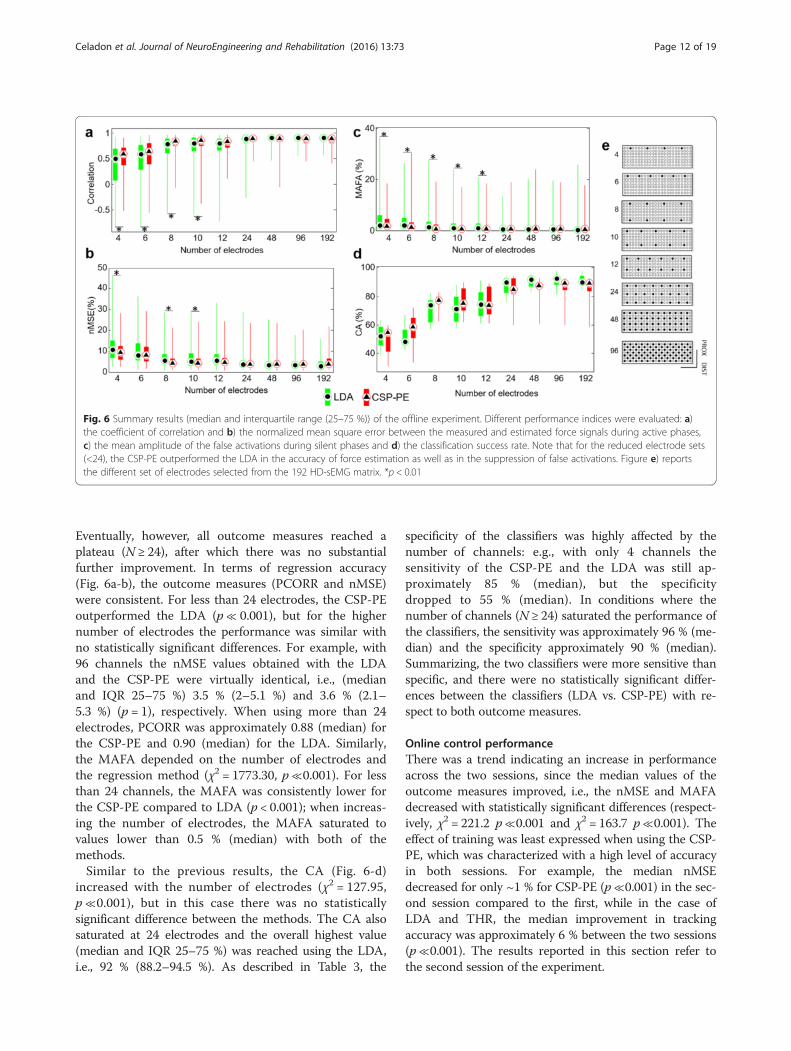
Eventually, however, all outcome measures reached aplateau (N ≥ 24), after which there was no substantialfurther improvement. In terms of regression accuracy(Fig. 6a-b), the outcome measures (PCORR and nMSE)were consistent. For less than 24 electrodes, the CSP-PEoutperformed the LDA (p≪ 0.001), but for the highernumber of electrodes the performance was similar withno statistically significant differences. For example, with96 channels the nMSE values obtained with the LDAand the CSP-PE were virtually identical, i.e., (medianand IQR 25–75 %) 3.5 % (2–5.1 %) and 3.6 % (2.1–5.3 %) (p = 1), respectively. When using more than 24electrodes, PCORR was approximately 0.88 (median) forthe CSP-PE and 0.90 (median) for the LDA. Similarly,the MAFA depended on the number of electrodes andthe regression method (χ2 = 1773.30, p≪0.001). For lessthan 24 channels, the MAFA was consistently lower forthe CSP-PE compared to LDA (p < 0.001); when increas-ing the number of electrodes, the MAFA saturated tovalues lower than 0.5 % (median) with both of themethods.Similar to the previous results, the CA (Fig. 6-d)
increased with the number of electrodes (χ2 = 127.95,p≪0.001), but in this case there was no statisticallysignificant difference between the methods. The CA alsosaturated at 24 electrodes and the overall highest value(median and IQR 25–75 %) was reached using the LDA,i.e., 92 % (88.2–94.5 %). As described in Table 3, the
specificity of the classifiers was highly affected by thenumber of channels: e.g., with only 4 channels thesensitivity of the CSP-PE and the LDA was still ap-proximately 85 % (median), but the specificitydropped to 55 % (median). In conditions where thenumber of channels (N ≥ 24) saturated the performance ofthe classifiers, the sensitivity was approximately 96 % (me-dian) and the specificity approximately 90 % (median).Summarizing, the two classifiers were more sensitive thanspecific, and there were no statistically significant differ-ences between the classifiers (LDA vs. CSP-PE) with re-spect to both outcome measures.
Online control performanceThere was a trend indicating an increase in performanceacross the two sessions, since the median values of theoutcome measures improved, i.e., the nMSE and MAFAdecreased with statistically significant differences (respect-ively, χ2 = 221.2 p≪0.001 and χ2 = 163.7 p≪0.001). Theeffect of training was least expressed when using the CSP-PE, which was characterized with a high level of accuracyin both sessions. For example, the median nMSEdecreased for only ~1 % for CSP-PE (p≪0.001) in the sec-ond session compared to the first, while in the case ofLDA and THR, the median improvement in trackingaccuracy was approximately 6 % between the two sessions(p≪0.001). The results reported in this section refer tothe second session of the experiment.
Fig. 6 Summary results (median and interquartile range (25–75 %)) of the offline experiment. Different performance indices were evaluated: a)the coefficient of correlation and b) the normalized mean square error between the measured and estimated force signals during active phases,c) the mean amplitude of the false activations during silent phases and d) the classification success rate. Note that for the reduced electrode sets(<24), the CSP-PE outperformed the LDA in the accuracy of force estimation as well as in the suppression of false activations. Figure e) reportsthe different set of electrodes selected from the 192 HD-sEMG matrix. *p < 0.01
Celadon et al. Journal of NeuroEngineering and Rehabilitation (2016) 13:73 Page 12 of 19

Figure 7 displays representative results from one sub-ject illustrating the quality of proportional tracking whenusing the three methods. Figure 7-a depicts the referenceand generated trajectories for each finger, including bothsilent and active sections, whereas Fig. 7-b zooms intothe active phases only. The subject successfully trackedthe reference trajectory during different segments (i.e.,slopes and constant levels at 80 % MVC) and rates ofchange of the trapezoidal activation profiles. In the caseof the CSP-PE and the LDA, the trajectory was wellreconstructed during both flexion and extension move-ments. The control using the LDA resulted in more falseactivations compared to the CSP-PE (e.g., see index andmiddle fingers in Fig. 7-a), but the difference between
the two methods was not so pronounced as in the offlineexperiments. The results for the thresholding (THR)were similar, as indicated by the outcome measurescomputed over the trial (Fig. 7-b), except for the ringfinger for which the subject was unable to control theextension. The subject effort to activate the ring exten-sion resulted in the false activation of the index flexion.The inability to control certain movements using theTHR was also observed in other subjects; in the secondsession, three out of nine subjects were not able to acti-vate a certain finger movement. This problem was notobserved with the two machine learning approaches.Group data are represented in Fig. 8-a, which shows
the summary results across control methods for the
Fig. 7 Representative results from one subject during online target task. The figure illustrates the quality of proportional tracking during theonline test when using the three methods (CSP-PE, LDA and THR). The task for the subject was to activate the fingers, one at a time, tracking areference trapezoidal trajectory. a The continuous colored and gray lines are the estimated and reference trajectories for each finger, includingboth silent (zero level) and active phases (trapezoids). b Active phases concatenated, with the indicated nMSE of estimation. With THR, falseactivations were more frequent and the ring extension could not be properly estimated
Table 3 Percentage of selectivity and specificity across different electrode subset extracted from the HD-sEMG matrix. The values arereported as median and interquartile range (25–75 %)
CH Sensitivity (%) Specificity (%)
CSP-PE Median andIQR (25–75 %)
LDA Median andIQR (25–75 %)
CSP-PE Median andIQR (25–75 %)
LDA Median andIQR (25–75 %)
4 85 (81–86) 84 (82–87) 55 (45–61) 54 (47–58)
6 87 (84–89) 84 (82–86) 59 (51–72) 52 (48–58)
8 93 (92–93) 92 (87–93) 81 (77–83) 75 (66–80)
10 92 (91–95) 91 (88–93) 79 (74–87) 74 (69–81)
12 92 (89–96) 92 (89–94) 81 (70–88) 77 (71–84)
24 95 (94–98) 97 (94–98) 86 (84–93) 91 (85–94)
48 96 (95–97) 97 (96–98) 90 (86–92) 92 (88–95)
96 96 (95–97) 97 (96–98) 90 (87–92) 92 (89–94)
192 96 (95–97) 97 (96–98) 90 (87–92) 91 (90–94)
Celadon et al. Journal of NeuroEngineering and Rehabilitation (2016) 13:73 Page 13 of 19

eight finger movements during the online experiment.The statistical test showed that the finger movementsand methods were significant factors as well as theirinteraction (χ2 = 43.52, p≪0.01). There were no statisti-cally significant differences between the methods for thesame as well as across fingers. However, there was atrend indicating that the THR was the weakest controlapproach, i.e., the median nMSE as well as its IQR rangewere consistently higher compared to that of the LDAand the CSP-PE.The results for the false activations complement those
for the accuracy of tracking. The median MAFA was thehighest for the THR consistently for all the movements,although there were no significant differences betweenthe methods. However, some movements were con-trolled with substantially lower MAFA, for example, theMAFA for the index and middle finger extension waslow with all three methods (e.g., compare to indexflexion). Yet, the post hoc comparison did not reveal anysignificant differences between the specific methodsacross the movement classes.
The experiment was performed twice on consecutivedays in order to evaluate the effect of training, and redu-cing the bias between subjects. Nevertheless, the lack ofexperience in EMG control may explain the overallvariability between subjects showed in Fig. 8-c. For goodcontrol, the subject needs to activate each finger consist-ently, generating reproducible patterns of muscle activa-tion that can be discriminated by the classifier/regressor.In addition, the patterns need to correspond to the onesgenerated during the training. The ability to reliablyexecute such patterns is likely subject dependent andtwo sessions were not enough for the consistency ofcontrol to improve and converge to a similar level acrosssubjects. The figure shows the results for the accuracy oftracking (nMSE) for each subject as a radar graph, whereeach spoke represents one of the target finger move-ments and the length of the spokes was normalized tothe maximum nMSE. This representation reveals partici-pants who reached high level of accuracy with: i) all thethree methods (s2 and s4), ii) the CSP-PE and the LDA(s1, s5, s6, s7 and s8), iii) the CSP-PE and the THR (s9)
Fig. 8 Summary results of the online experiment. a) Median and interquartile range (25–75 %) of the normalized mean square error betweentrapezoidal reference trajectory and control signals during active phases, and the mean amplitude of the false activations during silent phasesacross control methods for the eight finger movements. Overall, the online performance of the two machine learning methods was similar andbetter than the THR. b) Median and interquartile range (25–75 %) of the summary results shown in A. c Radar graph with the results for the accuracyof tracking (nMSE) for each subject. Each spoke represents one of the target finger movements and the length of the spokes is normalized to themaximum value of nMSE. The radar graphs for THR are characterized with spikes indicating larger errors, but the spikes are limited to some fingers(one, two typically), while for the other fingers the performance is actually comparable to that of the machine learning methods. *p < 0.01. The resultsof the distribution analysis are reported as circles ° p < 0.01
Celadon et al. Journal of NeuroEngineering and Rehabilitation (2016) 13:73 Page 14 of 19

and finally iv) the CSP-PE only (s3). It is reasonable toexpect that with practice, the online performance of thealgorithms can be further improved, producing a morecoherent behavior across participants [54]. The initialuncertainty and variability of control and substantialimprovement due to training are characteristic formyoelectric control in general [55]. Regarding THR, formost subjects and movements the performance wassimilar to that of the CSP-PE and the LDA. However, asindicated by the few pronounced spikes in the radarplot, there were one or two specific movements in somesubjects (see s1, s5, s7, and s8) that were substantiallymore difficult to control using THR. The performancedropped significantly in these few cases, decreasing theoverall average accuracy and increasing the overallvariability.In the summary results shown in Fig. 8-b, the nMSE
obtained with the CSP-PE, LDA and THR (median andIQR 25–75 %) was 3.6 % (2.5–5 %), 2.5 % (1.5–3.9 %)and 3.8 % (1.8–10.8 %), respectively. There was no statis-tically significant difference between the CSP-PE and theLDA, whereas the two machine learning methods weresignificantly different compared to the THR (p < 0.01).Furthermore, the CSP-PE and LDA exhibited similardispersion, which was significantly lower compared toTHR. The same trend holds for the MAFA. As shownin Fig. 8-b, the MAFA of the CSP-PE, LDA and THRwas 0.7 % (0.2–2.2 %), 0.6 % (0.2–2.6) and 2.2 %(0.24–9.9 %), respectively. Both the CSP-PE and theLDA performed similarly, and they differed signifi-cantly with respect to THR (p < 0.01) both in medianand dispersions.The general similarity in the performance of the CSP-
PE and the LDA was also confirmed by the CA (medianand IQR 25–75 %), which were 91 % (87.7–91.7 %) forthe CSP-PE and 90.3 % (89.4–93.25 %) for the LDA withno statistically significant differences.
DiscussionThe experiments demonstrated that the finger activationcould be successfully decoded for different target activa-tion profiles. Overall, the experiments demonstrated amore stable performance of the CSP-PE across thetested conditions. The CSP-PE exhibited less improve-ment between different sessions and outperformed theTHR in online control and the LDA in offline tests.Furthermore, the study showed that a simple method,exploiting the topographical information about the indi-vidual finger activity from the HD-sEMG, provided inmost cases regression accuracy similar to the patternrecognition techniques. However, THR lacked robust-ness in the sense that performance was not consistentacross subjects and fingers.
Offline and online proportional controlIn the offline experiment, the performance of the CSP-PE and the LDA increased with the number of channels,saturating to a stable level for more than 24 electrodes.Importantly, for less than 24 electrodes, the CSP-PEoutperformed the LDA consistently in all outcome mea-sures, except CA, yielding more accurate force estimatesin active phases and better suppression of false activa-tions in the silent phases. This confirmed the hypothesisthat the mathematical properties of the CSP-PE, asdescribed in [8], make this method especially effective inthe context of selective finger activation. Increasing thenumber of channels evened out the performance of thetwo methods with respect to the quality of tracking inthe active phase, but did not change the superiority ofthe CSP-PE in filtering out the activations during thesilent phases.The results of the online experiments were in accord-
ance with the insights from the offline tests. The trackingaccuracy was similar with both the CSP-PE and the LDA,with the median nMSE of approximately 3.6 and 2.6 % inonline experiment, and 3.6 and 3.5 % respectively in off-line experiment (48 electrodes). As explained in Methods,the online experiment resulted in more than 24 electrodesselected by the experimenter. According to the trendsrevealed offline (Fig. 6), this number of channels wasenough to even out the performances of the two algo-rithms. In addition, during online tests the subjects coulduse visual feedback to adapt the activations during thetrial, as demonstrated in [40].The CSP-PE exhibited more stable accuracy across the
tested conditions. The performance was good from thebeginning and similar across the two successive sessions,providing the least improvement in outcome measures.Furthermore, the CSP-PE produced better performancefor lower number of channels (Fig. 6) compared to LDA.This means that the CSP-PE might be less sensitive tothe subjective factors than the LDA, which producedsimilar accuracy but only for the high number of chan-nels and after a session of practice.
Electrode reductionIn the offline experiment, the electrodes were selected as aregular grid, without any relation to the specific finger ac-tivation patterns. Regular electrode grids are convenientfor practical implementation and allow simple mounting,since they can be realized as extensible uniform braceletsthat are simply wrapped around the forearm [56]. Futureinvestigations will further address the minimization of thenumber of channels, determining acceptable electrodelocations and optimizing electrode-recording configura-tions, using the established methods for feature reduction[57–59]. Importantly, the results of the present study(offline tests) demonstrate that the number of electrodes
Celadon et al. Journal of NeuroEngineering and Rehabilitation (2016) 13:73 Page 15 of 19

can be decreased substantially (from 192 to 10) withoutsignificantly compromising the performance. This is anoptimistic result implying that the proposed methodscould be translated into the clinical context using multi-channel EMG braces comprising practical dry electrodes.
Thresholding for online controlThe online experiment demonstrated the feasibility ofsimple thresholding for proportional control of individ-ual fingers, albeit with some limitations. Exploiting adense array of detection points provided by HD-sEMGinterface, distinct areas of focal sEMG activity could beidentified for each finger [35], with an overlap in somecases due to anatomical constraints and crosstalk [37].When the areas overlapped, the experimenter did notselect the electrodes from the intersection. The THRmethod has a low computational cost and there is notraining; after the electrode selection, the experimenterprovided only a fast calibration of the thresholds associ-ated with each finger movement. However, the THRmethod also exhibited some drawbacks. In the secondsession, three out of nine subjects were not able to acti-vate a certain finger movement, due to a significantoverlap, i.e., the activity of finger involved in the taskprojected strongly to neighboring areas triggeringthereby other fingers. The THR was also less successfulin suppressing the false activations. Nevertheless, whenthe subjects were able to control the finger, the trackingaccuracy was actually comparable to the performancesof the two machine learning approaches. Therefore, theTHR is not universally applicable. However, it can beused successfully in some subjects or with a reducednumber of movement classes, controlling only thosefingers (or finger groups) characterized with distinct andseparate areas of activity.
Application for rehabilitationThe aforementioned conclusions could provide usefulguidelines for the translation of the tested methods tothe clinical context, targeting dexterous control of handrehabilitation robots. For example, stroke patients haveimpaired motor functions characterized with patho-logical synergies. At the hand level, this is expressed as adifficulty in selectively activating individual fingers [34].The methods developed in the present study could be ap-plied to implement a user-driven control of a rehabilitationsystem. For example, the activation signals estimated foreach finger, as demonstrated in the online tests, could beused as the control signals in the isometric mode ofAmadeo, i.e., to implement the functionality of the forcejoystick, as when playing simple video games by producingappropriate isometric forces. More generally, the estimatedsignals could be used to trigger and/or modulate the move-ment of the Amadeo finger motors (dynamic control) [60].
Since the Amadeo system offers a set of therapeutic indi-vidual finger exercises, the future perspective is to integratethe individual finger myocontrol developed in the presentstudy with these motivational tasks, creating thereby an in-novative, engaging and user-responsive training program.Considering the future clinical application and followingthe results of the offline analysis, the control could beimplemented with substantially less electrodes, whichwould allow using a practical EMG bracelet such asMyoband [56] combined with the CSP-PE. Alternatively,even a full HD-sEMG electrode system could be usedpractically if integrated in a textile garment (e.g., [37]). Im-portantly, for online control of Amadeo, the arm/hand willbe supported exactly as in the present study, and it istherefore not required to train/test the algorithms with thearm in different positions, as usual in prosthetics toincrease the robustness of the classification/regressionacross arm postures [61, 62].Another potential application is the extension of the
methods to the control of dexterous hand prostheses. Atthe current level, the methods tested in the present studycould not be directly translated for general prostheticapplication, as the fingers are controlled sequentially, oneby one. However, such a controller could be used tosupport some specific functions (e.g., typing on a keyboard)exploiting the individual finger actuation available in themodern prostheses (e.g. i-Limb [63]). For example, key-board typing could be implemented through classification(on-off), or the estimated force could be mapped to thefinger velocity, allowing proportional control of the speedof finger flexion/extension (instead of force). The latter isnot essential for typing, but it could allow the subject totype faster as he/she becomes more trained and skilled incontrol. More importantly, the present study demonstratesthe feasibility of achieving fine and selective control ofindividual fingers, across a comprehensive number of tasks(force profiles) and with a reduced set of electrodes. Never-theless, the control of a prosthesis requires a more naturaland functional approach, and the future work will be tostudy the simultaneous regression of multiple fingers(including the thumb) using HD-sEMG setup.The translation into the clinical context faces a number
of challenges, which will be addressed in future work. Forexample, as explained above, stroke patients can havesignificant reduction in muscle forces and impairedcoordinative control. Due to weak activity and patho-logical synergies [64], the activation patterns for the indi-vidual finger movements are likely to be significantly lessdiscriminative and thereby more difficult to classify andestimate. In this context, nevertheless, an adaptive trainingcan be envisioned in which the patient and the systemcoadapt [65] and evolve through the process of recovery.Initially, the system can estimate a subset of movements,limited to those that can be well discriminated. This can
Celadon et al. Journal of NeuroEngineering and Rehabilitation (2016) 13:73 Page 16 of 19

be used to start the training, promoting the initialrecovery, and when the activity maps become betterdifferentiated, new movements can be included.Finally, as already pointed out in myoelectric control for
prosthetics [66], it would be of interest for clinical applica-tions to minimize the time and effort (subjects and staff)need for the training. Ideally, the training/calibrationshould be short and easy and without the need for fre-quent retraining. In the present study, we demonstratedthat a reasonable quality of control can be achieved withan in-session training. The training is especially simpleand easy to understand in the case of THR, since itreduces to selecting the channels with strong activation ina color map and then visually adjusting the thresholds(average time 10 min in THR vs. 20 min for LDA/CSP-PE). Therefore, the THR might also allow for an easyretraining across sessions. However, in the ideal case, theretraining would not be necessary. This was not tested inthe present study but it is certainly an important futuregoal. The robustness of the methods in terms of retrainingcould be assessed by testing the control across sessionsusing the same, previously collected data (no retraining).In this context, the use of HD-sEMG interface might beparticularly beneficial as an increased resolution contrib-utes with redundant information, and this can be used toincrease robustness. An illustrative demonstration is pro-vided in [67] by extracting features reducing the sensitivityto electrodes shifts.
ConclusionThe present study investigated methods for selectiveestimation of individual finger movements, motivatedby the final aim of implementing an online protocolfor dexterous finger control using a hand rehabilita-tion robot. We detected the intention to move a sin-gle finger from electromyographic signals providingproportional control while reducing the simultaneous co-activations of other fingers during both offline and onlineexperiments. The insights from the present study can beused to guide the implementation of a practical myoelec-tric system for dexterous control in hand rehabilitation ro-botics and prosthetics. More specifically, the resultsdemonstrated that despite the abundance of detectionpoints in HD-sEMG, a simple method based on threshold-ing (THR) exhibited serious drawbacks, and that thereforethe pattern recognition is still the method of choice forrobust practical implementations. Next, provided that therecording is above a certain resolution (>24 channels),either of the pattern recognition methods (CSP-PE andLDA) can be selected to implement the control. In thiscase, information redundancy compensates for thefavorable mathematical properties of the CSP-PE vs. LDA.Finally, if only a reduced number of electrodes is available(≤12), the CPS-PE is the recommended approach.
AbbreviationsCA, classification accuracy; CSP-PE, common spatial pattern proportional estimator;DIP, distal interphalangeal; HD-sEMG, high density surface electromyography; IE,index extension; IF, index flexion; IQR, interquartile range; LDA, linear discriminantanalysis; LE, little extension; LF, little flexion; MAFA, mean amplitude of the false ac-tivations; MCP, metacarpophalangeal; ME, middle extension; MF, middle flexion;MVC, maximum voluntary contraction; nMSE, normalized mean square error;PCORR, Pearson correlation coefficient; PIP, proximal interphalangeal; RE, ring ex-tension; RF, ring flexion; RMS, root mean square; THR, thresholding
AcknowledgementsThe authors would like to acknowledge Andrei Ninu for his help in designingexperiments, preparing the experimental setup and collecting the data.
FundingThis research was supported by the European Commission under theMYOSENS (FP7-PEOPLE-2011-IAPP-286208) project and by the GermanMinistry for Education and Research (BMBF) via the Bernstein FocusNeurotechnology (BFNT) Gottingen under Grant 01GQ0810.
Availability of data and materialsData will not be shared at this stage, because of a large volume of data andonline storage restrictions.
Authors’ contributionsDF, SD, IB, PA and NC conceived and designed the experiments. NC and SDimplemented the protocol and conducted the experiments. NC analyzed thedata. DF, SD, PA and NC participated in writing the manuscript. All authorsread and approved the final manuscript.
Competing interestsThe authors declare that they have no competing interests.
Consent for publicationNot applicable.
Ethics approval and consent to participateEach subject signed an informed consent form before commencing theexperiment, which was approved by the Ethical Committee of the UniversityMedical Center Göttingen (document: 32/2/16).
Author details1Center for Sustainable Futures@PoliTo, Fondazione Istituto Italiano diTecnologia, Torino, Italy. 2Institute for Neurorehabilitation Systems, UniversityMedical Center Göttingen, Göttingen, Germany. 3Tyromotion GmbH, Graz,Austria.
Received: 6 November 2015 Accepted: 12 July 2016
References1. Schieber MH, Poliakov AV. Partial inactivation of the primary motor cortex
hand area: effects on individuated finger movements. J Neurosci. 1998;18:9038–54.
2. Oby ER, Ethier C, Miller LE. Movement representation in the primary motorcortex and its contribution to generalizable EMG predictions.J Neurophysiol. 2013;109:666–78.
3. Asghari Oskoei M, Hu H. Myoelectric control systems—a survey biomed.Signal Process Control. 2007;2:275–94.
4. Fougner A, Stavdahl O, Kyberd PJ, Losier YG, Parker PA. Control of upperlimb prostheses: terminology and proportional myoelectric control-a review.IEEE Trans Neural Syst Rehabil Eng. 2012;20:663–77.
5. Nielsen JLG, Holmgaard S, Jiang N, Englehart KB, Farina D, Parker PA.Simultaneous and proportional force estimation for multifunctionmyoelectric prostheses using mirrored bilateral training. IEEE Trans BiomedEng. 2011;58(3 PART 1):681–8.
6. Muceli S, Member S, Farina D, Member S. Simultaneous and proportionalestimation of hand kinematics from EMG during mirrored movements atmultiple degrees-of-freedom. IEEE Trans Neural Syst Rehabil Eng. 2012;20:371–8.
Celadon et al. Journal of NeuroEngineering and Rehabilitation (2016) 13:73 Page 17 of 19

7. Jiang N, Englehart KB, Parker PA. Extracting simultaneous and proportionalneural control information for multiple-DOF prostheses from the surfaceelectromyographic signal. IEEE Trans Biomed Eng. 2009;56:1070–80.
8. Amsuess S, Gobel P, Graimann B, Farina D. A multi-class proportionalmyocontrol algorithm for upper limb prosthesis control: validation in real-life scenarios on amputees. IEEE Trans neural Syst Rehabil Eng. 2014;4320(c):1–11.
9. Cipriani C, Zaccone F, Micera S, Carrozza MC. On the shared control of anEMG-controlled prosthetic hand: analysis of user–prosthesis interaction. IEEETrans Robot. 2008;24:170–84.
10. Belter JT, Segil JL, Dollar AM, Weir RF. Mechanical design and performancespecifications of anthropomorphic prosthetic hands: A review. J Rehabil ResDev. 2013;50:599.
11. Chiri A, Vitiello N, Giovacchini F, Roccella S, Vecchi F, Carrozza MC.Mechatronic design and characterization of the index finger module of ahand exoskeleton for post-stroke rehabilitation. IEEE/ASME TransMechatronics. 2012;17:884–94.
12. Tong KY, Member S, Hu XL, Fung KL, Wei XJ, Rong W, Susanto EA. An EMG-driven exoskeleton hand robotic training device on chronic stroke subjectstask training system for stroke rehabilitation. IEEE Int Conf Rehabil Robot.2011;2011:5975340.
13. Wege A, Kondak K, Hommel G. Force control strategy for a handexoskeleton based on sliding mode position control. IEEE/RSJ Int Conf IntellRobot Syst. 2006:4615–4620.
14. Zhang X, Zhou P. High-density myoelectric pattern recognition. IEEE TransBiomed Eng. 2012;59:1649–57.
15. Al-Timemy AH, Bugmann G, Escudero J, Outram N. Classification of fingermovements for the dexterous hand prosthesis control with surfaceelectromyography. IEEE J Biomed Heal Informatics. 2013;17:608–18.
16. Merletti R, Parker PA. Electromyography: Physiology, Engineering, and Non-Invasive Applications. 2004.
17. Shrirao N a, Reddy NP, Kosuri DR. Neural network committees for fingerjoint angle estimation from surface EMG signals. Biomed Eng Online.2009;8:2.
18. Antfolk C, Cipriani C, Controzzi M, Carrozza MC, Sebelius F. Using EMG forreal-time prediction of joint angles to control a prosthetic hand equippedwith a sensory feedback system. J Med Biol Eng. 2010, 30:399–406.
19. Hioki M, Kawasaki H. Estimation of Finger Joint Angles from sEMG Using aNeural. ISRN Rehabil. 2012, 2012.
20. Ngeo JG, Tamei T, Shibata T. Continuous and simultaneous estimation offinger kinematics using inputs from an EMG-to-muscle activation model.J Neuroeng Rehabil. 2014;11:1–14.
21. Castellini C, Koiva R. Using surface electromyography to predict single fingerforces. 2012 4th IEEE RAS EMBS Int. Conf. Biomed. Robot. Biomechatronics.2012:1266–1272.
22. Gijsberts A, Bohra R, Sierra González D, Werner A, Nowak M, Caputo B, RoaM a, Castellini C. Stable myoelectric control of a hand prosthesis using non-linear incremental learning. Front Neurorobot. 2014;8(February):8.
23. Ravindra V, Castellini C. A comparative analysis of three Non-invasivehuman-machine interfaces for the disabled. Front Neurorobot. 2014;8(October):1–10.
24. Taub E, Uswatte G, Elbert T. New treatments in neurorehabilitation foundedon basic research. Nat Rev Neurosci. 2002;3:228–36.
25. Biitefisch C, Hummelsheim H, Denzler P. Repetitive training of isolatedmovements improves the outcome of motor rehabilitation of the centrallyparetic hand. J Neurol Sci. 1995;130:59–68.
26. Langhorne P, Bernhardt J, Kwakkel G. Stroke rehabilitation. Lancet. 2011;377:1693–702.
27. Johansson BB. Current trends in stroke rehabilitation. A review with focuson brain plasticity. Acta Neurol Scand. 2011;123:147–59.
28. Merians AS, Poizner H, Boian R, Burdea G, Adamovich S. Sensorimotortraining in a virtual reality environment: does it improve functional recoverypoststroke? Neurorehabil. Neural Repair. 2006;20:252–67.
29. Norouzi-Gheidari N, Archambault PS, Fung J. Effects of robot-assistedtherapy on stroke rehabilitation in upper limbs: systematic review andmeta-analysis of the literature. J Rehabil Res Dev. 2012;49:479–96.
30. Colombo R, Pisano F, Micera S, Mazzone A, Delconte C, Carrozza MC, Dario P,Minuco G. Robotic techniques for upper limb evaluation and rehabilitation ofstroke patients. IEEE Trans Neural Syst Rehabil Eng. 2005;13:311–24.
31. Mulas M, Folgheraiter M, Gini G. An EMG-Controlled Exoskeleton for HandRehabilitation. 9th Int. Conf. Rehabil. Robot. 2005. ICORR 2005. 2005:371–374.
32. Dipietro L, Ferraro M, Palazzolo JJ, Krebs HI, Volpe BT, Hogan N. Customizedinteractive robotic treatment for stroke: EMG-triggered therapy. IEEE TransNeural Syst Rehabil Eng. 2005;13:325–34.
33. Prange GB, Jannink MJ a, Groothuis-Oudshoorn CGM, Hermens HJ, IJzermanMJ. Systematic review of the effect of robot-aided therapy on recovery ofthe hemiparetic arm after stroke. J Rehabil Res Dev. 2006;43:171.
34. Schieber MH, Lang CE, Reilly KT, McNulty P, Sirigu A. Selective activation ofhuman finger muscles after stroke or amputation. Adv Exp Med Biol. 2009;629:559–75.
35. Leijnse JN a L, Campbell-Kyureghyan NH, Spektor D, Quesada PM.Assessment of individual finger muscle activity in the extensor digitorumcommunis by surface EMG. J Neurophysiol. 2008;100:3225–35.
36. Gallina A, Botter A. Spatial localization of electromyographic amplitudedistributions associated to the activation of dorsal forearm muscles. FrontPhysiol. 2013;4(December):367.
37. Gazzoni M, Celadon N, Mastrapasqua D, Paleari M, Margaria V, Ariano P.Quantifying forearm muscle activity during wrist and finger movements bymeans of multi-channel electromyography. PLoS One. 2014;9:e109943.
38. Peleg D, Braiman E, Yom-Tov E, Inbar GF. Classification of finger activation for usein a robotic prosthesis arm. IEEE Trans Neural Syst Rehabil Eng. 2002;10:290–3.
39. Tenore FVG, Ramos A, Fahmy A, Acharya S, Etienne-Cummings R, ThakorNV. Decoding of individuated finger movements using surfaceelectromyography. IEEE Trans Biomed Eng. 2009;56:1427–34.
40. Jiang N, Rehbaum H, Vujaklija I, Graimann B, Farina D. Intuitive, online,simultaneous, and proportional myoelectric control over two degrees-of-freedomin upper limb amputees. IEEE Trans Neural Syst Rehabil Eng. 2014;22:501–10.
41. Gijsberts A, Atzori M, Castellini C, Muller H, Caputo B. Movement error ratefor evaluation of machine learning methods for sEMG-based handmovement classification. IEEE Trans Neural Syst Rehabil Eng. 2014;22:735–44.
42. Birdwell JA, Hargrove LJ, Weir RF, Kuiken T a. Extrinsic finger and thumbmuscles command a virtual hand to allow individual finger and graspcontrol. IEEE Trans Biomed Eng. 2015;62:218–26.
43. Englehart K, Hudgins B. A robust, real-time control scheme for multifunctionmyoelectric control. IEEE Trans Biomed Eng. 2003;50:848–54.
44. Englehart K, Hudgins B, Parker PA, Member S. A wavelet-based continuousclassification scheme for multifunction myoelectric control. IEEE TransBiomed Eng. 2001;48:302–11.
45. Fukunaga K. Introduction to Statistical Pattern Recognition. 2013.46. Scheme EJ, Member S, Englehart KB, Member S, Hudgins BS, Member S.
Selective classification for improved robustness of myoelectric control undernonideal conditions. IEEE Trans Biomed Eng. 2011;58:1698–705.
47. Parker P, Englehart K, Hudgins B. Myoelectric signal processing for control ofpowered limb prostheses. J Electromyogr Kinesiol. 2006;16:541–8.
48. Hahne JM, Graimann B, Muller KR. Spatial filtering for robust myoelectriccontrol. IEEE Trans Biomed Eng. 2012;59:1436–43.
49. Tomioka R, Lemm S. Filters for Robust EEG. IEEE Signal Process Mag. 2008;2008:41–56.
50. Ramoser H, Müller-Gerking J, Pfurtscheller G. Optimal spatial filtering ofsingle trial EEG during imagined hand movement. IEEE Trans Rehabil Eng.2000;8:441–6.
51. Blankertz B, Dornhege G, Krauledat M, Müller K-R, Curio G. The non-invasiveBerlin Brain-Computer Interface: fast acquisition of effective performance inuntrained subjects. Neuroimage. 2007;37:539–50.
52. Lorrain T, Jiang N, Farina D. Influence of the training set on the accuracy ofsurface EMG classification in dynamic contractions for the control ofmultifunction prostheses. J Neuroeng Rehabil. 2011;8:25.
53. Lang CE. Human finger independence: limitations due to passivemechanical coupling versus active neuromuscular control. J Neurophysiol.2004;92:2802–10.
54. Jiang N, Vujaklija I, Rehbaum H, Member S, Graimann B. Is accuratemapping of EMG signals on kinematics needed for precise onlinemyoelectric control ? IEEE Trans Neural Syst Rehabil Eng. 2014;22:549–58.
55. Radhakrishnan SM, Baker SN, Jackson A. Learning a novel myoelectric-controlled interface task. J Neurophysiol. 2008;100:2397–408.
56. Myo armband. [https://www.thalmic.com/]. Accessed 12 May 2016.57. Geng Y, Zhang X, Zhang Y-T, Li G. A novel channel selection method for
multiple motion classification using high-density electromyography. BiomedEng Online. 2014;13:102.
58. Huang H, Zhou P, Li G, Kuiken T. Spatial filtering improves EMG classificationaccuracy following targeted muscle reinnervation. Ann Biomed Eng. 2009;37:1849–57.
Celadon et al. Journal of NeuroEngineering and Rehabilitation (2016) 13:73 Page 18 of 19

59. Hwang HJ, Hahne JM, Müller KR. Channel selection for simultaneousmyoelectric prosthesis control. 2014 Int. Winter Work. Brain-ComputerInterface. BCI. 2014;2014:056008.
60. “Amadeo solution”, Tyromotion GmbH [http://tyromotion.com/wp-content/uploads/2013/04/HartwigM-2014-The-Tyrosolution-Concept._EN.pdf].Accessed 12 May 2016.
61. Geng Y, Zhou P, Li G. Toward attenuating the impact of arm positions onelectromyography pattern-recognition based motion classification intransradial amputees. J Neuroeng Rehabil. 2012;9(74):1–11.
62. Fougner A, Scheme E, Member S, Chan ADC, Member S, Englehart K,Member S, Stavdahl Ø. Resolving the limb position effect in myoelectricpattern recognition. IEEE Trans Neural Syst Rehabil Eng. 2011;19:644–51.
63. “i-Limb Ultra: User Manual” Touch Bionics Ltd. [http://www.touchbionics.com/sites/default/files/files/i-limb ultra revolution user manual September2014.pdf]. Accessed 12 May 2016.
64. Li X, Liu J, Li S, Wang Y. Examination of hand muscle activation and motorunit indices derived from surface EMG in chronic stroke. IEEE Trans BiomedEng. 2014;61:2891–8.
65. Blank AA, French JA, Pehlivan AU, O’Malley MK. Current trends in robot-assisted upper-limb stroke rehabilitation: promoting patient engagement intherapy. Curr Phys Med Rehabil Reports. 2014;2:184–95.
66. Farina D, Member S, Jiang N, Rehbaum H, Member S, Holobar A, GraimannB, Dietl H, Aszmann OC. The extraction of neural information from thesurface EMG for the control of upper-limb prostheses: emerging avenuesand challenges. IEEE Trans Neural Syst Rehabil Eng. 2014;22:797–809.
67. Stango A, Member S, Negro F, Farina D, Member S. Spatial correlation ofhigh density EMG signals provides features robust to electrode number andshift in pattern recognition for myocontrol. IEEE Trans Neural Syst RehabilEng. 2015;23:189–98.
68. You K-J, Rhee K-W, Shin H-C. Finger motion decoding using EMG signalscorresponding various arm postures. Exp Neurobiol. 2010;19:54–61.
69. Cipriani C, Antfolk C, Controzzi M, Lundborg G, Rosen B, Carrozza MC,Sebelius F. Online myoelectric control of a dexterous hand prosthesis bytransradial amputees. IEEE Trans Neural Syst Rehabil Eng. 2011;19:260–70.
70. Khushaba RN, Kodagoda S, Takruri M, Dissanayake G. Toward improvedcontrol of prosthetic fingers using surface electromyogram (EMG) signals.Expert Syst Appl. 2012;39:10731–8.
71. Sebelius F, Eriksson L, Balkenius C, Laurell T. Myoelectric control of acomputer animated hand: a new concept based on the combined use of atree-structured artificial neural network and a data glove. J Med EngTechnol. 2006;30:2–10.
• We accept pre-submission inquiries
• Our selector tool helps you to find the most relevant journal
• We provide round the clock customer support
• Convenient online submission
• Thorough peer review
• Inclusion in PubMed and all major indexing services
• Maximum visibility for your research
Submit your manuscript atwww.biomedcentral.com/submit
Submit your next manuscript to BioMed Central and we will help you at every step:
Celadon et al. Journal of NeuroEngineering and Rehabilitation (2016) 13:73 Page 19 of 19