prosbot model and - upmc · prosbot – model and image controlled prostatic robot. m.-a. vitrani....
TRANSCRIPT
JID:IRBM AID:356 /FLA [m5+; v1.204; Prn:25/02/2015; 15:43] P.1 (1-8)
Disponible en ligne sur
IRBM ••• (••••) •••–•••
ScienceDirectwww.sciencedirect.com
ANR TECSAN 2015
PROSBOT – Model and image controlled prostatic robot
M.-A. Vitrani c, J. Troccaz b, A.-S. Silvent e, S.Y. Selmi b, J. Sarrazin a,b, D. Reversat c, E. Promayon b, C. Poquet c, P. Mozer d, G. Morel c, G. Fiard b,f, A. Moreau-Gaudry e, A. Leroy a,
M. Janier e, M. Baumann a,∗
a KOELIS SAS, Grenoble, Franceb TIMC laboratory, La Tronche, Francec ISIR laboratory, UPMC, Paris, France
d La Pitié Salpétrière hospital, Paris, Francee CIC IT, hospital CHU Grenoble, France
f Department of urology, hospital CHU Grenoble, France
Received 18 December 2014; received in revised form 27 January 2015; accepted 27 January 2015
Abstract
The PROSBOT project aims to improve the clinical gesture of prostate biopsy sampling through a pedagogic simulator and a robotic assistance system. The objective of the simulator is to improve the learning curve of systematic and targeted prostate biopsy acquisition through realistic simulations of the gesture and a multitude of pedagogic modules. This paper reports the developed versions of the simulator and their evaluation. The robotic assistance system, called Apollo, is a co-manipulated robotic probe holder that aims at improving the clinical gesture through several functions, amongst which are: a) locking the probe in a target position, b) providing haptic feed-back to reduce gland deformation and c) gravity compensation. Two cadaver studies have shown that the device does not negatively impact or disturb the clinical gestures (transparency), but that gravity compensation improves the ergonomics of the gesture and that the locking function helps considerably at maintaining a stable position during puncture. A clinical study is currently ongoing with the objective to prove that biopsy accuracy can be improved with the robot, both for systematic and targeted sampling. Finally, the Apollo project is in an advanced stage of industrialization and will become commercially available. The possibilities for industrialization of the simulator are currently evaluated through a follow-up study.© 2015 Elsevier Masson SAS. All rights reserved.
1. Introduction
Prostate cancer is the most frequent cancer and the second-most frequent cause of cancer-death in men. Several studies have shown that the current clinical practice in prostate cancer care has several major shortcomings [5,35]. The studies showed in particular that diagnosis is not sufficiently specific, which leads to a situation of over-treatment.
The current gold-standard for prostate cancer diagnosis are prostate biopsies, which are performed under 2D ultrasound (US) control. The prostate is accessed through the rectum using
* Corresponding author.E-mail address: [email protected] (M. Baumann).
http://dx.doi.org/10.1016/j.irbm.2015.01.0121959-0318/© 2015 Elsevier Masson SAS. All rights reserved.
a spring needle gun to take small samples of prostatic tissues. The needle gun is steered using a tubular biopsy guide that is rigidly attached on the ultrasound probe. The guide ensures that the puncture trajectory is fixed in the ultrasound acquisition plane, making it possible to visualize the prospective puncture path in the intra-operative ultrasound images. The clinician first orients and moves the probe such that the needle trajectory aims at the target, then he inserts the needle until the needle tip, which becomes visible in the ultrasound image when pushed into the rectal wall, is at the correct depth, and finally he trig-gers the spring gun to acquire a sample of the prostatic tissues. It is noteworthy that some clinicians prefer to access the gland through the perineum, but this approach is controversial due to its invasiveness and very rare.
JID:IRBM AID:356 /FLA [m5+; v1.204; Prn:25/02/2015; 15:43] P.2 (1-8)
2 M.-A. Vitrani et al. / IRBM ••• (••••) •••–•••
Because prostate carcinomas are most often not visible on intra-operative ultrasound images and thus not precisely local-izable, systematically distributed prostate samples are usually acquired during biopsy sessions. As a consequence, prostate cancer can be under-graded when, instead of the index tumor, only a low-grade secondary tumor is sampled, or it can be en-tirely overlooked. It is then often necessary to repeat the biopsy session when other indicators of prostate cancer presence like high prostate-specific antigene levels persist and cannot be ex-plained otherwise.
Once acquired, the prostate samples are histologically exam-ined to determine their Gleason cancer grading. Furthermore, the exact location of the tissue samples is lost after biopsy acquisition, due to the imprecise nature of the systematic pro-tocol and the manual biopsy gesture. These elements lead to a situation where, to lower the risk of cancer progression, pa-tients are treated radically, i.e. treatments address the entire gland instead of only the tissues affected by the tumor. As a result, after-effects like incontinence and impotence are fre-quent and they considerably affect the quality of life of the patients.
These findings led in the last years to a paradigm change in prostate cancer care. Clinicians introduced the new concepts of active surveillance, a conservative approach that consists in observing the evolution of low grade cancer rather than treat-ment, and focal cancer therapy, which aims at reducing the invasiveness of prostate interventions to allow treatment while preserving the patient [3,21]. A major requirement for these new strategies is to accurately localize prostate cancer. Ad-vances have been made notably in the domain of MR image analysis, where often suspicious lesions can be identified and directly targeted during biopsy sessions to get samples of the index tumor, and thus a more reliable evaluation of the can-cer grade [12,13,23,24,33,38,39]. Also, biopsy systems were developed that are able to record the location of the tissue samples, for example the Artemis system [40]. Koelis devel-oped the Urostation®, an image fusion and sample localiza-tion system that makes it possible to target MR lesions un-der transrectal ultrasound (TRUS) control and to record the precise sample location. The resulting 3D biopsy and cancer maps can be used for both active surveillance strategies, no-tably for repeated biopsy sessions on previously un-sampled or on suspicious regions, and for focal therapy planning [41,2]. Some research teams evaluated the possibility of biopsy acqui-sition under MR imaging control [22]. However, MR imaging with clinically satisfying resolution is still not real-time and it is very expensive compared to ultrasound imaging, pushing hospitals to minimize the duration of MR exams. Because of these technical and financial issues clinical usage of MR-based prostate biopsy systems is currently still limited to research projects.
A major issue of TRUS biopsy is the complexity of the sample acquisition gesture. Samples are acquired using an 18 gauge biopsy spring needle gun. The needle is placed using a biopsy guide that is rigidly mounted on the TRUS probe. The probe thus has a double purpose: guiding the biopsy needle and providing images of the prostate. The operator does not
have a fixed reference image that would allow him to accurately identify the prospective puncture path in the anatomic volume; he/she must mentally identify the location based on the moving 2D ultrasound images. Furthermore, probe motion moves and deforms the prostate, which makes it even more difficult to lo-cate the sampling site. As a result, experience and dexterity is required to place the needle such that its puncture path reaches the targeted tissues. Often the sample distribution of system-atic biopsies is not satisfying, leaving un-sampled regions in the anatomic volume. Also, it is difficult to place the needle such that its prospective puncture path yields optimal results for targeted biopsies, regardless whether the targets are suspi-cious lesions identified on MR images, or whether the goal is to reach previously un-sampled regions, or regions that need to be resampled when repeating biopsy sessions following the active surveillance paradigm.
The aim of the PROSBOT project is to improve the pre-cision and the repeatability of the biopsy acquisition gesture, to improve systematic biopsy sample distributions and to reach specific targets more accurately. Two axes of improvement were identified: training through simulation and robotic assistance. A ultra-realistic simulator was developed that incorporates mechanical models for prostate displacement and deformation caused by probe movements, realistic image generation using a prostate volume database and a multitude of pedagogic training modules simulate specific tasks that improve the dexterity of the operator. Also, a co-manipulated, image-guided robotic probe holder, named “Apollo”, was conceived, capable of gen-erating haptic assistance in function of the detected prostate deformation. Both systems are currently evaluated in clinical field studies with the final objective to integrate them into the Urostation® platform of Koelis. In the following sections we will summarize the methods, experiments and results that were obtained during this project.
2. Methods
2.1. Simulator
In the PROSBOT project, the need for simulation of US guided biopsy was twofold. One the one hand, simulation could drastically enhance medical education (see [18]). On the other hand biomechanical models could be very useful for providing predictions to image processing [19] or for force rendering dur-ing co-manipulation with a robot. Both aspects were studied in the framework of this project.
As introduced, ultrasound (US) guided biopsy is a difficult gesture requiring among others good hand-eye coordination, 3D representation abilities and good skills in US image un-derstanding. Thanks to our previous work [4] for 3D prostate biopsy mapping, a large database of patient 3D US images was available making possible the development of simulation tools where a virtual biopsy session could be performed on real pa-tient clinical data. To our knowledge, some simulators exist for prostate needle insertion but most of them are developed for prostate brachytherapy (see [37] for a more detailed literature

JID:IRBM AID:356 /FLA [m5+; v1.204; Prn:25/02/2015; 15:43] P.3 (1-8)
M.-A. Vitrani et al. / IRBM ••• (••••) •••–••• 3
Fig. 1. First version of the simulator.
review). Our simulator developed in the CamiTK1 framework integrates a database of anonymous patient data and images, a haptic device which allows the trainee to move a mock-up US probe and a software kernel which computes a new US im-age depending on the position of the probe. The simulator also includes specific exercises (US image understanding, 3D rep-resentation, ability to target a quadrant or a MRI target) and didactic material. It also provides two levels of guidance de-pending on the trainee’s skills; for instance a 3D visualization of the ultrasound image plane with respect to the prostate can be made available to the trainee to improve his/her understand-ing of probe movements. A score allows evaluating the progress of the trainee.
During the project, three versions of the simulator were de-veloped and experimented. Compared to the first version (see Fig. 1) and based on experimental results, the second version included a more realistic interaction device (anus and US probe mock-up – see Fig. 2). The first two versions computed the sim-ulated US images as a direct re-slice of the original patient 3D image, the third version of the system includes also the compu-tation of the deformation of the image based on the position of the probe and a physically-based biomechanical model running in real-time (see Fig. 3).
The deformation does not integrate a specific model of hu-man tissues and organs but considers the 3D US image globally as an elastic deformable volume (see [37]). This is very simi-lar to what Bajcsy et al. [1] proposed for elastic registration of images in the early stages of this research domain.
Regarding the second possible use of simulation (data gen-eration for robot control or image processing) our simple physically-based model is suitable for visualization in a sim-ulator but may be not accurate enough. To address these needs, a more formal biomechanical model of the prostate is also being studied. The objective is to provide a more accurate and realistic computation of prostate motion and deformation in real-time or
1 CamiTK is an open-source software environment for the development of Computer Assisted Medical Intervention applications. See http :/ /camitk .imag .frfor more details. Also see [15].
Fig. 2. Improvement of the interaction.
Fig. 3. Integration of image deformation.
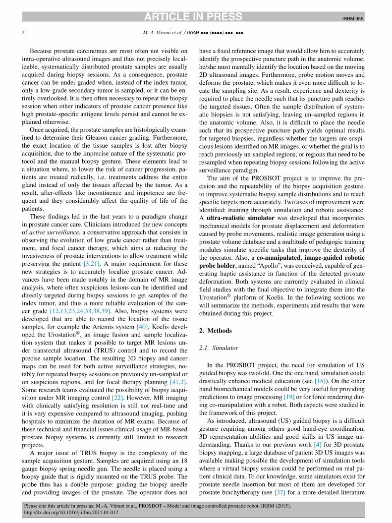
interactive time (i.e., a time compatible with an intra-operative application). A major effort has been made in order to compare different types of models (finite element models, mass-spring, mesh resolutions, constitutive law parameters, etc.) and to eval-uate them using ground truth data. A workflow (see Fig. 4) has been set-up enabling the acquisition and exploitation of data from a realistic phantom and measuring the error of each step of the pipeline.
Specific collision detection has also been developed for simulating the interactions of the US probe with the patient body.
2.2. Robotic probe holder
The surgical gesture and its impact on the diagnostic were first studied thanks to recorded data with Urostation® [25,26,6–11]. From that it appears that such a robotic system could
JID:IRBM AID:356 /FLA [m5+; v1.204; Prn:25/02/2015; 15:43] P.4 (1-8)
4 M.-A. Vitrani et al. / IRBM ••• (••••) •••–•••
Fig. 4. Pipeline for biomechanical evaluation.
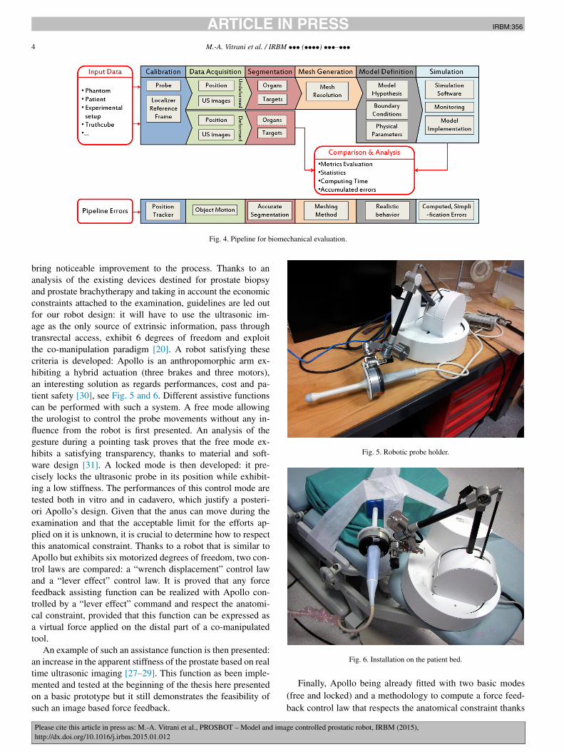
bring noticeable improvement to the process. Thanks to an analysis of the existing devices destined for prostate biopsy and prostate brachytherapy and taking in account the economic constraints attached to the examination, guidelines are led out for our robot design: it will have to use the ultrasonic im-age as the only source of extrinsic information, pass through transrectal access, exhibit 6 degrees of freedom and exploit the co-manipulation paradigm [20]. A robot satisfying these criteria is developed: Apollo is an anthropomorphic arm ex-hibiting a hybrid actuation (three brakes and three motors), an interesting solution as regards performances, cost and pa-tient safety [30], see Fig. 5 and 6. Different assistive functions can be performed with such a system. A free mode allowing the urologist to control the probe movements without any in-fluence from the robot is first presented. An analysis of the gesture during a pointing task proves that the free mode ex-hibits a satisfying transparency, thanks to material and soft-ware design [31]. A locked mode is then developed: it pre-cisely locks the ultrasonic probe in its position while exhibit-ing a low stiffness. The performances of this control mode are tested both in vitro and in cadavero, which justify a posteri-ori Apollo’s design. Given that the anus can move during the examination and that the acceptable limit for the efforts ap-plied on it is unknown, it is crucial to determine how to respect this anatomical constraint. Thanks to a robot that is similar to Apollo but exhibits six motorized degrees of freedom, two con-trol laws are compared: a “wrench displacement” control law and a “lever effect” control law. It is proved that any force feedback assisting function can be realized with Apollo con-trolled by a “lever effect” command and respect the anatomi-cal constraint, provided that this function can be expressed as a virtual force applied on the distal part of a co-manipulated tool.
An example of such an assistance function is then presented: an increase in the apparent stiffness of the prostate based on real time ultrasonic imaging [27–29]. This function as been imple-mented and tested at the beginning of the thesis here presented on a basic prototype but it still demonstrates the feasibility of such an image based force feedback.
Fig. 5. Robotic probe holder.
Fig. 6. Installation on the patient bed.
Finally, Apollo being already fitted with two basic modes (free and locked) and a methodology to compute a force feed-back control law that respects the anatomical constraint thanks

JID:IRBM AID:356 /FLA [m5+; v1.204; Prn:25/02/2015; 15:43] P.5 (1-8)
M.-A. Vitrani et al. / IRBM ••• (••••) •••–••• 5
Fig. 7. Experiments on a phantom.
to co-manipulation, exploiting its automatic functioning capa-bilities is proposed. An assistance to precise positioning featur-ing a loop on the ultrasonic image is implemented and prelim-inary tested in vitro: it allows to bring the line of sight of the biopsy needle near a target defined in the prostate with a satis-fying precision [32].
3. Experiments and results
3.1. Evaluation of the simulator and biomechanical models
The different versions of the educational simulator have been studied with non-clinicians, medicine students, residents and expert clinicians [36,14]. For each user, different elements are recorded concerning his/her ability to practice the exercises (image reading, accuracy to a target, ability to practice standard 12-cores protocol, etc.), the time he/she spent, his/her training history, etc. For the sake of simulator evaluation, each user also fills a questionnaire (see [36]). The first evaluation of the simu-lator was based on the experience of eight non-clinicians (PhD and master students) and proved the reliability and face valid-ity (realism judged by non-experts). The second evaluation of the simulator was based on the experience of 21 clinicians (14 medical students and 7 trained urologists) and proved the con-tent validity (realism judged by experts) and construct validity (scoring able to discriminate novice and expert). The collected comments led us to the development of the third version of the simulator. The image deformation was evaluated using data coming from a physical phantom and was also assessed qualita-tively by expert clinicians. A more complete evaluation of this last version of the educational simulator is planned for 2015 (see Section 4).
Concerning the biomechanical model for image processing or robot control, acquisition on a realistic deformable phantom with a 3D tracked US probe was performed (see Figs. 7 and 8). The real position of fiducials after deformation was extracted from US images and compared to the simulated ones. This en-abled us to finalize the pipeline and to develop all the image and information processing tools necessary for extensive simu-lation, and to perform all the data acquisition required for model comparisons.
Fig. 8. Simulation of probe motion and phantom deformation.
Fig. 9. Cadaver study.
In order to prepare the acquisition of patient data during prostate biopsy sessions with the PROSBOT robot,2 we also developed a new probe-robot calibration method exploiting in-formation resulting from image registration of a phantom [34].
3.2. In cadavero studies of the Apollo robot
Two experiments were performed at the Paris Surgical School. During each of these sessions, the robot’s installation and the basic control law were tested on cadaver by 3 urologists. The robot was under sterile drapes.
Positioning of the robot was evaluated for two cadaver po-sitions: left lateral decubitus and gynecologic position. For the installation, the robot base is roughly placed directly onto the table behind the legs of the cadaver (see Fig. 9) or on an ad-justable height stool between the legs.
Then the urologists were asked to insert the probe into the rectum, to scan the entire prostate and to mimic a biopsy ses-sion.
2 Let us mention that in this case the robot is used for acquiring the position of the US probe corresponding to US volumes of patients. The robot control does not depend on the model yet.
JID:IRBM AID:356 /FLA [m5+; v1.204; Prn:25/02/2015; 15:43] P.6 (1-8)
6 M.-A. Vitrani et al. / IRBM ••• (••••) •••–•••
For each trial, the robot workspace was sufficient to scan the prostate even with a rough installation. Furthermore, the urol-ogists acknowledged that the robot-assisted gesture was com-fortable and did not negatively impact or misguide the natural gesture. They particularly appreciated the gravity compensation of the probe provided by the robot. Indeed we observed that their probe handling was changed: instead of holding full hand as they do in clinical procedure, they held lightly, sometimes even with only 2 fingers.
These experiments showed the soundness of the proposed approach.
3.3. Clinical evaluation of the Apollo robot
Following the successful cadaver experiments clinical trials focusing on the free mode and the locked mode of the robot were prepared. The objective is to evaluate the practical usabil-ity and performance of the robot on the patient.
Essential requirements of the medical device directive 93/42/EEC needed to be respected, except for the aspects that are to be assessed during the clinical evaluation. An exhaustive risk analysis was performed to identify and control potential risks for patients and users. Furthermore, the robotic system was improved to comply with the applicable norms for medi-cal devices, in particular the norms for basic safety of medical electrical equipment (EN 60601-1) and for medical software lifecycle process (IEC 62304).
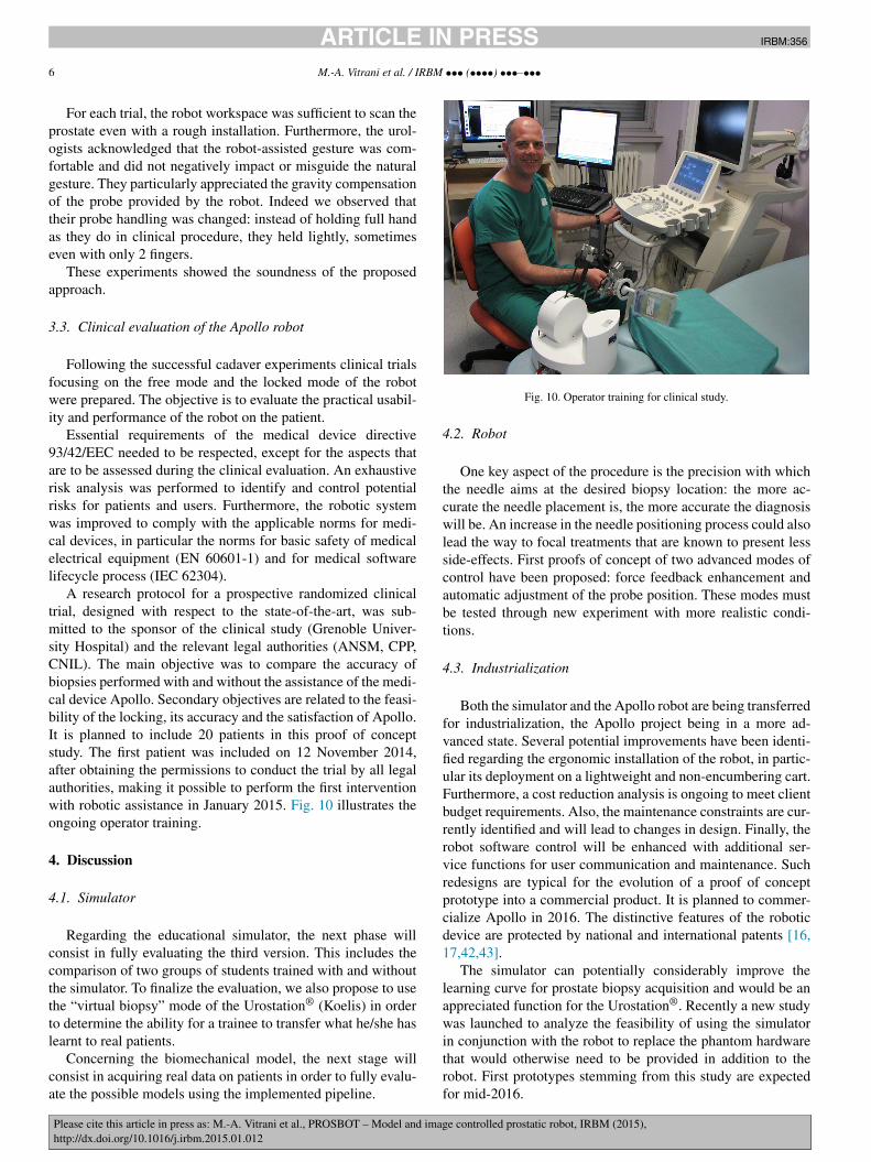
A research protocol for a prospective randomized clinical trial, designed with respect to the state-of-the-art, was sub-mitted to the sponsor of the clinical study (Grenoble Univer-sity Hospital) and the relevant legal authorities (ANSM, CPP, CNIL). The main objective was to compare the accuracy of biopsies performed with and without the assistance of the medi-cal device Apollo. Secondary objectives are related to the feasi-bility of the locking, its accuracy and the satisfaction of Apollo. It is planned to include 20 patients in this proof of concept study. The first patient was included on 12 November 2014, after obtaining the permissions to conduct the trial by all legal authorities, making it possible to perform the first intervention with robotic assistance in January 2015. Fig. 10 illustrates the ongoing operator training.
4. Discussion
4.1. Simulator
Regarding the educational simulator, the next phase will consist in fully evaluating the third version. This includes the comparison of two groups of students trained with and without the simulator. To finalize the evaluation, we also propose to use the “virtual biopsy” mode of the Urostation® (Koelis) in order to determine the ability for a trainee to transfer what he/she has learnt to real patients.
Concerning the biomechanical model, the next stage will consist in acquiring real data on patients in order to fully evalu-ate the possible models using the implemented pipeline.
Fig. 10. Operator training for clinical study.
4.2. Robot
One key aspect of the procedure is the precision with which the needle aims at the desired biopsy location: the more ac-curate the needle placement is, the more accurate the diagnosis will be. An increase in the needle positioning process could also lead the way to focal treatments that are known to present less side-effects. First proofs of concept of two advanced modes of control have been proposed: force feedback enhancement and automatic adjustment of the probe position. These modes must be tested through new experiment with more realistic condi-tions.
4.3. Industrialization
Both the simulator and the Apollo robot are being transferred for industrialization, the Apollo project being in a more ad-vanced state. Several potential improvements have been identi-fied regarding the ergonomic installation of the robot, in partic-ular its deployment on a lightweight and non-encumbering cart. Furthermore, a cost reduction analysis is ongoing to meet client budget requirements. Also, the maintenance constraints are cur-rently identified and will lead to changes in design. Finally, the robot software control will be enhanced with additional ser-vice functions for user communication and maintenance. Such redesigns are typical for the evolution of a proof of concept prototype into a commercial product. It is planned to commer-cialize Apollo in 2016. The distinctive features of the robotic device are protected by national and international patents [16,17,42,43].
The simulator can potentially considerably improve the learning curve for prostate biopsy acquisition and would be an appreciated function for the Urostation®. Recently a new study was launched to analyze the feasibility of using the simulator in conjunction with the robot to replace the phantom hardware that would otherwise need to be provided in addition to the robot. First prototypes stemming from this study are expected for mid-2016.

JID:IRBM AID:356 /FLA [m5+; v1.204; Prn:25/02/2015; 15:43] P.7 (1-8)
M.-A. Vitrani et al. / IRBM ••• (••••) •••–••• 7
5. Conclusion
The Prosbot project is a demonstration of a successful col-laboration of scientific laboratories with clinical and industrial partners. The two main objectives of the project, conceiving a co-manipulated probe holder and a simulator for prostate biopsy, were reached and the first results of the industrial trans-fers are very promising. Both devices will help to improve prostate cancer management through a steeper learning curve and a more accurate clinical gesture.
Acknowledgements
This project was partially supported by French state funds managed by the ANR within the Investissements d’Avenir programme (Labex CAMI) under reference ANR-11-LABX-0004 and through the PROSBOT project under reference ANR-11-TECS-0017. It further received funds from the French government agency Association Nationale de la Recherche et de la Technologie (CIFRE 2012/1247).
References
[1] Bajcsy R, Broit C. Matching of deformed images. In: Proc 6th int conf of pattern recognition. 1982.
[2] Rud E, Baco E, Eggesbø HB. MRI and ultrasound-guided prostate biopsy using soft image fusion. Anticancer Res 2012 Aug;32(8):3383–9.
[3] Bangma CH, Valdagni R, Carroll PR, van Poppel H, Klotz L, Hugosson J. Active surveillance for low-risk prostate cancer: developments to date. Eur Urol 2014 Nov 27. http://dx.doi.org/10.1016/j.eururo.2014.11.004[pii:S0302-2838(14)01178-6].
[4] Baumann M, Mozer P, Daanen V, Troccaz J. Prostate biopsy tracking with deformation estimation. Med Image Anal 2012;16(3):562–76.
[5] Barry Michael J. Screening for prostate cancer – the controversy that refuses to die. N Engl J Med March 26, 2009;360:1351–4. http://dx.doi.org/10.1056/NEJMe0901166.
[6] Coffin G, Torterotot C, Baumann M, Vitrani MA, Morel G, Chevreau G, et al. TRUS prostate biopsie cores: extent of the pierced area impacts the cancer detection rate. J Endourol 2011;25(9).
[7] Coffin G, Torterotot C, Vitrani MA, Baumann M, Chevreau G, Renard-Penna R, et al. Impact de la distribution des biopsies de prostate par voie endorectale sur le taux de détection de cancer dans les lobes droit et gauche. In: 105ème congrès de l’AFU (Association Française d’urolo-gie). 2011.
[8] Coffin G, Chevreau G, Renard-Penna R, Comperat E, Vitrani MA, Tortero-tot C, et al. Comparison between systematic and MRI targeted prostate biopsy for patient with no history of prostate cancer attending a first round of trans-rectal ultrasound biopsy procedure. J Endourol 2011;25(9).
[9] Coffin G, Chevreau G, Renard-Penna R, Comperat E, Vitrani MA, Tortero-tot C, et al. Systematic prostate biopsies may detect more insignificant cancer than MRI lesion target prostate biopsies. J Endourol 2011.
[10] Coffin G, Chevreau G, Renard-Penna R, Comperat E, Vitrani MA, Tortero-tot C, et al. Comparison between systematic and MRI targeted prostate biopsy for patient with no history of prostate cancer attending a first round of trans-rectal ultrasound biopsy procedure. In: Congrès de l’EAU (Euro-pean association of urology). 2012.
[11] Coffin G, Chevreau G, Renard-Penna R, Comperat E, Vitrani MA, Tortero-tot C, et al. Comparison between systematic and MRI targeted prostate biopsy for patient with no history of prostate cancer attending a first round of trans-rectal ultrasound biopsy procedure. In: 5th international sympo-sium on focal therapy and imaging in prostate & kidney cancer. 2012.
[12] Cornud F, Brolis L, Delongchamps NB, Portalez D, Malavaud B, Renard-Penna R, et al. TRUS-MRI image registration: a paradigm shift in the diagnosis of significant prostate cancer. Abdom Imaging
2013 Dec;38(6):1447–63. http://dx.doi.org/10.1007/s00261-013-0018-4[review].
[13] Delongchamps NB, et al. Prebiopsy magnetic resonance imaging and prostate cancer detection: comparison of random and targeted biopsies. J Urol 2013;189:493–9.
[14] Fiard G, Selmi SY, Promayon E, Vadcard L, Descotes JL, Troccaz J. Initial validation of a virtual reality learning environment for prostate biopsies: realism matters! J Endourol 2014;28(4):453–8.
[15] Fouard C, Deram A, Keraval Y, Promayon E. CamiTK: a modular frame-work integrating visualization, image processing and biomechanical mod-eling. In: Payan Y, editor. Soft tissue biomechanical modeling for com-puter assisted surgery. 2012. p. 323–54.
[16] National patent FR2980683A1. Dispositif de guidage d’un instrument médical inséré dans une voie naturelle ou une voie artificielle d’un pa-tient. Priority date: September 2011, publication date Sept. 2014.
[17] National patent application FR2983397A1. Dispositif d’assistance au po-sitionnement d’un instrument médical relativement à un organe interne d’un patient et procédé de commande d’un tel dispositif. Priority date: December 2011, publication date: June 2013.
[18] Haute Autorité de Santé. État de l’art (national et international) en matière de pratiques de simulation dans le domaine de la santé: Rapport de Mission de la Haute Autorité de santé. January 2012 [in French].
[19] Hu Y, Ahmed HU, Taylor Z, Allen C, Emberton M, Hawkes D, et al. MR to ultrasound registration for image-guided prostate interventions. Med Image Anal April 2012;16(3):687–703.
[20] Hungr N, Baumann M, Long J, Troccaz J. A 3-d ultrasound robotic prostate brachytherapy system with prostate motion tracking. IEEE Trans Robot Dec. 2012;28(6):1382–97.
[21] Klotz L, Vesprini D, Sethukavalan P, Jethava V, Zhang L, Jain S, et al. Long-term follow-up of a large active surveillance cohort of patients with prostate cancer. J Clin Oncol 2014 Dec 15. pii:JCO.2014.55.1192.
[22] Krieger Axel, Susil Robert C, Ménard Cynthia, Coleman Jonathan A, Fichtinger Gabor, Atalar Ergin, et al. Design of a novel MRI compatible manipulator for image guided prostate interventions. IEEE Trans Biomed Eng Feb 2005;52(2):306–13.
[23] Moore CM, et al. Image-guided prostate biopsy using magnetic resonance imaging-derived targets: a systematic review. Eur Urol 2013;63:125–40.
[24] Mozer P, Rouprêt M, Le Cossec C, Granger B, Comperat E, de Gorski A, Cussenot O, et al. First round of targeted biopsies us-ing magnetic resonance imaging/ultrasonography fusion compared with conventional transrectal ultrasonography-guided biopsies for the di-agnosis of localised prostate cancer. BJU Int 2015 Jan;115(1):50–7. http://dx.doi.org/10.1111/bju.12690. Epub 2014 Jul 27.
[25] Poquet-Torterotot C, Mozer P, Baumann M, Vitrani M-A, Morel G. Anal-ysis of endorectal probe kinematics during prostate biopsies. In: Hamlyn symposium. 2010.
[26] Poquet Torterotot C, Mozer P, Baumann M, Vitrani M-A. Analy-sis of prostate area sampled by TRUS prostate biopsy. J Endourol 2010:2093–157.
[27] Poquet Torterotot C, Vitrani M-A, Mozer P, Morel G. Ultrasound image-based comanipulation for enhanced perception of the contacts with a distal soft organ. In: IEEE international conference on robotics and biomemitics. (ROBIO). 2011 [“best student paper award” finalist].
[28] Poquet Torterotot C, Mozer P, Baumann M, Vitrani M-A, Morel G, Leroy A, et al. Dispositif d’assistance au positionnement d’un instrument médi-cal relativement à un organe interne d’un patient et procédé de commande d’un tel dispositif. 2011. FR2983397/WO2013083731.
[29] Poquet Torterotot C, Mozer P, Bauman M, Vitrani M-A, Morel G, Leroy A, et al. Dispositif de guidage d’un instrument médical inséré dans une voie naturelle ou une voie artificielle d’un patient. 2011. FR2980683/WO2013045645.
[30] Poquet C, Mozer P, Morel G, Vitrani M-A. A novel comanipulation device for assisting needle placement in ultrasound guided prostate biopsies. In: IEEE international conference on intelligent robots and systems (IROS). 2013. p. 4048–91.
[31] Poquet C, Mozer P, Vitrani M-A, Morel G. An endorectal ultrasound probe comanipulator with hybrid actuation combining brakes and motors. In: IEEE transactions on mechatronics (TMECH). 2013.

JID:IRBM AID:356 /FLA [m5+; v1.204; Prn:25/02/2015; 15:43] P.8 (1-8)
8 M.-A. Vitrani et al. / IRBM ••• (••••) •••–•••
[32] Poquet C, Vitrani M-A, Mozer P, Morel G. Achieving high precision in prostate biopsy thanks to robot closed loop control based on 3D ultrasound imaging. In: SURGETICA. 2014.
[33] Puech P, et al. Prostate cancer diagnosis: multiparametric MR-targeted biopsy with cognitive and transrectal US-MR fusion guidance ver-sus systematic biopsy – prospective multicenter study. Radiology 2013;268:461–9.
[34] Sarrazin J, Promayon E, Baumann M, Troccaz J. 3D ultrasound probe calibration using robotic arm and image registration. In: Proceedings of Surgetica’2014. 2014, http://surgetica2014-papers.imag.fr/2014-51-Johan_Sarrazin_TIMCIMAG.pdf.
[35] Schröder FH, Hugosson J, Roobol MJ, Tammela TLJ, Ciatto S, Ne-len V, et al. Screening and prostate-cancer mortality in a random-ized European study. N Engl J Med March 26, 2009;360:1320–8. http://dx.doi.org/10.1056/NEJMoa0810084.
[36] Selmi SY, Fiard G, Promayon E, Vadcard L, Troccaz J. A virtual reality simulator combining a learning environment and clinical case database for image-guided prostate biopsy. In: Proceedings of IEEE CBMS (26th IEEE international symposium on computer-based medical systems). 20–22 June, 2013. p. 179–84.
[37] Selmi S, Promayon E, Sarrazin J, Troccaz J. 3D interactive ultrasound image deformation for realistic prostate biopsy simulation. In: Bello F, Cotin S, editors. Proceedings of the 6th international symposium on biomedical simulation. LNCS, vol. 8789. Springer; 2014. p. 122–30.
[38] Siddiqui MM, et al. Magnetic resonance imaging/ultrasound-fusion biopsy significantly upgrades prostate cancer versus systematic 12-core transrectal ultrasound biopsy. Eur Urol November 2013;64(1):713–9. http://dx.doi.org/10.1016/j.eururo.2013.05.059.
[39] Sonn GA, et al. Value of targeted prostate biopsy using magnetic resonance-ultrasound fusion in men with prior negative biopsy and elevated prostate-specific antigen. Eur Urol 2014;65(4):809–15. http://dx.doi.org/10.1016/j.eururo.2013.03.025.
[40] Sonn GA, Filson CP, Chang E, Natarajan S, Margolis DJ, Macairan M, et al. Initial experience with electronic tracking of specific tumor sites in men undergoing active surveillance of prostate cancer. Urol Oncol 2014 Oct;32(7):952–7. http://dx.doi.org/10.1016/j.urolonc.2014.04.003. Epub 2014 Jul 11.
[41] Ukimura O, Desai MM, Palmer S, Valencerina S, Gross M, Abreu AL, et al. 3-Dimensional elastic registration system of prostate biopsy lo-cation by real-time 3-dimensional transrectal ultrasound guidance with magnetic resonance/transrectal ultrasound image fusion. J Urol 2012 Mar;187(3):1080–6. http://dx.doi.org/10.1016/j.juro.2011.10.124. Epub 2012 Jan 21.
[42] International patent WO2013045645A1. Device for guiding a medical instrument inserted into a natural duct or an artificial duct of a patient. Priority date: September 2011, publication date: April 2013.
[43] International patent WO2013083731A1. Assistive device for positioning a medical instrument relative to an internal organ of a patient. Priority date: December 2011, publication date: June 2013.