prospective, randomized study initial results on reading ... with the pelli-robson chart. all...
TRANSCRIPT

Original ArticleAdding access to a video magnifier to standard vision rehabilitation:initial results on reading performance and well-being from aprospective, randomized studyMary Lou Jackson, MD, Kimberly A. Schoessow, OTD, OTR/L, Alexandra Selivanova, BS, andJennifer Wallis, PhDAuthor affiliations: Department of Ophthalmology, Massachusetts Eye and Ear Infirmary, Harvard Medical School, Boston,Massachusetts
AbstractPurpose—Both optical and electronic magnification are available to patients with low vision. Electronicvideo magnifiers are more expensive than optical magnifiers, but they offer additional benefits, includingvariable magnification and contrast. This study aimed to evaluate the effect of access to a video magnifier(VM) added to standard comprehensive vision rehabilitation (VR).
Methods—In this prospective study, 37 subjects with central field loss were randomized to receive stand-ard VR (VR group, 18 subjects) or standard VR plus VM (VM group, 19 subjects). Subjects read the Inter-national Reading Speed Texts (IReST), a bank check, and a phone number at enrollment, at 1 month, andafter occupational therapy (OT) as indicated to address patient goals. The Impact of Vision Impairment(IVI) questionnaire, a version of the Activity Inventory (AI), and the Depression Anxiety and Stress Scale(DASS) were administered at enrollment, 1 month, after OT, 1 month later, and 1 year after enrollment.Assessments at enrollment and 1 month later were evaluated.
Results—At 1 month, the VM group displayed significant improvement in reading continuous print asmeasured by the IReST (P = 0.01) but did not differ on IVI, AI, or DASS. From enrollment to 1 month allsubjects improved in their ability to spot read (phone number and check; P < 0.01 for both). The VM groupimproved in their ability to find and read a number in a phone book more than the VR group at 1 monthafter initial consultation (P = 0.02). All reported better well-being (P = 0.02).
Conclusions—All subjects reported better well-being on the IVI. The VM group read faster and was bet-ter at two spot reading tasks but did not differ from the VR group in other outcome measures.
IntroductionIt is estimated that reading is difficult for over 85% ofpatients who attend vision rehabilitation consultations.1Reading is a priority skill for most individuals and isconsidered to be an important part of maintaining inde-pendence.2 Magnification plays a major role in address-ing the goal of reading for patients with central fieldloss, such as those with age-related macular degenera-tion (AMD), which has been estimated to affect over1.75 million individuals in the United States.3 Emergingresearch shows benefits of multidisciplinary rehabilita-
tion; however, it is not known what portion of rehabilita-tion success can be attributed to the various rehabilita-tion components: training, device acquisition, environ-mental modifications, education, visual skills, readingpractice, patient motivation, and caring rehabilitationpersonnel.4 Most previous research has reported pre-and post-rehabilitation function but has not quantifiedthe benefit attributable simply to device acquisition.Addressing the question of optimal rehabilitation,
Published March 31, 2017.Copyright ©2017. All rights reserved. Reproduction in whole or in part in any form or medium without expressed written permission of theDigital Journal of Ophthalmology is prohibited.doi:10.5693/djo.01.2017.02.001Correspondence: Mary Lou Jackson, MD, UBC/Eye Care Centre, 2550 Willow Street, Vancouver, BC, Canada (email: [email protected]).Clinical trial number: NCT01670643.Grants/Funding: Devices provided by Optelec USA.
Digital Journal of O
phthalmology, Vol. 23
Digital Journal of O
phthalmology, Vol. 23

including optimal reading device, is a priority in thisresearch.
Video camera magnifiers are a simple combination of avideo camera and a monitor; they allow magnification,enhanced image contrast, and a wide field for viewingtext. They are more expensive than traditional opticaldevices but are often chosen by a patient in comprehen-sive vision rehabilitation. A 2013 Cochrane Reviewexamined all available randomized and quasi-random-ized studies relevant to magnifying reading aids foradults with low vision (10 studies were included) andconcluded that the evidence was insufficient to assessthe effect of low-vision devices on reading performance,although there was some evidence that electronic devi-ces were associated with more improvement in readingspeed than optical devices were.5
The present study compared patients who receivedstandard visual rehabilitation (VR) with those who, inaddition to VR, were provided with a video magnifier(VM) at their initial contact with a multidisciplinaryvision rehabilitation service. VR included an initial con-sultation during which patients were educated aboutrehabilitation strategies, given information aboutremaining visual function, and shown a range of opticaland electronic devices, which they could purchase. Ourhypothesis was that access to an electronic video magni-fier device would improve objective reading perform-ance and patient-reported outcomes. This study evalu-ated results at enrollment, when all subjects used prere-habilitation devices, and at 1 month after enrollment,when subjects who had had access to a video magnifiercompleted reading assessment using the video magnifier.Those in the group without access to the VM used anydevice they may have acquired prerehabilitation or dur-ing the month since their initial consultation.
Materials and MethodsStudy DesignThis study was approved by the Massachusetts Eye andEar Institutional Review Board. Written informed con-sent was obtained from all participants. This pilot studywas a prospective, double-armed, randomized, control-led trial (Figure 1) comparing objective measurementsof reading and patient-reported quality of life, includingperceived reading ability, in subjects who had VMaccess in addition to standard VR (VM group) to sub-jects who only had VR (VR group). Randomizationoccurred after the initial consultation. All participantsunderwent an initial vision rehabilitation consultation,including visual function evaluation and discussion of
rehabilitation principles and options. Patients in the VMgroup received a desk video magnifier when they pre-sented for initial consultation. They were free to use thisdevice as much or as little as they wished during theensuing month. Patients in the VR group were free topurchase devices at any time, and they were advised thatthey would also receive a free video magnifier after thecompletion of rehabilitation training with an occupa-tional therapist (Figure 1). All subjects returned after 1month to begin an occupational therapy (OT) evaluationand subsequent training. The OT training consisted ofevaluation of visual performance, assessment of goalsand training with devices and task modification, asrequired. Subjects had objective reading assessments atthree time points: enrollment, 1 month after enrollment,and at the conclusion of OT. Subjects completed ques-tionnaires at five time points: enrollment, 1 month afterenrollment, at the conclusion of OT, 1 month after OTdischarge, and 1 year after enrollment. This studypresents findings from the first two time points of thestudy: from enrollment to 1 month after enrollment.
Patient SelectionPatients attending a multidisciplinary outpatient visionrehabilitation clinic at the Massachusetts Eye and Earfrom February 2010 to May 2011 were invited to partici-pate in this study. Inclusion criteria included age ≥40years, central visual field loss (defined as not respondingto 1 or more targets on macular perimetry testing in thebetter-seeing eye), no disease affecting the peripheral
Figure 1. Study flowchart. Results presented in this study arefrom enrollment to 1 month after enrollment (dashed box).
2
Digital Journal of O
phthalmology, Vol. 23
Digital Journal of O
phthalmology, Vol. 23

retina, visual acuity worse than 20/40 in each eye andbetter than 20/400 in the better seeing eye, cumulativescore of >20 (of 30) on the 6-question modified MiniMental State Examination (MMSE) questionnaire forvisually impaired, sufficient hearing to participate ininterviews, and no previous experience with vision reha-bilitation or video camera magnifiers.
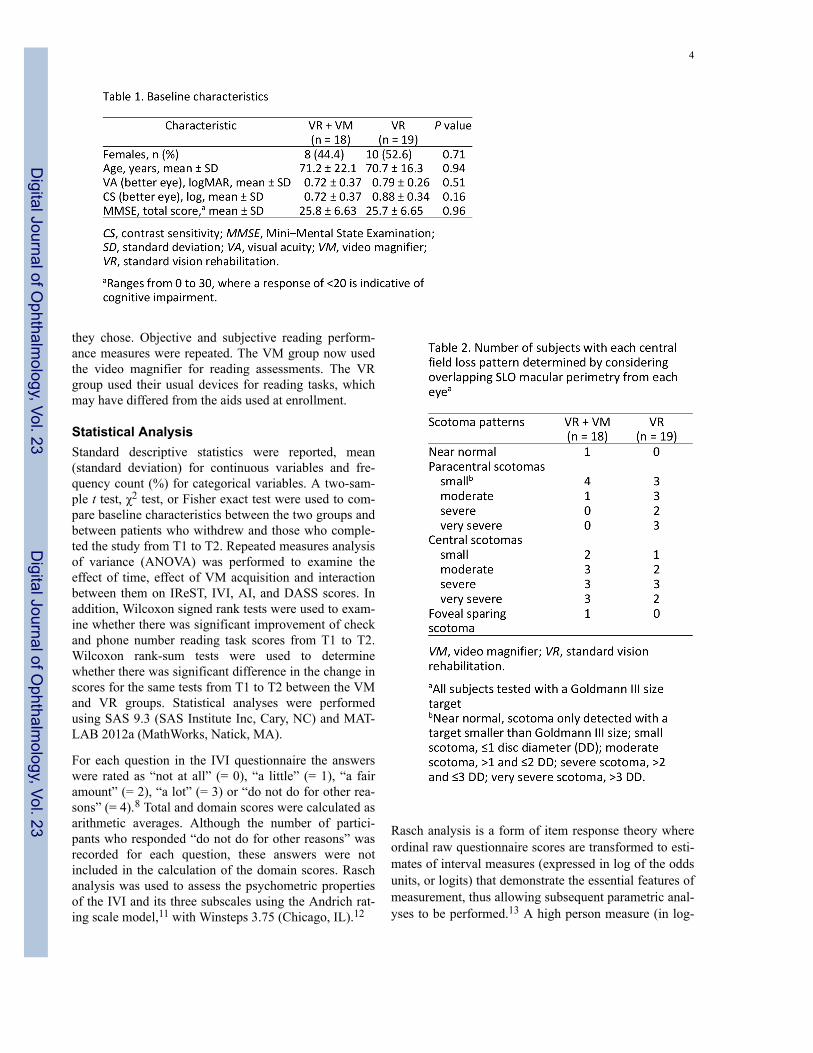
Clinical Visual Function TestsAs part of the initial consultation all subjects had refrac-tion and measurement of visual acuity using the EarlyTreatment Diabetic Retinopathy Study (ETDRS) chartor Snellen a projected chart. Contrast sensitivity wasmeasured with the Pelli-Robson chart. All subjectsunderwent scanning laser ophthalmoscope macular peri-metry to document fixation and the presence of centralscotoma(s) using the commercially available OPTOSmacular perimeter and a size III target testing the central20° of visual field. The extent of the scotoma was esti-mated by viewing results from both monocular perime-try tests and determining the scotoma that would over-lap, viewing binocularly. The size and location of thescotoma was then classified in five categories: (1) nearnormal, with scotoma only detected with a target smallerthan Goldmann III size; (2) small scotoma, ≤1 discdiameter (DD); (3) moderate scotoma, >1 and ≤2 DD;(4) severe scotoma, >2 and ≤3 DD; and (5) very severescotoma, >3 DD.
Visual Performance MeasuresReading speed was assessed in words per minute (wpm)using the International Reading Speed Texts (IReST), inwhich subjects read to the best of their ability a standar-dized paragraph of newspaper-sized text aloud.6 Sub-jects also had objective assessment of ability to read theamount on a bank check and a phone number on a pagefrom the phone book. The participants also were askedto identify the handwritten amount on the check. Thesame reading items were used at enrollment and after 1month; there was no variation in print size and contrast.At enrollment, subjects could hold the material at anydistance and use any devices they habitually used beforerehabilitation. The reading tasks were videotaped andafterward scored by two investigators as correct (score =3), correct but struggles (= 2), partly correct (= 1), orimpossible to complete (= 0). For the 1-month assess-ment, those in the VR group could again use any devicethey wished and those in the VM group used the videomagnifier.
Patient-reported MeasuresThe Impact of Vision Impairment questionnaire (IVI)and 10 reading questions from the Activity Inventory
(AI) were used as patient-reported measures.7,8 The IVIis a validated 28-item questionnaire designed to identifyvision-specific limitations in activities that affectpatients’ quality of life. The 28 questions fall into threedomains: mobility and independence (mobility), emo-tional well-being (well-being), and reading and access-ing information (reading). The AI first requires the par-ticipant to rate the importance of a goal (eg, read news-paper), and then rate the difficulty of the tasks nestedunder that goal (eg, read headlines) as “not difficult” (=0), “slightly difficult” (= 1), “moderately difficult” (= 2),“very difficult” (= 3), “impossible to do without some-one else’s help” (= 4) or “not applicable” (= 5).9 If agoal was not rated as important to the individual, then itwas not included in the final scoring. The Depression,Anxiety and Stress Scale (DASS) was administered toassess subjective well-being.10 The individual adminis-tering the questionnaires was not masked to groupassignment.
ProcedureAll patients had a consultation with an ophthalmologist(MLJ) who discussed the remaining visual function, theimpact of the visual deficit on visual performance,including reading, the advantages and drawbacks of thevarious device categories, options to acquire devices,and the recommendation of subsequent occupationaltherapy evaluation to address training to use devices,evaluation of success with devices, and opportunities tomodify tasks and strategies to improve visual perform-ance. Randomization to receive a VM at enrollment or atthe end of vision rehabilitation was according to a com-puter-generated numerical series. After half of thepatients had been enrolled, an adaptive design was adop-ted to ensure that the two groups were comparable invisual acuity.
At enrollment, all subjects completed the questionnairesand were videotaped completing the reading tasks usingtheir usual prerehabilitation devices or glasses. Patientsrandomized to the video magnifier group were shownbasic operation and set-up of the desk model video mag-nifier (Optelec, ClearView+, 2.6× to 50× magnification,17″ display). All subjects were able to purchase hand-held magnifiers or reading glasses at any time during therehabilitation process (standard VR). Subjects wereaware that their reading performance would be tested 1month later.
All subjects returned 1 month after their initial consulta-tion and study enrollment. During the 1-month intervalthe subjects who had had access to the VM had beenusing it as much as they wished, for whatever purpose
Jackson et al. 3
Digital Journal of O
phthalmology, Vol. 23
Digital Journal of O
phthalmology, Vol. 23
they chose. Objective and subjective reading perform-ance measures were repeated. The VM group now usedthe video magnifier for reading assessments. The VRgroup used their usual devices for reading tasks, whichmay have differed from the aids used at enrollment.
Statistical AnalysisStandard descriptive statistics were reported, mean(standard deviation) for continuous variables and fre-quency count (%) for categorical variables. A two-sam-ple t test, χ2 test, or Fisher exact test were used to com-pare baseline characteristics between the two groups andbetween patients who withdrew and those who comple-ted the study from T1 to T2. Repeated measures analysisof variance (ANOVA) was performed to examine theeffect of time, effect of VM acquisition and interactionbetween them on IReST, IVI, AI, and DASS scores. Inaddition, Wilcoxon signed rank tests were used to exam-ine whether there was significant improvement of checkand phone number reading task scores from T1 to T2.Wilcoxon rank-sum tests were used to determinewhether there was significant difference in the change inscores for the same tests from T1 to T2 between the VMand VR groups. Statistical analyses were performedusing SAS 9.3 (SAS Institute Inc, Cary, NC) and MAT-LAB 2012a (MathWorks, Natick, MA).
For each question in the IVI questionnaire the answerswere rated as “not at all” (= 0), “a little” (= 1), “a fairamount” (= 2), “a lot” (= 3) or “do not do for other rea-sons” (= 4).8 Total and domain scores were calculated asarithmetic averages. Although the number of partici-pants who responded “do not do for other reasons” wasrecorded for each question, these answers were notincluded in the calculation of the domain scores. Raschanalysis was used to assess the psychometric propertiesof the IVI and its three subscales using the Andrich rat-ing scale model,11 with Winsteps 3.75 (Chicago, IL).12
Rasch analysis is a form of item response theory whereordinal raw questionnaire scores are transformed to esti-mates of interval measures (expressed in log of the oddsunits, or logits) that demonstrate the essential features ofmeasurement, thus allowing subsequent parametric anal-yses to be performed.13 A high person measure (in log-
4
Digital Journal of O
phthalmology, Vol. 23
Digital Journal of O
phthalmology, Vol. 23

its) indicates that a person possesses a high level of theassessed latent trait (eg, vision-related quality of life)and vice versa.14 Rasch analysis also provides signifi-cant insight into the psychometric properties of thescale.14
The AI was scored using an Excel spreadsheet (providedby the author of the AI, Robert W. Massof). For correctRasch scoring of all subscales the full AI questionnaireis required, and in this study only a shortened version ofthe AI questionnaire was administered. As a result, over-all “goals” were Rasch scored for further analysis.
The sum of all DASS-21 scores for each participant wasmultiplied by 2, according to recommended analysismethods, and this was used for analyses.
Two independent reviewers scored the videotapedpatients performance on reading the amount on a bankcheck and on reading a phone number.
ResultsA total of 37 patients (18 females; mean age, 72.9[range, 40–91]) were enrolled, 18 in the VM group and19 in the VR group (Figure 1). Demographic character-istics and visual function of these patients are provided
in Table 1. The groups were not significantly different inage, sex, visual acuity, contrast sensitivity, or MMSE,and the groups were roughly balanced in the type of cen-tral field loss (P = 0.11; see Table 2). One month afterenrollment, 6 subjects withdrew from the study (3 fromeach group), and those who withdrew from the study didnot significantly differ from those who continued in age,sex, visual acuity, contrast sensitivity, and MMSE (Table3). The most common diagnosis was AMD or juvenile-onset macular degeneration (73%). The remaining sub-jects had optic nerve disease (16%) and macular dystro-phy or other maculopathy (11%).
At enrollment, the reading speed was 0–125 wpm in theVM group and 0–96 wpm in the VR group (Table 4).This increased to 22–168 wpm in the VM group at 1month. In the VR group the range was 0–108 wpm at 1month. Subjects in the VM group read the IReST contin-uous print significantly faster at 1 month (interactionbetween time and group, P = 0.01; Figure 2).
Phone Number and Check Reading TasksAfter two raters had scored the videotaped reading per-formance of subjects, an inter-rater reliability analysisusing the kappa statistic was performed to determinerater consistency. The inter-rater reliability was substan-tial for both measures at enrollment and 1 month later.
Jackson et al. 5
Digital Journal of O
phthalmology, Vol. 23
Digital Journal of O
phthalmology, Vol. 23

The kappa coefficient was 0.80 for the check readingtask and 0.72 for the phone number reading task atenrollment and was 0.63 and 0.85, respectively, at 1month (P < 0.001). An average score for each patient’sperformance based on the scores from the two reviewers
Figure 2. Mean changes in reading speed (IReST) in words perminute. The most significant improvement in reading speed wasobserved at 1 month in the video magnifier (VM) group, adjustedfor age, Geriatric Depression Scale, visual acuity, contrast sensitiv-ity, and central visual field (interaction between time and group, P= 0.01). Error bars represent standard error of the mean (SEM).
was used for further analyses. At enrollment, the twogroups did not differ in their ability to find and read aphone number in a phone book and the amount on ahandwritten check (P = 0.36 and P = 0.67, resp.; Table5). From enrollment to 1 month, all subjects improved intheir ability to find and read a phone number (P < 0.01for both), but the VM group improved their ability tofind and read a number more than the control group at 1month (P = 0.02; Figure 3). When all subjects were con-sidered, there was also improvement in ability to readthe amount on a check (P < 0.001), but neither groupimproved significantly better than the other at 1 month(P = 0.17).
Patient-reported MeasuresOverall, the composite IVI scale displayed suboptimalfit to Rasch model parameters. Although scale precisionand targeting of person ability to item difficulty wereexcellent, there was evidence of multidimensionality inthe scale. Specifically, the unexplained variance in thefirst contrast was >2.0 eigen values and three itemsexhibited misfit (infit MnSq >1.30). Inspection of thestandardized residual loadings for items (>0.4) indicatedthe possibility of at least two separate dimensions withinthe scale—“visual functioning” and “emotional.” DIFfor gender was also found. DIF indicates that a particu-
6
Digital Journal of O
phthalmology, Vol. 23
Digital Journal of O
phthalmology, Vol. 23

lar subgroup (eg, sex, age) systemically responds differ-ently to an item despite having the same underlying abil-ity level and therefore indicates the presence of bias. Inour sample, males systematically found the question thatenquired about feeling lonely or isolated because of eye-sight (question 23 on IVI) more difficult than females,even though they shared the same level of ability. Takentogether, this evidence suggested that the compositescore of the IVI should not be used in further statisticalanalyses, and the three subscales of the IVI were subse-quently analyzed separately. Both the reading and themobility subscales displayed excellent psychometricproperties, with good precision and targeting, and noevidence of multidimensionality or DIF. Each scale hadtwo misfitting questions, but as these only marginallyexceeded the acceptable infit MnSq cut-off of 1.30 logitsthese questions were retained so as not to lose importantitem content. Initial analysis of the well-being subscalerevealed good fit to Rasch model parameters, withordered thresholds, adequate precision, good targetingand no evidence of multidimensionality. However, ques-tions 23 (“Have you felt lonely or isolated because ofyour eyesight?”) and 25 (“In the past month, how oftenhave you worried about your eyesight getting worse?”)displayed DIF for sex and visual impairment, respec-tively. Therefore, questions 23 and 25 were iterativelyremoved to resolve the DIF, which resulted in excellentpsychometric properties of the remaining 6 questions ofthe well-being subscale.
Figure 3. Difficulty scores for reading tasks (change from enroll-ment to 1 month later). For ease of viewing, individual difficultyscores at enrollment were subtracted from 1 month later to give adifference in individual difficulty scores, and the averages of thedifference scores are represented below. The VM group improvedmore than the control group in reading a phone number at 1 month(P = 0.02). Error bars represent SEM.
Repeated measures ANOVA was performed to examinethe effect of group, time, and interaction among them onIVI Rasch domain scores (well-being, reading, andmobility) while controlling for age, Geriatric DepressionScale, visual acuity, contrast sensitivity, and central vis-ual field. There was no significant interaction effect forall three IVI Rasch domain scores (P = 0.77, 0.06, and0.10, resp.). IVI well-being domain score changed sig-nificantly over time considering all subjects (main effectof time, P = 0.02; Table 6). The IVI reading and mobi-lity domain scores did not significantly change over time(main effect of time, P = 0.30 and 0.07, resp.; Table 6).The AI person measures for “goals” did not significantlychange from enrollment to 1 month later. No significantchanges in mood as measured with the DASS wereobserved.
DiscussionThis study assessed the impact of adding access to a VMfrom the first day that a patient had contact with thevision rehabilitation clinic to standard comprehensiveVR. Those in the VM group did read continuous printfaster after 1 month and did spot read more accurately;however, patient-reported outcomes were not statisti-cally different in this group compared to those in the VRgroup.
Reading rehabilitation is part of comprehensive VR.Based on the American Academy of OphthalmologyPreferred Practice Pattern Guidelines, comprehensiveVR addresses five areas that may be affected by visionimpairment: reading, activities of daily living, safety,participation in activities despite vision loss, and psy-chosocial well-being.15 Rehabilitation includes a varietyof strategies, psychological supports, education, devicesand training. The patient is an active participant in therehabilitation process. Devices are recommended to thepatient, depending on a patient’s visual function andunique goals. Limited previous research has shown thatvision rehabilitation improves function and subjectivewell-being and that reading performance, in particular,improves with vision rehabilitation.4 Stelmack et al16
found significant improvement in reading ability in agroup of visually impaired veterans who received inten-sive VR compared to a waitlist control group. The inter-vention included vision examination, counseling, andprovision of low vision devices as well as six weeklytraining sessions. Reading performance was measured atbaseline and 4 months later. Jackson et al17 observedsmaller reading improvement 1 year after comprehen-sive rehabilitation using the same measure as Stelmacket al.16 One difference between the two studies is in the
Jackson et al. 7
Digital Journal of O
phthalmology, Vol. 23
Digital Journal of O
phthalmology, Vol. 23

provision of free devices. In Stelmack et al,16 studydevices were provided to subjects at no cost. In the Jack-son et al,17 study rehabilitation did not include free pro-vision of devices for all subjects. Devices were providedby local or state agencies for some patients or could bepurchased by patients if they chose. Neither study sepa-rated the effect of training on reading from the effect ofusing devices. Pijnacker et al18 reviewed methods totrain reading performance and found various trainingmethods beneficial (no strong support for one method)but noted that a limitation of all studies was that it wasnot possible to disentangle the benefit of training fromthe benefit of devices. A systematic review of eccentricviewing also noted that a failure to separate effects ofeccentric viewing training from effects of using low-vision devices was one reason that reviewed studiesfailed to meet criteria for well-designed trials.19
In this study we offered the VM to subjects in the inter-vention group at their first contact with the clinic so thateffects of training would be separated temporally fromreceiving the device. We offered all subjects standardVR so that any improvement in reading that might beattributed to education about rehabilitation strategies,information about remaining visual function, or simplycontact with caring professionals would be balancedbetween the two groups. Tasks used as objective readingtasks in this study approximate “real world” goals suchas reading continuous print, phone numbers, or amounts
on bank checks. A crossover design, although an excel-lent study design to evaluate single-device use, was notselected for this pilot study because the transient use of adevice may not approximate the real-world situationwhere one has access to device(s) indefinitely to use asmuch as one chooses.
Subjects were free to purchase devices that were demon-strated to them at any time in the rehabilitation process,as is typical for standard VR. Some patients are earlyadopters and quickly make choices of devices that canaddress their goals once options are outlined for them.Others require training or additional time until theychoose to purchase and use devices.
As was hypothesized, access to a VM improved reading.It allowed improvement on reading of continuous printand fine print. Several findings, however, are of note.First, the objective improvement in reading speed is notparalleled in patient-reported subjective improvement inreading performance. Also, the patient-reported qualityof life is not different in those who received the expen-sive video magnifier, than in those subjects who simplyhad the first step in the comprehensive vision rehabilita-tion process, the initial consultation. Improvement inwell-being 1 month after the consultation was foundwhen all subjects were considered. Those who hadreceived the VM did not have greater gain in mood orperceived reading performance on the IVI reading sub-
8
Digital Journal of O
phthalmology, Vol. 23
Digital Journal of O
phthalmology, Vol. 23

scale or AI reading questions. It is possible that receiv-ing an expensive device at no cost reduces motivationand perceived success. Perhaps the 53-wpm improve-ment in reading speed on average in the subjects whoreceived the video magnifier is not sufficient for sub-jects to perceive true success in day-to-day activities. Itmay be that the reading tasks, despite measured success,were still perceived as sufficiently difficult to not bereported as “successful.”
Considering all subjects, there was improvement in abil-ity to read a check amount and a phone number overtime. The VM group improved their ability to find andread a number in a phone book more than the VR groupat 1 month (P = 0.02) but not a check amount (P = 0.17).This difference may reflect the relative success of sub-jects with reading checks at enrollment, that a videomagnifier is not required for such a spot reading task, orthat the number of enrolled subjects is not great enoughto discover whether true differences exist. The phonenumber reading task was more difficult at enrollmentthan the check reading task, which may have allowed aceiling effect for the check reading task.
Patients presenting for rehabilitation in this study hadextreme difficulty reading. Reading rates are known todecrease with age and with eye disease. Legge et al20
reported that reading rates drop to one-fifth to one-thirdthe rates of normally sighted adults in the setting of age-related ocular disease. Patients with low vision and cen-tral visual field loss read on average <50 wpm whenusing magnifiers. Individuals without vision loss wouldbe anticipated to read up to 300 wpm.21 The increase inreading speed with the video magnifier was significantbut did not approach normal reading speeds. This mayhave contributed to the finding that subjective reports ofreading performance did not increase in those subjectswith access to the VM.
Previous literature has outlined some benefit of videocamera magnifiers. A Cochrane Review concluded thatelectronic devices tended to be preferred to opticalones.5 More recently, a retrospective review by Nguyenet al22 of 530 vision rehabilitation patients reportedimproved reading speeds in patients who were prescri-bed low vision aids; 42% of the group received videocamera magnifiers. It is not stated whether there was adifference between those receiving video camera magni-fiers or other aids. A recent trial in the Netherlandsshowed that use of a VM decreased reported difficultieswith reading and fine work, but patient training on theuse of the VM provided no more benefit than supplierprovided instructions.23 A current cross-over design
study is comparing the use of a portable VM to usingoptical magnifiers (p-EVES).24
In this study there was a significant improvement inreported well-being when all subjects were consideredand results adjusted for vision and demographic charac-teristics. The IVI well-being subscale includes questionsabout how much the subject perceives their vision toimpact their ability to cope and emotions such as frustra-tion. It is reasonable that the initial consultation, as theinitial step in comprehensive vision rehabilitation, whereoptions and future plans to assist the individual are out-lined, would positively impact one’s perception of abil-ity to cope. In 2007 Lamoureux et al25 reported signifi-cant improvement in IVI well-being subscale after reha-bilitation as well as improvement on both total score andreading subscales. It is of interest that no improvementon the mobility subscale was reported previously usingthis instrument post multidisciplinary low-vision reha-bilitation.25,26 Although not significant, our results didshow improvement on the mobility score. This changeon the mobility score may seem counterintuitive,because the intervention group received a readingdevice; however, two comments by study subjects maybe informative. One subject reported that he was notusing the bus when he enrolled in the study because hecould not read the bus schedules, but once he had thedevice and could easily read the schedule he returned togoing out on the bus. Another subject reported that hewas more active planting a garden once he had the VMand could read seed packages. Reading is an integralpart of many activities of daily living, and it is feasiblethat enhanced reading could augment activity and mobi-lity.
Limitations of this study include the small sample size,that an adaptive design was not used to balance the sco-toma patterns in each group, that subjects receiving onlystandard care knew they would receive a VM after thecompletion of rehabilitation training, that we did notexplicitly encourage all subjects to choose the device oftheir choosing for each reading task, that the same spotreading tasks were repeated, and that there was no con-trol group that did not receive any rehabilitation. With-holding care raises ethical issues as even waitlist con-trols have been shown to have deterioration in self-reported visual performance. Hence, using standard careas a comparison group is practical and fairer. Patients inthe VM group may have had more comorbidities affect-ing reading practice or device use.
This exploratory study raises further issues to be investi-gated in future confirmatory research. Future studieswill consider the time frame after one month (T3–T5).
Jackson et al. 9
Digital Journal of O
phthalmology, Vol. 23
Digital Journal of O
phthalmology, Vol. 23

Future studies can measure the time that patients usetheir devices at home for reading and other tasks todetermine whether improvement in reading performanceis related to practice with the device or even readingpractice. It can also consider whether training with thedevice changes patient-reported reading performance.Macular perimetry testing and categorization of scotomapatterns remains largely unstandardized in clinical set-tings and future consensus on testing and quantificationof central visual field will be beneficial.
References1. Owsley C, McGwin G Jr, Lee PP, Wasserman N, Searcey K. Char-
acteristics of low-vision rehabilitation services in the United States.Arch Ophthalmol 2009;127:681-9.
2. Kaldenberg, J.; Flax, M. Evaluation and Intervention for Deficits inReading and Writing. In: Warren, M., editor. Self Paced ClinicalCourse: Low Vision: Occupational Therapy Evaluation and Inter-vention with The Older Adult Bethesda: AOTA Press; 2008.
3. Friedman DS, O’Colmain B, Muñoz B, et al. Eye Diseases Preva-lence Research Group. Prevalence of age-related macular degenera-tion in the United States. Arch Ophthalmol 2004;122:564-72.
4. Binns AM, Bunce C, Dickinson C, et al. How effective is low visionservice provision? a systematic review. Surv Ophthal2012;57:34-65.
5. Virgili G, Acosta R, Grover LL, et al. Reading aids for adults withlow vision. Cochrane Database Syst Rev 2013;10:CD003303.
6. Trauzettel-Klosinski S, Dietz K. Standardized assessment of readingperformance: the New International Reading Speed Texts IReST.Invest Ophthalmol Vis Sci 2012;53:5452-61.
7. Lamoureux EL, Pallant JF, Pesudovs K, Hassell JB, Keeffe JE. TheImpact of Vision Impairment Questionnaire: an evaluation of itsmeasurement properties using Rasch analysis. Invest OphthalmolVis Sci 2006;47:4732-41.
8. Weih LM, Hassell JB, Keeffe J. Assessment of the impact of visionimpairment. Invest Ophthalmol Vis Sci 2002;43:927-35.
9. Massof RW, Ahmadian L, Grover LL, et al. The Activity Inventory:an adaptive visual function questionnaire. Optom Vis Sci2007;84:763-74.
10. Brown TA, Chorpita BF, Korotitsch W, Barlow DH. Psychometricproperties of the Depression Anxiety Stress Scales (DASS) in clin-ical samples. Behav Res Ther 1997;35:79-89.
11. Andrich D. A rating formulation for ordered response categories.Psychometrika 1978;43:561-73.
12. Linacre, JM. A User’s Guide to Winsteps: Rasch-Model ComputerProgram Chicago: Mesa Press; 2002.
13. Mallinson T. Why measurement matters for measuring patientvision outcomes. Optom Vis Sci 2007;84:675-82.
14. Lamoureux E, Pesudovs K. Vision-specific quality-of-life research:a need to improve the quality. Am J Ophthalmol 2011;151:195-7.
15. American Academy of Ophthalmology Vision Rehabilitation Com-mittee. Vision Rehabilitation for Adults San Franciso, CA: Ameri-can Academy of Ophthalmology; 2012. Preferred Practice Pat-tern® Guidelines. Available at: www.aao.org/ppp.
16. Stelmack JA, Tang XC, Reda DJ, Rinne S, Mancil RM, MassofRW, LOVIT Study Group. Outcomes of the Veterans Affairs LowVision Intervention Trial (LOVIT). Arch Ophthalmol2008;126:608-17.
17. Jackson ML, Wallis J, Schoessow K, Drohan B, Williams K. Vis-ual function in the “oldest-old” 1 year after comprehensive visionrehabilitation. J Am Geriatr Soc 2012;60:183-5.
18. Pijnacker J, Verstraten P, van Damme W, Vandermeulen J, Steen-bergen B. Rehabilitation of reading in older individuals with mac-ular degeneration: a review of effective training programs. Neuro-psychol Dev Cogn B Aging Neuropsychol Cogn 2011;18:708-32.
19. Gaffney AJ, Margrain TH, Bunce CV, Binns AM. How effective iseccentric viewing training? a systematic literature review. Ophthal& Physio Opt 2014;34:427-437.
20. Legge GE, Pelli DG, Rubin GS, Schleske MM. Psychophysics ofreading—I. Normal vision. Vision Res 1985;25:239-52.
21. Akutsu H, Legge GE, Ross JA, Schuebel KJ. Psychophysics ofreading—X. Effects of age-related changes in vision. J Gerontol1991;46:325-31.
22. Nguyen NX, Weismann M, Trauzettel-Klosinski S. Improvementof reading speed after providing of low vision aids in patients withage-related macular degeneration. Acta Ophthalmol2009;87:849-53.
23. Burggraaff MC, van Nispen RM, Hoeben FP, Knol DL, van RensGH. Randomized controlled trial on the effects of training in theuse of closed-circuit television on reading performance. InvestOphthalmol Vis Sci 2012;53:2142-50.
24. Taylor J, Bambrick R, Dutton M, et al. The p-EVES study designand methodology: a randomised controlled trial to compare porta-ble electronic vision enhancement systems (p-EVES) to opticalmagnifiers for near vision activities in visual impairment. Ophthal-mic Physiol Opt 2014;34:558-72.
25. Lamoureux EL, Pallant JF, Pesudovs K, Rees G, Hassell JB,Keeffe JE. The effectiveness of low-vision rehabilitation on partic-ipation in daily living and quality of life. Invest Ophthalmol VisSci 2007;48:1476-82.
26. Wang BZ, Pesudovs K, Keane MC, Daly A, Chen CS. Evaluatingthe effectiveness of multidisciplinary low-vision rehabilitation.Optom Vis Sci 2012;89:1399-408.
10
Digital Journal of O
phthalmology, Vol. 23
Digital Journal of O
phthalmology, Vol. 23