protease‑antiprotease imbalance in bronchiectasis
TRANSCRIPT

This document is downloaded from DR‑NTU (https://dr.ntu.edu.sg)Nanyang Technological University, Singapore.
Protease‑antiprotease imbalance inbronchiectasis
Oriano, Martina; Amati, Francesco; Gramegna, Andrea; De Soyza, Anthony; Mantero, Marco;Sibila, Oriol; Chotirmall, Sanjay Haresh; Voza, Antonio; Marchisio, Paola; Blasi, Francesco;Aliberti, Stefano
2021
Oriano, M., Amati, F., Gramegna, A., De Soyza, A., Mantero, M., Sibila, O., Chotirmall, S.H., Voza, A., Marchisio, P., Blasi, F. & Aliberti, S. (2021). Protease‑antiprotease imbalancein bronchiectasis. International Journal of Molecular Sciences, 22(11), 5996‑.https://dx.doi.org/10.3390/ijms22115996
https://hdl.handle.net/10356/152081
https://doi.org/10.3390/ijms22115996
© 2021 by the authors. Licensee MDPI, Basel, Switzerland. This article is an open accessarticle distributed under the terms and conditions of the Creative Commons Attribution (CCBY) license (https://creativecommons.org/licenses/by/4.0/).
Downloaded on 17 Jan 2022 22:48:15 SGT

International Journal of
Molecular Sciences
Review
Protease–Antiprotease Imbalance in Bronchiectasis
Martina Oriano 1,2, Francesco Amati 1, Andrea Gramegna 1,2, Anthony De Soyza 3, Marco Mantero 1,2 ,Oriol Sibila 4, Sanjay H. Chotirmall 5 , Antonio Voza 6, Paola Marchisio 1,7 , Francesco Blasi 1,2
and Stefano Aliberti 1,2,*
�����������������
Citation: Oriano, M.; Amati, F.;
Gramegna, A.; De Soyza, A.; Mantero,
M.; Sibila, O.; Chotirmall, S.H.; Voza,
A.; Marchisio, P.; Blasi, F.; et al.
Protease–Antiprotease Imbalance in
Bronchiectasis. Int. J. Mol. Sci. 2021,
22, 5996. https://doi.org/10.3390/
ijms22115996
Academic Editor: S. Lorraine Martin
Received: 21 April 2021
Accepted: 29 May 2021
Published: 1 June 2021
Publisher’s Note: MDPI stays neutral
with regard to jurisdictional claims in
published maps and institutional affil-
iations.
Copyright: © 2021 by the authors.
Licensee MDPI, Basel, Switzerland.
This article is an open access article
distributed under the terms and
conditions of the Creative Commons
Attribution (CC BY) license (https://
creativecommons.org/licenses/by/
4.0/).
1 Respiratory Unit and Cystic Fibrosis Adult Center, Fondazione IRCCS Ca’ Granda Ospedale MaggiorePoliclinico, 20122 Milan, Italy; [email protected] (M.O.); [email protected] (F.A.);[email protected] (A.G.); [email protected] (M.M.); [email protected] (P.M.);[email protected] (F.B.)
2 Department of Pathophysiology and Transplantation, Università degli Studi di Milano, 20122 Milan, Italy3 Population and Health Science Institute, NIHR Biomedical Research Centre for Ageing & Freeman Hospital,
Newcastle University, Newcastle NE2 4HH, UK; [email protected] Respiratory Department, Hospital Clinic, IDIBAPS, CIBERES, 08036 Barcelona, Spain; [email protected] Lee Kong Chian School of Medicine, Nanyang Technological University, Singapore 639798, Singapore;
[email protected] Emergency Department, IRCCS Humanitas Research Teaching Hospital, 20122 Milan, Italy;
[email protected] Paediatric Highly Intensive Care Unit, Fondazione IRCCS Ca’ Granda Ospedale Maggiore Policlinico,
20122 Milan, Italy* Correspondence: [email protected]; Tel.: +39-0250320627 or +39-3394171538; Fax: +39-0250320625
Abstract: Airway inflammation plays a central role in bronchiectasis. Protease–antiprotease balanceis crucial in bronchiectasis pathophysiology and increased presence of unopposed proteases activitymay contribute to bronchiectasis onset and progression. Proteases’ over-reactivity and antiproteasedeficiency may have a role in increasing inflammation in bronchiectasis airways and may lead toextracellular matrix degradation and tissue damage. Imbalances in serine proteases and matrix-metallo proteinases (MMPs) have been associated to bronchiectasis. Active neutrophil elastase hasbeen associated with disease severity and poor long-term outcomes in this disease. Moreover, highlevels of MMPs have been associated with radiological and disease severity. Finally, severe deficiencyof α1-antitrypsin (AAT), as PiSZ and PiZZ (proteinase inhibitor SZ and ZZ) phenotype, have beenassociated with bronchiectasis development. Several treatments are under study to reduce proteaseactivity in lungs. Molecules to inhibit neutrophil elastase activity have been developed in both oralor inhaled form, along with compounds inhibiting dipeptydil-peptidase 1, enzyme responsible forthe activation of serine proteases. Finally, supplementation with AAT is in use for patients withsevere deficiency. The identification of different targets of therapy within the protease–antiproteasebalance contributes to a precision medicine approach in bronchiectasis and eventually interrupts anddisrupts the vicious vortex which characterizes the disease.
Keywords: bronchiectasis; proteases; neutrophilic inflammation
1. Introduction
Bronchiectasis is a chronic respiratory disease characterized by an irreversible patho-logical dilation of the bronchi associated with a chronic syndrome of cough, sputumproduction and recurrent respiratory infections [1]. Bronchiectasis prevalence and inci-dence are increasing worldwide [2]. The highest prevalence and incidence of this diseasehave been reported in the UK with a prevalence in 2013 of 566.1 per 100,000 in women and485.5 per 100,000 in men and an incidence of 35.2 per 100,000 person-years in women and26.9 per 100,000 person-years in men [3]. Other data report an incidence of 362 patientsper 100,000 population and a prevalence of 48.1 person-years per 100,000 in 2012 in Cat-alonia and 163 per 100,000 population, with an incidence of 16.3 person-years in 2020 in
Int. J. Mol. Sci. 2021, 22, 5996. https://doi.org/10.3390/ijms22115996 https://www.mdpi.com/journal/ijms

Int. J. Mol. Sci. 2021, 22, 5996 2 of 15
Italy [2,4]. Bronchiectasis can be caused by several different genetic and acquired condi-tions [5]. In terms of pathophysiology, one of the current paradigms for bronchiectasisonset and progression is represented by a vicious vortex in which bacterial infection, airwayinflammation, lung tissue disruption, and impaired mucous clearance are regarded as themajor components [6,7]. Each of those may be the entry point of the vortex, which eventu-ally leads to bronchiectasis progression [8]. For instance, infection with non-tuberculousmycobacteria (NTM) induces a local inflammatory status which determines parenchymalstructural damage [9,10]. Ciliary dysfunction and failure of the muco-ciliary clearance,as demonstrated in patients with primary ciliary dyskinesia (PCD) and cystic fibrosis(CF), increases the risk of pulmonary infections and airway inflammation leading to thechronicity of the vortex [11–13]. Systemic immune diseases, such as immunodeficiencyor extra-pulmonary autoimmune diseases, may cause a delayed resolution of respiratoryinfections followed by inflammation and structural lung damage, determining the entry inthe pathophysiological vicious cycle [14,15].
Airway inflammation plays a central role in chronic respiratory diseases and, es-pecially, in bronchiectasis [16–18]. The vicious vortex has been identified as the targetof treatments in bronchiectasis and several approaches were adopted or are in study totarget inflammation in this disease. Inflammatory biomarkers were associated with diseaseseverity, clinical outcomes and are nowadays targets of treatment [16–18]. Proteases aremolecules with several mechanisms of action in bronchiectasis, ranging from fighting infec-tions, regulating inflammation and being involved in tissue remodeling [19,20]. Alterationsin protease–antiprotease balance have been associated with bronchiectasis and are nowa-days considered as targets of treatment [17,21,22]. Although also bacteria may produceproteases that may be involved in the protease–antiproteases balance in bronchiectasis, wedecided to focus only on proteases produced by the host [23,24].
2. Search Strategy and Selection Criteria
We searched PubMed for articles published in or translated into the English languagebetween 1 January 2010 and 1 April 2021, using combinations of the following terms:“proteases”, “bronchiectasis”, “treatable traits”, “personalised medicine”, “proteases treat-ment”, “antiproteases”, “lungs”. We determined relevance based on content. We alsofound articles through authors’ personal files and from references cited in retrieved articles.The final reference list was generated based on relevance to this personal view.
3. Airway Inflammation in Bronchiectasis
Neutrophilic inflammation represents the major response to infectious triggers inbronchiectasis airways [25]. Neutrophils are abundant in bronchiectasis airways, andneutrophilic inflammatory effectors are frequently secreted by these cells, thus contributingto the inflammatory status and suppressing external threats [26]. Several enzymes areassociated with neutrophilic inflammation in bronchiectasis, and a high number of proteinscoordinate neutrophil recruitment into the lung [27]. Once in the lungs, neutrophils hin-der microbial infection through the secretion of several effectors, including cathelicidins,myeloperoxidases (MPO), serin proteases, lactotransferrin and cytokines, interleukin (IL)-1α, IL-1β, tumour necrosis factor (TNF)-α, IL-8, IL-12β and others [26,28,29]. Imbalances incytokines interplay in bronchiectasis airways are associated with both disease severity, asin the case of IL-1β, and worse radiological involvement, as in the case of IL-8 [30,31]. Fur-thermore, neutrophils form extracellular traps (NETs) through the extrusion of chromatinDNA, histones and bactericidal proteins in order to actively kill bacteria. A recent studyhas confirmed through proteomics analysis the high presence of NETs proteins in sputumand their strong association with disease severity [32]. Another recent study has alsodemonstrated the presence of bactericidal proteins such as LL-37, a cathelicidin-derivedpeptide with a broad spectrum of antimicrobial activity, in sputum samples of bronchiecta-sis patients [33]. The balance between inflammatory and anti-inflammatory cytokines is

Int. J. Mol. Sci. 2021, 22, 5996 3 of 15
one of the determinants of disease status in bronchiectasis, and a dysregulated signalingmay perpetuate the inflammatory status [34,35].
However, up to one third of bronchiectasis patients might have an eosinophilic im-mune response [36–39]. Recent experiences focused on the importance of the identificationof eosinophilic inflammation as a treatable trait in bronchiectasis, underlying the roleof inflammatory response in bronchiectasis [39,40]. Proteases may also have a role inthe eosinophilic response, and further studies will be needed to unravel this aspect ofbronchiectasis pathophysiology.
4. An Overview on Proteases and Antiproteases in the Airways
The balance between different inflammatory effectors in the airways is fundamentalin bronchiectasis, and its disruption may be associated with disease severity and pro-gression [34,35]. Among inflammatory effectors contributing to infection resolution inbronchiectasis, proteases play the central role to directly fight micro-organisms invasionand regulate other inflammatory effectors. Both proteases and antiproteases regulatephysiological processes, including regeneration, repair and fighting local infections [41].High levels of proteases, as well as decreased production of antiproteases, are involved inthe pathophysiology of bronchiectasis and contribute to the onset and the sustaining ofthe vicious vortex. Several experiences reported an imbalance in serine proteases, matrixmetalloproteinases and cysteine proteases in bronchiectasis with an associated pulmonarydysfunction [17,18,20,42].
4.1. Serine Proteases
Neutrophil serine proteases, which belong to the chymotrypsin family, are stockedin an activated form in the azurophil granules of neutrophils and secreted upon noxiousstimuli in the lungs. Neutrophil elastase (NE), cathepsin G (Cat-G) and proteinase 3(PR3) are the main serine proteases. They are produced as zymogens during neutrophilicdifferentiation and activated before or during transports to granules cathepsin C, alsoknown as dipeptidyl peptidase 1 (DPP1). These proteases are involved in the non-oxidativepathway of intracellular and extracellular pathogen destruction both in free form attachedto the cellular membrane and in NETs. Intracellularly serine proteases support the digestionof phagocyted microorganisms within phagolysosomes [41].
In the extracellular environment serine proteases degrade bacterial virulence fac-tors [41]. They are released extracellularly in active form, and are able to bind and trimbacterial flagellin, depolarize bacterial membranes, inhibit protein synthesis, activategrowth factors through proteolysis, cleave adhesion molecules and contribute to lympho-cyte activation [43].
These enzymes also have a role in the activation/inactivation of cytokines andchemokines. They take part in the regulation of both production and activation of IL-8 along with TNFα, and IL-1β, directly or through the interaction with specific receptors(TLR or PAR) which are able to initiate transcription cascade. NE is also responsible for theactivation of other inflammation effectors (Figure 1) [41].
Cat-G clears pathogens, modifies chemokines and cytokines and, thus, regulatesinflammation [44]. Interestingly, Cat-G have been associated to poor P. aeruginosa clearancein CF mouse airways [45].
PR3 is an enzyme able to cleave structural proteins for tissue remodelling, to regulateimmune response to bacterial triggers through cleavage of antibacterial peptides, activationof pro-inflammatory cytokines and regulation of cellular processes. PR3 is also active incleaving C1 inhibitor, IL-8, TNF, IL-1, TGF [46].

Int. J. Mol. Sci. 2021, 22, 5996 4 of 15Int. J. Mol. Sci. 2021, 22, x FOR PEER REVIEW 4 of 16
Figure 1. Mechanism of action of proteases and antiproteases, along with their potential treatments. DPP1: Dipeptidyl peptidase 1; AAT: α1 antitrypsin; TMPs: tissue inhibitors of metalloproteinases; MMPs: Matrix metalloproteinases; IL: interleukin; TNF: tumour necrosis factor.
Neutrophil Elastase NE is a 218-amino-acids long protein coded in ELANE (Elastase, Neutrophil Ex-
pressed) gene and is the most abundant and studied serine protease [44]. NE may have both an intracellular and an extracellular mechanism of action, and the extracellular NE may exert its function as membrane bound or soluble protein [44]. Soluble NE action is further regulated by the presence of different inhibitors including α1 antitrypsin (AAT).
Serine proteases and specifically NE secretion have been associated with chronic in-fection with Gram-negative bacteria, including Pseudomonas aeruginosa [44]. Experiments conducted on mice suffering from P. aeruginosa-induced pneumonia demonstrated that the absence of NE was associated with decreased levels of pro-inflammatory cytokines, including TNF-α, macrophage inflammatory protein-2 (MIP-2), and IL-6 in the lungs. NE together with the modulation of cytokine expression contributes to the host protection against P. aeruginosa [47]. Although NE and proteases in general have a physiological and beneficial role in lungs, high levels of this protease may cause tissue damage, increased mucus production, decreased ciliary beating rate, and enhanced lung epithelium damage [48]. NE-dependent structural damage may lead to irreversible airway dilation and, thus, the development of bronchiectasis [49,50]. Antiproteases also have a major role in regu-lating proteases and hence their deficiency may contribute to the prolonged uncontrolled protease activity leading to subsequent inflammatory responses and tissue damage.
4.2. Matrix Metalloproteinases Matrix metalloproteinases (MMPs) are zinc-dependent proteases which are able to
degrade collagen. They are classified based on the specific substrate, and MMP-8 and MMP-9 have been reported as neutrophil MMP [51]. MMPs are synthesized in an inactive form and may be either secreted as pro-proteins or activated intracellularly by convert-ases. MMPs tissue inhibitors of metalloproteinases (TIMPs) are direct inhibitors of MMPs and, when secreted, are able to bind 1:1 their target and suppress MMPs activity. MMPs are responsible for tissue remodeling and involved in pulmonary immunity [52]. MMPs may act on a large variety of substrates, including inflammatory effectors. For instance, IL-1β, TNFα, IL-8 are cytokines that may be activated or potentiated by MMPs in lungs
Figure 1. Mechanism of action of proteases and antiproteases, along with their potential treatments. DPP1: Dipeptidylpeptidase 1; AAT: α1 antitrypsin; TMPs: tissue inhibitors of metalloproteinases; MMPs: Matrix metalloproteinases;IL: interleukin; TNF: tumour necrosis factor.
Neutrophil Elastase
NE is a 218-amino-acids long protein coded in ELANE (Elastase, Neutrophil Ex-pressed) gene and is the most abundant and studied serine protease [44]. NE may haveboth an intracellular and an extracellular mechanism of action, and the extracellular NEmay exert its function as membrane bound or soluble protein [44]. Soluble NE action isfurther regulated by the presence of different inhibitors including α1 antitrypsin (AAT).
Serine proteases and specifically NE secretion have been associated with chronicinfection with Gram-negative bacteria, including Pseudomonas aeruginosa [44]. Experimentsconducted on mice suffering from P. aeruginosa-induced pneumonia demonstrated thatthe absence of NE was associated with decreased levels of pro-inflammatory cytokines,including TNF-α, macrophage inflammatory protein-2 (MIP-2), and IL-6 in the lungs. NEtogether with the modulation of cytokine expression contributes to the host protectionagainst P. aeruginosa [47]. Although NE and proteases in general have a physiological andbeneficial role in lungs, high levels of this protease may cause tissue damage, increased mu-cus production, decreased ciliary beating rate, and enhanced lung epithelium damage [48].NE-dependent structural damage may lead to irreversible airway dilation and, thus, thedevelopment of bronchiectasis [49,50]. Antiproteases also have a major role in regulatingproteases and hence their deficiency may contribute to the prolonged uncontrolled proteaseactivity leading to subsequent inflammatory responses and tissue damage.
4.2. Matrix Metalloproteinases
Matrix metalloproteinases (MMPs) are zinc-dependent proteases which are able todegrade collagen. They are classified based on the specific substrate, and MMP-8 andMMP-9 have been reported as neutrophil MMP [51]. MMPs are synthesized in an inactiveform and may be either secreted as pro-proteins or activated intracellularly by convertases.MMPs tissue inhibitors of metalloproteinases (TIMPs) are direct inhibitors of MMPs and,when secreted, are able to bind 1:1 their target and suppress MMPs activity. MMPs areresponsible for tissue remodeling and involved in pulmonary immunity [52]. MMPs mayact on a large variety of substrates, including inflammatory effectors. For instance, IL-1β,

Int. J. Mol. Sci. 2021, 22, 5996 5 of 15
TNFα, IL-8 are cytokines that may be activated or potentiated by MMPs in lungs [49].MMP-9 is able to cut IL-1β pro-protein and inhibit the activity of IL-1β [50]. IL-8 activitymay be enhanced 10 times after MMP-9 activation [53]. MMPs are also involved in theregulation of serine proteases. On one hand, MMP-8 and MMP-9 degrade AAT and restorethe activity of inhibited serine proteases, mostly NE [54]. On the other hand, NE inactivatesTMP1 and leads to MMP-9 activation [20]. Although MMPs play a crucial role in pulmonaryimmunity through the activation of defensins and mediation of inter cellular signaling,altered levels of these enzymes may determine a degradation of the extracellular matrix.As mentioned, protease overactivity may affect lung integrity, resulting in bronchiectasisdevelopment or contribute to progression.
4.3. Cysteine Proteases
Cysteine proteases are part of the papain family and constitutively expressed in manytissues. Cathepsins are part of the cysteine proteases family. Cathepsin C, also known asDPP1, is mostly expressed in myeloid cells and it is responsible for fibronectin and collagenstype I, III, and IV cleavage [55,56]. DPP1 has also a role in the activation of serine proteasesin neutrophils precursors, and targeting this protease may hinder pathological serineproteases activity [57]. Due to the fundamental role of DPP1 in serine proteases activation,molecules were developed and tested in bronchiectasis in order to treat NE activity inbronchiectasis, with promising results [57]. Other cysteine proteases such as cathepsinS, B or L have been associated with cystic fibrosis (CF) and bronchiectasis severity in CF;however, no data have been published so far in patients with non-CF bronchiectasis [42,56].
4.4. Antiproteases4.4.1. α1 Antitrypsin (AAT)
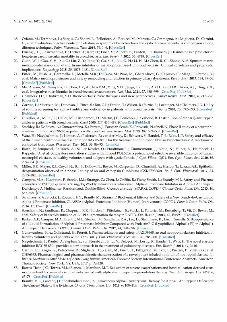
AAT is an albumin-like antiprotease secreted by hepatocytes, immune cells andbronchial epithelial cells [58]. AAT is able to inactivate through irreversible binding severalproteases including serin proteases and it has a high affinity for NE. AAT is an irreversibleinhibitor of serine proteases. When AAT docks serine proteases, the reactive central loopis cleaved in a high energy state and it is able to modify its conformation distorting andaltering the protease conformation, see Figure 2 [59]. AAT is coded in the SERPINA1(Serine peptidase inhibitor, clade A member 1) gene and polymorphisms in this gene areassociated with functional, dysfunctional, deficient or null variants of the protein and withlow levels of AAT in serum. Although more than one hundred polymorphisms in theAAT gene have been reported in literature, the most common variants associated withAAT serum deficiency are Z and S allele. The number of rare variants represents a largevariability in AAT production and functionality. These polymorphisms, sometimes withunknown function, may further be associated with AAT deficiency with an increasednumber of patients who might be affected by this condition [60,61]. Z allele is responsiblefor a mutated isoform of AAT and has been associated with the most severe deficiency inserum showing around 10% of the serum concentration of the physiological variants. Sallele is responsible for a milder deficiency, associated with nearly 60% of physiologicalAAT concentration in serum [61]. Carriers of pathological variants of AAT proteins withlow AAT serum concentration suffer from a rare genetic condition named α1 antitrypsindeficiency (AATD). Patients with Z AAT allele have 5 times less physiological concentrationof AAT and increased neutrophilic presence in lungs maybe because of an excess inchemoattractants [62]. A lack of AAT in the lungs may lead to unopposed protease activityand to tissue damage [62].

Int. J. Mol. Sci. 2021, 22, 5996 6 of 15Int. J. Mol. Sci. 2021, 22, x FOR PEER REVIEW 6 of 16
Figure 2. AAT mechanism of inhibition. (A) AAT binds NE, the reactive central loop is cleaved in a high energy state followed by a change of conformation (B) and the formation of an AAT-NE com-plex.
4.4.2. Antileukoprotease (ALP) Superfamily The antileukoprotease (ALP) superfamily includes enzymes secreted in the lung that
show an inhibitory effect on airway proteases. These proteins are synthesized and se-creted locally in the lung in response to primary cytokines such as IL-1 and TNF [63]. This superfamily includes elafin and secretory leukocyte proteinase inhibitor (SLPI).
SLPI. SLPI inhibits NE, Cat-G, trypsin, chymotrypsin and chymase, with a high af-finity to NE that is its major target. SLPI is believed to have an anti-inflammatory and anti-bacterial role [63].
Elafin. Elafin is a protein which inhibits a more limited number of proteases in com-parison to SLPI. Its main targets are NE and PR3. In addition to its inhibitory effect, elafin seems to be active against P. aeruginosa [63].
5. The Role of Proteases and Antiproteases in Bronchiectasis 5.1. Neutrophil Elastase in Bronchiectasis
NE represents a key biomarker in bronchiectasis, and levels of active NE (aNE) meas-ured in bronchoalveolar lavage fluid of bronchiectasis patients during stable state are higher compared to healthy controls [64,65]. From a microbiological point of view, NE directly correlates with bacterial load in sputum of patients chronically infected by P. ae-ruginosa [31]. From a clinical point of view, aNE is associated with a higher rate of exacer-bations in bronchiectasis. aNE concentration increases during bronchiectasis exacerba-tions and decreases during and after antibiotic treatment [31,66,67]. Although we are fo-cused on aNE, older methods for NE analysis included non-specific methods, not able to differentiate among serin proteases. In addition, older experiences reported NE concen-tration instead of activity, along with AAT-NE complex, higher in patients compared to healthy controls [18].
A few years ago, Chalmers and co-workers demonstrated an association between ac-tive NE levels in sputum of stable-state bronchiectasis adults and disease severity (evalu-ated through the Bronchiectasis Severity Index), pulmonary function, dyspnea, radiolog-ical severity and poor outcomes during follow-up, including exacerbations and hospital-ization [68]. These data have been recently confirmed across two southern European co-
Figure 2. AAT mechanism of inhibition. (A) AAT binds NE, the reactive central loop is cleaved in ahigh energy state followed by a change of conformation (B) and the formation of an AAT-NE complex.
4.4.2. Antileukoprotease (ALP) Superfamily
The antileukoprotease (ALP) superfamily includes enzymes secreted in the lung thatshow an inhibitory effect on airway proteases. These proteins are synthesized and secretedlocally in the lung in response to primary cytokines such as IL-1 and TNF [63]. Thissuperfamily includes elafin and secretory leukocyte proteinase inhibitor (SLPI).
SLPI. SLPI inhibits NE, Cat-G, trypsin, chymotrypsin and chymase, with a highaffinity to NE that is its major target. SLPI is believed to have an anti-inflammatory andanti-bacterial role [63].
Elafin. Elafin is a protein which inhibits a more limited number of proteases incomparison to SLPI. Its main targets are NE and PR3. In addition to its inhibitory effect,elafin seems to be active against P. aeruginosa [63].
5. The Role of Proteases and Antiproteases in Bronchiectasis5.1. Neutrophil Elastase in Bronchiectasis
NE represents a key biomarker in bronchiectasis, and levels of active NE (aNE)measured in bronchoalveolar lavage fluid of bronchiectasis patients during stable stateare higher compared to healthy controls [64,65]. From a microbiological point of view,NE directly correlates with bacterial load in sputum of patients chronically infectedby P. aeruginosa [31]. From a clinical point of view, aNE is associated with a higher rateof exacerbations in bronchiectasis. aNE concentration increases during bronchiectasisexacerbations and decreases during and after antibiotic treatment [31,66,67]. Although weare focused on aNE, older methods for NE analysis included non-specific methods, notable to differentiate among serin proteases. In addition, older experiences reported NEconcentration instead of activity, along with AAT-NE complex, higher in patients comparedto healthy controls [18].
A few years ago, Chalmers and co-workers demonstrated an association betweenactive NE levels in sputum of stable-state bronchiectasis adults and disease severity (evalu-ated through the Bronchiectasis Severity Index), pulmonary function, dyspnea, radiologicalseverity and poor outcomes during follow-up, including exacerbations and hospitaliza-tion [68]. These data have been recently confirmed across two southern European co-horts [18]. Patients were divided into three groups based on concentrations of aNE insputum (low aNE as aNE < 7 µg/mL, medium aNE between 7 and 20 µg/mL and highabove 20 µg/mL). aNE concentration directly correlated with disease severity, and it isinversely associated with quality of life [17,18,68]. The rate of patients with poor lung
Int. J. Mol. Sci. 2021, 22, 5996 7 of 15
function was higher in the medium and in the high aNE groups compared to the low aNEones. aNE levels were also higher in patients with chronic infection especially caused byP. aeruginosa [17]. A point of care assay has been recently developed to evaluate aNE insputum and tested in bronchiectasis patients [16]. This test showed a good performance inidentifying in few minutes patients at higher risk of airway infection and exacerbations.Finally, NE has been progressively identified as a relevant target for future treatments inbronchiectasis with pharmaceutical companies working on molecules able to inhibit thisprotease [57].
NE action in degrading elastin produces peptides including desmosine and isodesmo-sine, which are molecules frequently measured in serum and urine to evaluate NE activity.Circulating desmosine directly correlated with dyspnea, quality of life, radiological involve-ment and disease severity in bronchiectasis, and inversely with FEV1 (Forced ExpiratoryVolume in the 1st second) [21]. Moreover, serum desmosine concentration also correlatedwith sputum concentration of aNE [21]. Finally, high concentration of serum desmosinewas also associated with all-cause mortality, respiratory death in the first 3 years, andcardiovascular death after 3 years of follow-up in bronchiectasis [69].
5.2. Matrix Metalloproteinases in Bronchiectasis
Imbalances in MMPs levels as well as MMPs-TMPs ratios have been identified inbronchiectasis [70]. Several MMPs are associated with bronchiectasis. Taylor and colleaguesreported increased values of MMP-1, MMP-3, MMP-7, MMP-8, and MMP-9 and TIMP-2 and -4 levels, as well as MMP-8/TIMP-1 and MMP-9/TIMP-1 ratios in patients vs.healthy controls: Moreover, MMP-2 and MMP-8 are increased in patients with H. influenzae-dominated microbiota compared to those dominated by P. aeruginosa [70]. Guan andcolleagues reported increased levels of MMP-8, MMP-9 and MMP-9/TIMP-1 ratio inpatients with bronchiectasis and found an association between those levels and bothdisease and radiological severity. Notably, they also found MMP-9 levels increasing duringexacerbations [70]. Similar data were found in patients with bronchiectasis in general andin those with primary ciliary dyskinesia with researchers showing an association betweenhigh levels of MMPs and poor lung function [71].
5.3. Airway Proteases and Microbial Community in Bronchiectasis
The evaluation of airway inflammation alone in bronchiectasis limits our understand-ing of disease pathophysiology. A recent study evaluated the interaction between microbialcommunity and inflammatory biomarkers [15]. Among 185 adult patients with stable-statebronchiectasis enrolled in a cross-sectional study, those with aNE≥20 µg/mL had higherdisease severity compared to those with low aNE levels. High levels of active NE werecorrelated with low intra-patient (α) diversity, high levels of Pseudomonas genus, as well ashigh detection of P. aeruginosa in sputum by molecular biology; Streptococcus, Haemophilus,and Staphylococcus co-occurred instead in the low aNE group [17]. Another recent experi-ence evaluated integrative microbiomics in bronchiectasis and researchers demonstratedthe efficacy of this technique in understanding bronchiectasis exacerbations [72]. Differentclusters were identified through interaction networks involving fungi, viruses and bacteria.Pseudomonas interactome was different based on the exacerbations rate, suggesting anassociation between Pseudomonas interactome and exacerbation risk. This experience alsogave insight into exacerbations through shotgun metagenomics, confirming multi-biome in-teractions and the association of networks with exacerbations [72]. This approach has onlybeen applied to multi-biome analysis, not including airway inflammation. Multi-omicsapproaches are nowadays focusing on chronic respiratory diseases in order to integratedifferent analyses such as microbiome, transcriptomic, metatranscriptomic, metabolomics,proteomic and others, to investigate host pathogen interaction, in order to identify peculiarendotypes in these patients [70].
MMPs have also been studied in the context of lung microbiota in bronchiectasispatients. MMPs quantification and 16s rRNA gene sequencing were conducted in induced
Int. J. Mol. Sci. 2021, 22, 5996 8 of 15
sputum of 86 bronchiectasis patients and 8 healthy controls as well as clinical assessment.This experience showed increased MMP-2 and MMP-8 in patients H. influenzae-dominatedcompared to those with a P. aeruginosa-dominated microbiome [73].
5.4. Antiproteases in Bronchiectasis
In addition to proteases, also the role of antiproteases is under study in bronchiectasis.Genetic AAT deficiency has been associated with bronchiectasis, along with the associationrecently found between low saliva SLPI levels and bronchiectasis severity index and lowsputum SLPI, exacerbation frequency and longer time to the next exacerbation [33].
α1 Antitrypsin Deficiency (AATD) in Bronchiectasis
Very few experiences evaluated prevalence of AATD in bronchiectasis patients. AATserum concentration has been recently evaluated in two different cohorts from the UK:2.5% of patients in Scotland and 7.1% in England had serum levels of AAT less than1 g/L. Among the 1600 patients, 0.5% had PiZZ (proteinase inhibitor ZZ), 0.4% PiSZ (pro-teinase inhibitor SZ) or PiS (proteinase inhibitor SZ), and 3% PiMZ (proteinase inhibitorMZ) phenotype. This screening identified severe AATD in less than 1% of patients withbronchiectasis [74], confirming data from a previous study conducted in France in 2000 [75].Another study considering PiZ patients reported 27% of these patients with clinically signif-icant bronchiectasis. These patients showed high airway disease and more severe emphy-sema [76]. Although genetic deficiency of AAT is frequently associated with bronchiectasis,secondary deficiencies may also potentially contribute to bronchiectasis development.
6. Treatments of Protease–Antiprotease Imbalance in Bronchiectasis
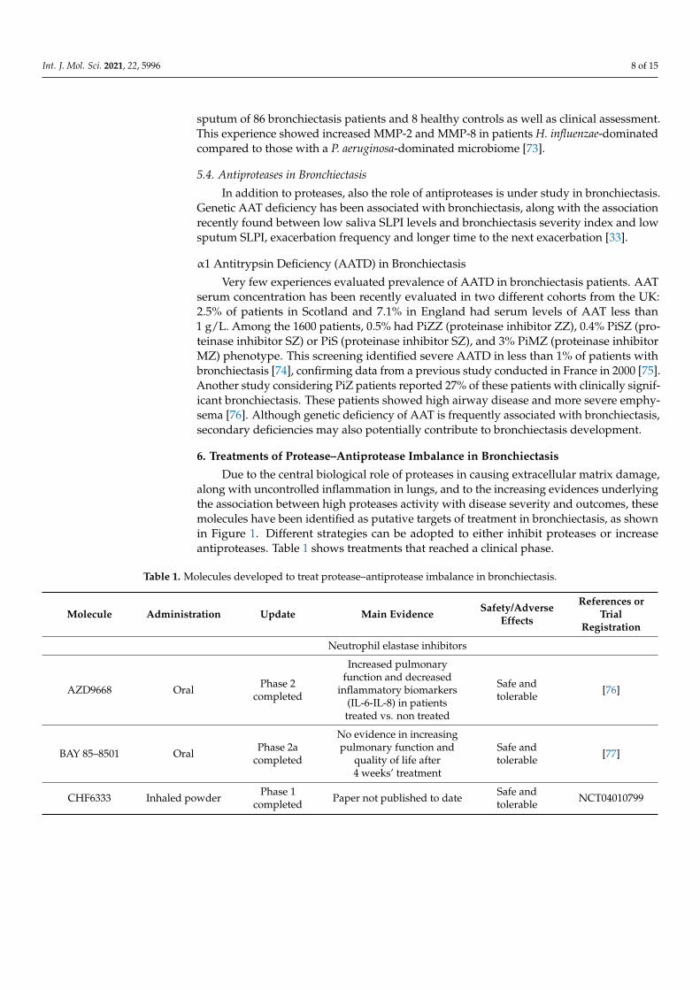
Due to the central biological role of proteases in causing extracellular matrix damage,along with uncontrolled inflammation in lungs, and to the increasing evidences underlyingthe association between high proteases activity with disease severity and outcomes, thesemolecules have been identified as putative targets of treatment in bronchiectasis, as shownin Figure 1. Different strategies can be adopted to either inhibit proteases or increaseantiproteases. Table 1 shows treatments that reached a clinical phase.
Table 1. Molecules developed to treat protease–antiprotease imbalance in bronchiectasis.
Molecule Administration Update Main Evidence Safety/AdverseEffects
References orTrial
Registration
Neutrophil elastase inhibitors
AZD9668 Oral Phase 2completed
Increased pulmonaryfunction and decreased
inflammatory biomarkers(IL-6-IL-8) in patientstreated vs. non treated
Safe andtolerable [76]
BAY 85–8501 Oral Phase 2acompleted
No evidence in increasingpulmonary function and
quality of life after4 weeks’ treatment
Safe andtolerable [77]
CHF6333 Inhaled powder Phase 1completed Paper not published to date Safe and
tolerable NCT04010799

Int. J. Mol. Sci. 2021, 22, 5996 9 of 15
Table 1. Cont.
Molecule Administration Update Main Evidence Safety/AdverseEffects
References orTrial
Registration
Neutrophil elastase inhibitors
POL6014 Inhaled Phase 1completed
No data available onbronchiectasis
Safe andtolerable [78]
Cathepsin C/DPP1 inhibitors
GSK2793660 Oral Phase 1 -Terminatedbecause of
adverse events[79]
Brensocatib Oral Phase 2completed
Effective in reducing time tothe first exacerbation and
rate of severe exacerbations
Safe andtolerable [57]
AATD therapy
Prolastin C Intravenous Post-marketingEffective in increasing AATserum levels. It reduces the
decline in lung density.
Safe andtolerable [80]
API-GLASSIA Intravenous Post-marketing Bioequivalent to Prolastin-C Safe andtolerable [81]
Zemaira Intravenous Post-marketing
Dose confirmed, higherdose may be associated with
greater effect. Preventsemphysema in AATD
patients
Safe andtolerable [82]
Liquid Alpha1-PI Intravenous Post-marketing Bioequivalent to Prolastin-C Safe andtolerable [83]
6.1. Active Neutrophil Elastase Inhibitors
Over the past decade, different molecules have been developed to target NE in bronchiectasis.AZD9668. AZD9668 is an oral reversible inhibitor of NE. The most recent experience
reported a small phase II double-blind controlled clinical trial on 38 bronchiectasis pa-tients [75]. Subjects who underwent 4 weeks of treatment with this inhibitor showed anincrease in pulmonary function, with a trend to a reduction of inflammatory biomarkers(IL-8- IL-6) in patients who undergo treatment vs. placebo. No difference was found interms of sputum volume and quality of life [84]. This inhibitor was also tested in CF andCOPD with inconclusive results [9].
BAY 85–8501. Another NE inhibitor which has been recently developed and testedin bronchiectasis is BAY 85–8501. This is a reversible and selective NE inhibitor activein animal models of acute lung injury. In phase I, no adverse effects were reported,and the drug was well tolerated at increasing dosage in healthy volunteers [85]. In arecently published phase 2a controlled randomized double-blind clinical trial, ninety-fourbronchiectasis patients were treated for 4 weeks with oral BAY 85–8501. Although bothtolerability and safety were proved in these patients along with a decrease in aNE in blood,no difference in terms of pulmonary function, quality of life and inflammatory and tissuedamage biomarkers including desmosine in both sputum and urine were detected betweenpatients treated with this molecule and placebo. The authors concluded that a larger studywith a prolonged time treatment is needed to better evaluate clinical outcomes [77].
CHF6333. Different administration strategies have been adopted to increase thebiodisponibility of the compounds at the target, including new molecules for NE inhibitiondelivered through inhalation. CHF6333 is the first inhaled candidate for NE inhibitionin bronchiectasis as well as in CF. Preclinical data showed high power of the compound

Int. J. Mol. Sci. 2021, 22, 5996 10 of 15
in inhibiting NE and data ex vivo on both bronchiectasis and CF bronchoalveolar lavagefluid samples show a higher inhibitory power of this molecule compared to BAY 85–8501.A phase I randomized clinical trial including CF and bronchiectasis patients has beenconcluded in 2020 (clinicaltrial.gov ID NCT04010799), although no data are currentlyavailable [86].
POL6014. POL6014 is a second inhaled compound recently proven to be safe inhealthy subjects and CF patients. This molecule, safe and well tolerated in the two studygroups, was reported to be successfully delivered to lungs with a poor systemic uptake [78].A double-blind, randomized, controlled, clinical trial has been completed in 2020 in CFpatients, although no data are currently available (clinicaltrial.gov ID NCT03748199).
6.2. DPP1 Inhibitors
While studies aimed at directly inhibiting NE in bronchiectasis patients have beencarried out, a new approach aimed at targeting the enzyme responsible for serine proteasesactivation has been proposed (Figure 3).
Int. J. Mol. Sci. 2021, 22, x FOR PEER REVIEW 11 of 16
Figure 3. Structure of DPP1 (A), brensocatib (B) and GSK2793660 (C).
6.3. AAT Supplementation Clinical trials aimed at investigating the efficacy of AAT supplementation reported a
beneficial effect of this treatment in terms of change of pulmonary density, decreased number of exacerbations/year and improved survival [87,88]. Several trials reported in-creased serum levels and anti-elastase capacity of these treatments in patients with AATD, confirming the stability of AAT serum levels in patients treated with intravascular AAT supplementation without serious adverse effects along with a positive effect of supple-mentation therapy on clinical endpoints as lung function decline, exacerbations risk and mortality [82,88]. All treatments listed in Table 1 represent different formulation of AAT augmentation therapy developed and available on the market. All the products resulted to be biosimilar to Prolastin C and no specific data on bronchiectasis patients with AATD are available.
6.4. Future Perspectives Heterogeneous results were observed in the trials studying proteases inhibitors and
antiproteases activity enhancers. Although the treatments characteristics are fundamental for the mechanism of action, along with the delivery method, the selection of the correct population to test and subsequently treat with a protease inhibitor is fundamental for the success of the treatment. The development of point-of-care analysis for proteases activity detection along with an increasing knowledge of patients endotype associated with high proteases in airways is a positive step, helping both clinicians and scientists in the selec-tion of the correct population [16,17]. The development of these tests may be very im-portant for a quick and broad evaluation of aNE in patients and the selection of the most suitable population to treat. As mentioned, not all patients have a neutrophilic inflamma-tion phenotype in stable state, and the heterogeneous results from clinical trials may be due to an inaccurate selection of the target population, including patients not able to re-spond to treatment, due to low values of aNE in lower airways. Moreover, a long-term effect at both efficacy and safety level of protease inhibition needs to be considered, and drug development is fundamental in order to achieve inflammatory inhibition without compromising host defenses, as well as inducing potential adverse effects in other depart-ments, as previously observed [57,73].
Different situations are associated with exacerbation. These treatments have a crucial role in reducing exacerbations risk and disease progression. We previously reported the role of these molecules in decreasing exacerbation risk in patients with high levels of aNE; however, future studies should focus on the use of these compounds in inhibiting aNE in patients with low baseline levels of aNE, which usually increases during exacerbations.
Figure 3. Structure of DPP1 (A), brensocatib (B) and GSK2793660 (C).
Brensocatib. Brensocatib is an oral reversible inhibitor of DPP1, the enzyme responsiblefor serin proteases inhibition that has been associated with decreased serin proteases levelsin healthy controls. Recently, Chalmers and colleagues published the results from a phaseII, randomized, double-blind, placebo-controlled clinical trial in bronchiectasis patients [57].Of 256 patients, 87 were assigned to receive placebo, 82 to receive 10 mg of brensocatib daily,and 87 to receive 25 mg of brensocatib for 24 weeks. Researchers found an improvement inclinical outcomes in treated vs. non-treated patients. Patients treated with two differentdoses of brensocatib showed an increased time to first exacerbation and a decreasedincidence rate of exacerbations compared to placebo. These patients also showed a lowerrate of severe exacerbations, lower negative change in FEV1 after bronchodilation, and adecrease in sputum aNE levels compared to the placebo ones. NE activity was restored tobaseline levels after 4 weeks from the end of the drug administration [57]. However, theincidence of dental and skin adverse events was higher with both the brensocatib dosescompared with placebo.
GSK2793660. Another molecule was previously developed to inhibit DPP1. AlthoughGSK2793660 was able to inhibit the majority of DPP1, downstream serine proteases activitywas inhibited in just 20% of the cases [79]. Additional studies were prematurely terminatedbecause of adverse effects.

Int. J. Mol. Sci. 2021, 22, 5996 11 of 15
6.3. AAT Supplementation
Clinical trials aimed at investigating the efficacy of AAT supplementation reporteda beneficial effect of this treatment in terms of change of pulmonary density, decreasednumber of exacerbations/year and improved survival [87,88]. Several trials reportedincreased serum levels and anti-elastase capacity of these treatments in patients withAATD, confirming the stability of AAT serum levels in patients treated with intravascularAAT supplementation without serious adverse effects along with a positive effect ofsupplementation therapy on clinical endpoints as lung function decline, exacerbationsrisk and mortality [82,88]. All treatments listed in Table 1 represent different formulationof AAT augmentation therapy developed and available on the market. All the productsresulted to be biosimilar to Prolastin C and no specific data on bronchiectasis patients withAATD are available.
6.4. Future Perspectives
Heterogeneous results were observed in the trials studying proteases inhibitors andantiproteases activity enhancers. Although the treatments characteristics are fundamentalfor the mechanism of action, along with the delivery method, the selection of the correctpopulation to test and subsequently treat with a protease inhibitor is fundamental forthe success of the treatment. The development of point-of-care analysis for proteasesactivity detection along with an increasing knowledge of patients endotype associatedwith high proteases in airways is a positive step, helping both clinicians and scientistsin the selection of the correct population [16,17]. The development of these tests may bevery important for a quick and broad evaluation of aNE in patients and the selection ofthe most suitable population to treat. As mentioned, not all patients have a neutrophilicinflammation phenotype in stable state, and the heterogeneous results from clinical trialsmay be due to an inaccurate selection of the target population, including patients notable to respond to treatment, due to low values of aNE in lower airways. Moreover,a long-term effect at both efficacy and safety level of protease inhibition needs to beconsidered, and drug development is fundamental in order to achieve inflammatoryinhibition without compromising host defenses, as well as inducing potential adverseeffects in other departments, as previously observed [57,73].
Different situations are associated with exacerbation. These treatments have a crucialrole in reducing exacerbations risk and disease progression. We previously reported therole of these molecules in decreasing exacerbation risk in patients with high levels of aNE;however, future studies should focus on the use of these compounds in inhibiting aNE inpatients with low baseline levels of aNE, which usually increases during exacerbations.
The effort of the scientific community in endotyping bronchiectasis patients may pavethe way to new targets of treatment and contribute to the precision medicine approachadopted for the management of this disease. Proteases and the proteases–anti-proteasesimbalance represent some of the most interesting targets in bronchiectasis to be explored infuture studies.
7. Conclusions
Protease–antiprotease balance is crucial in bronchiectasis pathophysiology. Increasedpresence of unopposed proteases activity may contribute to bronchiectasis onset and pro-gression. The identification of different targets of therapy within the protease–antiproteasebalance contributes to a precision medicine approach in bronchiectasis and eventuallyinterrupts the vicious vortex, which characterizes the progression of the disease. Datafrom the clinical trials on DPP1 and the new trials ongoing on NE inhibitors seem verypromising. However, future studies are needed to better understand the host pathogeninteraction in bronchiectasis, to better characterize patients with different inflammatorypatterns, to understand the role of potential treatments in use in other respiratory diseasesand to identify new treatable traits in bronchiectasis.

Int. J. Mol. Sci. 2021, 22, 5996 12 of 15
Author Contributions: Conceptualization, S.A. and M.O.; writing—original draft preparation, S.A.and M.O.; writing—review and editing, F.A., A.G., A.D.S., M.M., A.V., O.S., S.H.C., P.M., and F.B.;supervision, S.A. All authors have read and agreed to the published version of the manuscript.
Funding: This research received no external funding.
Conflicts of Interest: M.O., F.A., A.G., M.M., O.S., S.H.C., A.V. and P.M. declare no conflict of interest.F.B. reports grants and personal fees from AstraZeneca, grants from Bayer, grants and personal feesfrom Chiesi, grants and personal fees from GlaxoSmithKline, personal fees from Grifols, personal feesfrom Guidotti, personal fees from Insmed, grants and personal fees from Menarini, personal fees fromNovartis, grants and personal fees from Pfizer, personal fees from Zambon, personal fees from Vertex,outside the submitted work. S.A. reports personal fees from Bayer Healthcare, personal fees fromGrifols, personal fees from AstraZeneca, personal fees from Zambon, grants and personal fees fromChiesi, grants and personal fees from Insmed, personal fees from GlaxoSmithKline, personal feesfrom Menarini, personal fees from ZetaCube Srl, grants from Fisher & Paykel, outside the submittedwork. A.D.S. reports grants, speakers fees and travel support from AstraZeneca, Bayer, Chiesi, Forestlabs, GSK, Insmed, Pfizer, Medimmune, Novartis and Zambon outside the submitted work.
References1. Polverino, E.; Goeminne, P.C.; McDonnell, M.J.; Aliberti, S.; Marshall, S.E.; Loebinger, M.R.; Murris, M.; Cantón, R.; Torres, A.;
Dimakou, K.; et al. European Respiratory Society guidelines for the management of adult bronchiectasis. Eur. Respir. J. 2017, 50,1700629. [CrossRef]
2. Aliberti, S.; Sotgiu, G.; Lapi, F.; Gramegna, A.; Cricelli, C.; Blasi, F. Prevalence and incidence of bronchiectasis in Italy. BMC Pulm.Med. 2020, 20, 15. [CrossRef]
3. Quint, J.K.; Millett, E.R.C.; Joshi, M.; Navaratnam, V.; Thomas, S.L.; Hurst, J.R.; Smeeth, L.; Brown, J.S. Changes in the incidence,prevalence and mortality of bronchiectasis in the UK from 2004 to 2013: A population-based cohort study. Eur. Respir. J. 2016, 47,186–193. [CrossRef] [PubMed]
4. Monteagudo, M.; Rodríguez-Blanco, T.; Barrecheguren, M.; Simonet, P.; Miravitlles, M. Prevalence and incidence of bronchiectasisin Catalonia, Spain: A population-based study. Respir. Med. 2016, 121, 26–31. [CrossRef] [PubMed]
5. Lonni, S.; Chalmers, J.D.; Goeminne, P.C.; McDonnell, M.J.; Dimakou, K.; De Soyza, A.; Polverino, E.; Van de Kerkhove, C.;Rutherford, R.; Davison, J.; et al. Etiology of Non-Cystic Fibrosis Bronchiectasis in Adults and Its Correlation to Disease Severity.Ann. Am. Thorac. Soc. 2015, 12, 1764–1770. [CrossRef] [PubMed]
6. Flume, P.A.; Chalmers, J.D.; Olivier, K.N. Advances in bronchiectasis: Endotyping, genetics, microbiome, and disease heterogene-ity. Lancet 2018, 392, 880–890. [CrossRef]
7. Cole, P.J. Inflammation: A two-edged sword—The model of bronchiectasis. Eur. J. Respir. Dis. Suppl. 1986, 147, 6–15.8. Amati, F.; Simonetta, E.; Gramegna, A.; Tarsia, P.; Contarini, M.; Blasi, F.; Aliberti, S. The biology of pulmonary exacerbations in
bronchiectasis. Eur. Respir. Rev. 2019, 28, 190055. [CrossRef]9. Faverio, P.; Stainer, A.; Bonaiti, G.; Zucchetti, S.C.; Simonetta, E.; Lapadula, G.; Marruchella, A.; Gori, A.; Blasi, F.; Codecasa, L.;
et al. Characterizing Non-Tuberculous Mycobacteria Infection in Bronchiectasis. Int. J. Mol. Sci. 2016, 17, 1913. [CrossRef]10. Bonaiti, G.; Pesci, A.; Marruchella, A.; Lapadula, G.; Gori, A.; Aliberti, S. Nontuberculous Mycobacteria in Noncystic Fibrosis
Bronchiectasis. Biomed. Res. Int. 2015, 2015, 197950. [CrossRef] [PubMed]11. Knowles, M.R.; Zariwala, M.; Leigh, M. Primary Ciliary Dyskinesia. Clin. Chest Med. 2016, 37, 449–461. [CrossRef]12. Contarini, M.; Shoemark, A.; Rademacher, J.; Finch, S.; Gramegna, A.; Gaffuri, M.; Roncoroni, L.; Seia, M.; Ringshausen, F.C.;
Welte, T.; et al. Why, when and how to investigate primary ciliary dyskinesia in adult patients with bronchiectasis. Multidiscip.Respir. Med. 2018, 13, 26. [CrossRef]
13. Balázs, A.; Mall, M.A. Mucus obstruction and inflammation in early cystic fibrosis lung disease: Emerging role of the IL-1signaling pathway. Pediatr. Pulmonol. 2019, 54, S5–S12. [CrossRef]
14. Ramzi, N.; Jamee, M.; Bakhtiyari, M.; Rafiemanesh, H.; Zainaldain, H.; Tavakol, M.; Rezaei, A.; Kalvandi, M.; Zian, Z.;Mohammadi, H.; et al. Bronchiectasis in common variable immunodeficiency: A systematic review and meta-analysis. Pediatr.Pulmonol. 2020, 55, 292–299. [CrossRef]
15. Wang, D.; Zhang, J.; Lau, J.; Wang, S.; Taneja, V.; Matteson, E.L.; Vassallo, R. Mechanisms of lung disease development inrheumatoid arthritis. Nat. Rev. Rheumatol. 2019, 15, 581–596. [CrossRef]
16. Shoemark, A.; Cant, E.; Carreto, L.; Smith, A.; Oriano, M.; Keir, H.R.; Perea, L.; Canto, E.; Terranova, L.; Vidal, S.; et al. Apoint-of-care neutrophil elastase activity assay identifies bronchiectasis severity, airway infection and risk of exacerbation. Eur.Respir. J. 2019, 53, 1900303. [CrossRef]
17. Oriano, M.; Gramegna, A.; Terranova, L.; Sotgiu, G.; Sulaiman, I.; Ruggiero, L.; Saderi, L.; Wu, B.; Chalmers, J.D.; Segal, L.N.;et al. Sputum Neutrophil Elastase associates with microbiota and P. aeruginosa in bronchiectasis. Eur. Respir. J. 2020, 56, 2000769.[CrossRef] [PubMed]

Int. J. Mol. Sci. 2021, 22, 5996 13 of 15
18. Gramegna, A.; Aliberti, S.; Sibila, O.; Di Francesco, C.; Sotgiu, G.; Perea, L.; Terranova, L.; Oriano, M.; Pilocane, T.; Saderi, L.; et al.Sputum neutrophil elastase in bronchiectasis: A Southern European cohort study. Eur. Respir. J. 2020, 56, 2001702. [CrossRef][PubMed]
19. Korkmaz, B.; Attucci, S.; Juliano, M.A.; Kalupov, T.; Jourdan, M.L.; Juliano, L.; Gauthier, F. Measuring elastase, proteinase 3 andcathepsin G activities at the surface of human neutrophils with fluorescence resonance energy transfer substrates. Nat. Protoc.2008, 3, 991–1000. [CrossRef] [PubMed]
20. Elkington, P.T.G.; Friedland, J.S. Matrix metalloproteinases in destructive pulmonary pathology. Thorax 2006, 61, 259–266.[CrossRef]
21. Chalmers, J.D.; Moffitt, K.L.; Suarez-Cuartin, G.; Sibila, O.; Finch, S.; Furrie, E.; Dicker, A.; Wrobel, K.; Elborn, J.S.; Walker, B.; et al.Neutrophil elastase activity is associated with exacerbations and lung function decline in bronchiectasis. Am. J. Respir. Crit. CareMed. 2017, 195, 1384–1393. [CrossRef] [PubMed]
22. Keir, H.R.; Shoemark, A.; Dicker, A.J.; Perea, L.; Pollock, J.; Giam, Y.H.; Suarez-Cuartin, G.; Crichton, M.L.; Lonergan, M.;Oriano, M.; et al. Neutrophil Extracellular Traps are Increased in Severe Bronchiectasis and Reduced by Long-Term AzithromycinTreatment. Lancet 2020. [CrossRef]
23. Potempa, J.A.N.; Banbula, A.; Travis, J.I.M. Role of bacterial proteinases in matrix destruction and modulation of host responses.Periodontol. 2000 2000, 24, 153–192. [CrossRef] [PubMed]
24. Sandri, A.; Ortombina, A.; Boschi, F.; Cremonini, E.; Boaretti, M.; Sorio, C.; Melotti, P.; Bergamini, G.; Lleo, M. Inhibition ofPseudomonas aeruginosa secreted virulence factors reduces lung inflammation in CF mice. Virulence 2018, 9, 1008–1018. [CrossRef][PubMed]
25. Dente, F.L.; Bilotta, M.; Bartoli, M.L.; Bacci, E.; Cianchetti, S.; Latorre, M.; Malagrinò, L.; Nieri, D.; Roggi, M.A.; Vagaggini, B.;et al. Neutrophilic Bronchial Inflammation Correlates with Clinical and Functional Findings in Patients with Noncystic FibrosisBronchiectasis. Mediat. Inflamm. 2015, 2015, 642503. [CrossRef] [PubMed]
26. Weiss, S.J. Tissue destruction by neutrophils. N. Engl. J. Med. 1989, 320, 365–376. [CrossRef]27. Craig, A.; Mai, J.; Cai, S.; Jeyaseelan, S. Neutrophil Recruitment to the Lungs during Bacterial Pneumonia. Infect. Immun. 2009, 77,
568–575. [CrossRef]28. Weissmann, G.; Smolen, J.E.; Korchak, H.M. Release of Inflammatory Mediators from Stimulated Neutrophils. N. Engl. J. Med.
1980, 303, 27–34. [CrossRef]29. Rossaint, J.; Zarbock, A. Tissue-Specific Neutrophil Recruitment into the Lung, Liver, and Kidney. J. Innate Immun. 2013, 5,
348–357. [CrossRef]30. Aliberti, S.; Lonni, S.; Dore, S.; Mcdonnell, M.J.; Goeminne, P.C.; Dimakou, K.; Fardon, T.C.; Rutherford, R.; Pesci, A.;
Restrepo, M.I.; et al. Clinical phenotypes in adult patients with bronchiectasis. Eur. Respir. J. 2016, 47, 1113–1122. [CrossRef]31. Chalmers, J.D.; Smith, M.P.; McHugh, B.J.; Doherty, C.; Govan, J.R.; Hill, A.T. Short- and Long-Term Antibiotic Treatment Reduces
Airway and Systemic Inflammation in Non–Cystic Fibrosis Bronchiectasis. Am. J. Respir. Crit. Care Med. 2012, 186, 657–665.[CrossRef] [PubMed]
32. Keir, H.R.; Shoemark, A.; Dicker, A.J.; Perea, L.; Pollock, J.; Giam, Y.H.; Suarez-Cuartin, G.; Crichton, M.L.; Lonergan, M.;Oriano, M.; et al. Neutrophil extracellular traps, disease severity, and antibiotic response in bronchiectasis: An international,observational, multicohort study. Lancet Respir. Med. 2021. [CrossRef]
33. Sibila, O.; Perea, L.; Cantó, E.; Shoemark, A.; Cassidy, D.; Smith, A.H.; Suarez-Cuartin, G.; Rodrigo-Troyano, A.; Keir, H.R.;Oriano, M.; et al. Antimicrobial peptides, disease severity and exacerbations in bronchiectasis. Thorax 2019, thoraxjnl-2018-212895.[CrossRef] [PubMed]
34. Fuschillo, S.; De Felice, A.; Balzano, G. Mucosal inflammation in idiopathic bronchiectasis: Cellular and molecular mechanisms.Eur. Respir. J. 2008, 31, 396–406. [CrossRef] [PubMed]
35. Angrill, J.; Agustí, C.; De Celis, R.; Filella, X.; Rañó, A.; Elena, M.; De La Bellacasa, J.P.; Xaubet, A.; Torres, A. Bronchialinflammation and colonization in patients with clinically stable bronchiectasis. Am. J. Respir. Crit. Care Med. 2001, 164, 1628–1632.[CrossRef]
36. Martinez-Garcia, M.A.; Posadas, T.; Sotgiu, G.; Blasi, F.; Saderi, L.; Aliberti, S. Role of inhaled corticosteroids in reducingexacerbations in bronchiectasis patients with blood eosinophilia pooled post-hoc analysis of 2 randomized clinical trials. Respir.Med. 2020, 172, 106127. [CrossRef]
37. Perea, L.; Cantó, E.; Suarez-Cuartin, G.; Aliberti, S.; Chalmers, J.D.; Sibila, O.; Vidal, S. A Cluster Analysis of BronchiectasisPatients Based on the Airway Immune Profile. Chest 2020, 159, 1758–1767. [CrossRef]
38. Martinez-Garcia, M.A.; Posadas, T.; Sotgiu, G.; Blasi, F.; Saderi, L.; Aliberti, S. Repeteability of Circulating Eosinophil Measuresand Inhaled Corticosteroids Effect in Bronchiectasis: A Post Hoc Analysis of a Randomized Clinical Trial. Arch. Bronconeumol.2020, 56, 681–683. [CrossRef]
39. Aliberti, S.; Sotgiu, G.; Martinez Garcia, M.-A. Blood eosinophils do not predict inhaled budesonide response in bronchiectasis.Eur. Respir. J. 2020, 56, 2002210. [CrossRef]
40. Rademacher, J.; Konwert, S.; Fuge, J.; Dettmer, S.; Welte, T.; Ringshausen, F.C. Anti-IL5 and anti-IL5Rα therapy for clinicallysignificant bronchiectasis with eosinophilic endotype: A case series. Eur. Respir. J. 2020, 55, 1901333. [CrossRef]
41. López-Boado, Y.S.; Espinola, M.; Bahr, S.; Belaaouaj, A. Neutrophil serine proteinases cleave bacterial flagellin, abrogating its hostresponse-inducing activity. J. Immunol. 2004, 172, 509–515. [CrossRef]

Int. J. Mol. Sci. 2021, 22, 5996 14 of 15
42. Small, D.M.; Brown, R.R.; Doherty, D.F.; Abladey, A.; Zhou-Suckow, Z.; Delaney, R.J.; Kerrigan, L.; Dougan, C.M.;Borensztajn, K.S.; Holsinger, L.; et al. Targeting of Cathepsin S Reduces Cystic Fibrosis-like Lung Disease. Eur. Respir.J. 2019, 1801523. [CrossRef]
43. Segal, A.W. Europe PMC Funders Group How Neutrophils Kill Microbes. Annu. Rev. Immunol. 2007, 2. [CrossRef]44. Korkmaz, B.; Horwitz, M.S.; Jenne, D.E.; Gauthier, F. Neutrophil Elastase, Proteinase 3, and Cathepsin G as Therapeutic Targets in
Human Diseases. Pharmacol. Rev. 2010, 62, 726–759. [CrossRef] [PubMed]45. Sedor, J.; Hogue, L.; Akers, K.; Boslaugh, S.; Schreiber, J.; Ferkol, T. Cathepsin-G interferes with clearance of Pseudomonas aeruginosa
from mouse lungs. Pediatr. Res. 2007, 61, 26–31. [CrossRef]46. Crisford, H.; Sapey, E.; Stockley, R.A. Proteinase 3; a potential target in chronic obstructive pulmonary disease and other chronic
inflammatory diseases. Respir. Res. 2018, 19, 180. [CrossRef]47. Benabid, R.; Wartelle, J.; Malleret, L.; Guyot, N.; Gangloff, S.; Lebargy, F.; Belaaouaj, A. Neutrophil elastase modulates cytokine
expression: Contribution to host defense against pseudomonas aeruginosa-induced pneumonia. J. Biol. Chem. 2012, 287,34883–34894. [CrossRef] [PubMed]
48. Tosi, M.F.; Zakem, H.; Berger, M. Neutrophil elastase cleaves C3bi on opsonized pseudomonas as well as CR1 on neutrophils tocreate a functionally important opsonin receptor mismatch. J. Clin. Investig. 1990, 86, 300–308. [CrossRef] [PubMed]
49. Rubio, F.; Cooley, J.; Accurso, F.J.; Remold-O’Donnell, E. Linkage of neutrophil serine proteases and decreased surfactantprotein-A (SP-A) levels in inflammatory lung disease. Thorax 2004, 59, 318–323. [CrossRef] [PubMed]
50. Gramegna, A.; Amati, F.; Terranova, L.; Sotgiu, G.; Tarsia, P.; Miglietta, D.; Calderazzo, M.A.; Aliberti, S.; Blasi, F. Neutrophilelastase in bronchiectasis. Respir. Res. 2017, 18, 1–13. [CrossRef]
51. Van Wart, H.E.; Birkedal-Hansen, H. The cysteine switch: A principle of regulation of metalloproteinase activity with potentialapplicability to the entire matrix metalloproteinase gene family. Proc. Natl. Acad. Sci. USA 1990, 87, 5578–5582. [CrossRef][PubMed]
52. Lemaître, V.; D’Armiento, J. Matrix metalloproteinases in development and disease. Birth Defects Res. Part C Embryo Today Rev.2006, 78, 1–10. [CrossRef] [PubMed]
53. Van den Steen, P.E.; Proost, P.; Wuyts, A.; Van Damme, J.; Opdenakker, G. Neutrophil gelatinase B potentiates interleukin-8tenfold by aminoterminal processing, whereas it degrades CTAP-III, PF-4, and GRO-α and leaves RANTES and MCP-2 intact.Blood 2000, 96, 2673–2681. [CrossRef] [PubMed]
54. Liu, Z.; Zhou, X.; Shapiro, S.D.; Shipley, J.M.; Twining, S.S.; Diaz, L.A.; Senior, R.M.; Werb, Z. The serpin alpha1-proteinaseinhibitor is a critical substrate for gelatinase B/MMP-9 in vivo. Cell 2000, 102, 647–655. [CrossRef]
55. Wolters, P.J.; Laig-Webster, M.; Caughey, G.H. Dipeptidyl peptidase I cleaves matrix-associated proteins and is expressed mainlyby mast cells in normal dog airways. Am. J. Respir. Cell Mol. Biol. 2000, 22, 183–190. [CrossRef] [PubMed]
56. Wolters, P.J.; Chapman, H.A. Importance of lysosomal cysteine proteases in lung disease. Respir. Res. 2000, 1, 170–177. [CrossRef]57. Chalmers, J.D.; Haworth, C.S.; Metersky, M.L.; Loebinger, M.R.; Blasi, F.; Sibila, O.; O’Donnell, A.E.; Sullivan, E.J.; Mange, K.C.;
Fernandez, C.; et al. Phase 2 Trial of the DPP-1 Inhibitor Brensocatib in Bronchiectasis. N. Engl. J. Med. 2020, 383, 2127–2137.[CrossRef]
58. Fagerhol, M.K.; Laurell, C.-B. The polymorphism of “prealbumins” and α1-antitrypsin in human sera. Clin. Chim. Acta 1967, 16,199–203. [CrossRef]
59. Stoller, J.K.; Aboussouan, L.S. A review of α1-antitrypsin deficiency. Am. J. Respir. Crit. Care Med. 2012, 185, 246–259. [CrossRef]60. Veith, M.; Tüffers, J.; Peychev, E.; Klemmer, A.; Kotke, V.; Janciauskiene, S.; Wilhelm, S.; Bals, R.; Koczulla, A.R.; Vogelmeier, C.F.;
et al. The Distribution of Alpha-1 Antitrypsin Genotypes Between Patients with COPD/Emphysema, Asthma and Bronchiectasis.Int. J. Chron. Obstruct. Pulmon. Dis. 2020, 15, 2827–2836. [CrossRef]
61. Ferrarotti, I.; Thun, G.A.; Zorzetto, M.; Ottaviani, S.; Imboden, M.; Schindler, C.; Von Eckardstein, A.; Rohrer, L.; Rochat, T.;Russi, E.W.; et al. Serum levels and genotype distribution of α1-antitrypsin in the general population. Thorax 2012, 67, 669–674.[CrossRef]
62. Kelly, E.; Greene, C.M.; Carroll, T.P.; McElvaney, N.G.; O’Neill, S.J. Alpha-1 antitrypsin deficiency. Respir. Med. CME 2011, 4, 1–8.[CrossRef]
63. Sallenave, J.M. The role of secretory leukocyte proteinase inhibitor and elafin (elastase-specific inhibitor/skin-derived antileuko-protease) as alarm antiproteinases in inflammatory lung disease. Respir. Res. 2000, 1, 87–92. [CrossRef] [PubMed]
64. Vandivier, R.W.; Fadok, V.A.; Hoffmann, P.R.; Bratton, D.L.; Penvari, C.; Brown, K.K.; Brain, J.D.; Accurso, F.J.; Henson, P.M.Elastase-mediated phosphatidylserine receptor cleavage impairs apoptotic cell clearance in cystic fibrosis and bronchiectasis. J.Clin. Investig. 2002, 109, 661–670. [CrossRef] [PubMed]
65. Nakamura, H.; Abe, S.; Shibata, Y.; Yuki, H.; Suzuki, H.; Saito, H.; Sata, M.; Kato, S.; Tomoike, H. Elevated levels of cytokeratin19 in the bronchoalveolar lavage fluid of patients with chronic airway inflammatory diseases—A specific marker for bronchialepithelial injury. Am. J. Respir. Crit. Care Med. 1997, 155, 1217–1221. [CrossRef] [PubMed]
66. Ip, M.; Shum, D.; Lauder, I.; Lam, W.K.; So, S.Y. Effect of antibiotics on sputum inflammatory contents in acute exacerbations ofbronchiectasis. Respir. Med. 1993, 87, 449–454. [CrossRef]
67. Sibila, O.; Laserna, E.; Shoemark, A.; Keir, H.R.; Finch, S.; Rodrigo-Troyano, A.; Perea, L.; Lonergan, M.; Goeminne, P.C.; Chalmers,J.D. Airway Bacterial Load and Inhaled Antibiotic Response in Bronchiectasis. Am. J. Respir. Crit. Care Med. 2019, 200, 33–41.[CrossRef]
Int. J. Mol. Sci. 2021, 22, 5996 15 of 15
68. Oriano, M.; Terranova, L.; Sotgiu, G.; Saderi, L.; Bellofiore, A.; Retucci, M.; Marotta, C.; Gramegna, A.; Miglietta, D.; Carnini,C.; et al. Evaluation of active neutrophil elastase in sputum of bronchiectasis and cystic fibrosis patients: A comparison amongdifferent techniques. Pulm. Pharmacol. Ther. 2019, 59, 1–6. [CrossRef]
69. Huang, J.T.-J.; Kuzmanova, E.; Dicker, A.; Keir, H.; Finch, S.; Aliberti, S.; Fardon, T.; Chalmers, J. Desmosine is a predictor oflong-term cardiovascular mortality in bronchiectasis. Eur. Respir. J. 2020, 56, 4718. [CrossRef]
70. Guan, W.-J.; Gao, Y.-H.; Xu, G.; Lin, Z.-Y.; Tang, Y.; Gu, Y.-Y.; Liu, G.-H.; Li, H.-M.; Chen, R.-C.; Zhong, N.-S. Sputum matrixmetalloproteinase-8 and -9 and tissue inhibitor of metalloproteinase-1 in bronchiectasis: Clinical correlates and prognosticimplications. Respirology 2015, 20, 1073–1081. [CrossRef]
71. Pifferi, M.; Bush, A.; Caramella, D.; Metelli, M.R.; Di Cicco, M.; Piras, M.; Gherarducci, G.; Capristo, C.; Maggi, F.; Peroni, D.;et al. Matrix metalloproteinases and airway remodeling and function in primary ciliary dyskinesia. Respir. Med. 2017, 124, 49–56.[CrossRef] [PubMed]
72. Mac Aogáin, M.; Narayana, J.K.; Tiew, P.Y.; Ali, N.A.B.M.; Yong, V.F.L.; Jaggi, T.K.; Lim, A.Y.H.; Keir, H.R.; Dicker, A.J.; Thng, K.X.;et al. Integrative microbiomics in bronchiectasis exacerbations. Nat. Med. 2021, 27, 688–699. [CrossRef] [PubMed]
73. Chalmers, J.D.; Chotirmall, S.H. Bronchiectasis: New therapies and new perspectives. Lancet Respir. Med. 2018, 6, 715–726.[CrossRef]
74. Carreto, L.; Morrison, M.; Donovan, J.; Finch, S.; Tan, G.L.; Fardon, T.; Wilson, R.; Furrie, E.; Loebinger, M.; Chalmers, J.D. Utilityof routine screening for alpha-1 antitrypsin deficiency in patients with bronchiectasis. Thorax 2020, 75, 592–593. [CrossRef][PubMed]
75. Cuvelier, A.; Muir, J.F.; Hellot, M.F.; Benhamou, D.; Martin, J.P.; Bénichou, J.; Sesboüé, R. Distribution of alpha(1)-antitrypsinalleles in patients with bronchiectasis. Chest 2000, 117, 415–419. [CrossRef] [PubMed]
76. Stockley, R.; De Soyza, A.; Gunawardena, K.; Perrett, J.; Forsman-Semb, K.; Entwistle, N.; Snell, N. Phase II study of a neutrophilelastase inhibitor (AZD9668) in patients with bronchiectasis. Respir. Med. 2013, 107, 524–533. [CrossRef]
77. Watz, H.; Nagelschmitz, J.; Kirsten, A.; Pedersen, F.; van der Mey, D.; Schwers, S.; Bandel, T.-J.; Rabe, K.F. Safety and efficacyof the human neutrophil elastase inhibitor BAY 85-8501 for the treatment of non-cystic fibrosis bronchiectasis: A randomizedcontrolled trial. Pulm. Pharmacol. Ther. 2019, 56, 86–93. [CrossRef]
78. Barth, P.; Bruijnzeel, P.; Wach, A.; Sellier Kessler, O.; Hooftman, L.; Zimmermann, J.; Naue, N.; Huber, B.; Heimbeck, I.;Kappeler, D.; et al. Single dose escalation studies with inhaled POL6014, a potent novel selective reversible inhibitor of humanneutrophil elastase, in healthy volunteers and subjects with cystic fibrosis. J. Cyst. Fibros. Off. J. Eur. Cyst. Fibros. Soc. 2020, 19,299–304. [CrossRef]
79. Miller, B.E.; Mayer, R.J.; Goyal, N.; Bal, J.; Dallow, N.; Boyce, M.; Carpenter, D.; Churchill, A.; Heslop, T.; Lazaar, A.L. Epithelialdesquamation observed in a phase I study of an oral cathepsin C inhibitor (GSK2793660). Br. J. Clin. Pharmacol. 2017, 83,2813–2820. [CrossRef]
80. Campos, M.A.; Kueppers, F.; Stocks, J.M.; Strange, C.; Chen, J.; Griffin, R.; Wang-Smith, L.; Brantly, M.L. Safety and Pharma-cokinetics of 120 mg/kg versus 60 mg/kg Weekly Intravenous Infusions of Alpha-1 Proteinase Inhibitor in Alpha-1 AntitrypsinDeficiency: A Multicenter, Randomized, Double-Blind, Crossover Study (SPARK). COPD J. Chronic Obstr. Pulm. Dis. 2013, 10,687–695. [CrossRef]
81. Sandhaus, R.A.; Stocks, J.; Rouhani, F.N.; Brantly, M.; Strauss, P. Biochemical Efficacy and Safety of a New, Ready-to-Use, LiquidAlpha-1-Proteinase Inhibitor, GLASSIA (Alpha1-Proteinase Inhibitor (Human), Intravenous). COPD J. Chronic Obstr. Pulm. Dis.2014, 11, 17–25. [CrossRef]
82. Seersholm, N.; Sandhaus, R.; Chapman, K.R.; Burdon, J.; Piitulainen, E.; Stocks, J.; Tortorici, M.; Rosenberg, T.; Vit, O.; Bexon, M.;et al. Safety of bi-weekly infusion of A1-PI augmentation therapy in RAPID. Eur. Respir. J. 2015, 46, PA999. [CrossRef]
83. Barker, A.F.; Campos, M.A.; Brantly, M.L.; Stocks, J.M.; Sandhaus, R.A.; Lee, D.; Steinmann, K.; Lin, J.; Sorrells, S. Bioequivalenceof a Liquid Formulation of Alpha(1)-Proteinase Inhibitor Compared with Prolastin®-C (Lyophilized Alpha(1)-PI) in Alpha(1)-Antitrypsin Deficiency. COPD J. Chronic Obstr. Pulm. Dis. 2017, 14, 590–596. [CrossRef]
84. Gunawardena, K.A.; Gullstrand, H.; Perrett, J. Pharmacokinetics and safety of AZD9668, an oral neutrophil elastase inhibitor, inhealthy volunteers and patients with COPD. Int. J. Clin. Pharmacol. Ther. 2013, 51, 288–304. [CrossRef]
85. Nagelschmitz, J.; Kaufel, D.; Stephan, S.; von Nussbaum, F.; Li, V.; Delbeck, M.; Lustig, K.; Bandel, T.; Watz, H. The novel elastaseinhibitor BAY 85-8501 provides a new approach in the treatment of pulmonary diseases. Eur. Respir. J. 2014, 44, 3416.
86. Carnini, C.; Brogin, G.; Patacchini, R.; Miglietta, D.; Stefani, M.; Finch, H.; Fitzgerald, M.; Fox, C.; Puccini, P.; Villetti, G.; et al.CHF6333: Pharmacological and pharmacokinetic characterization of a novel potent inhaled inhibitor of neutrophil elastase. InB80-A: Mechanisms and Models of Acute Lung Injury; American Thoracic Society International Conference Abstracts; AmericanThoracic Society: New York, NY, USA, 2017; p. A4420.
87. Barros-Tizón, J.C.; Torres, M.L.; Blanco, I.; Martínez, M.T. Reduction of severe exacerbations and hospitalization-derived costsin alpha-1-antitrypsin-deficient patients treated with alpha-1-antitrypsin augmentation therapy. Ther. Adv. Respir. Dis. 2012, 6,67–78. [CrossRef] [PubMed]
88. Brantly, M.L.; Lascano, J.E.; Shahmohammadi, A. Intravenous Alpha-1 Antitrypsin Therapy for Alpha-1 Antitrypsin Deficiency:The Current State of the Evidence. Chronic Obstr. Pulm. Dis. 2018, 6, 100–114. [CrossRef] [PubMed]