proteases (caseinase and elastase), hemolysins, … · 2003-04-07 · hyaluronidases, mucinases and...
TRANSCRIPT

Brazilian Journal of Microbiology (2002) 33:157-162ISSN 1517-8382
157
PROTEASES (CASEINASE AND ELASTASE), HEMOLYSINS, ADHESION AND SUSCEPTIBILITYTO ANTIMICROBIALS OF STENOTROPHOMONAS MALTOPHILIA ISOLATES OBTAINED
FROM CLINICAL SPECIMENS
Doroti de Oliveira Garcia1,2*; Jorge Timenetsky2; Marina Baquerizo Martinez2,3; Waldemar Francisco2,4;Sumiko I. Sinto4,5; Roberto Mitio Yanaguita2,6
1 Laboratório Clínico do Instituto Dante Pazzanese de Cardiologia, São Paulo, SP, Brasil. 2 Departamento de Microbiologia doInstituto de Ciências Biomédicas da Universidade de São Paulo, São Paulo, SP, Brasil. 3 Departamento de Análises Clínicas e
Toxicológicas da Faculdade de Ciências Farmacêuticas da Universidade de São Paulo, São Paulo, SP, Brasil. 4 Laboratório Fleury,São Paulo, SP, Brasil. 5 Seção de Microbiologia, Laboratório Central do Hospital das Clínicas da Faculdade de Medicina da
Universidade de São Paulo, São Paulo, SP, Brasil. 6 Universidade Bandeirantes de São Paulo (UNIBAN), São Paulo, SP, Brasil.
Submitted: November 20, 2000; Returned to authors for corrections: November 29, 2001; Approved: April 08, 2002
ABSTRACT
Forty-six S. maltophilia isolates obtained from hospital clinical specimens were studied for protease (caseinaseand elastase) production, hemolytic activity, adhesion to HEp-2 cells, plastic and glass. Susceptibility toantimicrobial agents was also evaluated. The majority of isolates were obtained from respiratory tract secretionsof patients using medical devices. All the isolates grown overnight were able to hydrolyze casein at 30ºC and37ºC. After 72h, all the isolates hydrolyzed elastase at 30ºC and 40 isolates (87%) at 37ºC. Most of the isolatespresented hemolytic activity after 96h of incubation at both temperatures. Rabbit blood showed the hightesthemolytic activity, after 96h 61% and 98% of tested isolates presented β-hemolysis at 30ºC and 37ºC, respectively.All isolates were susceptible to trimethoprim-sulfametoxazole and were resistant to most β-lactams tested. Bythe dilution method, S. maltophilia showed a high susceptibility to ticarcillin-clavulanate and a lowersusceptibility to ciprofloxacin than the agar diffusion. The isolates showed adhesion to HEp-2 cells, plasticand glass. The proteolytic activities and adhesion to inanimate surfaces detected in S. maltophilia can berelated to the pathogenesis of this bacterium and/or medical device colonization which favors the developmentof nosocomial infections.
Key words: Stenotrophomonas maltophilia, proteases, elastase, hemolysins, adhesion, antimicrobial agents.
INTRODUCTION
After a period of uncertain taxonomy, the non-fermentativeGram-negative rod previously known as Pseudomonasmaltophilia and Xanthomonas maltophilia has beenreclassified as Stenotrophomonas maltophilia (17).
Stenotrophomonas maltophilia is frequently isolated fromclinical specimens and is characterized by a negative oxidasereaction, esculin hydrolysis, motility and DNase tests (8). S.maltophilia is found in a variety of environments and can be
* Corresponding author. Mailing address: Seção de Bacteriologia do Instituto Adolfo Lutz, Av. Dr. Arnaldo, 351, Cerqueira César. 01246-902, SãoPaulo, SP, Brasil. Phone: (+5511) 3068-2896, Fax: (+5511) 3085-3505. E-mail: [email protected]
isolated from water, soil, sewage, raw milk, frozen fish, andhospital disinfectant solutions (13). Several hospitalenvironment isolates have been recovered from water faucets,respirators, sinks, catheters and the hands of hospital personnel(18).
S. maltophilia is considered to be an opportunisticpathogen. Nosocomial colonization and infection are the mostcommon occurrences. This specie is frequently cultured astransient flora from hospitalized patients, but is also able tocause bacteremia, septicemia, endocarditis, conjunctivitis,

158
D.O. Garcia et al.
mastoiditis, meningitis, postoperative wounds, abscesses,urinary tract infections and pneumonia (7,9,12,21).
This microorganism is becoming an important nosocomialpathogen (6,13,22) and has an intrinsic resistance to most ofβ-lactam antimicrobial agents. When S. maltophilia is isolatedfrom patients with underliyng defficiencies in host defensemechanisms, there is convincing evidence for its potentialpathogenicity (21). However, little is known about virulencefactors associated with this microorganism (6,18).
The majority of the isolates produce several extracellularenzymes, such as cytolitic toxins (hemolysins), lipases, chitinases,hyaluronidases, mucinases and proteases, including elastase(18). The expression of elastase is variable and correlated withthe virulence of S. maltophilia (3).
Considering these characteristics, this study presents thedetection of proteolytic (casein and elastase) and hemolyticactivity, adhesion to HEp-2 cells, to plastic and to glass, andantimicrobial susceptibility of clinical isolates of S. maltophiliaisolated from four medical centers in the city of São Paulo, SP,Brazil.
MATERIALS AND METHODS
MicroorganismsForty-three S. maltophilia isolates were obtained from
clinical specimens of patients from the Dante PazzaneseInstitute of Cardiology, the Clinical Hospital of São Paulo andthe University Hospital, São Paulo, Brazil. Three isolates wereobtained from non-hospitalized patients at Fleury Laboratory,São Paulo, Brazil. Thirty-two (70%) of these isolates wereobtained from respiratory tract secretions (the majorityobtained from intubed patients in Post-operatory Unit), five(10%) from post-operative wounds, three (7%) from urine,three (7%) from catheters, one (2%) from blood, one (2%)from cerebral spinal fluid (CSF), and one (2%) from vaginalsecretion. Clinical manifestations are listed in Table 1. Themicroorganisms were identified by biochemical tests, such asoxidation of glucose and lactose, oxidase reaction, growth onMacConkey agar, esculin hydrolysis, motility and DNase
(Difco Laboratories, Detroit, Michigan, USA) (8), just toconfirm the previous identification. The isolates were storedat -20ºC in 2YT Broth (Difco Laboratories, Detroit, Michigan,USA) with glycerol (20). S. maltophilia ATCC 13637 was usedas a control.
Casein HydrolysisS. maltophilia isolates were previously grown overnight in
Tryptic Soy Broth (TSB) at 30ºC and 37ºC. The isolates wereinoculated on nutrient agar (Difco Laboratories, Detroit,Michigan, USA) with 0.3% of casein (Sigma) and incubatedovernight at 30ºC and 37ºC (several concentrations of caseinwere previously tested, data not published). Casein hydrolysiswas visualized by the application of 30% tricloroacetic acid onthe agar surface. A transparent halo around the bacterial growthwas considered as being a positive reaction. Strains of Bacillussubtilis and Escherichia coli ATCC 25922 were used as positiveand negative controls, respectively.
Elastin HydrolysisPlates with nutrient agar (meat extract, peptone and Noble
Agar) (Difco Laboratories, Detroit, Michigan, USA) - elastin(Sigma) were prepared as previously described (19). S.maltophilia isolates, previously grown overnight in BouillonBroth (Merck, Darmstadt, Germany) at 30 and 37ºC, wereinoculated by a single streak on elastin-nutrient agar andincubated at 30ºC and 37ºC/48-72 hours. The elastase activitywas visualized by elastin degradation halos around the bacterialgrowth. Strains of Pseudomonas aeruginosa ATCC 27853 andE. coli ATCC 25922 were used as positive and negative controls,respectively.
HemolysinsS. maltophilia isolates were inoculated on Blood Agar Base
plates (Difco Laboratories, Detroit, Michigan, USA) with pig,sheep, bovine, guinea pig or human defibrinated blood at 5%and incubated at 30ºC and 37ºC for 24 to 96 hours. Hemolysisaround the bacterial growth was observed.
Adhesion to HEp-2 cells and plasticThe adhesion assay was based on the technique described
by Cravioto (5). HEp-2 cell monolayers were obtained from 105
cells inoculated in 4 well disposable microplates (Nunc,Naperville, IL, USA). Bacterial suspensions (1:25) diluted inMinimal Eagle Medium (MEM) were added to the cellmonolayers and the system was incubated at 37ºC for 3 hours.Enteropathogenic E. coli (EPEC) E2348/68 (used as standardfor localized adhesion) (2) and Burkholderia cepacia ATCC25416 were used as positive and negative adhesion controls,respectively.
The adhesion to plastic was done in the same way as theadhesion assay described above, but without HEp-2 cells.
Table 1. Clinical manifestations of S. maltophilia obtained fromclinical specimens.
1- in 19 cases � S. maltophilia was the sole isolate;2- S. maltophilia was the sole isolate.
Stenotrophomonas maltophilia in clinical specimens
159
Adhesion to glass coverslipsGlass coverslips were dipped in bacterial cultures grown in
Tryptic Soy Broth (TSB) (Difco Laboratories, Detroit, Michigan,USA) and incubated at 37ºC for 3h. After washed 3 times withphosphate-buffered saline (PBS) (pH 7.2), the coverslips werefixed with methanol and stained with May-Grünwald at 2% for 5minutes and by Giemsa for 20 minutes. The glass coverslipswere fixed on glass slides and examined by light microscopy.
Agar Diffusion TestThe antimicrobial susceptibility testing was performed
according to standard methodologies (1). The followingantimicrobial agents (Cecon, Centro de Controle e Produtospara Diagnóstico Ltda., São Paulo, Brazil) were tested: amikacin(30µg), carbenicillin (100µg), cefoperazone (75µg), cefotaxime(30µg), ceftazidime (30µg), ceftriaxone (30µg), ciprofloxacin(5µg), chloramphenicol (30µg), gentamicin (10µg), imipenem(10µg), netilmicin (30µg), trimethoprim/sulfamethoxazole (25µg)and tobramycin (10µg).
Antimicrobial susceptibility was interpreted followingbreakpoints for P. aeruginosa, according to guidelines fromNational Committee for Clinical Laboratory Standards (NCCLS)(15).
E. coli ATCC 25922, P. aeruginosa ATCC 27853 andEnterococcus faecalis ATCC 29212 were used as controls.
Agar Dilution TestThe agar dilution tests were performed according to
standard methodologies (14). The following antimicrobialagents were included: ceftazidime (Glaxo), ciprofloxacin(Bayer), chloramphenicol (Searle), imipenem (Prodomis),ticarcillin/clavulanate (Beecham), trimethoprim/ sulfamethoxazole(Roche). Fresh stock solutions were previously prepared andfrozen at -20ºC. Mueller-Hinton (Difco Laboratories, Detroit,Michigan, USA) agar with antimicrobials were used for thesusceptibility tests and the interpretations were done inaccordance to breakpoints for P. aeruginosa from NCCLS (15).
RESULTS
All S. maltophilia isolates were negative for the oxidasetest, grew as lactose negative colonies on MacConkey agar,were motile and hydrolysed both esculin and DNA.
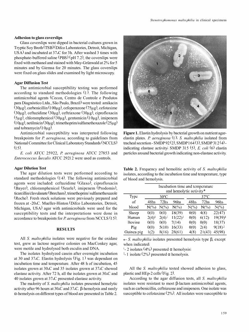
The isolates hydrolyzed casein after overnight incubationat 30 and 37ºC. Elastin hydrolysis (Fig. 1) was dependant onincubation time and temperature. After 48 h of incubation, 45isolates grown at 30ºC and 35 isolates grown at 37ºC showedelastase activity. After 72 h, all the isolates grown at 30ºC and40 isolates grown at 37ºC presented elastase activity.
The majority of S. maltophilia isolates presented hemolyticactivity after 96 hours at 30ºC and 37ºC. β-hemolysis and rarelyα-hemolysis on different types of blood are presented in Table 2.
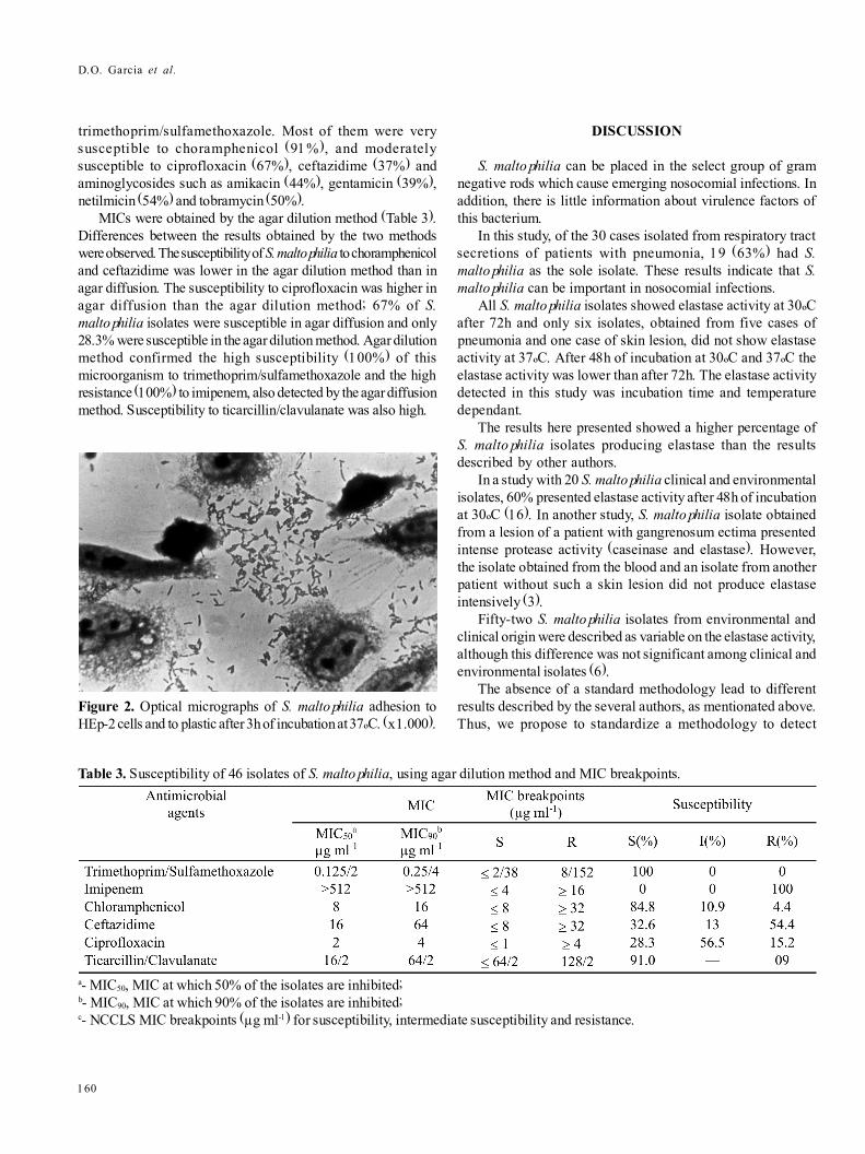
All the S. maltophilia tested showed adhesion to glass,plastic and HEp-2 cells (Fig. 2).
According to the agar diffusion tests, all S. maltophiliaisolates were resistant to most β-lactam antimicrobial agents,such as carbenicillin, ceftriaxone and imipenem. One isolate wassusceptible to cefotaxime (2%). All isolates were susceptible to
Figure 1. Elastin hydrolysis by bacterial growth on nutrient agar-elastin plates. P. aeruginosa (1); S. maltophilia isolated fromtracheal secretion - SMDP 92 (2), SMDP 164 (3), SMDP 312 (4) -indicating elastase activity; SMDP 315 (5), E. coli (6) elastinparticles around bacterial growth indicating non-elastase activity.
Table 2. Frequency and hemolitic activity of S. maltophiliaisolates, according to the incubation time and temperature, typeof blood and hemolysis.
*- S. maltophilia isolates presented hemolysis type β, exceptwhen indicated;1- 2 isolates (4%) presented α hemolysis;2- 1 isolate (2%) presented α hemolysis.
160
D.O. Garcia et al.
trimethoprim/sulfamethoxazole. Most of them were verysusceptible to choramphenicol (91%), and moderatelysusceptible to ciprofloxacin (67%), ceftazidime (37%) andaminoglycosides such as amikacin (44%), gentamicin (39%),netilmicin (54%) and tobramycin (50%).
MICs were obtained by the agar dilution method (Table 3).Differences between the results obtained by the two methodswere observed. The susceptibility of S. maltophilia to choramphenicoland ceftazidime was lower in the agar dilution method than inagar diffusion. The susceptibility to ciprofloxacin was higher inagar diffusion than the agar dilution method; 67% of S.maltophilia isolates were susceptible in agar diffusion and only28.3% were susceptible in the agar dilution method. Agar dilutionmethod confirmed the high susceptibility (100%) of thismicroorganism to trimethoprim/sulfamethoxazole and the highresistance (100%) to imipenem, also detected by the agar diffusionmethod. Susceptibility to ticarcillin/clavulanate was also high.
DISCUSSION
S. maltophilia can be placed in the select group of gramnegative rods which cause emerging nosocomial infections. Inaddition, there is little information about virulence factors ofthis bacterium.
In this study, of the 30 cases isolated from respiratory tractsecretions of patients with pneumonia, 19 (63%) had S.maltophilia as the sole isolate. These results indicate that S.maltophilia can be important in nosocomial infections.
All S. maltophilia isolates showed elastase activity at 30ºCafter 72h and only six isolates, obtained from five cases ofpneumonia and one case of skin lesion, did not show elastaseactivity at 37ºC. After 48h of incubation at 30ºC and 37ºC theelastase activity was lower than after 72h. The elastase activitydetected in this study was incubation time and temperaturedependant.
The results here presented showed a higher percentage ofS. maltophilia isolates producing elastase than the resultsdescribed by other authors.
In a study with 20 S. maltophilia clinical and environmentalisolates, 60% presented elastase activity after 48h of incubationat 30ºC (16). In another study, S. maltophilia isolate obtainedfrom a lesion of a patient with gangrenosum ectima presentedintense protease activity (caseinase and elastase). However,the isolate obtained from the blood and an isolate from anotherpatient without such a skin lesion did not produce elastaseintensively (3).
Fifty-two S. maltophilia isolates from environmental andclinical origin were described as variable on the elastase activity,although this difference was not significant among clinical andenvironmental isolates (6).
The absence of a standard methodology lead to differentresults described by the several authors, as mentionated above.Thus, we propose to standardize a methodology to detect
Figure 2. Optical micrographs of S. maltophilia adhesion toHEp-2 cells and to plastic after 3h of incubation at 37ºC. (x 1.000).
a- MIC50, MIC at which 50% of the isolates are inhibited;b- MIC90, MIC at which 90% of the isolates are inhibited;c- NCCLS MIC breakpoints (µg ml-1) for susceptibility, intermediate susceptibility and resistance.
Table 3. Susceptibility of 46 isolates of S. maltophilia, using agar dilution method and MIC breakpoints.

Stenotrophomonas maltophilia in clinical specimens
161
elastase activity of S. maltophilia. The methodology of thisstudy was based on the one described by Rust et al. (19), andshowed to be very efficient. We observed that the detection ofthis enzymatic activity was better after 72h of incubation at30ºC.
Hemolytic activity of S. maltophilia isolates showed avariety of patterns and was blood type, temperature, andincubation time dependant. Most of the S. maltophilia isolatesexpressed hemolytic activity only after 96 hours at 30ºC and37ºC. Most isolates also showed hemolytic activity on rabbitblood agar (61% after 96h at 30ºC and 98% after 96h at 37ºC).With the exception of pig blood, all types of blood presentedbetter hemolytic activity, although less intense, at 37ºC. After48h at 30ºC, 4% of S. maltophilia isolates presented hemolysison human blood agar and 2% on rabbit blood agar. These datadiffer from results described in another study (16) in which thehemolysis was observed, after 72h of incubation at 30ºC, in40% of isolates on pig blood, 60% on human blood, and 95% onrabbit blood agar. After 96h of incubation at 30ºC, hemolysiswas observed in all S. maltophilia isolates on sheep blood and25% on bovine blood agar. However, the hemolytic activityrestricted to the area under the colony was also considered(16). In this study, such hemolysis was not considered because,after long time of incubation, this effect can be due to bacterialmetabolic products.
S. maltophilia isolates showed adhesion to HEp-2 cells,plastic, and glass. The adhesion to HEp-2 cells suggests that S.maltophilia can adhere to other epithelial cells. This may explainthe presence of S. maltophilia in the respiratory tract cells.Most isolates were obtained from nosocomial infections whichin several cases medical devices were being used by the patients.The adhesion to plastic and glass can represent an importantstep in the bacterial colonization in immunosupressed patientsand/or using medical devices. The adhesion to inert surfacesrarely comes to the attention of investigators interested in thepathogenesis of human infections, except in the colonization ofimplantable medical devices (4).
Jucker et al. (11) described the adhesion to plastic of S.maltophilia isolated from urinary catheter from a patient with asuspicious urinary infection. They demonstrated that this S.maltophilia isolate was positively charged.
The susceptibility to antimicrobials from both methodologiesshowed differences as described in literature (6). There aredifficulties in determining the susceptibility of S. maltophiliabecause the methodologies are not standardized. The diffusionagar method was used because it still is the most commonlyused test in clinical laboratories, although not recommendedby NCCLS (14). It is important to emphasize the highsusceptibility to trimetoprim/sulfamethoxazole and the highresistance to imipenem presented by this group ofmicroorganisms, since the majority of gram negative rodsshowed opposite results to these antimicrobial agents. In
addition, it is important to emphasize that the agar dilutionmethod is the one which offers the most reliable results.
We conclude in this study that the elastase activity isincubation time and temperature dependant. Due to the differentresults obtained among several authors, we propose tostandardize the methodology to detect the elastase activity ofS. maltophilia. This bacterium adheres to plastic, glass and onto HEp-2 cells. Trimetoprim/sulfamethoxazole continues beingthe drug of choice for the treatment of S. maltophilia infections.The microorganism with these characteristics confirms the trendscited that S. maltophilia can become an important nosocomialpathogen.
Studies are under progress to characterize external structuresthat can be responsible for the described adhesions.
ACKNOWLEDGMENTS
This study was supported by Fundação de Amparo àPesquisa do Estado de São Paulo (FAPESP) 93/4695-8. We thankStella Maria Guida for providing the S. maltophilia isolatesfrom the Hospital Universitário of the Universidade de São Paulo,São Paulo, SP, Brazil, Dr. Leonard Mayer (Center for DiseasesControl - CDC, Atlanta, USA) for providing S. maltophiliaATCC13637 and Burkholderia cepacia ATCC25416 and CelyS.A. Medeiros for sending clinical data of S. maltophilia isolatesobtained from patients from Instituto Dante Pazzanese deCardiologia.
RESUMO
Proteases (caseinase e elastase), hemolisinas, adesãoe sensibilidade a antimicrobianos em cepas de
Stenotrophomonas maltophilia isoladas de amostrasclínicas
Quarenta e seis amostras de S. maltophilia obtidas deamostras clínicas foram estudadas quanto à produção deprotease (caseinase e elastase), atividade hemolítica, adesão acélulas HEp-2, ao plástico e ao vidro. A sensibilidade aos agentesantimicrobianos também foi avaliada. A maioria das amostrasfoi obtida de secreções do trato respiratório de pacientes emuso de �dispositivos� médicos. Todas as amostras foramcapazes de hidrolisar a caseína após o crescimento a 30ºC e37ºC por 16-18hs. Após 72 hs, todas as amostras apresentaramatividade hemolítica após 96hs de incubação em ambas astemperaturas. A maior atividade hemolítica foi verificada com osangue de coelho; após 96hs, 61% e 98% das amostrasapresentaram b-hemólise a 30ºC e 37ºC, respectivamente. Todasas amostras foram sensíveis ao sulfametoxazol-trimetoprim eresistentes à maioria dos antimicrobianos b-lactâmicos testados.Através do método de diluição em ágar, S. maltophilia mostrouuma alta sensibilidade à ticarcilina-ácido clavulânico e uma

162
D.O. Garcia et al.
menor sensibilidade à ciprofloxacina do que pelo método dedifusão em ágar. As amostras mostraram adesão às células HEp-2,ao plástico e ao vidro. A atividade proteolítica e adesão asuperfícies inanimadas detectadas em S. maltophilia podemestar relacionadas à patogênese desta bactéria. A colonizaçãode �dispositivos� médicos favorece o desenvolvimento deinfecções hospitalares.
Palavras-chave: Stenotrophomonas maltophilia, proteases,elastase, hemolisinas, adesão, agentes microbianos.
REFERENCES
1. Bauer, A.W.; Kirby, W.M.M.; Sherris, J.C. et al. Antibioticsusceptibility testing by a single disc method. Am. J. Clin. Pathol.,145: 45-495, 1966.
2. Baldini, M.M.; Kaper, J.B.; Levine, M.M.; Candy, D.C.; Moon, H.W.Plasmid-mediated adhesion in enteropathogenic Escherichia coli. J.Pediatr. Gastroenterol. Nutr., 2(3): 534-538, 1983.
3. Bottone, E.J.; Reitano, M.; Janda, M.; Troy, K.; Cuttner, J.Pseudomonas maltophilia Exoenzyme Activity as Correlate inPathogenesis of Ecthyma Gangrenosum. J. Clin. Microbiol., 24:995-997, 1986.
4. Christensen, G.D.; Baldassari, L.; Simpson, W.A. Methods forStudying Microbial Colonization of Plastics. Methods Enzimol., 253:477-500, 1995.
5. Cravioto, A.; Gross, R.J.; Scotland, S.M.; Rowe, B. An adhesivefactor found in strains of Escherichia coli belonging to the tradicionalinfantile enteropathogenic serotypes. Curr. Microbiol., 3: 95-99,1979.
6. Denton, M.; Kerr, K.G. Microbiological and Clinical Aspects ofInfection Associated with Stenotrophomonas maltophilia. Clin.Microbiol. Rev., 11: 57-80, 1998.
7. Fujita, J.; Yamadori, I.; Xu, G.; Hojo, S.; Negayama, K.; Miyawaki,H.; Yamaji, Y.; Takahara, J. Clinical features of Stenotrophomonasmaltophilia pneumonia in immunocompromised patients. Respir.Med., 90: 35-38, 1996.
8. Gilardi, G.L. Pseudomonas and Related Genera. In: Ballows A. (eds.).Manual of Clinical Microbiology. American Society for Microbiology,Washington, 1991, p.429-441.
9. Gopalakrishnan, R.; Hawley, B.; Czachor, J.S.; Markert, R.J.;Bernstein, J.M. Stenotrophomonas maltophilia infection andcolonization in the intensive care units of two community hospitals:A study of 143 patients. Heart Lung, 28: 134-141, 1999.
10. Jaffar-Bandjee, M.D.; Lazdunski, A.; Bally, M.; Carrère, J.;Chazalette, J.P.; Galabert, C. Production of Elastase, Exotoxin A,and Alkaline Protease in Sputa during Pulmonary Exacerbation ofCystic Fibrosis in Pacients Chronically Infected by Pseudomonasaeruginosa. J. Clin. Microbiol., 33: 924-949, 1995.
11. Jucker, B.A.; Harms, H.; Zehnder, A.J.B. Adhesion of the positivelycharged bacterium Stenotrophomonas (Xanthomonas) maltophilia70401 to glass and teflon. J. Bacteriol., 178: 5472-5479, 1996.
12. Julve, R.; Rovira, E.; Belda, A.; Prat, J.; Escoms, R.; Albert, A.;Gonzalvo, F. Espectro clínico de la infección por Stenotrophomonas(Xanthomonas) maltophilia. An. Med. Interna., 15: 476-480, 1998.
13. Marshall, W.F.; Keating, M.R.; Anhalt, J.P.; Steckenberg, J.M.Xanthomonas maltophilia: An emerging nosocomial pathogen. MayoClin. Proc., 1097-1104, 1989.
14. National Committee for Clinical Laboratory Standards. Methodsfor dilution antimicrobial susceptibility tests for bacteria that growaerobically. Approved Standard M7-A4. National Committee forClinical Laboratory Standards. Villanova, PA, 1997.
15. National Committee for Clinical Laboratory Standards. Performancestandards for antimicrobial disk susceptibility tests - Sixt Edition;Approved Standard. M2-A6. National Committee for ClinicalLaboratory Standards. Villanova, PA, 1997.
16. O�Brien, M.; Davis, G.H.G. Enzymatic profile of Pseudomonasmaltophilia. J. Clin. Microbiol., 16: 417-421, 1982.
17. Palleroni, N.J.; Bradbury, J.F. Stenotrophomonas, a new bacterialgenus for Xanthomonas maltophilia (Hugh, 1980) Swings et al.1983. Int. J. Syst. Bacteriol., 43: 606-609, 1993.
18. Robin, T.; Janda, J.M. Pseudo-, Xantho-, Stenotrophomonas: anemerging pathogen in search of genus. Clin. Microbiol. News, 18: 9-16, 1996.
19. Rust, L.; Messing, C.R.; Iglewski, B.H. Elastase Assays. MethodsEnzimol., 235: 561-562, 1994.
20. Sambrook, J.; Fritsh, E.F.; Maniatis, T. Molecular Cloning - ALaboratory Manual, 2nd Ed. Cold Spring Harbor Laboratory Press,Cold Spring Harbor, NY, 1989.
21. Schock, P.E.; Cunha, B.A. Pseudomonas maltophilia. Infect.Control., 4: 169-172, 1987.
22. Yao, J.D.C.; Louie, M.; Louie, L.; Goodfellow, J.; Simon, A.E.Comparison of E test and agar diluition for antimicrobial susceptibilitytesting of Stenotrophomonas (Xanthomonas) maltophilia. J. Clin.Microbiol., 33: 1428-1430, 1995.