protein-losing nephropathy in small animals -
TRANSCRIPT

Protein-losingNephropathy in SmallAnimals
Meryl P. Littman, VMD
The prevalence of protein-losing nephropathy (PLN) in the general population is muchgreater in dogs than cats but is largely unknown and probably higher than currentlyrecognized.1–3 Renal failure is arguably the most common organ failure in dogs andcats. The prevalence of glomerular lesions, mostly immune-mediated glomerulone-phritis (IMGN), was found in 43% to 90% of random dogs.1,3 Increased urineprotein/creatinine ratio (UPC), as an indicator of glomerular disease, is a negative pre-dictor of outcome.4–7 Microalbuminuria (MA) is detected in about 25% of all dogs andcats, increasing with age (36% in dogs 9–11 years, 49% in dogs �12 years, 39% incats�12 years, and 65% of cats�16 years),8 but its clinical significance is not known.When the first insult to the nephron is at the glomerulus, proteinuria occurs, which ulti-mately damages the rest of the nephron. By the time end-stage renal disease (ESRD)is discovered, the initiating glomerular cause may go undetected. Because proteinuriadecreases with nephron dropout and decreased glomerular filtration, hypoalbumine-mia may no longer exist or it may be masked by dehydration. Therefore, glomerulardisease as the initiating cause of ESRD may go unrecognized.Renal biopsy results may not settle the question of chicken-or-egg regarding
whether glomerular versus tubular damage (chronic interstitial nephritis) was theprimary cause, because both are often seen in end-stage kidney samples. Evenwhen renal biopsies are taken earlier in the disease process, pathologists’ interpreta-tions using routine histopathology techniques do not necessarily agree.9 There isinherent subjectivity with visual analysis of membrane thickening or mesangial cellnumbers present. Tissue sections traditionally cut at 5 to 6 mm for light microscopyare too thick for careful examination of renal lesions. Therefore, the incidence ofsubtypes of glomerulonephritis reported may not be accurate, and treatment proto-cols that might work for a particular subset (for instance, steroids or cyclosporine)may not seem beneficial because these cases were not properly identified.
Clinical Studies-Philadelphia, University of Pennsylvania School of Veterinary Medicine, 3900Delancey Street, Philadelphia, PA 19104-6010, USAE-mail address: [email protected]
KEYWORDS
� Proteinuria � Glomerular disease � Glomerulonephritis� Glomerulosclerosis � Amyloidosis
Vet Clin Small Anim 41 (2011) 31–62doi:10.1016/j.cvsm.2010.09.006 vetsmall.theclinics.com0195-5616/11/$ – see front matter � 2011 Elsevier Inc. All rights reserved.

With the advancement of technology, there are now sensitive and specific methodsto detect and monitor proteinuria and abnormalities can be identified earlier in thedisease process. The source of proteinuria can be localized and the cause character-ized via diagnostic tests; the trend can be followed and stability or disease progres-sion can be monitored. Kidney biopsies can be safely taken percutaneously withultrasound guidance, sophisticated methodology can be used with light microscopy(LM) examination of thin (3–4 mm) tissue sections, and the glomerular lesions can becharacterized by transmission electron microscopy (TEM), immunofluorescence (IF),and immunohistochemistry (IHC). Specific treatments may be recommended forspecific causes, as well as symptomatic and supportive therapies to reduce protein-uria, hypertension, risk of thromboembolic events, edema/effusions, and progressionof renal failure.
NORMAL GLOMERULAR STRUCTURE AND FUNCTION
The normal glomerulus is a complicated, elegant sieve, filtering 20% of the cardiacoutput, producing liters of ultrafiltrate per day, allowing water and small moleculesto cross the fenestrated vascular endothelial barrier by the force of transcapillary pres-sure, to penetrate the glomerular basement membrane (GBM), traverse the podocyteslit diaphragm (SD), and enter into the glomerular filtrate while holding back largermolecules based on their size and electrical charge. The endothelial cell glycocalyxis negatively charged; the underlying supportive GBM is made up of collagen typeIV, laminins, nidogen, and negatively charged glycosaminoglycans.10 Podocyte footprocesses are attached to the GBM via cell membrane receptors (a3b1 integranslinked to talin, vinculin, and paxillin, and a- and b-dystroglycans linked to utrophin).10
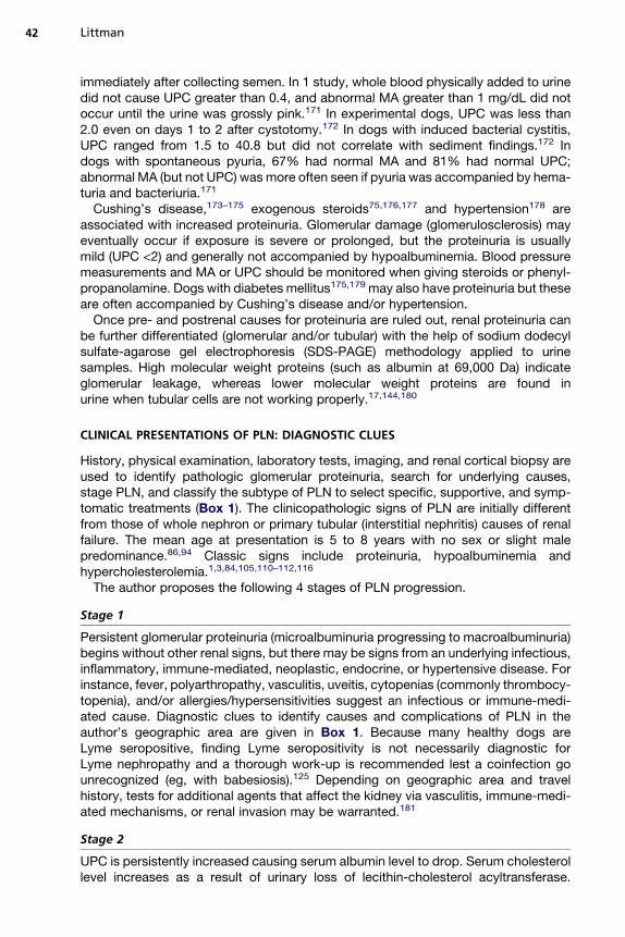
Recently the structure and function of a myriad of molecules in the glomerular filtrationbarrier of the SD (ie, the 25- to 40-nm wide pore between the foot processes) havebeen reviewed (Fig. 1).11 Produced by podocytes, these molecules work in concertto form a dynamic three-dimensional complex at the SD; they translate outside-insidesignaling, control calcium influx, and rearrange the actin cytoskeleton within the podo-cytes to cause their contraction and modification of their morphology as well as theintricate architecture of their interdigitating foot processes and SD aperture, thussensing and reacting to a changing environment. Normally very few proteins withmolecular weight of albumin (69,000 Da) or higher get passed into glomerular filtrate,especially if they are negatively charged as is albumin. The few proteins that do passthrough into the glomerular filtrate are normally reabsorbed and degraded by tubularcells and their lysosomes, but this work can cause tubular cell damage.12
GENETIC ABNORMALITIES ASSOCIATED WITH PLN
Genetic mutations producing 1 or more abnormal molecules at the SD or GBM maylead to immediate malfunction of the integrity of the permselective barrier, or toa susceptibility to injury by environmental triggers, or allow increased entrapment ofcirculating immune complexes (CIC), which may cause later onset proteinuria.Although not yet discovered in dogs and cats, more than 100 different mutationshave been identified in NPHS1, the gene for nephrin (the major SD transmembraneadhesion protein of the immunoglobulin superfamily)13; more than 40 mutations inNPHS2, the gene for podocin (a stomatin family member closely associated withnephrin at the SD); and various mutations in other genes including NPHS3 (phospho-lipase C31), ACTN4 (a-actinin 4), CD2AP (CD-2 associated protein), TRPC6 (transientreceptor potential cation channel 6), WT 1 (WT 1 protein), LAMB2 (laminin b-2), theNEPH 1-3 complex, several mitochondrial genes, MYH9 (nonmuscle myosin11A
Littman32

heavy chain), and many more (too numerous to mention here).11,14 Genetic abnormal-ities of the SD have been associated with many types of phenotypic expression (ie,mild to severe proteinuria); histopathology showing minimal change disease to severefocal segmental glomerulosclerosis (FSGS); onset that is congenital/infantile, child-hood, or adult onset; inheritance that is autosomal recessive, dominant, possiblywith low, medium, or high penetrance; and some genetic abnormalities include extra-renal abnormalities (eg, neurologic, orthopedic, or genital).11,15 At times complexinheritance such as a triple hit (homozygosity for 1 allele and heterozygosity foranother) or a 4-allelic hit (homozygosity at 2 sites) might be involved for phenotypicexpression.14,15 The expression of the phenotype may not be easily explained byjust the presence of 1 or more genetic mutations but by the interplay of the entiremolecular background.14
Genetic causes of PLN are usually steroid-resistant. Many breeds are predisposedto PLN (Table 1), and their genetic defects may someday be discovered to involvepodocytopathies that interfere with the normal development and maintenance of thestructure and function of the GBM or SD. Onset of PLN because of genetic causes
Fig. 1. The glomerular filtration barrier. (A) Overview of the structural components,including capillary endothelial cells, GBM, and podocyte foot processes (FP). The SDconnects neighboring foot processes. Blue lines within the podocytes symbolize their actincytoskeleton. (B) Molecules related to the nephrin-NEPH-podocin complex at the SD. Greenarrows indicate effector pathways that have been proposed to be involved in the regulationof actin cytoskeleton reorganization. Only a subset of known molecules and interactions areshown. (C) Molecules at the podocytes-GBM interface and linkage to the FP actin cytoskel-eton. Adhesion receptors expressed at the basal site of FP include integrin a3b1 and dystro-glycan. Only a subset of known molecules and interactions are shown. DAG, diacylglycerol;dys, dystroglycan; FAK, focal adhesion kinase; ILK, integrin-linked kinase; IP3, inositol 1,4,5-triphosphate; pax, paxillin; PI3K, phosphoinositide-3 kinase; pod, podocin; syn, synaptopo-din. (From Zenker M, Machuca E, Antignac C. Genetics of nephrotic syndrome: new insightsinto molecules acting at the glomerular filtration barrier. J Mol Med 2009;87:850; withpermission.)
Protein-losing Nephropathy in Small Animals 33

Table 1Breeds predisposed to glomerular pathogenic proteinuria
Breed Disease Characterization
American foxhound16–19 MPGN secondary to leishmaniasis Breed is at risk for leishmaniasis
Basenji20,21 Glomerulopathy with SIIPD DDX, Fanconi syndrome
Beagle22,23 Primary glomerulopathy22
Amyloidosis23May present up to 8 y, at least5 to 11 y
Bernese mountain dog24–27 MPGN AR � sex-linked modifier gene, F/Mw4, 2–5 y of age
Boxer28 Reflux nephropathy with segmental hypoplasia Onset <5 y of age
Brittany spaniel29 Primary glomerulopathy AR, associated with complement deficiency
Bull terrier30–35 Primary glomerulopathy AD model of Alport syndrome, average 3.5 y, up to 10 yDDX, polycystic kidney disease (also AD)
Bullmastiff36 FSGS AR, 2.5–11 y
Dalmatian37 Hereditary nephropathy ADmodel of Alport syndrome, onset 18mo (8mo to 7 y)
Doberman pinscher Primary glomerulopathy38,39
Also IMGN caused by sulfa40–42<3 y
English cocker spaniel43–46 Hereditary nephropathy AR model of Alport syndrome, 10–24 moAllele-specific PCR test to identify carrier dogs, OptiGen
English foxhound47 Amyloidosis 4 to 8 y
French mastiff (Bordeaux)48 Juvenile glomerulopathy Cystic glomerular atrophy, glomerular hypercellularity,<2 y
German shepherd49–52 IMGN (MCD) secondary to Ehrlichia canis infection Cell-mediated immunity abnormalityExperimental Beagle model is not as severely affected
Golden retriever53–59 IMGN caused by Lyme nephritis(Borrelia burgdorferi)53–57
JRD58,59
Most Lyme-positive dogs, even retrievers, do not getLyme nephritis; average age 5.6 � 2.6 y
Experimental beagle model does not get Lyme nephritisJRD, <3 y of age, may have proteinuria,
hypoalbuminemia, hypercholesterolemia
Gordon setter60 Juvenile nephropathy May have proteinuria, hypoalbuminemia, <3 y
Greyhound61–64 GN vasculopathy (skin, renal) 6 mo to 6 y
Littman
34

Labrador retriever53–57 IMGN caused by Lyme nephritis (Borrelia burgdorferi) Most Lyme-positive dogs, even retrievers, do not getLyme nephritis; average age 5.6 � 2.6 y
Experimental beagle model does not get Lyme nephritis
Mixed Navasota dog and kindred65,66 Primary glomerulopathy X-linked dominant Alport syndrome, 6 to 18 mo
Newfoundland67 Glomerulosclerosis AR, 2 to 12 moDDX, cystinuria (post-renal proteinuria, AR� DNA
marker available)
Norwegian elkhound68,69 Periglomerular fibrosis plus tubulointerstitial disease Mode of inheritance not known3 mo to 4 y
Pembroke Welsh corgi70 Primary glomerulonephropathy Littermates presented at 3 and 5 months of age; similarto Doberman
Rottweiler71 Primary glomerulopathy <1 y of age, atrophic glomerulopathy, massiveproteinuria
Samoyed and kindred72–78 Primary glomerulopathy Alport syndrome, X-linked recessive (an allele-specificPCR test is available for carrier Samoyeds, VetGen)
Males die at 2–15 mo; carrier females: high urinaryprotein at 2–3 mo of age but do not progress
Shar pei79–83 Amyloidosis Mean 4.1 y; M/F 1:2.5
Shetland sheepdog53–57 IMGN caused by Lyme nephritis (Borrelia burgdorferi) Most Lyme-positive dogs do not get Lyme nephritis;average age 5.6 � 2.6 y
Experimental beagle model does not get Lyme nephritis
Soft-coated wheaten terrier84–91 FSGS vs IMGN84–89
JRD90,91
Unknown inheritance, F/M 5 1.6:1PLN average 6.3 � 2.0 y; PLE/PLN combined average 5.9
� 2.2 y2/12 dogs with JRD had proteinuria
Abyssinian and Siamese cats92–97 Amyloidosis 1–5 yProteinuria variable (medullary vs glomerular
involvement)
Abbreviations: AD, autosomal dominant; AR, autosomal recessive; DDX, differentiate this from another type of renal proteinuria seen in this breed (as noted);FSGS, focal segmental glomerulosclerosis; GN, glomerulonephritis; IMGN, immune-mediated glomerulonephritis; JRD, juvenile renal disease (renal dysplasia);MCD, minimal change disease; MPGN, membranoproliferative glomerulonephritis; PCR, polymerase chain reaction; PLE, protein-losing enteropathy; SIIPD, smallintestinal immunoproliferative disease.
Data from Lees GE. Familial renal disease in dogs. In: Ettinger SJ, Feldman EC, editors. Textbook of veterinary internal medicine. 7th edition. St. Louis (MO):Saunders (Elsevier); 2010. p. 2058–62.
Protein-lo
singNephropathyin
SmallAnim
als
35

is usually young to middle age,98 but variable expression and incomplete penetrancemodes of inheritance may allow for later onset. For most breeds, specific geneticmutations are yet to be identified. It is hoped that, using DNA saved from animalswith well-characterized phenotypes, future genome-wide association studies(GWAS) with new SNP chip technology followed by fine mapping (gene sequencing)of areas of interest that are found, specific markers for these defects will be discov-ered so that by a simple polymerase chain reaction (PCR) test, carriers of at-risk geneswill be identified and breeding of a dominant individual or 2 at-risk recessive carriersmay be avoided. By identifying genes involved and realizing their function, the under-lying physiologic defects will be better understood and appropriate therapeutic proto-cols can be planned.Alport syndrome (hereditary nephritis) affects the production and maintenance of
the GBM as a result of abnormal collagen IV production and assembly. Normallycollagen IV is made up of heterotrimers of different types of chains, numbered a1-6.In Alport syndrome, there may be insufficient amounts or abnormalities in subtypesa3-5 chains produced. Various mutations of the encoding genes (COL4A3,COL4A4, and COL4A5) lead to nanomechanic GBM failure, and in humans may affectthe inner ear and eye as well. The abnormal GBM thickening or basket weave appear-ance and ultrastructural splitting of the lamina densa is seen by TEM, often with intra-membranous electron dense deposits. With light microscopy alone, these cases maybe misinterpreted as a type of glomerulonephritis (eg, membranoproliferative) or renalcortical hypoplasia. In humans, more than 350 mutations have been found affectingCOL4A5 (coding for a5) on the X-chromosome.65 Mutations on the autosomal genesCOL4A3 and COL4A4 are usually recessive. The primary glomerulopathies affectingbull terrier, Dalmatian, English cocker spaniel, Samoyed and Navasota mixbreeddogs are types of Alport syndrome and their mode of inheritance has been identified(see Table 1). By gene sequencing, carrier Samoyeds were found to have a prematurestop codon caused by a single nucleotide substitution in exon 35 on the gene COL4A5on the X-chromosome that codes for the a5 chain. In X-linked recessive Alportsyndrome in Samoyeds, affected males have proteinuria by 4 months and ESRD at8 to 10 months; carrier females are proteinuric early but do not progress to renalfailure. In contrast, mixed breed dogs from Navasota, Texas, were found to have anX-linked dominant COL4A5 defect as a result of a 10-bp deletion on exon 9 thatcauses a frame shift and premature stop codon in exon 10; carriers of both sexesshow early onset of proteinuria by 6 months and ESRD at 6 to 18 months.65 Therehas been high clinical variability found in autosomal dominant types of Alportsyndrome in humans.99 Treatment is nonspecific (see later discussion). DNAscreening tests exist for Samoyed and English cocker spaniel breeds. Early screeningby MA, and early treatment may slow progression. Progression to renal failure occursbefore age 2 years in affected Samoyed and English cocker spaniel dogs, but is morevariable (up to 10 years) in Dalmatians and bull terriers.Familial renal amyloidosis in Shar pei, beagles, English foxhounds, and Abyssinian
and Siamese cats is often primarily medullary without gross proteinuria but progressesto renal failure. Medullary renal biopsies are not recommended because of the risk ofhemorrhage. Familial amyloidosis in Shar pei has earlier onset than reactive amyloid-osis (mean age 4.1 years, M/F 5 1:2.5); only 25% to 43% have proteinuria but 64%had some glomerular involvement, thus renal cortical biopsies are still helpful (amyloidstains with Congo Red). Recurrent fever/swollen hock syndrome/increased interleukin(IL)-6 is seen in Shar pei, similar to familial Mediterranean fever.Several hereditary types of collagenofibrotic nephropathies (type I, type III, periodic
acid-Schiff [PAS] negative) and fibronectin glomerulopathies (PAS positive) have been
Littman36

described in humans with PLN associated with massive infiltration of collagen or fibro-nectin fibrils in the mesangium and subendothelium.100 Collagenofibrotic glomerulo-nephropathy (collagen III) has been described in 2 unrelated young dogs withPLN101,102 and nonamyloid fibrillary glomerulonephritis in a nephrotic cat and a youngdog.103,104 Without special stains and EM, these biopsies would have beenmisread asother forms of glomerular disease.Many dog breeds98 are predisposed to juvenile renal disease (renal dysplasia), poly-
cystic renal disease, Fanconi syndrome, and so forth, which are not primary glomer-ulopathies but in some dogs cause proteinuria, possibly hypoalbuminemia, and/orhypercholesterolemia, mimicking changes seen with primary glomerular disease.Breeds predisposed to primary glomerulopathies as well as other familial renaldiseases that need to be differentiated include the bull terrier, golden retriever, andsoft-coated wheaten terrier (SCWT) (see Table 1). Also listed are breeds with higherrisk for immune-mediated glomerular disease, possibly triggered by infection (eg,Lyme nephritis in retrievers, leishmaniasis in American foxhounds, ehrlichiosis inGerman shepherds), by drugs (eg, sulfa in Doberman pinschers), or by other hyper-sensitivities (possibly food allergies in SCWT).
ACQUIRED CAUSES OF GLOMERULAR LEAKAGE OF PROTEIN
Acquired PLN is sporadically seen in any breed and is often caused by IMGN, reactiveamyloidosis, or glomerulosclerosis (GS). Comprehensive descriptions (TEM and/or IFin addition to LM analysis) of renal lesions in several hundred clinical cases of PLNwere described 17 to 40 years ago.1–3,105–114 Because newly emerging infectiousdiseases (especially tick-borne) and new genetic predispositions may change thespectrum of disease with time, comprehensive examinations of renal cortical biopsieson our current patients with PLN need to be done so that predominant types ofglomerular lesions as presented to veterinarians in various locations are known, treat-ment protocols for properly identified subsets of PLN can be investigated, and individ-uals treated appropriately.In general, glomerular lesions are common. In dogs with and without clinical signs of
renal disease, 90% had glomerular lesions in 1 study.107 Among dogs with renaldisease, 52% had glomerular lesions in another study.106 The population at risk forPLN was middle-aged to older dogs, with slightly more males represented. Glomeru-lonephritis and amyloidosis were described more often than other types of glomerularlesions in dogs and cats.1–3,105–116 Membranoproliferative glomerulonephritis (MPGN)was common in dogs (presumably immune-mediated, and possibly postinfectious, asis seen in people in developing countries). Membranous nephropathy (MN) was themost common lesion in cats with PLN, but in general, PLN is not common incats.113,114
IMGN
Pathologists describe lesions depending on how much of each glomerulus is involved(eg, segmental, global), how many glomeruli are involved in the sample (eg, focal,diffuse), and whether there is inflammatory cell infiltration or mesangial cellular prolif-eration. Immune complex (antigen-antibody) deposits can involve immunoglobulin (Ig)A, IgG, and/or IgM, with or without complement (C3). The complexes can be CIC or beformed in situ as antigens are caught and bind antibody secondarily. The antigensinvolved are rarely identified but are sought indirectly by history, clinical presentation,by serologic tests for antibodies, by using culture, cytology, and PCR for antigensassociated with infections, and by searching for inflammatory disease and neoplasia.
Protein-losing Nephropathy in Small Animals 37

The true cause is often unproved because immunohistochemistry or elution studies onthe glomerular complexes are rarely done. Immune complex deposition causesinflammation (glomerulonephritis) through a variety of mediators, inflammatory cells,complement and platelet activation, renin-angiotensin-aldosterone (RAA) system acti-vation, and numerous humoral and cellular responses that influence the progressionversus resolution by mesangial phagocytosis.1,3,117
MPGN is the most common form of IMGN in dogs with a mean age of 10.5 years andno sex predilection.110 It is uncommon in the cat.118 Most common in dogs is type 1MPGN with immune complexes seen as lumpy-bumpy deposits by EM and IF on thesubendothelial side of the GBM (mesangiocapillary GN) and/or in the mesangium.Linear deposits that would indicate true autoimmune disease (systemic lupus erythe-matosus) have not been described in dogs and cats. The granular deposits of IMGNmay stain positive for complement and IgA, IgG, and/or IgM combinations. By LM,the complexes make the GBM appear thickened or duplicated (railroad) but if tissuesections are cut thickly at 5 to 6 mm, MPGN may be overdiagnosed.MPGN has been associated with sulfa drugs (mostly in Dobermans40–42), neoplasia,
inflammatory diseases, and with many types of infectious diseases, such as chronicbacterial infection (endocarditis, bartonellosis,119 brucellosis120), arthropod-borne(anaplasmosis [suspected],121 babesiosis,122–126 Lyme borreliosis,53–57 ehrlichio-sis,49–52 hepatozoonosis,127,128 leishmaniasis,16–19 Rocky Mountain spotted fever(RMSF)129), viral diseases (canine adenovirus I,130 feline leukemia virus,131 felineimmunodeficiency virus [suspected],132–134 feline infectious peritonitis [FIP]135) orparasitic diseases (Dirofilaria/Wolbachia,136–140 heterobilharziasis [schistosomi-asis],141 trypanosomiasis1,3) in which carrier status and chronic immune stimulationfrom antigenic variation occurs. In leishmaniasis, high antihistone antibodies arepresent and associated with MPGN142; histones are cationic and are implicated inbinding the CIC to the GBM, perhaps as a planted antigen. Some diseases may causeproteinuria as a result of vasculitis (RMSF, anaplasmosis, bartonellosis, ehrlichiosis,greyhound vasculopathy, leptospirosis, FIP) or renal infiltration (toxoplasmosis, cryp-tococcosis, systemic fungal infections, neoplasia), and not necessarily IMGN.Although dogs seropositive for leptospirosis were described as having IMGN,143
coinfections (eg, heartworms, leishmaniasis, and so forth) may have played a role;leptospirosis is generally considered to cause tubular rather than glomerular protein-uria, or vasculitis.144–146 Another spirochetal disease, Lyme borreliosis, has beenassociated with MPGN involving Lyme-specific immune complexes, accompaniedby tubular necrosis/regeneration and interstitial nephritis, sometimes with glycosuriacaused by tubular disease.53–56 Lyme nephritis may be seen in any breed but mostlyin Labrador and golden retrievers and Shetland sheepdogs, and has a younger onsetat 5.6 � 2.6 years compared with other dogs with PLN at 7.1 � 3.6 years. There maybe specific Borrelia strains or genetic predispositions for this form of Lyme diseasebecause most Lyme-positive dogs (even retrievers) remain asymptomatic and donot show proteinuria or PLN.57 Thus, when a dog with PLN happens to be Lyme posi-tive, it should be checked for coinfections and other causes of PLN, because Lymeseropositivity indicates tick exposure and not necessarily a diagnosis of Lymenephritis.54
Bernese mountain dogs have a genetic predisposition (see Table 1) for MPGN thatis no longer believed to be associated with Lyme seropositivity. The mode of inheri-tance is autosomal recessive, possibly with an X-linked modifier (M/F ratio 51:4).24–27 Another type of MPGN is seen in congenital complement deficiency in Brit-tany spaniels29; normal complement levels were found in 49 other dogs with acquiredPLN.147
Littman38

Treatment of type I MPGN involves treating the underlying infectious, inflammatory,or neoplastic disease process. Antiplatelet drugs may also be tried to decreaseplatelet activation, which seems to be involved in the inflammatory cascade. Some-times IMGN is also treated with immunosuppressive protocols (see later discussion).Membranous nephropathy (MN) is a form of IMGN characterized by severe protein-
uria (similar to that seen with reactive amyloidosis) and seen more often in males. Theglomerular damage is caused by complement-dependent mechanisms and not inflam-matory cell infiltration because the immune complexes are found on the subepithelial(podocyte) side of the GBM, away from the capillaries. Unbound circulating antibodymay react to antigens that are fixed on the urinary side of the GBM. It may be a trueautoimmune disease; in humans it is associated with underlying immunologic defects.It is the secondmost common lesion in dogs (M/F5 1.75:1,mean age 8 years, range 1–14 years). Because of the severe proteinuria, it is often accompanied by the nephroticsyndrome, identified in 14 (30%) of 46 proteinuric dogs (mean 6.5 years). Survival timesfor dogs with MN ranged from 4 days to 3 years. In cats with PLN, MN is the mostcommon lesion (M/F 5 6:1, mean age 3.6 years, range 1–7 years). In a study of 24cats with MN, 46% died or were euthanized shortly after diagnosis; 17% survived 4to 10 months, 33% survived 2.5 to 6 years (3/8 of the long-term survivors were givensteroids). Cats with only IgG and/or C3 deposits had longer survival than those thathad IgA or IgM deposits. Spontaneous remissions have been reported but more studyneeds to be done to validate treatment regimes and prognostic factors. In MN inhuman patients, immunosuppressive treatment with pulse or alternate-day steroidsand alkylating agents (cyclophosphamide, chlorambucil) for 6 months helps, althoughrelapses are common. Cyclosporine also helps in two-thirds of cases.1,112–114
Familial MN is seen in Doberman pinschers (<3 years old). By LM the GBM looksthickened, possibly showing spikes on the epithelial side that do not take up silverstain. Deposits are seen by IF and TEM and the granularity may be so intense thatthe beaded pattern almost seems linear. Mesangia may also stain for IgG and C3,which are more common than IgM and/or IgA in dogs. TEM can stage the engulfmentand resolution process.Proliferative glomerulonephritis (PGN), also known as endocapillary or mesangial
PGN, has been described in 2% to 16% of dogs with PLN (mean 7–9 years).1 It isseen in humans with lupus, IgA nephropathy, or as a postinfectious GN (eg, afterstreptococcal or staphylococcal infection) in which GN follows an infection withouta carrier status, which may be why there is no membranous component. The mesan-gial proliferation is defined as 4 or more mesangial or mononuclear cells per area,often with increased mesangial matrix seen. By IF and TEM there are fine granulardeposits of IgG and/or IgM, subepithelial in the BM and in the mesangium. Treatmentis to remove the source antigen.IgA nephropathy may appear just as mesangial proliferative GN by LM but the
immune deposits are seen by TEM and stain for IgA (more than for IgG or IgM) byIF.148 Because IgA is dimeric in dogs, it may be trapped nonspecifically in the mesan-gium in 47% to 85% of normal dogs.109,149,150 IgA nephropathy may be associatedwith hepatic and gastrointestinal disease149; treatment may depend on removingthe underlying cause. IgA positivity (and less so IgM) was seen in some glomeruli inSCWT with protein-losing enteropathy (PLE)/PLN and food allergies,87 but it is notyet proved whether the glomerular lesions in that breed are primarily immune medi-ated or sclerotic (FSGS) with secondary deposition of immune complexes.Minimal change disease (MCD) is common in children (often steroid responsive but
relapses are common) but rarely described in veterinary literature.151 Ehrlichiosis andthe drug masitinib were shown to cause MCD.49,152 This nil disease shows no
Protein-losing Nephropathy in Small Animals 39

morphologic lesions by LM, but there is foot process effacement seen by TEM exam-ination, and increased vimentin staining by IF. Loss of the anionic charge at the GBMcauses massive proteinuria and often nephrotic syndrome.
Reactive Amyloidosis
Reactive amyloidosis may be seen in any breed (dog > cat) and is often associatedwithglomerular deposition and severe proteinuria caused by extracellular deposition ofpolymerized serum amyloid A protein (SAA, an acute phase reactant made by the liver)into b-pleated sheets, seen as homogeneous eosinophilic thickening at the GBM andmesangium, staining red with Congo Red stain. This is 1 subset of PLN that can bediagnosed by LM. Other organs may be affected (eg, liver and spleen), becomingfriable and hemorrhage easily (biopsy is not recommended). Among dogs with PLN,23% had amyloidosis in 1 study.116 Chronic infectious, inflammatory, or neoplasticdisease was found in 32% to 53% of cases (mean age 9.2 years, M/F 5 1:1.7) withbeagles, collies, and Walker hounds predisposed.153 Nephrotic syndrome is commonbecause proteinuria is severe. Prognosis is poor; 58%of dogs were euthanized or diedsoon after diagnosis and only 8.5% survived for 1 year or more.154 Colchicine(0.01–0.03 mg/kg by mouth every 24 hours) may help decrease hepatic productionof SAA but may cause gastrointestinal upset. Dimethylsulfoxide (DMSO, 90 mg/kgby mouth 3 times a week or subcutaneous injections diluted 1:4 with sterile water) isantiinflammatory, decreases interstitial fibrosis, and may improve renal function anddecrease proteinuria, but it causes garlic breath and may cause nausea/anorexia.155
The odorless and tasteless metabolite of DMSO, methylsulfonylmethane (MSM),may be used in place of DMSO.
Glomerulosclerosis
The prevalence of glomerulosclerosis (GS) increases with age.1 It may be a primary(genetic) disease as in some forms of FSGS or it may be secondary to hypertension(eg, as a result of hyperadrenocorticism or steroid use) or any glomerular injury,such as an end-stage lesion. It may be underdiagnosed and misdiagnosed asMPGN. Nonspecific trapping of complexes in sclerotic/fibrotic areas may be seenby IF. In humans there are 5 subtypes, each with different prognoses, but these arepoorly characterized in dogs and cats. Genetic structural defects of the SD or circu-lating permeability factors may functionally alter the permselectivity of the GBM,and predispose for indolent immune complex deposition, damage, and sclerosis. Glo-merulosclerosis secondary to hyperadrenocorticism or hypertension rarely causessevere enough proteinuria to cause hypoalbuminemia.
DETECTING PROTEINURIA, THE HALLMARK OF PLN
Annual screening for proteinuria is recommended in healthy dogs as part of annualhealth care.7 In particular, breeds with genetic risks for PLN should be screened earlyand often, especially if used for breeding. The earliest warning of glomerular disease ismicroalbuminuria (MA), defined as 1 to 30 mg albumin/dL, which can be detected byspecies-specific enzyme-linked immunosorbent assays for albuminuria such as thein-house semi-quantitative E.R.D. (HESKA) test or by quantitative MA by a referencelaboratory. The sensitive MA test is useful as a first-line screening agent for geneticPLN (eg, SCWT,89 Samoyeds74) or acquired glomerular damage (eg, in Lyme- orheartworm-positive dogs57,139).Microalbuminuria is often associated with age and with systemic diseases.156,157 In
older dogs, MA may be too sensitive to be helpful compared with the UPC test.
Littman40

Looking for persistence as well as trend of progression or stability is important beforeassigning clinical significance to MA, because many if not most older dogs have low tomoderate positive MA, possibly as a result of normal aging of the kidney or infectious/inflammatory/neoplastic/vascular insults.Urinary dipstick tests for protein are less sensitive than MA, picking up more than
30 mg/dL, and are less specific for albuminuria, showing false-negative and false-positive results compared with MA. Dipstick false-positive readings may occur withhigh pH, hematuria, pyuria, and/or bacteriuria, and more often with feline than caninesamples.158 In 1 study, when dipstick and urine specific gravity (USG) were usedtogether, dogs with a USG greater than 1.012 and 11 by dipstick were likely nonpro-teinuric; but for those with11 dipstick and USG less than or equal to 1.012, proteinuriashould be further assessed by use of the UPC ratio.159 Both dipstick and urine turbiditywith sulfasalicylic acid (SSA) testing for proteinuria were less specific and gave false-positive results, whereas UPC testing showed higher specificity but less sensitivity,with some false-negative results.160 Multistix PRO dipsticks, read by a Clinitek 50analyzer (Bayer Corporation), were more sensitive but less specific than SSA testingfor proteinuria in dogs (but not a good alternative for cats); manual calculation of theUPC is done with the dipstick’s estimated urinary creatinine level.161 Another in-houseanalyzer, the IDEXX VetTest, showed strong association for UPC results with the refer-ence Vitros 50 instrument,162 however not all laboratories use similar methodology,and inter-laboratory comparisons may be difficult.163 Any UPC test may be increasedby Bence Jones and other nonalbumin proteins in the urine. A urine albumin/creatinineratio can also be done.164
Macroproteinuria is generally defined using UPC measurements. Borderline UPCvalues in nonazotemic animals (0.5–1.0 in dogs and 0.4–1.0 in cats) should be moni-tored for persistence and trend of progression. Larger amounts (UPC of >1.0) shouldbe investigated and localized as to the source (prerenal, renal, or postrenal); renalproteinuria is then categorized as functional or pathologic (glomerular, tubular, orinterstitial).2,7 Investigation of macroproteinuria is always recommended if azotemiaexists. Therapeutic intervention is recommended for nonazotemic animals at UPCgreater than or equal to 2.0, but is often started at lower values if a breed-associatedcause is suspected and progression is expected without early intervention. Forazotemic animals, intervention is recommended at UPC greater than or equal to0.5 (dogs) and UPC greater than or equal to 0.4 (cats).Once macroproteinuria is found, UPC is the standard test for quantitation, moni-
toring, and for comparisons. There was no statistical difference found in the measure-ment of UPC between free-catch and cystocentesis samples.165 Day-to-day variationof the UPC was seen in female dogs with stable glomerular proteinuria caused byX-linked hereditary nephropathy.166 This study showed that significant differences inUPC to indicate progression of disease or failure of intervention would have to begreater than 35% variance at high UPC near 12, and greater than 80% variance ata low UPC near 0.5. This may be true for other forms of PLN (not just mosaics). Tomini-mize costs of averaging results from 3 samples, equal pooling of 3 urine samples for 1determinationwas found to be as valid as averaging results from 3 samples (� 20%).167
Moderate exercise does not cause MA to increase.168 Contrary to what was seenwith another inflammatory marker (C-reactive protein), degree of MA showed nocorrelation with degree of periodontal disease, and there was no change in USG,MA, or UPC before and after dental treatment169; positive MA in 12.4% of dogsneeding dental work may be related to their age.Because whole ejaculate (not just sperm) physically added to urine may increase
dipstick proteinuria,170 it is not recommended to collect urine samples for MA testing
Protein-losing Nephropathy in Small Animals 41
immediately after collecting semen. In 1 study, whole blood physically added to urinedid not cause UPC greater than 0.4, and abnormal MA greater than 1 mg/dL did notoccur until the urine was grossly pink.171 In experimental dogs, UPC was less than2.0 even on days 1 to 2 after cystotomy.172 In dogs with induced bacterial cystitis,UPC ranged from 1.5 to 40.8 but did not correlate with sediment findings.172 Indogs with spontaneous pyuria, 67% had normal MA and 81% had normal UPC;abnormal MA (but not UPC) was more often seen if pyuria was accompanied by hema-turia and bacteriuria.171
Cushing’s disease,173–175 exogenous steroids75,176,177 and hypertension178 areassociated with increased proteinuria. Glomerular damage (glomerulosclerosis) mayeventually occur if exposure is severe or prolonged, but the proteinuria is usuallymild (UPC <2) and generally not accompanied by hypoalbuminemia. Blood pressuremeasurements and MA or UPC should be monitored when giving steroids or phenyl-propanolamine. Dogs with diabetes mellitus175,179 may also have proteinuria but theseare often accompanied by Cushing’s disease and/or hypertension.Once pre- and postrenal causes for proteinuria are ruled out, renal proteinuria can
be further differentiated (glomerular and/or tubular) with the help of sodium dodecylsulfate-agarose gel electrophoresis (SDS-PAGE) methodology applied to urinesamples. High molecular weight proteins (such as albumin at 69,000 Da) indicateglomerular leakage, whereas lower molecular weight proteins are found inurine when tubular cells are not working properly.17,144,180
CLINICAL PRESENTATIONS OF PLN: DIAGNOSTIC CLUES
History, physical examination, laboratory tests, imaging, and renal cortical biopsy areused to identify pathologic glomerular proteinuria, search for underlying causes,stage PLN, and classify the subtype of PLN to select specific, supportive, and symp-tomatic treatments (Box 1). The clinicopathologic signs of PLN are initially differentfrom those of whole nephron or primary tubular (interstitial nephritis) causes of renalfailure. The mean age at presentation is 5 to 8 years with no sex or slight malepredominance.86,94 Classic signs include proteinuria, hypoalbuminemia andhypercholesterolemia.1,3,84,105,110–112,116
The author proposes the following 4 stages of PLN progression.
Stage 1
Persistent glomerular proteinuria (microalbuminuria progressing to macroalbuminuria)begins without other renal signs, but there may be signs from an underlying infectious,inflammatory, immune-mediated, neoplastic, endocrine, or hypertensive disease. Forinstance, fever, polyarthropathy, vasculitis, uveitis, cytopenias (commonly thrombocy-topenia), and/or allergies/hypersensitivities suggest an infectious or immune-medi-ated cause. Diagnostic clues to identify causes and complications of PLN in theauthor’s geographic area are given in Box 1. Because many healthy dogs areLyme seropositive, finding Lyme seropositivity is not necessarily diagnostic forLyme nephropathy and a thorough work-up is recommended lest a coinfection gounrecognized (eg, with babesiosis).125 Depending on geographic area and travelhistory, tests for additional agents that affect the kidney via vasculitis, immune-medi-ated mechanisms, or renal invasion may be warranted.181
Stage 2
UPC is persistently increased causing serum albumin level to drop. Serum cholesterollevel increases as a result of urinary loss of lecithin-cholesterol acyltransferase.
Littman42

Box 1
Clues to find causes and complications for a dog with PLN in a Lyme-endemic area; treatment
ideas
History should include
Signalment, pedigree, family history, coat or color type (eg, coloring for Labradors: black,yellow, chocolate)
Travel history, tick exposure
History of prior treatment of tick-borne disease such as Lyme disease
Medication exposure (sulfa, masitinib), vaccination exposure
Polyuria/polydypsia? Vomiting? Appetite? Weight loss?
History of lower urinary tract signs (pollakiuria, stranguria, accidents)
History of lameness, dyspnea, blindness, effusions/edema, neurologic events
History of allergies, inflammatory bowel disease, PLE, Addison disease (SCWT)
Physical examination should include
Body weight, body condition score, hydration status
Temperature, femoral pulses, respiration
Mucous membranes: check for anemia, petechiation
Ophthalmologic examination including fundic examination
Peripheral edema? Ascites?
Ausculation for murmur, dyspnea, muffling
Lymphadenopathy?
Abdominal palpation, organomegaly?
Neurologic examination
If lameness: joint swelling/effusion? Which joints? Pulses?
Blood pressure measurements (multiple)
Clinical pathology, microbiology, parasitology, immunology samples
Blood samples for
Complete blood count (CBC)
Biochemical profile
Coagulation profile, thromboelastography
Blood cultures if indicated
SNAP-4Dx (IDEXX) for heartworm antigen and antibodies against Borrelia burgdorferi(C6 quantitation if positive), Ehrlichia canis/chaffeensis, Anaplasma phagocytophilum/platys; do additional SNAP test (convalescent) 2 weeks into the illness if the history isacute; get quantitative titers if positive (0 and 6 months after treatment)
Ehrlichia PCR to check for Ehrlichia ewingii antigen, if indicated
Bartonella Western blot, culture/PCR, titers
Babesia spp PCR (for novel spp), titers (B canis, B gibsoni, B microti); get additional titers(convalescent) 2 weeks into the illness if the history is acute
Rocky Mountain spotted fever acute/convalescent titers (if the history is acute; RMSFdoes not cause a carrier status)
Protein-losing Nephropathy in Small Animals 43

Leptospira titers (get additional titer [convalescent] 2 weeks into the illness if the historyis acute)
Brucella, Leishmania, Trypanosoma tests, and so forth as indicated
Coombs, antinuclear antibody titer, rheumatoid factor, perinuclear antineutrophilcytoplasmic antibody, and so forth as indicated.
Save additional EDTA whole blood for future PCR testing and for DNA banking or DNAtest, if available
Consider samples for antithrombin III, C3, CIC levels, and so forth
Urine samples for
Urinalysis
Urine culture
Urine protein/creatinine ratio (UPC)
Urine SDS-PAGE
Cytology of
Joint fluid cytology/culture
Lymph node aspirate
Bone marrow aspirate
Effusions
Imaging studies
Chest radiographs
Abdominal ultrasound
Echocardiogram if indicated
Radiographs of joints if lameness present
Renal cortical biopsy for TEM, IF, and thin-section LM (via percutaneous ultrasound-guided Tru-cut needle)
Control hypertension if present; discontinue antithrombotics 3–7 days before biopsy
Contact Dr George Lees (email [email protected], tel. 979-845-2351, fax 979-845-6978)at the Texas Veterinary Renal Pathology Service before planning sample collection to getthe renal biopsy kit with its special fixatives, tools, and shipment label (if you already havea kit, check that the fixatives are still in date), packing and return shipping instructions, andto coordinate the best date for the procedure to be done because the samples must bereceived on ice by overnight shipment. Dr George Lees, Building 508, Room 120, VeterinaryTeaching Hospital, Texas A&M University, College Station, TX 77843
Consider saving a frozen kidney sample for future elution studies
Therapeutic considerations
Standard therapy
Doxycycline 10 mg/kg/d, pending infectious disease results (1 month; longer for Lymenephritis)
Angiotensin-converting enzyme (ACE) inhibition: enalapril (Enacard) 0.5–1.0 mg/kgevery 12 to 24 hours, or benazepril (Lotensin) 0.25–0.5 mg/kg every 12 to 24 hours
Low antithrombotic dose of aspirin if albumin �2.5 g/dL, 1.0 mg/kg every 24 hours
Omega-3 fatty acid supplement
Antihypertensives are added to ACE inhibitor if dog is still hypertensive (eg, amlodipine[Norvasc] 0.2–0.4 mg/kg every 12 hours)
Littman44
The dyslipidemia occurring with hypoalbuminemia also includes increased hepaticactivity of several enzymes leading to decreased high-density lipoprotein andincreased low-density lipoprotein and triglycerides.182 Serious events caused bycomplications of PLN can occur before azotemia or polyuria/polydipsia exist andare more common in Stage 2 than Stage 1 PLN (eg, hypertension, thromboembolicevents, and/or nephrotic syndrome with ascites/edema).Causes of hypertension are multifactorial and include activation of the RAA system,
abnormal salt and water handling, decreased renal production of vasodilatory prosta-glandins and kinins, and increased arteriolar sensitivity to circulating vasoconstric-tors.183 Hypertensive target organ damage may be the reason for presentation tothe veterinarian. Damage includes blindness caused by retinal hemorrhage/detach-ment, cardiovascular disease (left ventricular hypertrophy, epistaxis, arterioscle-rosis/atherosclerosis), neurologic abnormalities (cerebrovascular accidents orstroke), and renal changes (glomerulosclerosis, proteinuria, pressure diuresis). Self-perpetuation of hypertension is caused by glomerulosclerosis and increased totalperipheral resistance as a result of vascular damage (arteriosclerosis/atherosclerosis).Risk for hypertensive target organ damage increases as blood pressure increases;severe risk is seen at blood pressure measurements (BPM) greater than or equal to180/120 mm Hg, moderate risk at 160 to 179/100 to 119 mmHg, mild risk at 150 to159/95 to 99 mm Hg, and minimal risk at less than 150/95 mm Hg.183 Roughly 60%
Other therapies for renal disease (eg, dietary modification, phosphate binder,gastroprotectant, antiemetic, and so forth)
Immunosuppressive therapy
If biopsy results (TEM, IF, thin-section LM) show compelling evidence of active immunecomplex deposition and inflammation, then immunosuppressive medications should beconsidered
As a rule-of-thumb, if <50% of the glomeruli are open and/or >50% show glomerularobsolescence, and if the tubulointerstitial lesion is characterized by diffuse fibroticchanges (as opposed to cellular inflammatory changes), immunosuppressive protocolsmay be ineffective and possibly contraindicated
If the patient is decompensating rapidly, consider starting a protocol while renal biopsyresults are pending
There are no blinded treatment studies; the following protocols are offered anecdotallydepending on owner considerations, patient tolerance, and so forth. Continue tomonitor blood pressure, UPC, CBC, chemistry panel, and so forth. every 1–4 weeks,depending on the severity/stability of clinical signs, and continue to look for anunderlying cause that may present itself with time or as a result of immunosuppression
Protocol 1: Methylprednisolone sodium succinate (Solu-Medrol) 5 mg/kgintravenously every 24 hours � 2 days; cyclophosphamide (Cytoxan) 200 mg/m2
intravenous bolus first day. Check white blood cells in 1 week; cyclophosphamide isrepeated every 2 weeks for a maximum of 6 cycles; Solu-Medrol is only used whenthe first cycle of cyclophosphamide is given
Protocol 2 (barring financial constraints): Methylprednisolone sodium succinate asprotocol 1; mycophenolate mofetil (CellCept) 10 mg/kg intravenously or by mouthevery 12 hours long-term
Protocol 3: Methylprednisolone sodium succinate as protocols 1 and 2; azathioprine(Imuran) 2 mg/kg by mouth every 24 hours � 7 days, then every 48 hours long-term
With thanks, Box 1 is derived from discussions with Drs Nicola Mason, Reid Groman, and Tabi-tha Hutton at the University of Pennsylvania School of Veterinary Medicine.
Protein-losing Nephropathy in Small Animals 45

to 90% of dogs and cats with renal disease are hypertensive and it is associated withpoor outcome.5,183,184
The risk for thromboembolic events in patients with PLN is well recognized.1,3,84,116
Mechanisms causing hypercoagulability in patients with PLN include urinary loss ofantithrombin (AT, which has similar size and charge as albumin) and platelet hypersen-sitivity as a result of hypoalbuminemia.185–188 Spontaneous vascular damage causedby hypertension or vasculitis and iatrogenic damage caused by venipuncture andcatheter placement may initiate thrombus formation. Both arterial and venous throm-boembolic (TE) events have been associated with PLN. Life-threatening TE eventsmay affect the heart, central nervous system, lung, aortic bifurcation (saddlethrombus), portal/mesenteric/splenic veins, and so forth, causing dyspnea, lameness,abdominal distress, collapse, or sudden death. One report found 22% of dogs withPLN had TE events.116 In SCWT, 10/84 dogs (12%) with PLN and 11/62 dogs (18%)with combined PLE/PLN were believed to have TE events.84
The drop in plasma colloid oncotic pressure caused by hypoalbuminemia allowsvascular fluid to be lost to the interstitium (as a result of Starling forces), perhapsleading to peripheral edema and signs of third spacing (dyspnea from pleural effusion;ascites from abdominal effusion; tamponade from pericardial effusion). Arterial hyper-tension and/or vasculitis from associated infectious, inflammatory, or immune-medi-ated disease may increase the risk for edema/effusions. Nephrotic syndrome(proteinuria, hypoalbuminemia, hypercholesterolemia, and edema/effusions)1,3,105,112
may be seen in cases of Stage 2 PLN, and although dramatic, has not been associatedwith decreased survival compared with cases of non-nephrotic canine PLN.189 InSCWT, 9/67 dogs (13%) with PLN and 23/58 (40%) of dogs with PLE/PLN hadeffusions.84
Stage 3
Risks continue as in Stage 2, but now azotemia begins. As a result of glomerulotubularimbalance, there may be little or no concentrating defect and therefore no perceivedpolyuria/polydipsia (PU/PD). In SCWT dogs with PLN (or PLE/PLN), average biochem-ical findingswere serumcreatinine5 5.4� 4.1mg/dL (4.6� 3.3), BUN5 95� 73mg/dL(86� 61), albumin5 2.2� 0.4 g/dL (1.8� 0.4), cholesterol5 399� 126 mg/dL (311�128), phosphorus 5 8.9 � 6.3 mg/dL (8.4 � 4.6), UPC 5 5.3 � 3.1 (7.1 � 4.7), and theaverage USG was 1.023 �.011 (1.022 � 0.012).84 The UPC may decrease withincreasing azotemia, but this is not necessarily a good sign, because there are fewerworking nephrons leaking protein.
Stage 4
ESRD now includes isosthenuria and PU/PD, vomiting, weight loss, and other signs ofchronic renal failure, or in some acute cases (eg, Lyme nephritis), possibly oliguria/anuria. The UPCmay drop further because of fewer working nephrons. Serum albuminmay normalize or hypoalbuminemia may be masked by dehydration. There may beglycosuria and/or renal tubular acidosis as a result of tubular damage and decreasedreabsorption of glucose and bicarbonate from the glomerular filtrate.
RENAL CORTICAL BIOPSY
To characterize the type of glomerular damage causing PLN, it is recommended totake renal cortical samples early in the process, before fibrosis or end-stage changesobscure the original lesion. By TEM examination, glomerular basement membraneultrastructural (hereditary) defects can be seen and not misdiagnosed as acquired
Littman46

MNorMPGN.With TEM, immune complex deposits can be seen and localized as sub-endothelial versus subepithelial versus mesangial. With IF and special stains,complexes can be associated with C3 or immunoglobulin subtypes IgA, IgG, and/orIgM. Without knowing what types of subsets are being treated, which protocolswork for which disease cannot be studied. Perhaps various subtypes of glomerulardisease can be associated with specific prognoses and response to treatments; forexample, IMGN and MCD may respond to steroids and/or immunosuppressives,whereas hereditary nephritis and FSGS may not. In human adults, although completeand partial remissions were seen with advocated alternate-day steroid/alkylatingagents, a Cochrane systematic review of 18 randomized trials with 1025 patientsshowed no long-term benefit for the use of immunosuppressives for idiopathicMN190 but found that immunosuppressives (especially steroids) helped decreaseprogression to renal failure in 13 studies with 623 people with IgA nephropathy.191
Such information is not yet available in veterinary medicine. Only 1 controlled study(with cyclosporine) has been done, and the statistics were unable to show a responsein dogs with PLN to cyclosporine probably because subtypes were not differentiatedadequately or in high enough numbers.9 Pathologists often disagree about light micro-scopic evaluations, especially when sections are cut too thickly as they are for otherroutine histopathology. For breeds with inherited forms of PLN, characterization ofphenotype subtype is important to decrease the risk that sporadic (nongenetic) PLNcases are admixed into genome-wide association studies. The challenge for progressin veterinary medicine demands that comprehensive diagnostic testing is done to fullycharacterize and classify PLN subtypes, and to validate or negate the use of specifictreatment protocols for specific entities.192
The when, why, and how of procuring renal cortical biopsies are described in detailelsewhere.193–199 The procedure involves planning several days beforehand. Hyper-tension should be controlled and antithrombotics should be stopped at least a fewdays, preferably a week, before to avoid hemorrhage. Contact the Texas VeterinaryRenal Pathology Service ([email protected], tel 979-845-2351, fax 979-845-6978) to receive instructions and special materials (see Box 1). With anesthesia andultrasound guidance, 2 to 4 Tru-cut renal cortical biopsies are taken percutaneously,checked by magnification for evidence of glomeruli, and prepared properly for TEM(1-mm cubes in chilled 3% glutaraldehyde), IF (1-mm cubes in chilled Michel transportmedium), and for thin-section LM (longer core in 10% formalin). As more information isobtained and shared, more will be learned about PLN subtypes and their response tovarious treatment protocols. Until then, results will help us make logical choices basedon what is known in other species.
MANAGEMENT OF PLN
Specific therapy may include antibiotics for bacterial or rickettsial infections, antipro-tozoals for babesiosis, treatment of heartworm infection, chemotherapy or debulkingfor neoplasia, and so forth (if the underlying cause of PLN is known), and avoidance ofpossible trigger antigens (eg, sulfa in Dobes, food allergies in SCWT) (see Box 1). Inour area, doxycycline 10 mg/kg/d is often given for 1 month even without firm cause;Lyme-positive dogs may be given doxycycline much longer (because only 85%–90%are cleared in 1 month).Nonspecific but standard of care for all patients with PLN includes use of an ACE
inhibitor to decrease proteinuria77,200–204 and a low antithrombotic dose ofaspirin205–207 to help lower the risk of serious TE events and perhaps decrease inflam-mation and progression to renal failure.
Protein-losing Nephropathy in Small Animals 47
An ACE inhibitor such as enalapril (Enacard) or benazepril (Lotensin) decreasesproteinuria by dilating both the efferent as well as afferent arterioles at the glomerulus,thereby lowering the glomerular filtration pressure. The ACE inhibitor therapy shouldbe given to all cases of PLN, whether they are hypertensive or not. If the animal isalso hypertensive, the ACE inhibitor may also help decrease the blood pressurea bit, but if needed, a calcium channel blocker (eg, amlodipine [Norvasc]) may beadded to further lower BPM. When ACE inhibitors are used for cardiac patients, thereis concern that increased azotemia may occur; this is because cardiac patients oftenhave low cardiac output and poor renal perfusion, which can drop further with ACEinhibitor drugs. However, in cases of PLN, when cardiac output is normal and bloodpressure is normal or often high, the use of ACE inhibitors is actually renoprotective,and even higher doses of ACE inhibitors can be used without impairing GFR. Anotherpast question was whether to avoid enalapril in cases of azotemic PLN because it iscleared by the kidney, and whether to use benazepril instead which is cleared bythe liver. There seems to be no clinical advantage; having an increased blood leveland activity of enalapril may be a good thing for these cases. Other drugs that mightbe added if proteinuria is not responding include angiotensin II receptor blockers(ARB) such as losartan (Cozaar) or telmisartan (Micardis), an aldosterone receptorantagonist such as spironolactone (Aldactone), or a renin inhibitor such as aliskiren(Tekturna).204 Although these inhibitors of the RAA system may help the kidney bydecreasing proteinuria as well as by decreasing inflammation and fibrosis,3,204 theymay increase the serum potassium level, which in 50% of renal cases may alreadybe increased when eating renal diets. If potassium levels do not lower after changingto another renal diet formulation then a home-prepared reduced-potassium diet maybe useful, especially for patients needing these drugs.205
An antithrombotic dose of aspirin is important for all animals with hypoalbuminemiabecause of the risk for thromboembolism. Aspirin decreases production of throm-boxane A2, which not only helps inhibit platelet aggregation to decrease the risk ofthromboembolism206–208 but because platelet activation is part of the inflammatoryprocess that increases renal damage, aspirin may help decrease proteinuria andfibrosis, as did a thromboxane synthase inhibitor in studies of heartworm-inducedPLN.209–211 The lowest dose for inhibition of platelet function in dogs seems to be1.0 mg/kg/d.207,208 The recommended dose for cats is 5 mg per cat every 72 hours.212
Other TE preventive drugs to be studied include clopidogrel (Plavix) and the anticoag-ulant warfarin (Coumadin). Heparin (fractionated or unfractionated) is less useful inpatients with PLN because heparin works by binding to AT, which is low in cases ofPLN because of urinary loss. Thrombolytics that have been used when TE eventsoccur include streptokinase (Streptase)213 and tissue plasminogen activator.214
Samoyed dogs with X-linked hereditary PLN lived 53% longer when fed a dietrestricted in protein, lipid, calcium, and phosphorus.78 Sodium restriction is recom-mended because dogs with PLN are at risk for hypertension and may be salt sensi-tive.3 Omega-3 fatty acids are antiinflammatory and were found to berenoprotective, decreasing the progression of renal failure.215 Anecdotally, the immu-nomodulating Chinese herb Astragalus membranaceus (Astragalus propinquus orhuang qi) helped 2 people with idiopathic MN and nephrotic syndrome achievecomplete remission, after little or no response for years trying more standard treat-ments (ACE inhibitors, ARB, spironolactone, aliskiren, prednisone, cyclosporine,mycophenolate).216,217 In 1 patient, nephrosis returned on stopping the herb, andcomplete remission was achieved again on its reintroduction.216 She took the herbfor 1 year and has remained in remission for 4 years. This herb warrants furtherinvestigation.
Littman48

Especially before and during intravenous therapy and anesthesia to get renalcortical biopsy samples, an animal with low colloid osmotic pressure may requirecrystalloid therapy for dehydration and also colloid therapy, such as hydroxyethylstarch (Hetastarch), to decrease risk of edema/effusions. Other therapies for renaldisease (phosphate binder, gastroprotectant, antiemetic, appetite stimulant, and soforth) are used as needed (see articles by Linda Ross; and David J. Polzin elsewherein this issue for further exploration of this topic). Recently the use of sodium bicar-bonate was shown to slow progression of hypertensive nephropathy, even if thepatient was not acidotic.218 Studies in veterinary medicine need to be done.Perhaps the most controversial topic is whether (and when) to use immunosuppres-
sive therapy for dogs and cats with PLN. There are no controlled studies that showbenefit; there is only 1 randomized study done in canine field cases (and none incats) that actually showed decreased survival in the cyclosporine-treated group(11 months) compared with placebo (16 months).9 However, side effects, smallnumbers, and incomplete subtyping of phenotypes may have played a role, soperhaps cyclosporine could be of benefit in some cases. The immunosuppressantscyclosporine (Neoral, Sandimmune, Atopica) and tacrolimus (FK506 or Prograf) arecalcineurin inhibitors that inhibit T-lymphocyte signal transduction and the transcrip-tion of IL-2 and related cytokines. Cyclosporine slowed disease progression in hered-itary X-linked PLN in Samoyeds.76 This is possible because in addition to itsimmunosuppressive properties, calcineurin inhibitors act to stabilize the actin cyto-skeleton of the podocyte via synaptopodin and TRPC6 regulation.10,11 Other drugsused for nephrotic syndrome may also have actions at the podocyte level (eg, corti-coids, ACE inhibitors, COX2 inhibitors, and mizoribine).10 A case report did showbenefit of use of mycophenolate (CellCept) in a dog with IMGN.219 Mycophenolate,a product of the fungus Penicillium, is an immunosuppressant (not a calcineurin inhib-itor) that acts by reducing guanine nucleotides in lymphocytes, thereby inhibiting DNAsynthesis and guanosine triphosphate-dependent metabolism.The most commonly used immunosuppressant/antiinflammatory combinations in
veterinary medicine are corticosteroids, which suppress T- and B-cell proliferation,cell-mediated/humoral immunity, inflammatory mediator/cytokine production, phago-cytosis, respiratory burst, and neutrophil/macrophage emigration and function.Steroids are beneficial for human patients with IgA nephropathy,191 MCD,1 and chil-dren with MPGN,220,221 however, solid evidence is lacking for the use of steroids foradults with MN.190 If renal biopsy shows active inflammation and immune-mediateddisease, steroids may have a role in veterinary patients with PLN. However, the blinduse of steroids for PLN cannot be recommended, because steroids increase protein-uria,75,173–177 and increase the risk for TE events, hypertension, glomerulosclerosis,and gastric ulceration, all of which may already exist in the patient with PLN.Controlled studies of steroid use in patients with known subtypes of PLN need tobe done.Immunosuppressive alkylating agents such as cyclophosphamide (Cytoxan) and
chlorambucil (Leukeran) are used for humans with IgA nephropathy191 and pulsetherapy with steroids for MPGN,221 lupus, and MN.1 Alkylating agents interfere withDNA/RNA replication/transcription. They decrease white blood cells and antibodyproduction, but the exact mechanisms are still unclear.Other immunosuppressive medications such as azathioprine (Imuran, a purine
antagonist), sirolimus (Rapamune), methotrexate (MTX), interferon, tumor necrosisfactor a antibodies such as inflixamab (Remicade) or etanercept (Enbrel), IV-IgG,and monoclonal antibodies (eg, directed against IL-2) are not generally used forhuman patients with PLN and have not been studied in veterinary cases of PLN.
Protein-losing Nephropathy in Small Animals 49

Apheresis or plasmapheresis has been advocated to remove antibodies, CIC, andcirculating permeability factors that may cause PLN, for instance, in MCD.222
Although colchicine and DMSO may be suggested for treatment of amyloidosis,there are no veterinary studies to support their benefit.155
If dogs with PLN have renal transplants, they may need 1 or both kidneys removedto help lessen loss of proteins. They could have relapse disease in the transplantedkidney, or if they have an inherited SD defect, they may produce antibodies to thenew antigens they are not tolerant to (eg, nephrin if they did not make nephrinbefore).10 Patients with PLN having hemodialysis require higher doses of heparinbecause of their low antithrombin levels.Monitoring of blood pressure, UPC, CBC, and biochemical parameters is important
every 1 to 4weeks, depending on the severity or stability of signs.With time, or becauseof immunosuppressive therapy, infectious/inflammatory/neoplastic diseases mayreveal themselves and the clinician needs to be watchful for the underlying cause tobe unmasked or for an additional diagnosis (eg, urinary tract infection) while on immu-nosuppressive therapy. If the dog has been treated for an infectious disease such asLyme disease, C6 antibody quantitation at 6 months is done to compare with theprevious baseline, and to get a new baseline for comparison in the future should thedog show lameness or recurrence of signs. If relapse or reinfection is suspected basedon an increase in C6 Quant, then doxycycline may need to be repeated.
PREVENTION
As genetic predispositions are discovered for early or late-onset PLN, breeding ques-tions will arise. DNA banking will be helpful along with classification of PLN subtypes,to find genetic marker tests by GWA studies and avoid breeding carriers with oneanother (for recessive traits). Even without knowing the mode of inheritance, earlydetection by frequent screening of dogs (by MA or UPC) before breeding is mostimportant. In some cases DNA tests are already available (see Table 1). In SCWTdogs, annual screening tests are recommended whether bred or not, including at leastMA and serum albumin, and if the owners can afford it, more thorough screening ofCBC, Chemscreen, urinalysis, UPC, � fecal alpha-1 protease inhibitor testing (forPLE). Starting ACE inhibitors early is recommended if the MA or UPC is found withan inactive urinary sediment. Tick control is advocated for all dogs that live or travelto areas with tick-borne diseases that can cause illness.
SUMMARY
Genetic and acquired defects of glomerular permselectivity allow for proteinuria andmay lead to PLN. The clinicopathologic abnormalities (proteinuria, hypoalbuminemia,hypercholesterolemia) seen with this type of nephron dysfunction are initially differentfrom those seen with whole nephron loss or primary tubular disease. Morbidity andmortality from complications of PLN may be severe even before progression toazotemia and renal failure occur, including thromboembolic events, nephroticsyndrome with edema/effusions, and hypertensive target organ damage. Leakageof plasma proteins into the glomerular filtrate can damage tubular cells and eventuallyaffect the function of the entire nephron. Detection, localization, and treatment ofproteinuria are important to decrease the clinical signs and complications of PLNand to decrease the likelihood of progression to renal failure. Thorough diagnosticwork-ups to characterize the underlying causes and to comprehensively describeglomerular lesions by transmission electron microscopy, immunofluorescence, and
Littman50

thin-section light microscopy help to identify subsets of glomerular disease and studytheir response to specific treatment protocols.
REFERENCES
1. Vaden SL. Glomerular diseases. In: Ettinger SJ, Feldman EC, editors. Textbookof veterinary internal medicine. 7th edition. St. Louis (MO): Saunders (Elsevier);2010. p. 2021–36.
2. Vaden SL, Brown CA. Glomerular disease. In: Bonagura JD, Twedt DC, editors.Kirk’s current veterinary therapy XIV. St. Louis (MO): Saunders (Elsevier); 2009.p. 863–8.
3. Grauer GF. Glomerulonephropathies. In: Nelson RW, Couto CG, editors. Smallanimal internal medicine. 4th edition. St. Louis (MO): Mosby (Elsevier); 2009.p. 637–44.
4. Jacob F, Polzin DJ, Osborne CA, et al. Evaluation of the association betweeninitial proteinuria and morbidity rate or death in dogs with naturally occurringchronic renal failure. J Am Vet Med Assoc 2005;226(3):393–400.
5. Wehner A, Hartmann K, Hirschberger J. Associations between proteinuria,systemic hypertension and glomerular filtration rate in dogs with renal andnon-renal diseases. Vet Rec 2008;162(5):141–7.
6. Syme HM, Markwell PJ, Pfeiffer D, et al. Survival of cats with naturally occurringchronic renal failure is related to severity of proteinuria. J Vet Intern Med 2006;29(3):528–35.
7. Lees GE, Brown SA, Elliott J, et al. Assessment and management of proteinuriain dogs and cats: 2004 ACVIM Forum Consensus Statement (Small Animal).J Vet Intern Med 2005;19(3):377–85.
8. Heska Corporation data. Prevalence of microalbumuminuria in veterinary clinicstaff-owned dogs, 2003. Available at: http://www.heska.com/Documents/RenalHealthScreen/erd_data.aspx; Prevalence of microalbuminuria in cats,2003. Available at: http://www.heska.com/Documents/RenalHealthScreen/erd_datacat.aspx. Accessed July 24, 2010.
9. Vaden SL, Breitschwerdt EB, Armstrong PJ, et al. The effects of cyclosporineversus standard care in dogs with naturally occurring glomerulonephritis.J Vet Intern Med 1995;9(4):259–66.
10. Lowik MM, Groenen PJ, Levtchenko EN, et al. Molecular genetic analysis of po-docyte genes in focal segmental glomerulosclerosis – a review. Eur J Pediatr2009;168(11):1291–304.
11. Zenker M, Machuca E, Antignac C. Genetics of nephrotic syndrome: newinsights into molecules acting at the glomerular filtration barrier. J Mol Med2009;87(9):849–57.
12. Russo LM, Bakris GL, Comper WD, et al. Renal handling of albumin: a criticalreview of basic concepts and perspective. Am J Kidney Dis 2002;39(5):899–919.
13. Schoeb DS, Chernin G, Heeringa SF, et al. Nineteen novel NPHS1 mutations ina worldwide cohort of patients with congenital nephrotic syndrome (CNS).Nephrol Dial Transplant 2010;25(9):2970–6.
14. Caridi G, Trivelli A, Sanna-Cherchi S, et al. Familial forms of nephrotic syndrome.Pediatr Nephrol 2010;25(2):241–52.
15. Santin S, Garcia-Maset R, Ruiz P, et al. Nephrin mutations cause childhood- andadult-onset focal segmental glomerulosclerosis. Kidney Int 2009;76(12):1268–76.
16. Gaskin AA, Schantz P, Jackson J, et al. Visceral leishmaniasis in a New Yorkfoxhound kennel. J Vet Intern Med 2002;16(1):34–44.
Protein-losing Nephropathy in Small Animals 51

17. Zatelli A, Borgarelli M, Santilli R, et al. Glomerular lesions in dogs infected withLeishmania organisms. Am J Vet Res 2003;64(5):558–61.
18. Poli A, Abramo F, Mancianti F, et al. Renal involvement in canine leishmaniasis. Alight microscopic, immunohistochemical and electron-microscopy study.Nephron 1991;57(4):444–52.
19. Costa FAL, Goto H, Saldanha LCB, et al. Histopathologic patterns of nephro-pathy in naturally acquired canine visceral leishmaniasis. Vet Pathol 2003;49(6):677–84.
20. Breitschwerdt EB. Immunoproliferative enteropathy of Basenjis. Semin Vet MedSurg (Small Anim) 1992;7(2):153–61.
21. Bartges JW.Gee – It’s GN: proteinuria. In: Proceedings of the Atlantic Coast Veter-inary Conference. Available at: http://www.vin.com/Members/Proceedings/Proceedings.plx?CID=ACVC2006&PID=pr14336&Print=1&O=VIN. AccessedJuly 14, 2010.
22. Rha JY, Labato MA, Ross LA, et al. Familial glomerulonephropathy in a litter ofbeagles. J Am Vet Med Assoc 2000;216(1):46–50.
23. Bowles MH, Mosier DA. Renal amyloidosis in a family of beagles. J Am Vet MedAssoc 1992;201(4):569–74.
24. Reusch C, Hoerauf A, Lechner J, et al. A new familial glomerulonephropathy inBernese mountain dogs. Vet Rec 1994;134(16):411–5.
25. Minkus G, Breuer W, Wanke R, et al. Familial nephropathy in Bernese MountainDogs. Vet Pathol 1994;31(4):421–8.
26. Gerber B, Eichenberger S, Haug K, et al. Association of urine protein excretionand infection with Borrelia burgdorferi sensu lato in Bernese Mountain dogs. Vet J2009;182(3):487–8.
27. Gerber B, Haug K, Eichenberger S, et al. Follow up of Bernese Mountain dogsand other dogs with serologically diagnosed Borrelia burgdorferi infection: whathappens to seropositive animals? BMC Vet Res 2009;5:18.
28. Chandler ML, Elwood C, Murphy KF, et al. Juvenile nephropathy in 37 boxerdogs. J Small Anim Pract 2007;48(12):690–4.
29. Cork LC, Morris JM, Olson JL, et al. Membranoproliferative glomerulonephritis indogs with a genetically determined deficiency of the third component ofcomplement. Clin Immunol Immunopathol 1991;60(3):455–70.
30. Hood JC, Savige J, Hendtlass A, et al. Bull terrier hereditary nephritis: a modelfor autosomal dominant Alport syndrome. Kidney Int 1995;47:758–65.
31. Hood JC, Robinson WF, Clark WF, et al. Proteinuria as an indicator of early renaldisease in bull terriers with hereditary nephritis. J Small Anim Pract 1991;32(5):241–8.
32. Hood JC, Savige JA, Dowling J, et al. Ultrastructural appearance of renal andother basement membranes in the bull terrier model of autosomal dominanthereditary nephritis. Am J Kidney Dis 2000;36(2):378–91.
33. Hood JC, Dawling J, Bertram JF, et al. Correlation of histopathological featuresand renal impairment in autosomal dominant Alport syndrome in Bull terriers.Nephrol Dial Transplant 2002;17(11):1897–908.
34. Burrows AK, Malik R, Hunt GB, et al. Familial polycystic kidney disease in bullterriers. J Small Anim Pract 1994;35(7):364–9.
35. O’Leary CA, MacKay BM, Malik R, et al. Polycystic kidney disease inBull Terriers: an autosomal dominant inherited disorder. Aust Vet J 1999;77(6):361–6.
36. Casal ML, Dambach DM, Meister T, et al. Familial glomerulonephropathy in theBullmastiff. Vet Pathol 2004;41(4):319–25.
Littman52

37. Hood JC, Huxtable C, Naito I, et al. A novel model of autosomal dominantAlport syndrome inDalmatian dogs. Nephrol Dial Transplant 2002;17(12):2094–8.
38. Wilcock BP, Patterson JM. Familial glomerulonephritis in Doberman Pinscherdogs. Can Vet J 1979;20(9):244–9.
39. Picut CA, Lewis RM. Juvenile renal disease in the Doberman pinscher: ultra-structural changes of the glomerular basement membrane. J Comp Pathol1987;97(5):587–96.
40. Giger U, Werner LL, Millichamp NJ, et al. Sulfadiazine-induced allergy in six Do-berman Pinschers. J Am Vet Med Assoc 1985;186(5):479–84.
41. Vasilopulos RJ, Mackin A, Lavergne SN, et al. Nephrotic syndrome associatedwith administration of sulfadimethoxine/ormetoprim in a dobermann. J SmallAnim Pract 2005;46(5):232–6.
42. Trepanier LA. Idiosyncratic toxicity associated with potentiated sulfonamides inthe dog. J Vet Pharmacol Ther 2004;27(3):129–38.
43. Lees GE, Wilson PD, Helman RG, et al. Glomerular ultrastructural findingssimilar to hereditary nephritis in four English cocker spaniels. J Vet Intern Med1997;11(2):80–5.
44. Lees GE, Helman RG, Homco LD, et al. Early diagnosis of familial nephropathyin English cocker spaniels. J Am Anim Hosp Assoc 1998;34(3):189–95.
45. Lees GE, Helman RG, Kashtan CE, et al. A model of autosomal recessive Alportsyndrome in English cocker spaniel dogs. Kidney Int 1998;54(3):706–19.
46. Davidson AG, Bell RJ, Lees GE, et al. Genetic cause of autosomal recessivehereditary nephropathy in the English cocker spaniel. J Vet Intern Med 2007;21(3):394–401.
47. Mason NJ, Day MJ. Renal amyloidosis in related English foxhounds. J SmallAnim Pract 1996;37(6):255–60.
48. Lavoue R, van der Lugt JJ, Day MJ, et al. Progressive juvenile glomeruloneph-ropathy in 16 related French Mastiff (Bordeaux) dogs. J Vet Intern Med 2010;24(2):314–22.
49. Codner EC, Caceci T, Saunders GK, et al. Investigation of glomerular lesions indogs with acute experimentally induced Ehrlichia canis infection. Am J Vet Res1992;53(12):2286–91.
50. Codner EC, Maslim WR. Investigation of renal protein loss in dogs withacute experimentally induced Ehrlichia canis infection. Am J Vet Res 1992;53(3):294–9.
51. Nyindo M, Huxsoll DL, Ristic M, et al. Cell-mediated and humoral immuneresponses of German shepherd dogs and beagles to experimental infectionwith Ehrlichia canis. Am J Vet Res 1980;41(2):250–4.
52. de Castro MB, Machado RZ, de Aquino LP, et al. Experimental acute caninemonocytic ehrlichiosis: clinicopathological and immunopathological findings.Vet Parasitol 2004;119(1):73–86.
53. Dambach DM, Smith CA, Lewis RM, et al. Morphologic, immunohistochemical,and ultrastructural characteristics of a distinctive renal lesion in dogs putativelyassociated with Borrelia burgdorferi infection: 49 cases (1987–1992). Vet Pathol1997;34(2):85–96.
54. Littman MP, Goldstein RE, Labato MA, et al. ACVIM small animal consensusstatement on Lyme disease in dogs: diagnosis, treatment, and prevention.J Vet Intern Med 2006;20(2):422–34.
55. Chou J, Wunschmann A, Hodzic E, et al. Detection of Borrelia burdorferi DNA intissues from dogs with presumptive Lyme borreliosis. J Am Vet Med Assoc2006;229(8):1260–5.
Protein-losing Nephropathy in Small Animals 53

56. Sanders NA. Canine Lyme nephritis. In: Proceedings of the 18th ACVIM Forum.Seattle (WA), May 25–28, 2000. p. 627–8.
57. Goldstein RE, Cordner AP, Sandler JL, et al. Microalbuminuria and comparisonof serologic testing for exposure to Borrelia burgdorferi in nonclinical Labradorand Golden Retrievers. J Vet Diagn Invest 2007;19(3):294–7.
58. Kerlin RL, Van Winkle TJ. Renal dysplasia in golden retrievers. Vet Pathol 1995;32(3):327–9.
59. de Morais HS, DiBartola SP, Chew DJ. Juvenile renal disease in goldenretrievers: 12 cases (1984–1994). J Am Vet Med Assoc 1996;209(4):792–7.
60. Reilly CM, Munson L, Bell JS. Progressive juvenile nephropathy in GordonSetters. Vet Pathol 2007;44(5):740.
61. Carpenter JL, Andelman NC, Moore FM, et al. Idiopathic cutaneous and renalglomerular vasculopathy of greyhounds. Vet Pathol 1988;25(6):401–7.
62. Hertzke DM, Cowan LA, Schoning P, et al. Glomerular ultrastructural lesions ofidiopathic cutaneous and renal glomerular vasculopathy of greyhounds. VetPathol 1995;32(5):451–9.
63. Cowan LA, Hertzke DM, Fenwick BW, et al. Clinical and clinicopathologic abnor-malities in greyhounds with cutaneous and renal glomerular vasculopathy: 18cases (1992–1994). J Am Vet Med Assoc 1997;210(6):789–93.
64. Bjotvedt G, Hendricks GM, Brandon TA. Hemodynamic basis of renal arterio-sclerosis in young greyhounds. Lab Anim Sci 1988;38(1):62–7.
65. Cox ML, Lees GE, Kashtan CE, et al. Genetic cause of X-linked Alport syndromein a family of domestic dogs. Mamm Genome 2003;14(6):396–403.
66. Lees GE, Helman RG, Kashtan CE, et al. New form of X-linked dominant hered-itary nephritis in dogs. Am J Vet Res 1999;60(3):373–83.
67. Koeman JP, Biewenga WJ, Gruys E. Proteinuria associated with glomeruloscle-rosis and glomerular collagen formation in three Newfoundland dog littermates.Vet Pathol 1994;31(2):188–93.
68. Finco DR. Familial renal disease in Norwegian elkhound dogs: physiologic andbiochemical examinations. Am J Vet Res 1976;37(1):87–91.
69. Finco DR, Duncan JD, Crowell WA, et al. Familial renal disease in Norwegianelkhound dogs: morphologic examinations. Am J Vet Res 1977;38(7):941–7.
70. McKay LW, Seguin MA, Ritchey JW, et al. Juvenile nephropathy in two relatedPembroke Welsh corgi puppies. J Small Anim Pract 2004;45(11):568–71.
71. Cook SM, Dean DF, Golden DL, et al. Renal failure attributable to atrophicglomerulopathy in four related rottweilers. J Am Vet Med Assoc 1993;202(1):107–9.
72. Zheng K, Thorner PS, Marrano P, et al. Canine X chromosome-linked hereditarynephritis: a genetic model for human X-linked hereditary nephritis resulting froma single base mutation in the gene encoding the a5 chain of collagen type IV.Proc Natl Acad Sci U S A 1994;91(9):3989–93.
73. Jansen B, Valli VE, Thorner P, et al. Samoyed hereditary glomerulonephropathy:serial clinical and laboratory (urine, serum biochemistry and hematology)studies. Can J Vet Res 1987;51(3):387–93.
74. Lees GE, Jensen WA, Simpson DF, et al. Persistent albuminuria precedes onsetof overt proteinuria in male dogs with X-linked hereditary nephropathy. J VetIntern Med 2002;16(3):353.
75. Lees GE, Willard MD, Dziezyc J. Glomerular proteinuria is rapidly but reversiblyincreased by short-term prednisone administration in heterozygous (carrier)female dogs with X-linked hereditary nephropathy. J Vet Intern Med 2002;16(3):352.
Littman54

76. Chen D, Jefferson B, Harvey SJ, et al. Cyclosporine A slows the progressiverenal disease of Alport syndrome (X-linked hereditary nephritis): results froma canine model. J Am Soc Nephrol 2003;14(3):690–8.
77. Grodecki KM, Gains MJ, Baumal R, et al. Treatment of X-linked hereditarynephritis in Samoyed dogs with angiotensin-converting enzyme (ACE) inhibitor.J Comp Pathol 1997;117(3):209–25.
78. Valli VE, Baumal R, Thorner P, et al. Dietary modification reduces splitting ofglomerular basement membranes and delays death due to renal failure incanine X-linked hereditary nephritis. Lab Invest 1991;65(1):67–73.
79. DiBartola SP, Tarr MJ, Webb DM, et al. Familial renal amyloidosis in ChineseShar Pei dogs. J Am Vet Med Assoc 1990;197(4):483–7.
80. May C, Hammill J, Bennett D. Chinese Shar Pei fever syndrome: a preliminaryreport. Vet Rec 1992;131(25–26):586–7.
81. Rivas AL, Tintle L, Kimball ES, et al. A canine febrile disorder associated withelevated interleukin-6. Clin Immunol Immunopathol 1992;64(1):36–45.
82. Rivas AL, Tintle L, Meyers-Wallen V, et al. Inheritance of renal amyloidosis inChinese Shar Pei dogs. J Hered 1993;84(6):438–42.
83. Johnson KH, Sletten K, Hayden DW, et al. AA amyloidosis in Chinese Shar Peidogs: immunohistochemical and amino acid sequence analysis. Int J Exp ClinInvest 1995;2(2):92–9.
84. Littman MP, Dambach DM, Vaden SL, et al. Familial protein-losing enteropathyand protein-losing nephropathy in soft-coated wheaten terriers: 222 cases(1983–1997). J Vet Intern Med 2000;14(1):68–80.
85. Vaden SL, Hammerberg B, Davenport DJ, et al. Food hypersensitivity reactionsin soft-coated wheaten terriers with protein-losing enteropathy or protein-losingnephropathy or both: gastroscopic food sensitivity testing, dietary provocation,and fecal immunoglobulin E. J Vet Intern Med 2000;14(1):60–7.
86. Vaden SL, Sellon RK, Melgarejo LT, et al. Evaluation of intestinal permeabilityand gluten sensitivity in soft-coated wheaten terriers with familial protein-losingenteropathy, protein-losing nephropathy, or both. Am J Vet Res 2000;61(5):518–24.
87. Afrouzian M, Vaden SL, Harris T, et al. Immune complex mediated proliferativeand sclerosing glomerulonephritis in Soft Coated Wheaten Terriers (SCWT): isthis an animal model of IgA nephropathy or IgM mesangial nephropathy?J Am Soc Nephrol 2001;12:670A.
88. Allenspach K, Lomas B, Wieland B, et al. Evaluation of perinuclear anti-neutro-philic cytoplasmic autoantibodies as an early marker of protein-losing entero-pathy and protein-losing nephropathy in Soft Coated Wheaten Terriers. Am JVet Res 2008;69(10):1301–4.
89. Vaden SL, Jensen W, Longhofer S, et al. Longitudinal study of microalbuminuriain soft-coated wheaten terriers. J Vet Intern Med 2001;15(3):300.
90. Nash AS, Kelly DF, Gaskell CJ. Progressive renal disease in soft-coated wheatenterriers: possible familial nephropathy. J Small Anim Pract 1984;25(8):479–87.
91. Eriksen K, Grondalen J. Familial renal disease in soft-coated wheaten terriers.J Small Anim Pract 1984;25(8):489–500.
92. Chew DJ, DiBartola SP, Boyce JT, et al. Renal amyloidosis in related Abyssiniancats. J Am Vet Med Assoc 1982;181(2):139–42.
93. DiBartola SP, Benson MD, Dwulet FE, et al. Isolation and characterization ofamyloid protein AA in the Abyssinian cat. Lab Invest 1985;52(5):485–9.
94. DiBartola SP, Tarr MJ, Benson MD. Tissue distribution of amyloid deposits inAbyssinian cats with familial amyloidosis. J Comp Pathol 1986;96(4):387–98.
Protein-losing Nephropathy in Small Animals 55
95. Boyce JT, DiBartola SP, Chew DJ, et al. Familial renal amyloidosis in Abyssiniancats. Vet Pathol 1984;21(1):33–8.
96. Niewold TA, van der Linde-Sipman JS, Murphy C, et al. Familial amyloidosis incats: Siamese and Abyssinian AA proteins differ in primary sequence andpattern of deposition. Amyloid 1999;6(3):205–9.
97. Godfrey DR, Day MJ. Generalised amyloidosis in two Siamese cats: sponta-neous liver haemorrhage and chronic renal failure. J Small Anim Pract 1998;39(9):442–7.
98. Lees GE. Familial renal disease in dogs. In: Ettinger SJ, Feldman EC, editors.Textbook of veterinary internal medicine. 7th edition. St. Louis (MO): Saunders(Elsevier); 2010. p. 2058–62.
99. Pescucci C, Mari F, Longo I, et al. Autosomal-dominant Alport syndrome: naturalhistory of a disease due to COL4A3 or COL4A4 gene. Kidney Int 2004;65(5):1598–603.
100. Herrera GA, Turbat-Herrera EA. Renal diseases with organized deposits: analgorithmic approach to classification and clinicopathologic diagnosis. ArchPathol Lab Med 2010;134(4):512–31.
101. Kamile J, Yasuno K, Ogihara K, et al. Collagenofibrotic glomerulonephropathywith fibronectin deposition in a dog. Vet Pathol 2009;46(4):688–92.
102. Kobayashi R, Yasuno K, Ogihara K, et al. Pathological characterization of colla-genofibrotic glomerulonephropathy in a young dog. J Vet Med Sci 2009;71(8):1137–41.
103. Cavana P, Capucchio MT, Bovero A, et al. Noncongophilic fibrillary glomerulo-nephritis in a cat. Vet Pathol 2008;45(3):347–51.
104. Brown PJ, Skuse AM, Tappin SW. Pulmonary haemorrhage and fibrillary glomer-ulonephritis (pulmonary-renal syndrome) in a dog. Vet Rec 2008;162(4):486–7.
105. Center SA, Smith CA, Wilkinson E, et al. Clinicopathologic, renal immunofluores-cent, and light microscopic features of glomerulonephritis in the dog: 41 cases(1975–1985). J Am Vet Med Assoc 1987;190(1):81–90.
106. Macdougall DF, Cook T, Steward AP, et al. Canine chronic renal disease: prev-alence and types of glomerulonephritis in the dog. Kidney Int 1986;29(6):1144–51.
107. Muller-Peddinghaus R, Trautwein G. Spontaneous glomerulonephritis in dogs. I.Classification and immunopathology. Vet Pathol 1977;14(1):1–13.
108. Muller-Peddinghaus R, Trautwein G. Spontaneous glomerulonephritis in dogs. II.Correlation of glomerulonephritis with age, chronic interstitial nephritis and ex-trarenal lesions. Vet Pathol 1977;14(2):121–7.
109. Koeman JP, Biewenga WJ, Gruys E. Proteinuria in the dog: a pathomorphologi-cal study of 51 proteinuric dogs. Res Vet Sci 1987;43(3):367–78.
110. Vilafranca M, Wohlsein P, Trautwein G, et al. Histological and immunohistologicalclassification of canine glomerular disease. Zentralbl Veterinarmed A 1994;41(8):599–610.
111. Biewenga WJ, Gruys E. Proteinuria in the dog: a clinicopathological study in 51proteinuric dogs. Res Vet Sci 1986;41(2):257–64.
112. Jaenke RS, Allen TA. Membranous nephropathy in the dog. Vet Pathol 1986;23(6):718–33.
113. Nash AS, Wright NG, Spencer AJ, et al. Membranous nephropathy in the cat:a clinical and pathological study. Vet Rec 1979;105(4):71–7.
114. Wright NG, Nash AS, Thompson H, et al. Membranous nephropathy in the catand dog: a renal biopsy and follow-up study of sixteen cases. Lab Invest1981;45(3):269–77.
Littman56

115. Grauer GF. Canine glomerulonephritis: new thoughts on proteinuria and treat-ment. J Small Anim Pract 2005;46(10):469–78.
116. Cook AK, Cowgill LD. Clinical and pathological features of protein-losingglomerular disease in the dog: a review of 137 cases (1985–1992). J AmAnim Hosp Assoc 1996;32(4):313–22.
117. Nangaku M, Couser WG. Mechanisms of immune-deposit formation and themediation of immune renal injury. Clin Exp Nephrol 2005;9(3):183–91.
118. Asano T, Tsukamoto A, Ohno K, et al. Membranoproliferative glomerulonephritisin a young cat. J Vet Med Sci 2008;70(12):1373–5.
119. Breitschwerdt EB, Maggi RG, Chomel BB, et al. Bartonellosis: an emerginginfectious disease of zoonotic importance to animals and human beings. J VetEmerg Crit Care 2010;20(1):8–30.
120. Margolis G, Forbus WD, Kerby GP, et al. Glomerulonephritis occurring in exper-imental brucellosis in dogs. Am J Pathol 1947;23(6):983–93.
121. Granick JL, Armstrong PJ, Bender JB. Anaplasma phagocytophilum infec-tion in dogs: 34 cases (2000–2007). J Am Vet Med Assoc 2009;234(12):1559–65.
122. Pomianowski A, Lew S, Kuleta Z, et al. Peritoneal dialysis in a dog with acuterenal failure caused by the infection with Babesia canis. Pol J Natur Sc 2008;23(1):257–67.
123. Cavalcante LFH, Neuwald EB, Mello FP, et al. Nephrotic syndrome in dog asso-ciated Babesia canis. Acta Scientiae Veterinariae 2006;34(3):335–8.
124. Camacho AT, Guitian FJ, Pallas E, et al. Azotemia and mortality among Babesiamicroti-like infected dogs. J Vet Intern Med 2004;18(2):141–6.
125. Slade DJ, Lees GE, Berridge BR, et al. Resolution of a proteinuric nephropathyassociated with Babesia gibsoni infection in a dog. J Am Anim Hosp Assoc, inpress.
126. Kim HJ, Park C, Jung DI, et al. A case of protein losing nephropathy in a doginfected with canine Babesia gibsoni. Korean J Vet Res 2006;46(1):77–81.
127. Baneth G, Weigler B. Retrospective case-control study of hepatozoonosis indogs in Israel. J Vet Intern Med 1997;11(6):365–70.
128. Macintire DK, Vincent-Johnson N, Dillon AR, et al. Hepatozoonosis in dogs: 22cases (1989–1994). J Am Vet Med Assoc 1997;210(7):916–22.
129. Quigg RJ, Gaines R, Wakely PE Jr, et al. Acute glomerulonephritis in a patientwith Rocky Mountain Spotted Fever. Am J Kidney Dis 1991;17(3):339–42.
130. Hervas J, Gomez-Villamandos JC, Perez J, et al. Focal mesangial-sclerosingglomerulonephritis and acute-spontaneous infectious canine hepatitis; struc-tural, immunohistochemical and subcellular studies. Vet Immunol Immunopathol1997;57(1/2):25–32.
131. Glick AD, Horn RG, Holscher M. Characterization of feline glomerulonephritisassociated with viral-induced hematopoietic neoplasms. Am J Pathol 1978;92(2):321–32.
132. Poli A, Abramo F, Taccini E, et al. Renal involvement in feline immunodeficiencyvirus infection: a clinicopathological study. Nephron 1993;64(2):282–8.
133. Baxter KJ, Levy JK, Edinboro CH, et al. Renal disease in cats infected with felineimmunodeficiency virus. J Vet Intern Med 2010;24(3):677.
134. Avila A, Reche A Jr, Kogika MM, et al. Occurrence of chronic kidney disease incats naturally infected with immunodeficiency virus. J Vet Intern Med 2010;24(3):760.
135. Hayashi T, Ishida T, Fujiwara K. Glomerulonephritis associated with feline infec-tious peritonitis. Nippon Juigaku Zasshi 1982;44(6):909–16.
Protein-losing Nephropathy in Small Animals 57

136. Grauer GF, Culham CA, Cooley AJ, et al. Clinicopathologic and histologic eval-uation of Dirofilaria immitis-induced nephropathy in dogs. Am J Trop Med Hyg1987;37(3):588–96.
137. Kramer L, Simon F, Tamarozzi F, et al. Is Wolbachia complicating the patholog-ical effects of Dirofilaria immitis infections? Vet Parasitol 2005;133(2/3):133–6.
138. Ludders JW, Grauer GF, Dubielzig RR, et al. Renal microcirculatory and corre-lated histologic changes associated with dirofilariasis in dogs. Am J Vet Res1988;49(6):826–30.
139. Grauer GF, Oberhauser EB, Basaraba RJ, et al. Development of microalbuminu-ria in dogs with heartworm disease. J Vet Intern Med 2002;16(3):352.
140. Casey HW, Splitter GA. Membranous glomerulonephritis in dogs infected withDirofilaria immitis. Vet Pathol 1975;12(2):111–7.
141. Ruth J. Heterobilharzia americana infection and glomerulonephritis in a dog.J Am Anim Hosp Assoc 2010;46(3):203–8.
142. Ginel PJ, Camacho S, Lucena R. Anti-histone antibodies in dogs with leishman-iasis and glomerulonephritis. Res Vet Sci 2008;85(3):510–4.
143. Ortega-PachecoA,Colin-FloresRF,Gutierrez-BlancoE, et al. Frequencyand typeof renal lesions in dogs naturally infected with Leptospira species. Ann N YAcadSci 2008;1149:270–4.
144. Zaragoza C, Barrera R, Centeno F, et al. Characterization of renal damage incanine leptospirosis by sodiumdodecyl sulphate-polyacrylamide gel electropho-resis (SDS-PAGE) and Western blotting of the urinary proteins. J Comp Pathol2003;129(2–3):169–78.
145. Mastrorilli C, Dondi F, Agnoli C, et al. Clinicopathologic features and outcomepredictors of Leptospira interrogansAustralis serogroup infection in dogs: a retro-spective study of 20 cases (2001–2004). J Vet Intern Med 2007;21(1):3–10.
146. Sykes JE, Hartmann K, Lunn KF, et al. 2010 ACVIM/ISCAID SmallAnimal consensus statement on leptospirosis: diagnosis, epidemiology,treatment, and prevention. Draft is currently on the ACVIM. Available at: http://www.acvim.org/websites/acvim/File/Resources/Lepto_consensus_draft_July11_forcomment_2010.pdf. Accessed July 25, 2010, to be published J Vet InternMed 2011.
147. Acierno MJ, Labato MA, Stern LC, et al. Serum concentrations of the thirdcomponent of complement in healthy dogs and dogs with protein-losingnephropathy. Am J Vet Res 2006;67(7):1105–9.
148. Harris CH, Krawiec DR, Gelberg HB, et al. Canine IgA glomerulonephropathy.Vet Immunol Immunopathol 1993;36(1):1–16.
149. Miyauchi Y, Nakayama H, Uchida K, et al. Glomerulopathy with IgA deposition inthe dog. J Vet Med Sci 1992;54(5):969–75.
150. Biewenga WJ, Gruys E, Hendriks HG. Urinary protein loss in the dog: nephro-logical study of 29 dogs without signs of renal disease. Res Vet Sci 1982;33(3):366–74.
151. Vilafranca M, Wohlsein P, Leopold-Temmler B, et al. A canine nephropathyresembling minimal change nephrotic syndrome in man. J Comp Pathol 1993;109(3):271–80.
152. Sum SO, Hensel P, Rios L, et al. Drug-induced minimal change nephropathy ina dog. J Vet Intern Med 2010;24(2):431–5.
153. DiBartola SP, Tarr MJ, Parker AT, et al. Clinicopathologic findings in dogs withrenal amyloidosis: 59 cases (1976–1986). J Am Vet Med Assoc 1989;195(3):358–64.
Littman58

154. DiBartola SP, Benson MD. The pathogenesis of reactive systemic amyloidosis.J Vet Intern Med 1989;3(1):31–41.
155. Pressler BM, Vaden SL. Managing renal amyloidosis in dogs and cats. Vet Med2003;98(4):320–33.
156. Whittemore JC, Gill VL, Jensen WA, et al. Evaluation of the association betweenmicroalbuminuria and the urine albumin-creatinine ratio and systemic disease indogs. J Am Vet Med Assoc 2006;229(6):958–63.
157. Whittemore JC, Miyoshi Z, Jensen WA, et al. Association of microalbuminuriaand the urine albumin-to-creatinine ratio with systemic disease in cats. J AmVet Med Assoc 2007;230(8):1165–9.
158. Grauer GF, Moore LE, Smith AR, et al. Comparison of conventional urine proteintest strip method and quantitative ELISA for the detection of canine and felinealbuminuria. J Vet Intern Med 2004;18(3):418–9.
159. Zatelli A, Paltrinieri S, Nizi F, et al. Evaluation of a urine dipstick test for confirma-tion or exclusion of proteinuria in dogs. Am J Vet Res 2010;7(2):235–40.
160. Lyon SD, Sanderson MW, Vaden SL, et al. Comparison of urine dipstick, sulfo-salicylic acid, urine protein-to-creatinine ratio, and species-specific ELISAmethods for detection of albumin in urine samples of cats and dogs. J AmVet Med Assoc 2010;236(8):874–9.
161. Welles EG, Whatley EM, Hall AS, et al. Comparison of Multistix PRO dipstickswith other biochemical assays for determining urine protein (UP), urine creat-inine (UC), and UP:UC ratio in dogs and cats. Vet Clin Pathol 2006;35(1):31–6.
162. Kahn M, Fernandes P, Jensen M, et al. Comparison of the Vitros 250 and theIDEXX VetTest chemistry analyzer for urine protein:creatinine ratios in dogsand cats. J Vet Intern Med 2005;19(3):431.
163. Fernandes P, Kahn M, Yang V, et al. Comparison of methods used for deter-mining urine protein-to-creatinine ratio in dogs and cats. J Vet Intern Med2005;19(3):431.
164. Kuwahar Y, Nishii N, Takasu M, et al. Use of urine albumin/creatinine ratio forestimation of proteinuria in cats and dogs. J Vet Med Sci 2008;70(8):865–7.
165. Beatrice L, Nizi F, Callegari D, et al. Comparison of urine protein-to-creatinineratio in urine samples collected by cystocentesis versus free catch in dogs.J Am Vet Med Assoc 2010;236(11):1221–4.
166. Nabity MB, Boggess MM, Kashtan CE, et al. Day-to-day variation of the urineprotein:creatinine ratio in female dogs with stable glomerular proteinuria causedby X-linked hereditary nephropathy. J Vet Intern Med 2007;21(3):425–30.
167. LeVine DN, Zhang DW, Harris T, et al. The use of pooled vs. serial urine samplesto measure urine protein:creatinine ratios. Vet Clin Pathol 2010;39(1):53–6.
168. Gary AT, Cohn LA, Kerl ME, et al. The effects of exercise on urinary albuminexcretion in dogs. J Vet Intern Med 2004;18(1):52–5.
169. Rawlinson JE, Goldstein RE, Erb HN, et al. Tracking inflammatory and renalparameters in dogs pre- and post-treatment for periodontal disease. J Vet InternMed 2005;19(3):430–1.
170. Prober LG, Johnson CA, Olivier NB, et al. Effect of semen in urine specimens onurine protein concentration determined by means of dipstick analysis. Am J VetRes 2010;71(3):288–92.
171. Vaden SL, Pressler BM, Lappin MR, et al. Effects of urinary tract inflammationand sample blood contamination on urine albumin and total protein concentra-tions in canine urine samples. Vet Clin Pathol 2004;33(1):14–9.
Protein-losing Nephropathy in Small Animals 59

172. Bagley RS, Center SA, Lewis RM, et al. The effect of experimental cystitis andiatrogenic blood contamination on the urine protein/creatinine ratio in the dog.J Vet Intern Med 1991;5(2):66–70.
173. Ortega TM, Feldman EC, Nelson RW, et al. Systemic arterial blood pressure andurine protein/creatinine ratio in dogs with hyperadrenocorticism. J Am Vet MedAssoc 1996;209(10):1724–9.
174. Hurley KG, Vaden SL. Evaluation of urine protein content in dogs with pituitary-dependent hyperadrenocorticism. J Am Vet Med Assoc 1998;212(3):369–73.
175. Mazzi A, Fracassi F, Dondi F, et al. Ratio of urinary protein to creatinine andalbumin to creatinine in dogs with diabetes mellitus and hyperadrenocorticism.Vet Res Commun 2008;32(Suppl 1):S299–301.
176. Waters CB, Adams LG, Scott-Moncrieff JC, et al. Effects of glucocorticoidtherapy on urine protein-to-creatinine ratios and renal morphology in dogs.J Vet Intern Med 1997;11(3):172–7.
177. Schellenberg S, Mettler M, Gentillini F, et al. The effects of hydrocortisone onsystemic arterial blood pressure and urinary protein excretion in dogs. J VetIntern Med 2008;22(2):273–81.
178. Bacic A, Kogika MM, Barbaro KC, et al. Evaluation of albuminuria and its rela-tionship with blood pressure in dogs with chronic kidney disease. Vet Clin Pathol2010;39(2):203–9.
179. Struble AL, Feldman EC, Nelson RW, et al. Systemic hypertension and protein-uria in dogs with diabetes mellitus. J Am Vet Med Assoc 1998;213(6):822–5.
180. Zini E, Bonfanti U, Zatelli A. Diagnostic relevance of qualitative proteinuria evalu-atedbyuseof sodiumdodecyl sulphate-agarosegel electrophoresis andcompar-ison with renal histologic findings in dogs. Am J Vet Res 2004;65(7):964–71.
181. Littman MP. Diagnosis of infectious diseases of the urinary tract. In: Bartges JW,Polzin DJ, editors. Nephrology and urology of small animals. Ames (IA): Black-well; 2011. p. 241–52.
182. Laquaniti A, Bolignano D, Donato V, et al. Alterations in lipid metabolism inchronic nephropathies: mechanisms, diagnosis and treatment. Kidney BloodPress Res 2010;33(2):100–10.
183. Brown S, Atkins C, Bagley R, et al. ACVIM consensus statement: guidelines forthe identification, evaluation, and management of systemic hypertension indogs and cats. J Vet Intern Med 2007;21(3):542–58.
184. Finco DR. Association of systemic hypertension with renal injury in dogs withinduced renal failure. J Vet Intern Med 2004;18(3):289–94.
185. Green RA, Russo EA, Greene RT, et al. Hypoalbuminemia-related platelet hyper-sensitivity in two dogs with nephrotic syndrome. J Am Vet Med Assoc 1985;186(5):485–8.
186. Palmer KG, King LG, Van Winkle TJ. Clinical manifestations and associateddisease syndromes in dogs with cranial vena cava thrombosis: 17 cases(1989–1996). J Am Vet Med Assoc 1998;213(2):220–4.
187. Greco DS, Green RA. Coagulation abnormalities associated with thrombosis ina dog with nephrotic syndrome. Compend Cont Educ Pract Vet 1987;9(6):653–8.
188. Goncalves R, Penderis J, Chang YP, et al. Clinical and neurological characteris-tics of aortic thromboembolism in dogs. J Small Anim Pract 2008;49(4):178–84.
189. Klosterman ES, Moore GE, deBrito Galvao JF, et al. A case-control study ofnephrotic syndrome in dogs: 78 cases. J Vet Intern Med 2010;24(3):678.
190. Schieppati A, Perna A, Zamora J, et al. Immunosuppressive treatment for idio-pathic membranous nephropathy in adults with nephrotic syndrome. CochraneDatabase Syst Rev 2004;4:CD004293.
Littman60
191. Barakat R, Molony DA, Samuels JA. Immunosuppressive agents for treating IgAnephropathy. Cochrane Database Syst Rev 2003;4:CD003965.
192. Cowgill LD. Diagnostic assessment of proteinuric renal disease: the 21stcentury vision. In: ACVIM Forum Proceedings. 2009. Available at: http://www.vin.com/Members/proceedings/Proceedings.plx?CID=ACVIM2009&Category=&PID=51528&O=VIN. Accessed October 20, 2010.
193. Lees GE, Berridge BR. Renal biopsy – when and why. NAVC Clinician’s Brief2009;7(10):26–8.
194. Lees GE, Berridge BR, Clubb FJ. Evaluation of renal biopsy samples. NAVCClinician’s Brief 2009;7(10):67–9.
195. Lees GE, Berridge BR, Cianciolo RE. Renal biopsy stains. NAVC Clinician’s Brief2010;8(1):27–30.
196. Lees GE, Bahr A, Sanders MH. Performing renal biopsy. NAVC Clinician’s Brief2010;8(4):67–72.
197. Vaden SL, Levine JP, Lees GE, et al. Renal biopsy: a retrospective study ofmethods and complications in 283 dogs and 65 cats. J Vet Intern Med 2005;19(6):794–801.
198. Vaden SL. Renal biopsy of dogs and cats. Clin Tech Small Anim Pract 2005;20(1):11–22.
199. Vaden SL. Renal biopsy: methods and interpretation. Vet Clin North Am SmallAnim Pract 2004;34(4):887–908.
200. Brown SA, Finco DR, Brown CA, et al. Evaluation of the effects of inhibition ofangiotensin converting enzyme with enalapril in dogs with induced chronic renalinsufficiency. Am J Vet Res 2003;64(3):321–7.
201. Lefebvre HP, Laroute V, Concordet D, et al. Effects of renal impairment on thedisposition of orally administered enalapril, benazepril, and their active metab-olites. J Vet Intern Med 1999;13(1):21–7.
202. Tenhundfeld J, Wefstaedt P, Nolte IJA. A randomized controlled clinical trial ofthe use of benazepril and heparin for the treatment of chronic kidney diseasein dogs. J Am Vet Med Assoc 2009;234(8):1031–7.
203. Grauer GF, Greco DS, Getzy DM, et al. Effects of enalapril versus placebo asa treatment for canine idiopathic glomerulonephritis. J Vet Intern Med 2000;14(5):526–33.
204. Vaden SL. What’s on the horizon for management of proteinuria renal diseases?In: ACVIM Forum Proceedings. 2009. Available at: http://www.vin.com/Members/Proceedings/Proceedings.plx?CID=acvim2009&PID=pr51531&O=VIN. AccessedOctober 20, 2010.
205. Segev G, Fascetti AJ, Weeth LP, et al. Correction of hyperkalemia in dogs withchronic kidney disease consuming commercial renal diets by potassium-reduced home-prepared diet. J Vet Intern Med 2010;24(3):546–50.
206. Grauer GF, Rose BJ, Toolan LA, et al. Effects of low-dose aspirin and specificthromboxane synthetase inhibition on whole blood platelet aggregation andadenosine triphosphate secretion in healthy dogs. Am J Vet Res 1992;53(9):1631–5.
207. Rackear D, Feldman B, Farver T, et al. The effect of three different dosages ofacetylsalicylic acid on canine platelet aggregation. J Am Anim Hosp Assoc1988;24(1):23–6.
208. Shearer L, Kruth SA, Wood D. Effects of aspirin and clopidogrel on plateletfunction in healthy dogs. In: Proceedings of the 19th ECVIM-CA Congress.Available at: http://www.vin.com/Members/Proceedings/Proceedings.plx?CID=ecvim2009&PID=pr52275&O=VIN. Accessed July 10, 2010.
Protein-losing Nephropathy in Small Animals 61

209. Longhofer SL, Frisbie DD, Johnson HC, et al. Effects of thromboxane synthetaseinhibition on immune complex glomerulonephritis. Am J Vet Res 1991;52(3):480–7.
210. Grauer GF, Frisbie DD, Longhofer SL, et al. Effects of a thromboxane synthetaseinhibitor on established immune complex glomerulonephritis in dogs. Am J VetRes 1992;53(5):808–13.
211. Grauer GF, Frisbie DD, Snyder PS, et al. Treatment of membranoproliferativeglomerulonephritis and nephrotic syndrome in a dog with a thromboxanesynthetase inhibitor. J Vet Intern Med 1992;6(2):77–81.
212. Smith SA, Tobias AH, Jacob KA, et al. Arterial thromboembolism in cats: acutecrisis in 127 cases (1992–2001) and long-term management with low-doseaspirin in 24 cases. J Vet Intern Med 2003;17(1):73–83.
213. Ramsey CC, Burney DP, Macintire DK, et al. Use of streptokinase in four dogswith thrombosis. J Am Vet Med Assoc 1996;209(4):780–5.
214. Clare AC, Kraje BJ. Use of recombinant tissue-plasminogen activator for aorticthrombolysis in a hypoproteinemic dog. J Am Vet Med Assoc 1998;212(4):539–43.
215. Brown SA, Brown CA, Crowell WA, et al. Beneficial effects of chronic administra-tion of dietary omega-3 polyunsaturated fatty acids in dogs with renal insuffi-ciency. J Lab Clin Med 1998;131(5):447–55.
216. Ahmed MS, Hou SH, Battaglia MC, et al. Treatment of idiopathic membranousnephropathy with the herb Astragalus membranaceus. Am J Kidney Dis 2007;50(6):1028–32.
217. Leehey DJ, Casini T, Massey D. Remission of membranous nephropathy aftertherapy with Astragalus membranaceus. Am J Kidney Dis 2010;55(4):772.
218. Mahajan A, Simoni J, Sheather SJ, et al. Daily oral sodium bicarbonatepreserves glomerular filtration rate by slowing its decline in early hypertensivenephropathy. Kidney Int 2010;78(3):303–9.
219. Banyard MRC, Hassett RS. The use of mycophenolate mofetil in the treatment ofa case of immune-mediated glomerulonephritis in a dog. Aust Vet Pract 2001;31(3):103–6.
220. Alchi B, Jayne D. Membranoproliferative glomerulonephritis. Pediatr Nephrol2010;25(8):1409–18.
221. Yagi K, Yanagida H, Sugimoto K, et al. Clinicopathologic features, outcome, andtherapeutic interventions in four children with isolated C3 mesangial proliferativeglomerulonephritis. Pediatr Nephrol 2005;20(9):1273–8.
222. Yokoyama H, Wada T, Zhang W, et al. Advances in apheresis therapy for glomer-ular diseases. Clin Exp Nephrol 2007;11(2):122–7.
Littman62