proteomic analysis of airwayinflammation … · we investigated pharmacoproteomics of a...
TRANSCRIPT

PROTEOMIC ANALYSIS OF AIRWAY INFLAMMATION
IN MURINE ASTHMA MODELS
ZHAO JING
(B. MED., M. MED.)
A THESIS SUBMITTED FOR THE DEGREE OF
DOCTOR OF PHILOSOPHY
DEPARTMENT OF PHARMACOLOGY
NATIONAL UNIVERSITY OF SINGAPORE
2006

ii
ACKNOWLEDGEMENTS
Four years may be nothing more than a numeric symbol to many people, but
it means a lot to me. From a layman to a well-trained “skilled researcher”, I
have learned many things which are invaluable to my future career.
The project could not have been finished without Prof W.S. Fred Wong,
who not only served as my supervisor but also encouraged and challenged
me throughout my academic program. I will always remember the numerous
discussions with him, his serious attitude to every even arbitrary ideas coming
out of my mind, and his encouragement when the project got stuck (no matter
how often it happens). Not only how to fish, but also how to weave the fishnet
have I learned from him.
Without help and support from my colleagues and friends, Chui Hong Wong,
Hui Hwa Choo, Zhu Hua, Jasmine Chan, Amy Lin, Winston Liao, and Bao
Zhang, I would never have finished these laborious works smoothly.
Thanks also to Foo Tet Wei, Mok Lim Sum and Wang Xianhui for their
earnest service and advice in proteomics practice.
Last but not least, I would like to express sincere appreciation to the
Research Scholarship support from the NUS, which gave me this precious
chance of studying in Singapore.
Zhao Jing
Jun 2006

iii
TABLE OF CONTENTS
ACKNOWLEDGEMENTS ii
TABLE OF CONTENTS iii
SUMMARY vii
LIST OF TABLES ix
LIST OF FIGURES x
LIST OF ABBREVIATIONS xii
LIST OF PUBLICATIONS AND CONFERENCES ATTENDED xv
1. INTRODUCTION 1
1.1. Proteomics 2
1.1.1. Proteomics and genomics 2
1.1.2. Protein sample preparation 4
1.1.3. Protein separation 6
1.1.3.1 Two-dimensional electrophoresis 6
1.1.3.2 Chromatography 13
1.1.3.2.1 Two-dimensional approaches 14
1.1.3.2.2 Three-dimensional approaches 19
1.1.4. Mass spectrometry 19
1.1.4.1 Principles 20
1.1.4.2 Types of Mass spectrometry 23
1.1.5. Array-based proteomics 24
1.1.6. Applications of proteomics 26

iv
1.1.6.1 Expression proteomics 26
1.1.6.2 Functional proteomics 27
1.1.7. Proteomics in asthma 29
1.2. Asthma 33
1.2.1. Pathophysiology of asthma 33
1.2.1.1 Airway inflammation 33
1.2.1.1.1 Early response and late response 34
1.2.1.1.2 Chronic inflammation 35
1.2.1.2 Airway remodeling 39
1.2.1.2.1 Characteristics of airway remodeling 39
1.2.1.2.2 Mechanism of airway remodeling 44
1.2.2. Animal models 47
1.2.3. Treatment of asthma 51
1.2.3.1 Glucocorticoids 51
1.2.3.2 Other therapeutic agents 53
1.3. Rationale and objectives 57
2. MATERIALS AND METHODS 60
2.1. Mouse animal models 61
2.1.1. Acute asthma model 61
2.1.2. Chronic asthma model 61
2.2. Measurement of airway responsiveness 63
2.3. Immunoglobulin E measurement in serum 66

v
2.4. Histology 66
2.5. Immunohistochemistry 67
2.6. Quantitative analysis of the airways 68
2.7. BAL fluid cell counts 69
2.8. Proteomic analysis 70
2.8.1. Sample collection and preparation 70
2.8.2. Two-dimensional electrophoresis 74
2.8.3. Silver staining 75
2.8.4. Image analysis 76
2.8.5. In-gel digestion and MS analysis 76
2.9. Western blotting 77
2.10. RT-PCR 78
2.11. Chitinase Activity Assay 78
2.12. Statistics 79
3. PROTEOMIC ANALYSIS OF ACUTE AIRWAY INFLAMMATION IN A
MOUSE MODEL 81
3.1. Results 82
3.1.1. Acute mouse asthma model 82
3.1.2. 2-DE and image analysis of BAL fluid 85
3.1.3. Immunoblotting and RT-PCR 94
3.2. Discussion 97

vi
4. PROTEOMICS OF AIRWAY INFLAMMATION AND REMODELING IN
A CHRONIC MOUSE ASTHMA MODEL 104
4.1. Results 105
4.1.1. Serum IgE level and airway responsiveness 105
4.1.2. Airway inflammation and airway remodeling 108
4.1.3. Two-dimensional electrophoresis analysis 114
4.1.4. Gene Ontology Classification 123
4.2. Discussion 125
5. PHARMACOPROTEOMIC ANALYSIS OF DEXAMETHASONE IN AN
ACUTE MOUSE ASTHMA MODEL 134
5.1. Results 135
5.1.1. Dexamethasone inhibits airway inflammation in mice 135
5.1.2. Dexamethasone alters mouse BAL fluid proteome 138
5.1.3. Immunoblotting and RT-PCR 144
5.1.4. Dexamethasone reduces BAL fluid chitinase activity 146
5.2. Discussion 149
6. CONCLUSIONS 158
7. REFERENCES 162

vii
SUMMARY
Proteomic techniques evaluate levels and post-translational modifications
of a large number of proteins simultaneously. Currently, proteomics has
been used for studying human diseases in a wide variety of biomedical
areas including cardiovascular diseases, cancer, neurological disorders,
and respiratory diseases. The proteomic approach allows us to search for
new bio-markers and explore the pathogenesis of allergic airway
inflammation based on the analysis of protein expression differences
between healthy state and diseased state.
The purpose of this study was firstly to analyse and quantify the
alterations in global protein expression in bronchoalveolar lavage (BAL)
fluid from mice with acute allergic airway inflammation compared with
normal mice by employing a proteomic technology. A typical 2-dimensional
map of BAL fluid of mouse was constructed. Secondly, the protein profiles
from both BAL fluid and lung tissue from mice with chronic allergic airway
inflammation were compared with the samples from normal mice. A typical
2-dimensional map of lung tissue of mouse was also constructed. Finally,
we investigated pharmacoproteomics of a glucocorticoid drug,
dexamethasone, the most effective class of drug for treating asthma, in an
acute allergic mouse asthma model.
To achieve these objectives, representative animal models are required.
No single animal asthma model can simulate all features of human asthma.

viii
Therefore, we established two types of asthma model, the acute and the
chronic, to demonstrate different stages of asthma conditions in human.
The acute asthma model focuses on early asthmatic responses and
airway acute inflammation with mild structural changes, while the chronic
asthma model illustrates more features of airway structural alterations but
the acute airway inflammation is attenuated.
We have identified many classes of proteins which were for the first time
shown to be related to the pathophysiology of asthma. Our findings shed
new light on the exploration of new mechanisms of the development of
asthma. The identified proteins may be considered as potential biomarkers
for monitoring the progression of asthma or potential therapeutic targets
for novel drug development. Moreover, the pharmacoproteomics study
further broadens our understanding of the spectrum of gene target
regulation by steroids and may be useful for the new drug development.

ix
LIST OF TABLES
Table Title Page
1 List of various chromatographic methods 13
2 Allergic responses in different mouse strains 50
3 Glucocorticoid-regulated genes 53
4 Running protocols for IEF 74
5 Primers for RT-PCR analysis 80
6 Proteins identified in acute asthma model 90
7 Proteins identified in chronic asthma model 116
8 Proteins identified from dexamethasone-treated mice 140

x
LIST OF FIGURES
Figure Title Page
1 Schematic summary of steps in the LCM 7
2 Workflow for DIGE system 11
3 ICAT strategy for quantifying differential protein expression 17
4 Mass spectrometry technology for proteomics 22
5 Scheme of fluorescence resonance energy transfer (FRET) 25
6 Pathogenesis of asthma 30
7 Aerosol delivery system 62
8 The Buxco system 64
9 Computation of the airway responsiveness 65
10 Type of cells in BAL fluid 72
11 Scheme of proteomics workflow and instrumentation 73
12 Development of a murine acute asthma model 83
13 Representative 2-D gels of BAL fluid in acute asthma model 87
14 A representive searching result (part) 88
15 Differential BAL fluid protein expression in acute asthma model 91
16 Graphical representation of 24 identified proteins 92
17 A representative MALDI TOF-TOF MS spectrum 93
18 Western blot analysis of BAL fluid in acute asthma model 95
19 RT-PCR analysis in acute asthma model 96
20 Serum level of OVA-specific IgE in chronic asthma model 106

xi
21 Airway responsiveness in chronic asthma model 107
22 H&E staining for airway inflammation in chronic asthma mode 108
23 PAS staining for mucus production in chronic asthma model 110
24 Smooth muscle thickness in chronic asthma model 111
25 Masson Trichrome staining for collagen deposition 112
26 Immunohistochemistry staining for fibronectin 113
27 Representative 2-D gels in chronic asthma model 120
28 Protein grouping using Gene Ontology Annotation 124
29 Interactions between actin binding proteins and actin 129
30 Anti-inflammatory effects of dexamethasone 136
31 Histological examinations of effects of dexamethasone 137
32 Representative 2-D gels in dexamethasone experiment 139
33 Locations of identified proteins in dexamethasone experiment 141
34 Optical intensities of identified proteins 142
35 Western blot analysis in dexamethasone experiment 145
36 RT-PCR analysis of lung tissue in dexamethasone experiment 147
37 Chitinase bioactivity analysis in BAL fluid 148

xii
LIST OF ABBREVIATIONS AC affinity chromatography AEX anion-exchange chromatography AHR airway hyperresponsiveness AIP actin interacting protein AMP adenosine monophosphate AP alkaline phosphatase AP-1 activator protein 1 APP acute phase protein APS ammonium persulfate ASM airway smooth muscle BAL bronchoalveolar lavage fluid BCIP 5-bromo-4-chloro-3-indoyl-phosphate BSA bovine serum albumin CA carrier ampholyte CAE capillary array electrophoresis CC-10 Clara Cell protein 10 CE capillary electrophoresis CGE capillary gel electrophoresis CHAPS 3-[(3-cholamidopropyl)dimethylammonio]-1-propanesulfonate cHPLC capillary high-performance liquid chromatography CID collision-induced dissociation CIEF capillary isoelectric focusing CITP capillary isotachophoresis COX cyclooxygenase CREB cyclic AMP response element binding CZE capillary zone electrophoresis DAB 3, 3’-diaminobenzidine DEAE diethylaminoethyl DIGE difference in-gel electrophoresis DNFB dinitro-fluorobenzene DTT dithiothreitol ECM extracellular matrix ECP eosinophil cationic protein EDN eosinophil-derived neurotoxin EGFR epidermal growth factor receptor ELISA enzyme-linked immunosorbent assay ESI electrospray ionization tandem FEV1 forced Expiratory Volume in the first second FRET fluorescence resonance energy transfer FT-ICR Fourier transform ion cyclotron resonance GM-CSF granulocyte-macrophage colony-stimulating factor GC glucocorticoid

xiii
GOA gene ontology annotation GPx glutathione peroxidase GR glucocorticoid receptor GRE GC response element H&E Haematoxylin & Eosin HPLC high performance liquid chromatography HRP horseradish peroxidase ICAM intracellular adhesion molecule ICAT isotope-coded affinity tags IEC ion-exchange chromatography IEF isoelectric focusing IL interleukin IMAC immobilized metal-affinity chromatography IMS imaging mass spectrometry LABA long-acting β2-agonist LCM laser capture microdissection LCP lymphocyte cytosolic protein LT leukotriene LTRA leukotriene receptor antagonist Mab monoclonal antibody MALDI matrix-assisted laser desorption ionization MAPK Mitogen-activated protein kinase MARCKS myristoylated alanine-rich C kinase MBP major basic protein MCP monocyte chemoattractant protein MIP macrophage inflammatory protein MMP matrix metalloproteinase MS mass spectrometry MudPIT multidimensional protein identification technology NBT nitroblue tetrazolium NCBI National Center for Biotechnology Information NF-κB nuclear factor κB NK Neurokinin NOS nitric oxide synthase OVA ovalbumin PAGE poly-acrylamide gel electrophoresis PAS Periodic Acid-Schiff PBS Phosphate-buffered saline PK protein kinase PLA phospholipase A PMF peptide mass fingerprint PPM parts per million PP2A protein phosphatase 2 PTM post-translational modifications

xiv
PVDF polyvinylidene difluoride () QLT quadrupole ion trap Q-TOF quadrupole time-of-flight RANTES regulated upon activation normal T cell expressed and
secreted RBM reticular basement membrane RP reversed-phase RPLC reversed-phase liquid chromatography SCX strong cation-exchange SDS sodium dodecyl sulphate SEC size-exclusion chromatography SP-D surfactant protein D STAT signal transducers and activators of transcription TCA trichloroacetic acid TEMED tetramethylethylenediamine TGF transforming growth factor TIMP tissue inhibitor of metalloproteinase TNF tumor necrosis factor TOF time-of-flight TTBS tween 20-Tris-buffered saline VCAM vascular cell adhesion molecule VLA-4 very late activation antigen WBP whole body plethysmograph

xv
LIST OF PUBLICATIONS AND CONFERENCES ATTENTED
Publications
1. Zhao J, Zhu H, Wong CH, Leung KY, Wong WSF. (2005) Increased
lungkine and chitinase levels in allergic airway inflammation: a
proteomics approach. Proteomics 5(11):2799-807.
2. Zhao J, Yeong LH, Wong WSF. (2007) Dexamethasone alters
bronchoalveolar lavage fluid proteome in a mouse asthma model.
International Archives of Allergy and Immunology 142(3):219-229.
[Epub ahead of print].
3. Zhao J, Lin YZ, Yeong LH, Wong WSF. (2006) Proteomics of airway
remodelling in a chronic mouse asthma model. Manuscript-in-
Preparation.
Conference Abstracts
1. Zhao J, Wong CH, Wong WSF. Proteomic analysis of IL-4-stimulated
human airway smooth muscle cells. The Second International
Conference on Structural Biology and Functional Genomics, Dec 2-4,
2002, Singapore
2. Zhao J, Wong CH, Wong WSF. Proteomic analysis of IL-4-stimulated
human airway smooth muscle cells. The 2003 Drug Discovery &
Development Asia-Pacific Congress, Nov 3-5, 2003, Singapore
3. Zhao J, Zhu H, Wong CH, Leung KY, Wong WSF. Increased lungkine

xvi
and chitinase levels in allergic airway inflammation: a proteomics
approach. 9th Congress of the Asia Pacific Society of Respirology, Dec
10-13, 2004, Hong Kong
4. Zhao J, Zhu H, Wong CH, Leung KY, Wong WSF. Increased lungkine
and chitinase levels in allergic airway inflammation: a proteomics
approach. American Thoracic Society 2005, May 20-25, 2005, San
Diego, California, USA.
5. Zhao J, Yeong LH, Wong WSF. Dexamethasone alters
bronchoalveolar lavage fluid proteome in a mouse asthma model.
Combined Scientific Meeting 2005, Nov 4-6, 2005, Singapore

1
1. INTRODUCTION

2
1.1. Proteomics
1.1.1. Proteomics and genomics
The word ‘proteome’ was first coined in 1994 to describe all proteins content
present in a cell, tissue, or body fluid at a given time (Wilkins et al., 1996). The
study of the proteome, called proteomics, was proposed in 1995 and was
defined as the identification and characterization of the entire protein
complement, or proteome, of a biological system (Wasinger et al., 1995). The
definition of proteomics has changed greatly over time. Originally, it was
coined to describe the large-scale, high-throughput separation and
subsequent identification of proteins resolved by 2-dimensional
electrophoresis (2-DE). Currently, proteomics denotes nearly any type of
technology focusing upon proteins analysis, ranging from a single protein to
thousands in one experiment. Proteomics thus has replaced the phrase
‘protein science’ (Baak et al., 2005).
The growth of proteomics is a direct result of rapid advances made in
genome study. The first complete genome of an organism, Hemophilus
influenzae, was sequenced in 1995, 42 years after the landmark description of
the DNA double helix structure in 1953 (Guttmacher and Collins, 2003). Since
then, the DNA information has been acceleratively accumulated. To date,
about 360 complete genomes have been published which include around 300
bacterial genomes and over 40 eukaryotic genomes. Over 1500 new genome
sequencing projects are ongoing covering all kinds of organisms
(http://www.genomesonline.org/). Among these progresses, the completion of
Human Genome Project (officially announced on April 14, 2003) was

3
undoubtedly most exciting event in the genomic world which introduced the
‘post-genomic era’ (Guttmacher and Collins, 2003; Mocellin et al., 2004).
From genome to proteome: Human genome has 2.9 gigabases (Gb)
(International Human Genome Sequencing Consortium, 2001) as compared
with rat (2.75 Gb) (Rat Genome Sequencing Project Consortium, 2004),
mouse (2.6 Gb) (Mouse Genome Sequencing Project Consortium, 2002),
chicken (1 Gb) (International Chicken Genome Sequencing Consortium,
2004), and fruit fly (0.12 Gb) (Adams et al., 2000). Notwithstanding, these
genomes can encode similar number of genes (20,000-25,000 genes), in
contrast to 13,000 genes encoded in fruit fly (Adams et al., 2000). This
indicates that the absolute number of genes may not be enough to explain the
phenotype complexity.
The information encoded by genomes does not describe the dynamics of
the life processes occurring in cells and organisms. Healthy as well as
disease status are governed by complex processes, which start from the
activation of a limited number of genes located in the genome either on the
same chromosome or in different chromosome segments. Gene activation,
translation, transcription as well as posttranslational modifications give rise to
a multitude of mature proteins and peptides with different structures, which
are not directly deducible from genome sequences (Crameri, 2005). Although
proteins and peptides are products of gene expression, there are much more
different forms of functional proteins and peptides than genes encoding them.
As a consequence of the tightly regulated synthesis, posttranslational
modifications and degradation, every cell, tissue and organ of an organism
contains a complex pool of proteins, protein fragments and peptides

4
adequately modified to fulfill their biological functions. One gene can be
transformed to a whole family of gene products via alternative splicing and
other mechanisms. Furthermore, the correlation between mRNA and protein
concentrations has been demonstrated to be insufficient to predict protein
expression levels from quantitative mRNA data (Gygi et al., 1999b), partially
because the formation of proteins is regulated by multiple steps.
Therefore, the study of the genome or even mRNA levels (the transcriptome)
will reveal only a small spectrum of the responses to a particular stimulus.
mRNA levels tell us nothing about the regulatory status of the corresponding
proteins, whose activities and functions are subject to many endogenous
posttranslational modifications and other modifications by environmental
agents. Moreover, proteins, but not genes, are responsible for the phenotypes
of cells. Only through the study of proteins can protein modification be
characterized and the targets of drugs be identified.
1.1.2. Protein sample preparation
A typical proteomics experiment (such as protein expression profiling) can be
broken down into the following steps: (i) the separation and isolation of
proteins from a cell line, tissue, or organism; (ii) the acquisition of protein
structural information for the purposes of protein identification and
characterization; and (iii) database utilization (Graves and Haystead, 2002).
In a proteomics study, we normally start with a biological sample: a piece of
tissue or a flask of cultured cells. The sample then is usually pulverized,
homogenized, sonicated, or otherwise disrupted to yield a soup that contains
subcellular components and debris in an aqueous buffer or suspension. The

5
proteins are extracted from the mixture with the aid of detergents (e.g. 3-[(3-
cholamidopropyl)dimethylammonio]-1-propanesulfonate (CHAPS)) which help
to solubilize membrane proteins and aid their separation from lipids,
reductants (e.g. dithiothreitol (DTT)) which reduce disulfide bonds or prevent
protein oxidation, denaturing agents (e.g. urea) which disrupt protein-protein
interactions, secondary and tertiary structures by altering solution ionic
strength and pH, and enzymes (e.g. DNAse) which digest contaminating
nucleic acids, carbohydrates, and lipids (Rabilloud, 1996).
Because many components of biological samples can interfere with
analysis, it is imperative to remove them before study. Insoluble substances
can be removed by centrifugation. For 2D-PAGE and mass spectrometry, it is
necessary to remove salts before analysis. This can be achieved by dialysis,
size-exclusion filtering, protein precipitation, or reverse-phase (RP)
chromatography (Issaq et al., 2002a). Frequently, abundant proteins such as
albumin or immunoglobulins need to be removed first (Wu et al., 2005).
Complex samples need to be fractionated before analysis to obtain simpler
subfractions and to decrease the dynamic range of components, if possible.
For example, the dynamic range of concentrations in a plasma sample
exceeds 10 orders of magnitude (Hirsch et al., 2004), whereas a current one-
dimensional chromatography-mass spectrometry approach can only detect
proteins in a dynamic range of approximately 4 orders of magnitude (Hirsch et
al., 2004). Affinity purification is a powerful approach to reduce the complexity
of a sample by specifically isolating individual proteins or “protein complexes”
(Bauer and Kuster, 2003). These preparation steps are often more time
consuming than the subsequent analysis steps and influence the sensitivity

6
and discriminative power of mass spectrometry-based protein identification
(Issaq et al, 2002a).
Laser capture microdissection (LCM): The advent of LCM, first
introduced by Emmert-Buck et al. (Bonner et al., 1997), has enhanced our
ability to collect specific subpopulations of cells of interest from frozen or fixed
tissues under direct microscopic visualization (Figure 1). Two-dimensional
electrophoresis can greatly benefit by applying LCM because LCM method
significantly decreases the heterogeneity of the sample, especially in the field
of cancer research (Lawrie and Curran, 2005).
1.1.3. Protein separation
The idea behind separating protein mixtures is to take advantage of their
diversity in physical properties, especially isoelectric point and molecular
weight (O’farrel, 1975). The mixture may be separated into many fractions
which then are taken for proteolytic digestion followed either by further
separation of the peptide fragments or direct MS analysis of the peptides. The
methods being used for protein separation are mainly based on either
electrophoretic or chromatographic techniques, or a combination of these two
techniques (Issaq et al., 2005).
1.1.3.1. Two-dimensional electrophoresis (2-DE)
The current standard method for the separation of protein complex is still 2-
dimensional poly-acrylamide gel electrophoresis (2D-PAGE), a method
developed about 30 years ago (O’farrel, 1975). 2D-PAGE is actually a
combination of two different types of separation. In the first dimension, the

7
Figure 1. Schematic summarizing the steps in the LCM. LCM uses a laser beam and transfer film to lift the desired cells out of the tissue sample, separating the unwanted cells and tissue from the sample for analysis. The transparent transfer film is placed on the surface of the tissue sample, and by using a microscope the researcher selects a group of cells. The laser diode produces a pulsed laser beam that is directed onto the transfer film directly above the desired cluster of cells. The laser melts the transfer film, fusing the cells with the film. When the film is removed from the tissue sample the desired cells are taken with it, leaving behind unwanted tissue sections (http://www.bio.davidson.edu/courses/Molbio/MolStudents/spring99/lisa/Method1a.html)

8
proteins are resolved on the basis of isoelectric point (pI) by isoelectric
focusing (IEF). In the second dimension, focused proteins then are further
separated by SDS-PAGE according to their molecular weight.
A number of improvements have been made in 2-DE over the years.
Introduction of denaturing conditions for protein solubilization by adding urea
and detergent increased solubility and resolution significantly and is the basis
of modern 2-DE (O’farrel, 1975).
Carrier ampholyte (CA) had been used to generate pH gradient required for
IEF for a few years, however, it was difficult to obtain reproducible results
using CA-generated pH gradients for reasons such as pH gradient instability
over time, cathodic drift, and batch-to-batch variability of synthetic CAs (Gorg
et al., 2004). These shortcomings have been largely overcome by the
development of immobilized pH gradient (IPG) (Bjellqvist et al., 1982). The pH
gradients are prepared by co-polymerizing acrylamide monomers with bi-
functional Immobiline reagents, a series of ten chemically well defined
acrylamide derivatives containing carboxylic and tertiary amino groups. These
form a series of buffers with different pK values between pK 1 and 13.
Because the buffering groups are fixed, the gradient cannot drift and is not
influenced by the sample composition. The appearance of IPG is one of the
biggest improvements which greatly increases the resolution by the ability to
generate narrow pH gradients (Δ pH up to 0.001) (Gorg et al., 1988), and
loading capacity (Bjellqvist et al., 1993). Consequently, pre-manufactured IPG
strips and dedicated instruments are available as commercial products which
make 2-DE a reproducible and reliable method. Isoelectric focusing (IEF) with
IPGs is the current method of choice for the first dimension of 2-DE for most

9
proteomic applications.
More recently, narrow range IPG strips have been developed for the first
dimension of 2-DE (Hoving et al., 2000). The strips are typically of 1-3 pH
units wide with overlapping pH range, e.g. pH 4.5-5.5, 5-6, and 6.2-8.2. A
narrow pH gradient strip combines two advantages: it has a higher loading
capacity and allows increased spatial resolution.
Different staining methods have been developed for the protein detection,
such as Coomassie Blue staining, silver staining, radioactive labeling, and
fluorescence staining (Hirsch et al., 2004; Gorg et al., 2004). The detection
sensitivity varies from 100 ng of proteins for Coomassie staining to 200 fg of
proteins for radiography. Due to the shortcomings of the organic dyes,
radiolabelling and silver staining for visualization and quantitation of proteins,
fluorescent detection has increasingly gained popularity for proteomic analysis.
Two major approaches for the fluorescent detection, pre-electrophoretic
staining (Urwin and Jackson, 1993) and post-electrophoretic staining
(Berggren et al., 2002), are currently practiced.
To shorten the laborious procedures of 2-DE and eliminate the experimental
variation due to inhomogeneity in poly-acrylamide gels, electrical and pH
fields, and thermal fluctuations, a new methodology called Difference In-Gel
Electrophoresis (DIGE) was introduced by Unlu et al. (1997). In this method,
two fluorescent cyanine dyes (Cy3 and Cy5) are commonly used to label two
different samples which are subsequently mixed and run on the same gel.
These two dyes are mass- and charge- matched and they possess distinct
excitation and emission spectra. Due to the co-migration of both samples,
methodological variations in spot positions and densities are excluded, and,

10
consequently, image analysis is facilitated considerably. In later studies (Alban
et al., 2003), a third dye (Cy2) was used as an internal standard to provide a
link for inter-gel comparison and to facilitate a more robust statistical analysis
(Figure 2). However, the instrumentation requirement restricts the popular use
of this method. Sample loading capacity is also a concern.
As the 2-DE experiments result in large amounts of data, efficient use of the
2D techniques relies on powerful and user-friendly data analysis system. The
development of software for 2-DE gel image analysis is continuously ongoing.
The functions become more reliable, reproducible and automated from year to
year. A number of software packages have appeared in the last decade,
including Delta 2D (DECODON GmbH), GELLAB (Scanalytics), Melanie
(Geneva Bioinformatics), PD Quest (Bio-Rad Laboratories), Phoretix 2D
(Nonlinear Dynamics), Progenesis (Nonlinear Dynamics), AlphaMatch 2D
(Alpha Innotech), Image Master 2D (Amersham Pharmacia), Investigator HT
(Genomic Solutions), Z3 (Compugen), and Proteomeweaver (Definiens). New
software packages or new versions of current packages are being developed
which help us in terms of fast and automated image analysis with less manual
intervention (Dowsey et al., 2003).

11
Figure 2. Workflow for DIGE system showing co-separation of three fluorescently labeled samples separated on a single 2-D gel. One sample is an internal standard (Adapted from GE website http://www1.amershambiosciences.com) .

12
Proteomics is described as a high-throughput technique dealing with a large
amount of samples. Therefore the automation of as many steps in the analytic
process as possible is need. In addition to instrumental improvements such as
Proteam IEF (Bia-Rad Laboratories) which allows up to 24 strips to run at the
same time, a method called molecular scanner has been developed recently
(Binz et al., 2004). Using this method, all proteins of a 2-DE are first
simultaneously digested and electro-transferred onto a membrane which is
then directly scanned by MALDI-TOF MS (matrix-assisted laser desorption
ionization- time-of-flight mass spectrometry). After searching database using
software, a fully annotated 2-D map is created on-line. This method provides a
new direction for automation of proteomics.
Although 2-DE is labor intensive and has some limitations such as limited
dynamic range, it is still preferred for large-scale separation of complex
protein mixtures. One of its key strengths is its ability to allow investigators to
compare protein expression levels in cells under different environmental
conditions or treatments. It can separate thousands of proteins in one gel. In
fact, this technique offers the best possible resolution because it separates
proteins according to two independent physicochemical parameters: pI and
size. Furthermore, the gel acts as an efficient fraction collector that stores the
proteins until pickup. So 2-DE is currently the most important technique that
can be routinely applied for parallel quantitative expression profiling of large
sets of complex protein mixtures such as whole cell lysate (Gorg et al., 2004).

13
1.1.3.2. Chromatography
2-DE based proteomic approach provides an unparalleled resolving power;
however, it has some problems. The most important one is that its limited
dynamic range. Resolving proteins with extreme pI, e.g. above pH 10 or
below pH 3, is still difficult. Inability to detect low-copy proteins is always the
drawback of 2-DE. These limitations have inspired the development of
alternative technologies which could be either purely chromatographic or
hybrid chromatographic/electrophoretic methods. The applicable techniques
are mainly high performance liquid chromatography (HPLC) and capillary
electrophoresis (CE). Different modes of HPLC (reversed-phase, ion
exchange, size exclusion, affinity, hydrophobic interaction) and CE (capillary
zone, isoelectric focusing, isotachophoresis, affinity) (Table 1) and a
combination of both techniques provide numerous options for developing
liquid phase based strategies for the separation of complex mixtures of
proteins and peptides (Wang and Hanash, 2003; Issaq et al., 2005). The
advantage is that chromatographic methods are automatable, sensitive, and
reproducible.
Table 1. List of various chromatographic methods used to separate proteins based on their physical or chemical property (Issaq et al., 2005).
Technique Physical/chemical property
HPLC
Size exclusion chromatography (SEC) Stoke's radius
Hydrophobic interaction chromatography Hydrophobicity
Reversed-phase chromatography Hydrophobicity
Ion-exchange chromatography (IEC) Charge
Affinity chromatography Specific biomolecular interaction
CE Size/charge

14
In current chromatographic approaches, in most cases, the proteins are
digested into peptides prior to separation. The advantage is that peptides
(especially from membrane proteins) are more soluble in a wider range of
solvents and hence easier to separate than proteins. The disadvantage is the
tremendous increment in the number of peptides to be resolved.
Multidimensional separation methods are necessary to simplify such complex
mixtures to allow for the greatest number of peptides to be successfully
identified by subsequent MS analysis (Issaq et al., 2005).
1.1.3.2.1. Two-dimensional approaches
Chromatography was first used in 1960 by Moore and Lee to fractionate water
soluble proteins from liver by ion exchange chromatography on a
diethylaminoethyl (DEAE) cellulose column (Moore and Lee, 1960). Since
then, many different chromatographic and electrophoretic methods have been
developed for fractionating proteins and peptides.
SEC/RPLC: Opiteck and co-workers (1997, 1998) described a 2-
dimensional HPLC system that used size-exclusion chromatography (SEC)
followed by reversed-phase (RP)-HPLC for the isolation of over-expressed
proteins and proteome mapping of proteins extracted from E. coli.
IEC(SCX)/RPLC: One of the most successful approaches, though, is the
one by Yates and co-workers (Link et al., 1999), who developed an on-line
multidimensional protein identification technology (MudPIT) where the strong
cation exchange (SCX) column was coupled to RP-HPLC, to separate a
mixture comprised of a tryptic digest of 80S ribosomes isolated from yeast. In
a later study (Washburn et al., 2001), this group identified almost 1500

15
proteins extracted from a total yeast lysate using MudPIT strategy. Similar
procedures were used for the separation of peptides and proteins in human
plasma filtrate (Raida et al., 1999), plasma peptides (Richter et al., 1999),
blood ultrafiltrate (Schrader et al., 1997), and human urine (Heine et al., 1997).
Today the combination of ion exchange liquid chromatography (IEC) (mainly
SCX chromatography) in the first dimension and RP-HPLC in the second
dimension, are the dominant procedures for the separation of proteome
digests (Issaq et al., 2005).
RPLC/CE: Jorgenson and coworkers (Lewisa et al., 1997) have developed
an elegant on-line multicolumn RP-HPLC/capillary zone electrophoresis (CZE)
approach in which the first dimension was RP-HPLC coupled to the second
dimension CZE to analyze many fractions eluting from the HPLC column.
Issaq et al. (Issaq et al., 1999) used an off-line RP-HPLC/CE system for
separating a protein digest. The CE separation mode can be either CZE for
separation of peptides or capillary gel electrophoresis (CGE) for the
separation of proteins. The advantages of an on-line approach are speed and
high throughput; however, such approaches have their limitations. There are
stringent requirements, such as time requirement for the second dimension,
the size limitation of the columns used, and special instrumental requirement.
An off-line approach, although slower, and may lead to sample loss, has
several advantages such as easy to perform and larger sample loading (Issaq
et al., 2005).
Affinity/RPLC: Affinity chromatography is used when a specific subset of
complex mixture, such as glycopeptides and phosphopeptides, is concerned.
Regnier and coworkers used lectin affinity chromatography for the

16
identification of glycoproteins in complex mixtures derived from either human
blood serum or a cancer cell line (Geng et al., 2001). In another study from
the same laboratory, Cu2+ immobilized metal-affinity chromatography (IMAC)
in tandem with RPLC was applied to a yeast protein extract in which peptides
rich in histidyl residues were selected. Concanavalin A affinity
chromatography was used to extract glycopeptides from whole serum (Apffel
et al., 1996). Recently, Hunt and coworkers (Ficarro et al., 2002) reported a
separation strategy that enables characterization of phosphopeptides from
whole cell lysates in a single experiment by coupling IMAC with RPLC-MS.
One of major shortcomings of chromatography-based approaches is the
lack of quantitative information (Hirsch et al., 2004). To circumvent this
problem a method called isotope-coded affinity tags (ICAT) has been
developed. This approach was first introduced by Gygi et al (Gygi et al.,
1999a). The isotope-coded affinity tags reagent consists of three components
(Figure 3): a thiol-specific reactive group for alkylating Cys residues, a
polyether linker containing either eight hydrogen atoms (light) or eight
deuterium atoms (heavy), and a biotin group which is used to isolate ICAT-
labeled peptides during the chromatography phase. There is an 8 Da mass
difference between the same peptides from two different samples labeled with
either heavy ICAT or light ICAT. Thus in MS, the ratios between the intensities
of the lower and upper mass components of these pairs of peaks provide an
accurate measure of the relative abundance of the peptides (and hence the
proteins). This method offers a powerful approach for identification and
quantitation of proteins, including membrane and low-abundance proteins
(Turecek, 2002).

17

18
Figure 3. The ICAT strategy for quantitating differential protein expression (Gygi et al., 1999a). Two protein mixtures representing two different cell states have been treated with the isotopically light and heavy ICAT reagents, respectively; and an ICAT reagent is covalently attached to each cysteinyl residue in every protein. The protein mixtures are combined and proteolyzed to peptides, and ICAT-labeled peptides are isolated utilizing the biotin tag. A pair of ICAT-labeled peptides is chemically identical and there is an 8 Da mass difference measured in a scanning mass spectrometer (four m/z units difference for a doubly-charged ion). The protein is identified and the relative quantification is determined by the ratio of the peptide pairs.
CE/CE: An on-line combination of capillary isoelectric focusing (CIEF) with
transient capillary isotachophoresis/zone electrophoresis (CITP/CZE) in an
integrated platform was introduced (Mohan et al., 2003). Dovichi and
coworkers reported a CE/CE system for automated protein analysis (Michels
et al., 2002). An on-line 2-D CE system consisting of CIEF/capillary gel
electrophoresis (CGE) was also introduced (Yang et al., 2003).
IEF/RPLC: C. S. Lee and colleagues have developed and demonstrated a
system based on CIEF coupled to RP-HPLC (Chen et al., 2003a). Xiao and
co-workers (2004) used a liquid phase IEF device (Rotofor, Bia-Rad
Laboratories) in a novel ampholyte-free format followed by RPLC to analyze
tryptically digested serum sample. Recently, immobilized pH gradient (IPG)
isoelectric focusing is used as first dimension separation followed by RP-
HPLC (Cargile et al., 2005). An important advantage of using IPG technology
over other IEF formats such as liquid IEF is that the pH gradient in the
manufactured strips is known and highly reproducible, making prediction of
the pI range of a particular fraction relatively straightforward (Cargile et al.,
2005).
SDS-PAGE/RPLC and RPLC/SDS-PAGE: Simpson and co-workers have

19
developed a method, in which the membrane proteins were separated by
SDS-PAGE in the first dimension. The unstained gel was cut into slices,
digested and then analyzed by RP-HPLC in the second dimension (Simpson
et al., 2000). In another study, proteins obtained from whole yeast cell extracts
were fractionated by HPLC, and the resulting fractions were subjected to
SDS-PAGE separation (Oda et al., 1999).
1.1.3.2.2. Three-dimensional approaches
In the past, various technologies have shown promise as alternatives to 2-DE
in terms of resolution and high-throughput in proteomics. With the emergence
of the combination of nano-flow high-performance capillary LC–MS, attention
is now being refocused on utilization of multi-dimensional liquid-phase based
separation of proteins (Neverova and van Eyk, 2005). In cases where 2-D LC
approaches do not give satisfactory results, a third orthogonal separation
procedure, e.g. SCX, is used which would result in the increased resolution of
a larger number of peptides.
Different approaches have been developed such as IEF/SCX/RP (Issaq et
al., 2005), RP/SCX/RP (Hattan et al., 2005), SEC/RP/CZE (Moore and
Jorgenson, 1995), SEC/SCX/RP (Jacobs et al., 2004), and ICAT/SCX/RP
(Gygi et al., 2002).
1.1.4. Mass spectrometry
One of the earliest methods used for protein identification was
microsequencing by Edman chemistry to obtain N-terminal amino acid
sequences. Although Edman analysis was used with considerable success for

20
routine protein identification, the method is relatively slow and insensitive
(Pappin et al., 1995). Now Mass spectrometry (MS) has rapidly replaced
Edman sequencing for protein identification. The rapid evolution in mass
spectrometry, which was initiated by the development of the ionization
techniques like desorption/ionization (MALDI) and electrospray ionization
(ESI), has led to significant improvements in the central step of a proteomics
experiment, protein identification (Hirsch et al., 2004).
1.1.4.1. Principles
Mass spectrometers are based on a combination of three essential
components: (1) the ion source, which produces ions from the sample protein
mixture; (2) the mass analyzer, which resolves ionized analytes on the basis
of their mass/charge (m/z) ratio; and (3) the detector, which detects the ions
resolved by the mass analyzer. The data are then automatically recorded and
can be retrieved for manual or computer-assisted interpretation (Figure 4).
Ion source: The two most commonly used soft ionization techniques
introduced in the late 1980s (Karas and Hillenkamp, 1988; Fenn et al., 1989),
matrix-assisted laser desorption/ionization (MALDI) and electrospray
ionization (ESI), have provided the methods which allow the formation of ions
with low internal energies and thus without significant loss of sample integrity
(Guerrera and Kleiner, 2005). One advantage of MALDI over ESI is that
samples can be used directly without any purification after in-gel digestion
(Graves and Haystead, 2002)
Mass analyzer: After ionization, the sample reaches the mass analyzer
which separates ions by their m/z ratios. Currently four basic kinds of mass

21
analysers are being used: time-of-flight (TOF), ion trap, quadrupole, and
Fourier transform ion cyclotron resonance (FT-ICR) analyzers. All four differ
considerably in sensitivity, resolution, mass accuracy and the possibility to
fragment peptide ions. The combination of ion source, mass analyzer and
detector is usually determined by the application.
Protein identification: MS enables protein structural information, such as
peptide mass or amino acid sequence, to be obtained. Before mass
spectrometry, proteins are cleaved into peptides at specific, reproducible
points in their amino acid sequence using chemical agents or proteases.
Because of its highly reproducible cleavage on the COOH-terminal side of
arginine and lysine residues, trypsin is the proteolytic enzyme used most often.
By comparing the masses of peptides obtained from the proteolytic digestion
of an unknown protein, the peptide mass fingerprint (PMF), to the predicted
masses of peptides from the theoretical digestion of proteins in a database,
the protein identity can be determined (James et al., 1994).
With the use of two sequential mass analyzers (tandem mass spectrometry
or MS-MS), primary structural analysis of the amino acid sequence can be
obtained by fragmenting parent peptides into a series of small daughter
peptides (Biemann and Scoble, 1987). Peptide fragmentation is achieved by
preferential cleavage of the backbone bond of polypeptides upon collisional
activation with a gas collision-induced dissociation (CID) (Medzihradszky and
Burlingame, 1994). From the m/z of these daughter peptides, the sequence of
parent peptide can be deduced. This approach is also called de novo
sequencing.

22
Figure 4. Mass spectrometry technology for proteomics. Two ionization techniques are currently used for biomolecules. (a) Electrospray ionization (ESI) is used to volatilize and to ionize peptides and proteins from liquid samples, (b) Matrix-assisted laser-desorption ionization (MALDI), the analyte is evaporated by a pulsed ultraviolet laser into the gas phase. (c) Scheme of a MALDI-TOF instrument (d) Scheme of a MALDI-TOF/TOF instrument (Mocellin et al., 2004)

23
1.1.4.2. Types of MS
The names of the various instruments are derived from the name of their
ionization source and the mass analyzer. ESI is most frequently coupled to
different MS instruments such as ion traps, quadrupole, and hybrid tandem
mass spectrometers like quadrupole time-of-flight (Q-TOF) instruments.
MALDI is usually coupled to TOF analysers, which separate ions according to
their flight time down a field-free tube and measure the mass of whole
peptides.
MALDI-TOF is widely used to identify proteins by peptide mass
fingerprinting. If proteins cannot be identified by fingerprinting, they can then
be analyzed by tandem mass spectrometry or MS/MS in which a combination
of multiple MS analyzers are adopted to perform a multistage mass analysis
of ions. Common ESI-paired MS/MS are ESI-triple quadrupole, ESI-ion trap,
and ESI-Q-TOF. MS/MS can also be carried out using MALDI ionization, such
as MALDI-TOF-TOF and MALDI-Q-TOF. Some new hybrid-MS analyzers are
also developed recently including Q-ion trap, Q-Q-ion trap, and Fourier
transform ion cyclotron resonance (FT-ICR) (Yates, 2004). FT-ICR analysers
operate by trapping ions in a cell with a static magnetic field. This type of
analyser is the most powerful currently available in terms of mass resolution
and mass measurement accuracy (Guerrera and Kleiner, 2005). New
developments are quadrupole ion trap (QLT)-FTICR-MS (Syka et al., 2004)
and Q-Q-FTICR-MS (Yates, 2004) which may create huge impact on
proteomics technology.

24
1.1.5. Array-based proteomics
From a technical point of view, two main types of technological platform are
currently used for high-throughput proteomics research: MS-based
proteomics and microarray-based proteomics (Mocellin et al., 2004). More
recently, various protein-array gives promise to allow rapid interrogation of
protein activity on a global scale. These arrays may be based on reagents that
interact specifically with proteins, including antibodies, peptides and small
molecules (Tyers and Mann, 2003).
Protein microarray: Unlike DNA microarrays, which provide one measure
of gene expression (namely RNA levels), there is a need to implement protein
microarray strategies that address the many different features of proteins that
can be altered in disease. These include, on the one hand, determination of
their levels in biological samples and, on the other, determination of their
selective interactions with other biomolecules, such as other proteins,
antibodies, drugs or various small ligands (Hanash, 2003). However, the main
challenges of this approach are the lack of availability of antibodies, lack of
purified recombinant proteins, and potential cross-reactivity with affinity agents
(Baak et al., 2005).
Fluorescence resonance energy transfer (FRET): Interaction between
two proteins can be imaged by detecting FRET between donor and acceptor
fluorescent tags attached to the interacting proteins, which takes place
nonradiatively (Wouters et al., 2001). FRET monitors macromolecular
interactions and resolves the spatial and temporal dynamics of protein-protein
interactions in the living cell.

25
Figure 5. Scheme of fluorescence resonance energy transfer (FRET). Interactions between two proteins can be imaged by detecting FRET between donor and acceptor fluorophores. Excitation of a donor fluorophore in a FRET pair leads to quenching of donor emission and in an increased, sensitized, acceptor emission (Wouters et al., 2001).
Surface-enhanced laser desorption/ionization (SELDI): SELDI is an
affinity-based mass spectrometry method in which proteins (<20 kDa) are
selectively absorbed to a chemically modified surface (bait), and impurities
are removed by washing with buffer. The baits are comprised of standard
chromatographic supports which can be hydrophobic, cationic, anionic, or
normal phase; or biochemical molecules which can be purified proteins,
ligands, receptors, antibodies, enzymes, or DNA oligonucleotides (Issaq et al.,
2002b). The proteins that have interacted with the bait surface are then
analyzed by MALDI-TOF-MS. SELDI is advantageous for rapid screening of
minute samples, e.g. body fluid obtained from patients can be analyzed for
differences in protein profiling (Merchant and Weinberger, 2000).

26
1.1.6. Applications of proteomics
Proteomics deals with three fundamental biological issues: protein
characterization (including single protein identification and/or quantification
and sample protein profiling), protein–protein interactions (how proteins work
together to build up metabolic and signaling pathways), and posttranslational
modifications of proteins (how protein function can be activated, silenced or
modulated). Depending on the aim of the research, two main categories of
proteomics studies can be identified; namely, expression proteomics and
functional proteomics (Mocellin et al., 2004).
1.1.6.1. Expression proteomics
Expression proteomics, also called quantitative proteomics, addresses the
issue of protein expression levels in a given sample, such as a tumor, normal
tissue or body fluid; this approach is particularly useful for protein profiling
studies aimed at determining the difference between normal and diseased
tissues. The protein profile (or ‘signature’) of disease should allow
investigators to identify disease markers and, ultimately, disease-specific
and/or patient-tailored molecular targets for the development of effective
drugs. 2D-PAGE is still the most commonly used approach for protein
separation which can provide quantitative information directly in the gels. The
lack of quantitative information limits the use of LC-based approaches in the
quantitative proteomics. MS itself is relatively poor quantitative device
because in MS the amount of analyte in the sample does not correlate directly
with the ion-current intensity of its MS signal (Hirsch et al., 2004; Mocellin et
al., 2004; Guerrera and Kleiner, 2005). The use of isotope-labeling techniques,

27
such as ICAT, can overcome this shortage. In addition, array-based
techniques such as SELDI are also alternatives to 2D-PAGE.
Another development is the use of MALDI-TOF MS in profiling and imaging
proteins directly from thin tissue sections, a technology known as protein
profiling and imaging mass spectrometry (IMS) (Chaurand et al., 2004). IMS
provides specific information on the local molecular composition, relative
abundance and spatial distribution of peptides and proteins in the analyzed
tissue section, and can be used as a tool for the investigation of cellular
processes in both healthy and diseased tissues (Yanagisawa et al., 2003).
1.1.6.2. Functional proteomics
Functional proteomics includes the analysis of protein-protein interactions,
protein-non-protein (e.g. DNA, RNA or lipids) interactions and protein
posttranslational modifications, which will help researchers to dissect the
complex network of molecular pathways underlying cellular activities in health
and disease. These types of data should greatly improve our understanding of
the biological process of disease development and progression, thus fostering
the identification of not only single protein-specific therapeutic targets but also
pathway-specific targets (Mocellin et al., 2004).
Protein interactions: Three main techniques are used to study protein
interactions: protein microarrays, co-purification (immunoprecipitation) and the
yeast two-hybrid system (Mocellin et al., 2004). In immunoprecipitation, the
protein complex is pull down by corresponding antibody and then separated
by SDS-PAGE for MS analysis. Unfortunately, there are currently no
comprehensive antibody collections and many available antibodies do not

28
immunoprecipitate well or lack sufficient specificity. A more generic strategy is
to ‘tag’ the proteins of interest, for instance, with a sequence that is readily
recognized by an antibody specific for the tag. This facilitates
immunoprecipitation of the target-protein complex and its subsequent MS-
based analysis. On the contrary, array-based technologies are undergoing
rapid development and provide a high-throughout approach to detect the
interactions of protein with other molecules including proteins, DNA, enzyme
substrates and so on.
Protein modifications: Protein post-translational modifications (PTMs) are
important information complementary to protein abundances. A protein that
has experienced some form of post-translational modification exhibits a mass
increase or decrease relative to the molecular weight calculated from its
amino acid sequence, which can be detected by MS. Recently, attempts have
been made to define modifications on a proteome-wide scale by employing
some form of affinity selection to purify the subproteome’ (e.g. the
phosphoproteome) bearing this modification. Anti-phosphoserine and anti-
phosphothreonine antibodies (Gronborg et al., 2002), immobilized metal
affinity chromatography (IMAC) (Nuhse et al., 2003) have been used to enrich
the phosphoproteins for MS analysis.

29
1.1.7. Proteomics in asthma
Allergic asthma is a chronic disease which is characterized by chronic airway
inflammation, airway hyperresponsiveness (AHR) and airway obstruction
(Busse et al., 2003). Pathologies of airway remodeling are complex involving
the interaction of many cell types and mediators (Figure 6). To better
understand the underlying mechanisms, a thorough and global study of the
function and interaction of different proteins present in the asthmatic airway is
required and necessary. However, the most abundantly studied respiratory
disease using proteomics is lung cancer (Houtman and van den Worm, 2005).
Not until recently has proteomics gained more and more attention emerged as
a powerful tool for large-scale protein studies of complex diseases such as
asthma. Analysis of the airway and lung phenotypes of the different stages of
asthma at the proteome level has great potential for the development of new
therapeutic strategies.
Bronchoalveolar lavage (BAL) fluid has been used in diagnosis and
research of several inflammatory lung diseases, including emphysema,
pulmonary fibrosis, cystic fibrosis, pulmonary transplantation and acute lung
injury for many years (Hermans and Bernard, 1999). BAL fluid contains a wide
variety of cellular and protein components of the epithelial lining fluid of the
airways and alveoli. Distinct change in certain protein levels in BAL fluid has
been correlated to allergic airway inflammation. Many investigations have
been performed to build a 2D-PAGE database of human BAL fluid proteome
(Noel-Georis et al., 2002; Wattiez et al., 1999; 2000). Lindahl and co-workers
(Lindahl et al., 1999) found five previously unidentified protein spots, lipocalin-
1, cystatin S, transthyretin, immunoglobulin binding factor 11kDa fragment of

30
Figure 6. Pathogenesis of asthma showing the interplay among inflammatory cells such as T cells, B cells, eosinophils, mast cells and APCs, and airway-resident cells such as airway smooth muscle cells, epithelial cells, fibroblasts, and of their secreted products including cytokines, chemokines, growth factors and inflammatory mediators.(Wong and Leong, 2004)

31
albumin, which were significantly reduced in the nasal lavage and BAL fluid of
asthmatic patients. Wu and co-workers (2005) used LC-MS/MS approach to
analyze the proteome change in human BAL fluid after segmental allergen
challenge and identified about 160 proteins. However, the high discrepancies
in the data from these studies maybe due to small number of samples,
heterogenous disease conditions, and complex environmental factors make it
difficult to interpret the findings obtained from human samples. For instance,
only 4 patients with mild asthma and 3 healthy control subjects were recruited
in Wu’s study. A large scale proteomic investigation using recognized standard
for subject selection and experimental design is still lacked. Because patient
material is usually very limited from asthmatic subjects in contrast to cancer
tissues, using animal models of asthma is a preferred choice for basic
discovery research. Fajardo and co-workers (2004) reported a group of
hypoxia-sensitive proteins in C57BL/6 mice BAL fluid which was increased
after ovalbumin (OVA) sensitization and challenge. In line with our study, they
also identified Ym1, Ym2 and annexin A3. Signor and co-workers (2004) have
compared the BAL fluid proteome from rats challenged with OVA and
challenged with endotoxin (LPS). They have identified some highly abundant
proteins which were also identified in our study including surfactant proteins,
α-1-antitrypsin, haptoglobin, transthyretin and annexin A5. Although due to the
differences in animal species and experiment protocols, their findings differed
from each other, a higher extent of accordance have been shown in these
studies in animal than the studies in human. The consistency also validates
the advantages of animal models in asthma research, e.g. availability of large
amount of samples and homogeneity of genetic background.

32
In addition to BAL fluid, lung tissue is also an important sample to study.
Aulak et al studied nitrated proteins in rat lungs during inflammatory challenge
(Aulak et al., 2001). Houtman reported lung proteome alterations in a mouse
model for nonallergic asthma in which BALB/c mice were sensitized with
dinitro-fluorobenzene (DNFB) (Houtman et al., 2003). However, this study
showed a totally different spectrum of proteins from what we have found which
may suggest the development of atopic and nonatopic asthma have different
mechanisms. A group from Korean reported 15 proteins that were differentially
expressed in the lungs of BALB/c mice sensitized and challenged with OVA
(Jeong et al., 2005). 5 of 15 proteins in that study were also found in our study
including transthyretin, Ym1, Ym2 and ferritin light chain. However, the animal
models in these studies are all acute asthma models in which only short time
but high dose exposure to aerosolized antigen is involved. These acute animal
models are now well recognized that they cannot successfully demonstrate
airway structural alterations.
A few additional proteomics studies targeting at single cell type derived from
either primary cell culture or cell line culture (Westergren-Thorsson et al., 2001;
Malmstrom et al., 2003; Rosengren et al., 2005) have been reported. Besides,
proteomics offers a straightforward strategy for the identification of new
allergens and new IgE-binding proteins (Yu et al., 2003; Fujimura et al., 2004;
Haishima et al., 2004).

33
1.2. Asthma
1.2.1. Pathophysiology of asthma
Asthma has represented a serious threat to human health throughout the
world. Over the past 25 years, both the prevalence and severity of asthma,
including mortality, have risen substantially (Corry, 2002). Considering current
prevalence (150 million affected individuals) and its annual percentage
increase, it is estimated that by 2010 asthma will affect 300 million people
worldwide (Torres et al., 2005). Asthma is a disease that is difficult to manage
pharmacologically because current treatment does not resolve the disease
and is fundamentally aimed at counteracting episodes of bronchospasm and
controlling the underlying inflammation in the chronic process, a phase of the
disease that presents particular therapeutic difficulty (Torres et al., 2005). Thus
understanding the potential novel mechanisms underlying the development of
asthma becomes extremely important and urgent.
1.2.1.1. Airway inflammation
Airway inflammation has been widely demonstrated in all forms of asthma,
and an association between the extent of inflammation and the clinical
severity of asthma has been demonstrated in many studies (Bousquet et al.,
2000). The events that occur after allergen inhalation into the airways of
allergic subjects have been studied extensively. Allergen inhalation results in
the development of acute bronchoconstriction (the early asthmatic response)
during 2 hours and delayed bronchoconstriction response (the late asthmatic
response) with maximum bronchoconstriction between 6 and 8 hours after

34
allergen inhalation and persisting for up to 24 h (Babu and Arshad, 2001;
O’Byrne et al., 2004).
1.2.1.1.1. Early response and late response
The early-phase reaction is initiated by the activation of cells bearing allergen-
specific IgE receptors, especially mast cells (Bousquet et al., 2000).
Circulating IgE is captured by high-affinity IgE receptors (FcεRI) present on
mast cells/basophils. Exposure to antigens induce FcεRI cross-linking, which
results in mast cell degranulation and release of a variety of mediators such
as cysteinyl-leukotrienes (CysLTs), prostaglandins, platelet activation factor,
histamine, and bradykinin. These mediators cause vascular dilation,
increased permeability and attract cells into the tissue, thus leading to
inflammation. The immediate effects on the bronchial trees include
bronchoconstriction and increased bronchial responsiveness (Busse and
Lemanske, 2001).
A few hours after the allergen exposure, events initiated during the early
response result in accumulation of inflammatory cells, mucus hypersecretion,
and oedema formation. Mast cells release cytokines such as IL-3, IL-4, IL-5
and IL-6 which activate T and B lymphocytes, stimulate mast cells and attract
eosinophils, further augmenting the allergic reaction. Eosinophils,
lymphocytes, mononuclear cells, and neutrophils are also involved in this
stage (Nabe et al., 2005). Eosinophil has been shown to be essential for the
development of late response (Cieslewicz et al., 1999). Upon activation,
eosinophils release pre-formed and newly synthesized mediators such as
eosinophilic cationic protein (ECP), major basic protein (MBP), leukotrienes,

35
and postglandins. These and other mediators enhance inflammation and
cause epithelial damage. Further secretion of a host of cytokines, including IL-
3, IL-4, and IL-5, contribute to an ongoing inflammation leading to prolonged
mucus secretion and persistent bronchial hyperresponsiveness.
1.2.1.1.2. Chronic inflammation
With continued or repeated exposure to allergen, a state of chronic
inflammation persists, essentially a continued late-phase reaction. This
inflammatory reaction, however, is not solely IgE-dependent and other
mechanisms exist which may lead to a similar inflammation state. All cells in
the airways, including T-cells, eosinophils, mast cells, macrophages, epithelial
cells, fibroblasts, and even bronchial smooth muscle cells are involved in the
pathogenesis of asthma and become activated to synthesize proinflammatory
cytokines and chemokines (Bousquet et al., 2000).
T helper cell type 2 (Th2): Studies during the past years have revealed the
central role of the Th2 cells in the setting of allergic inflammation (Pascual and
Peters, 2005). The Th2 cells secrete a highly characteristic cytokine repertoire
that includes interleukin-4 (IL-4), IL-5, IL-9, and IL-13, all of which contribute
to the various manifestations of allergic inflammation and disease (Barrios et
al., 2006), among which IL-4/IL-13 are essential for B cell activation and
immunoglobulin isotype switch from IgM to IgE; IL-5 is pivotal for the growth,
differentiation, recruitment and survival of eosinophils (Renauld, 2001). There
is evidence of increased numbers of activated Th2 cells in asthmatic airways
which express IL-3, IL-4, IL-5, and GM-CSF (Bousquet et al., 2000; Pascual
and Peters, 2005). These cytokines are important in the continuation of

36
inflammation and the attraction of inflammatory cells including eosinophils,
mast cells and macrophages.
The relationship between Th1/Th2 cytokines imbalance and asthma has
been well established and supported by numerous studies. Atopic disease is
associated with a shift in immune responses away from a Th1 (IFN-γ) pattern
toward a Th2 (IL-4, IL-5, and IL-13) profile (Busse and Rosenwasser, 2003).
The importance of Th2 cytokines in asthma has been well recognized.
However, the therapeutic trials targeting at IL-4 and IL-5 using different
approaches have not yet proven to be successful in the treatment of
persistent asthma (O’Byrne at al., 2004). Although this information can not
exclude the importance of these cytokines, people are starting to reassess the
Th2 hypothesis in asthma. In fact, the predominant view that Th1 cytokines
are down-regulated in asthma is somewhat controversial. Decreased allergen-
induced IFN-γ production has been shown to be an important factor
contributing to ongoing severe asthma (Smart et al., 2002; Busse and
Rosenwasser, 2003). However, other studies have demonstrated that
increases in serum IFN-γ levels (Litonjua et al., 2003) or in T-cell IFN-γ
production in cell cultures (Koh et al., 2002) in asthmatics are associated with
a greater decline in lung function. In addition, the levels of IFN-γ-producing T
cells are increased in the airways of atopic asthmatic children and adults
(Brown et a., 2003) and allergen-specific stimulation of T cells from atopic
children increases both Th1-type (IFN-γ) and Th2-type cytokines (IL-5 and IL-
13) (Smart and Kemp, 2002).
Eosinophils: Eosinophils play an important effector role in asthma by the
release of proinflammatory mediators, cytotoxic mediators, growth factors,

37
and cytokines. Intraepithelial eosinophils are a distinctive feature of asthmatic
airway inflammation and numerous eosinophils are also present within the
airway wall (Kumar, 2001). Most allergic and nonallergic asthmatics, including
those with mild asthma, have a bronchial eosinophilia and there is a
significant association between eosinophil activation and asthma severity as
well as bronchial hyperresponsiveness (Bousquet et al., 2000). Once
activated, products from eosinophils contract human bronchial smooth muscle
increase vascular permeability, and induce airway hyperresponsiveness.
Eosinophils are deleterious in asthma by the release of highly toxic products
(major basic protein (MBP), eosinophil cationic protein (ECP), eosinophil-
derived neurotoxin (EDN), and oxygen free radicals) which induce the
shedding of the surface epithelium of the airways (Moqbel, 1996; Bousquet et
al., 2000; Wolthers, 2003).
Neutrophils: Neutrophils are recruited in acute inflammation of the airways
in mild to moderate asthma, but appear to play a relatively minor role (Norzila
et al., 2000). Kelly and co-workers (1988) showed a correlation between the
numbers of neutrophils and airway hyperresponsiveness, and the activity of
neutrophils was increased in asthmatic subjects compared with control
subjects. They may have more significance in severe form of asthma that may
be fatal. In addition, during an exacerbation of asthma, a marked increase in
neutrophils has been observed in sputum (Turner et al., 1995). Some other
reports confirmed the elevated neutrophils in airway mediated by IL-8 in
persistent, refractory asthma without eosinophilia (Gibson et al., 2001).
Cell recruitment and survival in the airway: The inflammatory cells
pertinent to airway inflammation are generated in the bone marrow. They then

38
transit through the circulation and migration rapidly into tissues. Endothelial
cells have a major role in inflammatory cell recruitment into the airway
submucosa (Luscinskas and Gimbrone, 1996). There is up-regulation of
intracellular adhesion molecules in the blood vessels promoting stickiness of
the endothelium to leukocytes and facilitating their passage across into the
tissues. The transmigration of inflammatory cells is a multi-step process
involving the interaction of a group of adhesion molecules. Rolling along
vessel wall is the first step which is mediated by selectins. Firm adherence of
cells to endothelium is the second step and involves other adhesion
molecules such as CD11a, CD11b, CD18, or very late activation antigen
(VLA-4) on leucocytes and intracellular adhesion molecule (ICAM)-1 or
vascular cell adhesion molecule (VCAM-1) on endothelial cells (Bochner,
2004; Vanderslice et al., 2004). Up-regulation of these adhesion molecules in
allergic inflammation is critical for the induction of the inflammatory response
(Bousquet et al., 2000).
Recruitment of cells into the airways is also dependent on cytokines, such
as IL-5 and granulocyte-macrophage colony-stimulating factor (GM-CSF),
which act on eosinophil recruitment, maturation, and increased survival.
Chemokines such as RANTES (regulated upon activation, normal T cell
expressed and secreted) and eotaxin also act on eosinophils and T cells to
enhance their recruitment (Lukacs, 2001; Schuh et al., 2003). In addition to be
potent leucocyte chemoattractants, chemokines are also cellular activating
factors and histamine-releasing factors, which makes them particularly
important in the pathogenesis of allergic inflammation (Zimmermann et al.,
2003). In particular, the eotaxin subfamily of chemokines and their receptor

39
CC chemokine receptor 3 have emerged as central regulators of the
asthmatic response.
1.2.1.2. Airway remodeling
Airway wall remodeling refers to a series of progressive structural changes in
the airway wall (Barrios et al., 2006). These changes most likely occur as a
result of repeated episodes of inflammation with production of matrix proteins
and growth factors by inflammatory cells. It is also possible that repeated
damage to the epithelium, with subsequent repair, may lead to airway
remodeling. Airway remodeling in asthma includes not only structural changes
but also fundamental changes in the relationships between and among
various airway constituents. Cell-cell interactions, the regulation of cells by
extracellular matrix, and the modulation of the composition of matrix by cells
have all been demonstrated to be altered (Pascual and Peters, 2005).
1.2.1.2.1. Characteristics of airway remodeling
Changes during airway remodeling involving epithelial goblet cell hyperplasia
and metaplasia, collagen deposition and thickening of the lamina reticularis,
smooth muscle hyperplasia, and proliferation of airway blood vessels and
nerves are consistently observed in ongoing disease, as well as in
postmortem asthmatic airways (Davies et al., 2003).
Mucus hypersecretion: Mucus production is an important feature of
asthma and contributes substantially to morbidity and mortality, especially in
more severe disease (Cohn, 2006). Many case reports and autopsy series
demonstrate that in most cases of fatal asthma, many of the small airways are

40
completely obstructed with mucus (Bousquet et al., 2000). In asthma,
epithelium is partly shed, ciliated cells appear swollen, and there is often loss
of cilia (Montefort et al., 1993). In fatal asthma, extensive epithelial shedding
is commonly observed (Bousquet et al., 2000) When epithelium is
reconstituted, there are greater numbers of goblet cells than in normal
epithelium. Goblet cell hyperplasia has been consistently demonstrated in
asthma, irrespective of whether the disease is mild or severe (Pascual and
Peters, 2005). Similarly, enlargement and hyperplasia of mucus glands found
in the subepithelium is also a frequent finding. Goblet cells and mucus glands
are the sources of mucin glycoproteins in the airways (Bousquet et al., 2000).
Epidermal growth factor receptor (EGFR) and IL-13 have been showed to be
necessary in a two-step model leading to metaplasia of ciliated epithelial cells
into goblet cells and overproduction of mucus in chronic asthma (Tyner et al.,
2006). Sixteen genes encoding for human mucins are currently recognized.
Currently, the gene products for MUC5AC and MUC5B are considered the
major mucins in airway secretions from normal subjects and from patients with
asthma or COPD (Kim et al., 2003). MUC5AC is considered the major goblet
cell mucin, with MUC5B the major submucosal gland mucin, but which is also
present in goblet cells (Rogers, 2003). Myristoylated alanine-rich C kinase
(MARCKS) is another key molecule regulating mucin exocytosis, a process
also involving cooperative interaction between protein kinase (PK) C and PKG
(Rogers, 2003).
Airway smooth muscle remodeling: Smooth muscle mass is usually
increased in large and/or small airways in both fatal and nonfatal cases of
asthma (Bousquet et al., 2000). Patients dying of status asthmaticus have a 2-

41
fold to 3-fold increase in the amount of airway smooth muscle (ASM) in
asthmatic airways by comparison to normal subjects (Barrios et al., 2006).
Several possible mechanisms could explain this finding: an increase in cell
number (hyperplasia) resulting from increased cell proliferation or a reduction
in apoptosis, cellular hypertrophy, and/or differentiation of other cell types into
ASM cells, with subsequent migration into the muscle bundle (Pascual and
Peters, 2005).
Majority of the available evidence supports ASM hyperplasia as an
important mechanism leading to increased ASM mass in the asthmatic airway
(Pascual and Peters, 2005). Many molecules can stimulate the proliferation of
airway smooth muscle and affect ASM mass, such as EGF, transforming
growth factor (TGF)-β, leukotriene (LT) D4, LTE4, IL-1β, and tumor necrosis
factor (TNF)-α (Hirst et al., 2004). Mitogenesis mediated by cytokines,
receptor tyrosine kinase ligands, and G protein–coupled receptor ligands
occurs through the phosphoinositide 3’-kinase (Page et al., 2000) or the
mitogen-activated protein kinase (MAPK) pathways (Orsini et al., 1999).
Cumulative evidence suggests that combinations of ligands can induce ASM
proliferation in vitro in a synergistic manner (Krymskaya et al., 2000). On the
other hand, there are also reports showing the ASM hypertrophy in asthmatic
autopsied airway (Ebina et al., 1993; Benayoun et al., 2003). Several
mediators are known to increase smooth muscle size including TGF-β, IL-1,
IL-6 and angiotension II (Stewart, 2001).
It has been proposed that myofibroblasts play a role in this smooth muscle
thickening and also in the reticular basement membrane thickening. Exposure
of the epithelium to allergen causes myofibroblasts to migrate toward the

42
smooth muscle or that smooth muscle cells migrate from the muscle bundles
toward the lamina reticularis in response to myofibroblast-derived
chemoattractants (Gizycki et al., 1997). Because myofibroblasts have a
phenotype that is essentially an intermediate between that of the fibroblast
and the smooth muscle cell, this raises the possibility that myofibroblasts
might contribute directly to increased smooth muscle mass because of their
phenotypic plasticity (Davies et al., 2003).
The effect of changes in ASM mass on airway resistance remains under
investigation. Intuitively an enhanced contractility is expected based on
increased ASM mass. However, in vitro studies have not consistently showed
that asthmatic ASM generates more force or becomes more sensitive to
constrictor agonists than normal ASM (Dulin et al., 2003). Another interesting
hypothesis is that airway wall thickening in asthmatic subjects results in a
greater reduction in luminal cross-section when normal force is applied
(Solway and Fredberg, 1997). Deng and co-workers (2005) raise a possibility
of a maladaptive positive feedback in asthma in which enhanced ASM tone
potentiates ASM remodeling and stiffening that, in turn, further increases
stress, thus forming a vicious cycle of worsening airway function.
Another hypothesis is that in response to allergen challenge in asthma,
there is de-differentiation of existing smooth muscle myocytes (the cells then
are best referred to as “fibromyocytes”), and the cells migrate to a
subepithelial site where they form new muscle of abnormal phenotype and
abnormal function (Jeffery, 2004). However there are not enough data to
support this hypothesis yet.
In addition to their contractile function, ASM has been shown to be involved

43
in regulating immune response through the release of several mediators,
including cytokines, chemokines, and growth factors (Joubert and Hamid,
2005).
Subepithelial fibrosis: The basement membrane underlying the bronchial
epithelial cell layer is composed of several layers: the basal lamina (referred
to as the ’true’ basement membrane) and the lamina reticularis (reticular
basement membrane, RBM) (Bousquet et al., 2000). Electron microscopy
demonstrated that the true basement membrane, the lamina rara and densa,
is normal in asthma. The “thickening” is the result of a dense fibrotic response
that occurs primarily in the lamina reticularis (Elias et al., 1999). Subepithelial
fibrosis is a distinctive feature of airway remodeling and contributes to the
thickened airway walls due to the deposition of collagen types I, III, and V,
fibronectin, and other extracellular matrix (ECM) proteins such as tenascin
and laminin (Humbles et al., 2004).
The pathophysiologic significance of subepithelial fibrosis is not clear. An
association between RBM thickness and FEV1 in adult asthma has been
described by some investigators (Chetta et al., 1997; Kasahara et al., 2002)
but not by others (Chu et al., 1998; Payne et al., 2003). Payne and co-workers
(2003) compared the thickness of the RBM in asthmatic children with that in
healthy children and adults with asthma. Interestingly, the thickness of the
RBM in asthmatic children was similar to that in asthmatic adults and nearly
twice as thick as that in control children, indicating that this feature of airway
remodeling occurs early in the course of asthma.
Blood vessels: Airway wall remodeling in asthma patients involves a
number of changes, including increased vascularity, vasodilatation, and

44
microvascular leakage (Vignola et al., 2003b). Evidences suggest that
angiogenesis occurs in chronic asthma, and the number and size of bronchial
vessels are moderately increased in patients with asthma compared with
normal controls (Salvato, 2001).
1.2.1.2.2. Mechanism of airway remodeling
The mechanisms underlying the airway remodeling is still unclear. Although
remodeling has been commonly considered as a consequence of repeated
long-standing inflammation (Elias et al., 1999), airway biopsy studies in young
children have shown tissue remodeling occur early in the course of the
disease, indicating that this process begins early in the development of
asthma and might occur in parallel with inflammation or even be required for
the establishment of persistent inflammation (Pohunek et al., 2005). The
structural alterations in airway are poorly responsive to corticosteroid which is
considered the most effective anti-inflammatory agent for asthma. Although
prolonged steroid therapy can greatly relieve airway hyperresponsiveness, the
airway remodeling is difficult to be completely reversed (Gono et al., 2003;
van den Toorn et al., 2003). That suggests that there are other mechanisms
existing in the process of airway remodeling independent on airway
inflammation. Airway remodeling may provide a possible explanation for
corticosteroid-resistant AHR and the accelerated decline in lung function
observed over time in adult asthma (Davies et al., 2003).
Eosinophil: Eosinophils are prominent in the airways of patients with
asthma (Bousquet et al., 2000). As effector cells, eosinophils are capable of

45
producing Th2 cytokines, chemokines, lipid mediators, and growth factors and
are also capable of causing an increase in mucus production, causing
subepithelial fibrosis, and altering ASM contractility (AHR) (Pascual and
Peters, 2005). However, whether eosinophils play a central in airway
remodeling and AHR is still controversial. Two recent studies which generated
eosinophil-deficient mice using different transgenic strategies show different
results. The Δdb1 GATA mice showed markedly decreased matrix deposition
and airway smooth muscle hyperplasia, but not mucus hypersecretion, AHR,
IL-4/IL-5 levels, IgE level, and Th2 cell number in the airways (Humbles et al.,
2004). On the other hand, PHIL mice were completely protected from
developing AHR and showed partial protection from airway mucous
metaplasia (Lee et al., 2004). That suggests that different components of
airway remodeling may have distinct mechanisms.
Epithelium-Mesenchymal Interactions: The airway epithelium plays an
important role in the pathogenesis of airway remodeling. In asthmatics, the
bronchial epithelium is highly abnormal, with structural changes involving the
separation of columnar cells from their basal attachments, and functional
changes including increased expression and release of pro-inflammatory
cytokines, growth factors, and enzymes (Vignola et al., 2003b). Injury of the
epithelium in allergic airway inflammation results in a localized and persistent
increase in EGFR in areas of damage (Lazaar and Panettieri, 2003). During
repair, epithelia also release other pro-fibrotic mediators such as TGF-β,
fibroblast growth factor and endothelin, which can directly regulate the
phenotypic and functional features of mesenchymal cells that are located
underneath the basement membrane, such as smooth muscle cells,

46
fibroblasts and myofibroblasts (Davies et al, 2003). Under the effects of
epithelial-derived growth factors, mesenchymal cells produce collagen,
reticular and elastic fibers, as well as proteoglycans and glycoproteins of the
amorphous ECM, all of which likely contribute to the thickened airway wall in
asthmatic subjects (Vignola et al., 2003b).
Proteases and Protease Inhibitors: Matrix metalloproteinases (MMPs)
are key regulators of the components of matrix and regulate the deposition of
collagen and cytokine release (Pascual and Peters, 2005). MMPs selectively
degrade ECM components. Tissue inhibitors of metalloproteinases are
endogenously expressed inhibitors of metalloproteinases and exist in
equilibrium with the metalloproteinases in the airway wall. Although the
mechanism is still under investigation and not accurately known, the
imbalance between MMP-9 and tissue inhibitor of metalloproteinase-1 (TIMP-
1) is considered a major theory to explain the progression of asthmatic airway
remodeling (Gueders et al., 2006). Various inflammatory cytokines including
TGF-β and growth factors play a pivotal role in MMP-9 production and
secretion (Ohbayashi and Shimokata, 2005). On the other hand, MMP-9 was
showed to be involved in mediating the effect of IL-13 on TGF-β production
(Lee et al., 2001). Matrix metalloproteinase inhibitor regulates inflammatory
cell migration by reducing ICAM-1 and VCAM-1 expression in a murine
asthma model (Lee et al., 2003). An additional mechanism regulating smooth
muscle proliferation is represented by the production of MMP-2, which has
been demonstrated to be an important autocrine factor that is required for
airway smooth muscle proliferation (Johnson and Knox, 1999).

47
1.2.2. Animal models
Animal models of asthma have contributed abundantly to the understanding of
the pathophysiologic mechanisms of asthma. Accordingly, they are of great
value for the development and preclinical evaluation of new chemical entities
in the search for new drug therapy. Various animal models including guinea
pigs, monkeys, dogs, rats, mice, hamsters, rabbits, dogs, sheep, horses and
non-human primates have been established in an attempt to provide insights
into the complex immunological and pathophysiological mechanisms of
human allergic diseases (Herz et al., 2003). An ideal animal asthma model
should resemble the major features of the human disease including high
levels of allergen-specific immunoglobulin production, airway inflammatory
infiltration, airway hyperresponsiveness, and chronic airway remodeling with
deterioration in lung function. The non-human primates may be the perfect
animal models of asthma from an anatomical point of view. They are also
considered to have a similar immune system to that of humans compared with
mice, rats, and guinea pigs. Most features of asthma have been demonstrated
in monkeys (Szelenyi, 2000). However, due to economic and ethical reasons
non-human primates are rarely used in asthma research. Among other
species, mice are increasingly being used for asthma research given the
obvious advantages. Firstly, the mouse is a non-endangered species that
offers wide availability of genetically characterized inbred strains at relatively
low cost. Secondly, this species allows for the application in vivo of an
extremely wide diversity of immunological tools, including genetic
manipulation (transgenic and knockout) and wide availability of immunological
reagents (Kips et al., 2003; Torres et al., 2005). Murine models can provide

48
some major characteristic features of asthma such as airway and lung tissue
eosinophilia, mucus hypersecretion, and AHR (Taube et al., 2004; Epstein,
2004). However, like other animal species, murine models also have
limitations. Mice do not spontaneously develop asthma. Because of
considerable physiological dissimilarities between primates and rodents,
some of the finding from murine models may be difficult to apply to humans.
Moreover, in contrast to humans, OVA-induced airway inflammation in BALB/c
and C57BL/6 mice does not involve significant eosinophil degranulation
(Malm-Erjefalt et al., 2001). Therefore, apart from the difficulties in transposing
murine to human data, it has to be borne in mind that mice are probably more
suitable for modeling a trait associated with asthma, rather than for modeling
the entire asthma phenotype (Kips et al., 2003).
Many experimental mouse models of asthma have been proposed. In
general, the induction protocols are based on systemic sensitization through
injection of an allergen to generate a memory immune response followed by
local re-exposure of the respiratory system to the same allergen. Often, during
sensitization, the allergen is administered along with an adjuvant or a
nonspecific immunopotentiator compound such as aluminum sulfate (alum) to
elicit a sufficiently strong immune response (Herz et al., 2003; Torres et al.,
2005). Currently, murine asthma models could be classified into acute allergic
model and chronic allergic model according to the sensitization/challenge
protocols. However, each model has its own inherent advantages and
shortcomings and no model is identical to the conditions found in human
disease (Tournoy et al., 2006).
Acute asthma model: For induction of experimental airway allergy,

49
different experimental protocols are being applied by different research groups.
Most frequently, a biphasic protocol consisting of a systemic sensitization
phase followed by an airway challenge phase is used for the induction of
allergen-specific IgE production, bronchial eosinophilia and
hyperresponsiveness in mice (Hellings and Ceuppens, 2004). The acute
models can represent most features of acute allergic responses which are
similar to the early asthmatic responses in humans. Because of their well-
characterized immune system, the availability of research products and
transgenic mice, mice models are attractive and useful for understanding the
Th2 immunity, evaluating the functional role of a given cell type or mediator by
in “knock-out” mice, and testing the therapeutic effects of drugs. However, the
intensity of such a Th2-biased immune response depends largely on the
genetic background of the mice, the route, dose and frequency of exposure to
the allergen, and the type of adjuvant being used in different studies (Table 2)
(Herz et al., 2004). That variance makes interpretation and comparison of
results more difficult. More importantly, acute model lacks chronic airway
remodeling changes including subepithelial fibrosis and smooth muscle
hyperplasia and hypertrophy, which are the critical features of asthma in
humans, due to the short-term experiment (Kumar and Foster, 2001; 2002).
Moreover, very often, high-dose allergen exposure is required for the
development of acute asthma model, which is not physiologically experienced
in humans.

50
Table 2. Development of allergic responses to allergens, airway inflammation, and altered lung function in different mouse strains (Herz et al., 2004)
Chronic asthma model: In most chronic mouse asthma models, mucus
hypersecretion, epithelial cell hyperplasia and thickening of the basement
membrane were found in the bronchi in response to repeated airway
challenges with allergens (Temelkovski et al., 1998). Low mass concentration
of aerosolized allergen is usually used for more than 6 weeks (Kumar and
Foster, 2002). However, allergen-induced increase in antigen-specific serum
IgE level and eosinophil number in BAL fluid is frequently subsided in these
models (Temelkovski et al., 1998; Palmans et al., 2000; Kumar and Foster,
2001). Some other reports show that prolonged antigen exposure actually
induced a tolerance response which may involve T cell profile shift
(Yiamouyiannis et al., 1999; Sakai et al., 2001; Schramm et al., 2004). That
may partially be attributed to different protocols used and different
concentrations of allergen in aerosol. Recently, mouse asthma model has
been used to study immune tolerance. For example, it has been shown that
oral antigen administration prior to systemic sensitization or prolonged antigen
challenge without systemic sensitization could induce tolerances (Torres et al.,
2005; Tournoy et al., 2006). However, a study in which the systemic
sensitization was omitted still demonstrated airway inflammation and
remodeling (Johnson et al., 2004). Nevertheless, though these chronic
asthma models may not be able to demonstrate all the chronic features

51
existing in humans, they are still useful in studying some specific aspects
such as airway remodeling. Meanwhile, physiological similarity makes the
chronic model a necessary tool for studying chronic asthma.
Additional experimental techniques that have proved to be particularly
valuable in studying the pathogenesis of asthma have been the generation of
knockout or transgenic animals. This approach has proven to be a powerful
method for defining the function of specific molecules. Studies using
transgenic and gene-ablated mice in such models have yielded some
fascinating insights into the pathogenesis of asthmatic inflammation and
airway remodeling (Torres et al., 2005). However, overexpression of some
molecules may introduce an artificial situation (kips et al., 2003). Meanwhile,
due to the large amount of mediators which are involved in the development
of asthma and functional redundancy of biological molecules, knock-out of
one target may show no effects on the physiologic changes.
1.2.3. Treatment of asthma
1.2.3.1. Glucocorticoids
Glucocorticoids (GCs) have been the predominant therapeutic class for mild,
moderate and severe persistent asthma due to their broad anti-inflammatory
properties during the past years (Umland et al., 2002). Glucocorticoids exert
their effects by binding to ubiquitously expressed glucocorticoid receptor (GR),
a member of the nuclear receptor family. GC-GR complex can recognize
specific DNA sequences called GC response element (GRE) which is often
found in the promoter region of target genes, thus directly alters gene

52
transcription. In addition to the direct interaction with DNA, the major anti-
inflammatory effects of GCs occur through protein-protein interaction between
GR and transcription factors, such as activator protein 1 (AP-1), nuclear factor
κB (NF-κB), signal transducers and activators of transcription (STAT) proteins
and cyclic adenosine monophosphate (AMP) response element binding
(CREB) proteins (Adcock and Caramori, 2001; Umland et al., 2002). These
factors regulate a number of genes involved in inflammation (Table 3).
However, the known spectrum of genes which can be regulated by GCs is far
from complete. In vivo and in vitro studies have suggested both enhancing
and suppressive effects of GC on the immune and inflammatory response,
and even more discrepancies have emerged (Galon et al., 2002).
Unfortunately, long-term administration of glucocorticoids can result in
multiple adverse effects. Meanwhile, 5% to 10% of asthmatic patients are
glucocorticoid-resistant and fail to respond to even high doses of oral steroids
(Ito et al., 2006). That drives scientists to try hard to develop novel drugs
using genomic screening and combinatorial chemistry in the last decade;
however, the result is disappointing. With the exception of the leukotriene
receptor antagonists and some monoclonal antibodies, no new developments
have been introduced into asthma therapy. Inhaled corticosteroids still remain
the most effective agents available for the symptomatic control of asthma and
improvement in pulmonary function (Gibson and Powell, 2006). There is no
evidence at the very moment showing that corticosteroids can be displaced in
the next ten years. The most promising approach is still to improve the
corticosteroids and make therapy with them even safer or find drugs like
corticosteroids with multiple functions (Amon et al., 2006). Therefore,

53
thoroughly understanding the mechanisms of the action of GCs becomes
urgent.
Table 3. Glucocorticoid-regulated genes (Adcock and Caramori, 2001). CC-10, Clara Cell protein 10; MIP, macrophage inflammatory protein; MCP, monocyte chemoattractant protein; iNOS, inducible nitric oxide synthase; COX, cyclooxygenase; cPLA, inducible phospholipase A2; and NK, Neurokinin. Increased gene transcription
Lipocortin-1 (annexin-1) β2-adrenoceptor Secretory leucocyte inhibitory protein, CC-10 IL-RII, IL-1ra IκB-α
Decreased gene transcription Cytokines
IL-1, IL-2, IL-3, IL-4, IL-5, IL-6, IL-8, IL-11, IL-13, TNF-α, GM-CSF Chemokines
RANTES, eotaxin, MIP-1α, MCP-1, MCP-3 Enzymes
iNOS, COX-2, cPLA2 Adhesion molecules
ICAM-1, VCAM-1 Receptors
IL-2R, NK1R 1.2.3.2. Other therapeutic agents
Currently, short-acting β2-agonists, anticholinergic agents, theophylline, and a
combination of these drugs are the standard first-line and second-line
bronchodilators (Buhl and Farmer, 2004). Inhaled short-acting β2-agonists are
usually the preferred bronchodilators, although some studies suggest that
anticholinergic bronchodilator drugs have comparable effects on lung function

54
and these agents could be also beneficial in preventing airway remodeling in
asthmatic airways (Kanazawa, 2006). New developments in this field include
the long-acting β2-agonists (LABAs) such as salmeterol and formoterol. In
addition to the long-lasting bronchodilating effect, the LABAs have shown
synergistic effects with corticosteroids. Potentially important interactions have
been observed between LABAs and inhaled corticosteroids. Long-acting β2-
agonists may affect GR nuclear localization through modulation of GR
phosphorylation and furthermore through priming of GR functions within the
nucleus by modifying GR or GR-associated protein phosphorylation.
Glucocorticoids in turn may regulate β2-adrenergic receptor function by
increasing its expression, acting through GREs, and, importantly, by reversing
β2-receptor down-regulation, thereby preventing desensitization (Adcock et al.,
2002). A synergistic effect on transcription factors and an inhibitory effect on
smooth muscle cell proliferation have been observed (Roth et al., 2002).
However, considerable concern remains over the use of LABAs that they may
mask the underlying airway inflammation (Currie et al., 2005).
Leukotriene receptor antagonists (LTRAs) are another group of drugs, such
as montelukast or zafirlukast, which can be used as add-on therapy for
patients who remain symptomatic despite inhaled corticoids treatment. LTRAs
demonstrate bronchodilatory and anti-inflammatory properties in addition to
an ability to attenuate AHR (Currie et al., 2005). Although it has been about 25
years after the discovery of leukotrienes, there is still a great need for more
clinical and translational research into the role of leukotrienes in asthma
(Dahlen, 2006).
Improved understanding on the cellular and molecular basis of asthma has

55
identified more potential candidates for drug development. During the past
few years, several intriguing new approaches to the treatment of asthma have
been tested. One that appears promising is the administration of a
recombinant humanized monoclonal antibody to human IgE (Omalizumab).
Omalizumab administered subcutaneously is an effective treatment for add-on
therapy in patients with poorly controlled, moderate-to-severe allergic asthma
and allergic rhinitis and reduces the requirement for inhaled corticosteroids
while protecting against disease exacerbation (Dodig et al., 2005). There has
been great acceptance of the use of Omalizumab in asthma practices to
assist patients achieve better control and reducing exacerbations (Marcus et
al., 2006). However, despite the demonstration of the efficacy of the drug,
there have also been skeptics, particularly in terms of the costs of therapy
compared to the benefits obtained (Marcus et al., 2006).
Th2 cytokines such as IL-4 and IL-5 are also attractive targets for potential
drug development. There are several potential ways of inhibiting cytokine
effects including blocking antibodies, soluble receptors, altering the balance of
certain cytokines, and antisense oligonucleotides. Anti-IL-5 mab (monoclonal
antibody) has been developed but the efficacy is controversial (Corry et al.,
1996; Foster et al., 1996; Leckie et al., 2000; Flood-Page et al., 2003a, b). The
disappointing findings do not necessarily exclude the usefulness of this
approach. Anti-IL-5 therapy may only benefit those patients in whom
eosinophilic inflammation contributes significantly to the signs and symptoms
of disease (Jonkers and van der Zee, 2005). Similarly, lack of clinic efficacy
prevents the further development of soluble IL-4 receptor alpha (IL-4Rα)
(Boushey, 2004). However, human and humanized blocking IL-4 monoclonal

56
antibodies are also currently in clinical development. Interleukin-13 remains a
highly relevant new target for treatment of chronic severe asthma, utilizing
either recombinant human IL-13Rα2 receptor as a molecule decoy or blocking
monoclonal antibodies. Both approaches are in clinical trial (Boushey, 2004).
Approaches aiming at the inhibition of the IL-4 pathway, such as interrupting
downstream IL-4 receptor signaling by targeting transcription factors such as
Stat6 and GATA-3, are also under investigation (Jonkers and van der Zee,
2005). Some immunostimulatory approaches targeted at the Th1/Th2 balance.
Using CpG oligodeoxynucleotides (Hussain and Kline, 2001) or recombinant
IL-12 (Bryan et al., 2000) could induce Th1-polarized immune responses and
inhibit Th2-mediated eosinophils recruitment. However, it is still a long way to
go to become a clinically-approved drug.
There are some other new targets undergoing extensive investigation from
both basic research and clinical trial. Evidences have showed that
thromboxane a2 (TxA2) is involved in the onset and development of allergic
diseases (Nagai et al., 2006). The efficacy of TxA2 inhibitor in asthma
treatment is still under discussion, but the effectiveness of TxA2 receptor
antagonist in allergic rhinitis is noteworthy. Phosphodiesterase-4 (PDE4) is an
important cAMP-metabolising enzyme in immune and inflammatory cells,
airway smooth muscle and pulmonary nerves. Selective inhibitors of this
enzyme show a broad spectrum of activity in animal models of COPD and
asthma. The drug targeting PDE4 or other PDE families like PDE7A are
anticipated (Giembycz and Smith, 2006; Kroegel and Foerster, 2007).

57
1.3. Rationale and objectives
Allergic asthma is a chronic airway disorder characterized by airway
inflammation, mucus hypersecretion, and airway hyperresponsiveness (Elias
et al. 2003). The prevalence and mortality rate of asthma has been increasing
worldwide for the past 2 decades. Our current understanding of the
pathogenesis of asthma is primarily a Th2 cytokine-mediated inflammatory
disorder, which involves IL-4, IL-5 and IL-13. Nevertheless, strategies targeted
at these cytokines failed to offer a striking benefit in the treatment of asthma,
especially in the modulation of airway hyperresponsiveness (Holt et al., 2004;
O'Byrne et al, 2004). There is an urgent need to identify additional and novel
therapeutic targets and biomarkers for the management of asthma.
BAL fluid is routinely collected from patients suffering from a variety of lung
diseases and from animal models of lung disorders such as asthma, for
diagnosis and study of pathogenesis (Hermans and Bernard, 1999). BAL fluid
contains a wide variety of cellular and protein components of the epithelial
lining fluid of the airways and alveoli. However, proteome analysis of
asthmatic BAL fluid is lacking so that potentially novel biomarkers and
pathogenic molecules for asthma remain to be identified. Proteomics is the
important global protein profiling technique applicable to biologic fluids such
as BAL fluid, which cannot be analyzed using nucleic acid-based approaches
(Noel-Georis et al., 2002; Hirsch et al., 2004). The purpose of the first part of
this study was to employ proteomics technology to analyze and quantify the
alterations of global protein expression in BAL fluid from mice with acute
allergic airway inflammation.
Airway remodelling is postulated to be a critical mechanism in the

58
maintenance and deterioration of chronic asthma, and the process of
remodelling does not clearly appear to be reversed by current regimen of
treatments, mainly anti-inflammatory drugs and bronchodilators (Bousquet et
al., 2000; Fulkerson et al., 2005). Various degree of airway remodeling has
been shown to present in most patients even when they are asymptomatic
(Gono et al., 2003; van den Toorn et al., 2003). Recently, several laboratories
have successfully described chronic murine asthma models with airway
remodeling and persistent airway inflammation after prolonged challenges
with low-dose allergen (Temelkovski et al., 1998; McMillan and Lloyd, 2004;
Shinagawa and Kojima, 2003; Wegmann et al., 2005; Pitchford et al., 2004).
To better understand the underlying mechanisms of airway remodeling and
airway inflammation in chronic condition, in the second part of this study, we
developed a chronic asthma model which replicated several key
characteristics of asthma, including airway wall inflammatory cell infiltration
and subepithelial fibrosis. The proteins from lung tissue together with BAL
fluid from mice were subjected to proteomics analysis.
Glucocorticoid is the most effective anti-inflammatory agent for asthma due
to the widespread anti-inflammatory properties. A great deal of effort has been
focused on trying to understand the cellular and molecular mechanisms of
action of GC. In vivo and in vitro studies have suggested both enhancing and
suppressive effects of GC on the immune and inflammatory response, and
even more discrepancies have emerged (Galon et al., 2002). Nevertheless,
the full spectrum of protein targets of glucocorticoid is still not understood.
Additional, while most patients respond well to conventional antiasthmatic
therapy, a small percentage, however, are steroid-resistant and difficult to

59
control (Virchow, 2006). The mechanism is unknown. Therefore, a
comprehensive study of global protein expression in allergic airways in
response to glucocorticoid therapy is needed. The purpose of the third part of
this study is to investigate the pharmacological effect of dexamethasone on
global protein expression in BAL fluid from a mouse asthma model, which
may ultimately help identify novel protein targets of glucocorticoid and
biomarkers for glucocorticoid therapy.

60
2. METHODS AND MATERIALS

61
2.1. Mouse asthma models
2.1.1 Acute asthma model
Male BALB/c mice 6-8 wk of age (Interfauna, East Yorkshire, UK) were
sensitized by intraperitoneal injections of 20 µg ovalbumin (OVA) (Grade V,
Sigma, St. Louis, MO) together with 4 mg Al(OH)3 in 0.1 ml saline on days 0
and 14. On days 18, 19, and 20, animals were challenged with 1% OVA
aerosol for 20 min using DeVilbiss ultrasonic nebulizer (Sunrise Medical
Respiratory Products, Somerset, PA) (Figure 7). Aerosol particle size is in 0.5
to 5 μm range. Control mice were sensitized and challenged with saline.
In the pharmacoproteomic study, dexamethasone phosphate (Sigma) was
dissolved in sterile saline and was given intratracheally at 1 or 5 mg/kg 2
hours before each OVA aerosol challenge on days 18, 19, and 20. Mice were
placed into the clear mouse box in the V700000 CDS 2000 anesthesia
machine (Surgivet, Waukesha, WI, USA) which will be ventilated with oxygen
and isoflurane anesthetic. Intratracheal administration of 100 μl of drug
solution was given to mice when they are under anesthesia. Control mice will
be given saline intraperitoneal injection and saline aerosol challenge. The
mice were sacrificed by cervical dislocation 24 hours after the last challenge.
2.1.2 Chronic asthma model
Male BALB/c mice 6-8 wk of age were sensitized by intraperitoneal injections
of 20 µg OVA (Sigma) together with 4 mg Al(OH)3 in 0.1 ml phosphate-
buffered saline (PBS) on days 0 and 14. From day 18, animals were
challenged with

62
Figure 7. Aerosol delivery system. Aerosol particle size is generated by the DeVilbiss ultrasonic nebulizer (Sunrise Medical Respiratory Products, Somerset, PA) in 0.5 to 5 μm size range. Up to 4 mice can be put into the chamber at one time.

63
0.5% OVA aerosol for 30 min/day on 3 days per week for 8 weeks. Control
group was also sensitized the same way with OVA but challenged with same
volume of sterile PBS for the same period. The mice were sacrificed 24 hours
by cervical dislocation after the last challenge.
2.2. Measurement of airway responsiveness
Airway responsiveness to inhaled methacholine was measured in conscious
and free-moving mice using a whole body plethysmograph (WBP) (Buxco,
Charon, CT) (Figure 8) 24 hours after the last aerosol challenge as described
previously (Duan et al., 2003). In chronic asthma model, airway
hyperresponsiveness to inhaled methacholine were measured at 2-, 4-, 6-,
and 8-week challenge time point. In brief, after 5 min of rest, mice were
exposed to PBS and incremental doses of nebulized methacholine (5, 10, 20,
40, and 80 mg/ml) subsequently. Each exposure consisted of a challenge
period lasting 3 min followed by a break period lasting 5 min. The parameter
Penh (enhanced pause) values were recorded every minute. Penh is a
dimensionless value that characterizes the expiratory shape change and was
defined as Penh = (Te/Tr-1)(PEF/PIF)(Figure 9) (Hamelmann et al., 1997). It
consists of two ratios: one compares the duration of the early expiration to the
duration of the late expiration; the other one compares the level of the peak
expiratory flow to the level of the peak inspiratory flow. The highest Penh
value obtained during each exposure was selected to represent the maximal
response of this exposure. The highest value obtained in PBS exposure was
regarded as the basal Penh value and other values were expressed as
percentages of the basal Penh value.

64
Figure 8. The Buxco system consists of (a) BioSystem XA for Windows software, (b) MAXII preamplifier unit, (c) whole body unrestrained plethysmograph, (d) bias flow regulator, (e) aerosol controller, (f) aerosol distribution chamber, and (g) ultrasonic nebulizer.

65
Figure 9. Computation of the airway responsiveness measured by whole body plethysmography. TI = inspiratory time (s), time from start of inspiration to end of inspiration; TE = expiratory time (s), time from end of inspiration to start of next inspiration; PIP = peak inspiratory pressure (ml/s), maximal negative box pressure occurring in one breath; PEP = peak expiratory pressure (ml/ s), maximal positive box pressure occurring in one breath; f = frequency (breaths/min), respiratory rate; Tr = relaxation time (s), time of the pressure decay to 36% of total box pressure during expiration (Hamelmann et al., 1997).
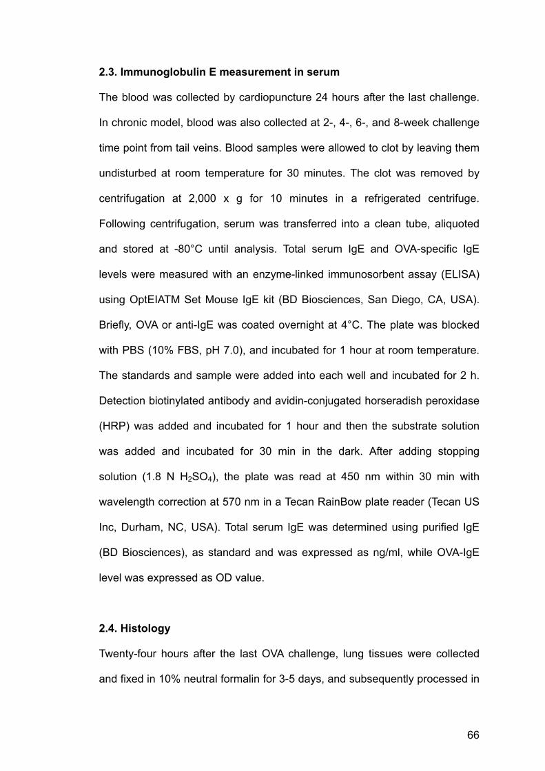
66
2.3. Immunoglobulin E measurement in serum
The blood was collected by cardiopuncture 24 hours after the last challenge.
In chronic model, blood was also collected at 2-, 4-, 6-, and 8-week challenge
time point from tail veins. Blood samples were allowed to clot by leaving them
undisturbed at room temperature for 30 minutes. The clot was removed by
centrifugation at 2,000 x g for 10 minutes in a refrigerated centrifuge.
Following centrifugation, serum was transferred into a clean tube, aliquoted
and stored at -80°C until analysis. Total serum IgE and OVA-specific IgE
levels were measured with an enzyme-linked immunosorbent assay (ELISA)
using OptEIATM Set Mouse IgE kit (BD Biosciences, San Diego, CA, USA).
Briefly, OVA or anti-IgE was coated overnight at 4°C. The plate was blocked
with PBS (10% FBS, pH 7.0), and incubated for 1 hour at room temperature.
The standards and sample were added into each well and incubated for 2 h.
Detection biotinylated antibody and avidin-conjugated horseradish peroxidase
(HRP) was added and incubated for 1 hour and then the substrate solution
was added and incubated for 30 min in the dark. After adding stopping
solution (1.8 N H2SO4), the plate was read at 450 nm within 30 min with
wavelength correction at 570 nm in a Tecan RainBow plate reader (Tecan US
Inc, Durham, NC, USA). Total serum IgE was determined using purified IgE
(BD Biosciences), as standard and was expressed as ng/ml, while OVA-IgE
level was expressed as OD value.
2.4. Histology
Twenty-four hours after the last OVA challenge, lung tissues were collected
and fixed in 10% neutral formalin for 3-5 days, and subsequently processed in

67
Leica Tp1020 tissue processor (Leica Microsystems, Nussloch, Germany) and
embedded in paraffin wax. The sequential procedure of procession was as
below: 10% formalin x 3h, 70% ethanol x 1h, 80% ethanol x 1h, 90% ethanol
x 1h, 100% ethanol x 1h, 100% ethanol x 1h, 100% ethanol x 1h, xylene x
3.5h, xylene x 3.5h, xylene x 3.5h, molten wax x 1.5h, and molten wax x 1.5h.
The embedded specimens were then cut into 5 μm sections, fixed upon the
slides, and stained with Haematoxylin & Eosin (H&E), Periodic Acid-Schiff
(PAS), or Masson Trichrome (all reagents were from Sigma). Briefly, slides
were deparaffinized with HistoClear (National Diagnostics, Atlanta, GA, USA)
and rehydrated by sequential passage through 100% ethanol, 90% ethanol,
70% ethanol, and distilled water. The sections were then stained using
different staining protocols: for H&E staining, in Harris Haematoxylin for 5 min,
then differentiation in 0.1% acid alcohol for 30 sec followed by counterstaining
in Eosin for1 min; for PAS staining, in Periodic Acid solution for 5 min, then in
Schiff’s reagent for 15 min followed by counterstaining in Gill 3 Haematoxylin
for 90 sec; for Masson Trichrome staining, in preheated Bouin’s solution at
56°C for 15 min, then sequential staining in Weigert’s Haematoxylin for 5 min
and in Fucshin solution for 5 min, and then rinsing in 1% Acetic acid for 5 min
followed by staining in Aniline Blue for 5 min. Slides were then dehydrated by
sequential passage through 70% ethanol, 90% ethanol, 100% ethanol, and
finally cleaned with HistoClear. Once dry, the slides were mounted with
HistoMount (National Diagnostics).
2.5. Immunohistochemistry
The detection of fibronectin was performed as previously described (Palmans

68
et al., 2000). Slides were incubated in 3% H2O2 in methanol for 10 min to
block endogenous peroxidase activity after deparaffinization and rehydration.
Then the sections were incubated in 0.4% pepsin solution (Sigma) in 0.01 N
HCl at 37°C for 15 min. After blocking with serum, the sections were
incubated with rabbit anti-mouse fibronectin antibody (US Biological,
Swampscott, MA) at a dilution of 1:40 for 30 min. Immunoreactivity was
detected by sequential incubations with a biotinylated secondary Ab,
VECTASTAIN Rabbit Elite ABC kit (Vector Laboratories, Burlingame, CA),
followed by 3, 3’-diaminobenzidine (DAB). The sections were counterstained
with Haematoxylin for 30 sec. Slides were then dehydrated by sequential
passage through 70% ethanol, 90% ethanol, 100% ethanol, and finally
cleaned with HistoClear. Once dry, the slides were mounted with HistoMount
(National Diagnostics).
2.6. Quantitative analysis of the airways
All slides were examined in a random blinded fashion. Evaluation of
hyperplasia of goblet cells and degree of peribronchial and perivascular
inflammation was based on a 5-point scoring system (Duan et al., 2004).
Briefly, the scoring system for goblet cell was: 0, no goblet cells; 1, <25% of
the epithelium; 2, 25–50%; 3, 50–75%; 4, >75%; the scoring system for
inflammation was: 0, no cells; 1, occasional cell cluster; 2, a ring of cells 1 cell
layer deep; 3, a ring of cells 2–4 cells deep; 4, a ring of cells >4 cells deep.
Scoring was performed in 5-10 different fields for each lung section. Masson
Trichrome stain and immunohistochemical stain analysis used Image-Pro Plus
software (Media Cybernetics, Silver Spring, MD) as described (Cho et al.,

69
2004). Briefly, nucleus in the lamina propria (defined as the region between
the epithelial basement membrane and the smooth muscle layer) and in the
whole epithelial layer were counted in the field and the length of the epithelial
basement membrane was measured. The data were used to calculate the
mean number of cells per 100 μm of epithelial basement membrane in the
lamina propria and epithelia (Temelkovski et al., 1998). Five bronchioles in
each slide were counted. The peribronchial Masson Trichrome staining and
fibronectin staining were expressed as the area of each staining per
micrometer length of basement membrane of bronchioles with 150–200 μm in
internal diameter. At least 5 bronchioles were counted in each slide. The
smooth muscle layer thickness was assessed at four predetermined
bronchiole sites (12, 3, 6, and 9 o’clock) in at least 5 bronchioles of similar
size (200-300 μm internal diameter) on each slide.
2.7. BAL fluid cell counts
The mice were sacrificed 24 hours after the last challenge. Tracheotomy was
performed, and then the lung was lavaged with two separated dosages of 0.6
ml of PBS via an intratracheal 18G cannula fixed in place with a ligature using
a syringe attached to the cannula. Cells were obtained by centrifugation (1000
x g for 5min). The supernatant was stored in -80°C for future proteomic
analysis. Red blood cells present in the cell pellet were lysed by adding 2ml of
0.87% NH4Cl solution for 10 minutes at room temperature and then
centrifuged again to remove the supernatant containing the lysed cells. The
resulting pellet was then resuspended in 1 ml of RPMI-1640 (containing 1%
BSA) and a total cell count was performed with a haemocytometer (10 μl BAL:

70
10 μl Trypan stain). Aliquots (105 cells/ 150 μl) of the lavage suspension were
then centrifuged by a Shandon Cytospin 3 (Thermo Electron Corporation,
Pittsburgh, PA) at 600 rpm for 10 min at room temperature. For cytological
examination, cytospin preparations were prepared, fixed, and stained in a
modified Wright stain (Duan et al., 2003). Differential cell count was then
performed on at least 500 cells (Figure 10).
2.8. Proteomics analysis
The general workflow of proteomic analysis involves sample preparation, first-
dimensional separation, equilibration, second-dimensional separation, staining,
imaging, image analysis, and protein identification (Figure 11).
2.8.1. Sample collection and preparation
BAL fluid samples were precipitated with trichloroacetic acid (TCA) (10% final
concentration) in an ice bath for 20 min, and subsequently centrifuged at 3500
rpm for 15 min at 4oC. The pellet was re-suspended in ice-cold acetone and
centrifuged as described above. The pellet was air-dried for a few minutes,
solubilized in the lysis buffer containing 8 M urea, 2 M thiourea, 4% 3-[(3-
cholamidopropyl)dimethylammonio]-1-propanesulfonate (CHAPS), 0.8%
Pharmalyte, 65 mM dithiothreitol (DTT), protease inhibitor cocktail, 5 μg/ml
DNase I, 20 μg/ml RNase A for 30 min at 4°C. The lysate was centrifuged at
20800 x g for 15 min. The supernatant was aliquoted and stored at -80°C until
further use. Protein concentration was measured by a modified Bradford
assay (Bradford, 1976). Briefly, a standard curve was made using bovine
serum albumin (BSA) which was prepared in the same lysis buffer as the

71
protein samples to be assayed with concentrations of 0, 5/30, 8/30, 10/30,
15/30, 20/30, 1 μg/ml. Pipet 30 μl of each standard and sample solution into a
clear tube and then 1 ml of diluted dye reagent (Bio-Rad Laboratories,
Hercules, CA, USA) (1 dye: 4 water) was added into the tube. The samples
were incubated at room temperature for 5 min and then the absorbance was
measure at 595 nm in a microplate reader (Tecan Sunrise, Salzburg, Austria).
Lung tissues were homogenized in the lysis buffer as mentioned above
using Heidolph Diax 900 homogenizer (Heidolph Instrument, Schwabach,
Germany) on ice and then leaved still for solubilization for 30 min at 4°C. The
protein solution was centrifuged at 20800 x g for 15 min. The supernatant was
aliquoted and stored at -80°C until further use. Protein concentration was also
measured above.

72
Figure 10. Type of cells typically found in BAL fluid. A, macrophage; B, eosinophil; C, lymphocyte; and D, neutrophil.
A
D
B
C
D B

73
Figure 11. Scheme of proteomics workflow and instruments. A, Reswelling tray for rehydration (Amersham Biosciences) B, IPGphor for first-dimensional electrophoresis (Amersham Biosciences) C, EttanTM DALTtwelve vertical system for second-dimensional electrophoresis (Amersham Biosciences) D, GS-800 imaging densitometer (Bio-Rad Laboratories) E, PDQUEST (Version 7.2) software (Bio-Rad Laboratories) F, Proteomics Analyzer with TOF-TOFTM optics work station (Applied Biosystems)
A B C D E F

74
2.8.2. Two-dimensional electrophoresis
The first- dimensional electrophoresis used Immobiline DryStrip (180 mm, pH
4-7 and pH 6-11) (Amersham Biosciences, San Francisco, CA, USA). For pH
4-7, the strips were rehydrated for 12h in 350 μl of solution including
rehydration solution with 8 M urea, 2% CHAPS, 0.5% IPG buffer, 0.0005%
Bromophen blue and protein sample in a reswelling tray (Amersham
Biosciences). For pH 6-11, the strips were only rehydrated in the rehydration
solution which was modified to 7 M urea, 2 M thiourea, 4% CHAPS, 2.5% DTT,
10% isopropanol, 5% glycerol, 2% IPG buffer pH6-11 according to Hoving and
co-workers (2002). The protein sample was loaded to the anodic side just
before electrophoresis. Equal protein loads (80 μg of BAL fluid protein, 120 μg
of lung protein) were used for all normal sample loading gels. Paper-bridge
loading method was used for high sample loading (>400 μg) of pH6-11 gels
(Sabounchi-Schutt et al., 2000). High sample loading was applied when the
preliminary analysis was finished and desired spots needed to be cut for MS
analysis. The isoelectrofocusing (IEF) was performed with an IPGphor
(Amersham Biosciences) at 20°C using the protocol listed in Table 4.
Table 4. Running protocols for IEF
Running Voltage & duration 200 V 500 V 4000 V 8000 V
Normal loadingpH4-7
0.5 h Step & Hold
1 h Gradient
1.5 h Gradient
48000 Vhr Step & Hold
High loading pH4-7
1 h Step & Hold
2 h Gradient
3 h Gradient
56000 Vhr Step & Hold
Normal loadingpH6-11
1 h Gradient
2 h Gradient
3 h Gradient
48000 Vhr Step & Hold
High loading pH6-11
2 h Gradient
4 h Gradient
6 h Gradient
56000 Vhr Step & Hold

75
The strips were then equilibrated for 15 min in 6 M urea, 30% glycerol, 2%
Sodium dodecyl sulphate (SDS), 50 mM Tris-Cl (pH 8.8), 1% DTT followed by
another 15 min equilibration in the same solution except that DTT was
changed to 2.5% iodoacetamine. The single-percentage 12% acrylamide gels
were prepared in EttanTM DALT Gel Caster (Amersham Biosciences). About
50 ml of gel solution containing 0.375 M Tris-Cl (pH 8.8), 0.1% SDS, 12%
acrylamide, 0.05% ammonium persulfate (APS), 0.0005% (v/v)
tetramethylethylenediamine (TEMED) was used for one gel. For second
dimensional separation, the equilibrated strips were transferred onto the top of
acrylamide gels and sealed with 0.5% agarose Gel electrophoresis was
carried out in an EttanTM DALTtwelve vertical system (Amersham
Biosciences) at room temperature. The electrophoresis condition was set at
2.5 W/per gel for 30 min followed by 10 W/per gel for about 4-5 h until the dye
front was 5 mm from the bottom. The gels were fixed in solution containing
40% (v/v) ethanol, and 10% (v/v) acetic acid for overnight.
2.8.3. Silver staining
Silver staining was performed according to the manufacturer’s instruction
(Amersham Biosciences). The gel containers were placed on a shaker
(Unimax 2010, Heidolph Instrument) at 30-40 rpm throughout the staining.
After fixation, the gels were sensitized with 30% (v/v) ethanol, 5% (w/v)
sodium thiosulfate, and 6.8% (w/v) sodium acetate for 30 min. After washing
with distilled water at 3 times for 5 min/each time, the gels were incubated in
0.25% (w/v) silver nitrate for 20 min. Then the gels were rinsed shortly with
distilled water at 2 times for 1 min/each time and developed in 2.5% (w/v)

76
sodium carbonate and 0.015% (w/v) formaldehyde until the desired intensity
of spots was achieved. Then the development was terminated with 1.46%
(w/v) EDTA-Na2 x 2H2O. The gels were scanned using GS-800 calibrated
imaging densitometer (Bio-Rad Laboratories) following washing with distilled
water at 3 times for 5 min/each time.
2.8.4. Image analysis
The PDQUEST (Version 7.2) software (Bio-Rad Laboratories) was used to
detect, quantify, and match spots. All spots were carefully reviewed by
operator to add missing spots, remove redundant spots, and rectify
mismatched spots. Every spot was given a quantity of OD (optical density)
fitted by Gaussian model. Because of potential variation in protein loading,
rehydration and staining time, therefore, the gel images were normalized by
total quantity of all matched spots to get rid of the effects of these non-
expression-related variations. Thus the quantity was expressed as parts per
million (PPM) of total OD of all valid spots. Only the spots which appeared in
all the gels of the same treatment group were selected for further analysis.
2.8.5. In-gel digestion and MS analysis
Protein spots of interest were excised from the stained gels and digested with
modified trypsin (Promega, Madison, WI, USA) according to the method
described before (Shevchenko et al., 1996). In general, in-gel digestion was
performed in an orderly sequence starting from destaining, dehydration,
drying, digestion, to extraction. Samples were analyzed using 4700
Proteomics Analyzer with TOF-TOFTM optics work station (Applied Biosystems,

77
Foster City, CA, USA). Both MS and MS/MS data were acquired with a
Nd:YAG laser with 200 Hz repetition rate and up to 4000 shots were
accumulated for each spectrum. MS/MS mode was operated with 2 keV
collision energy, mass range 800-3500 Da. Peptide mass fingerprints (PMF)
of the tryptic peptides from MALDI-TOF MS and MS/MS data on the
representative spots, together with the isoelectric points and molecular
weights, were used to search the National Center for Biotechnology
Information (NCBI) protein database with the programs Mascot at
http://www.matrixscience.com and MS-Fit at http://prospector.ucsf.edu.
2.9. Western blotting
Twenty μg of total protein lysate diluted in sample buffer (62.5 mM Tris-HCl
(pH=6.8), 10% glycerol, 5% 2-mercaptoethanol, 2% SDS, and 0.0125%
bromophenol blue) was resolved on SDS-PAGE. 9.8% gels were used for
Surfactant protein D (SP-D) was resolved in a 9.8% homogenous SDS-PAG,
and lungkine was resolved in a 15% homogenous SDS-PAG. Then the
proteins were electrotransferred to polyvinylidene difluoride (PVDF)
membrane using a semi-dry transblotter (ATTO Corp., Tokyo, Japan), and
followed by blocking in 5% non-fat milk dissolved in Tween 20-Tris-buffered
saline (TTBS) (1M Tris-HCl pH 7.5, 0.9% NaCl, 0.05% Tween 20) for 1.5 hour
at room temperature. The membranes were incubated with primary antibodies
against β-actin (1:5000), lungkine (1:1000) (R&D Systems, Minneapolis, MN)
or SP-D (Chemicon International, Temecula, CA) (1:1000) followed by alkaline
phosphatase (AP)-conjugated secondary antibodies (1:1000)
(DakoCytomation, Glostrup, Denmark). Proteins were visualized

78
colorimetrically by adding substrate NBT (nitroblue tetrazolium) and BCIP (5-
bromo-4-chloro-3-indoyl-phosphate) (Bia-Rad) in AP buffer (100 mM Tris-HCl
pH 9.5; 100 mM NaCl; 50 mM MgCl2). The gels were scanned and images
were analyzed by Quantity-One software (Bio-Rad).
2.10. RT-PCR
Total mRNA from BAL fluid cells using a RNA isolation kit (Ambion, Austin, TX)
and from lung tissues using TRIzol (Life Technologies, Gaithersburg, MD)
were extracted. First strand cDNA was synthesized from 1 μg RNA using oligo
(dT) 15 primers and 10 U AMV reverse transcriptase. PCR was performed in
a GeneAmp PCR system 2700 thermal cycler (Applied Biosystems). The
primers used and the product size are listed in Table 5. PCR products were
run in a 1.5% agarose gel, visualized under UV light and quantitated using
Gel-Pro imaging software (Media Cybernetics).
2.11. Chitinase activity assay
Chitinase bioactivity in BAL fluid was assayed using a fluorescence method as
described (Zhu et al., 2004). Briefly, 50 μl of non-precipitated BAL sample was
incubated at 37°C with 50 μl substrate 4-methylumbelliferyl-β-D-N,N’-
diacetylchitobioside (Sigma) in citrate/ phosphate buffer (0.1 M/ 0.2 M, pH 5.2),
at a concentration of 0.17 mM for 15 min. Then the reaction was stopped with
1 ml of glycine/ NaOH buffer (0.3 M, pH 10.6). The fluorescent product 4-
methylumbelliferone was measured using a SpectraMax Gemini EM
fluorimeter (Molecular Devices, Sunnyvale, CA) with excitation wavelength at
350 nm and emission at 445 nm. A 4-methylumbelliferone (Sigma) standard

79
curve was generated to quantify the enzyme activity.
2.12. Statistics
Changes in optical intensities of all matched protein spots between groups
(n=9) were subjected to ANOVA followed by two-tail Student’s t-test using
SPSS software (Chicago, IL). Data were expressed as mean ± SEM. The
critical level for significance was set at p < 0.05.

80
Table 5. Primers for RT-PCR analysis
Prod
uct S
ize
483
bp
624
bp
429
bp
537
bp
510
bp
426
bp
226
bp
301
bp
312
bp
317
bp
Rev
erse
5’-G
CTT
GA
CA
ATG
CTG
CTG
GTA
-3’
5’-C
AA
GC
ATG
GTG
GTT
TTAC
AG
GA
-3’
5’-A
TGG
TCC
TTC
CAG
TAG
GTA
ATA-
3’
5’-G
AG
TGC
GC
AG
GA
AA
CTT
AGG
-3’
5’-G
AA
GC
TCTC
CC
GTG
GTC
GTA
G-3
’
5’-C
AGA
GG
ACA
GG
AA
GG
TGA
GC
-3’
5’-G
CC
CAT
AGTG
GAG
TGG
GAT
AA
G-3
’
5’-T
GG
CTT
TGTT
CTG
GC
TCTT
T-3’
5’-A
TCG
AA
AAG
CC
CG
AA
AG
AG
T-3’
5’-G
GTT
ACAG
AG
GC
CAT
GC
AAT
-3’
Sequ
ence
s
Forw
ard
5’-T
GG
GTT
CTG
GG
CC
TAC
TATG
-3’
5’-C
TGG
AAT
TGG
TGC
CC
CTA
CA
-3’
5’-C
AGA
AC
CG
TCA
GAC
ATTC
ATTA
-3’
5’-T
GAC
TCTG
CTG
ATG
GTC
CAG
-3’
5’-G
CC
AA
GG
AG
CC
TCG
CC
TATT
CTC
AGG
-3’
5’-G
AG
TGA
CAT
TGC
AG
GA
AG
CA
-3’
5’-T
ATTC
CC
GC
GTT
AGTC
TGG
TG-3
’
5’-G
ACTC
CC
TGAA
GC
AGC
AAAC
-3’
5’-T
CA
AC
CC
CC
AG
CTA
GTT
GTC
-3’
5’-C
AGC
TCC
CTG
GTT
CTC
TCAC
-3’
Targ
ets
AM
Cas
e
Ym
1
Ym
2
Chi
totri
osid
ase
Gob
-5
MU
C5A
C
Lung
kine
PS
TPIP
IL-4
IL-1
3

81
3. PROTEOMIC ANALYSIS OF ACUTE AIRWAY
INFLAMMATION IN A MOUSE MODEL

82
3.1. Results
3.1.1. Acute mouse asthma model
By the end of the 21-day acute mouse asthma model, mice were sacrificed.
BAL fluid and blood was collected. Total cell count and eosinophil count were
dramatically increased (p < 0.05) in BAL fluid from OVA-treated mice as
compared to saline control mice. To a lesser extent, neutrophil, macrophage
and lymphocyte counts were also increased in allergic airway inflammation
(Figure 12A). Airway responsiveness, determined by Penh measurement
(Hamelmann et al., 1997), to inhaled methacholine was markedly increased (p
< 0.05) in OVA-challenged mice as compared to saline-challenged mice
(Figure 12B). Serum levels of total IgE and OVA-specific IgE were
substantially elevated in allergic airway inflammation (Figure 12C and D). In
addition, OVA aerosol challenge induced marked infiltration of inflammatory
cells, especially eosinophils, into the lamina propria, perivascular and
peribronchiolar areas as compared to saline control (Figure 12E, F and I).
Furthermore, OVA-challenged mice, but not saline-challenged mice,
developed marked goblet cell hyperplasia and mucus hypersecretion within
the bronchi (Figure 12G, H and J).

83
0
100
200
300
400
500
600
0 10 20 30 40 50 60 70 80Methacholine (mg/ml)
% P
enh
Saline (n=11)
OVA (n=11)
*
0
1
2
3
4
5
Total EOS NEU MAC LYM
Cel
l Cou
nt (x
105 ) Saline (n=11)
OVA (n=11)
*
*
*
**
0
200
400
600
800
1000
1200
1400
Saline (n=5) OVA (n=8)
Tota
l IgE
(ng/
ml)
*
0
0.2
0.4
0.6
0.8
1
1.2
1.4
Saline (n=5) OVA (n=8)
OVA
-IgE
(OD
)
*
B
DC
A
E F G H
0
1
2
3
4
Saline OVA
Muc
us S
core
*
J
0
1
2
3
4
5
Saline OVA
Infla
mm
atio
n Sc
ore
*
I

84
Figure 12. Development of a murine acute asthma model (A) BAL fluids were collected 24 h after the last saline or OVA aerosol challenge. Total and differential cell counts were performed. EOS, eosinophil; NEU, neutrophil; MAC, macrophage; LYM, lymphocyte. (B) Airway responsiveness of mice challenged with aerosolized saline or OVA for 3 consecutive days to increasing doses of inhaled methacholine was measured using a single-chamber whole-body plethysmograph. Serum levels of total IgE (C) and OVA-specific IgE (D) were analyzed using ELISA (saline, n=11; OVA, n=11). Histological examination of lung tissue eosinophilia (E and F) and mucus production (G and H) after the last challenge of saline aerosol (E and G) or OVA aerosol (F and H) was performed under a microscope at 400x magnification. Abundant cell infiltrates with high percentage of eosinophils were localized in the perivascular and peribronchiolar region of OVA-challenged mice. OVA-challenged mice also showed increased periodic acid-Schiff stain (PAS)-positive goblet cells. Quantitative analyses of inflammatory cell infiltration (I) and mucus production (J) in lung sections (n=4) were performed as described previously. Data are presented as means± SEM.*Significant difference from saline control, p < 0.05.

85
3.1.2. 2-DE and image analysis of BAL fluid
BAL fluid samples were obtained at the time when functional studies were
performed. Samples from control mice (n=9) and OVA-sensitized and
challenged mice (n=9) were resolved by 1-D electrophoresis on IPG gel strips
in pH 4-7 range, followed by 2-D electrophoresis on homogeneous 12% SDS-
PAGE. Identical protein patterns were observed on 2-D gels of both control
group and OVA group BAL fluids (Figure 13). Following image analysis, about
500 spots were consistently detected and matched on all 9 separate gels of
each treatment group (saline vs. OVA), and were selected for differential
protein expression analysis based on a criterion of at least 3-fold change in
optical density. At least 70%-80% of 500 spots were automatched by the
software and left spots were manually matched due to slightly alteration of
locations. A total of 28 protein spots were significantly altered (p< 0.05) in
airway inflammation, and were subjected to peptide mass fingerprinting by
MALDI-TOF MS. A representive searching result for spot 12 (SSP 8608)
which is gob-5 protein was shown (Figure 14). Twenty-four spots were
identified by MS and summarized in Table 6. The optical densities expressed
as mean ± SEM and the location of the identified protein spots were shown in
a master image (Figure 15). Graphical representation of these 24 identified
proteins whose levels changed in asthmatic mice compared with control mice
were showed as individual densities (Figure 16). A typical MALDI and MS/MS
spectrum is also shown (Figure 17).Several proteins existed in multiple spots
of similar molecular weights but of different isoelectric points, which may be a
result of post-translational modifications such as glycosylation or
phosphorylation (Graves and Haystead, 2002). We observed a marked

86
increase (p < 0.05) in some pro-inflammatory proteins in BAL fluid from
allergic airway such as lungkine, a recently described chemokine (Rossi et al.,
1999), gob-5 (Nakanishi et al., 2001), and a family of chitinases including Ym1,
Ym2 and acidic mammalian chitinase (AMCase) (Webb et al., 2001; Zhu et al.,
2004). We also detected a significant increase (p <0.05) in SP-D which may
afford protective action against airway inflammation (Crouch and Wright,
2001). On the other hand, the levels of some anti-inflammatory proteins such
as haptoglobin (Yang et al., 2000) and α-1-antitrypsin (Eden et al., 1997) were
dramatically decreased (p < 0.05) in allergic airway inflammation.

87
Figure 13. Representative 2-D gels of BAL fluid in acute asthma model samples corresponding to saline control (left panel) and OVA-treated (right panel). BAL fluids were separated by 2-DE using IPG at pH 4-7 and 12% SDS-PAGE. Proteins were stained with silver nitrate.
pH 4 7250
10
kDa
pH 4 7250
10
kDa
Saline OVA

88

89
Figure 14. A representive searching result (part) for spot 12 (SSP 8608) which is gob-5 protein.

90
Table 6. Proteins identified with significant expression level changes in BAL
fluid from asthmatic mice.
No. SSP# Protein name Accession ID
Coverage**
Score*
Fold No. of peptide
1 2007 Acidic mammalian chitinase (AMCase)
gi|37999744
49% 295 5.7 12
2 2330 Ym1 gi|285015 31% 120 42.5 11 3 3218 Ym1 gi|285015 39% 242 58 15 4 3227 Ym1 gi|285015 45% 109 16 15 5 3307 Ym1 gi|285015 29% 78 7.4 11 6 4207 Ym1 gi|285015 53% 121 4.2 19 7 4221 Ym1 gi|285015 53% 110 10.5 16 8 6203 Ym2 gi|2212390
7 46% 95 71.4 15
9 3004 Lungkine gi|5031129 35% 137 10 11 10 4010 Lungkine gi|5031129 36% 128 7.2 10 11 6011 Lungkine gi|5031129 34% 130 8.1 9 12 8608 gob-5 protein gi|7513665 22% 102 140 14 13 8630 gob-5 protein gi|7513665 27% 156 109 24 14 2703 Polymeric
immunoglobulin receptor
gi|2804246 31% 149 4.9 25
15 5020 Alpha-1-antitrypsin gi|90278 22% 78 -6 6 16 5312 Surfactant protein D gi|6524992 37% 108 40 12 17 6211 Surfactant protein D gi|6524992 35% 260 57.8 18 18 7209 Surfactant protein D gi|6524992 32% 318 39 16 19 8208 Surfactant Protein D gi|6524992 38% 81 19.7 11 20 3220 Haptoglobin gi|8850219 34% 371 -4.6 19 21 3430 Vitamin D-binding
protein precursor (DBP)
gi|111243 47% 280 -9 22
22 3204 γ-Actin gi|809561 37% 355 8 18 23 7123 Immunoglobulin light
chain variable region (Fragment)
gi|799373 27% 145 4.5 4
24 3012 PEST phosphatase interacting protein
gi|1857712 18% 80 20.5 12
* Score is -10 x Log (P), where P is the probability that the observed match is a random event. Protein score of greater than 75 is considered significant (p <0.05). ** Coverage refers to peptide sequence homology of at least 20% to the matched protein
# SSP is spot number given by software according the pI and MW. Fold means fold changes in OVA-mice compared with control mice. No. of peptide means the number of matched peptides.

91
Figure 15. Differential BAL fluid protein expression profile upon OVA sensitization and challenge. The 2-DE reference map is composed of dataset of 18 gels (n=9 for saline control and n=9 for OVA). The proteins were separated by 2-DE using IPG at pH 4-7. The boxed proteins were identified by peptide mass mapping by MALDI-TOF MS. The numbers represent the spot identification numbers listed in Table 5. Spot optical densities were expressed as mean ± SEM. The optical densities of saline control and OVA-treated group were compared using Student’s t test. *Significant difference from saline control, p < 0.05.

92
Figure 16. Graphical representation of the 24 identified proteins whose levels changed in asthmatic mice compared to control mice. The columns from left to right in each graph are densities of 9 control samples (blue column) and 9 asthmatic samples (red column). The digit beside the columns is the greatest PPM value (OD) for this protein among 18 gels.

93
Figure 17. A representative MALDI-TOF-TOF MS spectrum and subsequently MS/MS spectrum of Surfactant Protein D.
69 263 457 651 845 1039Mass (m/z)
223.3
0
10
20
30
40
50
60
70
80
90
100
% In
tens
ity
4700 MS/MS Precursor 984.564 Spec #1 MC=>BC=>AdvBC(32,0.5,
984.8027
101.0898
255.150684.0720
425.180770.0829 442.1877
272.1649
942.2531314.1469147.5309 430.2413 668.1866 875.3835538.273373.0719172.0847 949.8962360.2123 770.4724640.3446254.1118 864.0267494.0823 939.4499727.4112
69.0 437.2 805.4 1173.6 1541.8 1910.0Mass (m/z)
259.
0
10
20
30
40
50
60
70
80
90
100
% In
tens
ity
4700 MS/MS Precursor 1809.96 Spec #1 MC=>BC=>AdvBC(3
517.2515
86.1310370.1754
796.4009 1486.8080683.3224
570.2518 1052.52881648.8210
645.1956398.1780110.0869 1165.6396924.4422175.1249 469.2014 1237.6779758.2944 1016.4032112.1042 1664.74041489.0028556.2142300.1068 1241.6196739.2914884.8512 1747.1927538.0517 1050.8029 1523.3590389.1692199.0892
69 313 557 801 1045 1289Mass (m/z)
396.9
0
10
20
30
40
50
60
70
80
90
100
% In
tens
ity4700 MS/MS Precursor 1221.65 Spec #1 MC=>BC=>AdvBC(32,0
575.2684
110.0904
662.2947
947.4683
72.1033
809.3869243.1343784.3787 1075.5353785.3997438.1996120.0911 979.5063215.1629 767.363670.0834
342.1833 766.4186919.5055225.0743 429.1721 1093.5741558.2551 904.4669228.1490123.1007 739.4414 1071.5814388.1057 588.1970 938.6281483.2515 839.9477744.3339217.1290
799.0 1341.4 1883.8 2426.2 2968.6 3511.0Mass (m/z)
3706.3
0
10
20
30
40
50
60
70
80
90
100%
Inte
nsity
4700 Reflector Spec #1 MC=>BC=>AdvBC(32,0.5,0.1)=>NR(2.00)[BP = 2501.3, 3706]
2307.1094846.4947
2500.27932532.2761
1810.9457
902.5212 2516.27441221.65042030.0427
967.5378 1519.8286842.5573
1252.6152877.08031885.8224
2048.05741203.6339920.5247 2323.11771486.8013 2833.41501228.5793 2028.0310911.0296 2514.28491791.94971470.7819 2814.34622264.60841186.5425

94
3.1.3. Immunoblotting and RT-PCR
To verify the 2-DE findings, we conducted immunoblotting analyses and RT-
PCR on selected protein targets. As shown in Figure 18, the optical density of
protein spots in 2-DE were highly reproducible. Western blot analysis of BAL
fluids revealed significant increases (p < 0.05) in lungkine and SP-D protein
levels in allergic airway inflammation. Equal loadings of BAL fluid proteins
were confirmed using β-actin as internal control (data not shown). We also
noticed that lungkine mRNA expression was absent from BAL fluid cells which
comprise mainly of inflammatory cells like macrophage, eosinophil, neutrophil
and lymphocyte, but was strongly induced in lung tissues (Figure 18),
indicating that the source of lungkine is primarily from local lung
microenvironment such the bronchoepithelial cells. On the other hands, 2-DE
of BAL fluid and RT-PCR analysis of BAL fluid cells and lung tissues
demonstrated increased gene and protein expressions of Ym1, Ym2, AMCase
and gob-5 (Figure 18) in airway inflammation. The rank order for the basal
expression levels of the 3 chitinases identified was Ym1 >> AMCase >Ym2. In
fact, Ym2 level was undetectable in control BAL fluid. Upon OVA challenge, all
3 chitinase levels were markedly elevated. In addition, basal gob-5 protein
level in BAL fluid and mRNA level in BAL fluid cells were undetectable. Upon
OVA challenge, gob-5 gene and protein levels were markedly increased.

95
Figure 18. Western blot analysis of BAL fluids using monoclonal antibodies targeted at lungkine (A) and SP-D (B). Top panels indicate the locations and protein spots of lungkine and SP-D in 3 randomly selected 2-D gels of control BAL fluid and allergic airway BAL fluid. The middle panels show Western blot results of three control and three allergic airway BAL fluids. Proteins (20 μg/lane) were separated by SDS-PAGE and probed with anti-lungkine and anti-SP-D antibodies. The blot was developed by NBT/BCIP, and analyzed using Gel-Pro imaging software. The bottom panels show the semi-quantitative data (means ± SEM, n=3) of target protein band intensities between control group and OVA-treated group. Lungkine level was increased by 6.3-fold and SP-D level was increased by 3.0-fold in allergic airway inflammation. *Significant difference from saline control, p < 0.05.
0
100
200
300
400
500
Saline OVA
OD
Val
ue
*
0
50
100
150
200
250
300
Saline OVA
OD
Val
ue
*
Saline OVA Saline OVA
1 2 3 1 2 3 1 2 3 1 2 3
Saline 1 2 3 Saline 1 2 3
OVA 1 2 3 OVA 1 2 3
(A) Lungkine (B) SP-D

96
Figure 19. RT-PCR analysis of BAL fluid cells and lung tissues from control and OVA-treated mice targeting at gene expression of proteins that were found to have significantly altered expression levels in allergic airway inflammation by 2-DE. Total mRNA from BAL fluid cells and lung tissues were extracted using a RNA isolation kit and the PCR products were separated in a 1.5% agarose gel and visualized under UV light. The experiments were repeated for four times with similar pattern of results.
AMCase
Ym1
Ym2
Lungkine
Gob-5
β-Actin
CON OVA CON OVA CON OVA
BALF Cells Lung BALF 2-D

97
3.2. Discussion
The present study using 2-DE followed by MS-based protein analysis was able
to identify major alterations in the expression levels of a number of proteins in
BAL fluid collected from OVA-treated mice. During the course of this study,
there were 2 separate reports published on the proteomics of allergic airway
inflammation (Signor et al., 2004; Fajardo et al., 2004). However, their findings
are quite different from each other and are also dissimilar to our observations
in this study. In Fajardo’s report (2004), C57BL/6 mice were sensitized with
OVA on days 0 and 14 but challenged with 200 µg OVA on days 30, 33, and
35. In line with our study, they also identified Ym1 and Ym2 but surprisingly
without AMCase. The absence of AMCase may be due to technical missing or
the availability of AMCase gene information at the time of their study. In
Signor’s study N rats were sensitized with OVA subcutaneously on days 1, 15
and 22, and challenged with 0.3 mg/kg of OVA Intratracheally on day 29. They
have identified some proteins which were also identified in our study including
surfactant proteins, α-1-antitrypsin and haptoglobin. The differences in animal
species and experiment protocols may be the main cause leading to different
findings. Nevertheless, some extent of accordance can be observed given that
some proteins were identified by different groups. The advantages of our study
are that we used an established murine model of asthma (Duan et al., 2004)
and had large sample size (n=9 for control and n=9 for OVA) for BAL fluid
protein analysis to preclude false positive findings. Our present study reveals
for the first time using proteomics analysis of BAL fluid major alterations in
expression level of a newly described chemokine, lungkine, a family of
chitinases, gob-5, SP-D, and other proteins in allergic airway inflammation.

98
Lungkine is a member of ELR-CXC chemokine family (Rossi et al. 1999; Chen
et al., 2001). The CXC chemokine is characterized by the invariant four cysteine
residues at the N terminus with the first two cysteines separated by a
nonconserved amino acid. It is further classified by the presence or absence of
the tripeptide motif glutamic acid-leucine-arginine (ELR) immediately preceding
the CXC sequence (Olson et al., 2002). Lungkine, designated as CXCL15, has
been shown to distribute exclusively in lung, especially bronchoepithelial cells,
and be released into the airways. The expression of lungkine has previously
been shown to be up-regulated by lipopolysaccharide, N.brasiliensis worms,
aspergillus, and OVA sensitization and challenge (Rossi et al., 1999).
Our 2-DE findings clearly showed a dramatic increase in BAL fluid lungkine
level in allergic airway inflammation. The observation was further confirmed with
immunoblotting and RT-PCR analyses. Lungkine has been shown to regulate
neutrophil migration in the lungs (Rossi et al., 1999; Chen et al., 2001). The
present study also showed a slight but significant increase in neutrophil level in
BAL fluid obtained from asthmatic mice. The contributory role of neutrophils to
the pathogenesis of allergic airway inflammation has not been firmly established.
Nevertheless, cumulative evidence shows an allergen-specific early neutrophil
infiltration after allergen challenge in mouse asthma model (Taube et al., 2003).
During asthma exacerbation, a marked increase in neutrophils in sputum has
been demonstrated (Fahy et al., 1995). Additional studies on the role of lungkine
in the chemoattraction of other inflammatory cells and in asthma development
are warranted.
A family of chitinase proteins was found to be up-regulated in BAL fluid in
allergic airway inflammation. In contrast to results reported by Webb et al. (2001)

99
showing higher Ym2 level than Ym1 in the allergic murine lung, our 2-DE
analysis revealed more abundant expression of Ym1 than Ym2 in BAL fluid. It
has been shown that allergen-induced up-regulation of chitinases was dependent
on CD4+ T cells and IL-4 or IL-13 signaling (Webb et al., 2001; Welch et al., 2002;
Zhu et al., 2004). AMCase induction has also been observed in asthmatic
subjects. The catalytic activity of AMCase in the BAL fluid was found to be
enhanced in asthmatic mice. The contributory role of AMCase to allergic airway
inflammation has recently been confirmed as anti-AMCase serum decreased
eosinophil and lymphocyte infiltration, and airway hyperresponsivess in asthmatic
mice (Zhu et al., 2004). The role of Ym1 and Ym2 in airway inflammation is still
unclear. Ym1/Ym2 has been shown to be an eosinophil chemotactic factor
capable of regulating eosinophil trafficking with a potency equivalent to RANTES
but weaker than eotaxin (Webb et al., 2001; Chang et al., 2001). Ym1 is also able
to induce T lymphocyte chemotaxis (Chang et al., 2001). Furthermore, Ym1 and
Ym2 have been shown to bind specifically to glucosamine oligomers such as
chitobiose, chitortriose and chitotetraose, and to heparin/heparan sulfate
proteoglycans, and suggested to play a role in airway wall remodeling in allergic
asthma (Webb et al., 2001; Owhashi et al., 2000). A recent study reported that
intratracheal administration of an adenoviral vector that expressed ECF-L
(identical to Ym1) antisense suppressed airway hyperresponsiveness and
eosinophil infiltration in mouse model (Iwashita et al., 2006). These results
indicate that Ym1 may play a critical role in allergic inflammation and bronchial
asthma. Additional studies are required to determine if Ym1 and Ym2 are
pathogenic molecules and/or biomarkers for asthma.
The present study demonstrated for the first time that gob-5 protein can be

100
detected in BAL fluid. Gob-5 (alias mCLCA3), a member of the chloride channel,
calcium activated (CLCA) family, plays a critical role in active export of chloride
ions that leads to mucin secretion from airway epithelial cells such as goblet cells.
The levels of gob-5 and its human counterpart hCLCA1 were undetectable in
normal airway, but were strikingly increased in allergic airway (Nakanishi et al.,
2001; Gruber et al., 1998; Hoshino et al., 2002). Indeed, our findings showed a
significant surge in gob-5 expression not only in BAL fluid cells and lung lysates,
but also in BAL fluid in allergic airway. Expression of gob-5 or hCLCA1 in allergic
airway has been associated with significant induction of MUC5AC, a gene
responsible for goblet cell metaplasia and mucin production (Nakanishi et al.,
2001; Hoshino et al., 2002). Depletion of gob-5 protein using antisense
oligonucleotide suppressed mucus production, inflammatory cell infiltration, and
airway hyperresponsiveness in asthmatic mice (Nakanishi et al., 2001). In line
with a recent histological study showing gob-5 protein being secreted with mucin
granules from goblet cells (Leverkoehne and Gruber, 2002), we were able to
identify gob-5 in BAL fluid using 2-DE. Therefore, the notion of using gob-5 as a
biomarker in BAL fluid or induced sputum for asthma warrants further study.
Our 2-DE analysis also identified a striking increase in SP-D protein in allergic
airway inflammation. SP-D is a member of the collagenous calcium-dependent
lectin (collectin) family that includes SPA. Pulmonary SP-D is mainly synthesized
and secreted by airway epithelial cells and is involved in the modulation of
immune response via its carbohydrate recognition domain that specifically binds
glycoconjugates expressed on the surface of leukocytes such as alveolar
macrophages, and inhaled allergens (Crouch and Wright, 2001). The role of SP-
D in asthma is now being actively studied and appears to have some protective

101
functions. Our present findings are consistent with two previous studies showing
elevated BAL fluid level of SP-D in a rat model of asthma (Kasper et al., 2002)
and in patients with bronchial asthma (Cheng et al., 2000). It has been shown
that SP-D can bind to mite allergen (Dermatophagoides pteronyssinus, Der p) to
prevent its interaction with allergen-specific IgE, leading to decreased histamine
release from sensitized basophils (Wang et al., 1998). In a mouse allergic
bronchopulmonary aspergillosis model, SP-D has been shown to lower blood
eosinophilia, pulmonary infiltration, allergic specific IgE level and Th2 cytokine
levels (Madan et al., 2001). A recent study showed addition of rSP-D significantly
inhibited Th2 cell activation and IgE and chemokines productions after allergen
challenge in a mouse model (Haczku et al., 2006). Therefore, the observed
increase in SP-D BAL fluid level from allergic airway reflects a compensatory
anti-inflammatory response to lung inflammation.
Additional proteins were found to be up-regulated in BAL fluid from allergic
airway including polymeric immunoglobulin receptor (pIgR), immunoglobulin γ
light chain and γ-actin. PIgR is expressed on the basolateral surface of airway
epithelial cells capable of binding and transporting polymeric IgA and IgM to the
apical surface for mucosal immunity (Kim and Malik, 2003). The expression of
pIgR on human airway epithelium was found to be up-regulated by IL-4 and IFN-γ
(Ackermann et al., 1999). The level of pIgR in nasal lavage fluid has been shown
to increase in subjects with acute sinusitis (Casado et al., 2004). γ-actin is one of
six functional actin isoforms identified. While α-actin is predominantly expressed
in muscle cells and involved in muscular contraction, both β-actin and γ-actin are
ubiquitously expressed and function as critical cytoskeleton supporting various
biological activities (Khaitlina, 2001). The up-regulation of γ-actin in BAL fluid may

102
be due to increased cellular lysis in allergic airway. The relationship between
increased pIgR and γ-actin levels and asthma is unknown.
A few more proteins were found to be significantly down-regulated in allergic
airway inflammation. These include vitamin D-binding protein (DBP), haptoglobin
and α-1-antitrypsin. DBP is primarily a serum protein but is also present in the
fluid that lines alveolar epithelium (White and Cooke, 2000). DBP is an actin
scavenger that effectively reduces excessive actin polymerization and its ensuing
coagulopathy in response to tissue injury. It also enhances C5a-induced
neutrophil chemotaxis and mediates macrophage activation and subsequent
apoptosis (Gomme and Bertolini, 2004). Whether DBP is a pro- or anti-
inflammatory molecule in allergic airway remains to be answered. Haptoglobin is
mainly produced by the liver but the lung has been identified as a major
extrahepatic source of this protein (Yang et al., 2000). Cumulative evidence has
pointed out that haptoglobin has modulatory effects on immune response
(Langlois and Delanghe, 1996; Dobryszycka, 1997). In line with the observation
that serum haptoglobin levels were significantly lowered in a substantial portion
of patients with asthma and/or rhinitis (Piessens et al., 1984), we identified a
significant drop in BAL fluid haptoglobin level in allergic airways. However, the
precise role of haptoglobin in allergic airway inflammation is unknown. α-1-
Antitrypsin is a serine proteinase inhibitor which has been found in BAL fluid from
normal subjects (Noel-Georis et al., 2002; Signor et al., 2004; Wattiez et al.,
2000). α-1-Antitrypsin plays a critical role in inhibiting neutrophil elastase in the
airways and its deficiency can lead to severe pulmonary emphysema (Eden et al.,
1997). Studies showed that asthmatics with low levels of α-1-antitrypsin were
prone to develop airway hyperresponsiveness and reduced lung function (Eden

103
et al., 1997; von Ehrenstein et al., 2004). Vignola et al. (Vignola et al., 20031)
suggested that elastase/α1-antitrypsin imbalance may be important in the
development of airway remodelling. Consistent with those studies, we found a
major drop in α-1-antitrypsin in both BAL fluid and serum (data not shown).
Whether α-1-antitrypsin can be a new biomarker for evaluating asthma
progression requires further study.
In conclusion, we have demonstrated for the first time using proteomics
approach significant changes in the level of lungkine, a family of chitinases, gob-
5 and SP-D in BAL fluid from OVA-treated mice. AMCase and gob-5 have been
linked to the pathogenesis of asthma; whereas, the roles of lungkine, SP-D, Ym1
and Ym2 in allergic airway inflammation require further studies. In addition to
being considered as therapeutic targets, these proteins can also be developed as
biomarkers for airway inflammation.

104
4. PROTEOMICS OF AIRWAY INFLAMMATION AND
REMODELING IN A CHRONIC MOUSE ASTHMA
MODEL

105
4.1. Results
4.1.1. Serum IgE level and airway responsiveness
Serum OVA-specific IgE levels were measured at different time points when
mice were challenged with aerosolized OVA for 2, 4, 6, and 8 weeks. The
OVA-specific IgE level was substantially increased after a 2-week challenge.
However, the OVA-induced elevation of IgE levels gradually declined over a
period of 8-week of OVA aerosol challenge. Nevertheless, by the end of 8-
week challenge, the OVA-specific IgE levels remained significantly higher than
control mice (Figure 20). On the other hand, Figure 21 shows a gradual
decline in airway hyperresponsiveness to increasing concentrations of
methacholine in OVA-sensitized mice with prolonged OVA aerosol challenge
from 2 to 8 weeks. Airway responsiveness, determined by Penh measurement
(Hamelmann et al., 1997), was markedly increased (p < 0.05) at 2-week OVA
aerosol challenge (Figure 20). After challenge for 8 weeks, only the highest
dose (80 mg/ml) of methacholine induced an airway response with statistical
significance (p<0.05).

106
Figure 20. Serum levels of OVA-specific IgE were measured using ELISA (n=15 for each group) after mice were challenged with aerosolized OVA for 2, 4, 6, and 8 weeks. Data are expressed as mean of optic density (OD) ± SEM. *Significant difference from PBS control, p < 0.05.
00.10.20.30.40.50.60.70.8
PBS 2 weeks 4 weeks 6 weeks 8 weeks
OVA
-IgE
(OD
) *
*
* *

107
Figure 21. Airway responsiveness was measured after mice were challenged with aerosolized PBS or OVA for 2, 4, 6, and 8 weeks using a single-chamber whole-body plethysmograph (Buxco Technologies, Charon, CT) where mice were exposed to increasing doses of inhaled methacholine (n=5 for each group). Values are expressed as the ratio to basal Penh value. *Significant difference from PBS control, p < 0.05.
0
1
2
3
4
5
6
7
0 20 40 60 80
Methacholine (mg/ml)
100%
Penh
OVAPBS
*
* * *
6 weeks
0
1
2
3
4
5
6
7
0 20 40 60 80
Methacholine (mg/ml)
100%
Penh
OVAPBS
*
*
*
*2 weeks
0
1
2
3
4
5
6
7
0 20 40 60 80
Methacholine (mg/ml)
100%
Penh
OVAPBS
8 weeks
*
0
1
2
3
4
5
6
7
0 20 40 60 80
Methacholine (mg/ml)
100%
Penh
OVAPBS
*
*
* *
4 weeks

108
4.1.2. Airway inflammation and airway remodeling
Chronic OVA aerosol challenge induced increased inflammatory cell infiltration
in perivascular and peribronchial regions (Figure 22B). In contrast to acute
asthma models, cell infiltration into lamina propria and epithelial layer could
even be observed in this chronic asthma model (Figure 22D). On the other
hand, marked increase in PAS-positive stained areas in the epithelia indicative
of the hyperplasia/metaplasia of goblet cells was evident during chronic OVA
aerosol challenge (Figure 23). The thickness of the peribronchial smooth
muscle layer in OVA-mice was significantly greater than that in PBS-
challenged mice (15.7 μm ±1.3 versus 5.6 μm ± 0.5) (Figure 24). Finally,
collagen deposition was also examined using Masson Trichrome staining.
PBS-mice showed a thin layer of matrix in restricted peribronchiolar region;
whereas, OVA-mice showed a dramatic increase in both the extent and
intensity of collagen staining (Figure 25). Fibronectin is another important ECM
protein which can mediate contraction of collagen (Palmans et al., 2000).
Increased airway deposition of fibronectin was also observed in OVA-
challenged mice (Figure 26).

109
Figure 22. Histological examination of haematoxylin & eosin (H&E) staining for general airway inflammation (A, B) and intra-epithelia cell infiltration (C, D) in chronic experimental asthma from PBS challenged mice (A, C) and OVA challenged mice (B, D). Quantitation of (E) airway inflammation was expressed as cells in lamina propria and epithelial layer per 100 μm length of basement membrane of bronchioles with 150-200 μm in internal diameter. n=5-8 for each group *Significant difference from PBS control, p < 0.05
0
20
40
60
80
100
120
140
lamina propria epithelia
Cel
ls p
er 1
00 μ
m
PBSOVA
*
*
A B
C D
PBS OVA
E

110
Figure 23. Histological examination of periodic acid-Schiff (PAS) staining for goblet cell hyperplasia/metaplasia in chronic model from PBS challenged mice (A) and OVA challenged mice (B). Quantitation of (C) mucus production was performed as described. n=5-8 for each group *Significant difference from PBS control, p < 0.05
0
0.5
1
1.5
2
2.5
3
3.5
PBS OVA
Muc
us S
core
*
A B
C
PBS OVA

111
Figure 24. Histological examination of smooth muscle thickness (arrow) in chronic model from PBS challenged mice (A) and OVA challenged mice (B). Quantitation of (C) smooth muscle thickness was assessed at four predetermined bronchiole sites (12, 3, 6, and 9 o’clock) in at least 5 bronchioles of similar size (200-300 µm internal diameter) on each slide. n=5-8 for each group *Significant difference from PBS control, p < 0.05
A B
PBS OVA
C
0
4
8
12
16
20
PBS OVA
Smoo
th m
uscl
eth
ickn
ess
(μm
)
*

112
Figure 25. Histological examination of Masson Trichrome staining for subepithelial collagen deposition in chronic model from PBS challenged mice (A) and OVA challenged mice (B). Quantitation of (C) collagen deposition (green stained area) was expressed as the area of each staining per micrometer length of basement membrane of bronchioles with 150–200 µm in internal diameter. n=5-8 for each group *Significant difference from PBS control, p < 0.05
0
2
4
6
8
10
12
PBS OVA
Tric
hrom
e sa
tin(μ
m2 /μ
m)
*
A B
PBS OVA
C

113
Figure 26. Immunohistochemistry staining for fibronectin (arrow) in chronic model from PBS challenged mice (A) and OVA challenged mice (B). Quantitation of (C) fibronectin (brown stained area) was expressed as the area of each staining per micrometer length of basement membrane of bronchioles with 150–200 µm in internal diameter. n=5-8 for each group *Significant difference from PBS control, p < 0.05
0
0.2
0.4
0.6
0.8
1
1.2
1.4
PBS OVA
Fibr
onec
tin s
tain
(μm
2 /μm
)
*
A B
PBS OVA
C

114
4.1.3. Two-dimensional electrophoresis analysis
Lung tissue and BAL samples were obtained 24 h after the last OVA challenge.
Lung samples were resolved in IPG gel strips in both pH 4-7 and pH 6-11
ranges; whereas only pH 4-7 strips were used for separating BAL fluid
samples due to limited protein amounts in the lavage fluid. About 200-300 μg
of proteins can be extracted from each BAL fluid sample. Following image
analysis, about 1000 spots in the pH 4-7 lung sample image, 600-700 spots in
the pH 4-7 BAL fluid sample image, and 400-500 spots in the pH 6-11 lung
sample image were consistently detected and matched on all 10 separate gels
of each treatment group. The highly variable pattern beyond pH 8 made it
difficult to match. Totally over 100 spots were selected for differential protein
expression analysis based on a criterion of at least 2-fold change in optical
density with statistical significance between PBS control and OVA-challenged
mice (p<0.05). However, only 66 spots were successfully identified by MALDI-
TOF-TOF (Table 7A). Forty-six of them were identified from pH 4-7 gel for lung
tissue, 7 from pH 6-11 gel for lung tissue, and 13 from pH 4-7 gel for BAL fluid
(Figure 27, Table 7B, C). The failure of identifying the other spots was mainly
attributed to their low abundances. We noticed that the spots with normalized
density below 500 were difficult to be identified successfully. Among the 66
proteins identified, 41 (62%) proteins were up-regulated and 25 (38%) proteins
were down-regulated in OVA-mice compared with PBS-mice, in contrast to a
previous study in which the majority of proteins (80-90%) were up-regulated in
BAL fluid from asthmatic subjects (Wu et al., 2005). 12 different proteins from
BAL fluid were identified in chronic model compared with 13 of different

115
proteins in acute model. Among them, AMCase, Ym2, Gob-5, lungkine and
SP-D were identified in both models.

116
Table 7A. Proteins with significant expression level changes identified by MS classified by their Gene Ontology Annotation (GOA) functions.
Protein name A/N* Score# Coverage$ Fold& No. Peptide Cytoskeleton and related proteins
Lymphocyte cytosolic protein 1(LCP-1, L-plastin)
gi|31543113 858 57% 2.0 33 49
Lymphocyte cytosolic protein 1(LCP-1, L-plastin)
gi|31543113 967 45% 2.2 34 48
Actin related protein 2/3 complex, subunit 5
gi|37805157 84 52% 2.5 35 10
Capping protein (actin filament) muscle Z-line, beta (CapZ)
gi|6753262 159 31% 2.0 36 13
Msn, moesin gi|28703650 168 23% 5.2 37 24 Actin interacting protein 1 gi|6755995 234 43% -2.1 38 25 Keratin 7 gi|14861854 197 30% 2.1 42 19
Immune response
Resistin like- alpha (FIZZ1) gi|54611371 84 35% 10 39 5 Ym2 gi|22123907 394 42% 360 40 23 Ym2 gi|22123907 665 45% 31.3 41 24 Immunoglobulin kappa-chain variable region
gi|554065 79 33% 3.7 45 3
Immunoglobulin light chain variable region
gi|799373 80 36% 2.3 46 6
Ig kappa chain V-III region gi|37196720 151 58% 2.1 47 6 Ig kappa chain V-III region gi|37196720 80 32% 2.5 48 4 Ig kappa light chain variable region
gi|620021 90 50% 2.5 50 6
Immunoglobulin gamma-chain gi|440121 85 22% 16.2 51 9 Igh-4 protein (immunoglobulin heavy chain 4) (IgG1)
gi|23958196 120 20% 7.7 53 8
AMCase gi|37999744 295 49% 2.4 54 14 Ym2 gi|22123907 158 46% 71.5 55 18 Surfactant protein D gi|6524992 215 37% 2.1 64 14 Lungkine gi|5031129 128 36% 2.6 59 11 Lungkine gi|5031129 130 34% 2.3 60 12 BF, properdin gi|3986764 435 45% -3.8 65 20
Cellular metabolism
Protein phosphatase 2, regulatory subunit A, isoform alpha, PP2A
gi|8394027 221 40% -2.0 5 31
Serpinb6a protein (protease inhibitor 6)
gi|34784412 216 29% 2.3 6 15
Pyruvate carboxylase gi|6679237 126 20% -2.2 7 19 Pyruvate carboxylase gi|6679237 155 21% -2.3 8 17 Adenylate kinase 1 gi|10946936 257 51% 2.0 9 15 Phosphohistidine phosphatase gi|58037409 80 50% 2.0 10 9 Glutathione peroxidase 3 precursor
gi|1170039 194 36% 2.0 11 13
Superoxide dismutase 1, gi|45597447 469 44% -2.8 12 12 Ubiquitin-conjugating enzyme E2N
gi|18017605 80 33% -3.9 13 5

117
Table 7A–Continued
Protein name A/N* Score# Coverage$ Fold& Noa Peptideb Carboxylesterase 3 gi|175124887 467 35% -2.0 14 35 Carboxylesterase 3 gi|175124887 128 30% -2.3 15 25 Proteasome 26S non-ATPase regulatory subunit 11
gi|33585718 215 40% -2.0 16 24
Proteasome subunit beta type 9 precursor (LMP-2)
gi|17380240 305 43% 2.1 17 17
Apolipoprotein A-I gi|6753096 167 31% 2.8 20 13 Ribosomal protein S12 gi|63741520 224 42% -2.0 23 9 Vesicle amine transport protein 1
gi|55715891 203 42% -2.6 24 19
Myotrophin gi|6679961 196 32% -3.1 25 6 Ubiquitin-conjugating enzyme E2D 3
gi|13384718 75 36% -2.1 52 4
Acute phase protein
S100 calcium binding protein A8 (Calgranulin A)
gi|7305453 133 49% -4.5 31 8
S100 calcium binding protein A9 (Calgranulin B)
gi|6677837 242 41% -4.8 32 14
S100 calcium binding protein A11 (Calizzarin)
gi|21886811 140 57% -3.1 30 8
Transthyretin (prealbumin) gi|56541070 263 39% -2.6 56 10 Pregnancy zone protein (alpha-2-macroglobulin)
gi|85861184 490 43% -3.3 57 21
Serum amyloid P-component gi|38174334 414 44% -2.9 58 15 Extracellular matrix
Procollagen, type VI, alpha 1 gi|6753484 178 23% 2.0 43 15 Procollagen, type VI, alpha 1 gi|6753484 123 23& 2.2 44 12
Signal transduction
14-3-3 protein epsilon gi|31981925 394 66% -2.1 1 29 14-3-3 protein zeta/delta gi|76611326 77 25% -2.0 2 6 14-3-3 protein gamma gi|3065929 149 20% -2.0 3 8 14-3-3 protein beta gi|3065925 142 32% -2.3 4 13
Transport
Cytochrome c oxidase, subunit Va
gi|6680986 219 28% -2.2 18 8
Ubiquinol-cytochrome c reductase core protein 1
gi|46593021 1020 46% 2.1 19 32
Transferrin gi|20330802 147 24% 4.4 21 23 Transferrin gi|20330802 219 33% 2.4 22 28 Ferritin light chain 1 gi|6753914 734 50% 2.7 61 21 Hemopexin gi|23956086 218 39% 2.1 62 32 Gob-5 protein gi|7513665 661 33% 3.3 49 25 Gob-5 protein gi|7513665 115 28% 6.4 66 14
Calcium binding protein
Calpain, small subunit 1 gi|6753256 624 35% 2.0 27 14 Sorcin, Sri protein gi|13385076 385 40% 2.0 26 15 Annexin A3 gi|7304887 745 52% 2.1 29 24 Annexin A5 gi|1098603 513 71% 2.3 28 33 Annexin A5 gi|1098603 430 42% 2.5 63 23

118
Table 7B. Proteins with significant expression level changes identified by MS classified by their locations and tissue sources.
Proteins from lung (pH 4-7) No. Protein name No. Protein name 1 14-3-3 protein epsilon 24 vesicle amine transport protein 1 2 14-3-3 protein zeta/delta 25 myotrophin 3 14-3-3 protein gamma 26 Sorcin, Sri protein 4 14-3-3 protein beta 27 calpain, small subunit 1 5 protein phosphatase 2, regulatory
subunit A, isoform alpha, PP2A 28 annexin A5
6 Serpinb6a protein 29 annexin A3 7 pyruvate carboxylase 30 S100 calcium binding protein A11 8 pyruvate carboxylase 31 S100 calcium binding protein A8 9 adenylate kinase 1 32 S100 calcium binding protein A9 10 phosphohistidine phosphatase 33 lymphocyte cytosolic protein 1 11 Glutathione peroxidase 3 precursor 34 lymphocyte cytosolic protein 1 12 superoxide dismutase 1, soluble 35 Actin related protein 2/3 complex,
subunit 5 13 ubiquitin-conjugating enzyme E2N 36 capping protein (actin filament)
muscle Z-line, beta 14 Carboxylesterase 3 37 Msn, moesin 15 Carboxylesterase 3 38 Actin interacting protein 1 16 26S proteasome non-ATPase
regulatory subunit 11 39 Resistin like- alpha (FIZZ1)
17 Proteasome subunit beta type 9 precursor
40 Ym2
18 cytochrome c oxidase, subunit Va 41 Ym2 19 ubiquinol-cytochrome c reductase core
protein 1 42 keratin 7
20 apolipoprotein A-I 43 procollagen, type VI, alpha 1 21 transferrin 44 procollagen, type VI, alpha 1 22 transferrin 45 immunoglobulin kappa-chain
variable region 23 ribosomal protein S12 46 immunoglobulin light chain variable
region
Proteins from lung (pH 6-11) No. Protein name No. Protein name 47 Ig kappa chain V-III region 51 immunoglobulin gamma-chain 48 Ig kappa chain V-III region 52 ubiquitin-conjugating enzyme E2D 349 gob-5 protein 53 Igh-4 protein 50 Ig kappa light chain variable region
Proteins from BALF (pH 4-7) No. Protein name No. Protein name 54 AMCase 61 ferritin light chain 1 55 Ym2 62 Hemopexin 56 Transthyretin 63 Annexin V 57 pregnancy zone protein 64 Surfactant protein D 58 Serum amyloid P-component 65 BF 59 Lungkine 66 gob-5 protein 60 Lungkine

119
Table 7C. Summary of proteins with significant expression level changes identified by MS classified. pH 4-7 pH 6-11 Total Up-regulated Down-regulated Up-regulated Down-regulated Lung 25 21 6 1 53 BALF 9 4 13 Total 34 25 6 1 66 Table 7D. Comparison of proteins identified in acute model and in chronic model.
Chronic model Acute model AMCase AMCase Ym2 Ym1 Transthyretin Ym2 Lungkine Lungkine gob-5 protein gob-5 protein Surfactant protein D Surfactant protein D ferritin light chain 1 Alpha-1-antitrypsin pregnancy zone protein Polymeric immunoglobulin receptor Hemopexin Haptoglobin Annexin V Vitamin D-binding protein precursor (DBP)BF γ-Actin Serum amyloid P-component Immunoglobulin light chain variable region
(Fragment) PEST phosphatase interacting protein * NCBI accession number # Score is -10 x Log (P), where P is the probability that the observed match is
a random event. Protein score of greater than 75 is considered significant (p <0.05).
$ Coverage refers to peptide sequence homology of at least 20% to the matched protein
& up-regulated proteins in OVA-mice compared with control expressed as positive value (OVA/OS), down-regulation expressed as negative value (OS/OO)
a The number assigned to the spot corresponding to the location in Figure 27. b The number of matched peptides

120
4344
78
2122
3738
141516
24
41
4219
640
29
335
34
28
1 34
2
17 26
9 20
27
11
46
45
39 25
18
10
30 31
351213
2332
36
4 p H 7200
K D a
15 Lung
A

121
49
51 53
50
48
47
52
200
K D a
15
6 pH 11
Lung
B

122
Figure 27. Differentially expressed proteins in pH 4-7 gel for lung tissue (A), pH 6-11 gel for lung tissue (B), and pH 4-7 gel for BAL fluid sample (C) (n=10 for each group). The spot numbers are corresponding to the proteins in Table 7.
5654
63
62
59
61
60
5855
57
64
66
65
200
K D a
15
4 pH 7
B AL F lu id
C

123
4.1.4. Gene Ontology Classification
Proteins identified were categorized according to the Gene Ontology
Annotation (GOA) to define their cellular distribution and molecular function as
well as biological processes. Cellular component is the part of a cell or
extracellular environment of cells in which a gene product is a component.
Molecular function is the action characteristic of a gene product. Biological
process describes a phenomenon marked by changes that lead to a particular
result, mediated by one or more gene products (please refer to
http://www.ebi.ac.uk/GOA). From this database, 63.5% of M. musculus
proteome have been assigned to the terms of molecular function, 54% to the
terms of biological process, and 53.1% to the term of cellular component in
GO system. In this study, all the differentially expressed proteins identified
except Ym2 have assigned GO process terms. All 66 proteins were classified
and summarized (Figure 28) to show the distribution of identified proteins
using the GO indexing system. The protein complement covered a broad
range of protein functional classes associated with diverse biological
processes such as cytoskeleton, immune response, cell metabolism, signal
transduction, transport, and extracellular matrix.

124
Figure 28. Protein groupings of differentially expressed proteins in chronic mouse asthma model based on Gene Ontology Annotation. A, Cellular component is the part of a cell or extracellular environment of cells of which a gene product is a component. B, Molecular function is the action characteristic of a gene product. C, Biological process describes a phenomenon marked by changes that lead to a particular result, mediated by one or more gene products.
Cellular Component
nucleus, 5%cytoskeleton, 9%
intergral tomembrane , 15%
extracellularregion, 27%
cytoplam, 33%
unknow n & notassigned , 11%
A
Molecular Function
protein binding, 25%ion binding, 19%
nucleic acid binding,6%
hydrolase, 15%
oxidoreductase, 6%
ligase, 6% enzyme inhibitor, 6%
structural molecule,9%
other, 9%
B
Biological Process
transport, 11%
signal transduction,5%
cellular metabolism,29%
immune response,15%
cell communication,7%
other, 16%
cell grow th &proliferation, 9%
cytoskeletonorganization &biogenesis, 7%
C

125
4.2. Discussion
The application of proteomics to asthma research has not attracted much
attention until last few years. Different animal models of acute asthma have
been investigated using proteomics techniques. Houtman reported lung
proteome alterations in a mouse model for nonallergic asthma in which
BALB/c mice were sensitized with dinitro-fluorobenzene (DNFB) (Houtman et
al., 2003). However, this study showed a totally different spectrum of proteins
from what we have found which may be due to the different mechanisms
underlying the development of atopic and nonatopic asthma. A group from
Korean reported 15 proteins including Ym1, Ym2 that were differentially
expressed in the lungs of BALB/c mice sensitized and challenged with OVA
(Jeong et al., 2005). Interestingly Ym1 was also increased in the lung in their
acute asthma model compared with that only Ym2 but not Ym1 was increased
in the lung from chronic challenged mice in our study. That may suggests that
Ym2 may be more related to airway remodeling progress. Fajardo and co-
workers (2004) reported a group of proteins in C57BL/6 mice which was
increased after ovalbumin (OVA) sensitization and challenge. In line with our
study, they also identified Ym1 in BALF and Ym2 in the lung from OVA-treated
mice but failed to identify Ym2 in BALF. The major difference between these
studies and our study is that the animal models in these studies were all acute
asthma models in which only short time but high dose exposure to aerosolized
antigen is involved. These acute animal models are now well recognized that
they cannot successfully demonstrate airway structural alterations. Lack of
cytoskeleton proteins identified in these previous studies also demonstrated
the limitation of studying lung proteome alteration in acute asthma models.

126
In this study, a chronic mouse asthma model was developed and more than
60 proteins were identified with significant differential expression levels as
compared with control samples. 53 of 66 proteins were from the lung and 13
proteins were from BALF. The number of proteins up-regulated was slightly
higher than that of down-regulated proteins in OVA-mice (34: 25). For lung
tissue, the ratio was 25:21, and for BALF the ratio was 9: 4. However, in our
previous study in an acute asthma model, 21 of 24 proteins were up-regulated
in OVA-mice. That contrast may indicate the difference in the pathogenesis
between acute asthma model and chronic asthma model.
The actin cytoskeleton plays an important role in cell morphology and
motility. It has now become evident that dynamic rearrangements in the actin
cytoskeleton play a central role for inflammatory cell functions, in particular
with regard to their activation and migration. In this study, a group of
cytoskeleton-related proteins, including Lymphocyte cytosolic protein 1 (LCP-1,
L-plastin), actin related protein 2/3 complex (Arp2/3) and capping protein
muscle Z-line (CapZ), were up-regulated at 2-2.5 fold in asthmatic mice as
compared to control mice. L-plastin is a member of the plastin family which are
actin-bundling proteins facilitating microspike and filopodia formation (Samstag
et al., 2003). L-plastin is expressed exclusively in leukocytes and has been
shown to be able to regulate neutrophil and T lymphocyte migration and
activation (Chen et al., 2003b). A complex of seven proteins called Arp2/3
complex is the best characterized cellular initiator of actin filament nucleation
(Higgs and Pollard, 2001). Upon activation by promoting factors such as
WASp/Scar family proteins, Arp2/3 complex will initiate actin polymerization.
CapZ, an actin capping protein, binds the barbed ends of actin and stabilizes

127
the actin filament. Hence, CapZ can facilitate actin polymerization by forming
nuclei for elongation of filaments (dos Remedios et al., 2003). In addition to
anchoring actin, CapZ can bind protein kinase C (PKC) and promote PKC-
mediated control of myofilament activation and contractility (Pyle et al., 2002).
In our study, CapZ level was increased in chronic mouse asthma and that may
be a factor leading to increased airway stiffness. On the other hand, two
regulators of cytoskeleton formation, such as Actin interacting protein 1 (AIP1)
and protein phosphatase 2 (PP2A), were down-regulated in chronic asthma
model. AIP1 can bind to both actin and cofilin as a cofactor. Cofilin binds to G-
and F-actin and induces actin severing and depolymerization, working as an
actin depolymerization augmentator (Fujibuchi et al., 2005). Therefore, down-
regulation of AIP1 may inhibit the activity of cofilin and thus enhance actin
filament formation and stabilization. PP2A is a ubiquitous enzyme which has
been shown to dephosphorylate cofilin and thereby activate cofilin (Ambach et
al., 2000). The decrease of PP2A could also lead to enhanced actin formation.
These cytoskeleton-related proteins form a network to regulate the formation
and degradation of actin (Figure 29) and the regulation of this network in this
study showed a new insight into the roles of cytoskeleton-related protein in
development of chronic asthma.
As a structural protein, moesin plays a role as a cellular stabilizer, linking the
cellular actin cytoskeleton to the membrane and thus participating in the
control of cell shape, adhesion, locomotion, and signaling (Iontcheva et al.,
2004). There is increasing evidence for the involvement of moesin as a key
molecule in adhesion events during the inflammation process in different cell
types such as macrophage and lymphocyte (Barreiro et al., 2002). Taken

128
together, chronic asthma may be associated with differential expression of
certain cytoskeletons that may regulate inflammatory cell migration and airway
remodeling.
Keratin filaments constitute type I and type II intermediate filaments (IFs),
with at least 20 subtypes. It has been reported that keratins are distributed in a
tissue-specific manner and that immunohistochemistry of a certain keratin is
useful for the determination of tissues of origin in several types of tumors (Chu
and Weiss, 2002). The expression of keratin 7 is restricted to a subgroup of
glandular epithelial origin. Keratin 7 can be a marker for the discriminating
diagnosis of lung cancer (Ikeda et al., 2006). The up-regulation of keratin 7
may indicate hyperplasia of epithelial glands in chronically inflamed airway.
Collagen VI is one of major fibril-forming collagens. The increase of collagen
VI has been observed in both asthma patients and experimental asthma
(Palmans et al., 2002). However, we did not identify other types of collagens in
our gels. In another study, collagen VI was also the only collagen type which
was up-regulated by IL-13 in airway smooth muscle cells using proteomics
(data not shown). The possible reasons could include higher abundance of
collagen VI than other types (Kielty et al., 1991) and poor accessibility and
resolution of high-molecular mass proteins in 12% acrylamide gels. The failure
of identifying fibronectin in 2-d gels may also be attributed to its high molecular
mass (272 KDa).

129
Figure 29. Schematic diagram of the interactions between some actin binding proteins and actin filament. Actin nucleation (generation of new actin filaments) is initiated by binding of Wasp to the Arp2/3 complex. After dephosphorylated by PP2A, cofilin is activated and binds actin filament leading to break and depolymerization of actin filament. AIP1 binds to cofilin as a cofactor and facilitates the actin turnover. L-plastin bundles parallel actin filaments. CapZ binds the barbed end of the actin filament and terminates the actin polymerization and stabilizes the formed filament.
In line with our findings in the acute mouse asthma, gob-5 level was
dramatically increased in chronic mouse asthma. Gob-5 has been implicated
for mucus overproduction in allergic airway inflammation as discussed in last
chapter. Although a recent report using gob-5 knockout mice revealed that
gob-5 is not essential for mucus overproduction in a mouse asthma model
(Robichaud et al., 2005), more studies supported the indispensable role of
gob-5 in goblet cell hyperplasia and mucus hypersecretion (Long et al., 2006).
Studies also showed that gob-5 is a secreted non-integral membrane protein
serving as a signal molecule rather than an ion channel (Gibson et al., 2005;
Mundhenk et al., 2006), thus gob-5 is able to regulate cell infiltration in airway
inflammation (Long et al., 2006).
P PP2A
cofilinElongation
WASp/Arp2/3
actin L-plastin
AIP1
CapZ

130
Another interesting finding is the dramatic up-regulation of a group of
chitinase and FIZZ Family (ChaFF) proteins including Ym2, AMCase, and
Fizz1. Fizz1, also called hypoxia-induced mitogenic factor (HIMF), was a
newly found cytokine which has been shown to have mitogenic, angiogenic
and antiapoptotic effects (Wagner et al., 2004) and to stimulate smooth muscle
actin and collagen expression in fibroblasts (Liu et al., 2004). This is the first
time to show up-regulation of Fizz1 in lung tissue from experimental chronic
asthma model. AMCase has recently been shown by our laboratory to be up-
regulated in an acute mouse asthma model and its role in the pathogenesis of
asthma was investigated by Zhu and co-workers (2004). Ym2 (chitinase 3 like-
4), similar to Ym1 (chitinase 3 like-3), has been suggested to play a role in
airway wall thickening by its ability to recognize glycan or carbohydrate
structures on cell surface and in extracellular matrix (Webb et al., 2001). Two
homologues of Ym2 in human, GP-39 (chitinase 3 like-1) and YKL-39
(chitinase 3 like-2) were also increased in BAL fluid after antigen challenge
(Guttmacher and Collins, 2003). Unexpectedly, Ym1 level was not altered in
chronic mouse asthma in contrast to that both Ym1 and Ym2 were elevated in
acute asthma models (Jeong et al., 2005; Fajardo et al., 2004). Thus, AMCase,
Ym2 and Fizz1 could be considered as biomarkers of airway remodeling and
as potential therapeutic targets for asthma.
The role of oxidative stress in the development of asthma has recently been
extensively studied (Comhair et al., 2005b). Superoxide dismutase (SOD), a
critical anti-oxidative enzyme, has been shown to play a role in AHR and
airway remodeling (Comhair et al., 2005a, 2005b). SOD exists as three
isoforms: the cellular form (SOD1, Cu/ZnSOD), the mitochondrial form (SOD2,

131
MnSOD), and the extracellular form (SOD3, EC-SOD). It is the reduction of
SOD1 that has been correlated to airway epithelia apoptosis, airflow
obstruction and AHR (Comhair et al., 2005a, 2005b). In fact, over-expression
of SOD1 could decrease allergen-induced airway inflammation (Larsen et al.,
2000). Interestingly, the other two important anti-oxidative enzymes,
glutathione peroxidase (GPx) and catalase, did not show any correlations with
asthma development (Comhair et al., 2005a, 2005b; Clifton et al., 2005). In fact,
we observed an increased level of glutathione peroxidase 3 as compared with
a decreased level of SOD1 in chronic mouse asthma. Our findings also
suggest that at least SOD1 and GP-3 may play different and non-redundant
roles in oxidative process.
A group of acute phase proteins (APPs) were identified in this study. APPs
are a class of proteins that are synthetized mainly in the liver in response to
inflammation (Petersen et al., 2004). They can be classified according to the
magnitude of their increase (positive acute phase proteins such as haptoglobin,
hemopexin, serum amyloid A/ P, and C-reactive protein or decrease (negative
acute phase proteins such as albumin, transthyretin (prealbumin) and
apolipoprotein A-I (ApoA-I)) in serum concentrations during an acute phase
response. APPs have a wide range of activities that contribute to host defence:
they can directly neutralize inflammatory agents, help to minimize the extent of
local tissue damage, as well as participate in tissue repair and regeneration. In
this study, we observed a surprising discrepancy in the alterations of APPs
expression. Among positive APPs, a group of proteins related to ion
metabolism including hemopexin and ferritin were found to be up-regulated;
whereas, serum amyloid P-component, S100 calcium-binding proteins and

132
pregnancy zone protein (PZP) were down-regulated in chronic mouse asthma.
Among negative APPs, ApoA-I and transferrin were increased, however,
transthyretin was decreased. These findings may indicate the different
responses of APPs in chronic inflammatory scenario. APP synthesis is under
control by inflammatory mediators such as TNF-α, IL-1, IL-6, IL-11, IFN-γ and
TGF-β (Petersen et al., 2004). The differential expression of APPs may be due
to different cytokine profiles in chronic asthma. Understanding the roles of
these proteins in asthma development requires further studies.
A group of calcium binding proteins were up-regulated in OVA-treated mice
which included calpain, sorcin, and annexin A3 and A5 proteins. Excessive
activation of calpains has been implicated in the pathophysiology of
inflammation, trauma, and ischemia reperfusion injury (Tissier et al., 2004).
Inhibition of calpain activity could reduce acute and chronic inflammation in
animal models which may be due to attenuation of leukocyte endothelium
interactions by calpain inhibitor (Cuzzocrea et al., 2000; Tissier et al., 2004).
Sorcin has been shown to regulate cardiac contractility and overexpression of
sorcin can enhance cardiac contraction (Frank et al., 2005; Farrell et al., 2003).
We speculate that increased sorcin level may contribute to airway obstruction
in chronic asthma. Annexins belong to a class of Ca2-binding proteins
characterized by the property of binding to negatively charged phospholipid
membrane surface (Gerke et al., 2005). In humans, 12 annexin proteins
(annexin A1–A11, annexin A13) have been identified, but the functions of
annexins are not fully understood. Some annexins have been shown to be
involved in membrane-cytoskeleton organization, promoting exocytosis,
regulation of inflammation and apoptosis (Gerke et al., 2005). Annexin A3 has

133
been shown to promote granules aggregation in neutrophils (Le Cabec and
Maridonneau-Parini, 1994) and to induce angiogenesis (Park et al., 2005), and
annexin A5 shows anticoagulation effect (Gerke et al., 2005).
We have also observed a consistent down-regulation of four isoforms (beta,
gamma, epsilon, and zeta/delta) of 14-3-3 proteins in chronic mouse asthma.
14-3-3 proteins have been shown to bind to more than 200 different proteins
and regulate diverse cellular processes such as cell cycle, signaling
transduction, and apoptosis (van Heusden, 2005). Inactivation of 14-3-3
proteins has been shown to exacerbate cardiac hypertrophy and fibrosis in a
recent report (Gurusamy et al., 2005). At this moment, there is no evidence to
associate 14-3-3 proteins with asthma development directly, we speculate that
down-regulation of these four isoforms of 14-3-3 protein may be linked to
airway remodeling process such as smooth muscle and goblet cell
hypertrophy, or subepithelial fibrosis.
In this study, we identified over 100 spots which were significantly changed,
but only half of them could be identified by MS due to low abundances.
Therefore, in future using liquid chromatography combined with MS could be a
meaningful complement in analyzing those low-abundance proteins and
peptides (Crameri, 2005; Wu et al., 2005). In conclusion, this is the first report
of proteomic analysis of airway remodeling in the chronic experimental asthma
model. The identified proteins reflect the complexity of the pathophysiology of
asthma. Some proteins are the first time linked to the development of asthma.
This study provides new insight into the better understanding of the
mechanisms of airway remodeling in asthma.

134
5. PHARMACOPROTEOMIC ANALYSIS OF
DEXAMETHASONE IN AN ACUTE MOUSE ASTHMA
MODEL

135
5.1. Results
5.1.1. Dexamethasone Inhibits Airway Inflammation in Mice
BAL fluid was collected 24 hours after the last OVA aerosol challenge, and
total and eosinophil counts were performed. OVA/OVA mice showed
significantly (p < 0.05) higher total cell and eosinophil counts as compared
with saline-sensitized and -challenged (saline/saline) and OVA-sensitized and
saline-challenged (OVA/saline) control mice (Figure 30A). Dexamethasone (1
and 5mg/kg) substantially reduced the total cell number and eosinophils
recovered in BAL fluid as compared to OVA/OVA untreated control. Serum
was collected 24 hours after the last OVA aerosol challenge. Level of OVA-
specific IgE was determined using ELISA. Substantial elevation in OVA-
specific IgE was observed in OVA/OVA mice as compared with saline/saline
and OVA/saline controls (Figure 30B). Intratracheally given dexamethasone
dose-dependently lowered OVA-specific IgE levels by about 20% and 50% (p
< 0.05) at 1 and 5 mg/kg, respectively. Histological examination of mouse lung
tissue reveals that OVA aerosol challenge induced marked infiltration of
inflammatory cells, especially eosinophils, into lamina propria, perivascular
and peribronchiolar areas as compared to saline aerosol challenge (Figure
31A). Intratracheal dexamethasone (1 and 5mg/kg) dose-dependently
attenuated eosinophil-rich leukocyte infiltration as compared with OVA/OVA
untreated control (Figure 31A and E). On the other hand, OVA/OVA mice, but
not OVA/saline mice, developed marked goblet cell hyperplasia and mucus
hypersecretion within the bronchi in the lung (Figure 31B). The OVA-induced
mucus secretion was significantly abated by dexamethasone as compared to
OVA/OVA group (Figures 31B and 1D).

136
Figure 30. Anti-inflammatory effects of dexamethasone on mouse allergic airway inflammation. (A) BAL fluids were collected 24 hours after the last saline or OVA aerosol challenge. Total (empty bar) and eosinophil (filled bar) counts were performed as described. (B) Serum level of OVA-specific IgE was analyzed using ELISA. SS (saline/saline, n = 4), OS (OVA/saline, n = 4), OO (OVA/OVA, n = 9), D1 (dexamethasone-treated, 1 mg/kg, n = 16), D5 (dexamethasone-treated, 5mg/kg, n = 16). *Significant difference from OO group, p < 0.05.
A B
00.5
11.5
22.5
33.5
44.5
5
SS OS OO D1 D5
Cel
l Cou
nt (x
105)
***
**
0
0.2
0.4
0.6
0.8
1
1.2
1.4
SS OS OO D1 D5O
VA-Ig
E (O
D)
*

137
Figure 31. Histological examination of lung tissue cell infiltration (A) and mucus production (B) 24 hours after the last saline or OVA challenge was performed under a microscope at 400x magnification. Quantitative analyses of inflammatory cell infiltration (C, n = 4) and mucus production (D, n = 4) in lung sections were performed blinded as described. *Significant difference from OO group, p < 0.05.
A
B
OS OO D1 D5
OS OO D1 D5
C D
00.5
11.5
22.5
33.5
44.5
SS OS OO D1 D5
Infla
mm
tion
Scor
e
*
0
0.5
1
1.5
2
2.5
3
3.5
SS OS OO D1 D5
Muc
us S
core
**

138
5.1.2. Dexamethasone Alters Mouse BAL Fluid Proteome
BAL fluid samples were collected at the time when airway inflammation
parameters were examined. Samples from saline/saline (n = 9), OVA/saline (n
= 9). OVA/OVA (n = 9), OVA/OVA treated with dexamethasone 1 mg/kg (n = 9)
and OVA/OVA treated with dexamethasone 5 mg/kg (n = 9) were resolved by
2-DE. Similar protein patterns were observed on 2-DE of all groups studied
(Figure 32). Following image analysis, about 500 spots were consistently
detected and matched on all 9 separate gels of each group, and were
subjected to differential protein expression analysis. A total of 26 BAL fluid
protein species were significantly altered (p< 0.05) in mouse allergic airway
inflammation with and without dexamethasone treatment, and were subjected
to peptide mass fingerprinting by MALDI-TOF mass spectrometry. The 26
spots were identified by mass spectrometry and summarized in Table 8. In line
with our previous findings, allergic airway inflammation is associated with the
up-regulation of proteins such as the family of chitinases (AMCase, Ym1 and
Ym2), gob-5, lungkine, and surfactant protein-D (SP-D) and down-regulation
of proteins such as vitamin D-binding protein (DBP) and α-1-antitrypsin. The
location of identified protein spots were shown in a representative image
(Figure 33) and the quantitative changes of optical intensities of the identified
protein spots expressed as mean ± SEM were shown in Figure 34. AMCase,
Ym1, Ym2, and gob-5 have been shown to be related to airway inflammation,
hyperresponsiveness, and airway remodeling (Nakanishi et al., 2001;
Donnelly and Barnes, 2004; Zhu et al., 2004); whereas, SP-D may have
protective functions in the airways (Haczku et al., 2004). We have for the first
time shown that dexamethasone was able to dose-dependently inhibit the

139
OS
pH 4 7250
10
kDa
pH 4 7
OO
250
10
kDa
pH 4 7250
10
kDa
SS
D5
pH 4 7250
10
kDa
pH 4 7250
10
kDa
D1
Figure 32. Representative 2-D gels of BAL fluid samples corresponding to SS (saline/saline), OS (OVA/saline), OO (OVA/OVA), D1 (dexamethasone 1 mg/kg), D5 (dexamethasone 5mg/kg).

140
Table 8. Proteins with differential expression levels in BAL fluid from dexamethasone-treated mice.
No. Protein name Accession ID Dex 1 Fold-
change
Dex5 Fold-
change
No. peptide
1 AMCase gi|37999744 -1.3 -6.8 15 2 Ym1 gi|51315803 -2.9 -10.7 11 3 Ym1 gi|51315803 -2.3 -11.2 13 4 Ym1 gi|51315803 -1.1 -10.0 15 5 Ym1 gi|51315803 -2.5 -3.3 11 6 Ym1 gi|51315803 1.2 -2.7 17 7 Ym1 gi|51315803 -1.2 -5.5 15 8 Ym2 gi|22123907 -1.9 -12.8 24 9 Lungkine gi|5031129 -2.2 -7.8 12
10 Lungkine gi|5031129 -2.2 -4.0 14 11 Lungkine gi|5031129 -2.5 -5.3 16 12 gob-5 protein gi|7513665 -1.0 -5.8 14 13 gob-5 protein gi|7513665 -1.2 -3.7 19 14 Polymeric immunoglobulin
receptor gi|2804246
-1.7 -2.2 22
15 Alpha-1-antitrypsin gi|90278 4.6 4.7 8 16 Surfactant protein D gi|6524992 -2.0 -172.4 27 17 Surfactant protein D gi|6524992 -1.5 -4.1 33 18 Surfactant protein D gi|6524992 -1.7 -6.0 17 19 Surfactant Protein D gi|6524992 -2.4 -4.0 21 20 Haptoglobin gi|8850219 3.0 2.4 22 21 Vitamin D-binding protein
precursor (DBP) gi|111243
1.3 4.3 18
22 γ-Actin gi|809561 -1.2 -2.2 20 23 Immunoglobulin light chain
variable region (Fragment) gi|799373
-2.1 -1.9 6
24 Hemopexin gi|23956086 -1.3 -2.3 15 25 Hemopexin gi|23956086 -1.7 -2.9 16 26 PEST phosphatase
interacting protein gi|1857712
-103.5 -62.1 13

141
2
5
6 7 8
9 10
3
4
11
1
12
13
15
26
23
19 18 17
16
25 24
22 20
21
14
Figure 33. A representative gel image indicates the locations of all 26 BAL fluid protein spots that have been shown to be significantly altered by dexamethasone. Numbers correspond to the spot identification numbers listed in Table 7.

142
SS OS OO D1 D5
OD
1
*
SS OS OO D1 D5
OD
2
**
SS OS OO D1 D5
OD
3
**
SS OS OO D1 D5
OD
4
*
SS OS OO D1 D5
OD
5
**
SS OS OO D1 D5
OD
6
*
SS OS OO D1 D5
OD
7
*
SS OS OO D1 D5
OD
8
*
*
SS OS OO D1 D5
OD
9
*
*
SS OS OO D1 D5
OD
10
*
*
SS OS OO D1 D5
OD
11
*
*
SS OS OO D1 D5
OD
12
*
SS OS OO D1 D5
OD
13
*
SS OS OO D1 D5
OD
14
**
SS OS OO D1 D5
OD
15**
SS OS OO D1 D5
OD
16
*
*
SS OS OO D1 D5
OD
17
*
SS OS OO D1 D5
OD
18
*
*
SS OS OO D1 D5
OD
19
**
SS OS OO D1 D5
OD
20
**
SS OS OO D1 D5
OD
21
*

143
SS OS OO D1 D5
OD
22
*
SS OS OO D1 D5
OD
23
**
SS OS OO D1 D5
OD
24
*
SS OS OO D1 D5
OD
25
*
SS OS OO D1 D5
OD
26
* *
Figure 34. Histograms showing the optical intensities (OD) of identified protein spots among SS (saline/saline), OS (OVA/saline), OO (OVA/OVA), D1 (dexamethasone, 1 mg/kg) and D5 (dexamethasone, 5mg/kg) groups. Spot optical densities (OD) were expressed as mean ± SEM. OD of OVA/OVA control and dexamethasone 1 and 5 mg/kg treatment groups were compared using ANOVA followed by Student’s t-test. *Significant difference form OVA/OVA control, p < 0.05 (n = 9).

144
BAL fluid levels of AMCase, Ym1, Ym2 and gob-5 in allergic airways. In
addition, dexamethasone markedly suppressed BAL fluid levels of SP-D,
lungkine, polymeric immunoglobulin receptor (pIgR) and PSTPIP (PEST
domain-enriched protein tyrosine phosphatase) in airway inflammation.
Equally novel findings are that dexamethasone reversed the down-regulation
of DBP, haptoglobin and α-1-antitrypsin in allergic airway inflammation.
5.1.3. Immunoblotting and RT-PCR
To verify the 2-DE findings, we conducted immunoblotting and RT-PCR
analyses on selected protein targets. Western blot analysis of BAL fluids using
mAb targeted at SP-D and lungkine revealed significant increases (p < 0.05)
in SP-D and lungkine levels in allergic airway inflammation, and significant
decreases (p < 0.05) in SP-D and lungkine protein levels by dexamethasone
at 1 and 5 mg/ kg (Figures 35A-C). Equal loadings of BAL fluid proteins were
confirmed using β-actin as internal control. Upon OVA aerosol challenge, the
mRNA levels of three chitinase members (AMCase, Ym1 and Ym2) were
markedly elevated in lung tissues, which corroborate well with the protein up-
regulation observed in 2-DE. Likewise, dexamethasone also significantly
blocked all three chitinase mRNA expressions in allergic airway inflammation.
In contrast, chitotriosidase, another member of mammalian chitinases (Nio et
al., 2004), was not affected by allergic airway inflammation nor by
dexamethasone treatment. Consistent with 2-DE findings, the mRNA levels of
gob-5 and its downstream molecule MUC5AC were enhanced in airway
inflammation but were suppressed dose-dependently by dexamethasone.
Similarly, the intracellular protein PSTPIP mRNA level was increased in

145
Figure 35. Western blot analysis (A) of BAL fluids using mAbs targeted at lungkine (upper panel) and SP-D (lower panel). Proteins (10 μg/lane) were separated by SDS-PAGE. The blot was developed colorimetrically by NBT/BCIP and analyzed using Gel-Pro imaging software. Semi-quantitative analyses (B and C) (means ± SEM, n = 4) of target protein band intensities among SS (saline/saline), OS (OVA/saline), OO (OVA/OVA), D1 (dexamethasone, 1 mg/kg) and D5 (dexamethasone, 5mg/kg) groups. *Significant difference from OVA/OVA group, p < 0.05.
0
200
400
600
800
1000
SS OS OO D1 D5
OD
Val
ue *
0
500
1000
1500
2000
2500
3000
3500
SS OS OO D1 D5
OD
Val
ue **
B C
Lungkine
SP-D
SS OS OO D1 D5
β-actin
A

146
allergic airways and down-regulated by dexamethasone. Gene expression of
TH2 cytokines IL-4 and IL-13 was strongly induced in allergic airways and
was completely abolished by dexamethasone (Figure 36).
5.1.4. Dexamethasone Partially Reduces BAL Fluid Chitinase Activity
The chitinase bioactivity of BAL fluid from each group of mice was assayed.
As compared with saline/saline and OVA/saline controls, OVA/OVA group
demonstrated about 5-fold increase in BAL fluid chitinase activity.
Intratracheal dexamethasone partially inhibited the chitinase activity in a dose-
dependent manner. At 5 mg/kg dexamethasone, the inhibition was up to 30%
and was statistically significant from OVA/OVA untreated group (Figure 37).

147
Figure 36. mRNA expression of acidic mammalian chitinase, gob-5 and other protein targets in lung tissues from mouse asthma model. PCR products were separated in a 2% agarose gel and visualized under UV light. The experiments were repeated for three times with similar pattern of results.
Chitotriosidase
IL-13
PSTPIP
Lungkine
MUC5AC
β-actin
AMCase
Ym1
Ym2
SS OS OO D1 D5
IL-4
Gob-5

148
Figure 37. Chitinase bioactivity analysis in BAL fluid. Chitinase bioactivity was expressed as concentration (μM) of fluorescent product 4-methylumbelliferone generated during the process of the assay. SS (saline/saline, n=4), OS (OVA/saline, n=4), OO (OVA/OVA, n=9), D1 (dexamethasone 1 mg/kg, n=8), D5 (dexamethasone 5mg/kg, n=8). *Significant difference from OVA/OVA group, p < 0.05.
0
0.5
1
1.5
2
SS OS OO D1 D5Chi
tinas
e ac
tivity
(uM
)
*

149
5.2. Discussion
Glucocorticoid is the most effective anti-inflammatory agent for asthma. In
biopsy samples from patients with asthma, pro-inflammatory transcription
factors such as nuclear factor-κB (NF-κB) and activator protein-1 (AP-1) are
markedly activated (Barnes and Adcock, 1998; 2003), and the activity of
histone acetyltransferase (HAT) responsible for opening up chromatin favoring
gene expression is increased while the activity of histone deacetylase (HDAC)
responsible for closing up chromatin leading to gene repression is decreased
(Barnes and Adcock, 2003; Barnes et al., 2005). As a result, allergic airway
inflammation is characterized by increased pulmonary expression of pro-
inflammatory cytokines, chemokines, adhesion molecules and other
mediators (Barnes and Adcock, 2003). It is now believed that the primary
molecular anti-inflammatory mechanism of action of glucocorticoid is by
activating glucocorticoid receptors, leading to translocation of the
glucocorticoid receptors into the nucleus, and through protein interaction with
coactivators downstream of NF-κB and AP-1, inhibition of HAT activity and
stimulation of HDAC activity (Barnes and Adcock, 2003; Barnes et al., 2005).
Glucocorticoid is able to inhibit the expression of a wide array of inflammatory
mediators; however, the full spectrum of protein targets of glucocorticoid is not
understood. The present study utilized proteomics technology to explore
global differential protein expression in mouse BAL fluid in response to
intratracheally given dexamethasone. We have identified for the first time two
new protein targets, among many others, acidic mammalian chitinase and
gob-5, which can be regulated by dexamethasone and may be considered as
therapeutic targets and/or biomarkers for asthma.

150
Chitinases belong to the family 18 of glycosyl hydrolases that can cleave β-
glycosidic linkages between N-acetylglucosamine residues of chitin molecules
(Donnelly and Barnes, 2004; Owhasi et al., 2000). Chitin is not expressed in
mammals but is found abundantly in parasitic nematodes, crustaceans,
insects and fungi (Baak et al., 2005). Notwithstanding, at least seven
chitinases are found to be expressed in mammals, which include
chitotriosidase, AMCase, human cartilage glycoprotein-39 (HC-gp39), porcine
gp38K, mouse brp39, and mouse Ym1 and Ym2 (Nio et al., 2004). Human
chitotriosidase, AMCase and HC-gp39 have been linked to various
inflammatory diseases, but only chitotriosidase and AMCase possess
chitinolytic activity (Boot et al., 2001). Chitotriosidase has been shown to be a
biomarker for Gaucher disease (Vellodi et al., 2005). HC-gp39 has been
linked to the development of rheumatoid arthritis and osteoarthritis (Johansen
et al., 2001). We have recently reported a concurrent up-regulation of
AMCase, Ym1 and Ym2 proteins in BAL fluid from mouse allergic airway
inflammation using proteomics approach. Others have shown that TH2
cytokine IL-13 given intratracheally elevated Ym1 and Ym2 levels in BAL fluid
from mice in vivo (Webb et al., 2001) and IL-4 induced Ym1 expression in
mouse peritoneal macrophages in vitro (Welch et al., 2002). These findings
implicate a potential role of AMCase, Ym1 and Ym2 in asthma pathogenesis.
Indeed, a recent study demonstrated an increase in AMCase level in an IL-13-
dependent manner in a mouse asthma model and in asthmatic subjects, and
an anti-inflammatory effect in a mouse asthma model by using polyclonal
antibody targeted at AMCase or chitinase inhibitor allosamidin (Zhu et al.,
2004). Although Ym1 and Ym2 do not contain chitinolytic activity as the two

151
conserved acidic residues (Asp and Glu) key to chitinase activity are missing
in the catalytic domain, they have been shown to specifically bind to
carbohydrate moieties such as GlcN oligomers and other glycosaminoglycans
such as heparin and heparan sulfate to regulate eosinophil chemotaxis,
inflammation and tissue remodeling (Owhasi et al., 2000; Chang et al., 2001).
In this study, we observed that dexamethasone significantly blocked the
expression of AMCase, Ym1, and Ym2 proteins, but not chitotriosidase, in
BAL fluid and mRNA in lung tissues, and partially inhibited the chitinase
bioactivity in BAL fluid from OVA/OVA mice. The modest inhibitory effect of
dexamethasone on chitinase bioactivity in BAL fluid is likely due to the fact
that dexamethasone suppressed the expression of only AMCase but not
chitotriosidase, the other chitinase member having chitinolytic activity. As
AMCase, Ym1 and Ym2 have been implicated in TH2-mediated inflammatory
responses, the down-regulation of chitinase expression by dexamethasone
can be considered as a novel anti-inflammatory mechanism of action of
glucocorticoid in asthma. Cumulative evidence has shown that the expression
of chitinases (AMCase, Ym1 and Ym2) is controlled by TH2 cytokines such as
IL-4 and IL-13, and AMCase has been shown to mediate IL-13 effects in
pulmonary inflammatory cell infiltration and airway hyperresponsiveness (Zhu
et al., 2004; Webb et al., 2001; Welch et al., 2002). Therefore, it is likely that
the down-regulation of chitinase level in BAL fluid by dexamethasone is due to
the inhibition of IL-4 and IL-13 production as shown in the present study and
by others (Barnes and Adcock, 2003; Eum et al., 2003). In addition to its
chitinase bioactivity, AMCase may produce airway inflammation by regulating
cell chemotaxis and airway remodeling via its carbohydrate-binding capability

152
resembling its family members Ym1 and Ym2 (Vellodi et al., 2005; Chang et
al., 2001).
Gob-5 (alias mCLCA3), a member of the chloride channel, calcium
activated (CLCA) family, has previously been shown to play a critical role in
active export of chloride ions that leads to mucin secretion from airway
epithelial cells such as goblet cells. The levels of gob-5 and its human
counterpart hCLCA1 were undetectable in normal airway, but were strikingly
increased in allergic airway (Nakanishi et al., 2001; Hoshino et al., 2002;
Gruber et al., 1998). Expression of gob-5 or hCLCA1 in allergic airway has
been associated with significant induction of MUC5AC, a gene responsible for
goblet cell metaplasia and mucin production (Nakanishi et al., 2001; Hoshino
et al., 2002). Depletion of gob-5 protein using antisense oligonucleotide not
only suppressed mucus production, but also inflammatory cell infiltration and
airway hyperresponsiveness in asthmatic mice (Nakanishi et al., 2001). The
present study reveals that the elevation of gob-5 level in allergic airway
inflammation was detected not only in lung tissues but also in BAL fluid. In
contradiction, a recent study showed that gob-5 is a secreted non-integral
membrane protein and therefore is not an ion channel (Gibson et al., 2005),
and another report using gob-5 knockout mice revealed that gob-5 is not
essential for mucus overproduction in a mouse asthma model (Robichaud et
al., 2005). The discrepancies on the role of gob-5 in asthma from these
reports remain to be resolved. It is now speculated that gob-5, instead of
being a chloride channel per se, may play a role as a co-initiator or potentiator
in regulating chloride channel activity for mucus production (Gibson et al.,
2005; Robichaud et al., 2005). The present study clearly showed the dose-

153
dependent inhibitory effect of dexamethasone on gob-5 expression in BAL
fluid and in lung tissues, on MUC5AC mRNA expression in lung tissues, and
on mucus hypersecretion in allergic airway inflammation. The down-regulation
of gob-5 and MUC5AC expression by dexamethasone may be considered as
a novel anti-inflammatory mechanism of action of glucocorticoid in asthma. In
addition, gob-5 can be used as a biomarker in BAL fluid or induced sputum in
monitoring glucocorticoid therapy in asthma.
Aside from chitinases and gob-5, dexamethasone markedly altered BAL
fluid levels of many other proteins as summarized in Table 2. The significance
of pharmacological regulation of these proteins in asthma by dexamethasone
remains to be established. SP-D is a member of the collagenous calcium-
dependent lectin (collectin) family and is mainly synthesized and secreted by
airway epithelial cells (Haczku et al., 2004). In a mouse asthma model, SP-D
has been shown to lower blood eosinophilia, pulmonary inflammatory cell
infiltration, allergen-specific IgE level and TH2 cytokine levels (Madan et al.,
2001). SP-D appears to have an anti-inflammatory role in TH2-mediated
immune responses (Takahashi et al., 2006). In line with previous report
(Koopmans et al., 2004), our present study shows an enhanced BAL fluid
level of SP-D in allergic airway inflammation. Unexpectedly, dexamethasone
dose-dependently blocked the elevation of SP-D level in the mouse asthma
model. This is in contrast with the findings that dexamethasone can increase
SP-D expression in human fetal lung by a direct transcriptional up-regulation
of the gene (Rust et al., 1996; Dulkerian et al., 1996). However, it has also
been shown that TH2 cytokines IL-4 and IL-13 are potent stimulators of SP-D
production from airway epithelial cells (Homer et al., 2002; Cao et al., 2004),

154
and the increase of SP-D level in asthma mice was in a IL-4/IL-13-dependent
way (Haczku et al., 2006). Therefore, the mechanism by which
dexamethasone down-regulates SP-D in asthma is likely due to the overall
repression of TH2 cytokine production (2) including IL-4 and IL-13 as shown
in the present study. As such, SP-D can be a valuable biomarker not only for
asthma disease progression but also for monitoring the glucocorticoid effects
in asthma.
Lungkine is a member of the ELR-CXC chemokine family characterized by
a tripeptide motif glutamic acid-leucine-arginine (ELR) immediately preceding
the CXC sequence (Rossi et al., 1999). It has been shown to distribute
exclusively in mouse lung, especially bronchoepithelial cells, and regulate
neutrophil migration in the mouse airways (Chen et al., 2001). In the present
study, BAL fluid lungkine level was up-regulated in OVA/OVA mice and this
increased lungkine level was dramatically abated by dexamethasone in a
dose-dependent manner in 2-DE and immunoblotting/RT-PCR analyses.
Nevertheless, human homologue of lungkine has not been identified and,
therefore, its role in human asthma remains to be established. On the other
hand, dexamethasone down-regulated the elevated BAL fluid levels of
polymeric immunoglobulin receptor (pIgR), PSTPIP and γ-actin in allergic
airway inflammation in a dose-dependent manner. PIgR plays a critical role in
binding of and transcytosis of polymeric IgA and IgM from basolateral surface
of epithelial cells to the apical lumen, essential for mucosal immunity
(Phalipon and Corthesy, 2003). TH2 cytokine such as IL-4 has been shown to
stimulate pIgR expression in epithelial cells (Ackermann et al., 1999).
Suppression of pIgR BAL fluid level by dexamethasone in allergic asthma may

155
be due to the inhibition of IL-4 level. This is the first observation that
glucocorticoid can down-regulate pIgR expression in asthma and it may be
considered as a novel mechanism of glucocorticoid-mediated immune
suppression. PSTPIP (Cote et al., 2002) and γ-actin (Khaitlina, 2001) are both
intracellular proteins and their role in allergic airway inflammation is unknown.
PSTPIP (proline-serine-threonine phosphatase interacting protein) is a
scaffolding protein that has been shown to bind PTP-PEST (PEST domain-
enrich protein tyrosine phosphatase) and WASp (Wiskott-Aldrich syndrome
protein). A major biological function of this protein complex is to regulate actin
rearrangement which has been found to be critical in T cell activation and
fibroblast migration and cytokinesis (Badour et al., 2004; Angers-Loustau et
al., 1999). The present study reveals for the first time that dexamethasone
could inhibit the up-regulation of PSTPIP BAL fluid level in airway
inflammation. The significance of this inhibitory effect by glucocorticoid in
asthma remains to be determined. BAL fluid level of hemopexin was not
altered in allergic airway inflammation but was significantly abated by
dexamethasone treatment. Hemopexin is a hemoglobin-binding protein and
has antioxidant activity to protect cells against heme-mediated tissue damage
(Tolosano and Altruda, 2002). This observation implies that dexamethasone
may have a weakening effect on the allergic airways to resist oxidative stress.
Several BAL fluid proteins were found to be down-regulated in our mouse
asthma model but the down-regulation was halted by dexamethasone
treatment. These include vitamin D-binding protein (DBP), haptoglobin and α-
1-antitrypsin. DBP is primarily a serum protein but is also present in the fluid
that lines alveolar epithelium. DBP is an actin scavenger that effectively

156
reduces excessive actin polymerization and its ensuing coagulopathy in
response to tissue injury. It also enhances C5a-induced neutrophil chemotaxis
and mediates macrophage activation and subsequent apoptosis (Gomme and
Bertolini, 2004). Haptoglobin is another hemoglobin-binding protein mainly
produced by the liver but the lung has been identified as a major extrahepatic
source of this protein. Cumulative evidence has pointed out that haptoglobin
has immunomodulatory effects in inflammatory lung diseases including
allergic asthma (Yang et al., 2000). α-1-Antitrypsin is a serine proteinase
inhibitor which plays a critical role in inhibiting neutrophil elastase in the
airways, and its deficiency can lead to severe pulmonary emphysema (Eden
et al., 1997), and development of airway hyperresponsiveness and reduction
of lung function (von Ehrenstein et al., 2004). In addition, Vignola and
coworkers (Vignola et al., 20031) suggested that elastase/α-1-antitrypsin
imbalance may be important in the development of airway remodelling. Up-
regulation of these protective proteins in the inflamed airways by
dexamethasone implies potential novel anti-inflammatory mechanisms of
action of glucocorticoid in asthma therapy.
During the course of this study, there was a report published on the
pharmacoproteomics of dexamethasone in allergic airway inflammation (Roh
et al., 2004). However, in that study, lung tissues instead of BAL fluids were
used for proteomics analysis, and the protein profiling changes were
completely different from those reported in the present study. The
discrepancies observed between these two studies may be due to slightly
different sensitization and challenge protocols, different genders of BALB/c
mice, different running condition, and different kinds and sizes of samples

157
(lung tissue, n=4 vs. BAL fluid, n=9). More importantly, an acute mouse model
was used in Roh’s study but lung tissue was used as the protein source. As
we mentioned in the introduction, the major limitation of acute asthma model
is lack of airway structural alterations. That may be the reason that most
identified proteins only have minor changes in expression levels.
Nevertheless, they were still able to identify some proteins which were also
identified in our chronic asthma model study including keratin 7, vesicle amine
transport protein 1, annexin A3, apolipoprotein A-I and superoxide dismutase.
That may indicate the potential roles of these proteins in the development of
chronic asthma.
In conclusion, dexamethasone elicited complex pharmacological effects on
protein expression in allergic airway inflammation with some pro-inflammatory
mediators down-regulated and some protective mediators up-regulated. At
least 2 critical pathogenic molecules, namely AMCase and gob-5, have been
identified for the first time to be down-regulated by dexamethasone in allergic
airway inflammation. They may be considered as novel therapeutic targets for
asthma and/or biomarkers for monitoring glucocorticoid therapy for asthma.

158
6. CONCLUSIONS

159
Proteomics has attracted more and more attention from researchers due to its
unparalleled advantages in large-scale protein profiling. However, only limited
information can be obtained using proteomics approach in studying human
asthma, partially because of restricted availability of samples from asthmatic
patients compared with cancer sample (Houtman and van den Worm, 2005).
This study is to investigate both BAL fluid and lung proteome alterations in
either acute or chronic allergic airway inflammation in animal models for the
first time.
A few proteins were identified which have significant up- or down-regulation
including 24 proteins in BAL fluid from acute asthmatic mice, 13 proteins in
BAL fluid from chronic asthmatic mice, and 53 proteins in lung tissue from
chronic asthmatic mice. This comprehensive finding not only reflects the
complex pathophysiology of asthma, but also provides new insights for
uncovering novel molecules which can be potential therapeutic targets or
biomarkers fro asthma. One example is chitinase family proteins. When
AMCase was identified in this project, there was no report to show the
relationship between asthma and chitinase proteins. Just a couple of years
later, people seem to have accepted that the up-regulation of some chitinase
proteins could be an important characteristic of allergic asthma although the
functions of these proteins remain unclear. These proteins may open a new
window to better understanding of asthma.
Moreover, we also observed that about 26 proteins in BAL fluid were
regulated by dexamethasone. Although some changes could be predicted or
expected, some other changes are interesting and surprising. These proteins
could be related to the anti-inflammatory effect of dexamethasone, or steroid-

160
resistance phenomenon.
These findings from animal models prompt further studies in human in order
to validate and testify the correlation of these proteins such as gob-5 with the
development of asthma. A parallel project has been ongoing during last 2
years in which samples obtained from humans were used for proteomic
analysis. Unfortunately, it is quiet unlikely for us to get human BAL fluid and
lung biopsy in Singapore for many reasons. Alternatively we have collected
some serum and nasal lavage samples from patients with allergic rhinitis or
asthma. Due to the highly varied sample quality based on individual
conditions, e.g. the disease stages and administration of steroids, and limited
sample number, we have not got unambiguous conclusions so far. One way to
overcome this problem is to obtain samples from exogenous sources.
In addition to the validation study, we are very interested in characterizing
and exploring the physiological functions of the identified proteins in the
scenario of asthma. Currently, Ym proteins, AMCase and Fizz1 are our
primary targets mainly due to the dramatic alterations of their expression
levels observed in our previous studies and largely unknown biological
functions. However, that does not exclude the potential significances of other
proteins in asthma. Some cooperative projects have been set up in our
laboratory including raising monoclonal antibody, expressing proteins in E.coli
or mammalian cells system, and functional studies of these proteins in cell
culture and in our established mouse asthma models. Indeed, we have had
some interesting findings recently regarding the regulation of AMCase
expression and cellular distribution of AMCase in asthmatic mouse (data not
shown). The subsequential study is promising and anticipated.

161
The shortcomings of 2-DE-based approach prevented us from obtaining
more findings. Some spots with dramatic alterations in expression level shown
in gels have not been identified by MALDI-TOF MS due to their low
abundances. Limited pH and mass range which can be resolved in gels also
restrict the scope of proteins which can be identified. The lack of information
on the expression levels of pro-inflammatory cytokines (e.g. IL-4, 5 and 13)
from our proteomic data has been our concern throughout the study. There
are detection limitations of this 2-DE proteomics technology that are waiting to
be resolved. 2-DE is unable to identify proteins of low abundance and of low
molecular weight. The detection limit for silver staining of 2-D gel is about 1-10
ng (Hirsch et al., 2004; Fehniger et al., 2004); whereas, the levels of BAL fluid
IL-4, 5 and 13 are in the range of 10-100 pg/ml (Duan et al., 2004). Therefore,
we are unable to pick up changes of these low-abundant pro-inflammatory
mediators using our existing proteomics facility. In contrast, in this study we
were able to identify novel and unexpected protein targets of glucocorticoid
using proteomics approach. In contrast, in this study we were able to identify a
novel chemokine, lungkine, and a novel cytokine Fizz1 which may have
important role in the development of asthma using proteomics approach.
Nevertheless, even with these limitations, the power of proteomics is
undeniable. With predictable advances in technology in future, proteomics will
contribute more and more to the understanding of a complex disease such as
asthma.

162
7. REFERENCES

163
Ackermann LW, Wollenweber LA, Denning GM. IL-4 and IFN-gamma increase steady state levels of polymeric Ig receptor mRNA in human airway and intestinal epithelial cells. J Immunol 1999; 162: 5112-5118.
Adams MD, Celniker SE, Holt RA, Evans CA, Gocayne JD, Amanatides PG,
Scherer SE, Li PW, Hoskins RA, Galle RF, George RA, Lewis SE, Richards S, Ashburner M, Henderson SN, Sutton GG, Wortman JR, Yandell MD, Zhang Q, Chen LX, Brandon RC, Rogers YH, Blazej RG, Champe M, Pfeiffer BD, Wan KH, Doyle C, Baxter EG, Helt G, Nelson CR, Gabor GL, Abril JF, Agbayani A, An HJ, Andrews-Pfannkoch C, Baldwin D, Ballew RM, Basu A, Baxendale J, Bayraktaroglu L, Beasley EM, Beeson KY, Benos PV, Berman BP, Bhandari D, Bolshakov S, Borkova D, Botchan MR, Bouck J, Brokstein P, Brottier P, Burtis KC, Busam DA, Butler H, Cadieu E, Center A, Chandra I, Cherry JM, Cawley S, Dahlke C, Davenport LB, Davies P, de Pablos B, Delcher A, Deng Z, Mays AD, Dew I, Dietz SM, Dodson K, Doup LE, Downes M, Dugan-Rocha S, Dunkov BC, Dunn P, Durbin KJ, Evangelista CC, Ferraz C, Ferriera S, Fleischmann W, Fosler C, Gabrielian AE, Garg NS, Gelbart WM, Glasser K, Glodek A, Gong F, Gorrell JH, Gu Z, Guan P, Harris M, Harris NL, Harvey D, Heiman TJ, Hernandez JR, Houck J, Hostin D, Houston KA, Howland TJ, Wei MH, Ibegwam C, Jalali M, Kalush F, Karpen GH, Ke Z, Kennison JA, Ketchum KA, Kimmel BE, Kodira CD, Kraft C, Kravitz S, Kulp D, Lai Z, Lasko P, Lei Y, Levitsky AA, Li J, Li Z, Liang Y, Lin X, Liu X, Mattei B, McIntosh TC, McLeod MP, McPherson D, Merkulov G, Milshina NV, Mobarry C, Morris J, Moshrefi A, Mount SM, Moy M, Murphy B, Murphy L, Muzny DM, Nelson DL, Nelson DR, Nelson KA, Nixon K, Nusskern DR, Pacleb JM, Palazzolo M, Pittman GS, Pan S, Pollard J, Puri V, Reese MG, Reinert K, Remington K, Saunders RD, Scheeler F, Shen H, Shue BC, Siden-Kiamos I, Simpson M, Skupski MP, Smith T, Spier E, Spradling AC, Stapleton M, Strong R, Sun E, Svirskas R, Tector C, Turner R, Venter E, Wang AH, Wang X, Wang ZY, Wassarman DA, Weinstock GM, Weissenbach J, Williams SM, WoodageT, Worley KC, Wu D, Yang S, Yao QA, Ye J, Yeh RF, Zaveri JS, Zhan M, Zhang G, Zhao Q, Zheng L, Zheng XH, Zhong FN, Zhong W, Zhou X, Zhu S, Zhu X, Smith HO, Gibbs RA, Myers EW, Rubin GM, Venter JC. The genome sequence of Drosophila melanogaster. Science 2000; 287:2185-2195.
Adcock IM, Caramori G. Cross-talk between pro-inflammatory transcription
factors and glucocorticoids. Immunol Cell Biol 2001;79:376-384 Adcock IM, Maneechotesuwan K, Usmani O. Molecular interactions between
glucocorticoids and long-acting beta2-agonists. J Allergy Clin Immunol 2002; 110:S261-268.
Alban A, David SO, Bjorkesten L, Andersson C, Sloge E, Lewis S, Currie I. A
novel experimental design for comparative two-dimensional gel analysis: two-dimensional difference gel electrophoresis incorporating a pooled internal standard. Proteomics 2003; 3: 36-44.

164
Ambach, A., Saunus, J., Konstandin, M., Wesselborg, S., Meuer, S. C., Samstag, Y. The serine phosphatases PP1 and PP2A associate with and activate the actin-binding protein cofilin in human T lymphocytes. Eur. J. Immunol 2000; 30: 3422–3431.
Amon A, Pahl A, Szelenyi I. Can corticosteroids be beaten in future asthma
therapy? Pharmazie 2006; 61:122-124. Angers-Loustau A, Cote JF, Charest A, Downbenko D, Spencer S, Lasky LA,
Tremblay ML. Protein tyrosine phosphatase-PEST regulates focal adhesion disassembly, migration, and cytokinesis in fibroblasts. J Cell Biol 1999; 144:1019-1031.
Apffel A, Chakel JA, Hancock WS, Souders C, M'Timkulu T, Pungor E Jr.
Application of high-performance liquid chromatograph-electrospray ionization mass spectrometry and matrix-assisted laser-desorption ionization time-of-flight mass spectrometry in combination with selective enzymatic modifications in the characterization of glycosylation patterns in single-chain plasminogen activator. J Chromatogr A 1996; 732: 27-42.
Aulak KS, Miyagi M, Yan L, West KA, Massillon D, Crabb JW, Stuehr DJ.
Proteomic method identifies proteins nitrated in vivo during inflammatory challenge. Proc Natl Acad Sci U S A 2001; 98:12056-12061.
Baak JP, Janssen EA, Soreide K, Heikkilae R. Genomics and proteomics--the
way forward. Ann Oncol 2005;16: ii30-44. Babu KS, Arshad SH. IgE - a marker of late asthmatic response? Clin Exp
Allergy 2001; 31:182-185 Badour K, Zhang J, Shi F, Leng Y, Collins M, Siminovitch KA. Fyn and PTP-
PEST-mediated regulation of Wiskott-Aldrich syndrome protein (WASp) tyrosine phosphorylation is required for coupling T cell antigen receptor engagement to WASp effector function and T cell activation. J Exp Med 2004;199: 99-111.
Barnes PJ, Adcock IM, Ito K. Histone acetylation a deacetylation: importance
in inflammatory lung diseases. Eur Respir J 2005; 25: 552-563. Barnes PJ, Adcock IM. How do corticoteroids work in asthma? Ann Intern
Med 2003;139: 359-370. Barnes PJ, Adcock IM. Transcription factors and asthma. Eur Respir J
1998;12: 221-234. Barreiro O, Yanez-Mo M, Serrador JM, Montoya MC, Vicente-Manzanares M,
Tejedor R, Furthmayr H, Sanchez-Madrid F. Dynamic interaction of VCAM-1 and ICAM-1 with moesin and ezrin in a novel endothelial docking structure for adherent leukocytes. J. Cell Biol 2002; 157:1233-1245.

165
Barrios RJ, Kheradmand F, Batts L, Corry DB. Asthma: pathology and pathophysiology. Arch Pathol Lab Med 2006; 130: 447-451
Bauer A, Kuster B. Affinity purification-mass spectrometry. Powerful tools for
the characterization of protein complexes. Eur J Biochem 2003; 270: 570-578.
Benayoun L, Druilhe A, Dombret MC. Aubier M, Pretolani M. Airway structural alterations selectively associated with severe asthma. Am J Respir Crit Care Med 2003; 167: 1360-1368.
Berggren KN, Schulenberg B, Lopez MF, Steinberg TH, Bogdanova A,
Smejkal G, Wang A, Patton WF. An improved formulation of SYPRO Ruby protein gel stain: comparison with the original formulation and with a ruthenium II tris (bathophenanthroline disulfonate) formulation. Proteomics 2002; 2:486-498.
Biemann K and Scoble HA. Characterization by tandem mass spectrometry of
structural modifications in proteins. Science 1987; 237: 992-998 Binz PA, Muller M, Hoogland C, Zimmermann C, Pasquarello C, Corthals G,
Sanchez JC, Hochstrasser DF, Appel RD. The molecular scanner: concept and developments. Curr Opin Biotechnol 2004;15:17-23.
Bjellqvist B, Ek K, Righetti PG, Gianazza E, Gorg A, Westermeier R, Postel W.
Isoelectric focusing in immobilized pH gradients: principle, methodology and some applications. J Biochem Biophys Methods 1982; 6: 317-339
. Bjellqvist B, Sanchez JC, Pasquali C, Ravier F, Paquet N, Frutiger S, Hughes
GJ, Hochstrasser D. Micropreparative two-dimensional electrophoresis allowing the separation of samples containing milligram amounts of proteins. Electrophoresis 1993;14: 1375-1378.
Bochner BS. Adhesion molecules as therapeutic targets. Immunol Allergy
Clin North Am 2004; 24: 615-630 Bonner RF, Emmert-Buck M, Cole K, Pohida T, Chuaqui R, Goldstein S, Liotta
LA. Laser capture microdissection: molecular analysis of tissue. Science 1997; 21; 278:1481-1483.
Boot RG, Blommaart EFC, Swart E, Ghauharali-van der Vlug K, Bijl N, Moe C,
Place A, Aerts JMFG. Identification of a novel acidic mammalian chitinase distinct from chitotriosidase. J Biol Chem 2001; 276: 6770-6778.
Boushey HA. New and exploratory therapies for asthma. Chest 2003; 123:
439S-45S. Bousquet J, Jeffery PK, Busse WW, Johnson M, Vignola AM. Asthma. From
bronchoconstriction to airways inflammation and remodeling. Am J Respir Crit Care Med 2000; 161:1720-45.

166
Bradford MM. A rapid and sensitive method for the quantitation of microgram quantities of protein utilizing the principle of protein-dye binding. Anal Biochem 1976; 72: 248-254.
Brown V, Warke TJ, Shields MD, Ennis M. T cell cytokine profiles in childhood
asthma. Thorax 2003; 58: 311–316 Bryan SA, O’Connor BJ, Matti S, Leckie MJ, Kanabar V, Khan J, et al. Effects
of recombinant human interleukin-12 on eosinophils, airway hyper-responsiveness, and the late asthmatic response. Lancet 2000; 356:2149-2153.
Buhl R, Farmer SG. Current and future pharmacologic therapy of
exacerbations in chronic obstructive pulmonary disease and asthma. Proc Am Thorac Soc 2004;1:136-142.
Busse WW, Lemanske RF Jr. Asthma. New Engl J Med 2001; 344: 350-362. Busse WW, Rosenwasser LJ. Mechanisms of asthma. J Allergy Clin
Immunol 2003; 111:S799-804 Camon E, Magrane M, Barrell D, Binns D, Fleischmann W, Kersey P, Mulder
N, Oinn T, Maslen J, Cox A, Apweiler R. The Gene Ontology Annotation (GOA) project: implementation of GO in SWISS-PROT, TrEMBL, and InterPro. Genome Res. 2003;13: 662-672.
Cao Y, Tao JQ, Bates SR, Beers MF, Haczku A. IL-4 induces production of the
lung collectin surfactant protein-D. J Allergy Clin Immunol 2004;113: 439-444.
Cargile BJ, Sevinsky JR, Essader AS, Stephenson JL Jr, Bundy JL.
Immobilized pH gradient isoelectric focusing as a first-dimension separation in shotgun proteomics. J Biomol Tech 2005; 16:181-189.
Casado B, Pannell LK, Viglio S, Iadarola P, Baraniuk JN. Analysis of the
sinusitis nasal lavage fluid proteome using capillary liquid chromatography interfaced to electrospray ionization-quadrupole time of flight- tandem mass spectrometry. Electrophoresis 2004; 25:1386-1393.
Chang NC, Hung SI, Hwa KY, Kato I, Chen JE, Liu CH, Chang AC. A
macrophage protein, Ym1, transiently expressed during inflammation is a novel mammalian lectin. J Biol Chem 2001; 276:17497-17506.
Chaurand P, Sanders ME, Jensen RA, Caprioli RM. Proteomics in diagnostic
pathology: profiling and imaging proteins directly in tissue sections. Am J Pathol 2004;165:1057-1068.
Chen SC, Mehrad B, Deng JC, Vassileva G, Manfra DJ, Cook DN, Wiekowski
MT, Zlotnik A, Standiford TJ, Lira SA. Impaired Pulmonary Host Defense in Mice Lacking Expression of the CXC Chemokine Lungkine. J Immunol

167
2001; 166: 3362-3368. Chen J, Balgley BM, DeVoe DL, et al. Capillary isoelectric focusing-based
multidimensional concentration/ separation platform for proteome analysis. Anal Chem 2003a; 75: 3145–3152.
Chen H, Mocsai A, Zhang H, Ding RX, Morisaki JH, White M, Rothfork JM,
Heiser P, Colucci-Guyon E, Lowell CA, Gresham HD, Allen PM, Brown EJ. Role for plastin in host defense distinguishes integrin signaling from cell adhesion and spreading. Immunity 2003b; 19: 95-104.
Cheng G, Ueda T, Numao T, Kuroki Y, Nakajima H, Fukushima Y, Motojima S,
Fukuda T. Increased levels of surfactant protein A and D in bronchoalveolar lavage fluids in patients with bronchial asthma. Eur Respir J 2000; 16: 831-835.
Chetta A, Foresi A, Del Donno M, Bertorelli G, Pesci A, Olivieri D. Airways
remodeling is a distinctive feature of asthma and is related to severity of disease. Chest 1997; 111: 852-857.
Cho JY, Miller M, Baek KJ, Han JW, Nayar J, Lee SY, McElwain K, McElwain
S, Friedman S, Broide DH. Inhibition of airway remodeling in IL-5-deficient mice. J Clin Invest 2004;113: 551-560.
Chu HW, Halliday JL, Martin RJ, Leung DY, Szefler SJ, Wenzel SE. Collagen
deposition in large airways may not differentiate severe asthma from milder forms of the disease. Am J Respir Crit Care Med 1998; 158:1936-1944.
Chu PG, Weiss LM. Keratin expression in human tissues and neoplasms.
Histopathology 2002; 40: 403-439. Cieslewicz G, Tomkinson A, Adler A, Duez C, Schwarze J, Takeda K, Larson
KA, Lee JJ, Irvin CG, Gelfand EW. The late, but not early, asthmatic response is dependent on IL-5 and correlates with eosinophil infiltration. J Clin Invest 1999; 104: 301-308.
Clifton VL, Vanderlelie J, Perkins AV. Increased anti-oxidant enzyme activity
and biological oxidation in placentae of pregnancies complicated by maternal asthma. Placenta 2005; 26:773-779.
Cohn L. Mucus in chronic airway diseases: sorting out the sticky details. J
Clin Invest 2006; 116: 306-308 . Comhair SA, Ricci KS, Arroliga M, Lara AR, Dweik RA, Song W, Hazen SL,
Bleecker ER, Busse WW, Chung KF, Gaston B, Hastie A, Hew M, Jarjour N, Moore W, Peters S, Teague WG, Wenzel SE, Erzurum SC. Correlation of systemic superoxide dismutase deficiency to airflow obstruction in asthma. Am J Respir Crit Care Med 2005a; 172: 306-313.

168
Comhair SA, Xu W, Ghosh S, Thunnissen FB, Almasan A, Calhoun WJ, Janocha AJ, Zheng L, Hazen SL, Erzurum SC. Superoxide dismutase inactivation in pathophysiology of asthmatic airway remodeling and reactivity. Am J Pathol 2005b; 166: 663–674.
Corry DB. Emerging immune targets for the therapy of allergic asthma. Nat
Rev Drug Discov 2002; 1:55-64. Corry DB, Folkesson HG, Warnock ML, Erle DJ, Matthay MA, Wiener-Kronish
JP, et al. Interleukin 4, but not interleukin 5 or eosinophils, is required in a murine model of acute airway hyperreactivity. J Exp Med 1996; 183:109-117.
Cote JF, Chong PL, Theberge JF, Halle M, Spencer S, Lasky LA, Tremblay
ML. PSTPIP is a substrate of PTP-PEST and serves as a scaffold guiding PTP-PEST toward a specific dephosphorylation of WASP. J Biol Chem 2002; 277:2973-2986.
Crameri R. The potential of proteomics and peptidomics for allergy and
asthma research. Allergy 2005; 60:1227-1237 Crouch E, Wright JR. Surfactant proteins A and D and pulmonary host
defense. Annu Rev Physiol 2001; 63: 521-554. Currie GP, Lee DK, Srivastava P. Long-acting bronchodilator or leukotriene
modifier as add-on therapy to inhaled corticosteroids in persistent asthma? Chest 2005;128: 2954-2962.
Cuzzocrea S, McDonald MC, Mazzon E, Siriwardena D, Serraino I, Dugo L,
Britti D, Mazzullo G, Caputi AP, Thiemermann C. Calpain inhibitor I reduces the development of acute and chronic inflammation. Am J Pathol 2000; 157: 2065-2079
Dahlen SE. Treatment of asthma with antileukotrienes: first line or last resort
therapy? Eur J Pharmacol 2006;533:40-56. Davies DE, Wicks J, Powell RM, Puddicombe SM, Holgate ST. Airway
remodeling in asthma: new insights. J Allergy Clin Immunol 2003; 111: 215-225
Deng L, Fairbank NJ, Cole DJ, Fredberg JJ, Maksym GN. Airway smooth
muscle tone modulates mechanically induced cytoskeletal stiffening and remodeling. J Appl Physiol 2005; 99: 634-641.
Dobryszycka W. Biological functions of haptoglobin--new pieces to an old
puzzle. Eur J Clin Chem Clin Biochem 1997; 35: 647-654. Dodig S, Richter D, Cepelak I, Benko B. Anti-IgE therapy with omalizumab in
asthma and allergic rhinitis. Acta Pharm 2005; 55:123-138.

169
Donnelly LE, Barnes PJ. Acidic mammalian chitinase – a potential target for asthma therapy. Trend Pharmacol Sci 2004; 25: 509-511.
dos Remedios CG, Chhabra D, Kekic M, Dedova IV, Tsubakihara M, Berry DA,
Nosworthy NJ. Actin binding proteins: regulation of cytoskeletal microfilaments. Physiol Rev 2003; 83: 433-473.
Dowsey AW, Dunn MJ, Yang GZ. The role of bioinformatics in two-
dimensional gel electrophoresis. Proteomics 2003; 3:1567-1596. Duan W, Chan JH, Wong CH, Leung BP, Wong WSF. Anti-inflammatory
effects of mitogen-activated protein kinase kinase inhibitor U0126 in an asthma mouse model. J. Immunol 2004; 172, 7053-7059.
Duan W, Chan JHP, McKay K, Crosby JR, Choo HH, Leung BP, Karras JG,
and Wong WSF. Inhaled p38α motogen-activated protein kinase antisense oligonucleotide attenuates asthma in mice. Am J Respir Crit Care Med 2005;171: 571-578
Duan W, Kuo IC, Selvarajan S, Chua KY, Bay BH, Wong WSF.
Antiinflammatory effects of genistein, a tyrosine kinase inhibitor, on a guinea pig model of asthma. Am J Respir Crit Care Med 2003; 167:185-192.
Dulin NO, Fernandes DJ, Dowell M, Bellam S, McConville J, Lakser O,
Mitchell R, Camoretti-Mercado B, Kogut P, Solway J. What evidence implicates airway smooth muscle in the cause of BHR? Clin Rev Allergy Immunol 2003; 24: 73-84.
Dulkerian SJ, Gonzales LW, Ning Y, Ballard PL. Regulation of surfactant
protein D in human fetal lung. Am J Respir Cell Mol Biol 1996;15: 781-786.
Ebina M, Takahashi T, Chiba T, Motomiya M. Cellular hypertrophy and
hyperplasia of airway smooth muscles underlying bronchial asthma. A 3-D morphometric study. Am Rev Respir Dis 1993; 148: 720-726.
Eden E, Mitchell D, Mehiman B, Khouli H, Nejat M, Grieco MH, Turino GM.
Atopy, asthma, and emphysema in patients with severe α-1-antitrypsin deficiency. Am J Respir Crit Care Med 1997;156:68-74.
Edwan JH, Perry G, Talmadge JE, Agrawal DK. Flt-3 ligand reverses late
allergic response and airway hyper-responsiveness in a mouse model of allergic inflammation. J Immunol 2004; 172: 5016-5023.
Elias J. The relationship between asthma and COPD. Lessons from
transgenic mice. Chest 2004;126:111S-116S Elias JA, Lee CG, Zheng T, Ma B, Homer RJ, Zhu Z. New insights into the
pathogenesis of asthma. J Clin Invest 2003;111: 291-297.

170
Elias JA, Zhu Z, Chupp G, Homer RJ. Airway remodeling in asthma. J Clin
Invest 1999;104:1001-1006. Epstein MM. Do mouse models of allergic asthma mimic clinical disease? Int
Arch Allergy Immunol 2004;133: 84-100 Eum SY, Maghni K, Hamid Q, Eidelman DH, Campbell H, Isogai S, Martin JG.
Inhibition of allergic airways inflammation and airway hyperresponsiveness in mice by dexamethasone: role of eosinophils, IL-5, eotaxin, and IL-13. J Allergy Clin Immunol 2003;111: 1049-1061.
Fahy JV, Kim KW, Liu J, Boushey HA. Prominent neutrophilic inflammation in
sputum from subjects with asthma exacerbation. J Allergy Clin Immunol 1995;95: 843-852.
Fajardo I, Svensson L, Bucht A, Pejler G. Increased levels of hypoxia-
sensitive proteins in allergic airway inflammation. Am J Respir Crit Care Med 2004; 170: 477-484.
Farrell EF, Antaramian A, Rueda A, Gomez AM, Valdivia HH. Sorcin inhibits
calcium release and modulates excitation-contraction coupling in the heart. J Biol Chem 2003; 278: 34660-34666
Fehniger TE, Sato-Folatre JG, Malmstrom J, Berglund M, Lindberg C, Brange
C, Lindberg H, Marko-Varga G. Exploring the context of the lung proteome within the airway mucosa following allergen challenge. J Proteome Res 2004; 3: 307-320.
Fenn JB, Mann M, Meng CK, Wong SF, and Whitehouse CM. Electrospray
ionization for mass spectrometry of large biomolecules. Science 1989; 246: 64–71
Ficarro SB, McCleland ML, Stukenberg PT, Burke DJ, Ross MM, Shabanowitz
J, Hunt DF, White FM. Phosphoproteome analysis by mass spectrometry and its application to Saccharomyces cerevisiae. Nat Biotechnol 2002; 20:301-305.
Fleischmann RD, Adams MD, White O, Clayton RA, Kirkness EF, Kerlavage
AR, Bult CJ, Tomb JF, Dougherty BA, Merrick JM, et al. Whole-genome random sequencing and assembly of Haemophilus influenzae Rd. Science 1995; 269: 496-512
Flood-Page P, Menzies-Gow A, Kay AB, Robinson DS. Eosinophil's role
remains uncertain as anti-interleukin-5 only partially depletes numbers in asthmatic airway. Am J Respir Crit Care Med 2003a; 167:199-204
Flood-Page P, Menzies-Gow A, Phipps S, Ying S, Wangoo A, Ludwig MS,
Barnes N, Robinson D, Kay AB. Anti-IL-5 treatment reduces deposition of

171
ECM proteins in the bronchial subepithelial basement membrane of mild atopic asthmatics. J Clin Invest 2003b; 112:1029-1036
Foster PS, Hogan SP, Ramsay AJ, Matthaei KI, Young IG. Interleukin 5
deficiency abolishes eosinophilia, airways hyperreactivity, and lung damage in a mouse asthma model. J Exp Med 1996;183:195-201.
Frank KF, Bolck B, Ding Z, Krause D, Hattebuhr N, Malik A, Brixius K, Hajjar
RJ, Schrader J, Schwinger RH. Overexpression of sorcin enhances cardiac contractility in vivo and in vitro. J Mol Cell Cardiol 2005;;38: 607-615.
Fujibuchi T, Abe Y, Takeuchi T, Imai Y, Kamei Y, Murase R, Ueda N,
Shigemoto K, Yamamoto H, Kito K. AIP1/WDR1 supports mitotic cell rounding. Biochem Biophys Res Commun 2005; 327:268-275.
Fujimura T, Shigeta S, Kawamoto S, Aki T, Masubuchi M, Hayashi T et al.
Two-dimensional IgE-binding spectrum of Japanese cedar (Cryptomeria japonica) pollen allergens. Int Arch Allergy Immunol 2004;133:125–135.
Fulkerson PC, Rothenberg ME, Hogan SP. Building a better mouse model:
experimental models of chronic asthma. Clin Exp Allergy 2005; 35:1251-1253
Galon J, Franchimont D, Hiroi N, Frey G, Boettner A, Ehrhart-Bornstein M,
O'Shea JJ, Chrousos GP, Bornstein SR. Gene profiling reveals unknown enhancing and suppressive actions of glucocorticoids on immune cells. FASEB J 2002,16: 61-71.
Geng M, Zhang X, Bina M, Regnier F. Proteomics of glycoproteins based on
affinity selection of glycopeptides from tryptic digests. J Chromatogr B Biomed Sci Appl 2001; 752:293-306.
Gerke V, Creutz CE, Moss SE. Annexins: linking Ca2+ signalling to
membrane dynamics. Nat Rev Mol Cell Biol 2005; 6: 449-461. Gibson A, Lewis AP, Affleck K, meidrum E, Thompson N. hCLCA1 and
mCLCA3 are secreted non-integral membrane proteins and therefore are not ion channel. J Biol Chem 2005; 280: 27205-27212.
Gibson PG, Powell H. Initial corticosteroid therapy for asthma. Curr Opin
Pulm Med 2006; 12:48-53. Gibson PG, Simpson JL, Saltos N. Heterogeneity of airway inflammation in
persistent asthma evidence of neutrophilic inflammation and increased sputum interleukin-8. Chest 2001;119:1329-1336.
Giembycz MA, Smith SJ. Phosphodiesterase 7A: a new therapeutic target for
alleviating chronic inflammation? Curr Pharm Des 2006;12: 3207-20.

172
Gizycki MJ, Adelroth E, Rogers AV, O’Byrne PM, Jeffery PK. Myofibroblast involvement in the allergen-induced late response in mild atopic asthma. Am J Respir Cell Mol Biol 1997;16: 664-673.
Gomme PT, Bertolini J. Therapeutic potential of vitamin D-binding protein.
Trends Biotechnol 2004; 22: 340-345. Gono H, Fujimoto K, Kawakami S, Kubo K. Evaluation of airway wall
thickness and air trapping by HRCT in asymptomatic asthma. Eur Respir J 2003; 22: 965-971.
Gorg A, Postel W, Gunther S. The current state of two-dimensional
electrophoresis with immobilized pH gradients. Electrophoresis 1988; 9: 531-546.
Gorg A, Weiss W, Dunn MJ. Current two-dimensional electrophoresis
technology for proteomics. Proteomics 2004; 4:3665-3685. Graves PR, Haystead TA. Molecular biologist's guide to proteomics.
Microbiol Mol Biol Rev 2002; 66: 39-63 Gronborg M, Kristiansen TZ, Stensballe A, Andersen JS, Ohara O, Mann M,
Jensen ON, Pandey A. A mass spectrometry-based proteomic approach for identification of serine/threonine-phosphorylated proteins by enrichment with phospho-specific antibodies: identification of a novel protein, Frigg, as a protein kinase A substrate. Mol Cell Proteomics 2002; 1:517-527.
Gruber AD, Elble RC, Ji HL, Schreur KD, Fuller CM, Pauli BU. Genomic
Cloning, Molecular Characterization, and Functional Analysis of Human CLCA1, the First Human Member of the Family of Ca2+-Activated Cl−Channel Proteins. Genomics 1998; 54: 200-214.
Gueders MM, Foidart JM, Noel A, Cataldo DD. Matrix metalloproteinases
(MMPs) and tissue inhibitors of MMPs in the respiratory tract: potential implications in asthma and other lung diseases. Eur J Pharmacol 2006; 533:133-144.
Guerrera IC, Kleiner O. Application of mass spectrometry in proteomics.
Biosci Rep 2005;25: 71-93. Gurusamy N, Watanabe K, Ma M, Zhang S, Muslin AJ, Kodama M, Aizawa Y.
Inactivation of 14-3-3 protein exacerbates cardiac hypertrophy and fibrosis through enhanced expression of protein kinase C beta 2 in experimental diabetes. Biol Pharm Bull 2005; 28:957-962.
Guttmacher AE, Collins FS. Welcome to the genomic era. N Engl J Med 2003;
349:996-998. Gygi SP, Rist B, Gerber SA, Turecek F, Gelb MH, Aebersold R. Quantitative

173
analysis of complex protein mixtures using isotope-coded affinity tags. Nat Biotechnol 1999a;17: 994-999.
Gygi SP, Rist B, Griffin TJ, Eng J, Aebersold R. Proteome analysis of low-
abundance proteins using multidimensional chromatography and isotope-coded affinity tags. J Proteome Res 2002;1: 47-54.
Gygi SP, Rochon Y, Franza BR, and Aebersold R. Correlation between protein
and mRNA abundance in yeast. Mol Cell Biol 1999b;19: 1720–1730 Haczku A, Cao Y, Vass G, Kierstein S, Nath P, Atochina-Vasserman EN,
Scanlon ST, Li L, Griswold DE, Chung KF, Poulain FR, Hawgood S, Beers MF, Crouch EC. IL-4 and IL-13 form a negative feedback circuit with surfactant protein-D in the allergic airway response. J Immunol 2006; 176: 3557-3565.
Haczku A, Vass G, Kierstein S. Surfactant protein D and asthma. Clin Exp
Allergy 2004; 34:1815-1818. Haishima Y, Tsuchiya T, Tomitaka-Yagami A, Kano H, Matsunaga K. Proteomic
analysis of putative latex allergens. Int Arch Allergy Immunol 2004; 135: 3–11.
Hamelmann E, Schwarze J, Takeda K, Oshiba A, Larsen GL, Irvin CG,
Gelfand EW. Noninvasive measurement of airway responsiveness in allergic mice using barometric plethysmography. Am J Respir Crit Care Med 1997;156:766-775.
Hanash S. Disease proteomics. Nature 2003; 422: 226-232. Hattan SJ, Marchese J, Khainovski N, Martin S, Juhasz P. Comparative study
of [Three] LC-MALDI workflows for the analysis of complex proteomic samples. J Proteome Res 2005; 4:1931-1941.
Heine G, Raida M, Forssmann WG. Mapping of peptides and protein
fragments in human urine using liquid chromatography–mass spectrometry. J Chromatogr A 1997; 776:117-124.
Hellings PW, Ceuppens JL. Mouse models of global airway allergy: what have
we learned and what should we do next? Allergy 2004; 59: 914-919. Hermans C, Bernard A. Lung epithelium-specific proteins: characteristics and
potential applications as markers. Am J Respir Crit Care Med 1999;159:646-678.
Herz U, Renz H, Wiedermann U. Animal models of type I allergy using
recombinant allergens. Methods 2004; 32: 271-280. Higgs HN, Pollard TD. Regulation of actin filament network formation through
ARP2/3 complex: activation by a diverse array of proteins. Annu Rev

174
Biochem 2001;70: 649-676. Hirsch J, Hansen KC, Burlingame AL, Matthay MA. Proteomics: current
techniques and potential applications to lung disease. Am J Physiol Lung Cell Mol Physiol 2004;287:L1-23.
Hirst SJ, Martin JG, Bonacci JV, Chan V, Fixman ED, Hamid QA, et al. Proliferative aspects of airway smooth muscle. J Allergy Clin Immunol 2004; 114: S2-17.
Holgate ST. Cytokine and anti-cytokine therapy for the treatment of asthma
and allergic disease. Cytokine 2004; 28:152-157. Holt PG, Sly PD, Martinez FD, Weiss ST, Bjorksten B, von Mutius E, Wahn U.
Drug development strategies for asthma: in search of a new paradigm. Nat Immunol 2004 ;5:695-698.
Home RJ, Zheng T, Chupp G, He S, Zhu Z, Chen Q, Ma B, Hite RD, Gobran
LI, Rooney SA, Elias JA. Pulmonary type II cell hypertrophy and pulmonary lipoproteinosis are features of chronic IL-13 exposure. Am J. Physiol Lung Cell Mol Physiol 2002; 283:L52-L59.
Hoshino M, Morita S, Iwashita H, Sagiya Y, Nagi T, Nakanishi A, Ashida Y,
Nishimura O, Fujisawa Y, Fujino M. Increased Expression of the Human Ca2+-activated Cl- Channel 1 (CaCC1) Gene in the Asthmatic Airway. Am J Respir Crit Care Med 2002; 165:1132-1136.
Houtman R, van den Worm E. Asthma, the ugly duckling of lung disease
proteomics? J Chromatography B 2005; 815: 285-294. Houtman, R., Krijgsveld, J., Kool, M., Romijn, E. P., Redegeld, F. A., Nijkamp,
F. P., Heck, A. J., and Humphery-Smith, I. Lung proteome alterations in a mouse model for nonallergic asthma. Proteomics 2003; 3, 2008-2018
Hoving S, Gerrits B, Voshol H, Muller D, Roberts RC, van Oostrum J.
Preparative two-dimensional gel electrophoresis at alkaline pH using narrow range immobilized pH gradients. Proteomics 2002, 2,127-134.
Hoving S, Voshol H, van Oostrum J. Towards high performance two-
dimensional gel electrophoresis using ultrazoom gels. Electrophoresis 2000; 21: 2617-2621.
Humbles AA, Lloyd CM, McMillan SJ, Friend DS, Xanthou G, McKenna EE, et
al. A critical role for eosinophils in allergic airways remodeling. Science 2004; 305:1776-1779.
Hussain I, Kline JN. CpG oligodeoxynucleotides in asthma. Curr Opin
Investig Drugs 2001; 2:914-918. Ikeda S, Fujimori M, Shibata S, Okajima M, Ishizaki Y, Kurihara T, Miyata Y,
Iseki M, Shimizu Y, Tokumoto N, Ozaki S, Asahara T. Combined

175
immunohistochemistry of beta-catenin, cytokeratin 7, and cytokeratin 20 is useful in discriminating primary lung adenocarcinomas from metastatic colorectal cancer. BMC Cancer 2006 2; 6: 31-36
International Chicken Genome Sequencing Consortium. Sequence and
comparative analysis of the chicken genome provide unique perspectives on vertebrate evolution. Nature 2004; 432: 695-716.
International Human Genome Sequencing Consortium. Initial sequencing and
analysis of the human genome. Nature 2001; 409: 860-921 Iontcheva I, Amar S, Zawawi KH, Kantarci A, Van Dyke TE. Role for moesin in
lipopolysaccharide-stimulated signal transduction. Infect Immun 2004;72: 2312-2320.
Issaq HJ, Chan KC, Janini GM, Conrads TP, Veenstra TD. Multidimensional
separation of peptides for effective proteomic analysis. J Chromatogr B Analyt Technol Biomed Life Sci 2005; 817: 35-47.
Issaq HJ, Chan KC, Janini GM, Muschik GM. A simple two-dimensional high
performance liquid chromatography/high performance capillary electrophoresis set-up for the separation of complex mixtures. Electrophoresis 1999; 20:1533-1537.
Issaq HJ, Conrads TP, Janini GM, Veenstra TD. Methods for fractionation,
separation and profiling of proteins and peptides. Electrophoresis 2002a; 23:3048-3061.
Issaq HJ, Veenstra TD, Conrads TP, Felschow D. The SELDI-TOF MS
approach to proteomics: protein profiling and biomarker identification. Biochem Biophys Res Commun 2002b; 292: 587-592.
Ito K, Chung KF, Adcock IM. Update on glucocorticoid action and resistance. J
Allergy Clin Immunol 2006; 117: 522-543. Iwashita H, Morita S, Sagiya Y, Nakanishi A. Role of ECF-L on Airway
Hyperresponsiveness in a Murine Model of Allergic Asthma. Am J Respir Cell Mol Biol 2006; [Epub ahead of print]
Jacobs JM, Mottaz HM, Yu LR, Anderson DJ, Moore RJ, Chen WN, Auberry
KJ, Strittmatter EF, Monroe ME, Thrall BD, Camp DG 2nd, Smith RD. Multidimensional proteome analysis of human mammary epithelial cells. J Proteome Res 2004; 3: 68-75.
James P, Quadroni M, Carafoli E, and Gonnet G. Protein identification in DNA
databases by peptide mass fingerprinting. Protein Sci 1994; 3:1347–1350 Jeffery PK. Remodeling and inflammation of bronchi in asthma and chronic
obstructive pulmonary disease. Proc Am Thorac Soc 2004;1:176-183.

176
Jeong H, Rhim T, Ahn MH, Yoon PO, Kim SH, Chung IY, Uh S, Kim SI, Park CS. Proteomic analysis of differently expressed proteins in a mouse model for allergic asthma. J Korean Med Sci 2005; 20: 579-585
Johansen JS, Kirwan JR, Price PA, Sharif M. Serum YKL-40 concentrations in
patients with early rheumatoid arthritis: relation to joint destruction. Scand J Rheumatol 2001; 30:297-304.
Johnson JR, Wiley RE, Fattouh R, Swirski FK, Gajewska BU, Coyle AJ, et al..
Continuous exposure to house dust mite elicits chronic airway inflammation and structural remodeling. Am J Respir Crit Care Med 2004;169:378-385.
Johnson S, Knox A. Autocrine production of matrix metalloproteinase-2 is
required for human airway smooth muscle proliferation. Am J Physiol 1999; 277:L1109–L1117
Jonkers RE, van der Zee JS. Anti-IgE and other new immunomodulation-
based therapies for allergic asthma. Neth J Med 2005; 63:121-128. Joubert P, Hamid Q. Role of airway smooth muscle in airway remodeling. J
Allergy Clin Immunol 2005; 116: 713-716. Kanazawa H. Anticholinergic agents in asthma: chronic bronchodilator therapy,
relief of acute severe asthma, reduction of chronic viral inflammation and prevention of airway remodeling. Curr Opin Pulm Med 2006; 12: 60-67.
Karas M and Hillenkamp F. Laser desorption ionization of proteins with
molecular masses exceeding 10,000 daltons. Anal Chem 1988; 60: 2299–2301
Kasahara K, Shiba K, Ozawa T, Okuda K, Adachi M. Correlation between the
bronchial subepithelial layer and whole airway wall thickness in patients with asthma. Thorax 2002; 57: 242-246.
Kasper M, Sims G, Koslowski R, Kuss H, Thuemmler M, Fehrenbach H, Auten
RL. Increased surfactant protein D in rat airway goblet and Clara cells during ovalbumin-induced allergic airway inflammation. Clin Exp Allergy 2002; 32:1251-1258.
Kelly C, Ward C, Stenton CS, Bird G, Hendrick DJ, Walters EH. Number and
activity of inflammatory cells in bronchoalveolar lavage fluid in asthma and their relation to airway responsiveness. Thorax 1988; 43: 684-692.
Kelly EA, Jarjour NN. Role of matrix metalloproteinases in asthma. Curr Opin
Pulm Med 2003;9: 28-33. Khaitlina SY. Functional specificity of action isoforms. Int Rev Cytol 2001;
202: 35-98.

177
Kielty CM, Cummings C, Whittaker SP, Shuttleworth CA, Grant ME. Isolation
and ultrastructural analysis of microfibrillar structures from foetal bovine elastic tissues. Relative abundance and supramolecular architecture of type VI collagen assemblies and fibrillin. J Cell Sci 1991; 99: 797-807.
Kim KC, Hisatsune A, Kim do J, Miyata T. Pharmacology of airway goblet cell
mucin release. J Pharmacol Sci 2003; 92: 301-307. Kim KJ, Malik AB. Protein transport across the lung epithelial barrier. Am J
Physiol Lung Cell Mol Physiol 2003; 284: L247-259. Kips JC, Anderson GP, Fredberg JJ, Herz U, Inman MD, Jordana M, Kemeny
DM, Lotvall J, Pauwels RA, Plopper CG, Schmidt D, Sterk PJ, Van Oosterhout AJ, Vargaftig BB, Chung KF. Murine models of asthma .Eur Respir J 2003 ;22: 374-382.
Koh YI, Choi IS, Lee HC. Relationship between changes in interferon gamma
production by peripheral blood T cells and changes in peak expired flow rate in patients with chronic asthma. Clin Exp Allergy 2002; 32, 1734–1738
Koopmans JG, van der Zee JS, Krop EJM, Lopuhaa CE, Jansen HM,
Batenburg JJ. Serum surfactant protein D is elevated in allergic patients. Clin Exp Allergy 2004; 34:1827-1833.
Kroegel C, Foerster M. Phosphodiesterase-4 inhibitors as a novel approach
for the treatment of respiratory disease: cilomilast. Expert Opin Investig Drugs 2007;16:109-24.
Krymskaya VP, Orsini MJ, Eszterhas AJ, Brodbeck KC, Benovic JL, Panettieri
RA Jr, et al. Mechanisms of proliferation synergy by receptor tyrosine kinase and G protein-coupled receptor activation in human airway smooth muscle. Am J Respir Cell Mol Biol 2000; 23: 546-554.
Kumar RK, Foster PS. Murine model of chronic human asthma. Immunol Cell
Biol 2001; 79: 141-144. Kumar RK, Foster PS. Modeling allergic asthma in mice: pitfalls and
opportunities. Am J Respir Cell Mol Biol. 2002; 27: 267-272. Kumar RK. Understanding airway wall remodeling in asthma: a basis for
improvements in therapy? Pharmacol Ther 2001; 91: 93-104. Langlois MR, Delanghe JR. Biological and clinical significance of haptoglobin
polymorphism in humans. Clin Chem 1996; 42:1589-1600. Larsen GL, White CW, Takeda K, Loader JE, Nguyen DD, Joetham A, Groner
Y, Gelfand EW: Mice that overexpress Cu/Zn superoxide dismutase are resistant to allergen-induced changes in airway control. Am J Physiol

178
2000; 279: L350-359. Lawrie LC, Curran S. Laser capture microdissection and colorectal cancer
proteomics. Methods Mol Biol 2005; 293:245-253. Lazaar AL, Panettieri RA Jr. Is airway remodeling clinically relevant in asthma?
Am J Med 2003; 115: 652-659. Le Cabec V, Maridonneau-Parini I. Annexin 3 is associated with cytoplasmic
granules in neutrophils and monocytes and translocates to the plasma membrane in activated cells. Biochem J 1994; 303: 481-487.
Leckie MJ, Brinke AT, Khan J, Diamant Z, O’Connor B, Walls CM, et al.
Effects of an interleukin-5 blocking monoclonal antibody on eosinophils, airways hyper-responsiveness, and the late asthmatic response. Lancet 2000; 356:2144-2148.
Lee CG, Homer RJ, Zhu Z, Lanone S, Wang X, Koteliansky V, Shipley JM,
Gotwals P, Noble P, Chen Q, Senior RM, Elias JA. Interleukin-13 induces tissue fibrosis by selectively stimulating and activating transforming growth factor beta(1). J Exp Med 2001;194: 809-821.
Lee JJ, Dimina D, Macias MP, Ochkur SI, McGarry MP, O’Neill KR, et al.
Defining a link with asthma in mice congenitally deficient in eosinophils. Science 2004; 305:1773-1776.
Lee KS, Jin SM, Kim HJ, Lee YC. Matrix metalloproteinase inhibitor regulates
inflammatory cell migration by reducing ICAM-1 and VCAM-1 expression in a murine model of toluene diisocyanate-induced asthma. J Allergy Clin Immunol 2003;111: 1278-1284.
Leverkoehne I, Gruber AD. The murine mCLCA3 (alias gob-5) protein is
located in the mucin granule membranes of intestinal, respiratory, and uterine goblet cells. J Histochem Cytochem 2002 Jun; 50:829-838.
Lewisa KC, Opitecka GJ., Jorgenson JW., and Sheeley DM. Comprehensive
on-line RPLC-CZE-MS of peptides. J Am Soc Mass Spectrom 1997; 8: 495-504
. Lindahl M, Stahlbom B, and Tagesson C. Newly identified proteins in human
nasal and bronchoalveolar lavage fluids: potential biomedical and clinical applications. Electrophoresis 1999; 20: 3670–3676
Link AJ, Eng J, Schieltz DM, Carmack E, Mize GJ, Morris DR, Garvik BM,
Yates JR 3rd. Direct analysis of protein complexes using mass spectrometry. Nat Biotechnol 1999; 17: 676-682.
Litonjua AA, Sparrow D, Guevarra L, O'Connor GT, Weiss ST, Tollerud DJ.
Serum interferon-gamma is associated with longitudinal decline in lung

179
function among asthmatic patients: the Normative Aging Study. Ann Allergy Asthma Immunol 2003; 90, 422–428
Liu T, Dhanasekaran SM, Jin H, Hu B, Tomlins SA, Chinnaiyan AM, Phan SH.
FIZZ1 stimulation of myofibroblast differentiation. Am J Pathol 2004;164:1315-1326.
Long AJ, Sypek JP, Askew R, Fish SC, Mason LE, Williams CM, Goldman SJ.
Gob-5 Contributes to Goblet Cell Hyperplasia and Modulates Pulmonary Tissue Inflammation. Am J Respir Cell Mol Biol 2006 Apr 27 [Epub ahead of print]
Lukacs NW. Role of chemokines in the pathogenesis of asthma. Nat Rev
Immunol 2001;1:108-116. Luscinskas FW, Gimbrone MA Jr. Endothelial-dependent mechanisms in
chronic inflammatory leukocyte recruitment. Annu Rev Med 1996;47:413-421.
Madan T, Kishore U, Singh M, Strong P, Clark H, Hussain EJ, Reid KBM,
Sarma PU. Surfactant proteins A and D protect mice against pulmonary hypersensitivity induced by Aspergillus fumigatus antigens and allergens. J Clin Invest 2001; 107: 467-475.
Madan T, Kishore U, Singh M, Strong P, Clark H, Hussain EM, Reid KB,
Sarma PU. Surfactant proteins A and D protect mice against pulmonary hypersensitivity induced by Aspergillus fumigatus antigens and allergens. J Clin Invest 2001;107: 467-475.
Malm-Erjefalt M, Persson CG, Erjefalt JS. Degranulation status of airway tissue eosinophils in mouse models of allergic airway inflammation. Am J Respir Cell Mol Biol 2001; 24:352-359.
Malmstrom J, Tufvesson E, Lofdahl CG, Hansson L, Marko-Varga G,
Westergren-Thorsson G. Activation of platelet-derived growth factor pathway in human asthmatic pulmonary-derived mesenchymal cells. Electrophoresis 2003; 24: 276-285.
Marcus P; Practice Management Committee, American College of Chest
Physicians.Incorporating anti-IgE (omalizumab) therapy into pulmonary medicine practice: practice management implications. Chest 2006; 129: 466-474.
McMillan SJ, Lloyd CM. Prolonged allergen challenge in mice leads to
persistent airway remodelling. Clin Exp Allergy 2004; 34: 497-507. Medzihradszky KF and Burlingame AL. The advantages and versatility of a
high-energy collision-induced dissociation-based strategy for the sequence and structural determination of proteins. Methods Comp Methods Enzymol 1994; 6: 284–303.

180
Merchant M, Weinberger SR. Recent advancements in surface-enhanced
laser desorption/ionization-time of flight-mass spectrometry. Electrophoresis 2000; 21:1164-1177.
Michels DA, Hu S, Schoenherr RM, Eggertson MJ, Dovichi NJ. Fully
automated two-dimensional capillary electrophoresis for high sensitivity protein analysis. Mol Cell Proteomics 2002;1: 69-74..
Miescher SM, Vogel M. Molecular aspects of allergy. Mol Aspects Med 2002;
23: 413-462. Mocellin S, Rossi CR, Traldi P, Nitti D, Lise M. Molecular oncology in the post-
genomic era: the challenge of proteomics. Trends Mol Med 2004; 10: 24-32
Mohan D, Pasa-Tolic L, Masselon CD, Tolic N, Bogdanov B, Hixson KK, Smith
RD, Lee CS. Integration of electrokinetic-based multidimensional separation/concentration platform with electrospray ionization-Fourier transform ion cyclotron resonance-mass spectrometry for proteome analysis of Shewanella oneidensis. Anal Chem 2003; 75:4432-4440.
Montefort S, Djukanovic R, Holgate ST, Roche WR. Ciliated cell damage in
the bronchial epithelium of asthmatics and non-asthmatics. Clin Exp Allergy 1993; 23:185–189.
Moore AW Jr, Jorgenson JW. Comprehensive three-dimensional separation of
peptides using size exclusion chromatography/reversed phase liquid chromatography/optically gated capillary zone electrophoresis. Anal Chem 1995; 67: 3456-3463.
Moore B.W. and Lee R.H. Chromatography of rat liver soluble proteins and
localization of enzyme activities. J Biol Chem 1960; 235:1359 Moqbel R. Eosinophil-derived cytokines in allergic inflammation and asthma.
Ann N Y Acad Sci 1996; 796:209-217. Mouse Genome Sequencing Consortium. Initial sequencing and comparative
analysis of the mouse genome. Nature. 2002; 420: 520-562 Mundhenk L, Alfalah M, Elble RC, Pauli BU, Naim HY, Gruber AD. Both
cleavage products of the mCLCA3 protein are secreted soluble proteins. J Biol Chem 2006 Aug 8; [Epub ahead of print]
Nabe T, Zindl CL, Jung YW, Stephens R, Sakamoto A, Kohno S, Atkinson TP,
Chaplin DD. Induction of a late asthmatic response associated with airway inflammation in mice. Eur J Pharmacol 2005; 521:144-155.
Nagai H, Teramachi H, Tuchiya T. Recent advances in the development of

181
anti-allergic drugs. Allergol Int 2006; 55:35-42. Nakanishi A, Morita S, Iwashita H, Sagiya Y, Ashida Y, Shirafuji H, Fujisawa Y,
Nishimura O, Fujino M. Role of gob-5 in mucus overproduction and airway hyperresponsiveness in asthma. Proc Natl Acad Sci USA 2001;98:5175-5180.
Neverova I, Van Eyk JE. Role of chromatographic techniques in proteomic
analysis. J Chromatogr B Analyt Technol Biomed Life Sci 2005;815: 51-63.
Nio J, Fujimoto W, Konno A, Kon Y, Owhashi M, Iwanaga T. Cellular
expression of murine Ym1 and Ym2, chitinase family proteins, as revealed by in situ hybridization and immunohistochemistry. Histochem Cell Biol 2004; 121:473-482.
Noel-Georis, I., Bernard, A., Falmagne, P., and Wattiez, R. Database of
bronchoalveolar lavage fluid proteins. J. Chromatogr. B Anal. Technol. Biomed. Life Sci 2002; 771, 221 –236
Norzila MZ, Fakes K, Henry RL, Simpson J, Gibson PG. Interleukin-8
secretion and neutrophil recruitment accompanies induced sputum eosinophil activation in children with acute asthma. Am J Respir Crit Care Med 2000;161:769-774.
Nuhse TS, Stensballe A, Jensen ON, Peck SC. Large-scale analysis of in vivo
phosphorylated membrane proteins by immobilized metal ion affinity chromatography and mass spectrometry. Mol Cell Proteomics 2003; 2: 1234-1243.
O’farrel PH. High resolution two-dimensional electrophoresis of proteins. J
Biol Chem 1975; 250: 4007–4021. O'Byrne PM, Inman MD, Adelroth E. Reassessing the Th2 cytokine basis of
asthma. Trends Pharmacol Sci 2004; 25:244-248. Oda Y, Huang K, Cross FR, Cowburn D, Chait BT. Accurate quantitation of
protein expression and site-specific phosphorylation. Proc Natl Acad Sci USA 1999; 96: 6591-6596.
Ohbayashi H, Shimokata K. Matrix metalloproteinase-9 and airway
remodeling in asthma. Curr Drug Targets Inflamm Allergy 2005;4:177-181.
Olson TS, Ley K. Chemokines and chemokine receptors in leukocyte
trafficking. Am J Physiol Regul Integr Comp Physiol 2002; 283:R7-28. Opiteck GJ, Lewis KC, Jorgenson JW, Anderegg RJ. Comprehensive on-line
LC/LC/MS of proteins. Anal Chem 1997; 69:1518-1524.

182
Opiteck GJ, Ramirez SM, Jorgenson JW, Moseley MA 3rd. Comprehensive two-dimensional high-performance liquid chromatography for the isolation of overexpressed proteins and proteome mapping. Anal Biochem 1998; 258: 349-361.
Orsini MJ, Krymskaya VP, Eszterhas AJ, Benovic JL, Panettieri RA Jr, Penn
RB. MAPK superfamily activation in human airway smooth muscle: mitogenesis requires prolonged p42/p44 activation. Am J Physiol Lung Cell Mol Physiol 1999;277:L479-488.
Owhasi M, Arita H, Hayai N. Identification of a novel eosinophil chemotactic
cytokine (ECF-L) as a chitinase family protein. J Biol Chem 2000; 275:1279-1286.
Page K, Li J, Wang Y, Kartha S, Pestell RG, Hershenson MB. Regulation of
cyclin D(1) expression and DNA synthesis by phosphatidylinositol 3-kinase in airway smooth muscle cells. Am J Respir Cell Mol Biol 2000; 23:436-443.
Palmans E, Kips JC, Pauwels RA. Prolonged allergen exposure induces structural airway changes in sensitized rats. Am J Respir Crit Care Med 2000;161:627-635.
Palmans E, Pauwels RA, Kips JC. Repeated allergen exposure changes
collagen composition in airways of sensitised Brown Norway rats. Eur Respir J 2002;20:280-285.
Pappin DJC, Rahman D, Hansen HF, Jeffery W and Sutton CW. Methods in
Protein Structure Analysis (Atassi, M. Z. and Appella, E., eds.), 1995; pp. 161–173, Plenum Press, New York
Park JE, Lee do H, Lee JA, Park SG, Kim NS, Park BC, Cho S. Annexin A3 is
a potential angiogenic mediator. Biochem Biophys Res Commun 2005; 337:1283-1287.
Pascual RM, Peters SP. Airway remodeling contributes to the progressive loss
of lung function in asthma: an overview. J Allergy Clin Immunol 2005;116: 477-486
Payne DN, Rogers AV, Adelroth E, Bandi V, Guntupalli KK, Bush A, Jeffery PK.
Early thickening of the reticular basement membrane in children with difficult asthma. Am J Respir Crit Care Med 2003; 167:78-82.
Petersen HH, Nielsen JP, Heegaard PM. Application of acute phase protein
measurements in veterinary clinical chemistry. Vet Res 2004; 35:163-187 Phalipon A, Corthesy B. Novel functions of the polymeric Ig receptor: well
beyond transport of immunoglobulins. Trends Immunol 2003; 24:55-58. Piessens MF, Marien G, Stevens E. Decreased haptoglobin levels in
respiratory allergy. Clin Allergy 1984; 14: 287-293.

183
Pitchford SC, Riffo-Vasquez Y, Sousa A, Momi S, Gresele P, Spina D, Page
CP. Platelets are necessary for airway wall remodeling in a murine model of chronic allergic inflammation. Blood 2004; 103:639-647.
Plymoth, A., Lofdahl, C. G., Ekberg-Jansson, A., Dahlback, M., Lindberg, H.,
Fehniger, T. E., and Marko-Varga, G. Human bronchoalveolar lavage: biofluid analysis with special emphasis on sample preparation. Proteomics 2003; 3: 962 –972
Pohunek P, Warner JO, Turzikova J, Kudrmann J, Roche WR. Markers of
eosinophilic inflammation and tissue re-modelling in children before clinically diagnosed bronchial asthma. Pediatr Allergy Immunol 2005;16: 43-51.
Pyle WG, Hart MC, Cooper JA, Sumandea MP, de Tombe PP, Solaro RJ. Actin
capping protein: an essential element in protein kinase signaling to the myofilaments. Circ Res 2002; 90: 1299 –1306
Rabilloud T. Solubilization of proteins for electrophoretic analyses.
Electrophoresis 1996;17: 813-829 Raida M, Schulz-Knappe P, Heine G, Forssmann WG. Liquid chromatography
and electrospray mass spectrometric mapping of peptides from human plasma filtrate. J Am Soc Mass Spectrom 1999;10: 45-54.
Rat Genome Sequencing Project Consortium. Genome sequence of the
Brown Norway rat yields insights into mammalian evolution. Nature 2004 ; 428:493-521.
Renauld JC. New insights into the role of cytokines in asthma. J Clin Pathol
2001; 54:577-589. Richter R, Schulz-Knappe P, Schrader M, Standker L, Jurgens M, Tammen H,
Forssmann WG. Composition of the peptide fraction in human blood plasma: database of circulating human peptides. J Chromatogr B Biomed Sci Appl 1999; 726:25-35.
Robichaud A, Tuck SA, Kargman S, Tam J, Wong E, Abramovitz M, Mortimer
J, Burston HE, Masson P, Slipetz D, kennedy B, O’Neill G, Xanthoudakis S. Gob-5 is not essential for mucus overproduction in preclinical murine models of allergic asthma. Am J Respir Cell Mol Biol 2005; 33: 303–314
Rogers DF. The airway goblet cell. Int J Biochem Cell Biol 2003 Jan;35(1):1-
6. Roh GS, Shin Y, Seo SW, Yoon BR, Yeo S, Park SJ, Cho JW, Kwack K.
Proteome analysis of differential protein expression in allergen-induced asthmatic mice lung after dexamethasone treatment. Proteomics 2004; 4: 3318-3327.

184
Rosengren AT, Nyman TA, Lahesmaa R. Proteome profiling of interleukin-12
treated human T helper cells. Proteomics 2005; 5:3137-3141. Rossi DL, Hurst SD, Xu Y, Wang W, Menon S, Coffman RL, Zlotnik A.
Lungkine, a novel CXC chemokine, specifically expressed by lung bronchoepithelial cells. J Immunol 1999;162: 5490-5497.
Roth M, Johnson PR, Rudiger JJ, et al. Interaction between glucocorticoids
and β2-agonists on bronchial airway smooth muscle cells through synchronised cellular signalling. Lancet 2002; 360:1293–1299
Rust K, Bingle L, Mariencheck W, Persson A, Crouch EC. Characterization of
the human surfactant protein D promoter: transcriptional regulation of SP-D gene expression by glucocorticoids. Am J Respir Cell Mol Biol 1996; 14:121-130.
Sabounchi-Schutt F, Astrom J, Olsson I, Eklund A, Grunewald J, Bjellqvist B.
An immobiline DryStrip application method enabling high-capacity two-dimensional gel electrophoresis. Electrophoresis 2000; 21:3649-3656.
Sakai K, Yokoyama A, Kohno N, Hamada H, Hiwada K.Prolonged antigen
exposure ameliorates airway inflammation but not remodeling in a mouse model of bronchial asthma. Int Arch Allergy Immunol 2001;126:126-134.
Salvato G. Quantitative and morphological analysis of the vascular bed in
bronchial biopsy specimens from asthmatic and non-asthmatic subjects. Thorax 2001;56:902–906.
Samstag Y, Eibert SM, Klemke M, Wabnitz GH. Actin cytoskeletal dynamics in
T lymphocyte activation and migration. J Leukoc Biol 2003;73:30-48. Schrader M, Jurgens M, Hess R, Schulz-Knappe P, Raida M, Forssmann WG.
Matrix-assisted laser desorption/ionisation mass spectrometry guided purification of human guanylin from blood ultrafiltrate. J Chromatogr A 1997; 776:139-145.
Schramm CM, Puddington L, Wu C, Guernsey L, Gharaee-Kermani M, Phan
SH, Thrall RS. Chronic inhaled ovalbumin exposure induces antigen-dependent but not antigen-specific inhalational tolerance in a murine model of allergic airway disease. Am J Pathol 2004;164: 295-304.
Schuh JM, Blease K, Kunkel SL, Hogaboam CM. Chemokines and cytokines:
axis and allies in asthma and allergy. Cytokine Growth Factor Rev 2003; 14: 503-510.
Shevchenko A, Wilm M, Vorm O, Mann M. Mass spectrometric sequencing of
proteins from silver-stained polyacrylamide gels. Anal Chem 1996, 68:850-858.

185
Shinagawa K, Kojima M. Mouse model of airway remodeling: strain differences. Am J Respir Crit Care Med 2003;168: 959-967.
Signor L, Tigani B, Beckmann N, Falchetto R, Stoeckli M. Two-dimensional
electrophoresis protein profiling and identification in rat bronchoalveolar lavage fluid following allergen and endotoxin challenge. Proteomics 2004 ;4: 2101-2110
Simpson RJ, Connolly LM, Eddes JS, Pereira JJ, Moritz RL, Reid GE.
Proteomic analysis of the human colon carcinoma cell line (LIM 1215): development of a membrane protein database. Electrophoresis 2000; 21:1707-1732.
Smart JM, Horak E, Kemp AS, Robertson CF, Tang ML. Polyclonal and
allergen-induced cytokine responses in adults with asthma: resolution of asthma is associated with normalization of IFN-gamma responses. J Allergy Clin Immunol 2002;110: 450-456.
Smart, J.M. and Kemp, A.S. Increased Th1 and Th2 allergen induced cytokine
responses in children with atopic disease. Clin Exp Allergy 2002; 32: 796–802
Solway J, Fredberg JJ. Perhaps airway smooth muscle dysfunction
contributes to asthmatic bronchial hyperresponsiveness after all. Am J Respir Cell Mol Biol 1997; 17:144-146.
Stewart AG. Airway wall remodelling and hyperresponsiveness: modelling
remodelling in vitro and in vivo. Pulm Pharmacol Ther 2001; 14: 255-265. Syka JE, Marto JA, Bai DL, Horning S, Senko MW, Schwartz JC, Ueberheide
B, Garcia B, Busby S, Muratore T, Shabanowitz J, Hunt DF. Novel linear quadrupole ion trap/FT mass spectrometer: performance characterization and use in the comparative analysis of histone H3 post-translational modifications. J Proteome Res 2004; 3:621-626.
Szelenyi I. Animal models of bronchial asthma. Inflamm Res 2000 ;49: 639-
654. Takahashi H, Sano H, Chiba H, Kuroki Y. Pulmonary surfactant proteins A and
D: innate immune functions and biomarkers for lung diseases. Curr Pharm Des 2006; 12: 589-598.
Taube C, Dakhama A, Gelfand EW. Insights into the pathogenesis of asthma
utilizing murine models. Int Arch Allergy Immunol 2004;135:173-186. Taube C, Dakhama A, Takeda K, Nick JA, Gelfand EW. Allergen-specific early
neutrophil infiltration after allergen challenge in a murine model. Chest 2003;123: 410S-411S.
Temelkovski J, Hogan SP, Shepherd DP, Foster PS, Kumar RK. An improved

186
murine model of asthma: selective airway inflammation, epithelial lesions and increased methacholine responsiveness following chronic exposure to aerosolised allergen. Thorax 1998; 53: 849-856.
Tissier S, Lancel S, Marechal X, Mordon S, Depontieu F, Scherpereel A,
Chopin C, Neviere R. Calpain inhibitors improve myocardial dysfunction and inflammation induced by endotoxin in rats. Shock 2004; 21: 352-357
Tolosano E, Altruda F. Hemopexin: structure, function, and regulation. DNA
Cell Biol 2002; 21:297-306. Torres R, Picado C, de Mora F. Use of the mouse to unravel allergic asthma: a
review of the pathogenesis of allergic asthma in mouse models and its similarity to the condition in humans. Arch Bronconeumol 2005; 41:141-152.
Tournoy KG, Hove C, Grooten J, Moerloose K, Brusselle GG, Joos GF. Animal
models of allergen-induced tolerance in asthma: are T-regulatory-1 cells (Tr-1) the solution for T-helper-2 cells (Th-2) in asthma? Clin Exp Allergy 2006; 36: 8-20
Turecek F. Mass spectrometry in coupling with affinity capture-release and
isotope-coded affinity tags for quantitative protein analysis. J Mass Spectrom 2002; 37:1-14
Turner MO, Hussack P, Sears MR, Dolovich J, Hargreave FE. Exacerbations
of asthma without sputum eosinophilia. Thorax 1995; 50:1057-1061. Tyers M, Mann M. From genomics to proteomics. Nature 2003; 422:193-197. Tyner JW, Kim EY, Ide K, Pelletier MR, Roswit WT, Morton JD, Battaile JT,
Patel AC, Patterson GA, Castro M, Spoor MS, You Y, Brody SL, Holtzman MJ. Blocking airway mucous cell metaplasia by inhibiting EGFR antiapoptosis and IL-13 transdifferentiation signals. J Clin Invest 2006;116: 309-321.
Umland SP, Schleimer RP, Johnston SL. Review of the molecular and cellular
mechanisms of action of glucocorticoids for use in asthma. Pulm Pharmacol Ther 2002;15:35-50.
Unlu M, Morgan ME, Minden JS. Difference gel electrophoresis: a single gel
method for detecting changes in protein extracts. Electrophoresis 1997;18:2071-2077.
Urwin VE, Jackson P. Two-dimensional polyacrylamide gel electrophoresis of
proteins labeled with the fluorophore monobromobimane prior to first-dimensional isoelectric focusing: imaging of the fluorescent protein spot patterns using a cooled charge-coupled device. Anal Biochem 1993; 209: 57-62.

187
van den Toorn LM, Overbeek SE, Prins JB, Hoogsteden HC, de Jongste JC. Asthma remission: does it exist? Curr Opin Pulm Med 2003;9:15-20.
van Heusden GP. 14-3-3 proteins: regulators of numerous eukaryotic
proteins.IUBMB Life 2005;57: 623-629. Vanderslice P, Biediger RJ, Woodside DG, Berens KL, Holland GW, Dixon RA.
Development of cell adhesion molecule antagonists as therapeutics for asthma and COPD. Pulm Pharmacol Ther 2004;17: 1-10.
Vellodi A, Foo Y, Cole TJ. Evaluation of three biochemical markers in the
monitoring of Gaucher disease. J Inherit Metab Dis 2005; 28:585-592. Vignola AM, Bonanno A, Profita M, Riccobono L, Scichilone N, Spatafora M,
Bousquet J, Bonsignore G, Bellia V. Effect of age and asthma duration upon elastase and alpha1-antitrypsin levels in adult asthmatics. Eur Respir J 2003a; 22:795-801.
Vignola AM, Mirabella F, Costanzo G, Di Giorgi R, Gjomarkaj M, Bellia V,
Bonsignore G. Airway remodeling in asthma. Chest 2003b;123:417S-422S. Virchow JC. Therapy-resistant asthma--a distinct phenotype? Med Klin
(Munich). 2006;101:301-307. von Ehrenstein OS, Maier EM, Weiland SK, Carr D, Hirsch T, Nicolai T,
Roscher AA, von Mutius E. α-1 Antitrypsin and the prevalence and severity of asthma. Arch Dis Child 2004; 89:230-231.
Wagner KF, Hellberg AK, Balenger S, Depping R, Dodd-O J, Johns RA, Li D.
Hypoxia-induced mitogenic factor has antiapoptotic action and is upregulated in the developing lung: coexpression with hypoxia-inducible factor-2alpha. Am J Respir Cell Mol Biol 2004;31:276-82.
Wang H, Hanash S. Multi-dimensional liquid phase based separations in
proteomics. J Chromatogr B Analyt Technol Biomed Life Sci 2003; 787: 11-18.
Wang JY, Shieh CC, You PF, Lei HY, Reid KB. Inhibitory effect of pulmonary
surfactant proteins A and D on allergen-induced lymphocyte proliferation and histamine release in children with asthma. Am J Respir Crit Care Med 1998;158:510-518
Wang JY, Shieh CC, Yu CK, Lei HY. Allergen-induced bronchial inflammation
is associated with decreased levels of surfactant proteins A and D in a murine model of asthma. Clin Exp Allergy 2001; 31: 652 –662
Washburn MP, Wolters D, Yates JR 3rd. Large-scale analysis of the yeast
proteome by multidimensional protein identification technology. Nat Biotechnol 2001; 19: 242-247.

188
Wasinger VC, Cordwell SJ, Cerpa-Poljak A, Yan JX, Gooley AA, Wilkins MR, Duncan MW, Harris R, Williams KL, Humphery-Smith I. Progress with gene-product mapping of the Mollicutes: Mycoplasma genitalium. Electrophoresis 1995; 16:1090–1094.
Wattiez R, Hermans C, Bernard A, Lesur O, Falmagne P. Human
bronchoalveolar lavage fluid: two-dimensional gel electrophoresis, amino acid microsequencing and identification of major proteins. Electrophoresis 1999; 20: 1634 –1645
Wattiez R, Hermans C, Cruyt C, Bernard A, Falmagne P. Human
bronchoalveolar lavage fluid protein two-dimensional database: study of interstitial lung diseases. Electrophoresis 2000; 21: 2703 –2712
Webb DC, McKenzie AN, and Foster PS. Expression of the Ym2 lectin-binding
protein is dependent on interleukin (IL)-4 and IL-13 signal transduction: identification of a novel allergy-associated protein. J Biol Chem 2001; 276: 41969-41976
Wegmann M, Fehrenbach H, Fehrenbach A, Held T, Schramm C, Garn H,
Renz H. Involvement of distal airways in a chronic model of experimental asthma. Clin Exp Allergy 2005;35:1263-1271.
Welch JS, Escoubet-Lozach L, Sykes DB, Liddiard K, Greaves DR, Glass CK.
TH2 cytokines and allergic challenge induce Ym1 expression in macrophages by a STAT6-dependent mechanism. J Biol Chem 2002; 277:42821-42829.
Westergren-Thorsson G, Malmstrom J, Marko-Varga G. Proteomics -- the
protein expression technology to study connective tissue biology. J Pharm Biomed Anal 2001;24:815-824.
White P, Cooke N. The multifunctional properties and characteristics of
vitamin D-binding protein. Trends Endocrinol Metab 2000;11:320-327. Wilkins MR, Sanchez JC, Gooley AA, Appel RD, Humphery-Smith I,
Hochstrasser DF, and Williams KL. Progress with proteome projects: why all proteins expressed by a genome should be identified and how to do it. Biotechnol Genet Eng Rev 1996;13: 19-50
Wills-Karp M, Karp CL. Eosinophils in asthma: remodeling a tangled tale.
Science 2004;305:1726-1729. Wolthers OD. Eosinophil granule proteins in the assessment of airway
inflammation in pediatric bronchial asthma. Pediatr Allergy Immunol 2003 ;14:248-254.
Wong WSF, Leong KP. Tyrosine kinase inhibitors: a new approach for asthma.
Biochim Biophys Acta 2004; 1697:53-69.

189
Wouters FS, Verveer PJ, Bastiaens PI. Imaging biochemistry inside cells. Trends Cell Biol 2001;11: 203-211.
Wu J, Kobayashi M, Sousa EA, Liu W, Cai J, Goldman SJ, Dorner AJ, Projan
SJ, Kavuru MS, Qiu Y, Thomassen MJ. Differential proteomic analysis of bronchoalveolar lavage fluid in asthmatics following segmental antigen challenge. Mol Cell Proteomics 2005; 4:1251-1264
Xiao Z, Conrads TP, Lucas DA, et al. Direct ampholytefree liquid-phase
isoelectric peptide focusing: Application to the human serum proteome. Electrophoresis 2004; 25:128–133.
Yanagisawa K, Shyr Y, Xu BJ, Massion PP, Larsen PH, White BC, Roberts JR,
Edgerton M, Gonzalez A, Nadaf S, Moore JH, Caprioli RM, Carbone DP. Proteomic patterns of tumour subsets in non-small-cell lung cancer. Lancet 2003; 362: 433-439.
Yang C, Liu H, Yang Q, Zhang L, Zhang W, Zhang Y. On-line hyphenation of
capillary isoelectric focusing and capillary gel electrophoresis by a dialysis interface. Anal Chem 2003; 75:215-218.
Yang F, Ghio AJ, Herbert DC, Weaker FJ, Walter CA, Coalson JJ. Pulmonary
expression of the human haptoglobin gene. Am J Respir Cell Mol Biol 2000; 23:277-282.
Yates JR 3rd. Mass spectral analysis in proteomics. Annu Rev Biophys
Biomol Struct 2004; 33: 297-316. Yiamouyiannis CA, Schramm CM, Puddington L, Stengel P, Baradaran-
Hosseini E, Wolyniec WW, Whiteley HE, Thrall RS. Shifts in lung lymphocyte profiles correlate with the sequential development of acute allergic and chronic tolerant stages in a murine asthma model. Am J Pathol 1999;154:1911-1921.
Yu CJ, Lin YF, Chiang BL, Show LP. Proteomics and immunological analysis
of a novel shrimp allergen, Pen m 2. J Immunol 2003; 170: 445–453. Zhu Z, Zheng T, Homer RJ, Kim YK, Chen NY, Cohn L, Hamid Q, and Elias JA.
Acidic mammalian chitinase in asthmatic Th2 inflammation and IL-13 pathway activation. Science 2004; 304: 1678-1682
Zimmermann N, Hershey GK, Foster PS, Rothenberg ME. Chemokines in
asthma: cooperative interaction between chemokines and IL-13. J Allergy Clin Immunol 2003;111:227-42