proteomic analysis of lactobacillus nagelii in the
TRANSCRIPT
fmicb-10-00325 February 26, 2019 Time: 15:9 # 1
ORIGINAL RESEARCHpublished: 28 February 2019
doi: 10.3389/fmicb.2019.00325
Edited by:Giuseppe Spano,
University of Foggia, Italy
Reviewed by:Analia Graciela Abraham,
National University of La Plata,Argentina
Jie Yu,Inner Mongolia Agricultural University,
China
*Correspondence:Rudi F. Vogel
[email protected];[email protected]
†These authors have contributedequally to this work as joint first
authors
‡‡‡Present address:Jürgen Behr,
Leibniz-Institut fürLebensmittel-Systembiologie an der
Technischen Universität München,Freising, Germany
Specialty section:This article was submitted to
Food Microbiology,a section of the journal
Frontiers in Microbiology
Received: 19 December 2018Accepted: 07 February 2019Published: 28 February 2019
Citation:Bechtner J, Xu D, Behr J,
Ludwig C and Vogel RF (2019)Proteomic Analysis of Lactobacillus
nagelii in the Presenceof Saccharomyces cerevisiae Isolated
From Water Kefir and ComparisonWith Lactobacillus hordei.Front. Microbiol. 10:325.
doi: 10.3389/fmicb.2019.00325
Proteomic Analysis of Lactobacillusnagelii in the Presence ofSaccharomyces cerevisiae IsolatedFrom Water Kefir and ComparisonWith Lactobacillus hordeiJulia Bechtner1†, Di Xu1†, Jürgen Behr1,2†‡, Christina Ludwig2 and Rudi F. Vogel1*
1 Lehrstuhl für Technische Mikrobiologie, Wissenschaftszentrum Weihenstephan, Technische Universität München, Freising,Germany, 2 Bavarian Center for Biomolecular Mass Spectrometry, Freising, Germany
Water kefir is a slightly alcoholic and traditionally fermented beverage, which is preparedfrom sucrose, water, kefir grains, and dried or fresh fruits (e.g., figs). Lactobacillus (L.)nagelii, L. hordei, and Saccharomyces (S.) cerevisiae are predominant and stable lacticacid bacteria and yeasts, respectively, isolated from water kefir consortia. The growthof L. nagelii and L. hordei are improved in the presence of S. cerevisiae. In this workwe demonstrate that quantitative comparative proteomics enables the investigation ofinteractions between LAB and yeast to predict real-time metabolic exchange in waterkefir. It revealed 73 differentially expressed (DE) in L. nagelii TMW 1.1827 in the presenceof S. cerevisiae. The presence of the yeast induced changes in the changes in thecarbohydrate metabolism of L. nagelii and affected reactions involved in NAD+/NADHhomeostasis. Furthermore, the DE enzymes involved in amino acid biosynthesis orcatabolism predict that S. cerevisiae releases glutamine, histidine, methionine, andarginine, which are subsequently used by L. nagelii to ensure its survival in the waterkefir consortium. In co-culture with S. cerevisiae, L. nagelii profits from riboflavin, mostlikely secreted by the yeast. The reaction of L. nagelii to the presence of S. cerevisiaediffers from that one of the previously studied L. hordei, which displays 233 differentiallyexpressed proteins, changes in citrate metabolism and an antidromic strategy forNAD+/NADH homeostasis. So far, aggregation promotion factors, i.e., formation of aspecific glucan and bifunctional enzymes were only detected in L. hordei.
Keywords: Lactobacillus nagelii, Lactobacillus hordei, functional genome prediction, proteomic analysis,metabolism
INTRODUCTION
Water kefir is a slightly alcoholic, traditionally fermented beverage, which is prepared from sucrose,water, kefir grains, and dried or fresh fruits (e.g., figs). Water kefirs, originating from definitelydifferent sources, exhibit different species diversities. Still, the basic consortium, which mainlyconsists of lactic acid bacteria (LAB), acetic acid bacteria (AAB) and yeasts (Ward, 1891; Neveand Heller, 2002; Gulitz et al., 2011; Marsh et al., 2013; Laureys and De Vuyst, 2014) appears to
Frontiers in Microbiology | www.frontiersin.org 1 February 2019 | Volume 10 | Article 325

fmicb-10-00325 February 26, 2019 Time: 15:9 # 2
Bechtner et al. Interaction of Lactobacilli and Yeasts
be stable. L. hordei, L. nagelii, and S. cerevisiae are dominant LABand yeast species, respectively, isolated from water kefir grains(Gulitz et al., 2011; Stadie et al., 2013; Laureys and De Vuyst,2014). Among L. hordei and L. nagelii isolates from these waterkefirs L. hordei TMW 1.1822 and L. nagelii 1.1827 were the mostabundant isolates, which also produced dextrans and showedsynergisms with concomitant yeasts (Stadie et al., 2013; Xu et al.,2018, 2019a).
In contrast to milk kefir, there is only very limited researchon water kefir. Most of the available studies focused on itsspecies diversity (Ward, 1891; Pidoux, 1989; Neve and Heller,2002; Gulitz et al., 2011; Marsh et al., 2013; Laureys and DeVuyst, 2014; Martínez-Torres et al., 2017), or on the chemicaland structural composition of the water kefir grains (Horisberger,1969; Pidoux et al., 1988; Pidoux et al., 1990; Waldherr et al., 2010;Fels et al., 2018; Xu et al., 2018). To date, several attempts havebeen made to understand the interactions of the microorganismsin water kefir. For instance, Stadie et al. (2013) studied themetabolic interaction between LAB (L. hordei and L. nagelii)and yeasts (S. cerevisiae and Zygotorulaspora florentina) isolatedfrom water kefir and inferred, that the growth of L. hordei TMW1.1822 should be improved by nutrients produced by both yeasts,such as several amino acids (isoleucine, leucine, methionine,phenylalanine, tryptophan, tyrosine, and valine) and vitamin B6.
Another study explored the metabolite dynamics in a waterkefir fermentation. The major metabolites produced were ethanoland lactic acid during 192 h of fermentation. Glycerol, aceticacid, and mannitol were produced in low concentrations. Theprevailing volatile aroma compounds were ethyl acetate, isoamylacetate, ethyl hexanoate, ethyl octanoate, and ethyl decanoateafter 72 h (Laureys and De Vuyst, 2014). Further, the water kefirswere supplied with dried figs, apricots and raisins, respectively,as different nutrient sources delivering various concentrations.Also, the influence of oxygen has been investigated. It wasconcluded, that raisins led to low nutrient concentrations in thewater kefir formulation, which favored the growth of L. hilgardiiand Dekkera bruxellensis. In contrast, figs supplied the water kefirwith high nutrient concentrations, which favored the growth ofL. nagelii and S. cerevisiae. The presence of oxygen allowed theproliferation of AAB, resulting in high concentrations of aceticacid (Laureys et al., 2018). In addition, three main metabolicproducts were evaluated from the carbon flux from sucroseduring 192 h of fermentation (Martínez-Torres et al., 2017).After 24 h, lactic and acetic acid have been postulated to beinitially produced by L. hilgardii and subsequently produced byAcetobacter spp., mainly A. tropicalis. Ethanol was almost entirelyoxidized to acetic acid, which could be further dissimilated byAcetobacter species.
However, these studies only determined total metaboliteconcentrations produced by the microorganisms duringfermentation, but they did not reveal, how LAB, AAB and yeastsbenefit from or affect each other through dynamic metaboliteexchanges. Recently, we have shown that L. hordei TMW 1.1822is highly adapted to the water kefir environment (Xu et al., 2019a)and its sucrose rich but amino- and fatty acids poor conditions.In the presence of abundant sucrose, it produces a dextran,which specifically induces the aggregation of S. cerevisiae as to
ensure spatial proximity of the yeast cells in an initial step ofgranule formation (Xu et al., 2018). In a quantitative proteomicanalysis we could quantify 233 differentially expressed proteinsof L. hordei as its response to the co-culture with S. cerevisiae(Xu et al., 2019b). These were predicted to be involved incitrate and amino acids metabolism as well as maintenance ofNAD+/NADH homeostasis. It appears that L. hordei benefitsfrom S. cerevisiae by enhanced availability of amino acids, whileit alleviates acid stress of the yeast via metabolism of arginineprovided by the yeast.
In order to probe whether the response of L. hordei toS. cerevisiae and its role for the water kefir system are typicalor unique as compared to other water kefir lactobacilli, weinvestigated L. nagelii and compared its response to co-culturewith S. cerevisiae with that one of L. hordei.
MATERIALS AND METHODS
Strain Culture, Whole-GenomeSequencing, and Cell CountsL. nagelii TMW 1.1827 isolated from water kefir by Gulitz et al.(2011) was single-cultured anaerobically at 30◦C in modifiedMRS (mMRS) medium (Stolz et al., 1995). Genomic DNA wasisolated, as described previously (Xu et al., 2019a), and sent toGATC Biotech (Konstanz, Germany) for PacBio SingleMoleculeRealTime sequencing. The whole genome sequences wereannotated by the NCBI Prokaryotic Genome AnnotationPipeline and RAST, which is a SEED-based prokaryoticgenome annotation service using default settings (Aziz et al.,2008; Overbeek et al., 2013), as described previously (Xuet al., 2019a), and their key features were summarized inSupplementary Table S1.
S. cerevisiae TMW 3.221 was pre-cultured in YPG medium(Xu et al., 2019b). Single-cultivated S. cerevisiae, L. nagelii andco-cultivated L. nagelii TMW 1.1827 and S. cerevisiae TMW3.221 were prepared in water kefir medium (WKM) (Stadieet al., 2013). Cell counts were assessed by plating serial dilutionsof co-cultivated L. nagelii and S. cerevisiae on mMRS agarplates, supplemented with cycloheximide and YPG agar plates,supplemented with chloromycetin, respectively. In the same way,single-cultivated L. nagelii was plated on mMRS agar plates andsingle-cultivated S. cerevisiae on YPG agar plates, as describedpreviously by Xu et al. (2019b).
Chromatographic Analysis of AminoAcids, Sugars, and Organic Acids1% pre-cultured L. nagelii TMW 1.1827 and S. cerevisiaeTMW 3.221 were separately inoculated into chemically definedmedium (CDM) in triplicate, as described previously (Xu et al.,2019a). After 24 h of cultivation at 30◦C, 1 ml of each cultureand 1 ml of CDM as a control were mixed with 50 µlof 70% (v/v) perchloric acid (Sigma-Aldrich, St. Louis, MO,United States) and subsequently incubated overnight at 4◦C forprotein precipitation. After centrifugation (12,000 rpm, 10 min),the supernatant was collected and filtered by 0.2 µm PhenexTM
Frontiers in Microbiology | www.frontiersin.org 2 February 2019 | Volume 10 | Article 325

fmicb-10-00325 February 26, 2019 Time: 15:9 # 3
Bechtner et al. Interaction of Lactobacilli and Yeasts
Regenerated Cellulose Membrane (Phenomenex, Aschaffenburg,Germany) for the detection of amino acids and organic acidsas below. Amino acids were analyzed on a Dionex Ultimate3000 HPLC system (Dionex, Idstein, Germany) using a GeminiC18 column (Phenomenex, Aschaffenburg, Germany) with UVdetection at 338 and 269 nm. Quantification was executedemploying calibration adjustment by external HPLC gradestandards and the Chromeleon software version 6.80 (Dionex,Idstein, Germany).
Consumption and production of sugars and organic acidsof L. nagelii and S. cerevisiae grown in CDM for 24 h werequantified by a Dionex UltiMate 3000 HPLC system (Dionex,Idstein, Germany) with Rezex ROA-Organic Acid H+ column(Phenomenex, Aschaffenburg, Germany) and RI-101 detector(Shodex, München, Germany), as described previously (Xu et al.,2019a). For sugar analysis, 500 µl of each sample were mixedwith 250 µl of a 10% (w/v) ZnSO4
∗7H2O solution and afterwardadded with 250 µl 0.5 M NaOH. After incubation for 20 min at25◦C, the supernatant was obtained by centrifugation and filteredas described above. Analytes were separated at a constant flowrate of 0.7 ml/min with a column temperature of 85◦C for 30 min.Sulfuric acid (Rotipuran, Roth, Karlsruhe, Germany) solutionwith a concentration of 5 mM served as mobile phase.
Proteomic Sample Preparation andLabel-Free Quantitative ProteomicAnalysisCo-cultivated L. nagelii and S. cerevisiae, as well as single-cultured L. nagelii and S. cerevisiae were incubated anaerobicallyin WKM at 30◦C for 10 h in triplicate and prepared for proteomicanalysis, as previously described (Xu et al., 2019a). First ofall, these samples were treated with trichloroacetic acid (TCA,6.25% w/v), centrifuged (5,000 rpm, 5 min) at 4◦C, washedwith acetone and reconstituted in lysis buffer [8 M urea, 5 mMEDTA di-sodium salt, 100 mM (NH)4HCO3, 1 mM dithiothreitol(DDT)]. Subsequently, the cells were mechanically disruptedwith acid-washed glass beads (G8772, 425–600 µm, Sigma,Germany). Proteins were reduced with 10 mM DTT at 30◦Cfor 30 min, and subsequently carbamidomethylated with 55 mMchloroacetamide in the dark for 60 min. Finally, proteins weredigested by trypsin and desalted by C18 solid phase extractionusing Sep-Pak columns (Waters, WAT054960). Purified peptidesamples were dried in a SpeedVac concentrator (Acid-ResistantCentriVap Vacuum Concentrator, Labconco) and resuspended inan aqueous solution containing 1.9% acetonitrile and 0.1% formicacid to a final concentration of 0.25 µg/µl.
Generated peptides were analyzed on a Dionex Ultimate 3000nano LC system, coupled to a Q-Exactive HF mass spectrometer(Thermo Scientific, Bremen, Germany), as described previously(Xu et al., 2019b). Peptides were delivered to a trap column (75µm × 2 cm, self-packed with Reprosil-Pur C18 ODS-3 5 µmresin, Dr. Maisch, Ammerbuch, Germany) at a flow rate of5 µl/min in solvent A0 (0.1% formic acid in water). Peptideswere separated on an analytical column (75 µm × 40 cm,self-packed with Reprosil-Gold C18, 3 µm resin, Dr. Maisch,Ammerbuch, Germany), using a 120 min linear gradient from 4
to 32% solvent B (0.1% formic acid, 5% DMSO in acetonitrile)and solvent A1 (0.1% formic acid, 5% DMSO in water) at aflow rate of 300 nl/min. The mass spectrometer was operated indata dependent mode, automatically switching between MS1 andMS2 spectra. MS1 spectra were acquired over a mass-to-charge(m/z) range of 360–1,300 m/z at a resolution of 60,000 (at m/z200) using a maximum injection time of 50 ms and an AGCtarget value of 3e6. Up to 20 peptide precursors were isolated(isolation window 1.7 m/z, maximum injection time 25 ms, AGCvalue 1e5), fragmented by higher-energy collisional dissociation(HCD), using 25% normalized collision energy (Letort et al.,2002) and analyzed at a resolution of 15,000 with a scan rangefrom 200 to 2,000 m/z.
To enable differentiation of L. nagelii and S. cerevisiae proteinsand their identification, peptide and protein identification plusquantification were performed with MaxQuant (version 1.5.7.4)by searching the MS2 data against all protein sequences obtainedfrom UniProt – reference proteome S. cerevisiae S288c (6,724entries, downloaded 13.03.2017) and all protein sequences fromL. nagelii TMW 1.1827 (cf. section “Comparative GenomicFeatures and Growth Characteristics of L. nagelii in the Presenceof S. cerevisiae,” GenBank CP018180 – CP018183), using theembedded search engine Andromeda (Cox et al., 2011), aspreviously described (Xu et al., 2019a). Carbamidomethylatedcysteine was a fixed modification. Oxidation of methionine,and N-terminal protein acetylation were variable modifications.Precursor and fragment ion tolerances were 10 ppm and 20ppm, respectively. Label-free quantification and data matchingbetween consecutive analyses were enabled within MaxQuant.Search results were filtered for a minimum peptide length ofseven amino acids, 1% peptide and protein false discovery rate(FDR) plus common contaminants and reverse identifications.MaxQuant output files were further analyzed using Perseus(version 1.5.6.0) (Tyanova et al., 2016). iBAQ intensitieswere log2-transformed for further statistical analysis. NCBIannotation, PSORTb subcellular localization, SEED category(subcategory and subsystem) as previously annotated (cf. section“Strain Culture, Whole-Genome Sequencing, and Cell Counts”)were added to the matrix through identifier matching. Forthe comparison between two groups, t-tests were performed.Log2 fold change ≥ 2 or ≤ −2 and −Log10 P-value ≥ 2 (p-value ≤ 0.05) were considered to be significantly differentiallyexpressed proteins of L. nagelii TMW 1.1827 in the presence ofS. cerevisiae TMW 3.221.
Statistical Analysis and VisualizationA genomic atlas of L. nagelii TMW 1.1827 was generated usingArtemis and DNA plotter1 (Carver et al., 2008) as describedpreviously (Xu et al., 2019a). Subcellular localization of proteinswas predicted, using the tool PSORTb (Version 3.0.22) (Gardyet al., 2004; Yu et al., 2010). All the annotated EC numbersfrom RAST were imported into iPath 3.03 (Yamada et al., 2011)
1http://www.sanger.ac.uk/science/tools/artemis2http://www.psort.org/psortb/3https://pathways.embl.de/ipath3.cgi?map=metabolic
Frontiers in Microbiology | www.frontiersin.org 3 February 2019 | Volume 10 | Article 325

fmicb-10-00325 February 26, 2019 Time: 15:9 # 4
Bechtner et al. Interaction of Lactobacilli and Yeasts
for generating an overview of complete metabolic pathways andbiosynthesis of other secondary metabolites.
The sucrose metabolism, pyruvate metabolism, and aminoacid biosynthesis pathways of L. nagelii TMW 1.1827 wereconstructed based on the self-constructed overview on the keyreactions involved in sucrose metabolism, pyruvate metabolism,and amino acid biosynthesis pathways of L. hordei TMW 1.1822as described previously (Xu et al., 2019a). Enzymes involved ineach reaction step were manually checked, whether they werepresent in translated open reading frames (ORFs) annotatedfrom both, NCBI and RAST. The figure of the biosynthesis
pathways of amino acids and riboflavin was generated using theKEGG PATHWAY mapping tool4 by importing EC numbers onlyinvolved in amino acid biosynthesis and riboflavin metabolism.
Genomic differences between L. nagelii TMW 1.1827and L. hordei TMW 1.1822 were identified using BlastDiagnostic Gene findEr (BADGE) (Behr et al., 2016) undermodified settings. The “min_DMG_occurance” was set to0.00000000000001. The “megablast_perc_identity_cut” value wasset to 90, while both, the “megablast_within_groub_qscov” and
4http://www.genome.jp/kegg/tool/map_pathway1.html
TABLE 1 | Comparative genomic features of L. nagelii TMW 1.1827 with L. hordei TMW 1.1822.
Genomelength (Mbp)
GC content Number offeatures
Total numberof coding
sequencesplus plasmids
Total featurelength (Mbp)
Codingdensity (%)
L. nagelii TMW 1.1827 2.41 36.68 2232 2461 2.10 87.18
L. hordei TMW 1.1822 2.42 35 2268 2391 2.09 86.27
FIGURE 1 | Whole genome comparison as visualized by BRIG (Alikhan et al., 2011). CDS of the pan genome was used as reference and the genomes of bothmicroorganisms were aligned to this reference. As a result, the structures of the genomes and the pan genome did not reflect the physical structure of thechromosomes or plasmids. The core genome was approximately half of the pan genome and was detected from the beginning until about 1,300 kbp. Strain specificgenes were displayed in the range of approximately 1,300 kbp until the end.
Frontiers in Microbiology | www.frontiersin.org 4 February 2019 | Volume 10 | Article 325
fmicb-10-00325 February 26, 2019 Time: 15:9 # 5
Bechtner et al. Interaction of Lactobacilli and Yeasts
the “megablast_between_group_qscov” value was set to 0.90. Thedc_mode was enabled. Additionally, BADGE was run on proteinlevel using default protein-level options. The BADGE outputwas divided in pan and core genome. The genome comparisonwas graphically visualized by the BLAST Ring Image Generator(BRIG) (Alikhan et al., 2011) using the annotated and translatedORFs of the pan genome as reference. Furthermore, the genomicdifferences between L. nagelii TMW 1.1827 and L. nagelii DSM13675 were identified by BADGE using default settings.
Data DepositionThe whole-genome sequence of L. nagelii TMW 1.1827 wassubmitted to GenBank designated as BioSample SAMN06052354,referred to as accession numbers CP018180 to CP018183. Anadditional file containing all metadata of L. nagelii TMW 1.1827from NCBI and RAST annotation is deposited as SupplementaryMaterial. The mass spectrometry proteomics data have beendeposited to the ProteomeXchange via the PRIDE partnerrepository with the dataset identifier PXD0125135.
RESULTS AND DISCUSSION
Comparative Genomic Features andGrowth Characteristics of L. nagelii inthe Presence of S. cerevisiaeThe genomic size of L. nagelii TMW 1.1827 is 2.41 Mbp andexhibits a GC content of 36.68% (shown in Table 1). L. nageliiTMW 1.1827 exhibits a total number of 2,391 coding sequences(CDS), including all three plasmids (shown in comparison withL. hordei in Table 1 and visualized in Supplementary Figure S1).So far, the only published whole genome sequences of L. nageliistrains result from a comparative genomics project together with211 other LAB strains (Sun et al., 2015). L. nagelii DSM 13675isolated from wine, was associated to a different environmentthan water kefir and therefore faces different conditions. Thosedifferences in the adaptation to distinct environmental conditionswere also displayed in the genomes. For the two L. nagelii strainsfrom wine and water kefir the annotated differences could bereferred to genes related to carbohydrate metabolism, namelyenzymes of citrate and concomitant acetolactate metabolism,which were only found in the water kefir isolate L. nagelii TMW1.1827. Also, the water kefir isolate differed from the wine isolateby galactose PTS and metabolism including the tagatose pathway.As citrate and galactose are present or absent, respectively, in bothenvironments, a specific adaptation to the respective source ofisolation cannot be deduced from this. The genomic reflectionof environmental adaptation observed in strains of L. hordeiisolated from widely different environments of malted barley(DSM 19519; Sun et al., 2015) or water kefir TMW 1.1822;Xu et al., 2019a), respectively, was more decisive and markedlyresides in sucrose metabolism.
For comparative insights the whole genome sequences ofL. hordei TMW 1.1822 and L. nagelii TMW 1.1827 were
5http://proteomecentral.proteomexchange.org
compared to each other using BADGE. As visualized in Figure 1,the core genome of both microorganisms included 1,380 CDS,which displays 56.0% of the whole genome of L. hordei TMW1.1822 and 57.7% of the whole genome of L. nagelii TMW1.1827. The main components of the core genome were foundin the SEED categories of protein, carbohydrate and amino acidmetabolism. The accessory genome of L. hordei TMW 1.1822 ascompared to that one of L. nagelii TMW 1.1827 was dominatedby additional genes for carbohydrate and amino acid metabolism,and cell wall biosynthesis. Corresponding results were found forL. nagelii TMW 1.1827, except for the SEED category of cellwall formation, which was substituted by CDS involved in DNAmetabolism (shown in Figure 2). Since both microorganisms areassociated to water kefir, representing an environment rich insugar, it was not surprising, that L. nagelii TMW 1.1827 andL. hordei TMW 1.1822 mainly adapted to it by additional genescoding for carbohydrate metabolism.
While the cell yield of single cultivated L. nagelii TMW1.1827 was only slightly increased upon co-cultivation withS. cerevisiae after 8 and 12 h of fermentation (Figure 3A),it declined significantly slower in co-cultivated L. nagelii ascompared to single-cultivated L. nagelii until 24 h. I appearsthat the co-culture with S. cerevisiae preconditions L. nageliitoward an increased tolerance to the (e.g., increasingly acidic)environmental conditions. On the other hand, the cfu ofS. cerevisiae were reduced upon co-cultivation with L. nagelii(Figure 3B). This indicates that L. nagelii TMW 1.1827 affectsthe growth of S. cerevisiae much more than L. hordei TMW1.1822 (Xu et al., 2019b). To get insights into the reasons of thesedifferences, prediction of dynamic metabolite exchanges wereexplored by proteomics in this study for L. nagelii and comparedwith those previously determined for L. hordei (Xu et al., 2019b).
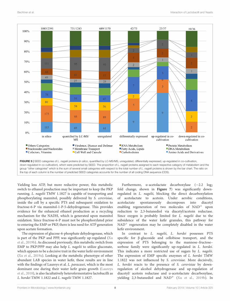
General Proteomic Analysis andOverview of Predicted CompleteMetabolic ActivitiesAs shown in Figure 4, 1,243 proteins of L. nagelii TMW1.1827 were identified and quantified by proteomic analysis,comprising about 52% of the genes annotated by whole genomeanalysis. A comprehensive overview of the complete metabolicpathways and significantly differentially expressed (DE) proteinsof L. nagelii in the presence of S. cerevisiae is provided inSupplementary Figure S2. As shown in Figure 5, there were 73DE proteins in L. nagelii regulated in the presence of S. cerevisiae.Those up/down-regulated proteins of L. nagelii were mostabundant in the SEED categories “amino acids and derivatives”(9 out of 69), “carbohydrates” (5 out of 93) “nucleosides andnucleotides” (4 out of 61) and “cofactors, vitamins” (7 out of 54)(shown in Figure 5).
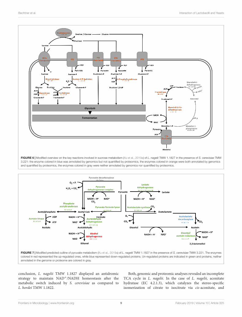
Sugar Transport and CarbohydrateMetabolismThe overview on the key reactions involved in sucrosemetabolism of L. nagelii is provided in Figure 6. L. nageliiencoded and expressed an MFS-transporter specific for sucroseuptake. As previously demonstrated, L. nagelii also produces
Frontiers in Microbiology | www.frontiersin.org 5 February 2019 | Volume 10 | Article 325

fmicb-10-00325 February 26, 2019 Time: 15:9 # 6
Bechtner et al. Interaction of Lactobacilli and Yeasts
FIGURE 2 | Annotated SEED categories of proteins, divided in the core and accessory genomes of L. hordei TMW 1.1822 and L. nagelii TMW 1.1827, which wasdone by BADGE analysis. The proportion of proteins assigned to each SEED category with respect to the total number of proteins is shown in the bar chart.
FIGURE 3 | Cell counts of L. nagelii TMW 1.1827 in single culture ( ) and co-cultivation with S. cerevisiae TMW 3.221 (N) (A). Cell counts of S. cerevisiae TMW3.221 in single culture (�) and in co-cultivation with L. nagelii TMW 1.1827 (�) (B).
a glucan from sucrose by an extracellular glucansucrase (Xuet al., 2018). The residual fructose can then be transportedinto the cell by a fructose specific PTS and simultaneousphosphorylation. Once inside the cell, the phosphorylatedfructose can directly enter the glycolytic pathway. All PTS,
namely for sucrose, glucose, fructose, mannose, sorbose,and mannitol uptake, were identified by proteomic analysisas constitutively expressed upon co-culture. Whole genomesequence analysis of L. nagelii TMW 1.1827 confirmed thepresence of the genes encoding all enzymes required for the EMP
Frontiers in Microbiology | www.frontiersin.org 6 February 2019 | Volume 10 | Article 325

fmicb-10-00325 February 26, 2019 Time: 15:9 # 7
Bechtner et al. Interaction of Lactobacilli and Yeasts
FIGURE 4 | Subcellular localization of L. nagelii proteins (in silico, quantified by LC-MS/MS, unregulated, differentially expressed, up-regulated in co-cultivation,down-regulated in co-cultivation), which were predicted by PSORTb. The proportion of L. nagelii proteins assigned to each respective subcellular compartment andthe group “unknown” with respect to the total number of L. nagelii proteins is shown by the bar chart. The table below shows the respective absolute numbers.
and PKP pathways (locus tags and IDs given in SupplementaryTable S2). Thus, L. nagelii TMW 1.1827 should also be consideredas facultatively heterofermentative, such as L. plantarum WCFS1and Lactococcus lactis (Kleerebezem and Hugenholtz, 2003;Kleerebezem et al., 2003). This is contrary to the fact that L. hordeiDSM 19519 and L. nagelii DSM 13675 were inferred as obligatelyhomofermentative strains according to their phenotype (Sunet al., 2015). However, those strains have been isolated fromdifferent environments.
To probe the principal fermentation type of L. nagelii TMW1.1827 we determined fermentation metabolites upon its growthin CDM to find 40.1 mM lactate and 6.9 mM acetate after24 h of fermentation. This way we could show for L. nageliiand also (previously) for L. hordei (Xu et al., 2019a) that thewater kefir isolates of these species are indeed different fromother ones even with respect to basic fermentation types. Thedata corroborate a homofermentative metabolism, in which
energy generation via EMP and recycling of NAD+ by reducingpyruvate to lactate is favored. The small amount of acetatemay reside from pyruvate by either generating formate viapyruvate formate lyase or by NADH and CO2 generation viathe pyruvate dehydrogenase complex. Subsequently, the resultingacetyl-CoA may be metabolized to acetate. However, the latteroption requires subsequent NAD+ recycling.
In the presence of S. cerevisiae, the 3-phosphoglycerate mutaseof L. nagelii TMW 1.1827 was significantly down-regulated. Aspostulated previously, the expression of this enzyme is linked tothe concentration of its substrate 3-phosphoglycerate (Smeianovet al., 2007). This indicates, that intermediates of early glycolyticsteps may be used for other metabolic reactions or hexoses mayrather enter PKP or PPP than EMP, resulting in less production of3-phosphoglycerate. At the same time, the alcohol dehydrogenase(EC 1.1.1.1, 3.5 log2 fold change) of L. nagelii was significantlyup-regulated in the presence of S. cerevisiae (shown in Figure 7).
Frontiers in Microbiology | www.frontiersin.org 7 February 2019 | Volume 10 | Article 325
fmicb-10-00325 February 26, 2019 Time: 15:9 # 8
Bechtner et al. Interaction of Lactobacilli and Yeasts
FIGURE 5 | SEED categories of L. nagelii proteins (in silico, quantified by LC-MS/MS, unregulated, differentially expressed, up-regulated in co-cultivation,down-regulated in co-cultivation), which were predicted by SEED. The proportion of L. nagelii proteins assigned to each respective category of metabolism and thegroup “other categories” which is the sum of several small categories with respect to the total number of L. nagelii proteins is shown by the bar chart. The ratio onthe top of each column is the number of predicted SEED categories accounts for the number of all coding DNA sequence (CDS).
Yielding less ATP, but more reductive power, this metabolicswitch to ethanol production may be important to keep the PKPrunning. L. nagelii TMW 1.1827 is capable of transporting andphosphorylating mannitol, possibly delivered by S. cerevisiae,inside the cell by a specific PTS and subsequent oxidation tofructose-6-P via mannitol-1-P-5-dehydrogenase. This providesevidence for the enhanced ethanol production as a recyclingmechanism for the NADH, which is generated upon mannitoloxidation. Since fructose-6-P must not be phosphorylated priorto entering the EMP or PKP, there is less need for ATP generationupon acetate formation.
The expression of glucose-6-phosphate dehydrogenase, whichis part of the PKP and PPP, was significantly up-regulated (Xuet al., 2019b). As discussed previously, this metabolic switch fromEMP to PKP/PPP may also help L. nagelii to utilize gluconate,which appears to be a decisive trait in the water kefir environment(Xu et al., 2019a). Looking at the metabolic phenotype of otherabundant LAB species in water kefir, these results are in linewith the findings of Laureys et al. L. paracasei, which is the mostdominant one during their water kefir grain growth (Laureyset al., 2018), is also facultatively heterofermentative lactobacilli asL. hordei TMW 1.1822 and L. nagelii TMW 1.1827.
Furthermore, α-acetolactate decarboxylase (−2.2 log2fold change, shown in Figure 7) was significantly down-regulated in L. nagelii, blocking the direct decarboxylationof acetolactate to acetoin. Under aerobic conditions,acetolactate spontaneously decomposes into diacetylenabling regeneration of two molecules of NAD+ uponreduction to 2,3-butanediol via diacetyl/acetoin reductase.Since oxygen is probably limited for L. nagelii due to thesubsidence of the water kefir granules, this pathway forNAD+ regeneration may be completely disabled in the waterkefir environment.
In contrast to L. nagelii, L. hordei possesses PTSspecific for β-glucoside and cellobiose transport, and theexpression of PTS belonging to the mannose–fructose–sorbose family were significantly up-regulated in L. hordei.This indicates a more restricted use of sugars by L. nagelii.The expression of EMP specific enzymes of L. hordei TMW1.1822 was not influenced by S. cerevisiae. More decisively,L. hordei reacts to the presence of S. cerevisiae by down-regulation of alcohol dehydrogenase and up-regulation ofdiacetyl/ acetoin reductase and α-acetolactate decarboxylase,yielding 2,3-butanediol and NAD+ (Xu et al., 2019b). In
Frontiers in Microbiology | www.frontiersin.org 8 February 2019 | Volume 10 | Article 325
fmicb-10-00325 February 26, 2019 Time: 15:9 # 9
Bechtner et al. Interaction of Lactobacilli and Yeasts
FIGURE 6 | Modified overview on the key reactions involved in sucrose metabolism (Xu et al., 2019a) of L. nagelii TMW 1.1827 in the presence of S. cerevisiae TMW3.221: the enzyme colored in blue was annotated by genomics but not quantified by proteomics, the enzymes colored in orange were both annotated by genomicsand quantified by proteomics, the enzymes colored in gray were neither annotated by genomics nor quantified by proteomics.
FIGURE 7 | Modified predicted outline of pyruvate metabolism (Xu et al., 2019a) of L. nagelii TMW 1.1827 in the presence of S. cerevisiae TMW 3.221. The enzymescolored in red represented the up-regulated ones, while blue represented down-regulated proteins. Un-regulated proteins are indicated in green and proteins, neitherannotated in the genome or proteome are colored in gray.
conclusion, L. nagelii TMW 1.1827 displayed an antidromicstrategy to maintain NAD+/NADH homeostasis after themetabolic switch induced by S. cerevisiae as compared toL. hordei TMW 1.1822.
Both, genomic and proteomic analyses revealed an incompleteTCA cycle in L. nagelii. In the case of L. nagelii, aconitatehydratase (EC 4.2.1.3), which catalyzes the stereo-specificisomerization of citrate to isocitrate via cis-aconitate, and
Frontiers in Microbiology | www.frontiersin.org 9 February 2019 | Volume 10 | Article 325

fmicb-10-00325 February 26, 2019 Time: 15:9 # 10
Bechtner et al. Interaction of Lactobacilli and Yeasts
isocitrate dehydrogenase (EC 1.1.1.42), which catalyzes theoxidative decarboxylation of isocitrate, producing 2-oxoglutarateand CO2, were significantly up-regulated (4.5 log2 fold change,2.0 log2 fold change). Despite its incompleteness, the TCAcycle is an important supplier for compounds involved in othermetabolic reactions and thus, isocitrate and 2-oxoglutarate maybe useful for amino acid metabolism in L. nagelii.
Water kefir is a challenging environment for its habitantsregarding low nutrient concentrations except for the excesssugar. Since lemon slices are added, it is not surprising thatmicroorganisms in water kefir use citrate as a nutrient. L. nageliiis capable of direct citrate import using malate permease. Onceinside the cell, citrate is converted by citrate lyase segregatingone molecule of acetate. The resulting oxaloacetate may then bedecarboxylated via oxaloacetate decarboxylase yielding pyruvateor is further used for amino acid biosynthesis. In contrast toL. nagelii, these enzymes are DE in L. hordei, which appearsto be positively influenced in its metabolism of citrate as anadditional carbon source upon co-culture with S. cerevisiae(Xu et al., 2019b). This may help to explain, why L. hordei ismore abundant in the water kefir consortium than L. nagelii(Gulitz et al., 2011).
Amino Acids Biosynthesis, Metabolism,and TransportThe concentration of amino acids in pure WKM is verylow (<0.004 mmol/l, respectively) (Stadie et al., 2013). Sorespective metabolite quantification is way too low to obtainconclusive data on amino acids metabolism in WKM, namelyon those metabolites, which are determinative for interactionof lactobacilli and yeasts. Indeed, this is a major reason touse quantitative proteomics for metabolic predictions. Thein silico analysis of the genome and proteome of L. nageliiTMW 1.1827 did not reveal any known homologs of acell wall proteinase (Prt). L. nagelii encodes the completeoligopeptide transport system OppABCDF (Tynkkynen et al.,1993; Detmers et al., 1998). Except from OppB, all genes ofboth annotated OppABCDF clusters were found to be present inthe proteome of L. nagelii. Despite lacking an expressed OppB,the growth of L. nagelii was not impaired. This phenomenonwas already described for other bacteria (Nepomuceno et al.,2007) indicating, that the function of OppB may be compensableby other trans-membrane proteins. In the presence of theyeast, the remaining proteins were widely un-regulated with theexception of OppF, which was significantly down-regulated inone cluster. Since OppF is responsible for coupling the energyof ATP hydrolysis with the import of oligopeptides, L. nageliimay reduce energy consumption caused by oligopeptide uptake.In contrast, L. hordei upregulated its OppABCDF system anda set of peptidases, suggesting that L. hordei benefits frompeptides, which are more readily available in the presence ofthe yeast (Xu et al., 2019b). Since water kefir provides verylimited resources of proteins and free amino acids, mainlyoriginating from dried fruits and the yeast, these findingsmay also explain the fact, that the growth of L. hordei isstimulated in co-culture.
From genomic annotation, L. nagelii encodes several aminoacid permeases and transporters. In the presence of S. cerevisiae,methionine aminopeptidase and amino acid permease weresignificantly up-regulated, suggesting that the yeast inducesamino acid uptake in L. nagelii. However, it was not possibleto specify from sequence comparison, which amino acidswere ingested by L. nagelii. Still, this may be solved by acloser look at amino acid synthesis pathways and auxotrophies.The genomic analysis of L. nagelii TMW 1.1827 revealedthe prototrophy for 13 amino acids and auxotrophy for 7amino acids (Table 2). According to the quantitative proteomicanalysis, nine enzymes of L. nagelii involved in histidine,methionine, glutamate, and arginine biosynthesis pathways wereall significantly up-regulated in the presence of S. cerevisiae(shown in Figure 8 and Table 3). Since those biosynthesispathways can also be used for amino acid catabolism, waterkefir microorganisms may profit from amino acids secreted
TABLE 2 | List of the auxotrophies of L. nagelii TMW 1.1827.
Name ofamino acid
Biosynthesisbased ongenome
Absent enzyme ofbiosynthesis of amino acidpathway
EC number
Alanine P None
Arginine P None
Asparagine P None
Aspartic acid P None
Cysteine P None
Glutamic acid P None
Glutamine P None
Glycine A Phosphoserine phosphataseThreonine aldolase
EC 3.1.3.3EC 4.1.2.48
Serine A Phosphoserine phosphatase EC 3.1.3.3
Histidine P None
Leucine A Ketol-acid reductoisomeraseDihydroxy-acid dehydratase
EC 1.1.1.86EC 4.2.1.9
Isoleucine A Citramalate synthaseKetol-acid reductoisomeraseDihydroxy-acid dehydratase
EC 2.3.1.182EC 1.1.1.86EC 4.2.1.9
Valine A Ketol-acid reductoisomeraseDihydroxy-acid dehydratase
EC 1.1.1.86EC 4.2.1.9
Lysine P None
Methionine P None
Proline P None
Phenylalanine A Prephenate dehydrataseAromatic-amino-acidtransaminase
EC 4.2.1.51EC 2.6.1.57
Tryptophan A AnthranilatephosphoribosyltransferaseAnthranilate synthaseIndole-3-glycerol phosphatesynthasePhosphoribosylanthranilateisomerase
EC 2.4.2.18
EC 4.1.3.27EC 4.1.1.48
EC 5.3.1.24
Tyrosine P None
Threonine P None
P represents prototrophy for amino acids, while A represents auxotrophyfor amino acids.
Frontiers in Microbiology | www.frontiersin.org 10 February 2019 | Volume 10 | Article 325

fmicb-10-00325 February 26, 2019 Time: 15:9 # 11
Bechtner et al. Interaction of Lactobacilli and Yeasts
FIGURE 8 | Biosynthesis of amino acids of L. nagelii TMW 1.1827 (A) and L. nagelii TMW 1.1827 in presence of S. cerevisiae TMW 3.221 (B). In (A), the redarrowed lines show the presence of enzymes annotated from genome. In (B), the red arrowed lines indicate up-regulated proteins.
TABLE 3 | Significantly differentially expressed proteins in L. nagelii TMW 1.1827 in response to S. cerevisiae TMW 3.221 involved in amino acid biosynthesis.
Number Enzyme EC number Log2 fold change(co-cultivation vs. single
culture)
−Log(P-value)
SEED subcategory
Up-regulated
1 Imidazoleglycerol-phosphatedehydratase
EC 4.2.1.19 4.6 3.5 Histidine biosynthesis
2 Histidinol-phosphate aminotransferase EC 2.6.1.9 4.4 3.8
3 Histidinol dehydrogenase EC 1.1.1.23 5.3 4.5
4 5-Methyltetrahydropteroyltriglutamate-homocysteine methyltransferase
EC 2.1.1.14 3.8 3.7 Methionine biosynthesis
5 Glutamate synthase EC 1.4.1.13 4.1 2.5 Glutamate, arginine biosynthesis
6 Acetylglutamate kinase EC 2.7.2.8 3.0 1.7 Arginine biosynthesis
7 Acetylornithine aminotransferase EC 2.6.1.11 8.7 3.3
8 Ornithine carbamoyltransferase EC 2.1.3.3 7.5 2.9
9 Argininosuccinate synthase EC 6.3.4.5 3.3 3.6
by the yeast, creating a symbiotic consortium. However, fromin silico analysis, the direction of a respective metabolicpathway remains speculative. Still, together with physiologicaldata on amino acid consumption and secretion of L. nageliiand S. cerevisiae, this can be solved for at least some of thepredicted cases.
As shown most prominently in Figure 9, S. cerevisiae secretedglutamine in high amounts, whereas L. nagelii consumed theamino acid at high levels via an up-regulated amino acidpermease involved in glutamine uptake. This suggests, that
L. nagelii, even though it is capable of producing glutamineby itself, profits from the glutamine provided by the yeastvia the up-regulated glutamate synthase. Since glutamine playsan important role in anaplerotic sequences of transaminationreactions in the biosynthesis of other amino acids, and alsoas a nitrogen carrier for the production of amino sugars andnucleotides, the uptake of this amino acid may be crucial topersist in the water kefir environment. L. nagelii was predictedto produce glutamate by itself via the up-regulated glutamatesynthase using glutamine and 2-oxoglutarate, which probably
Frontiers in Microbiology | www.frontiersin.org 11 February 2019 | Volume 10 | Article 325

fmicb-10-00325 February 26, 2019 Time: 15:9 # 12
Bechtner et al. Interaction of Lactobacilli and Yeasts
FIGURE 9 | Consumption of amino acids of L. nagelii TMW 1.1827 and S. cerevisiae TMW 3.221 isolated from water kefir grown in CDM after 24 h. Black barrepresents CDM, slash bar represents L. nagelii, gray bar represents S. cerevisiae.
results from the incomplete TCA cycle. This was consistentwith an un-regulated glutamine synthetase in the presence ofS. cerevisiae. As already described for Lactobacillus crispatusST1, this enzyme might exhibit additional functions, if displayedon the bacterial surface, which enable physical coherenceof the water kefir consortium under stressful conditions(Kainulainen et al., 2012). As a result, the yeast aggregationpromotion of L. hordei by its functional dextran (Xu et al.,2018) may be even enhanced by over expression of thisenzyme. Furthermore, among all amino acids, the productionof glutamate is of primary importance in the assimilationof nitrogen, representing a donor for amino groups in thesynthesis of other amino acids (Bernard and Habash, 2009;Dincturk et al., 2011).
Other amino acids were not produced, but partly consumedby the yeast after 24 h of fermentation in CDM. Therefore,it was not possible to determine real-time metabolic exchange(release/uptake) between L. nagelii and S. cerevisiae based onphysiological data. Still, the label-free quantitative proteomicanalysis enabled the investigation of the dynamic metabolicexchanges between microbial communities in water kefir.The DE enzymes involved in amino acid biosynthesis orcatabolism predict that S. cerevisiae releases glutamine, histidine,methionine, and arginine, which are subsequently used byL. nagelii to ensure its survival in the water kefir consortium.
Acid Tolerance by ADI PathwayFunctionally, the ADI pathway enables enhanced acid toleranceand energy provision in a variety of LAB genera such asLactobacillus, Lactococcus, Leuconostoc, and Weissella (Tononand Lonvaud-Funel, 2002; Fernández and Zúñiga, 2006; Rimauxet al., 2011). The system involves the three enzymes argininedeiminase (ADI), ornithine transcarbamylase (OTC), carbamatekinase (CK) and a transmembrane arginine/ornithine antiporter,which exchanges extracellular arginine against intracellular
ornithine. While ADI and OTC were present in both, thegenome and proteome of L. nagelii TMW 1.1827, CK andthe arginine/ornithine antiporter were only detectable in thegenome. Thus, it should be unable to convert carbamoyl-P to generate additional ATP in co-culture. In the energyrich environment of water kefir, this does not appear to bea disadvantage. Therefore, the fate of carbamoyl-P remainsunclear. However, only OTC was significantly up-regulated inL. nagelii in co-culture with S. cerevisiae. This reaction mayoccur in both directions yielding citrulline or ornithine andcarbamoyl-phosphate. L. nagelii did not encode any completealternative acid tolerance systems, e.g., the agmatine deiminase(AGDI) system or the glutamate decarboxylase (GAD) system.Except for neutralization upon ammonia formation via the ADIsystem, acidification appears limited by the switch from lactic andacetic acid production to ethanol formation, when L. nagelii andS. cerevisiae were co-cultivated.
In contrast, all respective enzymes of L. hordei involvedin ADI pathway were up-regulated in co-culture with theyeast. Although the fate of carbamoyl-phosphate and otherincidental compounds remains unclear, L. hordei likely producesammonia upon arginine hydrolysis to protect itself from pHstress by alkalization of its cytoplasm and proximal environment.Consequently, only L. hordei should reduce the acid stress for theyeast (Xu et al., 2019a).
Fatty Acid Biosynthesis and RiboflavinMetabolismAnother limit in the water kefir environment is the limitedavailability of fatty acids. L. nagelii TMW 1.1827 appearsto be deficient in FabB, which is a well studied 3-ketoacyl-ACP synthase for catalyzing the elongation reaction of fattyacid synthesis (Feng and Cronan, 2009), and additionally inFabA, which is hydroxyldecanoyl-ACP dehydratase/isomerase
Frontiers in Microbiology | www.frontiersin.org 12 February 2019 | Volume 10 | Article 325

fmicb-10-00325 February 26, 2019 Time: 15:9 # 13
Bechtner et al. Interaction of Lactobacilli and Yeasts
for the production of unsaturated fatty acids by manybacteria (Magnuson et al., 1993; Cronan and Rock, 1996).As demonstrated by Wang and Cronan (2004), FabF canfunctionally replace FabB, while FabZ adopts the functionof FabA. It was also reported that expression of Lactococcuslactis FabF can functionally replace both FabB and FabFin E. coli (Morgan-Kiss and Cronan, 2008). Due to lowsequence homologies, those enzymatic bi-functionalities are notpredictable by genome analysis. Since both microorganismsgrew to high cell densities in water kefir medium withoutany external fatty acids, those findings might also indicate theexistence of other functional homologs for FabB and FabA inL. nagelii. Co-cultivation with S. cerevisiae does not alter theexpression of any proteins involved in the fatty acid metabolismin both LAB (Supplementary Table S3). This indicates, thatthe beneficial effects of S. cerevisiae do not reside in a bilateralsupply with unsaturated fatty acids. This situation resembles theone in L. hordei, which only lacks FabA but also should expressfunctionally complementary alternatives (Xu et al., 2019a).
Moreover, there was a group of enzymes of L. nagelii, whichshowed decreased expression in response to the co-cultivationwith S. cerevisiae, which are involved in the biosynthesis ofriboflavin (as shown in Supplementary Figure S3). Riboflavinsynthase (EC 2.5.1.9), 6,7-dimethyl-8-ribityllumazine synthase(EC 2.5.1.78), 5-amino-6-(5-phosphoribosylamine) uracilreductase (EC 1.1.1.193), GTP cyclohydrolase II (EC 3.5.4.25)and 3,4-dihydroxy-2-butanone 4-phosphate synthase (EC4.1.99.12) were down-regulated in a range from −3.2 to −4.0log2 fold. Those enzymes connect the purine metabolismand pentose phosphate pathway to synthesize riboflavin.Since generally yeast produce group B vitamins (Emery et al.,1946; Zeidler et al., 2002), and riboflavin production by somelactobacilli (such as L. plantarum and L. fermentum) wasinducible (Burgess et al., 2006; Arena et al., 2014; Russo et al.,2014), this may be an evidence for the feeding of riboflavin fromS. cerevisiae to L. nagelii, supporting its growth and leading to astable water kefir consortium.
CONCLUSION
The label-free quantitative approach represents a powerful toolfor the identification and quantification of proteins to study thebacteria–yeast interaction of microorganisms involved in foodfermentation processes (Behr et al., 2007; Siragusa et al., 2014;Maeda et al., 2015). It may even be used to explore more complexcombinations or the complete water kefir system. However,with several (closely related) lactobacilli/yeasts in the systemthe sorting of proteins to species along sequence homologieswill probably be limited because of sequence similarities acrossspecies. So in turn one would probably not be able to see thespecific L. nagelii/L. hordei responses to S. cerevisiae any more,which are markedly different. So the reduction of the systemoffers also some advantage for a deeper understanding.
The predicted functional genome and the differentiallyexpressed proteins in the presence of S. cerevisiae TMW 3.221depicted the adaption of L. nagelii TMW 1.1827 to the water kefirconsortium and environment, although protein regulations were
less distinct than in L. hordei TMW 1.1822 (Xu et al., 2019b).Both microorganisms are highly efficient in degrading sucrose byan extracellular glucansucrase and subsequent fructose uptake,which may then enter EMP, PKP or mannitol metabolism. Asalready described for L. hordei, also L. nagelii appears to favorPKP over EMP, indicating a metabolic switch induced by analtered redox potential in the presence of S. cerevisiae. WhileL. nagelii remained widely un-affected in its citrate metabolism,the yeast stimulated L. hordei to use citrate as additional carbonsource and therefore, promoting its growth.
Both LAB profit from glutamine secreted by the yeast, whereasL. hordei also takes advantage of the provided glutamate. WhileL. hordei up-regulated all of its enzymes involved in the reductionof acid stress via ADI pathway, L. nagelii only altered theexpression of OTC. It was obvious, that both microorganismsreduced external acid stress by switching from lactate and acetateproduction to butanediol formation in the case of L. hordei andethanol production in the case of L. nagelii.
At first glance, the fatty acid metabolism of bothmicroorganisms appears to be impaired by the lack of oneor more genes coding for key fatty acid biosynthesis enzymes. Asit was already reported for other bacteria (Wang and Cronan,2004), it is likely, that the functional role of those enzymes maybe undertaken by other enzymes of the fatty acid biosynthesisgene cluster. This would explain, why both, L. hordei andL. nagelii, grew to high cell densities while facing an environmentinsufficient in unsaturated fatty acids. While S. cerevisiae TMW3.221 modulated the protein expression of L. hordei TMW1.1822 mainly in its carbohydrate metabolism, L. nagelii TMW1.1827 seems to profit from secreted riboflavin. With respectto the establishment of a consortium maintaining physicalproximity of lactobacilli and yeasts L. hordei appears to have amore prominent role as compared to L. nagelii as a result of itsunique dextran causing yeast aggregation and proteins involvedin adhesion functions.
DATA AVAILABILITY
The datasets generated for this study can be found in GenBank,CP018180–CP018183, and the ProteomeXchange via the PRIDEpartner repository with the dataset identifier PXD012513.
AUTHOR CONTRIBUTIONS
DX conducted the wet lab experiments and performed theprimary data analysis. CL helped with proteomic data analysesand deposition. JlB conducted detailed analysis and metabolicpredictions. JgB supervised data analyses. DX and JlB wrote thefirst draft of manuscript. JgB and RV established general layoutof experimental approach, supervised DX and JlB and did finaldiscussions and shaping of the manuscript.
FUNDING
This study was supported by the China Scholarship Councilin grant no. 201306820010, and the German Ministry
Frontiers in Microbiology | www.frontiersin.org 13 February 2019 | Volume 10 | Article 325

fmicb-10-00325 February 26, 2019 Time: 15:9 # 14
Bechtner et al. Interaction of Lactobacilli and Yeasts
of Economics and Technology (via AiF) and the WiFö(Wissenschaftsförderung der Deutschen Brauwirtschaft e.V.,Berlin) in project AiF 19180 N.
SUPPLEMENTARY MATERIAL
The Supplementary Material for this article can be foundonline at: https://www.frontiersin.org/articles/10.3389/fmicb.2019.00325/full#supplementary-material
FIGURE S1 | Genomic atlas of L. nagelii TMW 1.1827. Forward CDS (red),reverse CDS (blue), pseudogenes on both strands (black), tRNA and rRNA (darkgreen), % GC plot (yellow, high GC spike and green, low GC spike), GC skew[(G − C)/(G + C)] (gray).
FIGURE S2 | Overview of enzymatic activity of L. nagelii TMW 1.1827 in thecomplete metabolic and other pathways in presence of S. cerevisiae TMW 3.221:the nodes colored in bold red represent up-regulated, in bold blue representdown-regulated enzymes or proteins according to proteomic data, while nodescolored in thin red represent all the rest enzymes or proteins according to genomicannotation data presented in iPath 3.0.
FIGURE S3 | Overview of riboflavin metabolism of L. nagelii TMW 1.1827generated in KEGG mapper. The EC numbers colored in red show up-regulatedenzymes of L. nagelii in the presence of S. cerevisiae.
TABLE S1 | Annotated key features of the L. nagelii TMW 1.1827 genome.
TABLE S2 | List of annotated enzymes by NCBI and RAST involved incarbohydrate metabolism (glycolysis, pentose phosphate pathway, pyruvatemetabolism, and TCA cycle).
TABLE S3 | List of enzymes involved in fatty acid biosynthesis.
REFERENCESAlikhan, N. F., Petty, N. K., Zakour, N. L. B., and Beatson, S. A. (2011). BLAST
ring image generator (BRIG): simple prokaryote genome comparisons. BMCGenomics 12:402. doi: 10.1186/1471-2164-12-402
Arena, M. P., Russo, P., Capozzi, V., López, P., Fiocco, D., and Spano G. (2014).Probiotic abilities of riboflavin-overproducing Lactobacillus strains: a novelpromising application of probiotics. Appl. Microbiol. Biotechnol. 98, 7569–7581.doi: 10.1007/s00253-014-5837-x
Aziz, R. K., Bartels, D., Best, A. A., Dejongh, M., Disz, T., Edwards, R. A., et al.(2008). The RAST server: rapid annotations using subsystems technology. BMCGenomics 9:75. doi: 10.1186/1471-2164-9-75
Behr, J., Geissler, A. J., Schmid, J., Zehe, A., and Vogel, R. F. (2016). Theidentification of novel diagnostic marker genes for the detection of beer spoilingPediococcus damnosus strains using the BlAst diagnostic gene findEr. PLoS One11:e0152747. doi: 10.1371/journal.pone.0152747
Behr, J., Israel, L., Gänzle, M. G., and Vogel, R. F. (2007). Proteomicapproach for characterization of hop-inducible proteins in Lactobacillusbrevis. Appl. Environ. Microbiol. 73, 3300–3306. doi: 10.1128/AEM.00124-127
Bernard, S. M., and Habash, D. Z. (2009). The importance of cytosolic glutaminesynthetase in nitrogen assimilation and recycling. New Phytol. 182, 608–620.doi: 10.1111/j.1469-8137.2009.02823.x
Burgess, C. M., Smid, E. J., Rutten, G., and van Sinderen, D. (2006). A generalmethod for selection of riboflavin-overproducing food grade micro-organisms.Microb. Cell Fact. 5:24. doi: 10.1186/1475-2859-5-24
Carver, T., Thomson, N., Bleasby, A., Berriman, M., and Parkhill, J.(2008). DNAplotter: circular and linear interactive genomevisualization. Bioinformatics 25, 119–120. doi: 10.1093/bioinformatics/btn578
Cox, J., Neuhauser, N., Michalski, A., Scheltema, R. A., Olsen, J. V., and Mann, M.(2011). Andromeda: a peptide search engine integrated into the MaxQuantenvironment. J. Proteome Res. 10, 1794–1805. doi: 10.1021/pr101065j
Cronan, J. E., and Rock, C. O. (1996). Escherichia coli and Salmonellatyphimurium. Cell. Mol. Biol. 1, 612–636.
Detmers, F. J., Kunji, E. R., Lanfermeijer, F. C., Poolman, B., and Konings,W. N. (1998). Kinetics and specificity of peptide uptake by the oligopeptidetransport system of Lactococcus lactis. Biochemistry 37, 16671–16679. doi: 10.1021/bi981712t
Dincturk, H. B., Cunin, R., and Akce, H. (2011). Expression and functional analysisof glutamate synthase small subunit-like proteins from archaeon Pyrococcushorikoshii. Microbiol. Res. 166, 294–303. doi: 10.1016/j.micres.2010.03.006
Emery, W. B., McLeod, N., and Robinson, F. A. (1946). Comparativemicrobiological assays of members of the vitamin B complex in yeast and liverextracts. Biochem. J. 40, 426–432. doi: 10.1042/bj0400426
Fels, L., Jakob, F., Vogel, R. F., and Wefers, D. (2018). Structural characterizationof the exopolysaccharides from water kefir. Carbohydr. Polym. 189, 296–303.doi: 10.1016/j.carbpol.2018.02.037
Feng, Y. J., and Cronan, J. E. (2009). Escherichia coli unsaturated fatty acidsynthesis: complex transcription of the fabA gene and in vivo identificationof the essential reaction catalyzed by FabB. J. Biol. Chem. 284, 29526–29535.doi: 10.1074/jbc.M109.023440
Fernández, M., and Zúñiga, M. (2006). Amino acid catabolic pathways of lactic acidbacteria. Crit. Rev. Microbiol. 32, 155–183. doi: 10.1080/10408410600880643
Gardy, J. L., Laird, M. R., Chen, F., Rey, S., Walsh, C. J., Ester, M., et al. (2004).PSORTb v. 2.0: expanded prediction of bacterial protein subcellular localizationand insights gained from comparative proteome analysis. Bioinformatics 21,617–623. doi: 10.1093/bioinformatics/bti057
Gulitz, A., Stadie, J., Wenning, M., Ehrmann, M. A., and Vogel, R. F. (2011).The microbial diversity of water kefir. Int. J. Food Microbiol. 151, 284–288.doi: 10.1016/j.ijfoodmicro.2011.09.016
Horisberger, M. (1969). Structure of the dextran of the tibi grain. Carbohydr. Res.10, 379–385. doi: 10.1016/S0008-6215(00)80897-6
Kainulainen, V., Loimaranta, V., Pekkala, A., Edelman, S., Antikainen, J.,Kylväjä, R., et al. (2012). Glutamine synthetase and glucose-6-phosphateisomerase are adhesive moonlighting proteins of Lactobacillus crispatusreleased by cathelicidin LL-37. J. Bacteriol. 194, 2509–2519. doi: 10.1128/JB.06704-11
Kleerebezem, M., Boekhorst, J., Van Kranenburg, R., Molenaar, D., Kuipers, O. P.,Leer, R., et al. (2003). Complete genome sequence of Lactobacillus plantarumWCFS1. PNAS 100, 1990–1995. doi: 10.1073/pnas.0337704100
Kleerebezem, M., and Hugenholtz, J. (2003). Metabolic pathway engineering inlactic acid bacteria. Curr. Opin. Biotechnol. 14, 232–237. doi: 10.1016/S0958-1669(03)00033-8
Laureys, D., Aerts, M., Vandamme, P., and De Vuyst, L. (2018). Oxygen and diversenutrients influence the water kefir fermentation process. Food Microbiol. 73,351–361. doi: 10.1016/j.fm.2018.02.007
Laureys, D., and De Vuyst, L. (2014). Microbial species diversity, communitydynamics, and metabolite kinetics of water kefir fermentation. Appl. Environ.Microbiol. 80, 2564–2572. doi: 10.1128/AEM.03978-13
Letort, C., Nardi, M., Garault, P., Monnet, V., and Juillard, V. (2002). Caseinutilization by Streptococcus thermophilus results in a diauxic growth in milk.Appl. Environ. Microbiol. 68, 3162–3165. doi: 10.1128/AEM.68.6.3162-3165.2002
Maeda, K., Nagata, H., Ojima, M., and Amano, A. (2015). Proteomic andtranscriptional analysis of interaction between oral microbiota Porphyromonasgingivalis and Streptococcus oralis. J. Proteome Res. 14, 82–94. doi: 10.1021/pr500848e
Magnuson, K., Jackowski, S., and Rock, C. O. (1993). Regulation of fatty acidbiosynthesis in Escherichia coli. Microbiol. Rev. 57, 522–542.
Marsh, A. J., O’sullivan, O., Hill, C., Ross, R. P., and Cotter, P. D. (2013).Sequence-based analysis of the microbial composition of water kefir frommultiple sources. FEMS Microbiol. Lett. 348, 79–85. doi: 10.1111/1574-6968.12248
Martínez-Torres, A., Gutiérrez-Ambrocio, S., Heredia-Del-Orbe, P., Villa-Tanaca, L., and Hernández-Rodríguez, C. (2017). Inferring the role of
Frontiers in Microbiology | www.frontiersin.org 14 February 2019 | Volume 10 | Article 325

fmicb-10-00325 February 26, 2019 Time: 15:9 # 15
Bechtner et al. Interaction of Lactobacilli and Yeasts
microorganisms in water kefir fermentations. Int. J. Food Sci. Technol. 52,559–571. doi: 10.1111/ijfs.13312
Morgan-Kiss, R. M., and Cronan, J. E. (2008). The Lactococcus lactis FabF fatty acidsynthetic enzyme can functionally replace both the FabB and FabF proteins ofEscherichia coli and the FabH protein of Lactococcus lactis. Arch. Microbiol. 190,427–437. doi: 10.1007/s00203-008-0390-6
Nepomuceno, R., Tavares, M., Lemos, J., Griswold, A., Ribeiro, J., Balan, A.,et al. (2007). The oligopeptide (opp) gene cluster of Streptococcus mutans:identification, prevalence, and characterization. Oral Microbiol. Immunol. 22,277–284. doi: 10.1111/j.1399-302X.2007.00368.x
Neve, H., and Heller, K. J. (2002). The microflora of water kefir: a glanceby scanning electron microscopy. Kiel Milchwirtsch Forschungsber 54,337–349.
Overbeek, R., Olson, R., Pusch, G. D., Olsen, G. J., Davis, J. J., Disz, T., et al. (2013).The seed and the rapid annotation of microbial genomes using subsystemstechnology (RAST). Nucleic Acids Res. 42, D206–D214. doi: 10.1093/nar/gkt1226
Pidoux, M. (1989). The microbial flora of sugary kefir grain (the gingerbeerplant): biosynthesis of the grain from Lactobacillus hilgardii producing apolysaccharide gel. MIRCEN J. Appl. Microbiol. Biotechnol. 5, 223–238.doi: 10.1007/BF01741847
Pidoux, M., Brillouet, J. M., and Quemener, B. (1988). Characterization of thepolysaccharides from a Lactobacillus brevis and from sugary kefir grains.Biotechnol. Lett. 10, 415–420. doi: 10.1007/BF01087442
Pidoux, M., De Ruiter, G. A., Brooker, B. E., Colquhoun, I. J., and Morris, V. J.(1990). Microscopic and chemical studies of a gelling polysaccharide fromLactobacillus hilgardii. Carbohydr. Polym. 13, 351–362. doi: 10.1016/0144-8617(90)90035-Q
Rimaux, T., Vrancken, G., Pothakos, V., Maes, D., De Vuyst, L., and Leroy, F.(2011). The kinetics of the arginine deiminase pathway in the meat starterculture Lactobacillus sakei CTC 494 are pH-dependent. Food Microbiol. 28,597–604. doi: 10.1016/j.fm.2010.11.016
Russo, P., Capozzi, V., Arena, M. P., Spadaccino, G., Dueñas, M. T., López, P.,et al. (2014). Riboflavin-overproducing strains of Lactobacillus fermentum forriboflavin-enriched bread. Appl. Microbiol. Biotechnol. 98, 3691–3700. doi: 10.1007/s00253-013-5484-7
Siragusa, S., De Angelis, M., Calasso, M., Campanella, D., Minervini, F.,Di Cagno, R., et al. (2014). Fermentation and proteome profilesof Lactobacillus plantarum strains during growth under food-likeconditions. J. Proteomics 96, 366–380. doi: 10.1016/j.jprot.2013.11.003
Smeianov, V. V., Wechter, P., Broadbent, J. R., Hughes, J. E., Rodríguez, B. T.,Christensen, T. K., et al. (2007). Comparative high-density microarray analysisof gene expression during growth of Lactobacillus helveticus in milk versus richculture medium. Appl. Environ. Microbiol. 73, 2661–2672. doi: 10.1128/AEM.00005-07
Stadie, J., Gulitz, A., Ehrmann, M. A., and Vogel, R. F. (2013). Metabolicactivity and symbiotic interactions of lactic acid bacteria and yeastsisolated from water kefir. Food Microbiol. 35, 92–98. doi: 10.1016/j.fm.2013.03.009
Stolz, P., Vogel, R. F., and Hammes, W. P. (1995). Utilization of electron acceptorsby lactobacilli isolated from sourdough. Z. Lebensm. Unters. Forsch. 201, 402–410. doi: 10.1007/BF01193208
Sun, Z., Harris, H. M., Mccann, A., Guo, C., Argimón, S., Zhang, W., et al. (2015).Expanding the biotechnology potential of lactobacilli through comparativegenomics of 213 strains and associated genera. Nat. Commun. 6:8322.doi: 10.1038/ncomms9322
Tonon, T., and Lonvaud-Funel, A. (2002). Arginine metabolism by wineLactobacilli isolated from wine. Food Microbiol. 19, 451–461. doi: 10.1006/fmic.2002.0502
Tyanova, S., Temu, T., Sinitcyn, P., Carlson, A., Hein, M. Y., Geiger, T.,et al. (2016). The Perseus computational platform for comprehensive analysisof (prote) omics data. Nat. Methods 13, 731–740. doi: 10.1038/NMETH.3901
Tynkkynen, S., Buist, G., Kunji, E., Kok, J., Poolman, B., Venema, G., et al. (1993).Genetic and biochemical characterization of the oligopeptide transport systemof Lactococcus lactis. J. Bacteriol. 175, 7523–7532. doi: 10.1128/jb.175.23.7523-7532.1993
Waldherr, F. W., Doll, V. M., Meißner, D., and Vogel, R. F. (2010). Identificationand characterization of a glucan-producing enzyme from Lactobacillus hilgardiiTMW 1.828 involved in granule formation of water kefir. Food Microbiol. 27,672–678. doi: 10.1016/j.fm.2010.03.013
Wang, H., and Cronan, J. E. (2004). Functional replacement of the FabA andFabB proteins of Escherichia coli fatty acid synthesis by Enterococcus faecalisFabZ and FabF homologues. J. Biol. Chem. 279, 34489–34495. doi: 10.1074/jbc
Ward, H. M. (1891). The’ginger-beer plant,’and the organisms composing it: acontribution to the study of fermentation-yeasts and bacteria. Proc. R. Soc.Lond. 50, 261–265.
Xu, D., Bechtner, J., Behr, J., Eisenbach, L., Geißler, A. J., and Vogel,R. F. (2019a). Lifestyle of Lactobacillus hordei isolated from waterkefir based on genomic, proteomic and physiological characterization.Int. J. Food. Microbiol. 290, 141–149. doi: 10.1016/j.ijfoodmicro.2018.10.004
Xu, D., Behr, J., Geißler, A. J., Bechtner, J., Ludwig, C., and Vogel, R. F. (2019b).Label-free quantitative proteomic analysis reveals the lifestyle of Lactobacillushordei in the presence of Sacchromyces cerevisiae. Int. J. Food Microbiol. 294,18–26. doi: 10.1016/j.ijfoodmicro.2019.01.010
Xu, D., Fels, L., Wefers, D., Behr, J., Jakob, F., and Vogel, R. F. (2018). Lactobacillushordei dextrans induce Saccharomyces cerevisiae aggregation and networkformation on hydrophilic surfaces. Int. J. Biol. Macromol. 115, 236–242.doi: 10.1016/j.ijbiomac.2018.04.068
Yamada, T., Letunic, I., Okuda, S., Kanehisa, M., and Bork, P. (2011). iPath2. 0:interactive pathway explorer. Nucleic Acids Res. 39, W412–W415. doi: 10.1093/nar/gkr313
Yu, N. Y., Wagner, J. R., Laird, M. R., Melli, G., Rey, S., Lo, R., et al.(2010). PSORTb 3.0: improved protein subcellular localization predictionwith refined localization subcategories and predictive capabilities for allprokaryotes. Bioinformatics 26, 1608–1615. doi: 10.1093/bioinformatics/btq249
Zeidler, J., Ullah, N., Gupta, R. N., Pauloski, R. M., Sayer, B. G., and Spenser, I. D.(2002). 2‘-Hydroxypyridoxol, a biosynthetic precursor of vitamins B6 and B1 inyeast. J. Am. Chem. Soc. 124, 4542–4543. doi: 10.1021/ja012708z
Conflict of Interest Statement: The authors declare that the research wasconducted in the absence of any commercial or financial relationships that couldbe construed as a potential conflict of interest.
Copyright © 2019 Bechtner, Xu, Behr, Ludwig and Vogel. This is an open-accessarticle distributed under the terms of the Creative Commons Attribution License(CC BY). The use, distribution or reproduction in other forums is permitted, providedthe original author(s) and the copyright owner(s) are credited and that the originalpublication in this journal is cited, in accordance with accepted academic practice. Nouse, distribution or reproduction is permitted which does not comply with these terms.
Frontiers in Microbiology | www.frontiersin.org 15 February 2019 | Volume 10 | Article 325