proteomic analysis of protein deposits on worn daily wear ... · proteomic analysis of protein...
TRANSCRIPT
Proteomic analysis of protein deposits on worn daily wear siliconehydrogel contact lenses
Zhenjun Zhao,1,2,3 Xiaojia Wei,1,3 Yulina Aliwarga,1,3 Nicole A. Carnt,1,3 Qian Garrett,1,2,3 Mark D.P. Willcox1,2,3
1Institute for Eye Research, Sydney, NSW, Australia; 2School of Optometry and Vision Science, University of New South Wales,Sydney, NSW, Australia; 3Vision Cooperative Research Centre, Sydney, NSW, Australia
Purpose: Previous studies have demonstrated deposition of tear proteins onto worn contact lenses. In this study, we usedproteomic techniques to analyze the protein deposits extracted from worn daily wear silicone hydrogel contact lenses incombination with different lens care solutions.Methods: Worn lenses were collected and protein deposits extracted using urea and surfactant. Protein extracts weredesalted, concentrated, and then separated using one-dimensional gel electrophoresis. Individual protein components inextracts were identified using liquid chromatography combined with tandem mass spectrometry (LC-MS-MS) after trypsindigestion.Results: One-dimensional gel electrophoresis revealed that lysozyme and other small proteins (around 20 kDa) were themost abundant proteins in the extracts. LC-MS-MS revealed a wide array of proteins in lens extracts with lysozyme andlipocalin 1 being the most commonly identified in deposit extracts.Conclusions: Worn contact lenses deposit a wide array of proteins from tear film and other sources. Protein depositprofiles varied and were specific for each contact lens material.
Contact lens wear is an increasingly popular method ofvision correction with an estimated 140 million lens wearersworldwide in 2005 [1]. However, due to their direct contactwith the eye and tear film, contact lenses can lead to adverseevents in the eyes ranging from discomfort to seriousinfections [2-5]. It has been reported that following insertion,contact lenses adsorb/absorb components from the tear filmrapidly and/or progressively depending on lens materials andtype of deposit [6-8]. Deposition or contact lens spoilation hasbeen shown to change the physical and/or chemicalcharacteristics of the lens surface [9] and can affect lensclinical performance, contributing to discomfort during lenswear and adverse events [10,11]. Tear film deposition ishypothesized to negatively impact tear film structure andfunction [12,13]. It has also been proposed that tear filmdeposits irritate the eye, leading to adverse immunologicalresponses such as giant papillary conjunctivitis [10] andfacilitating adhesion and/or growth of bacteria on contactlenses surfaces [14], which would potentially result inmicrobial keratitis. Although the newly introduced siliconehydrogel contact lenses have been shown to accumulate muchless protein deposits than conventional hydrogel lensmaterials [15] and offer very good clinical performance [16,17], deposit-induced eye problems such as discomfort andgiant papillary conjunctivitis are still frequently reported with
Correspondence to: Dr. Zhenjun Zhao, Institute for Eye Research,Gate 14, Barker Street, North Wing, Rupert Myers Building, UNSWSydney, 2052, Australia; Phone: +61 2 93857593; FAX: +61 293857401; email: [email protected]
these contact lenses [18] and are the major reason ofdiscontinuing wear [19].
Products used in lens care regimens or lens care solutionsare specifically designed to reduce bacterial colonization andremove deposits from worn contact lenses [20]. Previousreports have demonstrated that the ability of these solutionsto remove deposits from silicone hydrogel contact lenses isaffected by lens materials [21,22]. Certain contact lens/lenscare solution combinations are more effective in depositionreduction. These studies generally investigated the overallamount of deposits on worn contact lenses. The effect of lenscare solutions on specific protein deposition profiles ondifferent contact lenses is unknown.
Deposits on worn lenses are mainly proteins and lipidsfrom tear fluid [15,23-27], the residual quantities of which arelens material-dependent and lens care solution-dependent[21]. Due to the small quantity of deposits on most siliconehydrogel lenses, studies have been generally limited todetection of total protein or specific proteins or lipids that bindavidly and in large quantities to particular lens types (i.e.,lysozyme to FDA Group IV hydrogel lenses [28]). Since tearfluid comprises many proteins [29], it is quite likely that avariety of proteins will deposit onto contact lenses. Indeed,some low abundant proteins have been detected in contact lensdeposits using antibody-based methods [30,31]. A systemicstudy of the proteome of contact lens deposits using standardproteomic methods, two-dimensional electrophoresiscombined with mass spectrometry, is hindered by very lowabundance of most proteins in the samples and according toour experience, by the interference of co-extracted lens
Molecular Vision 2008; 14:2016-2024 <http://www.molvis.org/molvis/v14/a238>Received 12 August 2008 | Accepted 30 October 2008 | Published 7 November 2008
© 2008 Molecular Vision
2016
materials, which causes “smearing” in SDS gels. So far, anextensive systematic proteomic study of the protein speciesdeposited onto worn contact lenses is absent in the literature.
Liquid chromatography combined with tandem massspectroscopy (LC-MS-MS) is a very sensitive proteinidentification method for biological samples [32]. It alsobypasses a gel separation step and can detect small peptides(<10 kDa) or proteins with very high or very low isoelectricpoints. Using this technique in the present study, we analyzedprotein deposits on four different silicone hydrogel contactlenses in combination with four different lens care solutions.
METHODSContact lenses and lens care solutions: All contact lensesexamined were commercially available (Table 1). Wornlenses were obtained from subjects who took part in clinicalstudies conducted within the Institute for Eye Research(Sydney, Australia). Prior to enrollment, all subjects signedinformed consents after the nature and possible consequencesof the study were explained. All experimental protocols werereviewed and approved by the Committee for ExperimentalResearch Involving Human Subjects at the Institute for EyeResearch and the University of New South Wales (Sydney,Australia) and complied with the Declaration of Helsinki forExperimentation on Humans (1975 and revised in 1983).
Studies were controlled, prospective, non-randomized,and non-concurrent. Wear schedules were daily wear (DW)with lenses being replaced on a monthly basis, except forsenofilcon A lenses with AQuify, ClearCare, or RepleniSHand for galyfilcon A lenses with RepleniSH, which were
replaced every two weeks. During lens wear, a lens caresolution, also commercially available (Table 2), was used bywearers to rinse lenses for 5 s without rubbing beforeovernight disinfection. Instruction was given to insert lensesstraight from the lens case. Compliance with lens care regimewas established at follow up visits. Participants were advisedto wear lenses at least five days per week and 6 h per day withno maximum wear time, providing they did not sleep in lenses.
While studies from three protocols over a period ofseveral years were combined, all participants were given thesame lens care and wear instructions apart from recommendedreplacement periods.
To avoid contamination, latex non-powdered surgicalgloves were worn when removing contact lenses frompatients’ eyes at the completion of lens wear schedule. Worncontact lenses were immediately soaked in 2-3 ml salinesolution in clean lens cases to remove any residual tears andstored temporarily at 4 °C. Lenses were separated from salinesolution the same day and transferred to −80 °C. The lenseswere analyzed within two months of collection.Chemicals and reagents: NuPAGE® 4%-12% Bis-Tris1.0 mm gels and NuPAGE® MES SDS Running buffer wereobtained from Invitrogen (Carlsbad, CA). α-cyano-4-hydroxycinnamic acid was purchased from Sigma (SaintLouis, MO). The SYPRO Ruby protein gel stain kit waspurchased from Molecular Probes (Eugene, OR). AmiconUltra centrifugal filter devices (4 ml, cut-off value 5 kDa) werepurchased from Millipore (Billerica, MA). Tris, glycerol,glycine, sodium dodecyl sulfate (SDS), and urea weresupplied by BDH (Poole, England). Tributyl phosphine (TBP)
TABLE 1. CHARACTERISTICS OF SILICONE HYDROGEL LENS MATERIAL USED IN THIS STUDY.
Contact lenses Lotrafilcon B Balafilcon A Galyfilcon A Senofilcon AProprietary Name O2Optix PureVision Acuvue Advance Acuvue OasysManufacturer CIBA Vision B&L J&J J&JWater content 0.33 0.36 0.47 0.38Dk 110 91 60 103Surface treatment 25 nm plasma coating Plasma oxidation None NoneInternal additive None None PVP PVPFDA group I III I I
B&L: Bausch & Lomb, J&J: Johnson & Johnson, Dk: oxygen permeability, PVP: polyvinyl pyrrolidone.
TABLE 2. COMPONENTS OF LENS CARE SOLUTIONS USED IN THIS STUDY.
Solution Manufacturer Surfactants PreservativeClearCare CIBA Vision Pluronic 17R4 Hydrogen peroxide 3%Opti-Free Express Alcon Poloxamine (Tetronic 1304) Polyquad 0.001%, Aldox 0.0005%Opti-FreeRepleniSH
Alcon TearGlyde (Poloxamine(Tetronic 1304) + C-9 ED3A)
Polyquad 0.001%, Aldox 0.0005%
AQuify CIBA Vision Sorbitol, Fluronic F127,Dexpanthenol
Polyhexanide 0.0001%
Molecular Vision 2008; 14:2016-2024 <http://www.molvis.org/molvis/v14/a238> © 2008 Molecular Vision
2017

and acrylamide were purchased from Bio-Rad (Hercules,CA). Bromophenol blue, sequencing grade modified trypsin,and PerfectPure reverse phase C-18 tip were purchased fromUnited States Biochemicals (Cleveland, OH), Promega(Madison, WI), and Eppendorf AG (Hamburg, Germany),respectively.Extraction of proteins from worn contact lenses: Proteinsaccumulated on worn contact lenses were extracted accordingto the method reported previously [33]. The extractionmethods recovered at least 75% of the proteins lysozyme,lactoferrin, and albumin in previous controlled studies [33].Normally, five lenses from five different patients within agroup were pooled to ensure there was sufficient protein inthe extracts for LC-MS-MS analysis. However, for balafilconA lenses, which accumulated more proteins than other lenses[33], one to two lenses each from a single patient were pooled.MS identification of protein in SDS gels: For gel separation ofproteins, 18 µl of protein extract was first mixed with 6 µl of4X sample buffer (0.125 M Tris-HCl, 2% SDS, 40% v/vglycerol, 0.8% bromophenol blue, pH 6.8) and incubated atambient temperature (AT) for 20 min. The extract (20 μl) wasloaded onto a NuPAGE® 4%-12% Bis-Tris 1.0 mm mini-gel.The stacking gel contained 4% acrylamide/bis.Electrophoresis was performed at 100 V in NuPAGE® MESSDS running buffer (50 mM MES, 50 mM Tris base, 0.1%SDS, 1 mM EDTA, pH 7.3). Protein bands were visualizedby silver staining or in the case where bands were to beidentified by mass spectrometry, stained with SYPRO Rubyaccording to the manufacturer’s instructions.
Protein bands were excised into 1 mm cubes before beingtransferred into 1.5 ml Eppendorf tubes. Samples werewashed with 200 µl 50% acetonitrile/50 mM ammoniumbicarbonate for 30 min at 37 °C, dehydrated with 100%acetonitrile, and then dried at 37 °C for 30 min. Utilizing amethod described by Herbert and colleagues [34], proteinswere reduced and alkylated with 50 µl freshly preparedsolution containing 5 mM tributyl phosphine and 10 mMacrylamide in 50 mM ammonium bicarbonate by incubationat AT for 1 h. The samples were washed, dehydrated, and driedagain as described above. The samples were digested for 17h at 37 °C with 50 µl of 6 µg/ml sequencing grade trypsin in50 mM ammonium bicarbonate. Resulting peptides wereextracted twice, each with 100 µl 10% acetonitrile/0.1%trifluoroacetic acid, and sonicated in a water bath for 10 min.Peptide extracts were combined and concentrated to 50 µl ina SpeedVac (Thermo Scientific, Waltham, MA) and thendesalted and further concentrated with Eppendorf PerfectPurereverse phase C-18 tip. Finally, peptides were eluted onto amatrix-assisted laser desorption/ionization (MALDI) targetplate with 1.5 µl matrix solution containing 8 mg/ml re-crystallized α-cyano-4-hydroxycinnamic acid in 70%acetonitrile/0.1% trifluoroacetic acid. Mass spectrometry(MS) data ranging from 700 to 3500 m/z were acquired
automatically with a Waters MicroMass M@LDI (Milford,MA) and calibrated for peptide mass fingerprintingidentification using ProteinLynx and MASCOT searchengines.LC-MS-MS analysis of protein extracts: Protein extracts(10 μl) were digested with trypsin (~200 nM) for 14 h at 37 °C.Resulting peptides were dissolved in formic acid andseparated by nano-LC using a Cap-LC autosampler system(Waters, Milford, MA). Samples (5 µl) were concentrated anddesalted onto a micro C18 precolumn (500 µm×2 mm,Michrom BioResources, Auburn, CA) with H2O:CH3CN(98:2, 0.05% heptafluorobutyric acid [HEBA]) at 15 μl/minute. After washing for 4 min, the precolumn wasautomatically switched (10 port valve; Valco, Houston, TX)into line with a fritless nano column manufactured accordingto Gatlin [35]. Peptides were eluted using a linear gradient ofH2O:CH3CN (98:2, 0.1% formic acid) to a differentH2O:CH3CN (50:50, 0.1% formic acid) at ~300 nl/min over30 min. The precolumn was connected via a fused silicacapillary (10 cm, 25 µ) to a low volume tee (UpchurchScientific, Oak Harbor, WA) where high voltage (HV; 2600V) was applied and the column tip positioned ~1 cm from theZ-spray inlet of a Quadrupole/time-of-flight (QTof) UltimaAPI hybrid tandem mass spectrometer (Micromass,Manchester, UK). Positive ions were generated byelectrospray, and the QTof operated in data dependentacquisition mode (DDA). A Tof MS survey scan was acquired(m/z 350–1700, 1 s), and the two largest multiple-charged ions(counts>20) were sequentially selected by Q1 for MS-MSanalysis. Argon was used as the collision gas, and optimumcollision energy was chosen (based on charge state and mass).Tandem mass spectra were accumulated for up to 8 s (m/z 50–2000). Peak lists were generated by MassLynx (version 4.0SP1, Micromass) using the Mass Measure program andsubmitted to the database search program, MASCOT (version2.1, Matrix Science, London, England). Search parameterswere precursor and product ion tolerances ±0.25 Da and 0.2Da, respectively; Met(O) specified as variable modification,enzyme specificity was trypsin, one missed cleavage waspossible and the non-redundant protein sequence database inNCBInr (July 2007) searched. For protein identification, thep value (probability that the observed match is a randomevent) was set at p≤0.05.Statistical analysis of data: To test for differences in the totalnumber of protein types or number of specific protein groupsthat were extracted from the same lens type after use ofdifferent lens care systems or same lens care system usingdifferent lenses, the proportions test was used withsignificance set at p<0.05.
RESULTSIdentification of gel bands: In an attempt to identify individualproteins in the protein extracts, samples were separated using4%-12% SDS gels and silver-stained. A single band around
Molecular Vision 2008; 14:2016-2024 <http://www.molvis.org/molvis/v14/a238> © 2008 Molecular Vision
2018
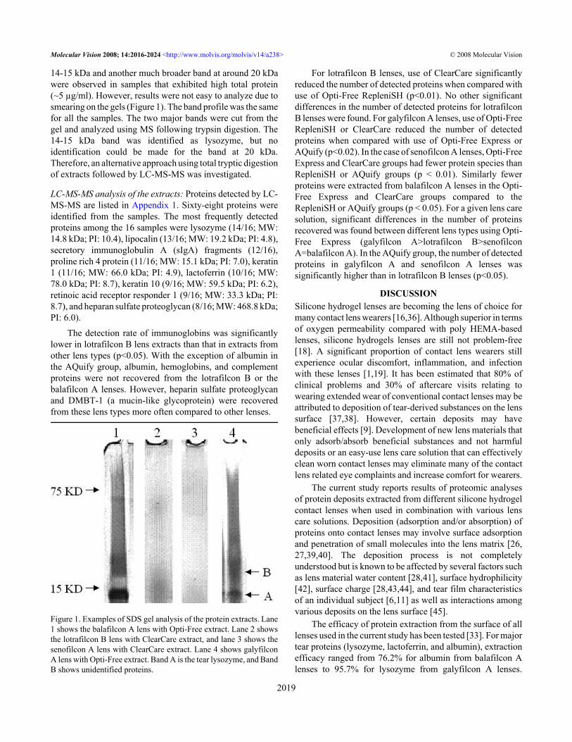
14-15 kDa and another much broader band at around 20 kDawere observed in samples that exhibited high total protein(~5 µg/ml). However, results were not easy to analyze due tosmearing on the gels (Figure 1). The band profile was the samefor all the samples. The two major bands were cut from thegel and analyzed using MS following trypsin digestion. The14-15 kDa band was identified as lysozyme, but noidentification could be made for the band at 20 kDa.Therefore, an alternative approach using total tryptic digestionof extracts followed by LC-MS-MS was investigated.
LC-MS-MS analysis of the extracts: Proteins detected by LC-MS-MS are listed in Appendix 1. Sixty-eight proteins wereidentified from the samples. The most frequently detectedproteins among the 16 samples were lysozyme (14/16; MW:14.8 kDa; PI: 10.4), lipocalin (13/16; MW: 19.2 kDa; PI: 4.8),secretory immunoglobulin A (sIgA) fragments (12/16),proline rich 4 protein (11/16; MW: 15.1 kDa; PI: 7.0), keratin1 (11/16; MW: 66.0 kDa; PI: 4.9), lactoferrin (10/16; MW:78.0 kDa; PI: 8.7), keratin 10 (9/16; MW: 59.5 kDa; PI: 6.2),retinoic acid receptor responder 1 (9/16; MW: 33.3 kDa; PI:8.7), and heparan sulfate proteoglycan (8/16; MW: 468.8 kDa;PI: 6.0).
The detection rate of immunoglobins was significantlylower in lotrafilcon B lens extracts than that in extracts fromother lens types (p<0.05). With the exception of albumin inthe AQuify group, albumin, hemoglobins, and complementproteins were not recovered from the lotrafilcon B or thebalafilcon A lenses. However, heparin sulfate proteoglycanand DMBT-1 (a mucin-like glycoprotein) were recoveredfrom these lens types more often compared to other lenses.
Figure 1. Examples of SDS gel analysis of the protein extracts. Lane1 shows the balafilcon A lens with Opti-Free extract. Lane 2 showsthe lotrafilcon B lens with ClearCare extract, and lane 3 shows thesenofilcon A lens with ClearCare extract. Lane 4 shows galyfilconA lens with Opti-Free extract. Band A is the tear lysozyme, and BandB shows unidentified proteins.
For lotrafilcon B lenses, use of ClearCare significantlyreduced the number of detected proteins when compared withuse of Opti-Free RepleniSH (p<0.01). No other significantdifferences in the number of detected proteins for lotrafilconB lenses were found. For galyfilcon A lenses, use of Opti-FreeRepleniSH or ClearCare reduced the number of detectedproteins when compared with use of Opti-Free Express orAQuify (p<0.02). In the case of senofilcon A lenses, Opti-FreeExpress and ClearCare groups had fewer protein species thanRepleniSH or AQuify groups (p < 0.01). Similarly fewerproteins were extracted from balafilcon A lenses in the Opti-Free Express and ClearCare groups compared to theRepleniSH or AQuify groups (p < 0.05). For a given lens caresolution, significant differences in the number of proteinsrecovered was found between different lens types using Opti-Free Express (galyfilcon A>lotrafilcon B>senofilconA=balafilcon A). In the AQuify group, the number of detectedproteins in galyfilcon A and senofilcon A lenses wassignificantly higher than in lotrafilcon B lenses (p<0.05).
DISCUSSIONSilicone hydrogel lenses are becoming the lens of choice formany contact lens wearers [16,36]. Although superior in termsof oxygen permeability compared with poly HEMA-basedlenses, silicone hydrogels lenses are still not problem-free[18]. A significant proportion of contact lens wearers stillexperience ocular discomfort, inflammation, and infectionwith these lenses [1,19]. It has been estimated that 80% ofclinical problems and 30% of aftercare visits relating towearing extended wear of conventional contact lenses may beattributed to deposition of tear-derived substances on the lenssurface [37,38]. However, certain deposits may havebeneficial effects [9]. Development of new lens materials thatonly adsorb/absorb beneficial substances and not harmfuldeposits or an easy-use lens care solution that can effectivelyclean worn contact lenses may eliminate many of the contactlens related eye complaints and increase comfort for wearers.
The current study reports results of proteomic analysesof protein deposits extracted from different silicone hydrogelcontact lenses when used in combination with various lenscare solutions. Deposition (adsorption and/or absorption) ofproteins onto contact lenses may involve surface adsorptionand penetration of small molecules into the lens matrix [26,27,39,40]. The deposition process is not completelyunderstood but is known to be affected by several factors suchas lens material water content [28,41], surface hydrophilicity[42], surface charge [28,43,44], and tear film characteristicsof an individual subject [6,11] as well as interactions amongvarious deposits on the lens surface [45].
The efficacy of protein extraction from the surface of alllenses used in the current study has been tested [33]. For majortear proteins (lysozyme, lactoferrin, and albumin), extractionefficacy ranged from 76.2% for albumin from balafilcon Alenses to 95.7% for lysozyme from galyfilcon A lenses.
Molecular Vision 2008; 14:2016-2024 <http://www.molvis.org/molvis/v14/a238> © 2008 Molecular Vision
2019
However, given the range of proteins found in the currentstudy, it is possible that a given protein may have a specialaffinity to a specific lens material, and therefore, we cannotconclude that the lack of protein extracted from a certain lens/solution combination is not due to this process.
Of the silicone hydrogel lens types examined in thecurrent study, balafilcon A is classified as an FDA group IIIlens (ionic, low water content) and has been found toaccumulate significantly greater amounts of protein depositsafter DW than other lenses [33]. To achieve a wettable surface,the lenses are treated by plasma oxidation producinghydrophilic glassy islands [46]. Silicate islands do notcompletely occlude the surface, resulting in a non-uniformsurface divided into hydrophobic and hydrophilic areas. It ispossible that this characteristic allows greater deposition ofproteins. The negative charge of the polymer (due to the acidiccarboxyl group in N-vinyl amino acid) would be expected toattract positively charged proteins such as lysozyme [47].Indeed, the ionic matrix is prone to accumulating proteindeposits, especially lysozyme [26,28,44]. Results in this paperreveal that the number of detected protein species in extractsfrom this lens type is not higher than the numbers found fromother lenses, indicating that the greater deposition seen withbalafilcon A lenses may be a general phenomenon and not aresult from accumulation of a particular protein species.
Distinct from balafilcon A lenses, the lotrafilcon B,galyfilcon A, and senofilcon A lenses are manufactured fromFDA group I materials (non-ionic, low water content). Thesurface of lotrafilcon B lenses is plasma-coated whereasgalyfilcon A and senofilcon A are free of surface coating.Galyfilcon A and senofilcon A lenses contain an internaladditive, polyvinyl pyrrolidone (PVP). The PVP may accountfor the common character of the two lenses to attract albumin,hemoglobins, and complement proteins and repelproteoglycans and mucins. The mechanism of the differencein attracting different proteins by different lens materials andsurface treatments and their clinical implications warrantfurther studies.
Components of lens care solutions may change thechemical and/or physical characteristics of the lens surfaceand/or stick to the surface or penetrate into the lens matrix.Indeed, the tested solutions affected the numbers of proteinsdetected for certain contact lens types, but their effects weredependent on lens materials. For example, Opti-Free Expressreduced the number of detected proteins in extracts ofsenofilcon A and balafilcon A lenses compared to the overallnumber of proteins detected in galyfilcon A lenses. Themechanism is unknown and also warrants further study todevelop a better lens care product.
Among the proteins identified in this paper, some havebeen detected in lens deposits by other researchers. Usingdifferent methods and extracts from various contact lenses,lysozyme [15,48-50], lipocalin [50,51], lactoferrin [50,52],
IgG, sIgA, IgM, complement C3 [53,54], IgE [55], secretoryphospholipase A2 [56], albumin [8,50], and vitronectin [31]have been found to deposit onto various lens materials.Interestingly, keratins 1 and 10, retinoic acid receptorresponder 1, and heparan sulfate proteoglycan were frequentlydetected in the current study but not reported by otherspreviously. The clinical significances of these deposits areunknown at present but warrant further studies.
A recent publication by Green-Church and Nicholsreported the first attempt to analyze contact lens deposits usingLC-MS-MS [50]. Less than 20 protein species were identifiedin this paper. We detected many more proteins (total 68)including some of the proteins detected in the previous study[50]. Green-Church and Nichols only examined galyfilcon Aor lotrafilcon B in combination with either AQuify or ReNuwith MoistureLoc (Bausch and Lomb, Rochester, NY). Whencomparing the galyfilcon A or lotrafilcon B lenses that wereused with AQuify to the results of the current investigation,the major differences are in the number of proteins identified(higher in the current investigation) and the presence oflacritin and secretoglobins (mammaglobins), which were notidentified in these particular lens/solution combinations in thepresent investigation. The Green-Church/Nichols study [50]did not wash the lenses after wear and analyzed lenses directlyfrom the eye. We have noted that if there is no washing beforethe extraction of proteins from lenses, the extract appears tobe very similar to the tear film (unpublished results). Perhapsthis is one reason for the differences noted between the twostudies. Also, the method of protein extraction from lenseswas different between the two studies, which may also affectprotein species recovered. In the current study, some of theproteins detected were not tear proteins such as keratins,which made up a significant proportion of the 68 protein typesidentified. These proteins may originate from the skin of thefingers or surrounding of the eyes that come into contact withcontact lenses when the participants perform their daily lenscare procedure and lens insertion.
The clinical trials from which the tested worn lenses werecollected were not designed solely for the deposit analysis.They had to meet other study requirements so the wear lengthsof these lenses were not the same (one month or two weeks,see Methods). It has been documented that the accumulatedamount of some deposits is wear length dependent [6,7].Different wear times would not impinge on the data analysisin the present study since this was a qualitative proteomicanalysis and did not attempt quantification of the proteinsidentified. However, some care should be taken whencomparing the results between lenses worn for different timeperiods.
Due to the use of pooled samples, the results in this studydo not give any information about variation between patientswithin a group. Previous reports have demonstrated thatdeposit accumulation on worn contact lenses is affected by
Molecular Vision 2008; 14:2016-2024 <http://www.molvis.org/molvis/v14/a238> © 2008 Molecular Vision
2020
tear film characteristics of individual subjects [6,11]. Thus, itis possible that patient-to-patient variability exists within thecurrent study. On the other hand, the use of pooled samplesshould reduce the component of patient-to-patient variationand reveal overall differences between lens/solution groups.The use of lesser numbers of pooled lenses in the balafilconA lens groups is a disadvantage in this aspect. The influenceof patient-to-patient variation will be addressed in subsequentresearch.
In conclusion, this study has shown that silicone hydrogellenses can adsorb/absorb proteins from the tear film and othersources. The accumulated deposition of individual proteintypes was dependent on the polymer materials from which thelenses had been made and lens care solutions used with thelenses.
ACKNOWLEDGMENTSWe thank Dr. Thomas Naduvilath for help with the statisticalanalysis of the data, Dr. Mai-Loan Huynh for MSidentification of proteins from 1D-PAGE gels, our clinicalteam for carrying out the clinical trials and collecting thesamples, and Dr. Judith Flanagan for her assistance inmanuscript preparation. This research was partly supportedby the Australian Federal Government through theCooperative Research Centres Program. The clinical trial wassponsored by CIBA Vision Corporation. Most massspectrometry was performed at the Bioanalytical MassSpectrometry Facilities within the Analytical Centre of theUniversity of New South Wales. Some parts of the resultswere presented as a poster in the Joint Fourth Asian andOceania Human Proteome Organisation Congress(AOHUPO) and in the Second Pacific-Rim InternationalConference on Protein Science (PRICPS).
REFERENCES1. Stapleton F, Keay L, Jalbert I, Cole N. The epidemiology of
contact lens related infiltrates. Optom Vis Sci 2007;84:257-72. [PMID: 17435509]
2. Willcox MD, Holden BA. Contact lens related cornealinfections. Biosci Rep 2001; 21:445-61. [PMID: 11900321]
3. Jones LW, Jones DA. Non-inflammatory corneal complicationsof contact lens wear. Cont Lens Anterior Eye 2001;24:73-9. [PMID: 16303457]
4. Dumbleton K. Noninflammatory silicone hydrogel contact lenscomplications. Eye Contact Lens 2003; 29:S186-9. [PMID:12772763]
5. Fonn D. Targeting contact lens induced dryness and discomfort:what properties will make lenses more comfortable. OptomVis Sci 2007; 84:279-85. [PMID: 17435511]
6. Jones L, Mann A, Evans K, Franklin V, Tighe B. An in vivocomparison of the kinetics of protein and lipid deposition ongroup II and group IV frequent-replacement contact lenses.Optom Vis Sci 2000; 77:503-10. [PMID: 11100888]
7. Carney FP, Nash WL, Sentell KB. The adsorption of major tearfilm lipids in vitro to various silicone hydrogels over time.Invest Ophthalmol Vis Sci 2008; 49:120-4. [PMID:18172083]
8. Luensmann D, Jones L. Albumin adsorption to contact lensmaterials: A review. Cont Lens Anterior Eye 2008;31:179-87. [PMID: 18603467]
9. Lorentz H, Rogers R, Jones L. The impact of lipid on contactangle wettability. Optom Vis Sci 2007; 84:946-53. [PMID:18049360]
10. Tan ME, Demirci G, Pearce D, Jalbert I, Sankaridurg P, WillcoxMD. Contact lens-induced papillary conjunctivitis isassociated with increased albumin deposits on extended wearhydrogel lenses. Adv Exp Med Biol 2002; 506:951-5. [PMID:12614016]
11. Jones L, Franklin V, Evans K, Sariri R, Tighe B. Spoilation andclinical performance of monthly vs. three monthly Group IIdisposable contact lenses. Optom Vis Sci 1996; 73:16-21.[PMID: 8867677]
12. Lorentz H, Jones L. Lipid deposition on hydrogel contactlenses: how history can help us today. Optom Vis Sci 2007;84:286-95. [PMID: 17435512]
13. Thai LC, Tomlinson A, Doane MG. Effect of contact lensmaterials on tear physiology. Optom Vis Sci 2004;81:194-204. [PMID: 15017179]
14. Butrus SI, Klotz SA. Contact lens surface deposits increase theadhesion of Pseudomonas aeruginosa. Curr Eye Res 1990;9:717-24. [PMID: 1980452]
15. Jones L, Senchyna M, Glasier MA, Schickler J, Forbes I, LouieD, May C. Lysozyme and lipid deposition on siliconehydrogel contact lens materials. Eye Contact Lens 2003;29:S75-9. [PMID: 12772737]
16. Sweeney DF, editor. Silicone hydrogels: the rebirth ofcontinuous wear contact lenses. Oxford: ButterworthHeinemann; 2000.
17. Guillon M, Maissa C. Use of silicone hydrogel material for dailywear. Cont Lens Anterior Eye 2007; 30:5-10. [PMID:17098464]
18. Riley C, Young G, Chalmers R. Prevalence of ocular surfacesymptoms, signs, and uncomfortable hours of wear in contactlens wearers: the effect of refitting with daily-wear siliconehydrogel lenses (senofilcon a). Eye Contact Lens 2006;32:281-6. [PMID: 17099389]
19. Richdale K, Sinnott LT, Skadahl E, Nichols JJ. Frequency ofand factors associated with contact lens dissatisfaction anddiscontinuation. Cornea 2007; 26:168-74. [PMID: 17251807]
20. Stevenson RW, Seal DV. Has the introduction of multi-purposesolutions contributed to a reduced incidence ofAcanthamoeba keratitis in contact lens wearers? A review.Cont Lens Anterior Eye 1998; 21:89-92. [PMID: 16303384]
21. Mok KH, Cheung RW, Wong BK, Yip KK, Lee VW.Effectiveness of no-rub contact lens cleaning on proteinremoval: a pilot study. Optom Vis Sci 2004; 81:468-70.[PMID: 15201721]
22. Caron P, St-Jacques J, Michaud L. Clinical discussion on therelative efficacy of 2 surfactant-containing lubricating agentsin removing proteins during contact lens wear. Optometry2007; 78:23-9. [PMID: 17208671]
23. Subbaraman LN, Glasier MA, Senchyna M, Sheardown H,Jones L. Kinetics of in vitro lysozyme deposition on siliconehydrogel, PMMA, and FDA groups I, II, and IV contact lensmaterials. Curr Eye Res 2006; 31:787-96. [PMID: 17050272]
Molecular Vision 2008; 14:2016-2024 <http://www.molvis.org/molvis/v14/a238> © 2008 Molecular Vision
2021
24. Okada E, Matsuda T, Yokoyama T, Okuda K. Lysozymepenetration in Group IV soft contact lenses. Eye Contact Lens2006; 32:174-7. [PMID: 16845262]
25. Maziarz EP, Stachowski MJ, Liu XM, Mosack L, Davis A,Musante C, Heckathorn D. Lipid deposition on siliconehydrogel lenses, part I: quantification of oleic Acid, oleic Acidmethyl ester, and cholesterol. Eye Contact Lens 2006;32:300-7. [PMID: 17099392]
26. Garrett Q, Laycock B, Garrett RW. Hydrogel lens monomerconstituents modulate protein sorption. Invest OphthalmolVis Sci 2000; 41:1687-95. [PMID: 10845587]
27. Garrett Q, Garrett RW, Milthorpe BK. Lysozyme sorption inhydrogel contact lenses. Invest Ophthalmol Vis Sci 1999;40:897-903. [PMID: 10102286]
28. Minarik L, Rapp J. Protein deposits on individual hydrophiliccontact lenses: effects of water and ionicity. CLAO J 1989;15:185-8. [PMID: 2776287]
29. de Souza GA, Godoy LM, Mann M. Identification of 491proteins in the tear fluid proteome reveals a large number ofproteases and protease inhibitors. Genome Biol 2006;7:R72. [PMID: 16901338]
30. Mann AM, Tighe BJ. The application of counterimmunoelectrophoresis (CIE) in ocular protein studies PartII: Kinin activity in the lens wearing eye. Cont Lens AnteriorEye 2002; 25:81-8. [PMID: 16303481]
31. Tighe BJ, Franklin V, Graham C, Mann A, Guillon M.Vitronectin adsorption in contact lens surfaces during wear.Locus and significance. Adv Exp Med Biol 1998;438:769-73. [PMID: 9634966]
32. Higgs RE, Knierman MD, Gelfanova V, Butler JP, Hale JE.Label-free LC-MS method for the identification ofbiomarkers. Methods Mol Biol 2008; 428:209-30. [PMID:18287776]
33. Zhao Z, Carnt NA, Aliwarga Y, Wei X, Naduvilath T, GarrettQ, Korth J, Willcox MDP. Care regimen & lens materialinfluence on in-vivo contact lens deposition. Optom Vis Sci.2008In press
34. Herbert BR, Molloy MP, Gooley AA, Walsh BJ, Bryson WG,Williams KL. Improved protein solubility in two-dimensionalelectrophoresis using tributyl phosphine as reducing agent.Electrophoresis 1998; 19:845-51. [PMID: 9629925]
35. Gatlin CL, Kleemann GR, Hays LG, Link AJ, Yates JR. Proteinidentification at the low femtomole level from silver-stainedgels using a new fritless electrospray interface for liquidchromatography-microspray and nanospray massspectrometry. Anal Biochem 1998; 263:93-101. [PMID:9750149]
36. Morgan PB, Efron N. A decade of contact lens prescribingtrends in the United Kingdom (1996–2005). Cont LensAnterior Eye 2006; 29:59-68. [PMID: 16580247]
37. Stenson S. Soft contact lens deposits. JAMA 1987; 257:2823.38. Brennan NA, Coles ML. Deposits and symptomatology with
soft contact lens wear. Int Contact Lens Clin 2000; 27:75-100.39. Garrett Q, Milthorpe BK. Human serum albumin adsorption on
hydrogel contact lenses in vitro. Invest Ophthalmol Vis Sci1996; 37:2594-602. [PMID: 8977473]
40. Luensmann D, Glasier MA, Zhang F, Bantseev V, Simpson T,Jones L. Confocal microscopy and albumin penetration intocontact lenses. Optom Vis Sci 2007; 84:839-47. [PMID:17873769]
41. Fowler SA, Korb DR, Allansmith MR. Deposits on soft contactlenses of various water contents. CLAO J 1985; 11:124-7.[PMID: 4006165]
42. Brash JL, Ten Hove P. Protein adsorption studies on 'standard'polymeric materials. J Biomater Sci Polym Ed 1993;4:591-9. [PMID: 8280673]
43. Minno GE, Eckel L, Groemminger S, Minno B, Wrzosek T.Quantitative analysis of protein deposits on hydrophilic softcontact lenses: I. Comparison to visual methods of analysis.II. Deposit variation among FDA lens material groups. OptomVis Sci 1991; 68:865-72. [PMID: 1766648]
44. Sack RA, Jones B, Antignani A, Libow R, Harvey H. Specificityand biological activity of the protein deposited on thehydrogel surface. Relationship of polymer structure to biofilmformation. Invest Ophthalmol Vis Sci 1987; 28:842-9.[PMID: 3570694]
45. Bontempo AR, Rapp J. Protein-lipid interaction on the surfaceof a hydrophilic contact lens in vitro. Curr Eye Res 1997;16:776-81. [PMID: 9255506]
46. Tighe B. Silicone Hydrogels - What are they and how shouldthey be used in everyday practice? Optician 1999; 218:31-2.
47. Garrett Q, Chatelier RC, Griesser HJ, Milthorpe BK. Effect ofcharged groups on the adsorption and penetration of proteinsonto and into carboxymethylated poly(HEMA) hydrogels.Biomaterials 1998; 19:2175-86. [PMID: 9884058]
48. Glasier MA, Keech A, Sheardown H, Subbaraman LN, JonesL. Conformational and quantitative characterization oflysozyme extracted from galyfilcon and senofilcon siliconehydrogel contact lenses. Curr Eye Res 2008; 33:1-11. [PMID:18214737]
49. Bontempo AR, Rapp J. Protein and lipid deposition ontohydrophilic contact lenses in vivo. CLAO J 2001; 27:75-80.[PMID: 11352452]
50. Green-Church KB, Nichols JJ. Mass spectrometry-basedproteomic analyses of contact lens deposition. Mol Vis 2008;14:291-7. [PMID: 18334948]
51. Baguet J, Sommer F, Claudon-Eyl V, Duc TM. Characterizationof lacrymal component accumulation on worn soft contactlens surfaces by atomic force microscopy. Biomaterials 1995;16:3-9. [PMID: 7718689]
52. Santos L, Rodrigues D, Lira M, Oliveira ME, Oliveira R, VilarEY, Azeredo J. The influence of surface treatment onhydrophobicity, protein adsorption and microbialcolonisation of silicone hydrogel contact lenses. Cont LensAnterior Eye 2007; 30:183-8. [PMID: 17291818]
53. Sack RA, Sathe S, Hackworth LA, Willcox MD, Holden BA,Morris CA. The effect of eye closure on protein andcomplement deposition on Group IV hydrogel contact lenses:relationship to tear flow dynamics. Curr Eye Res 1996;15:1092-100. [PMID: 8950503]
54. Richard NR, Anderson JA, Tasevska ZG, Binder PS. Evaluationof tear protein deposits on contact lenses from patients withand without giant papillary conjunctivitis. CLAO J 1992;18:143-7. [PMID: 1499118]
55. Zhao Z, Fu H, Skotnitsky CC, Sankaridurg PR, Willcox MD.IgE antibody on worn highly oxygen-permeable siliconehydrogel contact lenses from patients with contact lens-induced papillary conjunctivitis (CLPC). Eye Contact Lens2008; 34:117-21. [PMID: 18327049]
Molecular Vision 2008; 14:2016-2024 <http://www.molvis.org/molvis/v14/a238> © 2008 Molecular Vision
2022

56. Mochizuki H, Yamada M, Hatou S, Kawashima M, Hata S.Deposition of lipid, protein, and secretory phospholipase A2
on hydrophilic contact lenses. Eye Contact Lens 2008;34:46-9. [PMID: 18180684]
Molecular Vision 2008; 14:2016-2024 <http://www.molvis.org/molvis/v14/a238> © 2008 Molecular Vision
2023
Appendix 1. List of proteins identified in the protein extracts by LC-MS-MS.
To access the data, click or select the words “Appendix1.” This will initiate the download of a compressed (pdf)archive that contains the file.
Molecular Vision 2008; 14:2016-2024 <http://www.molvis.org/molvis/v14/a238> © 2008 Molecular Vision
The print version of this article was created on 30 October 2008. This reflects all typographical corrections and errata to thearticle through that date. Details of any changes may be found in the online version of the article.
2024