proteus spp. as putative gastrointestinal pathogens · proteus mirabilis undergoes swarming...
TRANSCRIPT

Proteus spp. as Putative Gastrointestinal Pathogens
Amy L. Hamilton,a,b Michael A. Kamm,a,b Siew C. Ng,c Mark Morrisond
aDepartment of Gastroenterology, St Vincent's Hospital, Melbourne, AustraliabDepartment of Medicine, The University of Melbourne, Melbourne, AustraliacDepartment of Medicine and Therapeutics, Institute of Digestive Disease, State Key Laboratory of DigestiveDisease, Li Ka Shing Institute of Health Science, Faculty of Medicine, The Chinese University of Hong Kong,Hong Kong, China
dThe University of Queensland Diamantina Institute, Faculty of Medicine, Translational Research Institute,Brisbane, Australia
SUMMARY . . . . . . . . . . . . . . . . . . . . . . . . . . . . . . . . . . . . . . . . . . . . . . . . . . . . . . . . . . . . . . . . . . . . . . . . . . . . . . . . . . . . . . . . 1INTRODUCTION . . . . . . . . . . . . . . . . . . . . . . . . . . . . . . . . . . . . . . . . . . . . . . . . . . . . . . . . . . . . . . . . . . . . . . . . . . . . . . . . . . 2CHARACTERISTICS OF THE PROTEUS GENUS . . . . . . . . . . . . . . . . . . . . . . . . . . . . . . . . . . . . . . . . . . . . . . . . 2
Pathogenic Features . . . . . . . . . . . . . . . . . . . . . . . . . . . . . . . . . . . . . . . . . . . . . . . . . . . . . . . . . . . . . . . . . . . . . . . . . . . 2Adhesion and mucosal attachment . . . . . . . . . . . . . . . . . . . . . . . . . . . . . . . . . . . . . . . . . . . . . . . . . . . . . . . . 4Urease. . . . . . . . . . . . . . . . . . . . . . . . . . . . . . . . . . . . . . . . . . . . . . . . . . . . . . . . . . . . . . . . . . . . . . . . . . . . . . . . . . . . . . . . . 6Hemolysins . . . . . . . . . . . . . . . . . . . . . . . . . . . . . . . . . . . . . . . . . . . . . . . . . . . . . . . . . . . . . . . . . . . . . . . . . . . . . . . . . . . 7Intracellular invasion and persistence . . . . . . . . . . . . . . . . . . . . . . . . . . . . . . . . . . . . . . . . . . . . . . . . . . . . . . 7Immune evasion . . . . . . . . . . . . . . . . . . . . . . . . . . . . . . . . . . . . . . . . . . . . . . . . . . . . . . . . . . . . . . . . . . . . . . . . . . . . . 8Endotoxin and flagellins. . . . . . . . . . . . . . . . . . . . . . . . . . . . . . . . . . . . . . . . . . . . . . . . . . . . . . . . . . . . . . . . . . . . . 8Conjugation, plasmid acquisition, and antibiotic resistance. . . . . . . . . . . . . . . . . . . . . . . . . . . . . . 9
Vaccine Candidates . . . . . . . . . . . . . . . . . . . . . . . . . . . . . . . . . . . . . . . . . . . . . . . . . . . . . . . . . . . . . . . . . . . . . . . . . . 10PROTEUS SPECIES AS GASTROINTESTINAL PATHOGENS . . . . . . . . . . . . . . . . . . . . . . . . . . . . . . . . . . 10
Colonization by Proteus Species . . . . . . . . . . . . . . . . . . . . . . . . . . . . . . . . . . . . . . . . . . . . . . . . . . . . . . . . . . . . . 10Proteus spp. in Gastroenteritis . . . . . . . . . . . . . . . . . . . . . . . . . . . . . . . . . . . . . . . . . . . . . . . . . . . . . . . . . . . . . . . 10Proteus spp. in the Upper Gastrointestinal Tract . . . . . . . . . . . . . . . . . . . . . . . . . . . . . . . . . . . . . . . . . . . 11Association with Hepatobiliary Disease . . . . . . . . . . . . . . . . . . . . . . . . . . . . . . . . . . . . . . . . . . . . . . . . . . . . . 11Pancreatic Disease . . . . . . . . . . . . . . . . . . . . . . . . . . . . . . . . . . . . . . . . . . . . . . . . . . . . . . . . . . . . . . . . . . . . . . . . . . . . 11Intestinal Disease . . . . . . . . . . . . . . . . . . . . . . . . . . . . . . . . . . . . . . . . . . . . . . . . . . . . . . . . . . . . . . . . . . . . . . . . . . . . . 12
Crohn’s disease . . . . . . . . . . . . . . . . . . . . . . . . . . . . . . . . . . . . . . . . . . . . . . . . . . . . . . . . . . . . . . . . . . . . . . . . . . . . . 12Other large intestinal diseases . . . . . . . . . . . . . . . . . . . . . . . . . . . . . . . . . . . . . . . . . . . . . . . . . . . . . . . . . . . . 13
Nosocomial Infections and Proteus Species Complicating Gastrointestinal Disease . . . . 14CONCLUSIONS . . . . . . . . . . . . . . . . . . . . . . . . . . . . . . . . . . . . . . . . . . . . . . . . . . . . . . . . . . . . . . . . . . . . . . . . . . . . . . . . . . 14ACKNOWLEDGMENTS . . . . . . . . . . . . . . . . . . . . . . . . . . . . . . . . . . . . . . . . . . . . . . . . . . . . . . . . . . . . . . . . . . . . . . . . . 15REFERENCES . . . . . . . . . . . . . . . . . . . . . . . . . . . . . . . . . . . . . . . . . . . . . . . . . . . . . . . . . . . . . . . . . . . . . . . . . . . . . . . . . . . . . 15AUTHOR BIOS . . . . . . . . . . . . . . . . . . . . . . . . . . . . . . . . . . . . . . . . . . . . . . . . . . . . . . . . . . . . . . . . . . . . . . . . . . . . . . . . . . . 19
SUMMARY Proteus species, members of the Enterobacteriaceae family, are usuallyconsidered commensals in the gut and are most commonly recognized clinically asa cause of urinary tract infections. However, the recent identification of Proteus spp.as potential pathogens in Crohn’s disease recurrence after intestinal resection servesas a stimulus to examine their potential role as gut pathogens. Proteus species pos-sess many virulence factors potentially relevant to gastrointestinal pathogenicity,including motility; adherence; the production of urease, hemolysins, and IgA pro-teases; and the ability to acquire antibiotic resistance. Gastrointestinal conditions thathave been linked to Proteus include gastroenteritis (spontaneous and foodborne), noso-comial infections, appendicitis, colonization of devices such as nasogastric tubes, andCrohn’s disease. The association of Proteus species with Crohn’s disease was particularlystrong. Proteus species are low-abundance commensals of the human gut that harborsignificant pathogenic potential; further investigation is needed.
KEYWORDS bacteriology, Crohn’s disease, Enterobacteriaceae, gastrointestinaldisease, infections, inflammatory bowel disease, Proteus
Published 13 June 2018
Citation Hamilton AL, Kamm MA, Ng SC,Morrison M. 2018. Proteus spp. as putativegastrointestinal pathogens. Clin Microbiol Rev31:e00085-17. https://doi.org/10.1128/CMR.00085-17.
Copyright © 2018 American Society forMicrobiology. All Rights Reserved.
Address correspondence to Michael A. Kamm,[email protected].
REVIEW
crossm
July 2018 Volume 31 Issue 3 e00085-17 cmr.asm.org 1Clinical Microbiology Reviews
on April 29, 2020 by guest
http://cmr.asm
.org/D
ownloaded from

INTRODUCTION
Proteus species are members of the Enterobacteriaceae family of bacteria. Mostcommonly, they are recognized clinically as a cause of urinary tract infections.
Although Proteus spp. are typically considered commensals in the gastrointestinal (GI)tract, their abundance as a proportion of the microbial community is very low (�0.05%)(1). As a result, their detection in disease states using 16S profiling, and possiblymetagenomics, may have rendered Proteus spp. undetectable due to bioinformaticabundance thresholds.
The recent identification of Proteus spp. as potential pathogens in Crohn’s diseaserecurrence after intestinal resection (2, 3) serves as a stimulus to examine their potentialrole as gut pathogens. This review aims to provide an overview of the genus Proteus interms of its known virulence factors as well as to collate the evidence surrounding therole of Proteus spp. in the pathophysiology of gastrointestinal diseases.
CHARACTERISTICS OF THE PROTEUS GENUS
Proteus spp. are Gram-negative bacteria belonging the Enterobacteriaceae familyand are common commensals of the gastrointestinal microbiota (4). The first isolateswere reported and characterized by Hauser in the late 19th century (5). The genus iscurrently comprised of Proteus mirabilis, P. vulgaris, P. penneri, P. hauseri, P. terrae, andP. cibarius, along with the unnamed genomospecies 4, 5, and 6 (6–10). In humans, allcurrent members of the genus, except for P. cibarius and P. terrae, have been isolatedfrom clinical specimens (4, 5, 7, 9, 11). Typically, the human gut is colonized by variouscombinations of P. vulgaris, P. mirabilis, and P. penneri, but they comprise less than0.05% of the gut microbiota of healthy subjects (Fig. 1) (1, 12). While Proteus spp. arewidely recognized as pathobionts and the gut is the reservoir of these bacteria, theresearch focus on this genus has been on their role in urinary tract infections ratherthan intestinal manifestations (13–15).
Many recent studies of the gut “microbiome” in health and disease have revealedthat there are gross alterations in the relative proportions of key bacterial taxa associ-ated with active disease, which is generically referred to as “dysbiosis.” One of thehallmarks of dysbiosis in the inflammatory bowel diseases (IBDs) is the populationexpansion of the phylum Proteobacteria, specifically the Enterobacteriaceae (16). Othergenera within the Enterobacteriaceae family, such as Escherichia, Shigella, Salmonella,and Klebsiella, have received due attention in this regard, while Proteus has not beencomprehensively investigated.
Pathogenic Features
Proteus species are short (1.5- to 2-�m) straight rods that demonstrate dimorphismas “swimming” and “swarming” forms, as do some other members of the Enterobacte-riaceae family (17). Swimmer cells predominate in liquid environments as single cellswith 4 to 10 peritrichous flagella (Fig. 2, bottom right) (5, 14). The swarming behaviorof Proteus species results in a characteristic bull’s-eye pattern on a plate culture, as aresult of a cyclic process of swarming and consolidation phases (18). When Proteus cellsare placed in a viscous environment or on a solid surface, they undergo differentiationto filamentous, multinucleated, highly flagellated swarmer cells (Fig. 2, top and bottomleft). Following this differentiation, a consolidation phase occurs, where the cells revertto a shorter morphotype, and metabolic preparation occurs prior to the next swarmingcycle (18).
Proteus mirabilis undergoes swarming differentiation at much higher concentrationsof agar (1.5 to 2%) than other swarming bacteria (19). When Proteus spp. swarm, thereis a dramatic increase in the production of secreted proteins, including virulence factorssuch as the protease ZapA (17, 20, 21). In vivo, swarmer cells have been demonstratedin mouse models of ascending urinary tract infection only infrequently, with theoccasional swarmer cell being isolated from the kidneys and bladder stones of infectedmice (22, 23). The swarming phenotype can occur under both aerobic and anaerobicconditions (24) and can be induced by the concentration of amino acids, in particular
Hamilton et al. Clinical Microbiology Reviews
July 2018 Volume 31 Issue 3 e00085-17 cmr.asm.org 2
on April 29, 2020 by guest
http://cmr.asm
.org/D
ownloaded from

FIG 1 Phylogenetic tree showing the species from the Enterobacteriaceae family that colonize the humangastrointestinal tract. GenBank accession numbers of the16S rRNA gene sequences are provided for eachspecies, and the family names are indicated. E. coli is highlighted in green, and the Proteus genus isshown in blue. (Reproduced from reference 133.)
Proteus spp. as Putative Gastrointestinal Pathogens Clinical Microbiology Reviews
July 2018 Volume 31 Issue 3 e00085-17 cmr.asm.org 3
on April 29, 2020 by guest
http://cmr.asm
.org/D
ownloaded from

glutamine (25, 26). It has also been demonstrated that a more acidic pH, as might beexpected to occur in the proximal small bowel and cecum, dramatically increasedswarming behavior (27–29). Swarming has been shown to be an important factor inintracellular invasion and persistence, with 15- to 20-fold more swarmer cells thanswimmer cells being capable of the intracellular invasion of uroepithelial cells (30).There is some evidence that swarming Proteus strains are more invasive in urinary tractmouse models than are swarm-defective mutant strains (22). Furthermore, a number ofmetabolites present in the intestinal tract have been shown to promote swarming,including choline, glutamine, and the most abundant polyamine in the gut, putrescine(25, 31–33). While we cannot yet conclude definitively that swarming behavior occursin the gut in vivo, the combination of a viscous surface (such as the gut mucosa), thehigh availability of glutamine and polyamines such as putrescine (26, 31, 33), andelectron acceptors for anaerobic respiration such as choline (32) makes it likely that thegut environment may be permissive for swarming.
Adhesion and mucosal attachment. Adhesion to epithelial surfaces is essential forthe pathogenesis of Proteus infections in both the urinary and gastrointestinal tracts.Sequencing of Proteus mirabilis strain HI4320 revealed 17 fimbrial gene sets (operons),more than any other bacterial genome currently characterized (34–36). Six of thefimbrial types that Proteus mirabilis can produce have been characterized (Table 1),including mannose-resistant Proteus-like fimbriae (MR/P fimbriae), mannose-resistantKlebsiella-like fimbriae (MR/K fimbriae), nonagglutinating fimbriae (NAF) (also known asuroepithelial cell adhesin [UCA]), ambient-temperature fimbriae (ATF), P. mirabilis P-likepili (PMP), and P. mirabilis fimbriae (PMF) (5, 36). These fimbriae and adhesins play amajor role in the formation of bacterial biofilms, a common complication of bothurinary and gastrointestinal instrumentation (37). It is likely that the MR/P and NAF/UCAfimbrial types are most important in gastrointestinal pathogenesis, due to their role inepithelial adhesion (38–41).
A comparison of the 17 individual chaperone-usher fimbrial operons across the 7sequenced P. mirabilis strains as well as 58 clinical isolates showed 99% conservation in13 of 17 fimbrial operons, demonstrating that these genes are highly conserved across
FIG 2 (Top) Strain of P. mirabilis inoculated twice, 1 h apart, demonstrating the macroscopic character-istic bull’s-eye pattern produced by periodic swarming. (Reproduced with permission from reference134.) (Bottom left) Interacting P. mirabilis swarmer cells; (bottom right) combination of swimmer andswarmer cells within a biofilm. (Both panels reproduced from reference 135 with permission fromElsevier.)
Hamilton et al. Clinical Microbiology Reviews
July 2018 Volume 31 Issue 3 e00085-17 cmr.asm.org 4
on April 29, 2020 by guest
http://cmr.asm
.org/D
ownloaded from

strains isolated from various clinical sites (36). Of these, it is likely that at least two, andup to six, of the characterized fimbriae can be assembled on the cell surface at any onetime (36, 42).
The regulation of motility and the expression of adhesion factors are tightly coupled;of the 17 fimbrial operons, at least 10 gene clusters possess a homolog of the mrpJgene, a repressor of motility (43). MrpJ downregulates the flagellar master regulatorflhDC via binding to the promoter sequence (43). When these cells revert back to theswimming morphology, the expression of MR/P and NAF fimbriae returns (37, 44).
TABLE 1 Fimbriae and pili expressed by Proteus speciesa
Fimbrialtype Structure Characteristic(s)
Contribution(s) to gastrointestinalpathogenicity Reference(s)
MR/P 7-nm–8-nm, “thick”channeled fimbriae
Important for epithelial cell adhesion Expression may allow mucosal celladhesion in the gastrointestinaltract and contribute to intestinalpersistence
38, 39, 69, 136
MR/P expression undergoes phase variation,allowing for a molecular “switch” thatturns on/off the expression of the mrpoperon depending on the environmentand oxygen availability, e.g., on inbladder colonization and off in kidneycolonization
The repeating structures offlagellin proteins contribute toimmunogenicity in the gut
MR/P fimbriae are potent immunogens andpotential vaccine candidates
MR/P� Proteus strains are more genotoxicand cytotoxic to bladder/kidney-derivedepithelial cells than MR/P� mutants
MR/P� Proteus strains are alsolikely more cytotoxic tointestinal epithelial cells
MR/K 4-nm–5-nm, “thin”nonchanneled fimbriae
Expression is more common in P. penneristrains than in P. mirabilis strains
P. penneri has not been linked togastrointestinal pathogenicity
42, 137
NAF/UCA 4-nm, “thin”nonchanneled fimbriae
Recognizes glycolipids, including asialo-GM1, asialo-GM2, lactosyl ceramide, andgalectin-3
40, 41, 138
Important for epithelial cell adhesion andcolonization
Not present in P. vulgaris
ATF Highly expressed at 23°C, moderatelyexpressed at 37°C
The structural subunit AtfA hassignificant homology with themajor subunit of type 1 fimbriaefrom other enteric pathogens,such as S. enterica serovar Typhi
37, 40, 139–141
Not considered to contribute to UTI orbiofilm formation; may be moreimportant for enteric colonization andenvironmental survival
Believed to be important for survival andpersistence outside a mammalian host, atroom temp
PMP Identified in uropathogenic P. mirabilisstrains isolated from dogs
142
Not present in P. vulgarisPmpA fimbrial subunits show high
homology to P-fimbriae fromuropathogenic Escherichia coli
Cell surface assembly has not beenconfirmed
PMF Important in biofilms and responsible forthe formation of higher-volume biofilms
37
aUTI, urinary tract infection.
Proteus spp. as Putative Gastrointestinal Pathogens Clinical Microbiology Reviews
July 2018 Volume 31 Issue 3 e00085-17 cmr.asm.org 5
on April 29, 2020 by guest
http://cmr.asm
.org/D
ownloaded from

Furthermore, the induction of MR/P fimbriae appears coupled to the availability ofoxygen (45). MR/P fimbriae are phase variant and can be “switched” on or off by asite-specific DNA recombinase (MrpI) that inverts a promoter region flanked by invertedrepeats. This “invertible element” switches on or off the MR/P fimbrial operon, depend-ing on the environmental conditions. There appears to be a growth advantage to MR/Pfimbrial expression under low-oxygen conditions, such as those that would be presentin the intestinal tract, likely contributing to the adhesiveness and persistence of Proteusspecies in the gut (45). The adaptation of Proteus species to mucosal surfaces by wayof both fimbrial expression (for adherence) and swarming motility could increase theinvasiveness, persistence, and pathogenicity of these species in the gut (Table 1 andFig. 3).
Urease. The urease enzyme is a microbiological adaptation to metabolize urea, themost abundant nitrogenous waste product of human metabolism (46). Urease gener-ates ammonia and carbonic acid as end products, with this ammonia providing a richsource of nitrogen for microbial metabolism in the gut (47). Urease confers a survivaladvantage to Proteus by providing nitrate for nonfermentative anaerobic respiration.This in turn promotes the population expansion of the Enterobacteriaceae (includingProteus) (48, 49). Additionally, as with Helicobacter pylori, the presence of this enzymelikely confers a survival advantage through increasing the local pH of the environment,allowing urease-positive organisms to survive in more-acidic environments such as the
FIG 3 Potentially important Proteus-related virulence factors in relation to anatomical disease location and disease. *, immune evasion includes the productionof the ZapA metalloprotease, O-antigens, and flagellin variation.
Hamilton et al. Clinical Microbiology Reviews
July 2018 Volume 31 Issue 3 e00085-17 cmr.asm.org 6
on April 29, 2020 by guest
http://cmr.asm
.org/D
ownloaded from

upper digestive tract. Proteus spp. have a wide pH range at which they are able to grow,from pH 5 to 10, with an optimal pH of 7 to 8 (9). Urease activity has been confirmedfor P. mirabilis, P. vulgaris, and P. penneri, and production is regulated by both theenvironmental concentration of the substrate (urea) and increased chromosomal tran-scription in swarming cells (50–52).
Colonization of the upper gastrointestinal tract by urease-producing Proteus speciesmay cause occasional false-positive results for the [13C]urea breath test (UBT), especiallyif the patient has already been treated with proton pump inhibitors and antibiotics forH. pylori eradication (53). Given that urease is a known contributor to the pathogenesisof H. pylori-mediated damage in the upper gastrointestinal tract, it may also contributeto the pathogenicity of Proteus spp. in the gut in the setting of environmentalperturbations, such as changes in pH.
Hemolysins. The Proteus genus produces two distinct cytotoxic hemolysins, HpmAand HlyA (51, 54, 55). P. mirabilis and most P. vulgaris strains produce only HpmA, mostP. penneri strains produce HlyA, and a few isolated P. vulgaris strains produce bothHpmA and HlyA (54, 56, 57). HpmA has been shown to lyse erythrocytes, bladderepithelial cells, B-cell lymphoma cells, and monocytes, while HlyA can lyse erythrocytes,fibroblasts, and neutrophils (55, 56). HpmA is a cell-associated hemolysin, encoded onthe hpm locus along with HpmB (an activator and chaperone of HpmA). The expressionof these hemolysin proteins is tightly coupled to the swimming-swarming cycle, withswarming cells being 18-fold more cytotoxic than swimmer cells (30). HpmA has alsobeen shown to lyse erythrocytes under anaerobic conditions and at multiple temper-atures (58). The contribution of hemolysins to gastrointestinal pathogenesis may occurthrough the lysis of innate immune cells, the induction of the NOD-like receptor protein3 (NLRP3) inflammasome, and downstream interleukin-1� (IL-1�) release (59).
Intracellular invasion and persistence. Intracellular invasion by Proteus mirabilis hasbeen assessed mainly by using cellular invasion assays in cell lines ranging fromuroepithelial cells to colonic cell types.
In a cell-based urinary tract model, swarming cells were 15-fold (0.18% intracellularentry) more invasive to uroepithelial cells than swimmer cells (0.012% intracellularentry) (30). After the invasion of uroepithelial cells, swarmer cells start to divide,develop septums, and differentiate back to an average of 50 to 300 swimmer cellswithin the cytoplasm (30). P. mirabilis has been identified as being intracellularlyinvasive in a number of cell lines (summarized in Table 2). There are differences in theintracellular invasion and uptake pathways depending on the cell type, with intracel-
TABLE 2 Cell culture lines capable of intracellular uptake of Proteus mirabilis
Cell line Cell type Reference(s)
CaCo-2a/CaCo-2BBea Colorectal adenocarcinoma/C2BBe1 (brushborder-expressing cells)
60, 62, 132,143, 144
Models for apical microvilli and tight junctions
HT29a Enterocytes, undifferentiated or differentiated(polarized, in the absence of glucose)
60, 62, 132,143, 145
HT29-18N2a Mucus-secreting differentiated cells 60, 132HT29-FUa Mucus-secreting differentiated cellsHT29-MTXa Mucus-secreting differentiated cells
INT407a Embryonic intestinal epithelial cells (possiblyHeLa-contaminated line)
61
HCT-8a Ileocecal colorectal adenocarcinomaT24 Transitional cell bladder carcinoma
HeLa Cervical carcinoma 146Vero African green monkey kidney cellsL-929 Mouse fibroblastsHuman blood lymphocytesaGastrointestinal cell line.
Proteus spp. as Putative Gastrointestinal Pathogens Clinical Microbiology Reviews
July 2018 Volume 31 Issue 3 e00085-17 cmr.asm.org 7
on April 29, 2020 by guest
http://cmr.asm
.org/D
ownloaded from
lular vacuoles containing P. mirabilis having a single membrane within urothelial cellsand a double membrane within intestinal cells (60, 61). These mechanisms maycontribute to effective intracellular colonization (cytoplasmic colonies), evasion of thehost immune system, and resistance to antibiotics (60).
Translocation of Gram-negative Enterobacteriaceae (including P. mirabilis) has beendemonstrated in vitro, in terms of both uptake and intracellular persistence. By usingCaco-2 and HT-29 cells (enterocyte cell lines), it was demonstrated that P. mirabilis notonly is invasive but also can survive intracellularly for �20 h without affecting theviability of enterocytes (62). The origin of the P. mirabilis isolate influences the inva-siveness of Proteus species in laboratory studies, with fecal isolates showing higherinvasion efficiencies (61).
Proteus mirabilis is a common cause of pathogenic infection of bladders augmentedwith bowel segments (enterocystoplasty). A study investigated the colonization ofcolonic cell lines by P. mirabilis as a model for stone formation in enterocystoplasty. TheHT29-18N2 cell line (a mucus-secreting intestinal goblet cell subclone of HT-29 [63])underwent widespread cellular destruction, with extensive intracellular bacterial colo-nies. Furthermore, upon confocal microscopy, there was colocalization of P. mirabilisand both human colonic mucin MUC2 and human gastric mucin (MUC5AC) (63). Thereis experimental evidence that Proteus spp. can invade at least the outer mucous layerof the gut and the inner layer to the epithelial surface if there are certain immunedefects. These defects include the absence of the Lypd8 protein or defects in innateimmunity (the T-bet�/� � Rag2�/� colitis [TRUC] mouse model) (64–66). Proteusmirabilis has many invasive characteristics; however, they remain to be directly char-acterized in the context of gastrointestinal disease.
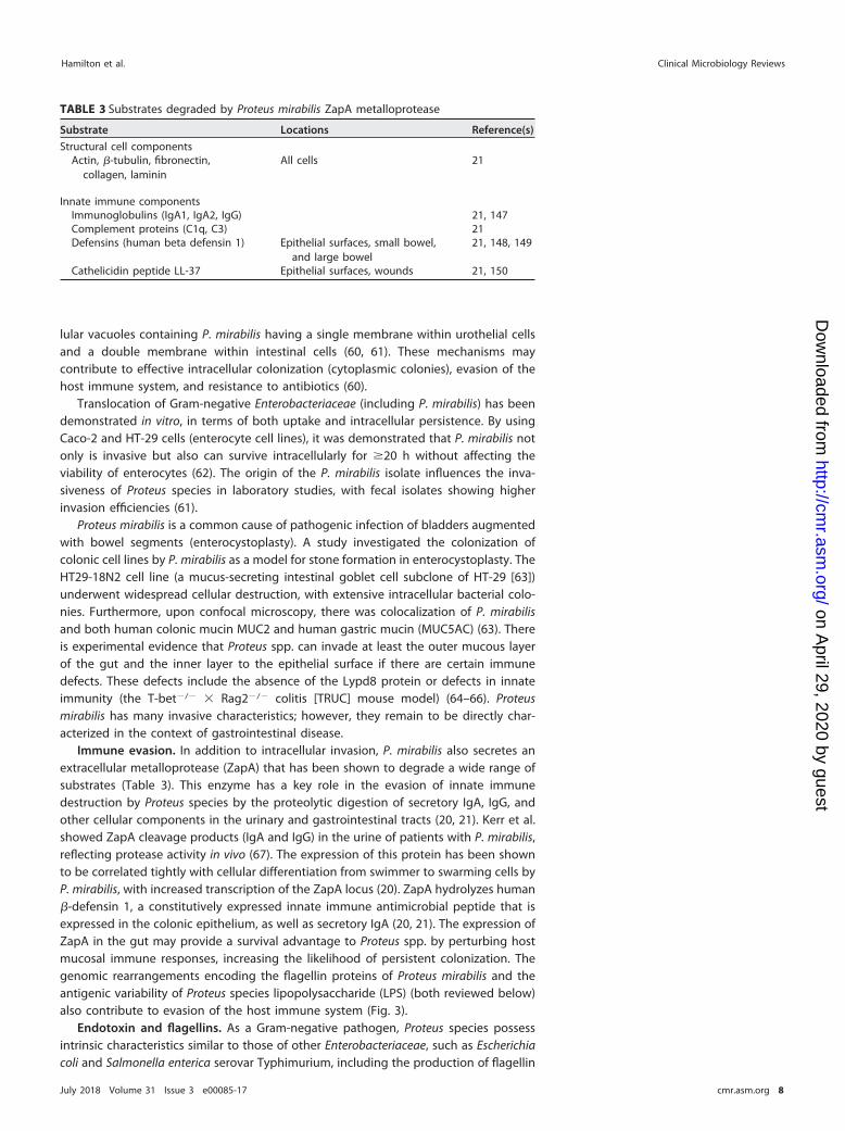
Immune evasion. In addition to intracellular invasion, P. mirabilis also secretes anextracellular metalloprotease (ZapA) that has been shown to degrade a wide range ofsubstrates (Table 3). This enzyme has a key role in the evasion of innate immunedestruction by Proteus species by the proteolytic digestion of secretory IgA, IgG, andother cellular components in the urinary and gastrointestinal tracts (20, 21). Kerr et al.showed ZapA cleavage products (IgA and IgG) in the urine of patients with P. mirabilis,reflecting protease activity in vivo (67). The expression of this protein has been shownto be correlated tightly with cellular differentiation from swimmer to swarming cells byP. mirabilis, with increased transcription of the ZapA locus (20). ZapA hydrolyzes human�-defensin 1, a constitutively expressed innate immune antimicrobial peptide that isexpressed in the colonic epithelium, as well as secretory IgA (20, 21). The expression ofZapA in the gut may provide a survival advantage to Proteus spp. by perturbing hostmucosal immune responses, increasing the likelihood of persistent colonization. Thegenomic rearrangements encoding the flagellin proteins of Proteus mirabilis and theantigenic variability of Proteus species lipopolysaccharide (LPS) (both reviewed below)also contribute to evasion of the host immune system (Fig. 3).
Endotoxin and flagellins. As a Gram-negative pathogen, Proteus species possessintrinsic characteristics similar to those of other Enterobacteriaceae, such as Escherichiacoli and Salmonella enterica serovar Typhimurium, including the production of flagellin
TABLE 3 Substrates degraded by Proteus mirabilis ZapA metalloprotease
Substrate Locations Reference(s)
Structural cell componentsActin, �-tubulin, fibronectin,
collagen, lamininAll cells 21
Innate immune componentsImmunoglobulins (IgA1, IgA2, IgG) 21, 147Complement proteins (C1q, C3) 21Defensins (human beta defensin 1) Epithelial surfaces, small bowel,
and large bowel21, 148, 149
Cathelicidin peptide LL-37 Epithelial surfaces, wounds 21, 150
Hamilton et al. Clinical Microbiology Reviews
July 2018 Volume 31 Issue 3 e00085-17 cmr.asm.org 8
on April 29, 2020 by guest
http://cmr.asm
.org/D
ownloaded from

and the proinflammatory cell wall component LPS (68, 69). A constituent of LPS,endotoxin (lipid A), is highly immunostimulatory (70). Endotoxin is sensed by the innateimmune system (specifically Toll-like receptor 4) and activates the downstream signal-ing of NF-�B (71). This, in turn, triggers a proinflammatory cascade (mediated by tumornecrosis factor alpha [TNF-�]) that can lead to acute sepsis (72, 73).
In addition to lipid A, Proteus species LPS includes O-antigens (polysaccharides thatare repeating oligosaccharide units, each made up of 2 to 8 sugar residues) that arehighly structurally diverse. Approximately 80 O-antigen serogroups have beenreported, derived from a total of 60 O-antigen gene clusters (11, 73). Antibodies toO-antigens are not uncommon in human sera, for example, 25% of blood donors haveanti-P. mirabilis antibodies to the O36 serogroup, which is just one of the manyserogroups (74). The expression levels of virulence factors, such as urease, proteases(ZapA), and hemolysin, can vary significantly between the O-antigen serogroups, withnegatively charged O-polysaccharide serogroups having higher ureolytic, proteolytic,and swarming activities (75). This heterogeneity of surface structures occurs across allProteus species.
In addition, bacterial flagellins, the repeating protein subunits from which flagellaare built, are highly immunogenic due to their three-dimensional structure (76). Bac-terial flagellin is sensed by Toll-like receptor 5, which activates a number of downstreaminflammatory pathways, including MyD88 (72, 76, 77). Recombination of the flagellingenes flaA and flaB, leading to hybrid flagellin proteins with significant antigenicvariation, also contributes to innate immune evasion by Proteus spp. (78, 79) Thesefeatures contribute to the overall pathogenicity of Proteus spp. via stimulation of thehost innate immune system by bacterial products (Fig. 3).
Conjugation, plasmid acquisition, and antibiotic resistance. Proteus species areinherently antibiotic resistant. Resistance to polymyxins is mediated via covalent mod-ifications of lipid A (80). The substitution of L-arabinoso-4-amine for either the Kdoresidue or the ester-linked lipid A phosphate moiety increases the overall charge of theusually negative LPS to zero, reducing the binding of cationic polymyxins (69, 80, 81).They also possess intrinsic resistance to colistin, tigecycline, and tetracycline (82).Sequencing of P. mirabilis strain HI4320 demonstrated the presence of genes for aconjugal transfer pilus, which allows the horizontal genetic transfer of plasmids encod-ing antibiotic resistance (35).
While most species of Proteus remain sensitive to a range of antibiotics, increasingrates of acquired antibiotic resistance in the Enterobacteriaceae are a growing problem(83). In 2007 to 2008, observations of P. mirabilis strains acquiring Salmonella genomicisland 1 (SGI1) were reported by groups in China and Palestine (84). SGI1 is a mobilegenomic element first identified in Salmonella Typhimurium that integrates into therecipient chromosome and carries multiple genes encoding resistance to streptomycin,trimethoprim, tetracycline, sulfonamides, chloramphenicol, fluoroquinolones, and abroad spectrum of �-lactam antibiotics (84). A further SGI1-positive P. mirabilis strain(NKU) has also been identified in Europe (84). Recently, an SGI1-positive P. mirabilisstrain acquired a plasmid containing the New Delhi metallo-�-lactamase 1 gene,leading to the identification of extensively drug-resistant (XDR) P. mirabilis strain PM58,which is resistant to all antibiotics used for Enterobacteriaceae except aztreonam (82).A recently sequenced P. mirabilis isolate (NO-051/03) from a patient with a soft tissueinfection in Europe had acquired the resistance genes for trimethoprim, �-lactams,phenicols, sulfonamides, and aminoglycosides (85).
In gastrointestinal disease, the antibiotic sensitivity profile of Proteus species isrelevant to pathogenicity under conditions that may be exacerbated by antibioticperturbation of the gut microbiome. Given the potential for a “bloom” of Enterobac-teriaceae (including Proteus species) both in the presence of an inflammatory process(86) and with a perturbation of the enteric environment via surgery (87), the use ofantibiotics should be considered a possible potentiating factor.
Proteus spp. as Putative Gastrointestinal Pathogens Clinical Microbiology Reviews
July 2018 Volume 31 Issue 3 e00085-17 cmr.asm.org 9
on April 29, 2020 by guest
http://cmr.asm
.org/D
ownloaded from
Vaccine Candidates
A full review of the treatment of Proteus infection is outside the scope of this review.Purified MR/P fimbrial proteins have been tested for their antigenic potential as
vaccine candidates (88, 89). There has been success by using the intranasal delivery ofMrpH, the fimbrial tip adhesin of P. mirabilis (88). A fusion protein comprised of MrpHand mannose-binding protein delivered intranasally provided 75% protection from P.mirabilis ascending urinary tract infection in a mouse model (88). A fusion proteincomprised of MrpH from P. mirabilis and FimH from uropathogenic E. coli deliveredintranasally with monophosphoryl lipid A (as an adjuvant) induced robust IgG and IgAresponses in mice (89). A clinical study of an inactivated bacterial cell suspension of fourbacterial species, including a strain of P. vulgaris, was trialed in 159 patients with ahistory of recurrent urinary tract infections, compared with 160 patients maintained onprophylactic sulfamethoxazole-trimethoprim (90). The group of patients who receivedthe vaccine had a mean number of 0.36 urinary tract infections in 3 months, versus 1.60for those receiving sulfamethoxazole-trimethoprim (P � 0.0001) (90).
PROTEUS SPECIES AS GASTROINTESTINAL PATHOGENSColonization by Proteus Species
Proteus species are known human digestive tract commensal organisms with abun-dances varying according to location (Fig. 3). Colonization occurs early. Infants fromSweden and Pakistan were assessed for Enterobacteriaceae based on mode of delivery(vaginal versus cesarean) and breastfeeding behavior (91). Cesarean births in Pakistanwere associated with Proteus species colonization within 3 days, with 11 of 21 cesarean-delivered and 1 of 9 vaginally delivered infants being positive for Proteus spp. (P �
0.049) (91).Zilberstein et al. cultured mucosal samples from the upper (n � 20) and lower (n �
24) digestive tracts of healthy controls. Proteus species were present in 8% of gastricsamples, 46% of duodenal and jejunal samples, 19% of ileal samples, 13% of cecalsamples, and 38% of samples from the transverse colon (12).
Müller compared the recovery of Proteus species from the stool specimens of 1,422healthy subjects. P. mirabilis was identified in 2.7% of healthy subjects, which is aprobable underestimate as the epithelial preference of P. mirabilis means that it is likelyundersampled in stool specimens (92). Proteus penneri and P. vulgaris were isolatedfrom 0.9% and 4.2%, respectively, of the same population (92). A smaller culture-basedstudy of 60 patients with gastrointestinal symptoms who tested negative for parasitesdemonstrated a colonization rate of 33% (20/60 patients) for P. vulgaris (93).
Proteus species, especially P. mirabilis, are often antibiotic resistant, conferring asurvival advantage when colonizing the gastrointestinal tract. In a study of multiple-drug-resistant Gram-negative bacteria in the rectum of long-term-care patients, 52drug-resistant strains were identified, 15 of which were P. mirabilis strains (94). A totalof 87% of patients colonized with P. mirabilis were also cocolonized with at least oneother resistant Gram-negative bacterium (range, 1 to 4 species; median, 2 species). Ofthe 15 patients with a resistant P. mirabilis strain, only 1 patient spontaneously clearedthe organism, compared to 30 to 75% clearance for other bacterial species (P � 0.007for clearance of other species versus P. mirabilis, as determined by a log rank test),demonstrating the ability of P. mirabilis to cause more-persistent colonization thanother Gram-negative species (94). Of the 15 P. mirabilis strains recovered, 13 weregenetically distinct, demonstrating the heterogeneity of P. mirabilis populations be-tween patients within the same health care facility (94).
Proteus spp. in Gastroenteritis
Proteus species have been associated with infectious gastroenteritis. In a comparisonof 1,271 patients with diarrhea, P. mirabilis was more prevalent in patients with diarrhea(10.8% in affected cases versus 2.7% in healthy subjects; P � 0.001) (92). However, thatstudy did not take into account the possible administration of antibiotics to affectedpatients or the potential bystander or overgrowth effect (92). P. mirabilis has also been
Hamilton et al. Clinical Microbiology Reviews
July 2018 Volume 31 Issue 3 e00085-17 cmr.asm.org 10
on April 29, 2020 by guest
http://cmr.asm
.org/D
ownloaded from

associated with foodborne gastroenteritis in an outbreak in Beijing, China, associatedwith the consumption of stewed pork (95). Shi et al. investigated genetic adaptation tothe digestive tract of P. mirabilis species isolated from the vomit and feces of patients,obtained at the time of a foodborne outbreak (96). When three clinical isolates werecompared to four local and reference strains of P. mirabilis, obtained from food, ahealthy subject, and two patients with urinary tract infections (including referencestrains HI4320 and BB2000), there was evidence of strain-level genetic adaptation to thedigestive tract (96). All seven isolates harbored drug resistance genes, but only thethree isolates (one from vomit, one from stool, and one from infected food) containeddigestive tract toxicity genes, including one with a complete type 4 secretion system(T4SS) not identified previously in P. mirabilis. Active horizontal gene acquisition hasbeen demonstrated for Proteus mirabilis, including a protein from Yersinia enterocolitica,a known GI pathogen and member of the Enterobacteriaceae family (96). In summary,Proteus species can be linked to diarrheal states, but their primary pathogenic role hasnot been confirmed.
Proteus spp. in the Upper Gastrointestinal Tract
Colonization of the upper gastrointestinal tract, including the esophagus and stom-ach, by Proteus species in infants and older adults has been reported, often associatedwith instrumentation of the oropharynx (97–101). In 13 infants with feeding tubeswithout gastrointestinal symptoms, Proteus was isolated from the throat in 8% ofpatients, from the gastric juice in 15% of patients, and from the duodenal fluid in 8%of patients (98). In elderly patients with nasogastric feeding tubes (NGTs), Proteusspecies were isolated from the oropharynx in 24% of patients and from gastric fluids in26% of patients (100). In another study, colonization of the oropharynx with Proteusspecies was present in 13% of patients with percutaneous endoscopic gastrostomy(PEG) and 21% of patients with NGTs, and colonization of the stomach was present in4% and 23% of the same patients, respectively (99).
Association with Hepatobiliary Disease
Early culture-based surveys of patients undergoing biliary surgery showed theoccasional isolation of Proteus species from the biliary tract (13% of bile samples) (102).P. mirabilis has also been recovered from bile obtained during endoscopic retrogradecholangiopancreatography (ERCP) in 6% of cultured samples (103).
In a metagenomic analysis using multitag pyrosequencing, the rectosigmoid mu-cosal community of healthy individuals was compared with that from patients withcirrhosis. Patients with cirrhosis had an elevated proportion of Proteus species com-pared with controls; the relative abundance in healthy controls was 0.0%, versus 0.1%in patients with cirrhosis (P � 0.00001) (104). Urease-producing microbes, such asProteus spp., in the gut are known to contribute to the pathogenesis of hepaticencephalopathy through the breakdown of urea to ammonia and carbonic acid (104,105).
In a series of patients undergoing liver resection, Proteus vulgaris bacteremia wasidentified in two patients, and polymicrobial infections were identified in eight patients(106). In the hepatobiliary tract, Proteus spp. are an uncommon cause of infection andare usually related to surgical interventions, such as ERCP or abdominal surgery.Implantable devices, such as stents, are also at risk of colonization and biofilm forma-tion (107).
Pancreatic Disease
There are isolated reports of Proteus species infections of the pancreas, including apatient with a large infected pancreatic pseudocyst compressing the common bileduct. The cystic contents were polymicrobial, including Proteus vulgaris, Morganellamorganii, Stenotrophomonas maltophilia, and Pseudomonas aeruginosa (108).
Proteus species were found in biofilms that form on biliary and pancreatic stentsplaced via ERCP in 14 of 100 stents (107).
Proteus spp. as Putative Gastrointestinal Pathogens Clinical Microbiology Reviews
July 2018 Volume 31 Issue 3 e00085-17 cmr.asm.org 11
on April 29, 2020 by guest
http://cmr.asm
.org/D
ownloaded from
Intestinal Disease
There are a number of reported links between Proteus spp. and various intestinalconditions, including small bowel intestinal overgrowth, Crohn’s disease, and ulcerativecolitis.
A small study of the downstream effects of small bowel ulceration caused bynonsteroidal anti-inflammatory drugs in rats identified a mixed population of E. coli andP. mirabilis. When treated with metronidazole, rats were protected from ulcer devel-opment (109). Proteus species were recovered from jejunal fluid in 11% of patients withsmall intestinal bacterial overgrowth syndrome (SIBOS), which often follows the expan-sion of facultative anaerobic bacterial communities (110). Viable bacterial translocationof Proteus mirabilis across the intact intestinal barrier has been demonstrated from thececal and colonic mucosa of a monoassociated mouse model, with bacteria beingadditionally isolated from the mesenteric lymph nodes and the liver (111).
Crohn’s disease. Recent research has implicated Proteus spp. in inflammatory boweldiseases, with evidence being derived both from patient-based microbiome surveysand mechanistic research. Ambrose et al. used culture-based techniques to comparethe recovery of pathogenic gut bacteria from the ileal serosa and mesenteric lymphnodes in 45 Crohn’s disease patients and 43 patients having surgery for other indica-tions (112). Overall, Crohn’s disease cases were more likely to have pathogenic bacteriarecovered from the small bowel serosa (12/45 [27%] patients versus 6/41 [15%]controls). Of the 12 patients with positive serosal cultures, 4/12 patients were positivefor Proteus spp. (33%). Additionally, involved and uninvolved mesenteric lymph nodeswere assessed: 15/45 patients (33%) with involved nodes had a positive culture, and ofthese samples, 1/15 (7%) grew Proteus spp. Eleven of 45 samples of uninvolved nodesharbored bacteria, with 1/11 (9%) being positive for Proteus species (112). Althoughthese data are not significant due to small numbers, they demonstrate the recovery ofProteus species from patients with Crohn’s disease, while the recovery of Proteus speciesfrom lymph nodes establishes bacterial translocation, in line with the other members ofthe Enterobacteriaceae family (113).
Two microbiome studies have linked an overabundance of Enterobacteriaceae andProteus spp. to Crohn’s disease. In a pediatric study, Proteus species comprised 3/18(16.7%) Gram-negative bacterial strains recovered from 12 Crohn’s disease patients,compared to the total absence of Proteus species recovered from patients with ulcer-ative colitis, indeterminate colitis, and lymphonodular hyperplasia and from controls(114). A microarray-based study comparing patients with active Crohn’s disease withmatched healthy controls identified an overrepresentation of Proteus species overall,and Proteus vulgaris in particular, and other members of the Proteobacteria phylum inpatients with active ileal disease requiring surgery (115).
A number of studies have addressed the mechanisms by which Proteus and otherEnterobacteriaceae may contribute to the development of inflammatory bowel diseases.The TRUC mouse model of ulcerative colitis has been used to demonstrate that Proteusmirabilis and Klebsiella pneumoniae can elicit colitis and that this propensity for thedevelopment of colitis can be transmitted to wild-type mice via microbiome transfer(65).
Crohn’s disease has been associated with changes in nitrogen metabolism. Ammo-nia produced from the breakdown of urea by bacterial urease provides a source ofnitrogen for respiration and amino acid synthesis by pathogenic facultative anaerobesfrom the Proteobacteria phylum (49).
Interactions between the Enterobacteriaceae, which include Proteus species, andfungi (Candida tropicalis) have recently been implicated in the dysbiosis that charac-terizes Crohn’s disease (116). When Crohn’s disease patients were compared withfirst-degree relatives of Crohn’s disease patients and healthy controls, bacterial dysbio-sis was identified in affected patients and unaffected first-degree relatives. Crohn’sdisease patients had an increased presence of Candida tropicalis. Complex interactionsbetween Enterobacteriaceae species (Serratia marcescens and E. coli) and Candida
Hamilton et al. Clinical Microbiology Reviews
July 2018 Volume 31 Issue 3 e00085-17 cmr.asm.org 12
on April 29, 2020 by guest
http://cmr.asm
.org/D
ownloaded from

tropicalis were confirmed in laboratory studies, showing that flagellated and/or fimbri-ated bacteria combined with fungal hyphae to form a robust biofilm, with these threespecies combined forming the thickest biofilm (P � 0.0001). It was postulated thatbiofilms enriched for immunomodulatory microbial components (lipopolysaccharidesand oligomannans, etc.) may perpetuate inflammation in dysbiotic patients throughthe induction of proinflammatory cytokine responses and of apoptosis. Proteus spp.were also strongly positively correlated with the abundance of Candida in patients withfamilial Crohn’s disease (r � 0.709; P � 0.005), raising the possibility that Proteus spp.are capable of the same interactions, although this was not demonstrated, possibly dueto their low abundance (116).
Seo et al. demonstrated that in the presence of intestinal injury and colonizationwith P. mirabilis, a marked proinflammatory IL-1� response occurs, via the activation ofthe NOD-like receptor protein 3 (NLRP3) inflammasome (59). This occurs only in thepresence of the P. mirabilis hemolysin HpmA, which appears to induce host macro-phage induction of NLRP3. Preexisting injury or inflammation was necessary for theinduction of NLRP3 activity and IL-1� by P. mirabilis (dextran sodium sulfate [DSS]-induced colitis), as inflammatory monocytes were required, suggesting that P. mirabilismay act to perpetuate and accelerate preexisting inflammation rather than induce it.Proteus mirabilis was as efficient at inducing IL-1� as pathogenic Salmonella spp., butthe presence of the HpmA hemolysin was essential for the induction of IL-1�. IL-1� hasbeen shown to be associated with disease activity in IBD patients (117, 118); however,in that study, most other anaerobic or facultative anaerobic commensals induced TNF-�expression but not IL-1� (59). When hemolysin expression levels were compared acrossP. mirabilis strains from multiple clinical sources (pyelonephritis, catheter-associated,and fecal isolates), fecal isolates had the highest hemolytic activity and had significantlyhigher hemolytic titers than those of the catheter-associated strain (P � 0.001) but notthose of the pyelonephritis strain (P � 0.065) (119).
Proteus species have been associated with the postoperative recurrence of Crohn’sdisease by two independent groups (2, 3). Metagenomic surveys of patients at the timeof surgery and 6 and 18 months postoperatively demonstrated that patients were morelikely to have disease recurrence in the presence of detectable Proteus genera (P �
0.008) and the absence of detectable Faecalibacterium (P � 0.001) (3). The combinationof detectable Proteus species and absent Faecalibacterium (�0.1%) in postoperativeileal biopsy specimens was associated with an increased risk of recurrence, with anodds ratio (OR) of 14 (95% confidence interval [CI], 1.7 to 110; P � 0.013). Smoking, anindependent risk factor for postoperative disease recurrence, was also associated withan increased presence of Proteus spp. (P � 0.0130) (3). In another study by Mondot et al.of 20 Crohn’s disease patients undergoing ileocolonic resection, the presence of aProteus mirabilis operational taxonomic unit (OTU) was predictive of recurrence at 6months postoperatively (2).
A recent review of consecutive Crohn’s disease patients with intra-abdominal ab-scesses as a result of active disease demonstrated infection with Proteus spp. in 4.8% ofcases, which was associated with high rates of quinolone resistance (120). Whether thepresence of Proteus spp. in association with postoperative recurrence is a primarypathogenic event or secondary to disease recurrence remains to be elucidated. How-ever, the association in both studies was established prospectively and longitudinally,with predictive association, making a pathogenic role more likely.
Other large intestinal diseases. Kanareykina et al. obtained samples (mouth, stom-ach, small intestine, and feces) from 65 patients with ulcerative colitis; performedculture-based enumeration; and identified Proteus mirabilis, Proteus vulgaris, or theclosely related species Morganella morganii or Providencia rettgeri (Fig. 1) in nearly allcases (121). In 40/65 patients, these species were recovered from more than oneanatomic site. A Proteus species protein “vaccine” was then administered and resultedin clinical improvement in moderate to severe cases of ulcerative colitis as well as adecrease in bacterial counts. However, no details of the vaccine composition or any
Proteus spp. as Putative Gastrointestinal Pathogens Clinical Microbiology Reviews
July 2018 Volume 31 Issue 3 e00085-17 cmr.asm.org 13
on April 29, 2020 by guest
http://cmr.asm
.org/D
ownloaded from

objective disease activity metrics were described, and the study overall was of lowquality (121).
A recent study of children with and without appendicitis showed increases in therelative abundances of 12 genera, of which Proteus species were the only representa-tives of the Enterobacteriaceae family (0.015% versus 0%; P � 0.028) (122).
Proteus bacteria have been implicated in the perpetuation of colonic inflammationin diversion colitis (123). Inflammatory conditions of the bowel increase the localconcentrations of inducible nitric oxide synthase, leading to high levels of nitrate thatcannot be metabolized, except by the microbiota (124). This favors the expansion ofbacteria that are able to metabolize nitrate under anaerobic conditions, leading to asurvival advantage and population expansion of Proteus spp. and other nitrate-reducing Enterobacteriaceae (48, 123).
Nosocomial Infections and Proteus Species Complicating Gastrointestinal Disease
Proteus species, especially P. mirabilis and P. vulgaris, are common causes of noso-comial opportunistic infections. Many patients with preexisting gastrointestinal dis-eases are liable to secondary Proteus infections, often in the context of polymicrobialinfections. Proteus species can also cause peritonitis following perforations of thegastrointestinal tract; in one report of 383 patients with peritonitis, Proteus species wereidentified in 87 (23%) patients (125).
Proteus species can colonize medical devices placed in the gastrointestinal tract,including ventriculoperitoneal shunts (126), nasogastric tubes (99, 100), biliary andpancreatic stents (107), and tracheoesophageal voice prostheses (127). Proteus bacteriahave been shown to be contaminants of gastroscopes and colonoscopes after insuffi-cient disinfection (128). Infections can also be acquired in the hospital setting due toenvironmental contamination, with P. vulgaris persisting on dry, hard surfaces for up to2 days (129). There are reports of hospital-based and community epidemics of infectionwith person-to-person spread, with most patients acquiring gastrointestinal carriageprior to infection (13, 130).
CONCLUSIONS
Proteus species are hardy, adaptable, and potentially pathogenic residents of thehuman gastrointestinal tract and have been underappreciated as a cause of gastroin-testinal disease. Host-microbe and microbe-microbe interactions by Proteus spp., andthe pathogenicity of this genus that may result from population expansion in responseto environmental changes, are emerging as important aspects of disease associatedwith this genus. The possible contribution of Proteus spp. to intestinal diseases andinfections has been somewhat neglected. Research into the virulence of Proteus spp. inthe urinary tract using the bacteriology of ileal conduits (131) and intestinal segments(60, 132) for bladder augmentation suggests that Proteus spp. should be examinedmore closely for their potential as gastrointestinal pathogens.
There is increasing evidence that Proteus species may play a role in inflammatorybowel disease through the direct action of the bacteria, compounded by host immuneevasion and perturbation. As Gram-negative organisms, Proteus species are intrinsicallyproinflammatory as a result of the production of lipopolysaccharide (LPS) and immu-nostimulatory flagellin proteins. There may be an association between Proteus speciesand inflammatory bowel disease, especially Crohn’s disease, mainly through populationexpansion and immune activation. Their low population abundance does not precludea potential large pathogenic effect.
Genetic characterization of enteric isolates compared to urinary tract isolates willbe important for determining the effect of virulence factors on gastrointestinalpathogenesis.
Research on the gut microbiome as an ecosystem is informing our understanding ofProteus species, yet there are still unanswered questions. These include obtainingconfirmation that Proteus species can swarm within the human gut and addressing the
Hamilton et al. Clinical Microbiology Reviews
July 2018 Volume 31 Issue 3 e00085-17 cmr.asm.org 14
on April 29, 2020 by guest
http://cmr.asm
.org/D
ownloaded from

effect of individual environmental changes (e.g., surgery, pH, or oxygen concentrations)on the mucosa-associated Proteus population.
ACKNOWLEDGMENTSA.L.H. was supported via a Dora Lush postgraduate scholarship from the National
Health and Medical Research Council (NHMRC). St Vincent’s Research Endowment Fundand the Australasian Gastro Intestinal Research Foundation supported M.A.K. TheUniversity of Queensland Diamantina Institute and The University of Queenslandsupported M.M. The Leona M. and Harry B. Helmsley Charitable Trust supported thiswork.
We have no conflicts of interest.M.A.K. and A.L.H. devised the concept. A.L.H. acquired data, screened papers,
interpreted the data, and wrote the manuscript. M.A.K., S.C.N., and M.M. providedcritical revision of the manuscript for important intellectual content.
REFERENCES1. Yatsunenko T, Rey FE, Manary MJ, Trehan I, Dominguez-Bello MG,
Contreras M, Magris M, Hidalgo G, Baldassano RN, Anokhin AP, HeathAC, Warner B, Reeder J, Kuczynski J, Caporaso JG, Lozupone CA, LauberC, Clemente JC, Knights D, Knight R, Gordon JI. 2012. Human gutmicrobiome viewed across age and geography. Nature 486:222–227.https://doi.org/10.1038/nature11053.
2. Mondot S, Lepage P, Seksik P, Allez M, Treton X, Bouhnik Y, ColombelJF, Leclerc M, Pochart P, Dore J, Marteau P, GETAID. 2016. Structuralrobustness of the gut mucosal microbiota is associated with Crohn’sdisease remission after surgery. Gut 65:954 –962. https://doi.org/10.1136/gutjnl-2015-309184.
3. Wright EK, Kamm MA, Wagner J, Teo SM, Cruz P, Hamilton AL, RitchieKJ, Inouye M, Kirkwood CD. 2017. Microbial factors associated withpostoperative Crohn’s disease recurrence. J Crohns Colitis 11:191–203.https://doi.org/10.1093/ecco-jcc/jjw136.
4. Penner JL. 2005. Genus XXIX. Proteus, p 745–753. In Brenner DJ, KriegNR, Staley JT, Garrity GM (ed), Bergey’s manual of systematic bacteri-ology, 2nd ed, vol 2. The Proteobacteria: part B, the Gammaproteobac-teria. Lippincott Williams & Wilkins, Philadelphia, PA.
5. Manos J, Belas R. 2006. The genera Proteus, Providencia, and Morganella,p 245–269. In Dworkin M, Falkow S, Rosenberg E, Schleifer K-H, Stacke-brandt E (ed), The prokaryotes. Springer, New York, NY.
6. Drzewiecka D. 9 January 2016. Significance and roles of Proteus spp.bacteria in natural environments. Microb Ecol https://doi.org/10.1007/s00248-015-0720-6.
7. O’Hara CM, Brenner FW, Steigerwalt AG, Hill BC, Holmes B, Grimont PA,Hawkey PM, Penner JL, Miller JM, Brenner DJ. 2000. Classification ofProteus vulgaris biogroup 3 with recognition of Proteus hauseri sp. nov.,nom. rev. and unnamed Proteus genomospecies 4, 5 and 6. Int J Syst EvolMicrobiol 50(Part 5):1869–1875. https://doi.org/10.1099/00207713-50-5-1869.
8. O’Hara CM, Brenner FW, Miller JM. 2000. Classification, identification,and clinical significance of Proteus, Providencia, and Morganella. ClinMicrobiol Rev 13:534 –546. https://doi.org/10.1128/CMR.13.4.534-546.2000.
9. Hyun DW, Jung MJ, Kim MS, Shin NR, Kim PS, Whon TW, Bae JW. 2016.Proteus cibarius sp. nov., a swarming bacterium from Jeotgal, a tradi-tional Korean fermented seafood, and emended description of thegenus Proteus. Int J Syst Evol Microbiol 66:2158 –2164. https://doi.org/10.1099/ijsem.0.001002.
10. Behrendt U, Augustin J, Sproer C, Gelbrecht J, Schumann P, Ulrich A.2015. Taxonomic characterisation of Proteus terrae sp. nov., a N2O-producing, nitrate-ammonifying soil bacterium. Antonie Van Leeuwen-hoek 108:1457–1468. https://doi.org/10.1007/s10482-015-0601-5.
11. Yu X, Torzewska A, Zhang X, Yin Z, Drzewiecka D, Cao H, Liu B, KnirelYA, Rozalski A, Wang L. 2017. Genetic diversity of the O antigens ofProteus species and the development of a suspension array for molec-ular serotyping. PLoS One 12:e0183267. https://doi.org/10.1371/journal.pone.0183267.
12. Zilberstein B, Quintanilha AG, Santos MA, Pajecki D, Moura EG, Alves PR,Maluf Filho F, de Souza JA, Gama-Rodrigues J. 2007. Digestive tract
microbiota in healthy volunteers. Clinics (Sao Paulo) 62:47–54. https://doi.org/10.1590/S1807-59322007000100008.
13. Chow AW, Taylor PR, Yoshikawa TT, Guze LB. 1979. A nosocomialoutbreak of infections due to multiply resistant Proteus mirabilis: role ofintestinal colonization as a major reservoir. J Infect Dis 139:621– 627.https://doi.org/10.1093/infdis/139.6.621.
14. Mobley HL, Belas R. 1995. Swarming and pathogenicity of Proteusmirabilis in the urinary tract. Trends Microbiol 3:280 –284. https://doi.org/10.1016/S0966-842X(00)88945-3.
15. Coker C, Poore CA, Li X, Mobley HLT. 2000. Pathogenesis of Proteusmirabilis urinary tract infection. Microbes Infect 2:1497–1505. https://doi.org/10.1016/S1286-4579(00)01304-6.
16. Mukhopadhya I, Hansen R, El-Omar EM, Hold GL. 2012. IBD—what roledo proteobacteria play? Nat Rev Gastroenterol Hepatol 9:219 –230.https://doi.org/10.1038/nrgastro.2012.14.
17. Armbruster CE, Mobley HLT. 2012. Merging mythology andmorphology: the multifaceted lifestyle of Proteus mirabilis. Nat RevMicrobiol 10:743–754. https://doi.org/10.1038/nrmicro2890.
18. Pearson MM, Rasko DA, Smith SN, Mobley HLT. 2010. Transcriptome ofswarming Proteus mirabilis. Infect Immun 78:2834 –2845. https://doi.org/10.1128/IAI.01222-09.
19. Rather PN. 2005. Swarmer cell differentiation in Proteus mirabilis. Envi-ron Microbiol 7:1065–1073. https://doi.org/10.1111/j.1462-2920.2005.00806.x.
20. Walker KE, Moghaddame-Jafari S, Lockatell CV, Johnson D, Belas R.1999. ZapA, the IgA-degrading metalloprotease of Proteus mirabilis, is avirulence factor expressed specifically in swarmer cells. Mol Microbiol32:825– 836. https://doi.org/10.1046/j.1365-2958.1999.01401.x.
21. Belas R, Manos J, Suvanasuthi R. 2004. Proteus mirabilis ZapA metallo-protease degrades a broad spectrum of substrates, including antimi-crobial peptides. Infect Immun 72:5159 –5167. https://doi.org/10.1128/IAI.72.9.5159-5167.2004.
22. Allison C, Emody L, Coleman N, Hughes C. 1994. The role of swarm celldifferentiation and multicellular migration in the uropathogenicity ofProteus mirabilis. J Infect Dis 169:1155–1158. https://doi.org/10.1093/infdis/169.5.1155.
23. Li X, Zhao H, Lockatell CV, Drachenberg CB, Johnson DE, Mobley HLT.2002. Visualization of Proteus mirabilis within the matrix of urease-induced bladder stones during experimental urinary tract infection.Infect Immun 70:389 –394. https://doi.org/10.1128/IAI.70.1.389-394.2002.
24. Alteri CJ, Himpsl SD, Engstrom MD, Mobley HLT. 2012. Anaerobicrespiration using a complete oxidative TCA cycle drives multicellularswarming in Proteus mirabilis. mBio 3:e00365-12. https://doi.org/10.1128/mBio.00365-12.
25. Armbruster CE, Hodges SA, Mobley HLT. 2013. Initiation of swarmingmotility by Proteus mirabilis occurs in response to specific cues presentin urine and requires excess L-glutamine. J Bacteriol 195:1305–1319.https://doi.org/10.1128/JB.02136-12.
26. Allison C, Lai H-C, Gygi D, Hughes C. 1993. Cell differentiation of Proteusmirabilis is initiated by glutamine, a specific chemoattractant for
Proteus spp. as Putative Gastrointestinal Pathogens Clinical Microbiology Reviews
July 2018 Volume 31 Issue 3 e00085-17 cmr.asm.org 15
on April 29, 2020 by guest
http://cmr.asm
.org/D
ownloaded from
swarming cells. Mol Microbiol 8:53– 60. https://doi.org/10.1111/j.1365-2958.1993.tb01202.x.
27. Nugent SG, Kumar D, Rampton DS, Evans DF. 2001. Intestinal luminalpH in inflammatory bowel disease: possible determinants and implica-tions for therapy with aminosalicylates and other drugs. Gut 48:571–577. https://doi.org/10.1136/gut.48.4.571.
28. Fujihara M, Obara H, Watanabe Y, Ono HK, Sasaki J, Goryo M, HarasawaR. 2011. Acidic environments induce differentiation of Proteus mirabilisinto swarmer morphotypes. Microbiol Immunol 55:489 – 493. https://doi.org/10.1111/j.1348-0421.2011.00345.x.
29. Pickard JM, Zeng MY, Caruso R, Nunez G. 2017. Gut microbiota: role inpathogen colonization, immune responses, and inflammatory disease.Immunol Rev 279:70 – 89. https://doi.org/10.1111/imr.12567.
30. Allison C, Coleman N, Jones PL, Hughes C. 1992. Ability of Proteusmirabilis to invade human urothelial cells is coupled to motility andswarming differentiation. Infect Immun 60:4740 – 4746.
31. Milovic V. 2001. Polyamines in the gut lumen: bioavailability andbiodistribution. Eur J Gastroenterol Hepatol 13:1021–1025. https://doi.org/10.1097/00042737-200109000-00004.
32. Jameson E, Fu T, Brown IR, Paszkiewicz K, Purdy KJ, Frank S, Chen Y.2016. Anaerobic choline metabolism in microcompartments pro-motes growth and swarming of Proteus mirabilis. Environ Microbiol18:2886 –2898. https://doi.org/10.1111/1462-2920.13059.
33. Sturgill G, Rather PN. 2004. Evidence that putrescine acts as an extra-cellular signal required for swarming in Proteus mirabilis. Mol Microbiol51:437– 446. https://doi.org/10.1046/j.1365-2958.2003.03835.x.
34. Schaffer JN, Pearson MM. 2015. Proteus mirabilis and urinary tractinfections. Microbiol Spectr 3:UTI-0017-2013. https://doi.org/10.1128/microbiolspec.UTI-0017-2013.
35. Pearson MM, Sebaihia M, Churcher C, Quail MA, Seshasayee AS,Luscombe NM, Abdellah Z, Arrosmith C, Atkin B, Chillingworth T,Hauser H, Jagels K, Moule S, Mungall K, Norbertczak H, RabbinowitschE, Walker D, Whithead S, Thomson NR, Rather PN, Parkhill J, MobleyHLT. 2008. Complete genome sequence of uropathogenic Proteus mi-rabilis, a master of both adherence and motility. J Bacteriol 190:4027– 4037. https://doi.org/10.1128/JB.01981-07.
36. Kuan L, Schaffer JN, Zouzias CD, Pearson MM. 2014. Characterization of17 chaperone-usher fimbriae encoded by Proteus mirabilis revealsstrong conservation. J Med Microbiol 63:911–922. https://doi.org/10.1099/jmm.0.069971-0.
37. Scavone P, Iribarnegaray V, Caetano AL, Schlapp G, Härtel S, Zunino P.2016. Fimbriae have distinguishable roles in Proteus mirabilis biofilm for-mation. Pathog Dis 74:ftw033. https://doi.org/10.1093/femspd/ftw033.
38. Scavone P, Villar S, Umpierrez A, Zunino P. 2015. Role of Proteusmirabilis MR/P fimbriae and flagella in adhesion, cytotoxicity and geno-toxicity induction in T24 and Vero cells. Pathog Dis 73:ftv017. https://doi.org/10.1093/femspd/ftv017.
39. Jansen AM, Lockatell V, Johnson DE, Mobley HLT. 2004. Mannose-resistant Proteus-like fimbriae are produced by most Proteus mirabilisstrains infecting the urinary tract, dictate the in vivo localization of bacteria,and contribute to biofilm formation. Infect Immun 72:7294–7305. https://doi.org/10.1128/IAI.72.12.7294-7305.2004.
40. Rocha SP, Pelayo JS, Elias WP. 2007. Fimbriae of uropathogenic Proteusmirabilis. FEMS Immunol Med Microbiol 51:1–7. https://doi.org/10.1111/j.1574-695X.2007.00284.x.
41. Lee KK, Harrison BA, Latta R, Altman E. 2000. The binding of Proteusmirabilis nonagglutinating fimbriae to ganglio-series asialoglycolipidsand lactosyl ceramide. Can J Microbiol 46:961–966. https://doi.org/10.1139/w00-083.
42. Adegbola RA, Old DC, Senior BW. 1983. The adhesins and fimbriae ofProteus mirabilis strains associated with high and low affinity for theurinary tract. J Med Microbiol 16:427– 431. https://doi.org/10.1099/00222615-16-4-427.
43. Pearson MM, Mobley HLT. 2008. Repression of motility during fimbrialexpression: identification of fourteen mrpJ gene paralogs in Proteusmirabilis. Mol Microbiol 69:548 –558. https://doi.org/10.1111/j.1365-2958.2008.06307.x.
44. Latta RK, Grondin A, Jarrell HC, Nicholls GR, Berube LR. 1999. Differen-tial expression of nonagglutinating fimbriae and MR/P pili in swarmingcolonies of Proteus mirabilis. J Bacteriol 181:3220 –3225.
45. Lane MC, Li X, Pearson MM, Simms AN, Mobley HL. 2009. Oxygen-limiting conditions enrich for fimbriate cells of uropathogenic Proteusmirabilis and Escherichia coli. J Bacteriol 191:1382–1392. https://doi.org/10.1128/JB.01550-08.
46. Mora D, Arioli S. 2014. Microbial urease in health and disease. PLoSPathog 10:e1004472. https://doi.org/10.1371/journal.ppat.1004472.
47. Rutherford JC. 2014. The emerging role of urease as a general microbialvirulence factor. PLoS Pathog 10:e1004062. https://doi.org/10.1371/journal.ppat.1004062.
48. Winter SE, Winter MG, Xavier MN, Thiennimitr P, Poon V, Keestra AM,Laughlin RC, Gomez G, Wu J, Lawhon SD, Popova IE, Parikh SJ, AdamsLG, Tsolis RM, Stewart VJ, Bäumler AJ. 2013. Host-derived nitrate boostsgrowth of E. coli in the inflamed gut. Science 339:708 –711. https://doi.org/10.1126/science.1232467.
49. Ni J, Shen T-CD, Chen EZ, Bittinger K, Bailey A, Roggiani M, Sirota-MadiA, Friedman ES, Chau L, Lin A, Nissim I, Scott J, Lauder A, Hoffmann C,Rivas G, Albenberg L, Baldassano RN, Braun J, Xavier RJ, Clish CB,Yudkoff M, Li H, Goulian M, Bushman FD, Lewis JD, Wu GD. 2017. A rolefor bacterial urease in gut dysbiosis and Crohn’s disease. Sci Transl Med9:eaah6888. https://doi.org/10.1126/scitranslmed.aah6888.
50. Mobley HL, Jones BD, Penner JL. 1987. Urease activity of Proteuspenneri. J Clin Microbiol 25:2302–2305.
51. Mobley HL, Chippendale GR, Swihart KG, Welch RA. 1991. Cytotoxicityof the HpmA hemolysin and urease of Proteus mirabilis and Proteusvulgaris against cultured human renal proximal tubular epithelial cells.Infect Immun 59:2036 –2042.
52. Mobley HL, Island MD, Hausinger RP. 1995. Molecular biology of mi-crobial ureases. Microbiol Rev 59:451– 480.
53. Osaki T, Mabe K, Hanawa T, Kamiya S. 2008. Urease-positive bacteria inthe stomach induce a false-positive reaction in a urea breath test fordiagnosis of Helicobacter pylori infection. J Med Microbiol 57:814 – 819.https://doi.org/10.1099/jmm.0.47768-0.
54. Swihart KG, Welch RA. 1990. The HpmA hemolysin is more commonthan HlyA among Proteus isolates. Infect Immun 58:1853–1860.
55. Swihart KG, Welch RA. 1990. Cytotoxic activity of the Proteus hemolysinHpmA. Infect Immun 58:1861–1869.
56. Senior BW. 1993. The production of HlyA toxin by Proteus penneristrains. J Med Microbiol 39:282–289. https://doi.org/10.1099/00222615-39-4-282.
57. Cestari SE, Ludovico MS, Martins FH, da Rocha SP, Elias WP, Pelayo JS.2013. Molecular detection of HpmA and HlyA hemolysin of uropatho-genic Proteus mirabilis. Curr Microbiol 67:703–707. https://doi.org/10.1007/s00284-013-0423-5.
58. Kaca W, Rozalski A. 1991. Characterization of cell-bound and cell-freehemolytic activity of Proteus strains. Eur J Epidemiol 7:159 –165. https://doi.org/10.1007/BF00237360.
59. Seo S-U, Kamada N, Muñoz-Planillo R, Kim Y-G, Kim D, Koizumi Y,Hasegawa M, Himpsl SD, Browne HP, Lawley TD, Mobley HLT, InoharaN, Núñez G. 2015. Distinct commensals induce interleukin-1� via NLRP3inflammasome in inflammatory monocytes to promote intestinal in-flammation in response to injury. Immunity 42:744 –755. https://doi.org/10.1016/j.immuni.2015.03.004.
60. Mathoera RB, Kok DJ, Verduin CM, Nijman RJM. 2002. Pathological andtherapeutic significance of cellular invasion by Proteus mirabilis in anenterocystoplasty infection stone model. Infect Immun 70:7022–7032.https://doi.org/10.1128/IAI.70.12.7022-7032.2002.
61. Oelschlaeger TA, Tall BD. 1996. Uptake pathways of clinical isolates ofProteus mirabilis into human epithelial cell lines. Microb Pathog 21:1–16. https://doi.org/10.1006/mpat.1996.0037.
62. Wells CL, van de Westerlo EMA, Jechorek RP, Erlandsen SL. 1996.Intracellular survival of enteric bacteria in cultured human enterocytes.Shock 6:27–34. https://doi.org/10.1097/00024382-199607000-00007.
63. Phillips TE, Huet C, Bilbo PR, Podolsky DK, Louvard D, Neutra MR. 1988.Human intestinal goblet cells in monolayer culture: characterization ofa mucus-secreting subclone derived from the HT29 colon adenocarci-noma cell line. Gastroenterology 94:1390 –1403. https://doi.org/10.1016/0016-5085(88)90678-6.
64. Garrett WS, Lord GM, Punit S, Lugo-Villarino G, Mazmanian SK, Ito S,Glickman JN, Glimcher LH. 2007. Communicable ulcerative colitis in-duced by T-bet deficiency in the innate immune system. Cell 131:33– 45. https://doi.org/10.1016/j.cell.2007.08.017.
65. Garrett WS, Gallini CA, Yatsunenko T, Michaud M, DuBois A, DelaneyML, Punit S, Karlsson M, Bry L, Glickman JN, Gordon JI, Onderdonk AB,Glimcher LH. 2010. Enterobacteriaceae act in concert with the gutmicrobiota to induce spontaneous and maternally transmitted colitis.Cell Host Microbe 8:292–300. https://doi.org/10.1016/j.chom.2010.08.004.
66. Okumura R, Kurakawa T, Nakano T, Kayama H, Kinoshita M, Motooka D,
Hamilton et al. Clinical Microbiology Reviews
July 2018 Volume 31 Issue 3 e00085-17 cmr.asm.org 16
on April 29, 2020 by guest
http://cmr.asm
.org/D
ownloaded from

Gotoh K, Kimura T, Kamiyama N, Kusu T, Ueda Y, Wu H, Iijima H, BarmanS, Osawa H, Matsuno H, Nishimura J, Ohba Y, Nakamura S, Iida T,Yamamoto M, Umemoto E, Sano K, Takeda K. 2016. Lypd8 promotesthe segregation of flagellated microbiota and colonic epithelia. Nature532:117–121. https://doi.org/10.1038/nature17406.
67. Kerr MA, Loomes LM, Senior BW. 1995. Cleavage of IgG and IgA in vitroand in vivo by the urinary tract pathogen Proteus mirabilis. Adv ExpMed Biol 371A:609 – 611. https://doi.org/10.1007/978-1-4615-1941-6_128.
68. Eaves-Pyles T, Murthy K, Liaudet L, Virag L, Ross G, Soriano FG, Szabo C,Salzman AL. 2001. Flagellin, a novel mediator of Salmonella-inducedepithelial activation and systemic inflammation: I kappa B alpha deg-radation, induction of nitric oxide synthase, induction of proinflamma-tory mediators, and cardiovascular dysfunction. J Immunol 166:1248 –1260. https://doi.org/10.4049/jimmunol.166.2.1248.
69. Rózalski A, Sidorczyk Z, Kotełko K. 1997. Potential virulence factors ofProteus bacilli. Microbiol Mol Biol Rev 61:65– 89.
70. Raetz CR, Whitfield C. 2002. Lipopolysaccharide endotoxins. Annu RevBiochem 71:635–700. https://doi.org/10.1146/annurev.biochem.71.110601.135414.
71. Akira S, Uematsu S, Takeuchi O. 2006. Pathogen recognition and innateimmunity. Cell 124:783– 801. https://doi.org/10.1016/j.cell.2006.02.015.
72. Takeuchi O, Akira S. 2010. Pattern recognition receptors and inflam-mation. Cell 140:805– 820. https://doi.org/10.1016/j.cell.2010.01.022.
73. Knirel YA, Perepelov AV, Kondakova AN, Senchenkova SN, Sidorczyk Z,Rozalski A, Kaca W. 2011. Structure and serology of O-antigens as thebasis for classification of Proteus strains. Innate Immun 17:70 –96.https://doi.org/10.1177/1753425909360668.
74. Arabski M, Grabowski S, Konieczna I, Kaca W, Kondakova AN, PerepelovAV, Senchenkova SN, Shashkov AS, Knirel YA. 2008. Serotyping ofclinical isolates belonging to Proteus mirabilis serogroup O36 andstructural elucidation of the O36-antigen polysaccharide. FEMS Immu-nol Med Microbiol 53:395– 403. https://doi.org/10.1111/j.1574-695X.2008.00440.x.
75. Stankowska D, Kwinkowski M, Kaca W. 2008. Quantification of Proteusmirabilis virulence factors and modulation by acylated homoserinelactones. J Microbiol Immunol Infect 41:243–253.
76. Hayashi F, Smith KD, Ozinsky A, Hawn TR, Yi EC, Goodlett DR, Eng JK,Akira S, Underhill DM, Aderem A. 2001. The innate immune response tobacterial flagellin is mediated by Toll-like receptor 5. Nature 410:1099 –1103. https://doi.org/10.1038/35074106.
77. López-Yglesias AH, Zhao X, Quarles EK, Lai MA, VandenBos T, Strong RK,Smith KD. 2014. Flagellin induces antibody responses through a TLR5-and inflammasome-independent pathway. J Immunol 192:1587–1596.https://doi.org/10.4049/jimmunol.1301893.
78. van der Woude MW, Bäumler AJ. 2004. Phase and antigenic variation inbacteria. Clin Microbiol Rev 17:581– 611. https://doi.org/10.1128/CMR.17.3.581-611.2004.
79. Murphy CA, Belas R. 1999. Genomic rearrangements in the flagellingenes of Proteus mirabilis. Mol Microbiol 31:679 – 690. https://doi.org/10.1046/j.1365-2958.1999.01209.x.
80. Olaitan AO, Morand S, Rolain J-M. 2014. Mechanisms of polymyxinresistance: acquired and intrinsic resistance in bacteria. Front Microbiol5:643. https://doi.org/10.3389/fmicb.2014.00643.
81. Vaara M, Vaara T, Jensen M, Helander I, Nurminen M, Rietschel ET,Mäkelä PH. 1981. Characterization of the lipopolysaccharide from thepolymyxin-resistant pmrA mutants of Salmonella typhimurium. FEBSLett 129:145–149. https://doi.org/10.1016/0014-5793(81)80777-6.
82. Qin S, Qi H, Zhang Q, Zhao D, Liu Z-Z, Tian H, Xu L, Xu H, Zhou M, FengX, Liu H-M. 2015. Emergence of extensively drug-resistant Proteusmirabilis harboring a conjugative NDM-1 plasmid and a novel Salmo-nella genomic island 1 variant, SGI1-Z. Antimicrob Agents Chemother59:6601– 6604. https://doi.org/10.1128/AAC.00292-15.
83. Iredell J, Brown J, Tagg K. 2016. Antibiotic resistance in Enterobacteriaceae:mechanisms and clinical implications. BMJ 352:h6420. https://doi.org/10.1136/bmj.h6420.
84. Doublet B, Poirel L, Praud K, Nordmann P, Cloeckaert A. 2010. Europeanclinical isolate of Proteus mirabilis harbouring the Salmonella genomicisland 1 variant SGI1-O. J Antimicrob Chemother 65:2260 –2262. https://doi.org/10.1093/jac/dkq283.
85. D’Andrea MM, Giani T, Henrici De Angelis L, Ciacci N, Gniadkowski M,Miriagou V, Torricelli F, Rossolini GM. 2016. Draft genome sequence ofProteus mirabilis NO-051/03, representative of a multidrug-resistantclone spreading in Europe and expressing the CMY-16 AmpC-type
beta-lactamase. Genome Announc 4:e01702-15. https://doi.org/10.1128/genomeA.01702-15.
86. Lupp C, Robertson ML, Wickham ME, Sekirov I, Champion OL, GaynorEC, Finlay BB. 2007. Host-mediated inflammation disrupts the intestinalmicrobiota and promotes the overgrowth of Enterobacteriaceae. CellHost Microbe 2:119 –129. https://doi.org/10.1016/j.chom.2007.06.010.
87. Alverdy JC, Hyoju SK, Weigerinck M, Gilbert JA. 2017. The gut micro-biome and the mechanism of surgical infection. Br J Surg 104:e14 – e23.https://doi.org/10.1002/bjs.10405.
88. Li X, Lockatell CV, Johnson DE, Lane MC, Warren JW, Mobley HLT. 2004.Development of an intranasal vaccine to prevent urinary tract infectionby Proteus mirabilis. Infect Immun 72:66 –75. https://doi.org/10.1128/IAI.72.1.66-75.2004.
89. Habibi M, Asadi Karam MR, Shokrgozar MA, Oloomi M, Jafari A, BouzariS. 2015. Intranasal immunization with fusion protein MrpH·FimH andMPL adjuvant confers protection against urinary tract infections causedby uropathogenic Escherichia coli and Proteus mirabilis. Mol Immunol64:285–294. https://doi.org/10.1016/j.molimm.2014.12.008.
90. Lorenzo-Gómez MF, Padilla-Fernández B, García-Criado FJ, Mirón-Canelo JA, Gil-Vicente A, Nieto-Huertos A, Silva-Abuin JM. 2013. Eval-uation of a therapeutic vaccine for the prevention of recurrent urinarytract infections versus prophylactic treatment with antibiotics. Int Uro-gynecol J 24:127–134. https://doi.org/10.1007/s00192-012-1853-5.
91. Adlerberth I, Carlsson B, de Man P, Jalil F, Khan SR, Larsson P, MellanderL, Svanborg C, Wold AE, Hanson LA. 1991. Intestinal colonization withEnterobacteriaceae in Pakistani and Swedish hospital-delivered infants.Acta Paediatr Scand 80:602– 610. https://doi.org/10.1111/j.1651-2227.1991.tb11917.x.
92. Müller HE. 1986. Occurrence and pathogenic role of Morganella-Proteus-Providencia group bacteria in human feces. J Clin Microbiol23:404 – 405.
93. Amin OM. 2011. The contribution of pathogenic bacteria to GI symp-toms in parasite-free patients. J Bacteriol Parasitol 2:109. https://doi.org/10.4172/2155-9597.1000109.
94. O’Fallon E, Gautam S, D’Agata EM. 2009. Colonization with multidrug-resistant gram-negative bacteria: prolonged duration and frequentcocolonization. Clin Infect Dis 48:1375–1381. https://doi.org/10.1086/598194.
95. Wang Y, Zhang S, Yu J, Zhang H, Yuan Z, Sun Y, Zhang L, Zhu Y, SongH. 2010. An outbreak of Proteus mirabilis food poisoning associatedwith eating stewed pork balls in brown sauce, Beijing. Food Control21:302–305. https://doi.org/10.1016/j.foodcont.2009.06.009.
96. Shi X, Lin Y, Qiu Y, Li Y, Jiang M, Chen Q, Jiang Y, Yuan J, Cao H, Hu Q,Huang S. 2016. Comparative screening of digestion tract toxic genesin Proteus mirabilis. PLoS One 11:e0151873. https://doi.org/10.1371/journal.pone.0151873.
97. Thomas S, Raman R, Idikula J, Brahmadathan N. 1992. Alterationsin oropharyngeal flora in patients with a nasogastric tube: a cohortstudy. Crit Care Med 20:1677–1680. https://doi.org/10.1097/00003246-199212000-00013.
98. Challacombe DN, Richardson JM, Anderson CM. 1974. Bacterial micro-flora of the upper gastrointestinal tract in infants without diarrhoea.Arch Dis Child 49:264 –269. https://doi.org/10.1136/adc.49.4.264.
99. Segal R, Dan M, Pogoreliuk I, Leibovitz A. 2006. Pathogenic colonizationof the stomach in enterally fed elderly patients: comparing percutane-ous endoscopic gastrostomy with nasogastric tube. J Am Geriatr Soc54:1905–1908. https://doi.org/10.1111/j.1532-5415.2006.00964.x.
100. Segal R, Pogoreliuk I, Dan M, Baumoehl Y, Leibovitz A. 2006. Gastricmicrobiota in elderly patients fed via nasogastric tubes for prolongedperiods. J Hosp Infect 63:79 – 83. https://doi.org/10.1016/j.jhin.2005.11.005.
101. Ehrenkranz NJ. 1970. Bacterial colonization of newborn infants andsubsequent acquisition of hospital bacteria. J Pediatr 76:839 – 847.https://doi.org/10.1016/S0022-3476(70)80363-8.
102. Lou MA, Mandal AK, Alexander JL, Thadepalli H. 1977. Bacteriology ofthe human biliary tract and the duodenum. Arch Surg 112:965–967.https://doi.org/10.1001/archsurg.1977.01370080063010.
103. Misra V, Misra SP, Singh PA, Dwivedi M, Verma K, Narayan U. 2009.Significance of cytomorphological and microbiological examination ofbile collected by endoscopic cannulation of the papilla of vater. IndianJ Pathol Microbiol 52:328 –331. https://doi.org/10.4103/0377-4929.54986.
104. Bajaj JS, Hylemon PB, Ridlon JM, Heuman DM, Daita K, White MB,Monteith P, Noble NA, Sikaroodi M, Gillevet PM. 2012. Colonic mucosal
Proteus spp. as Putative Gastrointestinal Pathogens Clinical Microbiology Reviews
July 2018 Volume 31 Issue 3 e00085-17 cmr.asm.org 17
on April 29, 2020 by guest
http://cmr.asm
.org/D
ownloaded from

microbiome differs from stool microbiome in cirrhosis and hepaticencephalopathy and is linked to cognition and inflammation. Am JPhysiol Gastrointest Liver Physiol 303:G675–G685. https://doi.org/10.1152/ajpgi.00152.2012.
105. Lai D, Gorbach SL, Levitan R. 1972. Intestinal microflora in patients withalcoholic cirrhosis: urea-splitting bacteria and neomycin resistance.Gastroenterology 62:275–279.
106. Wang X, Andersson R, Soltesz V, Bengmark S. 1992. Bacterial translo-cation after major hepatectomy in patients and rats. Arch Surg 127:1101–1106. https://doi.org/10.1001/archsurg.1992.01420090109016.
107. Vaishnavi C, Kapoor P, Kochhar R. 2014. Su1148. Bacterial biofilmsproduced in stents retrieved from patients with biliary and pancreaticdiseases. Gastroenterology 146:S-389. https://doi.org/10.1016/S0016-5085(14)61396-2.
108. Yeh CL, Lai KH, Lo GH, Lin CK, Hsu PI, Chan HH, Tsai WL, Lin CP. 2003.Endoscopic treatment in a patient with obstructive jaundice caused bypancreatic pseudocyst. J Chin Med Assoc 66:555–559.
109. Collins AJ, Reid J, Soper CJ, Notarianni LJ. 1995. Characteristics of ulcersof the small bowel induced by non-steroidal anti-inflammatory drugs inthe rat: implications for clinical practice. Br J Rheumatol 34:727–731.https://doi.org/10.1093/rheumatology/34.8.727.
110. Bouhnik Y, Alain S, Attar A, Flourie B, Raskine L, Sanson-Le Pors MJ,Rambaud JC. 1999. Bacterial populations contaminating the upper gutin patients with small intestinal bacterial overgrowth syndrome. Am JGastroenterol 94:1327–1331. https://doi.org/10.1111/j.1572-0241.1999.01016.x.
111. Wells CL, Erlandsen SL. 1991. Localization of translocating Escherichiacoli, Proteus mirabilis, and Enterococcus faecalis within cecal and colonictissues of monoassociated mice. Infect Immun 59:4693– 4697.
112. Ambrose NS, Johnson M, Burdon DW, Keighley MRB. 1984. Incidence ofpathogenic bacteria from mesenteric lymph nodes and ileal serosaduring Crohn’s disease surgery. Br J Surg 71:623– 625. https://doi.org/10.1002/bjs.1800710821.
113. O’Brien CL, Pavli P, Gordon DM, Allison GE. 2014. Detection of bacterialDNA in lymph nodes of Crohn’s disease patients using high throughputsequencing. Gut 63:1596 –1606. https://doi.org/10.1136/gutjnl-2013-305320.
114. Conte MP, Schippa S, Zamboni I, Penta M, Chiarini F, Seganti L, OsbornJ, Falconieri P, Borrelli O, Cucchiara S. 2006. Gut-associated bacterialmicrobiota in paediatric patients with inflammatory bowel disease. Gut55:1760 –1767. https://doi.org/10.1136/gut.2005.078824.
115. Mondot S, Kang S, Furet JP, Aguirre de Carcer D, McSweeney C,Morrison M, Marteau P, Doré J, Leclerc M. 2011. Highlighting newphylogenetic specificities of Crohn’s disease microbiota. Inflamm BowelDis 17:185–192. https://doi.org/10.1002/ibd.21436.
116. Hoarau G, Mukherjee PK, Gower-Rousseau C, Hager C, Chandra J,Retuerto MA, Neut C, Vermeire S, Clemente J, Colombel JF, Fujioka H,Poulain D, Sendid B, Ghannoum MA. 2016. Bacteriome and mycobiomeinteractions underscore microbial dysbiosis in familial Crohn’s disease.mBio 7:e01250-16. https://doi.org/10.1128/mBio.01250-16.
117. Leal RF, Planell N, Kajekar R, Lozano JJ, Ordás I, Dotti I, Esteller M,Masamunt MC, Parmar H, Ricart E, Panés J, Salas A. 2015. Identificationof inflammatory mediators in patients with Crohn’s disease unrespon-sive to anti-TNF� therapy. Gut 64:233–242. https://doi.org/10.1136/gutjnl-2013-306518.
118. Ligumsky M, Simon PL, Karmeli F, Rachmilewitz D. 1990. Role of inter-leukin 1 in inflammatory bowel disease— enhanced production duringactive disease. Gut 31:686 – 689. https://doi.org/10.1136/gut.31.6.686.
119. Mobley HL, Chippendale GR. 1990. Hemagglutinin, urease, and hemo-lysin production by Proteus mirabilis from clinical sources. J Infect Dis161:525–530. https://doi.org/10.1093/infdis/161.3.525.
120. Reuken PA, Kruis W, Maaser C, Teich N, Büning J, Preiß JC, SchmelzR, Bruns T, Fichtner-Feigl S, Stallmach A. 5 February 2018. Microbialspectrum of intra-abdominal abscesses in perforating Crohn’s disease:results from a prospective German registry. J Crohns Colitis https://doi.org/10.1093/ecco-jcc/jjy017.
121. Kanareykina SK, Misautova AA, Zlatkina AR, Levina EN. 1987. Proteusdysbioses in patients with ulcerative colitis. Nahrung 31:557–561.https://doi.org/10.1002/food.19870310570.
122. Jackson HT, Mongodin EF, Davenport KP, Fraser CM, Sandler AD,Zeichner SL. 2014. Culture-independent evaluation of the appendixand rectum microbiomes in children with and without appendicitis.PLoS One 9:e95414. https://doi.org/10.1371/journal.pone.0095414.
123. Neut C, Guillemot F, Colombel JF. 1997. Nitrate-reducing bacteria in
diversion colitis: a clue to inflammation? Dig Dis Sci 42:2577–2580.https://doi.org/10.1023/A:1018885217154.
124. Neut C, Bulois P, Desreumaux P, Membree J-M, Lederman E, GambiezL, Cortot A, Quandalle P, van Kruiningen H, Colombel J-F. 2002.Changes in the bacterial flora of the neoterminal ileum after ileocolonicresection for Crohn’s disease. Am J Gastroenterol 97:939 –946. https://doi.org/10.1111/j.1572-0241.2002.05613.x.
125. Hau T. 1990. Bacteria, toxins, and the peritoneum. World J Surg 14:167–175. https://doi.org/10.1007/BF01664869.
126. Liu KL, Lee TC, Lin MT, Chen SJ. 2007. Education and imaging.Gastrointestinal: abdominal abscess associated with a ventriculoperi-toneal shunt. J Gastroenterol Hepatol 22:757. https://doi.org/10.1111/j.1440-1746.2007.04964.x.
127. Ticac B, Ticac R, Rukavina T, Kesovija PG, Pedisic D, Maljevac B, StarcevicR. 2010. Microbial colonization of tracheoesophageal voice prostheses(Provox2) following total laryngectomy. Eur Arch Otorhinolaryngol 267:1579 –1586. https://doi.org/10.1007/s00405-010-1253-8.
128. Machado AP, Pimenta AT, Contijo PP, Geocze S, Fischman O. 2006.Microbiologic profile of flexible endoscope disinfection in two Brazilianhospitals. Arq Gastroenterol 43:255–258. https://doi.org/10.1590/S0004-28032006000400002.
129. Kramer A, Schwebke I, Kampf G. 2006. How long do nosocomialpathogens persist on inanimate surfaces? A systematic review. BMCInfect Dis 6:130. https://doi.org/10.1186/1471-2334-6-130.
130. Cherry WB, Lentz PL, Barnes LA. 1946. Implication of Proteus mirabilis inan outbreak of gastroenteritis. Am J Public Health Nations Health36:484 – 488. https://doi.org/10.2105/AJPH.36.5.484.
131. Chan RC, Reid G, Bruce AW, Costerton JW. 1984. Microbial colonizationof human ileal conduits. Appl Environ Microbiol 48:1159 –1165.
132. Mathoera RB, Kok DJ, Visser WJ, Verduin CM, Nijman RJ. 2001. Cellularmembrane associated mucins in artificial urine as mediators of crystaladhesion: an in vitro enterocystoplasty model. J Urol 166:2329 –2336.https://doi.org/10.1016/S0022-5347(05)65581-4.
133. Rajilic-Stojanovic M, de Vos WM. 2014. The first 1000 cultured speciesof the human gastrointestinal microbiota. FEMS Microbiol Rev 38:996 –1047. https://doi.org/10.1111/1574-6976.12075.
134. Rauprich O, Matsushita M, Weijer CJ, Siegert F, Esipov SE, Shapiro JA.1996. Periodic phenomena in Proteus mirabilis swarm colony develop-ment. J Bacteriol 178:6525– 6538. https://doi.org/10.1128/jb.178.22.6525-6538.1996.
135. Wilkerson ML, Niederhoffer EC. 1995. Swarming characteristics of Pro-teus mirabilis under anaerobic and aerobic conditions. Anaerobe1:345–350. https://doi.org/10.1006/anae.1995.1037.
136. Li X, Lockatell CV, Johnson DE, Mobley HLT. 2002. Identification of MrpIas the sole recombinase that regulates the phase variation of MR/Pfimbria, a bladder colonization factor of uropathogenic Proteus mira-bilis. Mol Microbiol 45:865– 874. https://doi.org/10.1046/j.1365-2958.2002.03067.x.
137. Yakubu DE, Old DC, Senior BW. 1989. The haemagglutinins and fim-briae of Proteus penneri. J Med Microbiol 30:279 –284. https://doi.org/10.1099/00222615-30-4-279.
138. Wray SK, Hull SI, Cook RG, Barrish J, Hull RA. 1986. Identification andcharacterization of a uroepithelial cell adhesin from a uropathogenicisolate of Proteus mirabilis. Infect Immun 54:43– 49.
139. Massad G, Bahrani FK, Mobley HL. 1994. Proteus mirabilis fimbriae: identi-fication, isolation, and characterization of a new ambient-temperaturefimbria. Infect Immun 62:1989–1994.
140. Zunino P. 2000. Virulence of a Proteus mirabilis ATF isogenic mutant isnot impaired in a mouse model of ascending urinary tract infection.FEMS Immunol Med Microbiol 29:137–143. https://doi.org/10.1111/j.1574-695X.2000.tb01516.x.
141. Massad G, Fulkerson JF, Jr, Watson DC, Mobley HL. 1996. Proteusmirabilis ambient-temperature fimbriae: cloning and nucleotide se-quence of the aft gene cluster. Infect Immun 64:4390 – 4395.
142. Bijlsma IG, van Dijk L, Kusters JG, Gaastra W. 1995. Nucleotide se-quences of two fimbrial major subunit genes, pmpA and ucaA, fromcanine-uropathogenic Proteus mirabilis strains. Microbiology 141(Part6):1349 –1357. https://doi.org/10.1099/13500872-141-6-1349.
143. Hess DJ, Henry-Stanley MJ, Erickson EA, Wells CL. 2002. Effect of tumornecrosis factor alpha, interferon gamma, and interleukin-4 on bacteria-enterocyte interactions. J Surg Res 104:88 –94. https://doi.org/10.1006/jsre.2002.6417.
144. Wells CL, Jechorek RP, Olmsted SB, Erlandsen SL. 1993. Effect of LPS on
Hamilton et al. Clinical Microbiology Reviews
July 2018 Volume 31 Issue 3 e00085-17 cmr.asm.org 18
on April 29, 2020 by guest
http://cmr.asm
.org/D
ownloaded from

epithelial integrity and bacterial uptake in the polarized humanenterocyte-like cell line Caco-2. Circ Shock 40:276 –288.
145. Wells CL, VandeWesterlo EM, Jechorek RP, Erlandsen SL. 1996. Effect ofhypoxia on enterocyte endocytosis of enteric bacteria. Crit Care Med24:985–991. https://doi.org/10.1097/00003246-199606000-00019.
146. Rozalski A, Dlugonska H, Kotelko K. 1986. Cell invasiveness of Proteusmirabilis and Proteus vulgaris strains. Arch Immunol Ther Exp (Warsz)34:505–512.
147. Senior BW, Loomes LM, Kerr MA. 1991. The production and activity in vivoof Proteus mirabilis IgA protease in infections of the urinary tract. J MedMicrobiol 35:203–207. https://doi.org/10.1099/00222615-35-4-203.
148. O’Neil DA, Porter EM, Elewaut D, Anderson GM, Eckmann L, Ganz T,Kagnoff MF. 1999. Expression and regulation of the human beta-defensins hBD-1 and hBD-2 in intestinal epithelium. J Immunol 163:6718 – 6724.
149. Ramasundara M, Leach ST, Lemberg DA, Day AS. 2009. Defensins andinflammation: the role of defensins in inflammatory bowel disease. JGastroenterol Hepatol 24:202–208. https://doi.org/10.1111/j.1440-1746.2008.05772.x.
150. Vandamme D, Landuyt B, Luyten W, Schoofs L. 2012. A comprehensivesummary of LL-37, the factotum human cathelicidin peptide. CellImmunol 280:22–35. https://doi.org/10.1016/j.cellimm.2012.11.009.
Amy L. Hamilton is a Clinical Scientist fromSt Vincent’s Hospital and the University ofMelbourne. She graduated from Monash Uni-versity, Melbourne, Australia, with a bachelorof science, with Honors in Microbiology andImmunology. She has worked on a numberof investigator-initiated clinical trials in gas-troenterology and is passionate about trans-lational research in inflammatory boweldisease as a way of improving long-termoutcomes for patients. She has a special in-terest in the gut microbiome and its role in the etiology of inflammatorybowel diseases, with this research focus allowing her to utilize bothbasic science and translational skills. She has completed her Ph.D. in theDepartment of Medicine at the University of Melbourne, addressing themicrobiology of Crohn’s disease after surgical resection. She has been amember of Professor Michael A. Kamm’s research group since 2010.
Michael A. Kamm is Gastroenterologist at StVincent’s Hospital Melbourne and Professo-rial Fellow at the University of Melbourne,Melbourne, Australia, since 2008. He waspreviously Chairman of Medicine and Di-rector of the Physiology and InflammatoryBowel Disease Units at St Mark’s Hospitalin London, United Kingdom, from 1989 to2008 and Professor of Gastroenterology atImperial College London. He treats andresearches in the fields of inflammatory boweldisease (IBD) and functional bowel disorders. Professor Kamm hasconducted multidisciplinary research, spanning basic science, transla-tional, and clinical aspects of gut disorders. In the field of the gutmicrobiota and IBD, he has studied mechanisms of inflammation, in-cluding recognition of bacterial antigens by dendritic cells and theimmunological and clinical therapeutic effects of putative prebioticsand probiotics. Most recently, he conducted investigator-initiated stud-ies into the perioperative management of Crohn’s disease (“POCER”study) and fecal microbiota transplantation in ulcerative colitis(“FOCUS” study), both changing outcomes for patients and identifyingspecific bacteria associated with clinical outcomes.
Siew C. Ng is Professor at the Department ofMedicine and Therapeutics, The Chinese Uni-versity of Hong Kong. She received her med-ical degree from St Bartholomew’s and RoyalLondon School of Medicine and obtainedher Ph.D. from Imperial College London. Dr.Ng’s main research interests include inflam-matory bowel disease, gut microbiota, andcolorectal cancer, aiming to further our un-derstanding of the pathogenesis of inflam-matory intestinal disorders via clinical trialsand translational research. She is President of the Hong Kong IBDSociety, Scientific Secretary of the International Organization of IBD, andmember of the Management Committee of the International IBD Ge-netics Consortium. She has published over 150 peer-reviewed papers ininternational journals, including Nature Genetics, Nature Communica-tions, Lancet, Gastroenterology, and Gut. She is a pioneer of IBD Epide-miologic and Microbiome research in Asia-Pacific. Her work has re-ceived over 20 Prestigious National and International Awards.
Mark Morrison is the Chair in Microbial Bi-ology and Metagenomics at The Universityof Queensland Diamantina Institute (UQDI),since October 2013. An authority in metag-enomic and molecular microbiology, he iscurrently Australia’s science representativeto the International Human MicrobiomeConsortium. Professor Morrison obtained hisPh.D. from the University of Illinois, Urbana,IL. After 20 years in the United States, hereturned to Australia in 2006 as CSIRO’s firstappointment via the Science Leader Scheme (leading the Gut HealthFlagship Research Program). Since 2007, he has held a Professorshipwithin the School of Chemistry and Molecular Biosciences, The Univer-sity of Queensland. Professor Morrison aims to translate genomic andmetagenomic data sets into sound biological frameworks, helping toproduce novel diagnostic, organismal, and enzyme-based technologies.While Professor Morrison’s focus was initially on microbial physiologyand metabolism, he has since attained international acclaim for hisefforts to successfully develop and use genomics and related methodsto study anaerobic “commensal” gut bacteria.
Proteus spp. as Putative Gastrointestinal Pathogens Clinical Microbiology Reviews
July 2018 Volume 31 Issue 3 e00085-17 cmr.asm.org 19
on April 29, 2020 by guest
http://cmr.asm
.org/D
ownloaded from