Pseudoangiomatous Stromal Hyperplasia - Department of Surgery at
16
Pseudoangiomatous Stromal Hyperplasia Nefertiti A. Brown, MD SUNY Downstate Medical Center Department of Surgery Morbidity and Mortality Conference August 30, 2012
The patient's left breast was positioned and images of the calcifications at 2 o'clock were obtained. Left breast mass, excision: Breast tissue showing sclerosing adenosis with microcalcifications. With usual ductal hyperplasia and apocrine cysts. (Fibrocystic)
Presenter
Presentation Notes
Hyperplasia of these cells originally reported by Vuitch MF, et al in 1986. Pseudoangiomatous stromal hyperplasia may present in a wide clinicopathologic spectrum ranging from incidental histologic finding to clinically palpable breast mass.
Presenter
Presentation Notes
PASH can affect persons in any age group, ranging from 12 to 75 years, but it occurs more commonly in premenopausal women. It was originally described in females and later on, it was documented in males in association with gynecomastia. PASH can occur as an isolated mass or may coexist with any breast lesion ranging from benign to malignant. PASH has been described in the breast stroma in association with gynecomastia in males. The widely accepted hypothesis in the literature is that the stromal hyperplasia in PASH results from an exaggerated, aberrant responsiveness of mammary myofibroblasts to hormonal stimuli. The hormonal influence may be endogenous or exogenous. The main hormone implicated to stimulate the myofibroblasts is progesterone. The nuclei of myofibroblasts in PASH have been shown to express progesterone receptors (PR). This makes sense, as the stromal changes identified within PASH are similar to breast changes during the luteal phase of menstruation
Presenter
Presentation Notes
This hyperplasia usually results in a slow-growing breast mass. On physical examination, tumorous PASH is a solitary, firm, painless, well-circumscribed, and freely mobile mass mimicking fibroadenoma. Sometimes, the mass may enlarge rapidly and mimic a malignant tumor. The differential diagnoses of PASH include fibroadenoma, low-grade angiosarcoma, myofibroblastoma, and mammary hamartoma. The most important differential diagnosis is angiosarcoma and it must be distinguished from PASH. Grossly, angiosarcoma is an infiltrative, haphazardly arranged in fascicles with interspersed,thick, hyalinized collagen bundles. Tumorous PASH with predominant cellular areas or fascicular foci is more likely to be confused with myofibroblastoma. It is believed that PASH and myofibroblastoma share common histogenesis and represent a spectrum of myofibroblastic proliferation. On immnohistochemistry, the spindle cells in PASH, as well as in myofibroblastoma, express myofibroblastic markers including vimentin, CD34, and SMA. The focal distribution of fascicular areas and presence of more typical areas of PASH adjacent to fascicular foci favor the diagnosis of tumorous PASH. The other feature that differentiates PASH from myofibroblastoma is the expression of progesterone receptors, in contrast to the expression of androgen receptors in myofibroblastoma. Fibroadenoma is the most common differential diagnosis on clinical and imaging studies. However, it can be easily distinguished on histology. Fibroadenoma is a benign, fibroepithelial tumor. It is composed of glandular elements in characteristic intralobular stroma. Fibroadenoma may show intracanalicular or pericanalicular pattern, depending upon the amount of stroma. In intracanalicular pattern, abundant stroma compresses the ducts with slitlike lumen. The ducts are open in pericanalicular pattern. The ducts are lined by an inner layer of cuboidal to low columnar epithelial cells and an outer layer of myoepithelial cells. The stroma may show myxoid change in fibroadenoma. Mammary hamartoma is an uncommon, benign neoplasm of breast. The distinctive histologic feature of mammary hamartoma is the presence of mature adipose tissue and nodular aggregates of mammary parenchyma. Tumorous PASH is differentiated from mammary hamartoma by the absence of adipose tissue within the mass.
Presenter
Presentation Notes
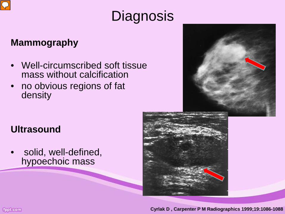
Mammography and ultrasonography are the 2 most frequently used modalities in clinical practice for breast imaging. The mammographic findings consist of a well-circumscribed, round to oval density without calcification. A solid, well-circumscribed, homogenous, hypoechoic mass as the most common appearance of tumorous PASH on ultrasonography. To confirm the diagnosis, a biopsy is required primarily to distinguish PASH from a low-grade angiosarcoma. The mammographic description of PASH is a round or ovoid, circumscribed or partially circumscribed mass. The sonographic feature is a hypoechoic mass. PASH is similar to a fibroadenoma in clinical and imaging features.
Presenter
Presentation Notes
PASH does not have any unique features on cytology that can help in making the accurate diagnosis. The major utility of cytologic examination lies in ruling out malignant lesion rather than in providing the definitive diagnosis. Sometimes, core needle biopsy is warranted in suspicious cases to exclude malignancy. Needle core biopsies have a sensitivity of 83% for diagnosing the lesion . However, careful correlation of histologic features with clinical and radiologic findings is required to ensure that the target lesion has been appropriately and adequately sampled.
Presenter
Presentation Notes
well-circumscribed tumor with a smooth external surface and a rubbery texture. The outer surface is usually smooth and unencapsulated, but sometimes resembles a capsule in some cases. The cut surface reveals a homogenous solid lesion with gray-white color and occasional cysts.
• Immunohistochemical analysis reveals a stroma that is uniformly positive for the CD34 antibody.
Normal breast tissue
Hargaden, et. al . AJR(2008 ).191(2):359-63
www.downstatesurgery.org
Presenter
Presentation Notes
Microscopically, the typical lesion is composed of complex anastomosing, slitlike empty spaces in a dense fibrous stroma . The spaces found in the lesion are not true vascular channels. Rather, they are caused by disruption and separation of stromal collagen fibers. These spaces are lined by a discontinuous layer of flat, benign spindle cells. No mitosis or nuclear atypia is seen. The stromal hyperplasia may involve perilobular as well as intralobular stroma. Positivity for CD34, actin and vimentin confirm the presence of myofibroblasts. This is in contrast to normal histology of breast tissue consists of the lobules. Within the lobules are small acini. Lobules are connected to intralobular ductules and interlobular ducts. Lobules are surrounded by loose connective tissue stroma sensitive to sex hormones
Clinical Implications
Breast mass
PASH Observation
Indeterminate Complete local excision
Cor
e B
iops
y
enlarging
www.downstatesurgery.org
Presenter
Presentation Notes
If PASH is diagnosed on the basis of core biopsy, there are no scientifically derived standards of treatment. There is an argument for leaving the mass if several well-positioned biopsies are performed and careful mammographic follow-up is undertaken, with excision if the lesion enlarges. This would depend on patient choice and the certainty of the diagnosis. If core biopsies are not diagnostic or suggestive of lowgrade angiocarcinoma, then surgical excision is warranted. Certainly, there is a potential for recurrence if a true PASH lesion is not completely excised (15-22%). However, after complete local excision, clinical follow-up evaluation probably does not need to extend beyond 3 years. Diffuse PASH occasionally presents a difficult treatment problem that may necessitate mastectomy.
Conclusions
• Hyperplasia of the mammary stroma • Possible hormonal etiology • Spectrum of disease • Increased awareness - can coexist with malignancy • Benign • Does not increase the risk of Breast CA
www.downstatesurgery.org
Presenter
Presentation Notes
PASH is hyperplasia of mammary stroma that represents spectrum of disease extending from focal, insignificant microscopic changes to cases where PHMS produces a breast mass. Increased awareness of PHMS and its clinicopathologic spectrum will allow its differentiation from other vascular tumors of the breast, especially low-grade angiosarcoma. A case with any suspicious features warrants further sampling because PASH can coexist with a malignant process and should not be accepted as a final diagnosis on the basis of core biopsy findings alone. Even though it behaves in a benign fashion, excision is largely the treatment of choice and is often a necessity for differentiating the condition histologically from angiosarcoma or other breast diseases.
Question 1 PASH occurs because A) Hyperplasia of fat B) Overexpression of androgen receptors C) Hyperplasia of glands D) Hyperplasia of myofibroblasts and
fibroblasts
www.downstatesurgery.org
Question 2
The best way to diagnose PASH is A) FNA B) Core needle biopsy C) Complete excision D) X-ray vision
www.downstatesurgery.org
Question 3 PASH A) Mimics other benign diseases B) Mimics cancer C) Requires mastectomy if diffuse D) All of the above E) None of the above
www.downstatesurgery.org
Question 4
PASH is A) Malignant B) Benign C) A rock band
www.downstatesurgery.org
References • Chung EM, Cube R, Hall GJ, González C, Stocker JT, Glassman LM. Breast masses in children
and adolescents: radiologic-pathologic correlation. Radiographics. 2009 May-Jun;29(3):907-31. • Choi YJ, Ko EY, Kook S. Diagnosis of pseudoangiomatous stromal hyperplasia of the breast:
ultrasonography findings and different biopsy methods. Yonsei Med J. 2008 Oct 31;49(5):757-64. • Cyrlak D, Carpenter PM. Breast imaging case of the day. Pseudoangiomatous stromal
Analysis of the mammographic and sonographic features of pseudoangiomatous stromal hyperplasia. AJR Am J Roentgenol. 2008 Aug;191(2):359-63.
• Milanezi MF, Saggioro FP, Zanati SG, Bazan R, Schmitt FC. Pseudoangiomatous hyperplasia of mammary stroma associated with gynaecomastia. J Clin Pathol. 1998;51(3):204–206.
• Mercado CL, Naidrich SA, Hamele-Bena D, Fineberg SA, Buchbinder SS. Pseudoangiomatous stromal hyperplasia of the breast: sonographic features with histopathologic correlation. Breast J. 2004;10(5):427–432.