pseudomeningocele refractory posterior fossa - cureus · figure 1: axial ct scan of a 35-year-old...
TRANSCRIPT

Received 11/10/2013 Review began 11/11/2013 Published 01/05/2014
© Copyright 2014Saigal et al. This is an open accessarticle distributed under the terms ofthe Creative Commons AttributionLicense CC-BY 3.0., which permitsunrestricted use, distribution, andreproduction in any medium, providedthe original author and source arecredited.
Rotational Pericranial Flap for Repair ofRefractory Posterior FossaPseudomeningoceleRajiv Saigal , Arnau Benet , William Hoffman , Lawrence R. Lustig , Steven W. Cheung ,Michael W. McDermott
1. Department of Neurological Surgery, University of California, San Francisco, CA 2. UCSF Dept ofNeurosurgery, UCSF Dept of Otolaryngology - Head and Neck Surgery 3. Division of Plastic Surgery,University of California, San Francisco, CA 4. UCSF Dept of Otolaryngology - Head and Neck Surgery 5.Dept of Otolaryngology - Head and Neck Surgery, University of California, San Francisco, CA 6.Department of Neurological Surgery, University of California, San Francisco, San Francisco, USA
Corresponding author: Rajiv Saigal, [email protected] Disclosures can be found in Additional Information at the end of the article
AbstractSkull base approaches requiring retro- or translabyrinthine petrosectomy are frequently usedfor vestibular schwannoma and cerebellopontine angle tumor removal. One postoperativechallenge is pseudomeningocele formation. Primary dural closure is often impractical due todural shrinkage from cautery, desiccation, and the fact that pre-sigmoid dura is difficult toapproximate with the retro-/translabyrinthine approach. Synthetic, allograft, and autograftdural substitutes have been used with varying success rates. Pericranial autograft is a desirablesubstitute due to lack of immunogenicity, flexibility, and wide availability adjacent to posteriorfossa lesions. Autologous fat has been used to pack the pre-sigmoid dural defect in order toeffect a watertight seal. However, no method is completely effective.
We present a technical note on use of a vascularized rotational pericranial flap technique forrepair of refractory pseudomeningocele after translabyrinthine petrosectomy. In ourexperience, this technique, when combined with temporary or permanent CSF diversion, allowsfor definitive dural closure and repair of pseudomeningocele.
Categories: Otolaryngology, Plastic Surgery, NeurosurgeryKeywords: pseudomeningocele, pericranium, autograft, pericranial flap, duraplasty, cerebellopontineangle tumor
IntroductionRepairing a pseudomeningocele after a skull base approach in association with retro- ortranslabyrinthine petrosectomy remains a challenging clinical problem. Prior studies haveexamined a variety of dural substitutes for use when primary dural closure is not possible [1-3]. Many techniques have been suggested, including use of a composite polyglactin 910 / poly-p-dioxanone mesh patch [4], monolayer and bilayer collagen matrices [5-9],polytetrafluoroethylene [10-13], crescent durotomies for midline suboccipital craniotomies[14], and autologous pericranium [1, 15]. Of these various dural substitutes, autologouspericranium is favorable due to lack of immunogenic response, low risk of infection, flexibility,and ease of use.
However, there is a paucity of technical reports on the use of a rotational pericranial flap for
1 2 3 4 5
6
Open Access TechnicalReport DOI: 10.7759/cureus.153
How to cite this articleSaigal R, Benet A, Hoffman W, et al. (January 05, 2014) Rotational Pericranial Flap for Repair ofRefractory Posterior Fossa Pseudomeningocele. Cureus 6(1): e153. DOI 10.7759/cureus.153

dural substitution. This technique is particularly well-suited for challenging posterior fossadural repair cases. As in other types of reconstructive surgery [16], a rotational flap offers thebenefit of preserving the vascular supply. Plastic and reconstructive surgeons have successfullyused vascularized pericranial flaps for reconstruction of congenital cranial malformations [17-18], repair of chronic scalp ulcers [19], dura mater reconstruction after decompressivecraniectomy complicated by infection [20], orbital [21-22] and nasal septum [23] reconstruction,repair of supratarsal sulcus depression [24], cranial base reconstruction after tumor resection[25], and frontal sinus fracture repair [26]. In this technical note, we utilize the principles ofplastic and reconstructive surgery in the implementation of a rotational pericranial flap forclosure of a complex posterior fossa wound.
As an illustrative case, we present a 35-year-old female with a history of a 4 cm rightcerebellopontine angle (CPA) schwannoma S/P right transcrural petrosal approach for resectionwith placement of a lumbar subarachnoid drain. The patient gave written approval for the useof her pictures and case history in this paper. The dura over the temporal lobe was closed withinterrupted and running 4-0 Polyglactin 910 suture. It was not possible to achieve a watertightclosure of the pre-sigmoid dura, so a collagen matrix dural substitute was placed as a sling toprevent excessive herniation of fat into the CPA and this layer was then covered with a fat graft(harvested from the abdomen) and fibrin sealant. The temporal bone flap was repositioned andsecured with titanium plates and screws. Bone reconstruction of the mastoidectomy defect andpetrous bone was completed with a porous polyethylene plate implant. Her lumbar drain wasopened on postoperative day one to drain a goal of 10-15 cc/hr, but was pulled by the patienton postoperative day two. She was discharged in good condition.
Two weeks postoperatively, she was readmitted with an enlarging pseudomeningocele andsigns of intracranial hypotension. A left ventriculoperitoneal shunt was placed as thepseudomeningocele was increasing in size. She was discharged in good condition, but returnedone week later with enlargement of the pseudomeningocele. A rotational pericranial flap isideal to repair a suboccipital wound such as this. Figure 1 shows the axial CT scan prior torepair.
2014 Saigal et al. Cureus 6(1): e153. DOI 10.7759/cureus.153 2 of 14

FIGURE 1: Axial CT scan of a 35-year-old female with history ofa 4 cm right vestibular schwannoma status-post transcruraltranspetrosal approach for resection.She subsequently developed a postoperative pseudomeningocele. Bone (A.) and brain (C.)windows show superior end of petrosectomy and porous polyethylene/titanium plate reconstructionwith surrounding pseudomeningocele. Bone (B.) and brain (D.) windows of a more inferior slicefurther demonstrate the extent of petrosectomy and pseudomeningocele.
In the suboccipital, retromastoid region, the occipital artery runs superficial to the superioroblique capitis muscle towards the supreme nuchal line. On its course, the occipital artery givesbranches to the suboccipital muscles: the longisimus capitis, the occipital belly of the occipital-frontal muscle, and the galea and pericranium of the occipital and posterior portion of theparietal and temporal bones. These branches can anastomose with the vertebral artery. Abovethe supreme nuchal line, the main trunk of the occipital artery pierces the occipital musclewithin the lateral two-thirds of the aponeurosis of the occipital-frontal muscle and becomessuperficial to it. However, many branches of variable diameter take off from the occipital arteryas it courses through the occipital muscle to feed the pericranium and the parietal bone. Thesedeep branches of the occipital artery are responsible for the blood supply of the proposedrotational flap.
2014 Saigal et al. Cureus 6(1): e153. DOI 10.7759/cureus.153 3 of 14

Technical ReportMethodsCSF diversion is recommended for a period of five days as the initial wound healing takes place.If CSF access is not yet available, placement of a lumbar subarachnoid or external ventriculardrain is recommended. The drain is left clamped during the procedure. Once CSF access isgained, the patient is positioned semi-lateral with the head turned contralateral to thelesion. The head is positioned on a foam donut head holder, as shown below.
FIGURE 2: Patient positioning
The skin is shaved and prepped in the usual sterile manner. The previous C-shaped skinincision is re-marked, and a straight line is added to allow access to the pericranium over theparietal and occipital bones (Figure 3). The inferior C-shaped incision is re-opened sharply, asshown in Figure 4 below.
2014 Saigal et al. Cureus 6(1): e153. DOI 10.7759/cureus.153 4 of 14

FIGURE 3: Skin incision
2014 Saigal et al. Cureus 6(1): e153. DOI 10.7759/cureus.153 5 of 14

FIGURE 4: Skin incision for opening of inferior C-shapedincision, prior to harvesting pericranium
Subcutaneous soft tissues are divided sharply, stopping short of the pericranium. There, thesubgaleal plane above the pericranium is opened with a combination of blunt dissection withMetzenbaum scissors and monopolar electrocautery with a fine Colorado tip. Skin hooks areplaced for retraction. The bone flap and porous polyethylene implants are removed. Valsalva isused to investigate the site of CSF leak. Figure 5 shows these various steps in more detail.
2014 Saigal et al. Cureus 6(1): e153. DOI 10.7759/cureus.153 6 of 14
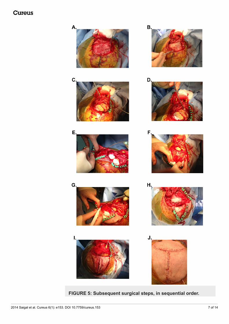
FIGURE 5: Subsequent surgical steps, in sequential order.
2014 Saigal et al. Cureus 6(1): e153. DOI 10.7759/cureus.153 7 of 14

A. The subgaleal layer is re-opened. B. The bone flap is removed. C, D. The epidural fat graft isexposed. E, F. Subgaleal dissection to widen the rostral cranial opening G. Monopolarelectrocautery is used to divide the edge of the pericranial flap. H. The pericranial flap is rotateddownwards to cover the site of tumor excision. I. The rostral cranial opening is reapproximated firstwith a galeal closure. J. Skin closure after duraplasty with rotational pericranial flap.
A rectangular pedicle of pericranium is divided with electrocautery anteriorly over the frontalbone, curving around medially under the scalp over the parietal and occipital bones to thesuperior nuchal line inferiorly and separated from the skull with a periosteal dissector. In orderto preserve the vascular supply, the base of the pedicle is the lateral two-thirds of the base ofthe flap at the superior nuchal line. The medial one-third "back cut" of the pericranium allowsfor an increased working length of the flap as it is rotated over the posterior fossa, pre-sigmoid,and inferior temporal dura. The pericranial patch is rotated and flapped down to cover the siteof leak and surrounding dura. The patch is secured in place with interrupted 4-0 polyglactin910 sutures at the anterior and inferior margins and covered with fibrin sealant. The wound iswashed with copious antibiotic irrigation. A subgaleal hemovac drain is placed. Galea isreapproximated with interrupted 3-0 polyglactin 910 and then closed with a running 2-0polyglactin 910. The skin is closed with a running 4-0 monofilament polybutester suture. Thefinal closure is shown in Figure 5J. The wound is then covered with bacitracin ointment, telfadressing, and a pressure head wrap is applied with a combination of fluffs, kerlex, and aflexinet. The CSF drain is opened to drain a goal of 10-15 mL per hour on postoperative dayone.
Cadaver studyA postmortem human head was prepared at the skull base and cerebrovascular laboratory toillustrate the surgical anatomy of the pericranial flap. The head was cut at C5 level, and thecarotid and vertebral arteries and the jugular veins were cannulated for embalming. The sametubing was used to inject red (arteries) and blue (veins) silicone to ease identification duringthe surgical simulation procedure.
A standard transmastoid-transcochlear approach was performed in a stepwise manner (Figure6). The scalp incision was extended anteriorly aproximately 2 cm around the pinna until thezygomatic process of the temporal bone. A second incision was performed at the superiorborder of the first incision towards the midline and 10 degrees posteriorly. The scalp wasreflected away from the longitudinal incision, and the gale, frontalis, and occipitalis muscleswere identified. Next, the frontal and parietal branches of the superior temporalis artery andthe supraorbital and the occipital arteries were exposed. The galea was then incised, and arectangular shaped pericranial flap was raised from anterior to posterior. This maneuverrequired cutting the suproaorbital artery and distal branches of the supratrochlear artery(Figure 7). Finally, the flap was incised from medial to lateral until the pedicle, which wascentered at the occipital artery in the lateral two-thirds of the occipital bone along the nuchalline (back cut technique). The flap was then rotated on its pedicle and placed in the mastoidcavity and floor of the middle fossa uniformly (Figure 8).
2014 Saigal et al. Cureus 6(1): e153. DOI 10.7759/cureus.153 8 of 14

FIGURE 6: Photograph of a cadaver-based surgical simulationprocedure for the proposed pericranial rotational flap (blueshading).A conventional transmastoid-transcochlear approach has been performed in the cadaver. Thepericranial flap has been exposed after splitting and reflecting the musculocutaneous layer (scalpand occipito-frontalis muscle). The occipital, superficial temporalis and supraorbital arteries wereexposed at the level of the proposed flap. The pericranial flap pedicle (green shading) is locatedalong the lateral two-thirds of the supreme occipital line. Arrowheads show the location of the backcut. Abbreviations: a, artery; br, branch; SS, sigmoid sinus; STA, superficial temporal artery.
2014 Saigal et al. Cureus 6(1): e153. DOI 10.7759/cureus.153 9 of 14

FIGURE 7: Photograph of a surgical simulation of thetransmastoid-transcochlear approach and pericranial rotationalflap elevation in cadaver.The pericranial flap was cut at its lateral, medial and anterior limits and prepared for the back cuttechnique (arrowheads) along the medial one-third of the supreme occipital line. Green label, flappedicle.
2014 Saigal et al. Cureus 6(1): e153. DOI 10.7759/cureus.153 10 of 14

FIGURE 8: Photograph of the flap positioning for closure of thetransmastoid defect.The posterior edge of the pericranial flap was back cut (arrowheads) until the pedicle around theoccipital artery and rotated laterally towards the mastoid defect. The flap covered the defectcompletely without any tension on the pedicle.
ResultsThe rotational pericranial flap allows for closure of complex suboccipital wounds, such asshown here, which were refractory to primary dural closure or other common dural substitutesand methods of CSF diversion. In the senior authors' (MWMcD, WGH) clinical experience, therotational pericranial flap has been used successfully for recalcitrant pseudomeningocelewound repair with success. Three such patients had no recurrence of theirpseudomeningoceles.
FIGURE 9: Cosmetic result after wound healing.A. Top view B. Superolateral view C. Posterior view
2014 Saigal et al. Cureus 6(1): e153. DOI 10.7759/cureus.153 11 of 14

DiscussionPericranial flaps are versatile and have long been used to reconstruct face, ear, and anteriorskull base defects [27]. Here, we described the vascularized, rotational pericranial flap forclosure of complex posterior fossa wounds. As pericranial harvest requires a larger incision, itmay not be desirable for upfront closure in all cases at the first operation. However, as theincision is hidden in hair-bearing scalp, it allows for excellent cosmesis once healed. PermanentCSF diversion with an indwelling shunt may be needed in some of these cases. We recommend aminimum of period of five days of temporary CSF diversion using a lumbar drain or externalventricular drain after the pericranial flap repair. These drains can be clamped to evaluate forreaccumulation of subgaleal fluid or ventricular enlargement.
Vascular supply considerations are important in this procedure. The deep (periosteal) branch ofthe occipital artery supplies the pericranial flap [28]. The occipital artery runs over the superioroblique capitis muscle and pierces the occipitofrontal muscle aponeurosis in a variable locationwithin the lateral two-thirds of the supreme occipital line. We find very important to respectthe lateral two-thirds of the flap at its pedicle to preserve the main trunk of the occipital arteryas well as many branches to the periostium. If a large periostium flap is needed, the occipitalartery can be further dissected from the occipital belly of the occipitofrontalis muscle toidentify and preserve more flap feeders. Also, we describe the back cut technique to allow therotation of the flap towards the mastoid defect without stretching the pedicle, which minimizesthe risk of flap retraction and wound dehiscence.
The cadaver simulation models the surgical technique, but with some important distinctions. Insurgery, the scalp is raised with the galea and the occipital and STA vessels would be in thegalea, not on the pericranial flap. The cadaver dissection was completed in order to show therelationship of the occipital artery to the flap as it is mobilized. The occipital artery can befollowed and dissected away from the galea as it continues to give perforators to the pericranialflap. Large vessels seen in the cadaver dissection show the arteries available to base thepericranial flap on. We stopped dissecting and transected the occipital artery after the lastobserved perforator. Thus, the artery is mobilized with the flap in the cadaver simulationto demonstrate the anatomical features difficult to see in surgery. In doing so, the simulationshows the maximal reach of the occipital artery and that there are perforators that attach it tothe flap and make it viable.
One area of potential concern is the possible devascularization of the scalp and long-termwound healing after a large scalp incision. Unlike the cadaver simulation, in surgery, theoccipital artery is not dissected from the galea so the scalp is not is not heavily devascularizedas it is shown in the cadaver image. Small penetrating branches of the artery below the base ofthe flap are likely enough to provide vascualrity for the flap over its length and provide thedesired clinical result. However, one could preserve the perforators until further anterior alongthe course of the occipital artery, as demonstrated in the cadaver. Most importantly, the lengthof occipital artery is available for vascular rescue if needed. We have not had any scalp necrosisor wound healing problems in any of the three patients who have undergone thisprocedure. The rotational pericranial flap aided wound healing since the fluid collection waseliminated.
ConclusionsSkull base approaches requiring retro- or translabyrinthine petrosectomy are associated withthe development of CSF collections in the postoperative period. Pseudomeningoceles thatpersist despite temporary or permanent CSF diversion may require additional surgical attemptsat closure of the CFS fistula. The posteriorly and inferiorly-based rotational parietal-occipitalpericranial flap described here works well, presumably by providing a living, vascular tissue.
2014 Saigal et al. Cureus 6(1): e153. DOI 10.7759/cureus.153 12 of 14

Surgeons should consider this technique for recurrent pseudomeningocele cases.
Additional InformationDisclosuresHuman subjects: Consent was obtained by all participants in this study. Animal subjects: Allauthors have confirmed that this study did not involve animal subjects or tissue. Conflicts ofinterest: In compliance with the ICMJE uniform disclosure form, all authors declare thefollowing: Payment/services info: All authors have declared that no financial support wasreceived from any organization for the submitted work. Financial relationships: All authorshave declared that they have no financial relationships at present or within the previous threeyears with any organizations that might have an interest in the submitted work. Otherrelationships: All authors have declared that there are no other relationships or activities thatcould appear to have influenced the submitted work.
References1. Lam FC, Kasper E: Augmented autologous pericranium duraplasty in 100 posterior fossa
surgeries--A retrospective case series. Neurosurgery. 2012, 71:ons302-307.10.1227/NEU.0b013e31826a8ab0
2. Parizek J, Mericka P, Nemecek S, Nemeckova J, Spacek J, Suba P, Sercl M: Posterior cranialfossa surgery in 454 children. Comparison of results obtained in pre-CT and CT era and aftervarious types of management of dura mater. Childs Nerv Syst. 1998, 14:426-39.
3. Moskowitz SI, Liu J, Krishnaney AA: Postoperative complications associated with duralsubstitutes in suboccipital craniotomies. Neurosurgery . 2009, 64:ons28-34.
4. Verheggen R, Schulte-Baumann WJ, Hahm G, Lang J, Freudenthaler S, Schaake T, Markakis E:A new technique of dural closure--Experience with a vicryl mesh . Acta Neurochirurgica. 1997,139:1074-1079.
5. Danish SF, Samdani A, Hanna A, Storm P, Sutton L: Experience with acellular human dura andbovine collagen matrix for duraplasty after posterior fossa decompression for Chiarimalformations. J Neurosurg. 2006, 104:16-20.
6. Litvack ZN, West GA, Delashaw JB, Burchiel KJ, Anderson VC: Dural augmentation: Part 1-Evaluation of collagen matrix allografts for dural defect after craniotomy. Neurosurgery.2009, 65:890-897.
7. Narotam PK, Qiao F, Nathoo N: Collagen matrix duraplasty for posterior fossa surgery:Evaluation of surgical technique in 52 adult patients. Clinical article. J Neurosurg. 2009,111:380-386.
8. Narotam PK, Reddy K, Fewer D, Qiao F, Nathoo N: Collagen matrix duraplasty for cranial andspinal surgery: A clinical and imaging study. J Neurosurg. 2007, 106:45-51.
9. Nathoo N, Narotam PK: Collagen matrix duraplasty. J Neurosurg. 2009, 110:192-193.10. Attenello FJ, McGirt MJ, Garces-Ambrossi GL, Chaichana KL, Carson B, Jallo GI: Suboccipital
decompression for Chiari 1 malformation: Outcome comparison of duraplasty with expandedpolytetrafluoroethylene dural substitute versus pericranial autograft. Childs Nerv Syst. 2009,25:183-190.
11. Shimizu S, Koizumi H, Kurita M, Utsuki S, Oka H, Fujii K: Duraplasty in the posterior fossausing a boat-shaped sheet of expanded polytetrafluoroethylene. Neurologia Medico-Chirurgica. 2007, 47:379-381.
12. Miyake S, Fujita A, Aihara H, Kohmura E: New technique for decompressive duraplasty usingexpanded polytetrafluoroethylene dura substitute--Technical note. Neurologia Medico-Chirurgica. 2006, 46:104-106.
13. Nakagawa S, Hayashi T, Anegawa S, Nakashima S, Shimokawa S, Furukawa Y: Postoperativeinfection after duraplasty with expanded polytetrafluoroethylene sheet. Neurologia Medico-Chirurgica. 2003, 43:120-124.
14. Panigrahi M, Krishnan SS, Varma DR: Crescent posterior fossa durotomy for occipito-marginal venous sinus preservation: A pilot study. Acta Neurochirurgica. 2012, 154:2115-2121.
2014 Saigal et al. Cureus 6(1): e153. DOI 10.7759/cureus.153 13 of 14

15. Stevens EA, Powers AK, Sweasey TA, Tatter SB, Ojemann RG: Simplified harvest of autologouspericranium for duraplasty in Chiari malformation Type 1. Technical note. J Neurosurg Spine.2009, 11:80-83.
16. Shonka DC, Jr., Potash AE, Jameson MJ, Funk GF: Successful reconstruction of scalp and skulldefects: Lessons learned from a large series. The Laryngoscope. 2011, 121:2305-2312.
17. Wolfe SA: The utility of pericranial flaps . Annals of Plastic Surgery. 1978, 1:147-153.18. Okumoto T, Iijima Y, Yoshimura Y: Treatment of cranium bifidum occultum of the
frontonasal region with a pericranial flap. J Plast Reconstr Aesthet Surg. 2012, 65:e64-66.19. Yoon SH, Burm JS, Yang WY, Kang SY: Vascularized bipedicled pericranial flaps for
reconstruction of chronic scalp ulcer occurring after cranioplasty. Arch Plast Surg. 2013,40:341-347. 10.5999/aps.2013.40.4.341
20. Takagi M, Kiyokawa K, Sakamoto A, Watanabe K, Rikimaru H, Inoue Y, Shirouzu T: Two-stagereconstructive surgery of a patient with head trauma resulting in extensive cranial bone anddura mater loss caused by postoperative infection: Usefulness of a pericranial flap for duramater reconstruction. J Craniofac Surg. 2006, 17:584-590.
21. Okumoto T, Koike G, Yoshimura Y: Secondary reconstruction of a mobile eye socket 30 yearsafter enucleation of the eyeball for retinoblastoma: A case report. JPRAS. 2013, Aug. 2,2013:S1748-6815. http://www.jprasurg.com/article/S1748-6815(13)00440-3/abstract.
22. Lari AR, Kanjoor JR, Vulvoda M, Katchy KC, Khan ZU: Orbital reconstruction following sino-nasal mucormycosis. Br J Plast Surg. 2002, 55:72-75.
23. Paloma V, Samper A, Cervera-Paz FJ: Surgical technique for reconstruction of the nasalseptum: The pericranial flap. Head Neck. 2000, 22:90-94.
24. Hobar PC, Burt JD, Masson JA, Merritt JA, Trawnik R: Pericranial flap correction of superiorsulcus depression in the anophthalmic orbit. J Craniofac Surg. 1999, 10:487-490.
25. Chang DW, Langstein HN, Gupta A, De Monte F, Do KA, Wang X, Robb G: Reconstructivemanagement of cranial base defects after tumor ablation. Plastic and reconstructive surgery.2001, 107:1346-1357.
26. Thaller SR, Donald P: The use of pericranial flaps in frontal sinus fractures . Ann Plast Surg.1994, 32:284-287.
27. Argenta LC, Friedman RJ, Dingman RO, Duus EC: The versatility of pericranial flaps . PlastReconstr Surg. 1985, 76:695-702.
28. Gadre AK, O'Leary M J, Zakhary R, Linthicum FH, House WF: The lateral skull base: A vascularperspective with clinical implications. Skull Base Surg. 1991, 1:110-116.
2014 Saigal et al. Cureus 6(1): e153. DOI 10.7759/cureus.153 14 of 14