pubertal maturation and sex effects on the default …...pubertal maturation and sex effects on the...
TRANSCRIPT
Ernst et al. Translational Psychiatry (2019) 9:103
https://doi.org/10.1038/s41398-019-0433-6 Translational Psychiatry
ART ICLE Open Ac ce s s
Pubertal maturation and sex effects on thedefault-mode network connectivityimplicated in mood dysregulationMonique Ernst1, Brenda Benson1, Eric Artiges2,3,4,5,6, Adam X. Gorka1, Herve Lemaitre 2,3, Tiffany Lago1,Ruben Miranda2, Tobias Banaschewski7, Arun L. W. Bokde8, Uli Bromberg9, Rüdiger Brühl10, Christian Büchel9,Anna Cattrell11, Patricia Conrod12,13, Sylvane Desrivières 11, Tahmine Fadai9, Herta Flor7,14, Antoine Grigis15,Juergen Gallinat16, Hugh Garavan17, Penny Gowland18, Yvonne Grimmer7, Andreas Heinz19, Viola Kappel20,Frauke Nees7,14, Dimitri Papadopoulos-Orfanos 15, Jani Penttilä21, Luise Poustka7,22, Michael N. Smolka 23,Argyris Stringaris1,24, Maren Struve25, Betteke M. van Noort20, Henrik Walter 19, Robert Whelan25, Gunter Schumann11,Christian Grillon1, Marie-Laure Paillère Martinot2,4,26,27 and Jean-Luc Martinot2,3,4,5, for the IMAGEN Consortium
AbstractThis study examines the effects of puberty and sex on the intrinsic functional connectivity (iFC) of brain networks, witha focus on the default-mode network (DMN). Consistently implicated in depressive disorders, the DMN’s function mayinteract with puberty and sex in the development of these disorders, whose onsets peak in adolescence, and whichshow strong sex disproportionality (females > males). The main question concerns how the DMN evolves with pubertyas a function of sex. These effects are expected to involve within- and between-network iFC, particularly, the salienceand the central-executive networks, consistent with the Triple-Network Model. Resting-state scans of an adolescentcommunity sample (n= 304, male/female: 157/147; mean/std age: 14.6/0.41 years), from the IMAGEN database, wereanalyzed using the AFNI software suite and a data reduction strategy for the effects of puberty and sex. Three midlineregions (medial prefrontal, pregenual anterior cingulate, and posterior cingulate), within the DMN and consistentlyimplicated in mood disorders, were selected as seeds. Within- and between-network clusters of the DMN iFC changedwith pubertal maturation differently in boys and girls (puberty-X-sex). Specifically, pubertal maturation predictedweaker iFC in girls and stronger iFC in boys. Finally, iFC was stronger in boys than girls independently of puberty.Brain–behavior associations indicated that lower connectivity of the anterior cingulate seed predicted higherinternalizing symptoms at 2-year follow-up. In conclusion, weaker iFC of the anterior DMN may signal disconnectionsamong circuits supporting mood regulation, conferring risk for internalizing disorders.
IntroductionPuberty and sex critically influence brain maturation in
adolescence (for review, see ref. 1,2). The pubertal rise in
sex steroids is thought to further refine the organizationalsex differences that are established early in life (for review,see ref. 3,4. These puberty-related effects are expected tocontribute to brain development and to promote sexdifferences in neural circuits. Consistent with this notion,puberty-related changes in brain functional organizationwould be predicted to reveal different trajectory patternsbetween girls and boys. The influence of puberty on brainfunction may also have a role in the emergence of
© The Author(s) 2019OpenAccessThis article is licensedunder aCreativeCommonsAttribution 4.0 International License,whichpermits use, sharing, adaptation, distribution and reproductionin any medium or format, as long as you give appropriate credit to the original author(s) and the source, provide a link to the Creative Commons license, and indicate if
changesweremade. The images or other third partymaterial in this article are included in the article’s Creative Commons license, unless indicated otherwise in a credit line to thematerial. Ifmaterial is not included in the article’s Creative Commons license and your intended use is not permitted by statutory regulation or exceeds the permitted use, you will need to obtainpermission directly from the copyright holder. To view a copy of this license, visit http://creativecommons.org/licenses/by/4.0/.
Correspondence: Monique Ernst ([email protected])1NIMH/NIH, Bethesda, MD, USA2INSERM, UMR 1000, Research unit “Neuroimaging and Psychiatry”, DIGITEOLabs, University Paris-Saclay, and University Paris Descartes, Gif sur Yvette,FranceFull list of author information is available at the end of the article.
1234
5678
90():,;
1234
5678
90():,;
1234567890():,;
1234
5678
90():,;
psychiatric disorders, for two reasons. First, incidencerates of psychiatric disorders peak in adolescence5, and,second, they exhibit striking sex differences6, such asfemale preponderance for depressive and anxiety dis-orders. In addition, a specific role of sex steroids, such ashigh levels of dehydroepi–androsterone in children, hasbeen associated with mental health problems (e.g., ref. 7).In contrast to the abundant neurodevelopmental
research probing neural changes with age and sex (e.g.,ref. 8–18), neuroimaging studies that query the effects ofpuberty on neural development, are relatively sparse19–26
(for review, see ref. 27). A primary focus of these neuro-developmental studies have targeted structural measuresof the brain (e.g., ref. 28–31 and task-based functionalmagnetic resonance imaging (fMRI)20–24). Relatively fewresting-state fMRI studies have investigated the effects ofage and sex in adolescence. Across the relative sparsity ofthese studies, many different methods and targets havebeen used, which make the identification of consistentpatterns too soon to draw. Some studies focus on specificstructures. For example, Alarcon et al.23 examined iFC ofsubregions of the amygdala (seeds) in 122 healthy youths(10–16 years). Their findings show opposite age-relatedchanges in connectivity in girls vs. boys, but these changesare not always in the same direction depending on theamygdala subregions. Others have conducted studies ofwhole-brain organization, using methods such as graphtheory (for review ref. 32). For example, Satterthwaite et al.examined sex differences in 9–22-year-old youths33. Theyreported sex differences in the organization of whole-brain connectivity (i.e., greater between-networks con-nectivity in boys, and greater within-networks con-nectivity in girls). This type of data underscore thepresence of sex differences in brain connectivity in youths,but does not speak specifically to the direction of sexdifferences in specific networks. Unfortunately, an ana-logous situation characterizes this type of research inadults (e.g., 34–36). Of note, a recent study using a largeadult sample from the human connectome (n= 820, 336females, 22–37 yo) identified the default-mode network(DMN) as being the best predictor of sex status, parti-cularly for couplings involving the fusiform gyrus andventromedial prefrontal cortex37. The direction of effectswas not detailed. Taken together, this brief survey of theliterature does not permit to integrate existing findingsinto specific hypotheses that could guide the presentwork. Finally, to our knowledge, no studies have yetinvestigated puberty-related changes in resting-statefunctional connectivity (referred to as “intrinsic Func-tional Connectivity” or iFC), particularly with respect tothe DMN, which is shown to have a central role in thedevelopment of psychopathology38, and seems to behighly sensitive to sex status37. Of note, one reason for the
sparsity of research on puberty stems from the difficultyof dissociating the effects of age from puberty.The present study takes advantage of a large community
cohort of adolescents, all ~14-year old39, to examine howpuberty and sex influence the iFC of three specific nodes.These three nodes have been selected for two reasons.First, their structural parameters have been associatedwith adolescent mood dysregulation40,41. Second, theybelong to the DMN42,43. As mentioned above, the DMNhas been consistently implicated in internalizing disorders(e.g., ref. 44–48 and it comprises midline cortical regionsthat systematically emerge in studies of mood disorders(see reviews and meta-analyses45,49–54). Therefore,understanding the effects of puberty and sex on thedevelopment of the DMN function might shed light onthe neural mechanisms conferring vulnerability to mooddysregulation in adolescence.Highly relevant to this question is the Triple-Network
Model55. This model proposes that dysfunction orimbalance among three core canonical networks ofresting-state fMRI might contribute to a number of psy-chiatric disorders. These networks consist of the DMN,the central-executive network and the salience network(SN). The DMN serves self-referential-related functionsand comprises regions in the anterior medial prefrontalcortex (PFC), posterior cingulate cortex, middle temporalcortex, and hippocampus47,48. The central-executive net-work supports working memory, decision-making, andcognitive control. This latter network is particularlyimportant for the regulation of emotion processing, whichitself depends largely on subcortical regions (e.g., amyg-dala). The central-executive network encompasses thedorsolateral PFC and dorsomedial PFC56–58. Finally, theSN supports the integration of internal and external sti-muli into emotional and behavioral responses. The corenodes of the SN include the insula and dorsal anteriorcingulate cortex59. The framework of the Triple-NetworkModel is used as a heuristic tool in the present work. Thisapproach emulates the widely use of neural systemsmodels to explain typical adolescent behaviors, such asincreased risk-taking, emotional lability, or social trans-formation60–64.The present work focuses on how puberty and sex affect
the DMN iFC, including couplings within and betweennetworks, especially the salience and central-executivenetworks of the Triple-Network Model. We hypothesizesex differences in the pubertal maturation effects onthe brain’s iFC. We also anticipate sex differences that areindependent of pubertal maturation1. However, we do notpredict pubertal maturation to influence brain con-nectivity similarly in males and females, because pubertyis by essence sexually dimorphic. Specific directionalhypotheses are difficult to predict, based on the lack of
Ernst et al. Translational Psychiatry (2019) 9:103 Page 2 of 14 103
prior work of typical changes of resting-state networkswith puberty. Finally, exploiting the 2-year behavioralfollow-up (16 yo) of this cohort, exploratorybrain–behavior analyses are expected to reveal associa-tions between the DMN iFC of the 14-year old andbehavioral dimensions of internalizing, externalizing, orsocial problems when these adolescents reach 16 years ofage.
Participants and methodsParticipantsThe IMAGEN consortium recruited over 2000 youths.
Only five sites opted to collect resting-state scans. IMAGENdata are available from a dedicated database: https://imagen2.cea.fr. Adolescents (n= 381) from the IMAGENsample39 underwent fMRI scanning in a resting state. Asample size of n= 381 subjects was expected to providesufficient power to detect reliable interaction effects ofpuberty by sex on iFC measures. Indeed, previous studieshave reported significant clinical group effects on measuresof intrinsic connectivity in pediatric samples smaller than n= 60 (e.g., ref. 65–67). Recruitment and assessment proce-dures, and exclusion and inclusion criteria are describedelsewhere in detail39. In brief, participants and their parentswere recruited via middle-schools in five European sites.Inclusion criterion was age between 13 and 15 years.Exclusion criteria were birth weight < 800 g, severe medicalconditions, bipolar disorder, treatment for schizophrenia,and major neuro-developmental disorders. All participantswere assessed for intelligence quotient (IQ) using theWechsler Abbreviated Scale of Intelligence68.Written informed assent and consent were obtained,
respectively, from all adolescents and their parents in
accordance with the ethics committees of the participatinginstitutions39 and the Declaration of Helsinki. Seventy-seven participants were excluded from the analysis owingto excessive head motion (i.e., > 30% of acquired Repeti-tion Time (TRs) with a frame-to-frame Euclidean normmotion derivative > 0.25mm; n= 72, 53 boys and 19 girls),poor spatial normalization by visual inspection (n= 2), orcorrupted data (n= 1). Two participants lacked pubertalscores. The excluded group (n= 77), compared to inclu-ded participants (n= 304), had more boys (T= 3.58, p <0.001), but was similar in age, puberty status, and IQ (allp’s > 0.1). Characteristics of the sample are presented inTable 1. Of note, participants overlapped slightly withthose of our previous structural studies (i.e., n= 21 incommon with40, n= 31 in common with Vulser et al.41,and n= 5 in common to all three samples).Behavioral data were collected again in this sample
2 years later, at age 16 years. The attrition rate was 17%(53 subjects were not tested at follow-up), leaving asample of 251 16-yo adolescents.
Behavioral assessmentsEvery participant completed a psychiatric assessment
via the Development and Well-Being Assessment(DAWBA, www.dawba.com). The DAWBA is a self-administered questionnaire consisting of open- and close-ended questions completed by the participants and theirparents that generates computerized probability levels ofmeeting DSM-IV and ICD-10 diagnoses, called “DAWBAbands” that are subsequently validated by experiencedclinicians69. As part of the DAWBA, participants alsocompleted a self-report inventory behavioral screeningquestionnaire, the Strengths and Difficulties questionnaire
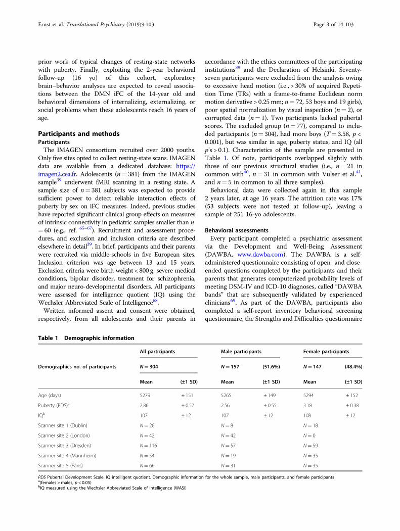
Table 1 Demographic information
All participants Male participants Female participants
Demographics no. of participants N= 304 N= 157 (51.6%) N= 147 (48.4%)
Mean (±1 SD) Mean (±1 SD) Mean (±1 SD)
Age (days) 5279 ± 151 5265 ± 149 5294 ± 152
Puberty (PDS)a 2.86 ± 0.57 2.56 ± 0.55 3.18 ± 0.38
IQb 107 ± 12 107 ± 12 108 ± 12
Scanner site 1 (Dublin) N= 26 N= 8 N= 18
Scanner site 2 (London) N= 42 N= 42 N= 0
Scanner site 3 (Dresden) N= 116 N= 57 N= 59
Scanner site 4 (Mannheim) N= 54 N= 19 N= 35
Scanner site 5 (Paris) N= 66 N= 31 N= 35
PDS Pubertal Development Scale, IQ intelligent quotient. Demographic information for the whole sample, male participants, and female participantsa(females > males, p < 0.05)bIQ measured using the Wechsler Abbreviated Scale of Intelligence (WASI)
Ernst et al. Translational Psychiatry (2019) 9:103 Page 3 of 14 103
(SDQ70), which gives a measure of severity of problemswithin internalizing, externalizing, and social behavioraldomains. Although the resting-state fMRI scanning onlyoccurred at age 14 years, the behavioral assessments wererepeated 2 years later, at age 16 years.
Pubertal statusPubertal status was assessed via self-report using the
Pubertal Development Scale (PDS)71. Previous researchhas demonstrated that the PDS exhibits good internalconsistency (median α= 0.77) and is highly correlatedwith physician ratings (Pearson’s R= 0.61)71,72.
BOLD fMRI data acquisition, preprocessing, and analysisMRI data were acquired at five sites using 3 T scanners:
Phillips (Dublin), General Electrics (London), and Sie-mens (Paris, Dresden, Mannheim). BOLD fMRI signalwas acquired across 40 interleaved slices using the fol-lowing parameters: TR= 2,200 ms; TE= 30 ms; flip angle= 75°; acquisition matrix= 64 × 64 × 40 with 2.4 mm slicethickness and 1mm slice gap yielding an acquisitionresolution 3.4 mm isotropic; 187 volumes collected over6.5 minutes. High-resolution anatomical images wereobtained using parameters based on the ADNI protocol,yielding a final voxel size of 1.1 × 1.1 × 1.1 mm.Preprocessing and analyses of BOLD fMRI data were
conducted using AFNI73. FreeSurfer version 5.374 wasemployed to segment the T1-weighted anatomical images.The i
first four volumes of the functional run were dis-carded to allow for steady-state equilibrium. Functionalvolumes were slice-time corrected, aligned, and co-registered to the participants’ corresponding anatomicalimage. Functional volumes were then normalized to theColin 27 Average Brain standardized template using3dQwarp, which is a nonlinear transformation, and spa-tially smoothed with a 6 mm Full-Width Half-Maximum(FWHM) Gaussian kernel. All coordinates are reported inthe Talairach and Tournoux system75.Acknowledging the debate as to whether global signal
should be regressed out of resting-state data sets, wedecided not to adopt this strategy. This decision wasbased on Saad et al.76 who suggest that global signalregression can introduce spurious correlations intoresting-state data sets, and advise against its use in pre-processing. In addition, we employed several strategies tominimize motion- and physiological-related variance,thus mitigating the need to apply additional measures, likeglobal signal regression, to minimize variance related tomotion and cardiac/respiratory processes (Power et al.77).The following nuisance signals were regressed from the
functional volumes: (1) six head motion parameters andtheir derivatives, (2) average time-series extracted from theventricles, (3) time-series from local white matter within a25-mm radius sphere surrounding each voxel using the
ANATICOR approach78, and (4) individual regressorscorresponding with a frame-to-frame Euclidean normmotion derivative ≥ 0.25mm, or volumes where ≥ 10% ofvoxels were determined to be outliers. This strategy fol-lowed the recommendations of Power et al.79.Three cortical regions of interest (ROIs) from the
DMN80 were selected as seeds. These three seeds wereretained because of their previously demonstrated asso-ciation with subthreshold elevated40, as well as depres-sed41 mood symptoms in two independent subsets of thecommunity cohort of 14 yo from the IMAGEN con-sortium (https://imagen.cea.fr39). MNI coordinates wereall converted to Talairach coordinates using the Yalemni2tal GUI (“MNI- Yale University” 2017). Specifically,cubic ROIs (3 mm × 3mm× 3mm) were created withinthe left pregenual ACC (lpgACC; Talairach: x=−12, y=36, z= 12), left medial PFC (lmPFC; Talairach: x=−2,y= 45, z= 16), and the left PCC (Talairach: x=−1, y=−47, z= 3481). The left side was selected because theregions identified in previous studies were on the left side.Generically, these cortical regions have been implicated inthe processing of salience and emotion encoding, parti-cularly in the context of social and self-referential infor-mation (mPFC)82, visual stimuli (PCC)83, and autonomicvisceral signals (pgACC)84.Subsequently, average time-series from seed ROIs were
extracted from the residualized functional images andwere used to calculate Pearson correlations between thetime-series from the seed ROIs and every voxel in thebrain. Resulting statistical images were Fisher-transformed for group analyses.ANCOVA models (3dMVM) were used to determine
the interaction of puberty-X-sex and the main effects ofeach factor85. All group-level ANCOVA analyses statisti-cally controlled for the effects of scanner site (Dublin,Dresden, London, Mannheim, and Paris), as well as age.Group-level analyses were limited to all gray matterregions as determined by the parcellation of the Colin 27Average Brain (i.e., CA_N27_ML atlas). Statisticalthresholding was calculated using 3dClustSim’s MonteCarlo simulation via updated versions of 3dFWHMx and3dClustSim to address the concerns of inflated falsepositive rates identified by Eklund86. These updatesincorporate a mixed autocorrelation function that bettermodels non-Gaussian noise structure87. The resultingmaps were thresholded to p < 0.005 two-tailed, k= 37,which represents a global cluster correction at p < 0.05.Finally, to examine more stringently potential effects ofgroup motion, we conducted an additional analysisincluding the additional covariate of individual averagemotion per TR. This analysis is presented in supplementalmaterial (Table S1).Average iFC values from clusters exhibiting a puberty-
X-sex interaction, or main effects of sex or puberty, were
Ernst et al. Translational Psychiatry (2019) 9:103 Page 4 of 14 103
extracted using 3dmaskave. Post hoc comparisons wereconducted within SPSS v24 and interactions were pro-bed using the PROCESS v21688 macro by calculating thebeta value for the relationship between puberty and iFCfor male and female participants, while controlling forage and scanner site. Levene’s test for equality of var-iances, and measures of skew and kurtosis are reportedin Supplemental Table S2. Importantly, all clustersexhibiting an interaction between puberty and sex arenormally distributed. Furthermore, although variance isnot equal between groups for all identified clusters,multiple regression analyses, which do not entailequality of variance assumptions, demonstrate thatidentified interactions remain significant within a mul-tiple regression framework (SupplementalTables S3–S4).
Brain–behavior analyses based on factorial approachTo reduce the number of variables, two sets of principal
components using SAS-9.4 program were extracted fromthe neural data at baseline (14 years old), and the beha-vioral data collected at 2-year follow-up (16 years old). Allthe iFC clusters significantly modulated by puberty and/orsex were entered into one factor analysis, and 15 items ofthe SDQ into a separate factor analysis. These factoranalyses used ones as prior communality estimates. Theprincipal axis method was employed to extract the com-ponents, and this was followed by a varimax (orthogonal)rotation.Only two DAWBA bands were retained for this analysis,
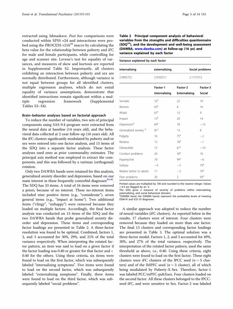
generalized anxiety disorder and depression, based on ourmain interest in these frequently comorbid diagnoses89,90.The SDQ has 33 items. A total of 16 items were removeda priori, because of no interest. These no-interest itemsincluded nine positive items (e.g., “considerate”), sevengeneral items (e.g., “impact at home”). Two additionalitems (“clingy”, “unhappy”) were removed because theyloaded on multiple factors. Accordingly, the final factoranalysis was conducted on 15 items of the SDQ and thetwo DAWBA bands that probe generalized anxiety dis-order and depression. These items and correspondingfactor loadings are presented in Table 2. A three-factorresolution was found to be optimal. Combined, factors 1,2, and 3 accounted for 30%, 29%, and 21% of the totalvariance respectively. When interpreting the rotated fac-tor pattern, an item was said to load on a given factor ifthe factor loading was 0.40 or greater for that factor and <0.40 for the others. Using these criteria, six items werefound to load on the first factor, which was subsequentlylabeled “internalizing symptoms”. Five items were foundto load on the second factor, which was subsequentlylabeled “externalizing symptoms”. Finally, three itemswere found to load on the third factor, which was sub-sequently labeled “social problems”.
A similar approach was adopted to reduce the numberof neural variables (iFC clusters). As reported below in theresults, 17 clusters were of interest. Four clusters wereremoved because they loaded on more than one factor.The final 13 clusters and corresponding factor loadingsare presented in Table 3. The optimal solution was athree-factor model. Factors 1, 2, and 3 accounted for 49%,30%, and 27% of the total variance, respectively. Theinterpretation of the rotated factor pattern, used the samethreshold as above, i.e., 0.40. Using these criteria, eightclusters were found to load on the first factor. These eightclusters were iFC clusters of the lPCC seed (n= 5 clus-ters) and of the lMPFC seed (n= 3 cluster), all of whichbeing modulated by Puberty-X-Sex. Therefore, factor-1was labeled PCC/mPFC-pubXsex. Four clusters loaded onthe second factor. All these clusters belonged to the lPCC-seed iFC, and were sensitive to Sex. Factor-2 was labeled
Table 2 Principal component analysis of behavioralvariables from the strengths and difficulties questionnaire(SDQ59), and the development and well-being assessment(DAWBA, www.dawba.com) at follow-up (16 yo) andvariance explained by each factor
Variance explained by each factor
Internalizing externalizing Social problems
2.9905752 2.9182511 2.1157313
Factor-1 Factor-2 Factor-3
Internalizing Externalizing Social
Somatic 52a 22 10
Worries 63a 8 16
Afraid 67a 13 9
Impact 72a 20 14
Depressionb 69a 18 −14
Generalized anxiety b 81a 13 8
Fidgety 16 75a −2
Restless 15 78a −1
Distractible 13 67a −10
Conduct problems 24 50a 16
Hyperactive 19 94a −6
Solitary −4 −3 79a
Relates better to adults 17 −3 70a
Peer problems 20 2 93a
Printed values are multiplied by 100 and rounded to the nearest integer. Values> 0.4 are flagged by an ‘a’The SDQ gives a measure of severity of problems within internalizing,externalizing, and social behavioral domainsbDAWBA band: the DAWBA bands represent the probability levels of meetingDSM-IV and ICD-10 diagnoses
Ernst et al. Translational Psychiatry (2019) 9:103 Page 5 of 14 103
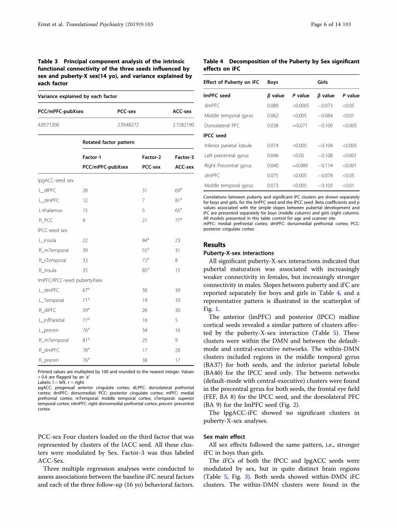
PCC-sex Four clusters loaded on the third factor that wasrepresented by clusters of the lACC seed. All these clus-ters were modulated by Sex. Factor-3 was thus labeledACC-Sex.Three multiple regression analyses were conducted to
assess associations between the baseline iFC neural factorsand each of the three follow-up (16 yo) behavioral factors.
ResultsPuberty-X-sex interactionsAll significant puberty-X-sex interactions indicated that
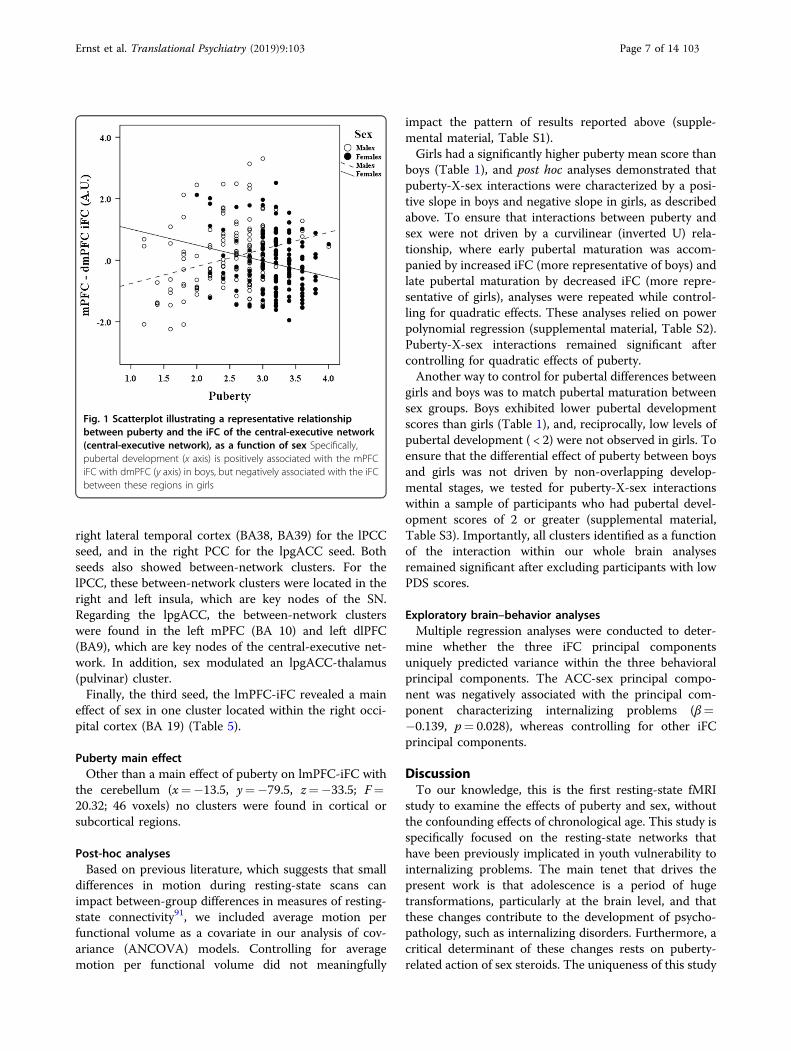
pubertal maturation was associated with increasinglyweaker connectivity in females, but increasingly strongerconnectivity in males. Slopes between puberty and iFC arereported separately for boys and girls in Table 4, and arepresentative pattern is illustrated in the scatterplot ofFig. 1.The anterior (lmPFC) and posterior (lPCC) midline
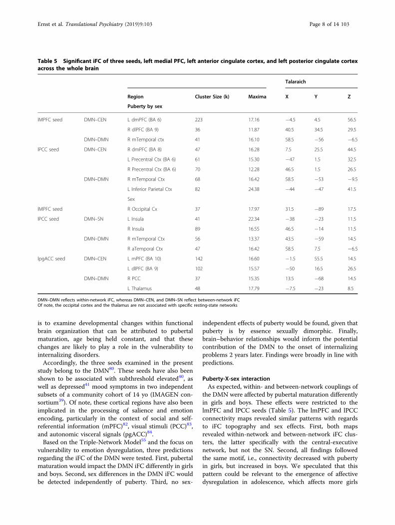
cortical seeds revealed a similar pattern of clusters affec-ted by the puberty-X-sex interaction (Table 5). Theseclusters were within the DMN and between the default-mode and central-executive networks. The within-DMNclusters included regions in the middle temporal gyrus(BA37) for both seeds, and the inferior parietal lobule(BA40) for the lPCC seed only. The between networks(default-mode with central-executive) clusters were foundin the precentral gyrus for both seeds, the frontal eye field(FEF, BA 8) for the lPCC seed, and the dorsolateral PFC(BA 9) for the lmPFC seed (Fig. 2).The lpgACC-iFC showed no significant clusters in
puberty-X-sex analyses.
Sex main effectAll sex effects followed the same pattern, i.e., stronger
iFC in boys than girls.The iFCs of both the lPCC and lpgACC seeds were
modulated by sex, but in quite distinct brain regions(Table 5, Fig. 3). Both seeds showed within-DMN iFCclusters. The within-DMN clusters were found in the
Table 3 Principal component analysis of the intrinsicfunctional connectivity of the three seeds influenced bysex and puberty-X sex(14 yo), and variance explained byeach factor
Variance explained by each factor
PCC/mPFC-pubXsex PCC-sex ACC-sex
4.8571200 2.9548272 2.7282190
Rotated factor pattern
Factor-1 Factor-2 Factor-3
PCC/mPFC-pubXsex PCC-sex ACC-sex
lpgACC-seed sex
L_dlPFC 28 31 69a
L_dmPFC 12 7 81a
L-thalamus 15 5 65a
R_PCC 9 21 77a
lPCC-seed sex
L_insula 22 84a 23
R_mTemporal 39 55a 31
R_sTemporal 33 72a 8
R_insula 35 85a 15
lmPFC/lPCC-seed pubertyXsex
L_dmPFC 67a 30 39
L_Temporal 71a 19 10
R_dlPFC 59a 26 30
L_infParietal 77a 16 5
L_precen 76a 34 16
R_mTemporal 81a 25 9
R_dmPFC 78a 17 28
R_precen 76a 38 17
Printed values are multiplied by 100 and rounded to the nearest integer. Values> 0.4 are flagged by an ‘a’Labels: l= left, r= rightpgACC: pregenual anterior cingulate cortex; dLPFC: dorsolateral prefrontalcortex; dmPFC: dorsomedial; PCC: posterior cingulate cortex; mPFC: medialprefrontal cortex; mTemporal: middle temporal cortex; sTemporal: superiortemporal cortex; rdmPFC: right dorsomedial prefrontal cortex; precen: precentralcortex
Table 4 Decomposition of the Puberty by Sex significanteffects on iFC
Effect of Puberty on iFC Boys Girls
lmPFC seed β value P value β value P value
dmPFC 0.080 <0.0005 −0.073 <0.05
Middle temporal gyrus 0.062 <0.005 −0.084 <0.01
Dorsolateral PFC 0.038 =0.071 −0.100 <0.005
lPCC seed
Inferior parietal lobule 0.074 <0.005 −0.104 <0.005
Left precentral gyrus 0.046 <0.05 −0.108 <0.001
Right Precentral gyrus 0.040 =0.089 −0.114 <0.001
dmPFC 0.075 <0.005 −0.078 <0.05
Middle temporal gyrus 0.073 <0.005 −0.103 <0.01
Correlations between puberty and significant iFC clusters are shown separatelyfor boys and girls, for the lmPFC seed and the lPCC seed. Beta coefficients and pvalues associated with the simple slopes between pubertal development andiFC are presented separately for boys (middle column) and girls (right column).All models presented in this table control for age and scanner sitemPFC: medial prefrontal cortex; dmPFC: dorsomedial prefrontal cortex; PCC:posterior cingulate cortex
Ernst et al. Translational Psychiatry (2019) 9:103 Page 6 of 14 103
right lateral temporal cortex (BA38, BA39) for the lPCCseed, and in the right PCC for the lpgACC seed. Bothseeds also showed between-network clusters. For thelPCC, these between-network clusters were located in theright and left insula, which are key nodes of the SN.Regarding the lpgACC, the between-network clusterswere found in the left mPFC (BA 10) and left dlPFC(BA9), which are key nodes of the central-executive net-work. In addition, sex modulated an lpgACC-thalamus(pulvinar) cluster.Finally, the third seed, the lmPFC-iFC revealed a main
effect of sex in one cluster located within the right occi-pital cortex (BA 19) (Table 5).
Puberty main effectOther than a main effect of puberty on lmPFC-iFC with
the cerebellum (x=−13.5, y=−79.5, z=−33.5; F=20.32; 46 voxels) no clusters were found in cortical orsubcortical regions.
Post-hoc analysesBased on previous literature, which suggests that small
differences in motion during resting-state scans canimpact between-group differences in measures of resting-state connectivity91, we included average motion perfunctional volume as a covariate in our analysis of cov-ariance (ANCOVA) models. Controlling for averagemotion per functional volume did not meaningfully
impact the pattern of results reported above (supple-mental material, Table S1).Girls had a significantly higher puberty mean score than
boys (Table 1), and post hoc analyses demonstrated thatpuberty-X-sex interactions were characterized by a posi-tive slope in boys and negative slope in girls, as describedabove. To ensure that interactions between puberty andsex were not driven by a curvilinear (inverted U) rela-tionship, where early pubertal maturation was accom-panied by increased iFC (more representative of boys) andlate pubertal maturation by decreased iFC (more repre-sentative of girls), analyses were repeated while control-ling for quadratic effects. These analyses relied on powerpolynomial regression (supplemental material, Table S2).Puberty-X-sex interactions remained significant aftercontrolling for quadratic effects of puberty.Another way to control for pubertal differences between
girls and boys was to match pubertal maturation betweensex groups. Boys exhibited lower pubertal developmentscores than girls (Table 1), and, reciprocally, low levels ofpubertal development ( < 2) were not observed in girls. Toensure that the differential effect of puberty between boysand girls was not driven by non-overlapping develop-mental stages, we tested for puberty-X-sex interactionswithin a sample of participants who had pubertal devel-opment scores of 2 or greater (supplemental material,Table S3). Importantly, all clusters identified as a functionof the interaction within our whole brain analysesremained significant after excluding participants with lowPDS scores.
Exploratory brain–behavior analysesMultiple regression analyses were conducted to deter-
mine whether the three iFC principal componentsuniquely predicted variance within the three behavioralprincipal components. The ACC-sex principal compo-nent was negatively associated with the principal com-ponent characterizing internalizing problems (β=−0.139, p= 0.028), whereas controlling for other iFCprincipal components.
DiscussionTo our knowledge, this is the first resting-state fMRI
study to examine the effects of puberty and sex, withoutthe confounding effects of chronological age. This study isspecifically focused on the resting-state networks thathave been previously implicated in youth vulnerability tointernalizing problems. The main tenet that drives thepresent work is that adolescence is a period of hugetransformations, particularly at the brain level, and thatthese changes contribute to the development of psycho-pathology, such as internalizing disorders. Furthermore, acritical determinant of these changes rests on puberty-related action of sex steroids. The uniqueness of this study
Fig. 1 Scatterplot illustrating a representative relationshipbetween puberty and the iFC of the central-executive network(central-executive network), as a function of sex Specifically,pubertal development (x axis) is positively associated with the mPFCiFC with dmPFC (y axis) in boys, but negatively associated with the iFCbetween these regions in girls
Ernst et al. Translational Psychiatry (2019) 9:103 Page 7 of 14 103
is to examine developmental changes within functionalbrain organization that can be attributed to pubertalmaturation, age being held constant, and that thesechanges are likely to play a role in the vulnerability tointernalizing disorders.Accordingly, the three seeds examined in the present
study belong to the DMN80. These seeds have also beenshown to be associated with subthreshold elevated40, aswell as depressed41 mood symptoms in two independentsubsets of a community cohort of 14 yo (IMAGEN con-sortium39). Of note, these cortical regions have also beenimplicated in the processing of salience and emotionencoding, particularly in the context of social and self-referential information (mPFC)82, visual stimuli (PCC)83,and autonomic visceral signals (pgACC)84.Based on the Triple-Network Model55 and the focus on
vulnerability to emotion dysregulation, three predictionsregarding the iFC of the DMN were tested. First, pubertalmaturation would impact the DMN iFC differently in girlsand boys. Second, sex differences in the DMN iFC wouldbe detected independently of puberty. Third, no sex-
independent effects of puberty would be found, given thatpuberty is by essence sexually dimorphic. Finally,brain–behavior relationships would inform the potentialcontribution of the DMN to the onset of internalizingproblems 2 years later. Findings were broadly in line withpredictions.
Puberty-X-sex interactionAs expected, within- and between-network couplings of
the DMN were affected by pubertal maturation differentlyin girls and boys. These effects were restricted to thelmPFC and lPCC seeds (Table 5). The lmPFC and lPCCconnectivity maps revealed similar patterns with regardsto iFC topography and sex effects. First, both mapsrevealed within-network and between-network iFC clus-ters, the latter specifically with the central-executivenetwork, but not the SN. Second, all findings followedthe same motif, i.e., connectivity decreased with pubertyin girls, but increased in boys. We speculated that thispattern could be relevant to the emergence of affectivedysregulation in adolescence, which affects more girls
Table 5 Significant iFC of three seeds, left medial PFC, left anterior cingulate cortex, and left posterior cingulate cortexacross the whole brain
Talaraich
Region Cluster Size (k) Maxima X Y Z
Puberty by sex
lMPFC seed DMN–CEN L dmPFC (BA 6) 223 17.16 −4.5 4.5 56.5
R dlPFC (BA 9) 36 11.87 40.5 34.5 29.5
DMN–DMN R mTemporal ctx 41 16.10 58.5 −56 −6.5
lPCC seed DMN–CEN R dmPFC (BA 8) 47 16.28 7.5 25.5 44.5
L Precentral Ctx (BA 6) 61 15.30 −47 1.5 32.5
R Precentral Ctx (BA 6) 70 12.28 46.5 1.5 26.5
DMN–DMN R mTemporal Ctx 68 16.42 58.5 −53 −9.5
L Inferior Parietal Ctx 82 24.38 −44 −47 41.5
Sex
lMPFC seed R Occipital Cx 37 17.97 31.5 −89 17.5
lPCC seed DMN–SN L Insula 41 22.34 −38 −23 11.5
R Insula 89 16.55 46.5 −14 11.5
DMN–DMN R mTemporal Ctx 56 13.37 43.5 −59 14.5
R aTemporal Ctx 47 16.42 58.5 7.5 −6.5
lpgACC seed DMN–CEN L mPFC (BA 10) 142 16.60 −1.5 55.5 14.5
L dlPFC (BA 9) 102 15.57 −50 16.5 26.5
DMN–DMN R PCC 37 15.35 13.5 −68 14.5
L Thalamus 48 17.79 −7.5 −23 8.5
DMN–DMN reflects within-network iFC, whereas DMN–CEN, and DMN–SN reflect between-network iFCOf note, the occipital cortex and the thalamus are not associated with specific resting-state networks
Ernst et al. Translational Psychiatry (2019) 9:103 Page 8 of 14 103
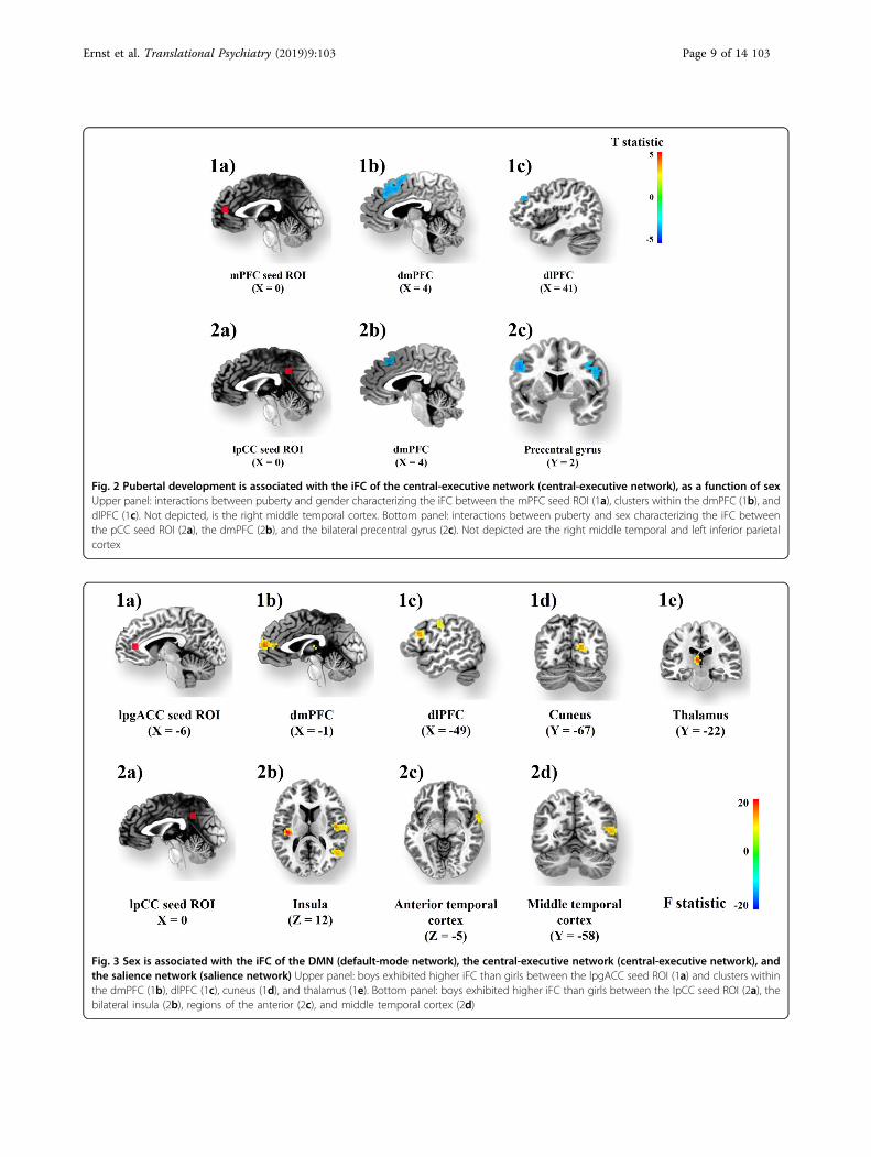
Fig. 2 Pubertal development is associated with the iFC of the central-executive network (central-executive network), as a function of sexUpper panel: interactions between puberty and gender characterizing the iFC between the mPFC seed ROI (1a), clusters within the dmPFC (1b), anddlPFC (1c). Not depicted, is the right middle temporal cortex. Bottom panel: interactions between puberty and sex characterizing the iFC betweenthe pCC seed ROI (2a), the dmPFC (2b), and the bilateral precentral gyrus (2c). Not depicted are the right middle temporal and left inferior parietalcortex
Fig. 3 Sex is associated with the iFC of the DMN (default-mode network), the central-executive network (central-executive network), andthe salience network (salience network) Upper panel: boys exhibited higher iFC than girls between the lpgACC seed ROI (1a) and clusters withinthe dmPFC (1b), dlPFC (1c), cuneus (1d), and thalamus (1e). Bottom panel: boys exhibited higher iFC than girls between the lpCC seed ROI (2a), thebilateral insula (2b), regions of the anterior (2c), and middle temporal cortex (2d)
Ernst et al. Translational Psychiatry (2019) 9:103 Page 9 of 14 103
than boys. Accordingly, the observed sexually dimorphicmaturational changes of the DMN might reflect changesin adaptive emotion regulation with puberty. The direc-tion of these changes might be protective in boys, orreflect vulnerability in girls for mood dysregulation.In support of this thesis, the exploratory brain–behavior
analysis across the whole sample revealed a negativeassociation between the spgACC iFC component andinternalizing symptoms 2 years later: The lower theconnectivity at 14 yo, the more severe were internalizingproblems at 16 yo. This relationship suggests that puberty,which is accompanied by decreased iFC in girls, mightamplify risk for internalizing symptoms in girls. Findingsfrom the literature using clinical samples or individuals atrisk for internalizing disorders could inform this inter-pretation. Unfortunately, results are inconsistent, in termsof topology, direction of group differences, methodology,and sample characteristics. A number of studies inpediatric samples have reported that internalizing symp-toms were associated with lower iFC in core networks. Forexample, Frost-Bellgowan et al. (2015)92 reported lowerDMN-iFC (with insula, ventral striatum, pre/post centralgyrus) in behavioral inhibited (at risk for internalizingdisorders) vs. healthy children (8–17 yo), and showed thatgirls (not boys) exhibited a negative correlation ofinternalizing-symptom severity with DMN-iFC. Anotherstudy of 8–12-yo children also showed that patients withdepression/anxiety (vs. healthy peers) had similarly loweriFC, but this time in the ventral attention network, whichwas the exclusive focus of this work66. In contrast, arelatively large study in a community sample of 112children (53 boys, 59 girls; mean age= 11.5 yo)93 focusedon the three networks of the triple-network model55 inrelation to internalizing symptoms (i.e., anxiety anddepression, and rumination). Findings revealed no sexdifferences in the iFCs of the DMN, SN, or CEN. How-ever, brain–behavior correlations identified a positivecorrelation between iFC within the SN and the levels ofinternalizing symptoms in girls, but not in boys. Specifi-cally, higher SN-iFC predicted more severe symptoms ingirls. In summary, this study93 reported a differentialeffect of sex on the association of SN-iFC with inter-nalizing symptoms, affecting only girls. Compared withour current study, this brain–behavior relationship93
concerned a different network (SN) and was in theopposite direction to our findings (greater iFC with highersymptoms, while we showed lower iFC with highersymptoms). Furthermore, it is difficult to compare theseresults with the present findings, because of the differ-ences in methodology (independent component analysis),and of the significantly younger sample (11yo). Collec-tively, these pediatric studies concur on the associationbetween dysfunction and internalizing symptoms, but thedirection of this dysfunction is inconsistent, as is the
specific couplings. In addition, the two studies probing theeffect of sex revealed that brain–behavior associationsconcerned mainly girls. This observation supports ourinterpretation that the puberty-related reduction in theDMN iFC in girls might carry vulnerability for inter-nalizing symptoms.Negative findings regarding the distinct iFC modulation
by puberty in girls and boys are notable. The lack of sexmodulation of the effects of puberty on the lpgACC seedconnectivity, and the lack of implication of the SN (par-ticularly the insula) and the amygdala were surprising.Indeed, their established role in the coding of salience andemotional responses (e.g., ref. 94,95, would be expected tocontribute to the increased emotional intensity and labilityin adolescence (e.g., ref. 60,64,96). These negative findingssuggest that the iFC of the nodes/circuits underlyingemotion/motivation processes do mature similarly in boysand girls. Although task-based fMRI studies have reportedpuberty-related changes of these structures in response toemotion/motivation probes (e.g., ref. 20,22,24), how sexinfluences these trajectories has not been reported.
Sex main effectIn fact, the present findings reveal sex effects, inde-
pendent of puberty, in the spgACC iFC and SN (insula).In all cases, connectivity was higher in boys than girls.These sex effects concerned mainly two of the three seeds,the lPCC and lpgACC.From the perspective of the Triple-Network Model, sex
impacted differently the between-network connectivity ofthe anterior (spgACC seed) and posterior (lPCC seed)DMN components (Table 5) (anterior vs. posterior hubsof the DMN97). Sex influenced the connectivity of theanterior DMN seed (spgACC) with the SN, and theconnectivity of the posterior seed (lPCC) with the central-executive network. In line with this distinction, as dis-cussed above, the anterior spgACC connectivity predictedinternalizing symptoms 2 years later (age 16 years),whereas the posterior lPCC component was not asso-ciated with any of the behavioral components.These differential effects of sex on the anterior and
posterior DMN iFC need to be examined more closely inthe context of the modulation of the Triple-NetworkModel, and might suggest a refinement of this model byconsidering regional functional specialization within theDMN, i.e., the anterior and posterior aspects. Accordingly,they suggest that the modulation by sex and pubertyaffects differently the anterior and posterior DMN com-ponents98, which likely play distinct roles in vulnerabilityto emotion dysregulation.
LimitationsThese findings are not without limitations. First, pub-
erty was measured by self-report (PDS71), without
Ernst et al. Translational Psychiatry (2019) 9:103 Page 10 of 14 103
physical examination or hormonal assays. However, thePDS has been shown to have good psychometric prop-erties and convergent validity from self- and physician-rated Tanner stages71,72. Second, as girls and boys were ofsimilar age, the average pubertal level was higher in girls.Two different control analyses were conducted to validatethe findings. First, the youngest boys were removed fromanalysis to equalize the puberty mean in girls and boys.Findings with these control analyses revealed that thepattern of results remained pretty much unchanged(supplemental material, Table S3). Second, we conductedmultiple regression analyses, which included quadraticterms for puberty across the whole sample. This approachexplored whether the effects of puberty were indeedcharacterized by two separate and opposite lines ofregression in girls and boys, while controlling for aninverse-U-curve pattern of pubertal maturation. Resultsshowed that the puberty-X-sex interaction was still sig-nificant (supplemental material, Table S2). Therefore, weare confident in the findings of different pubertal trajec-tories in boys and girls. Third, this study was cross-sec-tional, which is sub-optimal for the study ofdevelopmental changes. However, this limitation isleveraged by the relatively large sample size, and also thehomogeneous age that allowed us to dissociate the effectsof chronological changes from those specific to puberty.Fourth, this study focused on extending earlier brainstructural findings of neural risk for mood problems in 14yo. Accordingly, only three seeds were investigated, whichconstrained the yield of this study. However, thisapproach is specific to a behavioral domain that, we hope,will foster the formulation of models of vulnerability tointernalizing problems to be tested in the future.
ConclusionsThis study reveals that pubertal maturation influences
iFC differently in boys and girls, and that sex can impactiFC independently of puberty. First, because mood dys-regulation has its peak onset in adolescence, duringpubertal maturation, it is reasonable to consider a role ofpuberty in the rise of incidence rate of mood symptoms.Second, because mood dysregulation, particularlydepression and anxiety, occurs disproportionally in girls,sex is expected to uniquely modulate circuits of emotionregulation. For these reasons, analyses were focused onthree regions previously identified as conferring risk formood problems in adolescents. These three regions hap-pened to belong to the DMN, which is recognized to beperturbed in pathological mood disturbances. Findingsrevealed that both puberty-X-sex interaction and sexmain effects modulate within-network and between-network clusters of the DMN iFC. Notably, effects ofpuberty did not involve the amygdala or the SN, sug-gesting the notion that pubertal maturation might not
significantly affect the iFC of these key centers of emotionprocesses. This is in contrast to the effect of sex, whichdid impact DMN–SN iFC. Tentatively, stronger iFC inboys might suggest tighter emotional control, alsopotentially serving a protective role against emotion dys-regulation. Finally, the spgACC iFC significantly predictedinternalizing symptoms 2 years later, supporting theassociation of this network with mood dysregulation.
AcknowledgementsDr Stringaris receives royalties from the University of Cambridge for his book.Dr. Banaschewski served in an advisory or consultancy role for Actelion, HexalPharma, Lilly, Medice, Novartis, Oxford outcomes, PCM scientific, Shire, andViforpharma. He received conference support or speaker’s fee by Medice,Novartis, and Shire. He is/has been involved in clinical trials conducted by Shire& Viforpharma. He received royalities from Hogrefe, Kohlhammer, CIP Medien,Oxford University Press. The present work is unrelated to the above grants andrelationships. This work received support from the following funding sources:the European Union-funded FP6 Integrated Project IMAGEN (Reinforcement-related behavior in normal brain function and psychopathology) (LSHM-CT-2007-037286), the Agence Nationale de la Recherche ANR (project AF12-NEUR0008-01—WM2NA, and ANR-12-SAMA-0004), the Fondation de France(grants 2012-00033703 and 2017—0008L242), the Fondation pour laRecherche Médicale (grants DPA 20140229802 and DPP20151033945), theFédération pour la Recherche sur le Cerveau (AAP 2014), the MissionInterministérielle de Lutte-contre-les-Drogues-et-les-Conduites-Addictives(MILDECA), and the INSERM (interface grant to the the Assistance-Publique-Hôpitaux-de-Paris); The Academy of Finland (grant number 276612 to A.S.U);the Emil Aaltonen Foundation (grant to A.S.U.); the Jalmari and Rauha AhokasFoundation (grant to A.S.U.). Further support to the IMAGEN centers in UK andGermany was provided by the Horizon 2020 funded ERC Advanced Grant‘STRATIFY’ (Brain network based stratification of reinforcement-relateddisorders) (695313), ERANID (Understanding the Interplay between Cultural,Biological, and Subjective Factors in Drug Use Pathways) (PR-ST-0416-10004),BRIDGET (JPND: BRain Imaging, cognition Dementia and next generationGEnomics) (MR/N027558/1), the FP7 projects IMAGEMEND(602450; IMAgingGEnetics for MENtal Disorders), and MATRICS (603016), the InnovativeMedicine Initiative Project EU-AIMS (115300-2), the Medical Research CouncilGrant ‘c-VEDA’ (Consortium on Vulnerability to Externalizing Disorders andAddictions) (MR/N000390/1), the Swedish Research Council FORMAS, theMedical Research Council, the National Institute for Health Research (NIHR)Biomedical Research Centre at South London and Maudsley NHS FoundationTrust and King’s College London, the Bundesministeriumfür Bildung undForschung (BMBF grants 01GS08152; 01EV0711; eMED SysAlc01ZX1311A;Forschungsnetz AERIAL 01EE1406A, 01EE1406B), the DeutscheForschungsgemeinschaft (DFG grants SM 80/7–2, SFB 940/2), the MedicalResearch Foundation and Medical research council (grant MR/R00465X/1).Further support was provided by grants from: the National Institutes of Health,Science Foundation Ireland (16/ERCD/3797), USA (Axon, Testosterone, andMental Health during Adolescence; RO1 MH085772-01A1), and by NIHConsortium grant U54 EB020403, supported by a cross-NIH alliance that fundsBig Data to Knowledge Centres of Excellence. This work was supported by theEuropean Union-funded FP6 Integrated Project IMAGEN (LSHM-CT-2007-037286); the FP7 project IMAGEMEND (602450); the Innovative MedicineInitiative Project European Autism Interventions—A Multicentre Study forDeveloping New Medications (EU-AIMS) (115300-2); a Medical ResearchCouncil Programme (93558); the Swedish funding agency Formas; theWellcome Trust (University of Cambridge); the National Institute for HealthResearch (NIHR) Biomedical Research Centre at South London and MaudsleyNHS Foundation Trust and King’s College London; the Department of HealthUnited Kingdom; the Bundesministerium für Bildung und Forschung (BMBF)(01GS08152, 01EV0711, eMED SysAlc01ZX1311A, Forschungsnetz AERIAL); theFrench funding agency Agence Nationale de la Recherche (ANR) (ANR-12-SAMA-0004); Eranet—Neuron (AF12-NEUR0008-01—WM2NA); the Assistance-Publique-Hôpitaux-de-Paris and Insitut national de la santé et de la recherchémédicale (INSERM, interface grant); Paris-Descartes-University (collaborative-project-2010); Paris-Sud-University (IDEX-2012); the Fondation de France; theFondation pour la Recherche Médicale (DPA20140629802); the MissionInterministérielle de Lutte-contre-les-Drogues-et-les-Conduites-Addictives
Ernst et al. Translational Psychiatry (2019) 9:103 Page 11 of 14 103
(MILDECA); and the Intramural Research Program of the National Institute ofMental Health (ZIAMH002798).
Other Imagen Consortium collaborators (www.imagen-europe.com):J. Dalley, N. Subramaniam, D. Theobald, C. Bach, G. J. Barker, M. Fauth-Bühler, S.Millenet, R. Spanagel, L. Albrecht, N. Ivanov, M. Rapp, J. Reuter, N. Strache, A.Ströhle, J. B. Poline, Y. Schwartz, B. Thyreau, J. Ireland, J. Rogers, N. Bordas, Z.Bricaud, I. Filippi, A. Galinowski, F. Gollier-Briant, D. Hall, S. Havatzias, T. Jia, C.Mallik, C. Nymberg, B. Ruggeri, L. Smith, K. Stueber, L. Topper, H. Werts, R. BrühlR. A. Ihlenfeld, B. Walaszek, T. Hübner, K. Müller, T. Paus, S. Ripke, E. Mennigen, D.Schmidt, N. C. Vetter, V. Ziesch, D. Carter, C. Connolly, S. Nugent, J. Jones, J.Yacubian, S. Schneider, K. Head, N. Heym, C. Newman, Z. Pausova, A.Tahmasebi, D. Stephens.
Author details1NIMH/NIH, Bethesda, MD, USA. 2INSERM, UMR 1000, Research unit“Neuroimaging and Psychiatry”, DIGITEO Labs, University Paris-Saclay, andUniversity Paris Descartes, Gif sur Yvette, France. 3INSERM, UMR 1000, Facultéde médecine, University Paris-Saclay, DIGITEO Labs, Gif sur Yvette, France.4University Paris Descartes, Paris, France. 5Center for Neuroimaging Research(CENIR), Brain & Spine Institute, Paris, France. 6Psychiatry Department 91G16,Orsay Hospital, Paris, France. 7Department of Child and Adolescent Psychiatryand Psychotherapy, Central Institute of Mental Health, Medical FacultyMannheim, Heidelberg University, Mannheim, Germany. 8Discipline ofPsychiatry, School of Medicine and Trinity College Institute of Neurosciences,Trinity College, Dublin, Ireland. 9University Medical Centre Hamburg-Eppendorf, House W34, 3.OG, Hamburg, Germany. 10Physikalisch-TechnischeBundesanstalt, Abbestr. 2 - 12, Berlin, Germany. 11Medical Research Council -Social, Genetic and Developmental Psychiatry Centre, Institute of Psychiatry,Psychology & Neuroscience, King’s College London, London, United Kingdom.12Department of Psychological Medicine and Psychiatry, Institute of Psychiatry,Psychology & Neuroscience, King’s College London, London, United Kingdom.13Department of Psychiatry, Université de Montréal, CHU Ste Justine Hospital,Montréal, QC, Canada. 14Department of Psychology, School of Social Sciences,University of Mannheim, 68131 Mannheim, Germany. 15Neurospin,Commissariat à l’Energie Atomique, CEA-Saclay Center, Saclay, France.16Department of Psychiatry and Psychotherapy, University Medical CenterHamburg-Eppendorf, Martinistr. 52, 20246 Hamburg, Germany. 17Departmentsof Psychiatry and Psychology, University of Vermont, 05405 Burlington, VT,USA. 18Sir Peter Mansfield Imaging Centre School of Physics and Astronomy,University of Nottingham, University Park, Nottingham, United Kingdom.19Department of Psychiatry and Psychotherapy, Campus CharitéMitte, Charité-Universitätsmedizin Berlin, Charitéplatz 1, Berlin, Germany. 20Department ofChild and Adolescent Psychiatry Psychosomatics and Psychotherapy, CampusCharitéMitte, Charité-Universitätsmedizin Berlin, Charitéplatz 1, Berlin, Germany.21Department of Social and Health Care, Psychosocial Services AdolescentOutpatient Clinic, University of Tampere, Kauppakatu 14, Lahti, Finland.22Department of Child and Adolescent Psychiatry and Psychotherapy, MedicalUniversity of Vienna, Vienna, Austria. 23Department of Psychiatry andNeuroimaging Center, Technische Universität Dresden, Dresden, Germany.24Department of Child and Adolescent Psychiatry, Institute of Psychiatry,Psychology & Neuroscience, King’s College London, London, United Kingdom.25Department of Psychology, University College, Dublin, Ireland. 26AP-HP,Department of Child and Adolescent Psychiatry, Pitié-Salpêtrière Hospital, Paris,France. 27Sorbonne Universités, Paris, France
Conflict of interestAll the other authors declare no biomedical financial interests or potentialconflict of interest.
Publisher’s noteSpringer Nature remains neutral with regard to jurisdictional claims inpublished maps and institutional affiliations.
Supplementary information accompanies this paper at (https://doi.org/10.1038/s41398-019-0433-6).
Received: 3 May 2018 Revised: 11 October 2018 Accepted: 1 January 2019
References1. Blakemore, S. J., Burnett, S. & Dahl, R. E. The role of puberty in the developing
adolescent brain. Hum. Brain Mapp. 31, 926–933 (2010).2. Bale, T. L. & Epperson, C. N. Sex as a biological variable: who, what, when, why
and How. Neuropsychopharmacology 42, 386–396 (2017).3. Forbes, E. E. & Dahl, R. E. Pubertal development and behavior: Hormonal
activation of social and motivational tendencies. Brain Cogn. 72, 66–72 (2010).4. Byrne, M. L. et al. A systematic review of adrenarche as a sensitive period in
neurobiological development and mental health. Dev. Cogn. Neurosci. 25,12–28 (2017).
5. Kessler, R. C. et al. Lifetime prevalence and age-of-onset distributions of DSM-IV disorders in the National Comorbidity Survey Replication. Arch. Gen. Psy-chiatry 62, 593–602 (2005).
6. Kessler, R. C., Petukhova, M., Sampson, N. A., Zaslavsky, A. M. & Wittchen, H. U.Twelve-month and lifetime prevalence and lifetime morbid risk of anxiety andmood disorders in the United States. Int J. Methods Psychiatr. Res. 21, 169–184(2012).
7. Whittle, S. et al. Associations between early adrenarche, affective brain func-tion and mental health in children. Soc. Cogn. Affect Neurosci. 10, 1282–1290(2015).
8. Casey, B. J., Soliman, F., Bath, K. G. & Glatt, C. E. Imaging genetics and devel-opment: challenges and promises. Hum. Brain Mapp. 31, 838–851 (2010).
9. Casey, B. J., Duhoux, S. & Malter Cohen, M. Adolescence: what do transmission,transition, and translation have to do with it? Neuron 67, 749–760 (2010).
10. Giedd, J. N. et al. Development of the human corpus callosum duringchildhood and adolescence: a longitudinal MRI study. Progress euro-Psychopharmacol. Biol. Psychiatry 23, 571–588 (1999).
11. Matsuzawa, J. et al. Age-related volumetric changes of brain gray and whitematter in healthy infants and children. Cereb. Cortex 11, 335–342 (2001).
12. Shaw, P. et al. Neurodevelopmental trajectories of the human cerebral cortex.J. Neurosci. 28, 3586–3594 (2008).
13. Jung, R. E. & Haier, R. J. The Parieto-Frontal Integration Theory (P-FIT) ofintelligence: converging neuroimaging evidence. Behav. rain Sci. 30, 135–154(2007). discussion154-187.
14. Deary, I. J., Penke, L. & Johnson, W. The neuroscience of human intelligencedifferences. Nat. Rev. Neurosci. 11, 201–211 (2010).
15. De Bellis, M. D. et al. Sex differences in brain maturation during childhood andadolescence. Cereb. Cortex 11, 552–557 (2001).
16. Goldstein, J. M. et al. Normal sexual dimorphism of the adult human brainassessed by in vivo magnetic resonance imaging. Cereb. Cortex 11, 490–497(2001).
17. Lenroot, R. K. & Giedd, J. N. Sex differences in the adolescent brain. Brain Cogn.72, 46–55 (2010).
18. Nota, N. M. et al. Brain functional connectivity patterns in children and ado-lescents with gender dysphoria: Sex-atypical or not? Psychoneuroendocrinology86, 187–195 (2017).
19. Giedd, J. N. et al. Puberty-related influences on brain development. Mol. CellEndocrinol. 254-255, 154–162 (2006).
20. Forbes, E. E., Phillips, M. L., Silk, J. S., Ryan, N. D. & Dahl, R. E. Neural systems ofthreat processing in adolescents: role of pubertal maturation and relation tomeasures of negative affect. Dev. Neuropsychol. 36, 429–452 (2011).
21. Klapwijk, E. T. et al. Increased functional connectivity with puberty in thementalising network involved in social emotion processing. Horm. Behav. 64,314–322 (2013).
22. Moore, W. E. 3rd et al. Facing puberty: associations between pubertal devel-opment and neural responses to affective facial displays. Soc. Cogn. AffectNeurosci. 7, 35–43 (2012).
23. Alarcon, G., Cservenka, A., Fair, D. A. & Nagel, B. J. Sex differences in the neuralsubstrates of spatial working memory during adolescence are not mediatedby endogenous testosterone. Brain Res. 1593, 40–54 (2014).
24. Goddings, A. L., Burnett Heyes, S., Bird, G., Viner, R. M. & Blakemore, S. J. Therelationship between puberty and social emotion processing. Dev. Sci. 15,801–811 (2012).
25. Satterthwaite, T. D. et al. Impact of puberty on the evolution of cerebralperfusion during adolescence. Proc. Natl Acad. Sci. USA 111, 8643–8648(2014).
Ernst et al. Translational Psychiatry (2019) 9:103 Page 12 of 14 103

26. Cservenka, A., Stroup, M. L., Etkin, A. & Nagel, B. J. The effects of age, sex, andhormones on emotional conflict-related brain response during adolescence.Brain Cogn. 99, 135–150 (2015).
27. Vijayakumar, N., Op de Macks, Z., Shirtcliff, E. A. & Pfeifer, J. H. Puberty and thehuman brain: Insights into adolescent development. Neurosci. Biobehav Rev.92, 417–436 (2018).
28. Satterthwaite, T. D. et al. Neuroimaging of the Philadelphia neurodevelop-mental cohort. Neuroimage 86, 544–553 (2014).
29. Dennison, M. et al. Mapping subcortical brain maturation during adolescence:evidence of hemisphere- and sex-specific longitudinal changes. Dev. Sci. 16,772–791 (2013).
30. Vijayakumar, N. et al. Brain development during adolescence: a mixed-longitudinal investigation of cortical thickness, surface area, and volume. Hum.Brain Mapp. 37, 2027–2038 (2016).
31. Wierenga, L. M. et al. Unraveling age, puberty and testosterone effects onsubcortical brain development across adolescence. Psychoneuroendocrinology91, 105–114 (2018).
32. Ernst, M., Torrisi, S., Balderston, N., Grillon, C. & Hale, E. A. fMRI functionalconnectivity applied to adolescent neurodevelopment. Annu. Rev. Clin. Psychol.11, 361–377 (2015).
33. Satterthwaite, T. D. et al. Linked sex differences in cognition and functionalconnectivity in youth. Cereb. cortex 25, 2383–2394 (2015).
34. Philippi, C. L., Motzkin, J. C., Pujara, M. S. & Koenigs, M. Subclinical depressionseverity is associated with distinct patterns of functional connectivity forsubregions of anterior cingulate cortex. J. Psychiatr. Res. 71, 103–111 (2015).
35. Weissman-Fogel, I., Moayedi, M., Taylor, K. S., Pope, G. & Davis, K. D. Cognitiveand default-mode resting state networks: do male and female brains “rest”differently? Hum. Brain Mapp. 31, 1713–1726 (2010).
36. Zhang, C. et al. Sex and age effects of functional connectivity in early adult-hood. Brain Connect. 6, 700–713 (2016).
37. Zhang, C., Dougherty, C. C., Baum, S. A., White, T. & Michael, A. M. Functionalconnectivity predicts gender: evidence for gender differences in resting brainconnectivity. Hum. Brain Mapp. 39, 1765–1776 (2018).
38. Sato, J. R. et al. Default mode network maturation and psychopathology inchildren and adolescents. J. Child Psychol. Psychiatry 57, 55–64 (2016).
39. Schumann, G. et al. The IMAGEN study: reinforcement-related behaviour innormal brain function and psychopathology. Mol. Psychiatry 15, 1128–1139(2010).
40. Paillère Martinot, M. L. et al. White-matter microstructure and gray-mattervolumes in adolescents with subthreshold bipolar symptoms. Mol. Psychiatry19, 462–470 (2014).
41. Vulser, H. et al. Subthreshold depression and regional brain volumes in youngcommunity adolescents. J. Am. Acad. Child Adolesc. Psychiatry 54, 832–840(2015).
42. Raichle, M. E. The brain’s default mode network. Annu. Rev. Neurosci. 38,433–447 (2015).
43. Greicius, M. D., Krasnow, B., Reiss, A. L. & Menon, V. Functional connectivity inthe resting brain: a network analysis of the default mode hypothesis. Proc. NatlAcad. Sci. USA 100, 253–258 (2003).
44. Whitfield-Gabrieli, S. & Ford, J. M. Default mode network activity and con-nectivity in psychopathology. Annu Rev. Clin. Psychol. 8, 49–76 (2012).
45. Lichenstein, S. D., Verstynen, T. & Forbes, E. E. Adolescent brain developmentand depression: a case for the importance of connectivity of the anteriorcingulate cortex. Neurosci. Biobehav. Rev. 70, 271–287 (2016).
46. Yang, R. et al. Decreased functional connectivity to posterior cingulate cortexin major depressive disorder. Psychiatry Res.: Neuroimaging. 255, 15–23 (2016).
47. Raichle, M. E. et al. A default mode of brain function. Proc. Natl Acad. Sci. 98,676–682 (2001).
48. Buckner, R. L., Andrews-Hanna, J. R. & Schacter, D. L. The brain’s default net-work: anatomy, function, and relevance to disease. Ann. NY Acad. Sci. 1124,1–38 (2008).
49. Rive, M. M. et al. Neural correlates of dysfunctional emotion regulation inmajor depressive disorder. A systematic review of neuroimaging studies.Neurosci. Biobehav. Rev. 37, 2529–2553 (2013).
50. Pfeifer, J. H. & Peake, S. J. Self-development: Integrating cognitive, socio-emotional, and neuroimaging perspectives. Dev. Cogn. Neurosci. 2, 55–69(2012).
51. Pizzagalli, D. A. Frontocingulate dysfunction in depression: toward biomarkersof treatment response. Neuropsychopharmacology 36, 183–206 (2011).
52. Qin, P. & Northoff, G. How is our self related to midline regions and thedefault-mode network? Neuroimage 57, 1221–1233 (2011).
53. Nejad, A. B., Fossati, P. & Lemogne, C. Self-referential processing, rumination,and cortical midline structures in major depression. Front. Hum. Neurosci. 7,666 (2013).
54. Lichenstein, S. D., Verstynen, T. & Forbes, E. E. Adolescent brain developmentand depression: a case for the importance of connectivity of the anteriorcingulate cortex. Neurosci. Biobehav Rev. 70, 271–287 (2016).
55. Menon, V. Large-scale brain networks and psychopathology: a unifying triplenetwork model. Trends Cogn. Sci. 15, 483–506 (2011).
56. Corbetta, M. & Shulman, G. L. Control of goal-directed and stimulus-drivenattention in the brain. Nat. Rev. Neurosci. 3, 201–215 (2002).
57. Damoiseaux, J. S. S. et al. Consistent resting-state networks across healthysubjects. Proc. Natl Acad. Sci. USA 103, 13848–13853 (2006).
58. Rogers T. T., McClelland J. L. J. Semantic cognition: a parallel distributed pro-cessing approach. Cambridge, MA: MIT Press (2013).
59. Sridharan, D., Levitin, D. J. D. J. & Menon, V. A critical role for the right fronto-insular cortex in switching between central-executive and default-modenetworks. Proc. Natl Acad. Sci. 105, 12569–12574 (2008).
60. Ernst, M. The triadic model perspective for the study of adolescent motivatedbehavior. Brain Cogn. 89, 104–111 (2014).
61. Shulman, E. P. et al. The dual systems model: Review, reappraisal, and reaf-firmation. Dev. Cogn. Neurosci. 17, 103–117 (2016).
62. Blakemore, S. J. The social brain in adolescence. Nat. Rev. Neurosci. 9, 267–277(2008).
63. Galvan, A. Neural systems underlying reward and approach behaviors inchildhood and adolescence. Curr. Top. Behav. Neurosci. 16, 167–188 (2014).
64. Nelson, E. E., Leibenluft, E., McClure, E. B. & Pine, D. S. The social re-orientationof adolescence: a neuroscience perspective on the process and its relation topsychopathology. Psychol. Med. 35, 163–174 (2005).
65. Frost Bellgowan, J. et al. A neural substrate for behavioral inhibition in the riskfor major depressive disorder. J. Am. Acad. Child Adolesc. Psychiatry 54,841–848 (2015).
66. Sylvester, C. M. et al. Resting state functional connectivity of the ventralattention network in children with a history of depression or anxiety. J. Am.Acad. Child Adolesc. Psychiatry 52, 1326–1336.e1325 (2013).
67. Gabard-Durnam, L. J. et al. The development of human amygdala functionalconnectivity at rest from 4 to 23 years: a cross-sectional study. Neuroimage 95,193–207 (2014).
68. Axelrod, B. N. Validity of the Wechsler abbreviated scale of intelligence andother very short forms of estimating intellectual functioning. Assessment 9,17–23 (2002).
69. Goodman, R., Ford, T., Richards, H., Gatward, R. & Meltzer, H. The developmentand well-being assessment: description and initial validation of an integratedassessment of child and adolescent psychopathology. J. Child Psychol. Psy-chiatry. 41, 645–655 (2000).
70. Goodman, R. The Strengths and Difficulties Questionnaire: a research note. J.Child Psychol. Psychiatry 38, 581–586 (1997).
71. Petersen, A. C., Crockett, L., Richards, M. & Boxer, A. A self-report measure ofpubertal status: reliability, validity, and initial norms. J. Youth Adolesc. 17,117–133 (1988).
72. Siegel, J. M., Yancey, A. K., Aneshensel, C. S. & Schuler, R. Body image, perceivedpubertal timing, and adolescent mental health. J. Adolesc. Health 25, 155–165(1999).
73. Cox, R. W. AFNI: software for analysis and visualization of functional magneticresonance neuroimages. Comput. Biomed. Res. 29, 162–173 (1996).
74. Fischl, B. FreeSurfer. NeuroImage 62, 774–781 (2012).75. Talairach J., Tournoux P. Co-planar Stereotaxic Atlas of the Human Brain. New
York: Thieme (1988).76. Saad, Z. S. et al. Trouble at rest: how correlation patterns and group differences
become distorted after global signal regression. Brain Connect. 2, 25–32 (2012).77. Power, J. D., Plitt, M., Laumann, T. O. & Martin, A. Sources and implications of
whole-brain fMRI signals in humans. Neuroimage 146, 609–625 (2017).78. Jo, H. J., Saad, Z. S., Simmons, W. K., Milbury, L. A. & Cox, R. W. Mapping sources
of correlation in resting state FMRI, with artifact detection and removal.NeuroImage 52, 571–582 (2010).
79. Power, J. D. et al. Methods to detect, characterize, and remove motion artifactin resting state fMRI. Neuroimage 84, 320–341 (2014).
80. Broyd, S. J. et al. Default-mode brain dysfunction in mental disorders: a sys-tematic review. Biobehav Rev. 33, 279–296 (2009).
81. Buckner, R. L. et al. Cortical hubs revealed by intrinsic functional connectivity:mapping, assessment of stability, and relation to Alzheimer’s disease. J. Neu-rosci. 29, 1860–1873 (2009).
Ernst et al. Translational Psychiatry (2019) 9:103 Page 13 of 14 103

82. Davey, C. G., Pujol, J. & Harrison, B. J. Mapping the self in the brain’s defaultmode network. NeuroImage 132, 390–397 (2016).
83. Maddock, R. J., Garrett, A. S. & Buonocore, M. H. Posterior cingulate cortexactivation by emotional words: fMRI evidence from a valence decision task.Human. Brain Mapp. 18, 30–41 (2003).
84. Vogt, B. A. Pain and emotion interactions in subregions of the cingulate gyrus.Nat. Rev. Neurosci. 6, 533–544 (2005).
85. Chen, G., Adleman, N. E., Saad, Z. S., Leibenluft, E. & Cox, R. W. Applications ofmultivariate modeling to neuroimaging group analysis: a comprehensivealternative to univariate general linear model. NeuroImage 99, 571–588 (2014).
86. Eklund A., Nichols T. E., Knutsson H. Cluster failure: why fMRI inferences forspatial extent have inflated false-positive rates. Proc. Natl Acad. Sci.111:7900–7905 (2016).
87. Cox R. W., Reynolds R. C., Taylor P. A. AFNI and Clustering: false positive RatesRedux (2016).
88. Hayes A. F. PROCESS: A versatile computational tool for observed variablemediation, moderation, and conditional process modeling (2012).
89. Watson, D. Rethinking the mood and anxiety disorders: a quantitative hier-archical model for DSM-V. J. Abnorm. Psychol. 114, 522–536 (2005).
90. Sunderland, M., Mewton, L., Slade, T. & Baillie, A. J. Investigating differentialsymptom profiles in major depressive episode with and without generalizedanxiety disorder: true co-morbidity or symptom similarity? Psychol. Med. 40,1113–1123 (2010).
91. van Dijk, K. R. A., Sabuncu, M. R. & Buckner, R. L. The influence of headmotion on intrinsic functional connectivity MRI. NeuroImage 59, 431–438(2012).
92. Frost Bellgowan, J. et al. A neural substrate for behavioral inhibition in the riskfor major depressive disorder. J Am Acad. Adolesc. Psychiatry 54, 841–848(2015).
93. Ordaz, S. J. et al. Ruminative brooding is associated with salience networkcoherence in early pubertal youth. Soc. Cogn. Affect Neurosci. 12, 298–310(2017).
94. Burghy, C. A. et al. Developmental pathways to amygdala-prefrontal functionand internalizing symptoms in adolescence. Nat. Neurosci. 15, 1736–1741(2012).
95. Pessoa, L. A network model of the emotional brain. Trends Cogn. Sci. 21,357–371 (2017).
96. Luciana, M. Commentary on the special issue on the adolescent Brain:incentive-based striving and the adolescent brain. Neurosci. Biobehav Rev. 70,339–342 (2016).
97. Andrews-Hanna, J. R. J. R., Reidler, J. S. J. S., Sepulcre, J., Poulin, R. & Buckner, R. L.R. L. Functional-anatomic fractionation of the brain’s default network. Neuron65, 550–562 (2010).
98. Uddin, L. Q., Kelly, A. M. C., Biswal, B. B., Castellanos, F. X. & Milham, M. P.Functional connectivity of default mode network components: correlation,anticorrelation, and causality. Hum. Brain Mapp. 30, 1–25 (2009).
Ernst et al. Translational Psychiatry (2019) 9:103 Page 14 of 14 103