public health genetics and dental caries: a...
TRANSCRIPT
PUBLIC HEALTH GENETICS AND DENTAL CARIES:
A SUMMARY OF CURRENT RESEARCH AND A PROPOSED PUBLIC HEALTH
INTERVENTION
by
Kathleen Deeley
BS, University of Pittsburgh 2004
Submitted to the Graduate Faculty of
the Department of Human Genetics,
Graduate School of Public Health in partial fulfillment
of the requirements for the degree of
Master of Public Health
University of Pittsburgh
2013

ii
UNIVERSITY OF PITTSBURGH
GRADUATE SCHOOL OF PUBLICH HEALTH
This essay is submitted
by
Kathleen Deeley
on
August 10th 2013
and approved by
Essay Advisor:
Candace M. Kammerer, BS, PhD
Associate Professor
Human Genetics
Graduate School of Public Health
University of Pittsburgh
Essay Reader:
Alexandre R. Vieira, DDS, MS, PhD
Associate Professor
Oral Biology
School of Dental Medicine
University of Pittsburgh

iii
Copyright © by Kathleen Deeley
2013

iv
Kammerer, Candace M. BS, PhD
PUBLIC HEALTH GENETICS AND DENTAL CARIES:
A SUMMARY OF CURRENT RESEARCH AND A PROPOSED PUBLIC HEALTH INTERVENTION
Kathleen Deeley, MPH
University of Pittsburgh 2013
ABSTRACT
Dental caries is the most common oral disease worldwide. Caries is primarily caused by bacteria
that colonize the mouth and breaks down sugars to produce an acid that de-mineralizes the surface of
teeth. The resulting hole is called a carious lesion, and is the symptom used to diagnose this disease.
Untreated caries provides an opportunity for other bacteria to infect the inner structure of the tooth, and
potentially, get into the bloodstream. Caries is a multi-factorial disease that is affected by many host and
environmental factors such as: diet, access to dental care, saliva composition, immune response, air
quality, availability of fluoride, and genes.
Caries can be an expensive disease to treat. Since many people in the United States of America
do not have dental insurance and public resources in this area are scares, most of this cost comes directly
from the consumer. The effects of untreated caries, particularly in children, can be severe. Tooth pain
can lead to reduced food intake and loss of sleep. This can lead to poor nutrition and poor performance in
school, stunting the child’s physical and mental development. Teeth extracted due to caries can impair a
child’s ability to speak, as well as impact their sense of self-worth.
A focus on preventing caries will not only prevent these extreme outcomes, it will also help prevent
more serious chronic conditions. Poor diet and malnutrition contribute to severe caries as well as heart
disease, obesity, and diabetes. Preventing caries is of public health importance because it can have
severe short and long term outcomes, it is a financial burden on healthcare consumers, and it is easily
preventable. Genetic testing can play a role in preventing caries. If people know they have a higher
susceptibility to caries they can try to mitigate their increased susceptibility by changing their diet or
brushing more. If they know that they do not have a higher susceptibility, they may focus on other areas
of their health.

v
TABLE OF CONTENTS
1.0 Introduction……………………………………………………………...……………………………..1
1.1 Host-Related Factors……………………………………………………………………….…2
1.1.1 Dental Enamel…………………………………………….…..………………….2
1.1.2 Tooth Morphology……………………………………….…………………..…...2
1.1.3 Saliva………………………………………………………..….…...……..…..…3
1.1.4 Immunology……………………………………………………………......….….3
1.1.5 Hygiene...…………………………..……………………….…………..…..…….4
1.1.6 Gender……………...…………………………………………………......………4
1.2 Diet...………………………………………………………………………………..…………5
1.3 Bacteria…………………………………………………………………………...………....…5
1.4 Environment……………………………………………………………………..…….....……6
1.4.1 Water Fluoridation………………………………………………………….…….6
1.4.2 Air Quality and Asthma…………………………………………………………..7
1.4.3 Access to Care……………………………………………………………………8
1.5 Public Health Relevance…………………………………………………………....................8
2.0 Genetics of Caries Research…………………………………………………...……………………….9
2.1 Enamel Formation Genes…………………………………………..…………...…………..…9
2.1.1 Case Control Studies…………………………………………...………………...9
2.1.2 Tooth Cohort………………………………………….…………………………10
2.2 Fine Mapping of Associated Regions………………………………………………………..14
2.2.1 GWS……………………………………………………………………….…….15
2.2.2 Fine Mapping…………………………………………………………...…….…15
2.3 Detecting S. mutans in saliva…………………………………...…………………….…..…17
3.0 Application………………………………………………………………………………….…….…..21
Bibliography...……………………………………………………………………………...……..…...…..23

vi
LIST OF TABLES
Table 1: Allele p-values for SNPs in Five Study Population……………………………………………...12
Table 2: Significant Enamel Hardness Results, by Tooth Surface………………………………………..13
Table 3: Summary of 5q13.3 Genotyping…………………………………………………………………16
Table 4: p-value for 14q11.2 Genotyping…………………………………………………………………17
Table 5: Results from S. mutans Real Time Detection Assay…………………………………………….19

vii
LIST OF FIGURES
Figure 1: Surfaces of human teeth……………………………………………………………………....14
1
1.0 INTRODUCTION
Dental caries, commonly referred to as “dental cavities” in the US, is one of the most common
sequela in the world, affecting 60% to 90% of children worldwide (1). Caries occurs when bacteria
endogenous to the mouth break down sugars in the food we eat and produce an acid that de-mineralizes
the surface of teeth, the enamel, producing small holes. If caries is not treated, bacteria can invade the
inner structure of the tooth, the dentin, and cause an infection that can spread throughout the skull. In rare
cases, these kinds of infections can cause death (2). Caries is a multi-factorial disease that is affected by
many host and environmental factors. For instance, the morphology of the tooth will influence how
easily it is colonized by bacteria. The amount of bacteria in the mouth is a result of diet, immune
response, and saliva composition. Importantly, genes play a role in all of this (3). Non genetic factors,
such as oral hygiene and access to dental care, will also be addressed in this essay.
The role of genetics in development of caries is well established. Over thirty years of studying
monozygotic and dizygotic twins reared apart and together has shown that at least 40% of the risk for
caries is genetic (4). The specific genes involved remain unclear. In the past 10 years more research has
been done, by the Veira lab and others, into the contribution of genes involved in enamel formation to
caries. Genes involved in tooth morphology and immune response are also promising areas of research
(3).
Within this essay I will describe the various factors that influence an individual’s response to
caries. Typically, to simplify these kinds of discussions, caries is examined as an intersection of 3 factors:
host, diet, and bacteria. These factors exist within the context of other factors beyond the direct control of
the individual, like water quality and access to dental care, which for the sake of this essay will represent
the “environment.” I will also describe several projects I have participated in as manager of the Vieira
Lab in the School of Dental Medicine at the University of Pittsburgh. Based on the data gathered from
those projects and the available information on caries progression, I propose a public health intervention
to reduce the prevalence of dental caries.

2
1.1 HOST-RELATED FACTORS
1.1.1 Dental Enamel
Several decades ago, individuals who never seemed to get cavities were said to have “strong
teeth”, whereas individuals who were often afflicted with caries were said to have “soft teeth”. After the
proliferation of fluoride toothpaste, this concept was abandoned in favor of judgments about
hygiene. More recently, however, researchers have proposed that dental enamel strength may be one of
the potential explanations for differences in caries susceptibility among individuals. One example is the
genetic disorder Amelogenesis Imperfecta, a condition associated with pitting and thinning of the enamel,
and subsequently, with a higher caries experience. This condition is caused by defects in the amelogenin
and enamelin genes. Much of my early work in the lab of Dr. Vieira in the School of Dental Medicine at
the University of Pittsburgh has focused on genes involved in enamel formation: amelogenin,
ameloblastin, enamelin, tuftelin 1, and tuftelin interacting protein 11. All of these genes play different
roles in the development and organization of the cells that become teeth.
Amelogenin (AMELX) is involved in the formation of the enamel surface and makes up 90% of
the enamel matrix in humans. Enamelin (ENAM) is the next most common protein. Mutations in either
of these genes can lead to enamel defects, called hypoplasias, that can make the enamel surface easier for
bacteria to colonize (5). Ameloblastin (AMBN) is also present in the enamel matrix, but its role is more
about stabilizing the crystalline structure of the enamel matrix and repairing early damage (6). Tuftelin 1
(TUFT1) is found in developing and mature enamel tissue, specifically at the junction between the enamel
and the dentin of the tooth. It seems to be expressed very early in stages of mineralization, and may be
involved in the differentiation of the two areas of the tooth (7). It also may play a role in nuclear cell
signaling, as does tuftelin interacting protein 11 (TFIP11) (8).
1.1.2 Tooth Morphology
Investigating genes involved in enamel formation is a relatively recent occurrence (e.g., Deeley et
al., 2008). Dental genetics has traditionally focused on genes affecting tooth morphology and position in
the mouth (3). There are 4 main signaling pathways that affect the position teeth and general tooth

3
development: bone morphogenetic protein (BMP), fibroblast growth factor (FGF), sonic hedge hog
(SHH), and wingless-type MMTV integration site (WNT). Products of the genes in this pathway have an
impact on the enamel formation genes already mentioned. Alterations to the genes in these pathways are
known to have a variety of effects, such as complete tooth loss, or the development of extra teeth (9).
These developmental genes control when teeth emerge from the gums into the oral cavity and have an
impact on genetic susceptibility to caries because teeth that erupt first have more exposure to factors in
the oral environment that cause caries (10).
1.1.3 Saliva
One of the factors in the oral environment, saliva, does many things to help control caries. The
amount and nature of saliva in the mouth is determined on a genetic level by a group of genes called
aquaporins (11). Saliva helps create a neutral environment with a cycling carbonic acid/bicarbonate
system. It allows for ion exchange during re-mineralization by providing phosphate and calcium to the
teeth in a neutral pH for optimal reabsorption, a process helped by fluoride (12). It also helps to clean the
oral cavity by diluting and washing away any acid produced by the fermentation of sugars by cariogenic
bacteria. Saliva also raises the overall pH to a point where these bacteria cannot survive. The physical
characteristics of saliva are also important. Having a low saliva flow rate or thick saliva means that that it
does not cover all the teeth, it does not dilute anything, and in general, it is less effective in preventing
caries.
1.1.4 Immunology
In general, individuals with immune deficiencies have a higher incidence of caries (3). Many
studies have been done looking at the relationship between HLA types and caries. For example, there is a
clear association between HLA-DR and enamel defects (13) and Streptoccocus mutans antigens bind
more strongly to certain HLA-DR types than others (14). Both of these observations are consistent with
the possibility that HLA type influences caries susceptibility. However, there is no direct association
between caries experience and HLA type (3). Another way the body responds to S. mutans infections on
teeth is through the release of immunoglobulin A (IgA) (15). Higher levels of IgA have been noted in
patients with low caries experience. In addition to creating antibodies, the body also produces anti-
4
microbial, like alpha and beta defensin (16) and lactotransferrin. Lactotransferrin in particular has been
shown to destroy S. mutans in vitro (17). The amount of microbial or antibodies produced may be
dependent on genetic expression, though more research in this area is needed.
1.1.5 Hygiene
Basic dental hygiene is considered by dentists to be: brushing ones teeth twice a day with fluoride
toothpaste with an intact brush and flossing at least once a day with dental floss (18, 19). Though this
may seem like common knowledge, a 2008 survey by the American Dental Association (ADA) showed
that “1 in 5 Americans admits to not brushing twice a day” and 43% of people surveyed did not know
when to replace their toothbrushes (20). Despite the proven benefits of fluoride some people choose to
use more environmentally friendly alternatives, like sodium bicarbonate (baking soda) based mixes
(21). Fluoride is effective because it inhibits de-mineralization while promoting re-mineralization,
allowing for a more efficient response to caries inducing challenges (22). Sodium bicarbonate physically
removes plaque and raises the pH of the oral environment, discouraging the growth of acid producing
bacteria and facilitating re-mineralization (23). While oral rinses with a sodium bicarbonate solution has
been recommended, the ADA does not officially advocate using just sodium bicarbonate to brush one’s
teeth.
1.1.6 Gender
Gender also appears to have an impact on caries susceptibility because women, on average, have
more caries then men (24). There are many possible explanations for this difference. First of all, the X
chromosome carries the amelogenin gene that is critical in the development of enamel. There is a version
of amelogenin on the Y chromosome, but it does not function the same. For example, mutations in X-
linked amelogenin have a more severe phenotype than mutations in the Y-linked version. Also, women
have lower rates of salivary flow, lessening the mechanical and chemical benefits of saliva on the
teeth. The saliva in women’s mouths also has less immunoglobulin A (IgA), which has been shown to be
protective against caries (25). Lastly, permanent teeth erupt earlier in women, leaving their teeth exposed
to cariogenic agents in the oral cavity longer than those of men (10).

5
Hormones are also thought to play a role in the differential caries susceptibility between genders
because women typically experience more caries during pregnancy (24). Animal studies have shown that
an increase in estrogen in rats influences the thyroid gland and inhibits salivary flow, leading to more
caries, whereas an increase in androgens has no effect (24). People with conditions that cause reduced
salivary flow, such as hyperthyroidism, have been observed to have an increased amount of caries (24).
1.2 DIET
Ingestion of sugary foods contributes to caries development because monosaccharides,
disaccharides, and polysaccharides are broken down into simple sugars that feed the bacteria that
contribute to caries. Complex carbohydrates, such as: fiber, polyol-monosaccahrides, polyol-
disaccharides, and polyol-polysaccharides are not broken down as easily and do not influence caries
progression (26). However, a diet high in carbohydrates may contribute to obesity, which is then
associated with caries and tooth loss (27). This relationship between caries and obesity may simply be a
correlation, in other words, a poor diet may lead to obesity as well as caries. Alternatively, there may be a
causal relationship; that is, obesity may depress the immune system and thus increase the risk of
developing more caries (28).
1.3 BACTERIA
Within the mouth many species of bacteria contribute to the development of caries. After
brushing, various Streptococci species (collectively called mutans streptococci) begin to replicate and
colonize teeth (29). They form the foundation for the plaque that grows on the enamel surface. As more
food is ingested, acid-producing bacteria, such as Streptococcus mutans and Streptococcus sobrinus,
begin to thrive and colonize the plaque. The longer the bacteria remain on the plaque, the more they grow
and the more lactic acid they produce via the breakdown of sugars (29). This acid then attacks the

6
enamel, making a small hole that allows the acid to reach the inner dentin. At this point, a second group
of bacteria, the Lactobacillus family, will divide and digest the dentin in the inner tooth. If the cavity is
not repaired, the infection can spread through the roots and into the blood stream (29).
Most discussions of bacterial involvement with caries focus on S. mutans. However, S. mutans is
not the only bacteria responsible for the breakdown of enamel (29). While it is more acid tolerant than its
neighbors, other species of mutans streptococci have been observed to de-mineralize enamel, and the
presence of S. mutans in not always directly connected to caries. How these different species interact is
more important than how they function separately. For example, when there is one deep carious infection
caused by Lactobacillus species, new lesions do not often form. This is because some Lactobacillus
species, L. rhamnosous and L. reuteri, inhibit the growth of S. mutans (29).
People are not born with S. mutans; babies usually acquire these bacteria within the first or
second year of life (30). In the majority of cases, babies acquire the bacteria via vertical transmission
from their mother. Sometimes it is horizontal transmission from siblings. This process has an impact on
overall oral health, because children who acquire the bacteria earlier have more caries than children who
acquire it later (30). Some of the behaviors that spread the bacteria are innocuous habits of motherhood:
blowing over hot food, testing food before giving it to a child, and kissing. Interestingly, vertical
transmission is higher from mothers to daughters than from mothers to sons (30).
1.4 ENVIRONMENT
1.4.1 Water fluoridation
Before the advent of fluoridated toothpaste, water fluoridation greatly reduced the number of
caries in the populations of western countries. By some reports, it continues to reduce the gap between
health disparities within different socio-economic groups (31). Most of the fluoride we ingest is absorbed
into our bodies and approximately one-half of that is incorporated into bones. As permanent teeth are
developing, fluoride is incorporated into the enamel matrix, hardening the enamel that later develops (32).
Tooth enamel may be malformed from too much fluoride being absorbed into developing teeth, causing a
condition called fluorosis. It is rare, being primarily caused by poor nutrition or children habitually

7
swallowing toothpaste (31). The effects are mostly aesthetic, although severe forms result in pitting of
the enamel with dark brown staining (32). While fluoride plays an important role in the developing tooth,
its benefit is lost if the fully formed tooth is not exposed to fluoride continually throughout the
individual’s life (32). Fluoride also interferes with the ability of mutans streptococci to produce acid.
1.4.2 Air quality and Asthma
Although reports may vary, the consensus is that children with asthma have more caries
experience than children who do not have asthma (33). Our lab has investigated a possible genetic link
between the two conditions and found a very strong association with the ameloblastin gene (p=2.525e-
007). This effect is likely due to the medications asthmatics take. These medications typically cause
xerostomia, or dry mouth, which removes the protective effect of saliva. In addition, the constant drying
out of the front teeth and gums observed with mouth breathing has been associated with increased
incidence of gingivitis. Lastly, asthma medication is often acidic, and when taken in the form of an
inhaler, can cause some dental erosion (33).
Smoking leads to poor oral health and, in young children, second hand smoke is significantly
associated with an increase in early childhood caries experience (34), although the exact mechanism is
unknown. Tobacco smoke is an immunosuppressant, which may make mutans streptococci infections
worse. It also contains free radicals that draw vitamin C out of a person’s system, promoting the growth
of cariogenic bacteria. Children of smokers have higher rates of asthma and respiratory infection, that
subsequently may lead to children breathing through their mouths, a possible risk factor for dental caries,
as noted previously (34).
1.4.3 Access to care
Dental care varies greatly from person to person and place to place. In 2009, only 38.6% of the
general U.S. population had seen a general practice dentist during the course of the year (35). This low
rate is primarily due to two factors: a societal belief that general dental checkups are not important and a
lack of effective dental insurance. More than one-third of Americans have no dental insurance, private or
public, and many private policies are ineffective due to high co-pays (36). Most state Medicaid programs
do not offer basic dental care to adults, and only 38% of CHIP (children’s health insurance program)-
8
covered children see a dentist annually. Among the many barriers to obtaining health care with public
assistance, the largest is finding dentists who will accept Medicaid/CHIP (37). In order to save money,
many states cut back the reimbursements for dental care, to the point where the reimbursements do not
cover the basic costs of performing the dental procedure (35). For this reason, many dentists are
unwilling to accept patients covered by Medicaid or CHIP.
1.5 PUBLIC HEALTH RELEVANCE
With dental care, the focus is more on treating the symptoms of the illness instead of preventing
it. This ends up being a costly decision, as 5 to 10% of public health funding in industrialized nations is
spent on dental care. This makes oral diseases the 4th most expensive illness to treat in these countries
(38). In addition to cost saving, preventative care would have an impact on individual health beyond
improved oral health. Many of the risk factors for dental caries: poor diet, malnutrition, tobacco and
alcohol use, are the same for other chronic disease: cardiovascular disease, diabetes, cancer, chronic
obstructive pulmonary disease. Early intervention against these risk factors could help prevent the
severity of these diseases later in life (38).
Caries and tooth loss also have an impact on a person’s quality of life. In addition to contributing
to low self-esteem and depression, severe caries can lead to persistent pain, trouble chewing, and
problems with speech (38). A recent study in the United Kingdom found that 96% of 5 years-old had
untreated caries. This was related to failure to thrive symptoms that were reversed after the caries was
treated (39). Untreated caries can also contribute to loss of sleep due to pain, reduced food intake, more
missed school, and a higher risk of hospitalization (39). These effects are seen more in poor and
disadvantaged groups worldwide than in their more affluent counterparts (38). Women in developing
countries, where they have less access to health care, are more affected by oral health problems, as are
children, who are more susceptible to caries (38).

9
2.1 ENAMEL FORMATION GENES
As stated earlier, the focus of the Viera lab is to investigate the role of enamel formation genes in
caries. A standard measure of caries experience is to count the number of decayed, missing due to caries,
or filled due to caries, teeth that a person has. The sum of which is called DMFT (decayed, missing,
filled, teeth). DMFT (or dmft when referring to children’s teeth) will be used in this essay as a surrogate
for caries experience.
2.1.1 Case Control Studies
To look at the impact of these enamel formation genes on caries we designed a case control study
using available populations. The reasoning was that because DMFT varies widely among individuals (for
the many reasons noted previously), the best chance of detecting the genetic impact was to look at people
with extreme DMFTs. That is, the study comprised people in the same location, with the same access to
fluoridated water and healthcare, with a similar diet, except one group has not had any caries while the
other has had a lot. Our hypothesis was that there would be significant allelic differences in enamel
formation genes in the low DMFT group when compared to the high DMFT group.
The first population we studied was a group of 110 adult Guatemalans from families with a
history of cleft lip and palate (40). Samples from these individuals had been collected for a larger project
in an affiliated lab at the dental school. All individuals in this group lived in the same village, were of the
same ethnicity, and had a similar socioeconomic level, thus mitigating the effects of several non-genetic
factors. The individuals were categorized by their DMFT scores into two groups: high or “cases” (DMFT
≥ 3) and low or “controls” (DMFT ≤ 2). Initial results showed no associations between the
aforementioned genes and DMFT. However, when the definition of cases was restricted to those with
very high caries experience, DMFT >20, we saw an association (p-value= 0.002) with the rarer variant of
X-linked amelogenin, the single nucleotide polymorphisms (SNP) hCV2190967.
Our next study was performed on a group of 173 children from Istanbul (41). These samples
were collected by a visiting post-doctoral student for the purpose of testing enamel formation genes in a
2.0 GENETICS OF CARIES RESEARCH

10
child population. In this study, cases were defined as dmft ≥ 4 and controls were children with no caries.
Again, initial results showed no difference, but when cases were confined to children with high caries,
dmft > 8, a significant (p=0.01) overrepresentation of the C allele of SNP rs17878486 in the amelogenin
gene was seen. Overrepresentation of the T allele in SNP rs34538475 in the ameloblastin gene had a
borderline association with cases with a dmft > 10 (p=0.05), and the CT genotype of SNP rs3790506 in
tuftelin was overrepresented in cases with a dmft > 5 (p=0.05). These results are consistent with our
previous findings, and also suggest that the cutoff for caries affection status should be dmft ≥ 5
These observations raise a concern about this method of evaluating results. The dmft/DMFT
score of 3 or 4 that was initially considered “caries affected” in both these groups was chosen based on
general assumptions about caries progression. A DMFT score = 3 or 4 is not uncommon, but a higher
score would indicate a problem with the individual or their dental care habits. The fact that differences
were not observed until higher dmft/DMFT scores were considered could mean that this cut off is too
low. There is also the possibility that no one cut off will be valid, as these samples come from different
cultures with different access to dental care. In the Turkish population, a case definition with dmft ≥ 4
results in 86 cases and 90 controls, whereas when dmft ≥ 8 there were 49 cases and 127 controls. In the
Guatemalan population, a case DMFT ≥ 3 results in 66 cases and 44 controls, whereas a case DMFT ≥ 20
results in 7 cases and 103 controls.
2.1.2 Tooth Cohort
Our most recent study uses a different approach. In addition to DNA from our standard
population sets, DNA and extracted (due to caries) tooth samples were collected from patients at the
School of Dental Medicine at the University of Pittsburgh. These patients are typically older and from
low socioeconomic communities. The carious part of the teeth were cut off, the remaining enamel
mounted in a stand and subjected to artificial de- and re-mineralization. The hardness of the enamel was
measured at various points and this data was correlated to the results of DNA screening for variations in
enamel formation genes (42).
The DNA used in this screening came from a variety of counties. The reason for this is twofold.
First, the Vieira lab hosts many visiting graduate students from other countries who bring their own
samples. In this way, we have samples from Brazil, Turkey, the Philippines, and many other locations.
The second reason is because allele frequency varies between different ethnic groups. When analyzing
genotyping results, it helps to be working in a homogenous population. Countries like Brazil and the

11
United States have long histories of ethnic mixture, making it difficult to determine if allelic variations
are common or not. Populations with little mixture, like Guatemala and Turkey, have more stable
frequencies.
The main group in this screening is a set of samples from cleft patients and their families in the
Philippines. These samples came from rural, isolated areas without access to stable dental care or
fluoridated water. Next was the Turkish children samples already described. There were two sample sets
from Brazil, one from a pediatric dental clinic in Rio de Janeiro, and the other from hospitals in Curitiba,
a capital city in a southern state of Brazil. While both these sets are from large cities with free access to
dental care and fluoridated water, Curitiba is in a more rural area, and the sample set contained adults as
well as children. The Rio de Janeiro set was exclusively children. Finally, there was a set of samples
from the Patagonia region of Argentina composed of cleft patients and their families. This is also a rural
area with access to fluoridated water and public services.
In total, 1,831 DNA samples from these different populations were tested and 48 teeth (from 28
individuals) were examined. Again, a SNP in X-linked amelogenin, rs946252 was associated with high
caries experience (p=0.01). SNP rs4694075 in ameloblastin was also associated with high caries
experience (p=0.007). However, at least one SNP in all of the enamel formation genes studied, except for
tuftelin interacting protein 11, was associated with caries experience in at least one study population.
Table 1: Allele p-values for SNPs in Five Study Population
SNP Gene Filipino Turkish Brazilian
(Rio de
Janeiro)
Brazilian
(Curitiba)
Argentinean
rs946252 AMELX 0.01 0.04 0.31 0.22 0.2
rs4970957 TUFT1 0.53 0.5 0.04 0.8 0.03
rs4694075 AMBN 0.007 0.29 0.94 0.22 0.29
rs12640848 ENAM 0.52 0.73 0.29 0.04 0.76
rs5997096 TFIP11 0.76 0.61 0.24 0.61 0.78

12
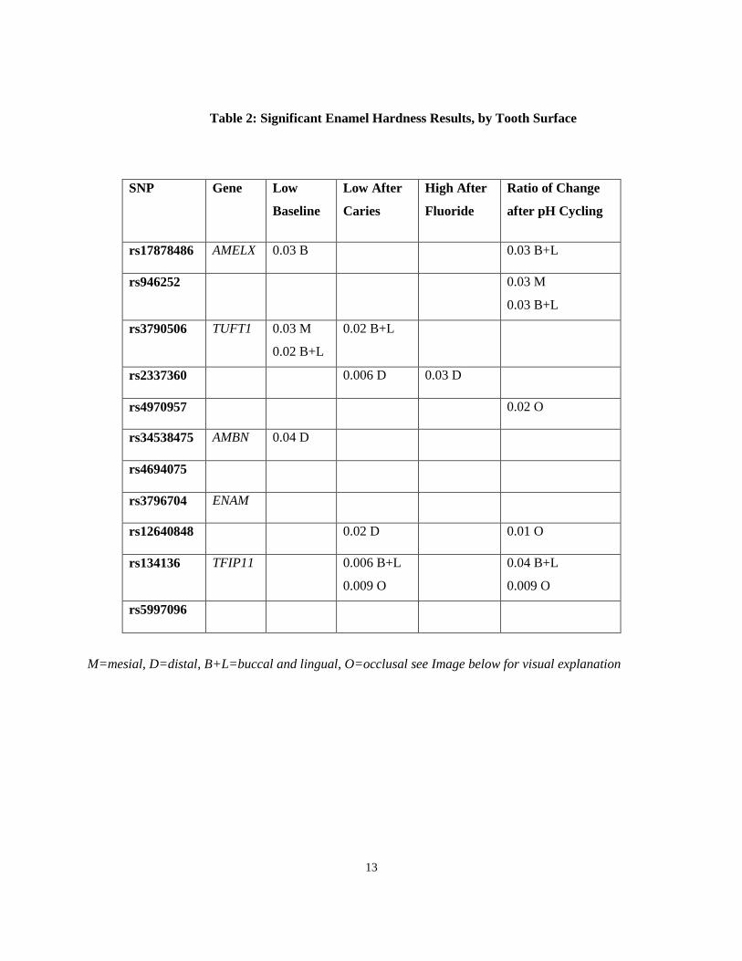
The enamel hardness association results were less specific, showing an association in every gene
studied in nearly every SNP used, as shown in the following table.
13
Table 2: Significant Enamel Hardness Results, by Tooth Surface
SNP Gene Low
Baseline
Low After
Caries
High After
Fluoride
Ratio of Change
after pH Cycling
rs17878486 AMELX 0.03 B 0.03 B+L
rs946252 0.03 M
0.03 B+L
rs3790506 TUFT1 0.03 M
0.02 B+L
0.02 B+L
rs2337360 0.006 D 0.03 D
rs4970957 0.02 O
rs34538475 AMBN 0.04 D
rs4694075
rs3796704 ENAM
rs12640848 0.02 D 0.01 O
rs134136 TFIP11 0.006 B+L
0.009 O
0.04 B+L
0.009 O
rs5997096
M=mesial, D=distal, B+L=buccal and lingual, O=occlusal see Image below for visual explanation

14
Figure 1: Surfaces of human teeth (43)
The amelogenin results are expected because it plays such a large role in enamel formation, but
the tuftelin results are interesting. Knowledge of the role tuftelin may play in the formation of the tooth is
limited and the results of our study may offer clues into its exact function. However, the teeth that were
assessed had been removed due to caries. Thus, there may have been underlying issues with the enamel
on these teeth. In addition to this, each population had its own limitations. The two populations from
Brazil had lower DMFTs overall than the other populations. The Turkish population and one of the
Brazilian populations was made up entirely of children, and we know that primary teeth respond to caries
differently than permanent teeth (44). The Argentinean and Filipino populations were from families
affected by cleft lip and/or palate, though the samples for this study were unaffected individuals. It is
unclear if having a family history of cleft lip and palate puts one at a higher risk of caries than the general
population.
2.2 FINE MAPPING OF ASSOCIATED REGIONS
While the studies focusing on enamel formation genes had promising results, they did not offer
definitive results regarding caries experience. Even when controlling (as much as possible) for outside
factors, no genes stood out as consistently being associated with high or low caries experience. For this
reason, other areas of the genome were investigated for possible genetic links to caries experience.

15
2.2.1 GWS
Our lab performed one of the first genome wide scans for genes related to caries (45). A panel of
392 fluorescent genotype markers was used to assay 46 families consisting of 458 total individuals. After
performing linkage analyses to detect co-segregation between the DNA markers and caries experience (as
measured by DMFT), significant evidence for linkage was found in five chromosomal regions: three for
low caries susceptibility: 5q13.3 (dominant midpoint LOD score=2.30), 14q11.2 (recessive single-point
LOD score=2.29), and Xq27.1 (non-parametric LOD p-value=0.00005); and two for high caries
susceptibility:13q31.1 (recessive single point LOD score=2.33 and recessive multipoint LOD score=2.20)
and 14q24.3 (recessive single-point LOD score=2.06). There are no enamel formation genes in any of
these locations. In fact, the genes and gene products found in the above mentioned regions are not known
to be related to caries formation or tooth development (45). This result is not unexpected because many
factors influence the onset and progression of caries. How these factors interact is unknown, but these
results can be used for generating additional hypotheses.
2.2.2 Fine Mapping
Currently, our lab is fine-mapping these 5 regions by first genotyping a population with unlinked
SNPs from each region, then determining which SNPs are associated with high or low caries experience,
and finally investigating the genes that contain those associated SNPs (45). The genotyping of 5q13.3
revealed 4 markers in 4 different genes that were significantly associated with low caries experience.

16
Table 3: Summary of 5q13.3 Genotyping
Name Abbreviation Marker p value
prostate androgen-regulated transcript 1 PART1 rs27565 0.00021
SWIM-type containing zinc finger 6 ZSWIM6 rs4700418 0.0089
cyclin B1 CCNB1 rs875459 0.0046
basic transcription factor 3 BTF3 rs6862039 0.0084
In this analysis, the definition of caries experience varied with age: low was defined as DMFT ≤2 in
children, ≤ 5 in teenagers, and ≤8 in adults.
None of these genes have a known function associated with caries. To further assess whether any
of these genes may be involved in caries, RNA extracted from whole saliva samples was tested for the
mRNA of these 4 genes. RNA expression was only observed for PART1 and BTF3, and only expression
of BTF3 was correlated with low caries experience. This gene is involved in transcription regulation and
cell cycle regulation (46), but how these processes may influence caries is unclear (47).
Despite the evidence for linkage in the GWS, the 13q31.1 chromosomal region had no associated
SNPs. In the 14q24.3 region, the gene ESRRB showed some promising results. ESRRB codes for a
protein that is similar to an estrogen receptor, but does not actually interact with estrogen; its complete
function is unknown. In mice, it plays an important role in the proliferation of stem cells (48). In
humans, however, it has only been associated with non-syndromic hearing loss (49).
Significant results were found in the next region, 14q11.2. Four SNPs were associated with low
caries experience in two of the four populations studied (50).

17
Table 4: p-value for 14q11.2 Genotyping
Filipino Turkish Brazilian
(Rio de Janeiro)
Guatemalan
rs1997532 0.04 0.0005 not significant 0.03*
rs1997533 0.02 0.01 not significant not significant
rs8011979 0.01 0.001 not significant not significant
rs7150049 0.02 0.001 0.05 not significant
* this result was significant only after adjusting for sex
As in the last study, the definition of low caries experience varied with age: dmft ≤2 in children, ≤5 in
teenagers, and ≤8 in adults. Unfortunately, all of these SNPs are in between T-cell receptor alpha
variable (TRAV) genes and may not actually code for anything. In general, TRAV genes help determine
the variability of the alpha region of T-cells, that is, they influence how well T-cells respond to infection
(50). The associated SNPs are closest to the gene TRAV4, and RNA expression of this gene was found in
the saliva of children and teenagers with low caries experience. The possible nature of the involvement of
TRAV4 in caries can only be hypothesized. The theory in our lab is that TRAV4 is involved in a higher
immune response, attacking S. mutans infections early so that cavities progress more slowly or not at all.
While none of these regions revealed any obvious genes that influence caries development, it did
provide further areas for investigation. It also highlighted some of the difficulties in dealing with a multi-
factorial disease. So many factors interact to create this disease that many genes with a variety of
functions could be involved in its acquisition and progression. Hypotheses generated from these results
need to be tested and replicated in additional studies.
2.3 DETECTING S. MUTANS IN SALIVA
Due to the strong influence of S. mutans on caries, our lab was interested in developing an assay
that would detect bacterial DNA in samples extracted from buffered saliva (51). Many such assays exist
for whole saliva, or for DNA extracted from whole saliva. The assay our lab developed was based on one

18
previously performed with DNA from whole saliva (52). The DNA our lab uses comes, primarily, from
saliva collected in kits that buffers saliva upon collection. The manufacturers of this kit claim that the
buffer not only neutralizes the DNAses in the saliva, but that it also suppresses and removes the majority
of bacteria from the saliva. Our assumption was that the buffer included in the saliva samples we worked
with would still, despite manufacturer’s claims, contain a measurable amount of bacterial DNA. Thus,
we created primers specific to the glucocyltransferase gene of S. mutans and ran an absolute
quantification real time PCR reaction with our samples.
This reaction works in the same manner as a standard PCR, primers for specific regions are
created, they match up to those regions, replicate them, then those copies get replicated with the copy in
the genome, causing an exponential increase in the amount of the genome region that the primer is
designed to find. If there is a lot of S. mutans in the sample, and as a result a lot of glucocyltransferase for
the primers to recognize, the number of replications per cycle of PCR will sharply increase early on. If
there is a small amount of S. mutans DNA in the sample, it will take many cycles before a significant
amount of replication is seen. Unfortunately, while this reaction can tell us how many reactions are
happening per cycle, we cannot accurately measure the specific amount of S. mutans DNA present in the
samples. This reaction can tell us, in general, if someone has a lot of S. mutans or not. Within this
constraint, as the following table shows, significant results were only found in populations containing
mostly children (51).

19
Table 5: Results from S. mutans Real Time Detection Assay
Population N Average
age
DMFT/dmft
average
Spearman’s
rho
T
statistic
Degrees
of
freedom
Two-
tailed
p-
value
Argentina 98 31.5 7.44 −0.0831 −0.82 96 0.41
Guatemala 109 28 6.87 0.07244 0.7513 107 0.45
Turkey 175 5.4 3.68 0.3328 3.23 84 0.002
Dental
Registry and
DNA
Repository
(DRDR)
666 42.9 15.98 0.02069 0.53 655 0.6
Cariology
Course
158 43.3 3.77 0.05079 −0.6352 156 0.53
Center for
Oral Health
Disparities in
Appalachia
(COHRA)
44 3.3 1.64 0.4343 3.161 43 0.003
20
This result may seem obvious, because children are more prone to caries and therefore to having
a detectable amount of S. mutans. It is also possible that there are carious lesions that can’t be seen by the
naked eye, which would elevate the level of S. mutans present. Another limitation is that this is just a
snapshot of one moment in the S. mutans profile of the volunteer. It tells us whether they had high or low
caries on the day they donated. We assume, for the purposes of research, that it represents a general
picture of the oral biome from which to extrapolate.
We can use this assay for other studies to categorize subjects into different groups. For example,
if someone has low caries and high S. mutans, we can design a study to look at other factors in their
body/lifestyle that will impact their caries outcome. Alternatively, using data on subjects with high caries
but no S. mutans, we can investigate what other bacteria in their mouth are creating the acid. We can also
assess their diet and investigate the level of proteins versus carbohydrates.
21
3.0 APPLICATION
As described previously, dental caries among the pediatric population is a serious public health
problem. Given my background in the genetics of dental caries, I propose a pilot public health
intervention to assess whether parent and child’s knowledge of the child’s possible increased
susceptibility to dental cares would affect the oral health habits of the child. As part of this proposal, a
genetic panel of SNPs associated with high or low caries risk will be developed. This panel would then
be tested with children from a pediatric clinic whose parents would be notified of the results of the test.
The parents and child will then be questioned after hearing the results if they plan on making any lifestyle
changes. They will be asked the same questions at their next two regular visits (6 months, 1 year later), to
see if they actually implemented any of these changes.
The primary resource required for this project is a pediatric clinic with dentists and patients
willing to participate. I propose the pediatric clinic at the University of Pittsburgh School of Dental
Medicine because it has been the site of other research projects in the past, so the staff is familiar with
dealing with research protocols. The people who attend to this clinic are typically from a lower social
economic status and from neighborhoods near the University. The children volunteering for this study
should be patients between the ages of 7 and 12. In this age range, children are beginning to get their
permanent dentition (adult teeth). As new teeth come in, parents will be more observant of their
children’s oral health habits.
Each volunteering patient will be given a standard dental exam and cleaning. The dentist will
then explain to the patient and their parents what constitutes good oral health, and what they need to do to
improve their oral health. Half of these volunteer will then be asked to provide a saliva sample, from
which DNA will be extracted. All parents and children will need to provide informed consent to allow
specific items from the child’s medical record to be accessed and for having their DNA used. The child’s
dmft/DMFT from their medical records will be used in this study as a measure of caries experience.
Other medical information, such as current medications and chronic medical conditions, will also be
noted. Parents will be asked what they expect the outcome of the testing to be and if they plan to make
any changes in their child’s current oral health care regime.
The panel will be designed to evaluate many of the above mentioned genes, as well as any
gathered from a more exhaustive literature search. Specific SNPs in each of these genes will be on the
panel. A laboratory would be required to create the physical panel, extract the DNA, and run the assay.

22
SNP results will be compared to DMFT scores to see if there are any statistically significant correlations.
Within a month of running the assay, the children and their parents will be notified of the results. They
will be asked again if they plan on making changes in their lifestyle. At the next routine visit with the
dentist for all the children in this study, 6 months after the recruiting visit, the child and the parents will
be asked if they made any of the changes in dental health care that they previously mentioned. The
child’s dmft/DMFT will be noted to see if there has been any improvement or not. This process will be
repeated at the next 6 month visit (one year after the genetic screening was implemented) to see if there is
any difference between the dmft/DMFTs of the children who got the genetic screening and those who
didn’t.
There are two goals with this project. The first is to assess if it is possible to design a predictive
genetic panel that could help customize oral health care. This panel could be used by larger entities like
Medicaid or dental insurance providers to help determine how better to spend resources on patients. For
example, whether preventive care or restorative care would be better for a specific patient.
The second goal is to assess whether knowledge of genetic testing results impacts individual
choices for health care. All the participants in this study will receive a comprehensive education about
the causes of dental caries, what each patient can do to prevent caries, and the evidence for a genetic
component to the disease. Those who receive genetic profile results may or may not act any differently
than those who did not. Less susceptible people may reduce brushing their teeth and get more cavities;
more susceptible may be more diligent about oral care and improve their oral health. An individuals
action depends on how he/she internalizes the risk. Because the underlying message, i.e., that everyone
still needs to brush their teeth at least twice a day, is the same for both groups, the parents of the children
who get the test result may not react at all to the results.
In the short term, the patients involved in this study along with their parents will have increased
general knowledge about oral health. Those who received the genetic screening will have knowledge
about their susceptibility to caries. In the medium term, patients and their parents will be reminded of
healthy lifestyle choices they could make to improve their child’s oral health. In the long term, this study
could help in addressing the larger question of whether genetic testing for more common diseases (such as
dental caries) is useful, that is, do individuals change their behaviors to mitigate risk. The study may also
indicate additional procedures that would increase its effectiveness.

23
BIBLIOGRAPHY
1. World Health Organization. Oral health, Fact sheet N°318. April 2012.
<http://www.who.int/mediacentre/factsheets/fs318/en/index.html> Accessed 19 June 2013.
2. Owing L. 2007 Mar. 5. ABC News.
<http://abcnews.go.com/Health/Dental/story?id=2925584&page=1> Accessed 2013 Mar. 9.
3. Shuler CF. Inherited risks for susceptibility to dental caries. J Dent Educ. 2001;65(10):1038-45.
4. Conry JP, Messer LB, Boraas JC, Aeppli DP, Bouchard TJ Jr: Dental caries and treatment
characteristics in human twins reared apart. Arch Oral Biol 1993; 38: 937–943.
5. Wright JT. The molecular etiologies and associated phenotypes of amelogenesis imperfecta. Am J Med
Genet A 2006;140:2547–2555.
6. Tamburstuen MV, Reseland JE, Spahr A, Brookes SJ, Kvalheim G, Slaby I, Snead ML, Lyngstadaas
SP. Ameloblastin expression and putative autoregulation in mesenchymal cells suggest a role in early
bone formation and repair. Bone 2011; 48:406-413.
7. Zhengkuan M.The human tuftelin gene: cloning and characterization.Gene 279 (2001) 181–196
8. Tannukit S. Identification of a novel nuclear localization signal and speckle-targeting sequence of
tuftelin-interacting protein 11, a splicing factor involved in spliceosome disassembly. Biochem Biophys
Res Commun. 2009 Dec 18;390(3):1044-50.
9. Bei M. Molecular genetics of tooth development. Genetics and Development. 2009; 19:504-510.
10. Ferraro M, Vieira AR. Explaining gender differences in caries: a multifactorial approach to a
multifactorial disease. Int J Dent. 2010;649643.
11. Wang X, et.al. Genetic and Environmental Factors Associated with Dental Caries in Children: The
Iowa Fluoride Study. Caries Res. 2012;46:177–184
12. Joana Cunha-Cruz, JoAnna Scott, Marilynn Rothen, Lloyd Mancl, Timothy Lawhorn, Kenneth
Brossel and Joel Berg. Salivary characteristics and dental caries: Evidence from general dental practices.
JADA 2013;144(5):e31-e40
13. Aguirre JM, Rodriquez R, Oribe D, Vitoria JC. Dental enamel defects in celiac patients. Oral Surg
1997;84:646-50.
14. Senpuku H, Yanagi K, Nisizawa T. Identification of Streptococcus mutans PAc peptide motif binding
with human MHC class II molecules (DRB1-0802, 1101, 1401 and 1405). Immunology 1998;95:322-30.
15. Sanui T, Gregory RL. Analysis of Streptococcus mutans biofilm proteins recognized by salivary
immunoglobulin A. Oral Microbiol Immunol. 2009 Oct;24(5):361-8.
16. Gomes Pde S, and Fernandes MH. Defensins in the oral cavity: distribution and biological role. J Oral
Pathol Med. 2010 Jan;39(1):1-9.

24
17. Fine DH, Toruner GA, Velliyagounder K, Sampathkumar V, Godboley D, Furgang D. A
lactotransferrin single nucleotide polymorphism demonstrates biological activity that can reduce
susceptibility to caries. Infect Immun. 2013 May; 81(5):1596-605.
18. Mayo Clinic Staff. 2013 May 14. Oral Health: Brush Up on Dental Care Basics.
<http://www.mayoclinic.com/health/dental/DE00003> Accessed 2013 June 11.
19. Graves RC, Disney JA, Stamm JW. Comparative effectiveness of flossing and brushing in reducing
interproximal bleeding. J Periodontol. 1989 May;60(5):243-7.
20. The Public Speaks Up on Oral Health Care: An ADA and Crest/Oral B Survey. October 2008. <
http://www.crest.com/ada-webcast/surveyfindings.pdf > Accessed 2013 June 11.
21. Beavan C. 2007 Mar. 20. No Impact Man.
<http://noimpactman.typepad.com/blog/2007/03/i_hereby_sacrif.html> Accessed 2013 Mar. 10.
22. Ten Cate JM. Contemporary perspective on the use of fluoride products in caries prevention.Br Dent
J. 2013 Feb 22;214(4):161-7.
23. Walsh LJ. Preventive dentistry for the general dental practitioner. Aust Dent J. 2000 Jun;45(2):76-82.
24. Lukacs JR, Largaespada LL. Explaining sex differences in dental caries prevalence: saliva, hormones,
and "life-history" etiologies. Am J Hum Biol. Jul-Aug;18(4):540-55. 2006
25. Lehner T, Clarry ED, Cardwell JE. Immunoglobulins in saliva and serum in dental caries. Lancet. Jun
17;1(7503):1294-6. 1967
26. Touger-Decker R, van Loveren C. Sugars and dental caries. Am J Clin Nutr. 2003 Oct;78(4):881S-
892S.
27. Yuan JC, et.al. Dentistry and obesity: a review and current status in U.S. predoctoral dental education.
J Dent Edu. 2012 Sep;76(9):1129-36.
28. Marshall TA and Eichenberger-Gilmore JM. Dental caries and childhood obesity: roles of diet and
socioeconomic status. Community Dent Oral Epidemiol. 35: 449–4582007
29. Marcotte H and Lavoie MC. Oral microbial ecology and the role of salivary immunoglobulin A.
Microbiol Mol Rev. 1998 Mar; 62(1):71-109.
30. Lapirattanakul J, Nakano K, Nomura R, Hamada S, Nakagawa I, and Ooshima T. Demonstration of
mother-to-child transmission of Streptococcus mutans using multilocus sequence typing. Caries Res.
2008;42(6):466-74.
31. McGrady MG, Ellwood RP, Maguire A, Goodwin M, Boothman N, Pretty IA. The association
between social deprivation and the prevalence and severity of dental caries and fluorosis in populations
with and without water fluoridation. BMC Public Health 2012, 12:1122
32. Palmer CA, Gilbert JA; Academy of Nutrition and Dietetics. Position of the Academy of Nutrition
and Dietetics: the impact of fluoride on health. J Acad Nutr Diet. 2012 Sep;112(9):1443-53.
33. Widmer RP. Oral health of children with respiratory diseases. Paediatr Respir Rev. 2010
Dec;11(4):226-32.
25
34. Leroy R, Hoppenbrouwers LR, Hoppenbrouwers K, Jara A, Declerck D. Parental smoking behavior
and caries experience in preschool children. Community Dent Oral Epidemiol 2008; 36: 249–257
35. American Dental Association. 2012 April 26. Breaking Down Barriers to Oral health for All
Americans: The Role of Finance.
<http://www.ada.org/sections/advocacy/pdfs/7170_Breaking_Down_Barriers_Role_of_Finance-FINAL4-
36. Sanders, B. 2012 Feb. 29. U.S. Senate Committee on Health, Education, Labor &
Pensions,Subcommittee on Primary Health and Aging. Dental crisis in america:The Need to Expand
Access. <http://www.sanders.senate.gov/imo/media/doc/dentalcrisis.report.pdf>. Accessed 2013 Mar. 10.
37. Center for Medicare and Medicaid Services. Dental Care. <http://www.medicaid.gov/Medicaid-
CHIP-Program-Information/By-Topics/Benefits/Dental-Care.html>. Accessed 2013 Mar. 9.
38. Petersen PE, Bourgeois D, Ogawa H, Estupinan-Day S, Ndiaye C. The global burden of oral diseases
and risks to oral health. Bull World Health Organ. 2005 Sep;83(9):661-9.
39. Sheiham A. Dental caries affects body weight, growth and quality of life in pre-school children. Br
Dent J. 2006 Nov 25;201(10):625-6.
40. Deeley K, Letra A, Rose EK, Brandon CA, Resick JM, Marazita ML, Vieira AR. Possible association
of amelogenin to high caries experience in a Guatemalan-Mayan population. Caries Res.2008;42(1):8-13.
41. Patir A, Seymen F, Yildirim M, Deeley K, Cooper ME, Marazita ML, Vieira AR. Enamel formation
genes are associated with high caries experience in Turkish children. Caries Res. 2008;42(5):394-400
42.Shimizu T, Ho B, Deeley K, Briseño-Ruiz J, Faraco IM Jr, Schupack BI, Brancher JA, Pecharki GD, Küchler EC, Tannure PN, Lips A, Vieira TC, Patir A, Yildirim M,Poletta FA, Mereb JC, Resick JM, Brandon CA, Orioli IM, Castilla EE, Marazita ML, Seymen F, Costa MC, Granjeiro JM, Trevilatto PC, Vieira AR. Enamel formation genes influence enamel microhardness before and after cariogenic
challenge. PLoS One. 2012;7(9). Epub 2012 Sep 24.
43. dozenist. "MandibularLeftFirstMolar08-15-06." Photograph. Wikimedia Commons. 15 August 2006.
<http://commons.wikimedia.org/wiki/File:MandibularLeftFirstMolar08-15-06.jpg>
44. Hayashi-Sakai S, Sakai J, Sakamoto M, Endo H. Determination of fracture toughness of human
permanent and primary enamel using an indentation microfracture method. J Mater Sci Mater Med. 2012
Sep;23(9):2047-54.
45. Vieira AR, Marazita ML, Goldstein-McHenry T. Genome-wide scan finds suggestive caries loci. J
Dent Res. 2008 May;87(5):435-9.
46. Green CD, Thompson PD, Johnston PG, El-Tanani MK. Interaction between transcription factor,
basal transcription factor 3, and the NH2-terminal domain of human estrogen receptor alpha. Mol Cancer
Res. 2007 Nov;5(11):1191-200.
47. Shimizu T, Deeley K, Briseño-Ruiz J, Faraco Jr IM, Poletta FA, Brancher JA, Pecharki GD, Küchler
EC, Tannure PN, Lips A, Vieira TC, Patir A, Yildirim M, Mereb JC, Resick JM, Brandon CA, Cooper
ME, Seymen F, Costa MC, Granjeiro JM, Trevilatto PC, Orioli IM, Castilla EE, Marazita ML, Vieira AR.
Fine-Mapping of 5q12.1-13.3 Unveils New Genetic Contributors to Caries. Caries Res. 2013 Jan
30;47(4):273-283.
26
48. Martello G, Sugimoto T, Diamanti E, Joshi A, Hannah R, Ohtsuka S, Göttgens B, Niwa H, Smith A.
Esrrb is a pivotal target of the Gsk3/Tcf3 axis regulating embryonic stem cell self-renewal. Cell Stem
Cell. 2012 Oct 5;11(4):491-504.
49. Ben Saïd M, Ayedi L, Mnejja M, Hakim B, Khalfallah A, Charfeddine I, Khifagi C, Turki K, Ayadi
H, Benzina Z, Ghorbel A, Castillo ID, Masmoudi S, Aifa MH. A novel missense mutation in the ESRRB
gene causes DFNB35 hearing loss in a Tunisian family. Eur J Med Genet. 2011 Nov-Dec;54(6):e535-41.
50. Briseño-Ruiz J, Shimizu T, Deeley K, Dizak PM, Ruff TD, Faraco IM Jr, Poletta FA, Brancher JA,
Pecharki GD, Küchler EC, Tannure PN, Lips A, Vieira TC, Patir A, Koruyucu M, Mereb JC, Resick JM,
Brandon CA, Letra A, Silva RM, Cooper ME, Seymen F, Costa MC, Granjeiro JM, Trevilatto PC, Orioli
IM, Castilla EE, Marazita ML, Vieira AR. Role of TRAV locus in low caries experience. Hum Genet.
2013 May 9.
51. Vieira AR, et.al.. Detection of Streptococcus mutans Genomic DNA in Human DNA Samples
Extracted from Saliva and Blood. ISRN Dent. 2011;2011:543561.
52. A. Yano, N. Kaneko, H. Ida, T. Yamaguchi, and N. Hanada. Real-time PCR for quantification of
Streptococcus mutans. FEMS Microbiology Letters, vol. 217, no. 1, pp. 23–30, 2002