publication du pr. baulieu dans pnas - avril 2014
TRANSCRIPT

Immunophilin FKBP52 induces Tau-P301L filamentousassembly in vitro and modulates its activity in amodel of tauopathyJulien Giustiniania,1, Béatrice Chambrauda,1, Elodie Sardina, Omar Dounanea, Kevin Guillemeaua, Hiroko Nakatania,Dominik Paquetb,c,2, Amina Kamahd, Isabelle Landrieud, Guy Lippensd, Etienne-Emile Baulieua,3, and Marcel Tawka,3
aInstitut National de la Santé et de la Recherche Médicale, Unité Mixte de Recherche 788, Université Paris XI, 94276 Le Kremlin Bicêtre, France; bGermanCenter for Neurodegenerative Diseases and cAdolf Butenandt Institute, Department of Biochemistry, Ludwig Maximilians University, 80336 Munich, Germany;and dCentre National de la Recherche Scientifique, Université de Lille 1, Unité Mixte de Recherche, 8576 Villeneuve-d’Ascq, France
Contributed by Etienne-Emile Baulieu, February 18, 2014 (sent for review January 31, 2014)
The Tau protein is the major component of intracellular filamentsobserved in a number of neurodegenerative diseases known astauopathies. The pathological mutant of Tau containing a proline-to-leucine mutation at position 301 (P301L) leads to severe humantauopathy. Here, we assess the impact of FK506-binding proteinwith a molecular mass of ∼52 kDa (FKBP52), an immunophilin pro-tein that interacts with physiological Tau, on Tau-P301L activity.We identify a direct interaction of FKBP52 with Tau-P301L and itsphosphorylated forms and demonstrate FKBP52’s ability to inducethe formation of Tau-P301L oligomers. EM analysis shows thatTau-P301L oligomers, induced by FKBP52, can assemble into fila-ments. In the transgenic zebrafish expressing the human Tau-P301L mutant, FKBP52 knockdown is sufficient to redrive defectiveaxonal outgrowth and branching related to Tau-P301L expressionin spinal primary motoneurons. This result correlates with a signif-icant reduction of pT181 pathological phosphorylated Tau andwith recovery of the stereotypic escape response behavior. Collec-tively, FKBP52 appears to be an endogenous candidate that di-rectly interacts with the pathogenic Tau-P301L and modulates itsfunction in vitro and in vivo.
FKBP | Tau assembly | Tau-P301L dementia
Tau is a MAP mainly found in neurons (1, 2), which becomeshyperphosphorylated in several neurodegenerative diseases
known as tauopathies and forms intraneuronal aggregates calledneurofibrillary tangles (NFTs) (3–5). The accumulation of Tauaggregates is a multistep process that involves transient species,and growing evidence suggests that not only the NFTs butequally small oligomeric species contribute to Tau-mediatedneurotoxicity (6–8). The pathological mutant of Tau con-taining a proline-to-leucine mutation at position 301 (P301L)is identified in frontotemporal dementia and Parkinsonismlinked to chromosome 17 (FTDP-17) (9). Larvae of thezebrafish overexpressing the Tau-P301L mutant protein showdefects in motoneuron development early on, and in thismodel, different Tau phosphoforms are associated with itspathological state (10).The FK506-binding protein with a molecular mass of ∼52 kDa
(FKBP52) belongs to the immunophilin family and presentsrotamase activity inhibited by FK506 binding (11). This enzy-matic activity catalyzes the isomerization of peptidyl-prolyl bondsbetween cis and trans conformations, and therefore influencestarget protein folding and function (12, 13). FKBP52 containsadditional functional domains, such as a tetratricopeptide repeatdomain that serves as a binding site for molecular chaperoneheat shock protein with a molecular mass of 90 kDa (14) and forwhich chaperone activity was observed (15).A novel role of FKBP52 has recently emerged because it binds
directly to tubulin and induces tubulin depolymerization in vitro(16). Biochemical characterization of FKBP52 showed that itinteracts with Tau, especially its hyperphosphorylated form, and
demonstrated an antagonist effect of FKBP52 on Tau’s functionin tubulin assembly (17). Here, we show that FKBP52 interactswith the pathological form of Tau-P301L and induces its oligo-merization and assembly into filaments. Using a transgeniczebrafish model for Tau-P301L tauopathy (10), we show thatFKBP52 knockdown attenuates pathological Tau activity toreestablish axonal outgrowth and branching in defective spinalprimary motoneurons. This result confirms the functional impli-cation of FKBP52 in modulating early pathological Tau activity,at least for axonal growth and motility. We propose that FKBP52is an important regulator of Tau conformational change and as-sembly, and thus may be instrumental in a therapeutic approachto the disease.
ResultsFKBP52 Binds Tau-P301L Mutant. The interaction between FKBP52and the Tau-P301L mutant was investigated using a dot blotassay. Purified recombinant protein (2.2 μg of Tau-P301L), phos-phorylated or not phosphorylated, was spotted on nitrocellulose
Significance
Growing evidence underlines the role attributed to abnormalforms of Tau in several neurodegenerative diseases known astauopathies, including Alzheimer’s disease, which are charac-terized by accumulation of oligomers and filamentous Tauinclusions in the CNS. We identify a direct interaction of theimmunophilin FK506-binding protein with a molecular mass of∼52 kDa (FKBP52) and the pathological mutant of Tau con-taining a proline-to-leucine mutation at position 301 (Tau-P301L), inducing Tau-P301L oligomerization and assembly intofilaments. This interaction has a considerable impact on theprogress of the tauopathy because some early aspects of thedisease are rescued, in vivo, by knocking down FKBP52. Ourresults open a new field for the study of FKBP52 pathophysi-ology in tauopathic dementias, including prediction of diseasephenotype and search for new types of antipathologicalTau treatments.
Author contributions: J.G., B.C., E.-E.B., and M.T. designed research; J.G., B.C., E.S., O.D.,K.G., H.N., A.K., and M.T. performed research; D.P. and I.L. contributed new reagents/analytic tools; J.G., B.C., G.L., and M.T. analyzed data; and J.G., B.C., G.L., E.-E.B., and M.T.wrote the paper.
The authors declare no conflict of interest.
Freely available online through the PNAS open access option.1J.G. and B.C. contributed equally to this work.2Present address: Laboratory of Brain Development and Repair, The Rockefeller Univer-sity, New York, NY 10065.
3To whom correspondence may be addressed. E-mail: [email protected] [email protected].
This article contains supporting information online at www.pnas.org/lookup/suppl/doi:10.1073/pnas.1402645111/-/DCSupplemental.
4584–4589 | PNAS | March 25, 2014 | vol. 111 | no. 12 www.pnas.org/cgi/doi/10.1073/pnas.1402645111

and incubated with purified human FKBP52 protein. A poly-clonal antibody against FKBP52 was used to monitor the amountof protein retained. As shown in Fig. 1, the amount of FKBP52retained by Tau-P301L was 18% (±4.5), whereas 55% (±10) ofFKBP52 was retained by the same amount of the phosphorylatedTau-P301L. Thus, FKBP52 binds Tau-P301L, especially in itsphosphorylated form.
FKBP52 Colocalizes with Tau-P301L in Human Neuronal Cells. In lightof the emerging concept of transcellular propagation of Tau(18), we seeded SH-SY5Y neuronal cells with exogenous Tau-P301L, labeled with a cyanine (Cy) 5.5 dye, and investigated thesubcellular localization of Tau-P301L and endogenous FKBP52.Fluorescence microscopy and confocal microscopy analysis showedthat Tau-P301L localization was concentrated in the perinuclearregion, with FKBP52 showing the intracellular colocalization ofboth proteins (Fig. 2 and Fig. S1).
FKBP52 Induces the Formation of Tau-P301L Oligomers in Vitro. Toinvestigate the ability of recombinant FKBP52 to induce struc-tural changes to Tau-P301L, we monitored the migration profileof these proteins using native gel electrophoresis. Fixed quantities ofTau-P301L were mixed with increasing amounts of FKBP52 andincubated for 30 min at 37 °C. OnWestern blotting carried out withan anti-Tau antibody, titration of Tau-P301L with FKBP52 ledto the formation of several bands having different electro-phoretic mobility from those of isolated Tau-P301L, whereasno modification occurred upon exposure of Tau-P301L tocontrol GST (Fig. 3A). However, the same samples analyzedwith an anti-FKBP52 antibody did not reveal any modificationin the migration profile, suggesting a structural modificationof Tau-P301L rather than a stable complex between FKBP52and Tau-P301L. A similar pattern of oligomer formation afterincubation of FKBP52 and phosphorylated Tau-P301L wasdetected by anti-Tau antibodies, AT8 or AT180, that recog-nize specific phosphorylated forms (Fig. 3B).
Tau-P301L Can Form Paired Helical-Like Filaments When Incubatedwith FKBP52. To see whether the oligomers obtained couldevolve into fibrillar structures, we first used light diffraction.When measuring the light scattering at 340 nm, we indeedobserved a signal when mixing FKBP52 and Tau-P301L for 30min at 37 °C, but not with both proteins individually (Fig. 4A).We analyzed the resulting samples using transmission EM,and we did observe fibrillar structures after incubation for 1 h,whereas no such structures could be found when individualprotein solutions were deposited on the grid. The resultingfibers appear to be short in length and twisted, with a width of17 nm (Fig. 4B). As such, they resemble the filaments ob-served in the brains of patients with FTDP-17, most of whichappear to be irregularly twisted ribbons with a width of about15 nm (19).
FKBP52 Knockdown Significantly Improves Axonal Growth andBranching Defects Observed in Primary Motoneurons of Tau-P301LTransgenic Fish. To test in vivo the effect of FKBP52/Tau-P301Linteraction, we used the zebrafish, cloned its full-length FKBP52cDNA, and showed its ubiquitous expression (Fig. S2). Forfunctional analysis, we took advantage of the transgenic Tau-P301L zebrafish model (for convenience, we use HuC-Tau in thefigures), which recapitulates several neurological defects of tauo-pathies, including axonal growth and branching defects seen in thespinal motoneurons (10, 20) (Fig. 5 D, F, and I). We lookedspecifically at the primary motoneurons that undergo axonogenesisat 17 h postfertilization (hpf), using znp1 antibodies, a pan-specificmarker of primary motor axons (21). We analyzed the first fiveoutgrowing caudal primary motoneurons (CaP) anterior to the endof the yolk extension (Fig. 5).To test the role of FKBP52 in this model, we used a knockdown
approach by injecting specific antisense morpholino (MO)against FKBP52. Injection of FKBP52 MO (0.4–0.6 pmol perembryo) or FKBP52 mRNA (200 pg) into WT embryos didnot lead to any defect regarding the length (ability to reachtheir most ventral target) or the number of branches in the CaPmotoneurons (Fig. 5 E and E′). Note that FKBP52 morphantsshow a slightly shorter and curved body axis (Fig. 5 A–C) but havenormal neurogenesis in the spinal cord (Figs. S3 and S4). CaPmotor axons of control 5-mismatch morphants, FKBP52 mor-phants, and FKBP52 mRNA were similar to WT at 48 hpf (Fig. 5D–E′). In the Tau-P301L transgenic model, only 57% of the CaP
Fig. 1. Binding of mutant Tau-P301L to FKBP52. (A) Representative dotblot analysis: (1) 0.5 μg of FKBP52 used at 100%, (2) 2.2 μg of mutant Tau-P301L, (3) 2.2 μg of phosphorylated mutant Tau-P301L, and (4) 5 μg of GSTused as negative control were dotted on nitrocellulose. The dots weredeveloped with FKBP52 antibody. (B) Amount of FKBP52 retained by Tauproteins was quantified using GeneTools software (Syngene). These resultsrepresent the data from four separate experiments. (C ) SDS/PAGE analysisof recombinant mutated Tau-P301L and P301L hyperphosphorylation (HP).IB, immunoblot.
Fig. 2. FKBP52 colocalizes with Tau-P301L in SH-SY5Y cells. Confocal mi-croscopy analyses of the colocalization with Z-stack imaging. Tau-P301L is inthe same plane as FKBP52 (arrowhead), showing the intracellular colocali-zation of both proteins (arrow). The fluorescence profiles confirmed that thetwo proteins colocalize. (Scale bars: 5 μM.)
Giustiniani et al. PNAS | March 25, 2014 | vol. 111 | no. 12 | 4585
NEU
ROSC
IENCE

axons reach their most ventral target in comparison to WT (Fig.5I; P < 0.05). They also show a randomized pattern of branchingregarding the number of collateral branches or fascicles per axon(Fig. 5F). However, striking changes occurred when injectingFKBP52 MO in the Tau-P301L transgenic embryos. Here, Tau-P301L/FKBP52 morphants readjust their axonal growth, whereby78% of the CaP axons now reach their most ventral target incomparison to HuC-Tau (Fig. 5I; P = 0.038). In this case, no sig-nificant difference is observed between WT and Tau-P301L/FKBP52 morphants (Fig. 5I; P > 0.05). They also reestablisha more close to normal distribution of collateral branching perCaP axon in comparison to Tau-P301L (t = −3.09, P = 0.01)(Fig. 5 G and G′ and Fig. S3). Injection of FKBP52 mRNA intoTau-P301L embryos aggravated their axonal defects. Here, only44% of CaP motoneurons reach their most ventral target incomparison to WT (Fig. 5I; P < 0.01). There was also a sig-nificant difference in the collateral numbers of branches in theTau-P301L/FKBP52 mRNA, whereby the number of CaPaxons showing fewer collateral branches is significantly increasedin comparison to Tau-P301L alone (Fig. 5 H and H′ and Fig. S3;t = −2.26, P = 0.01).
FKBP52 Knockdown Does Not Modify Neuronal Cell Death in Tau-P301L Spinal Cord. The Tau-P301L transgenic larvae show a sig-nificant increase in neuronal cell death in the spinal cord (10).
To assess whether FKBP52 knockdown might modify the neu-ronal cell death seen in the Tau-P301L transgenic embryos, weinjected FKBP52 MO and looked for cell death in the spinalcord using acridine orange (AO) labeling. No significant dif-ference was observed in the number of AO-positive cells be-tween Tau-P301L embryos and the Tau-P301L/FKBP52 MOsiblings at 72 hpf (t = 1.15, P = 0.273; Fig. 6).
FKBP52 Knockdown Differentially Regulates the Levels of PathologicalPhosphorylated Forms of Tau-P301L. To investigate a change in thephosphorylation state of pathological forms of Tau, we haveanalyzed, after FKBP52 knockdown, two different pathologicalphosphorylated forms of Tau-P301L (pT181 and pT231) byWestern blotting. Although there was no significant change inthe overall level of expression of Tau and the pT231 patholog-ical form, we observed a sharp decrease in the levels of thepathological phosphorylated form of Tau recognized by AT270(epitope pT181) (Fig. 7).
FKBP52 Knockdown Improves the Stereotypic Escape ResponseBehavior in the Tau-P301L Transgenic Fish. To test whether theimprovement seen in axonal branching and outgrowth afterFKBP52 knockdown translates into a functional recovery, andthus better motility, we injected FKBP52 MO into Tau-P301Ltransgenic embryos and analyzed the escape response behav-ior. Due to their altered motoneuron morphology, the Tau-P301L embryos show a slow or absent movement in most of thelarvae at 48 hpf (10). Although most control fish responded toa touch stimulus with a stereotypic escape response, mostmutated Tau-expressing fish showed a significantly reduced oreven absent response (Fig. 8 and Movies S1 and S2). However,the Tau-P301L/FKBP52 MO embryos showed a significantimprovement in their escape response (Fig. 8 and Movie S3).We saw that although Tau-P301L embryos move at an averagespeed of 527 units per second (n = 99), the speed of the Tau-P301L/FKBP52 MO embryos increased significantly to 1,750units per second (n = 102) (t = 4.62, P = 0.001), whereascontrol embryos moved at an average speed of 4,147 units persecond (n = 39).
DiscussionFKBP52/Tau-P301L Interaction Leads to Tau Oligomerization. Weshow that FKBP52 is able to interact physically with the Tau-P301L mutant. In this case, FKBP52 can induce a conforma-tional change of Tau-P301L and leads to its oligomerizationand filamentous assembly. This observation is in line with pre-vious data showing important and differential effects of FKBP52on the conformational state of other proteins, such as citrate
Fig. 3. FKBP52 promotes “in vitro” oligomerization of Tau-P301L. (A) Mixof Tau-P301L with increasing concentration of FKBP52 or with GST used asa control were analyzed by blue native PAGE electrophoresis and revealedby Western blotting using specific antibodies as indicated in the figure. (B)Ability of FKBP52 to induce conformational changes of Tau-P301L HP wasanalyzed as in A.
Fig. 4. Assembly of Tau-P301L into filaments. (A) Coincubation of FKBP52and Tau-P301L, at 37 °C for 30 min, induces light scattering, whereas eachprotein alone gives no detectable signal. abs, absorbance. (B) EM of FKBP52/Tau-P301L after 1 h of incubation at 37 °C.
4586 | www.pnas.org/cgi/doi/10.1073/pnas.1402645111 Giustiniani et al.

synthase (15) and alpha-synuclein (22). Interestingly, the Tau-P301L fibers induced by FKBP52 have an average width of 17nm, and their ultrastructure is reminiscent of those isolated frompatients with FTDP-17 (19).However, we failed to detect a stable Tau-P301L/FKBP52
complex, suggesting a rapid and short-lived interaction be-tween both partners that might involve the enzymatic prolylcis/trans isomerase of FKBP52. This activity links FKBPs tothe parvulin family of isomerases, such as Pin1, the importance ofwhich has already been reported in the pathological functionof Tau (23).
FKBP52/Tau-P301L Interaction Can Modulate Axonal Growth but NotCell Death at Early Stages of Tauopathy. We show that reducingFKBP52 levels can significantly enhance the growth of de-fective spinal primary motoneuron projections and branching,
as well as improving the motility of zebrafish larvae. More-over, the phosphorylation site pT181 is strongly decreasedin FKBP52 morphants. This result suggests that reducingFKBP52 levels may delay the early progression of pathologicalaxonal growth by acting on specific phosphorylation site(s)of Tau.We propose a model whereby lowering the levels of FKBP52
in this transgenic model of FTDP-17 tauopathy prevents Tau-P301L from adopting a conformation prone to phosphorylationand aggregation, thereby reducing the neurotoxicity of the trans-gene. Given that the cell death phenotype is not modified in themorphants at these early stages, the impact of this interactionaffects the defective microtubule dynamics and stability related tothis pathological Tau model. However, further experiments shouldshed some light on the impact of FKBP52 knockdown on celldeath at later stages.We also cannot rule out the possibility that the absence of
FKBP52 could lead to a release of endogenous physiologicalTau, allowing it to execute its normal function of microtubuleassembly. The latter hypothesis is in line with the one describedpreviously in PC12 cells (17).
FKBP52/Tau Interaction: The Early vs. Late Aspects of Tauopathy. Themechanism(s) linking FKBP52 and the late stage of the tauopathyprocess remains to be elucidated. Previously, we have shown thatFKBP52 via Tau can modulate microtubule stability in vitro (16)and that levels of FKBP52 protein, at the late stages of diseases,are dramatically decreased in the brains of patients with Alz-heimer’s disease and FTDP-17 (24). Here, we show, in a pre-liminary stage of tauopathy, that attenuating FKBP52 expressionreduces the toxicity of the transgene. Based on these findings,different roles for FKBP52 can be considered. In the early de-velopment of this tauopathy, FKBP52 may induce Tau oligomer
Fig. 5. FKBP52 modulates pathological Tau activity in spinal primary motoneurons. (A–C ) Lateral views of 48-hpf control embryos and FKBP52 mor-phant. Lateral views of znp1 antibody staining of control (D), FKBP52 morphant (E ), Huc-Tau (F ), and Huc-Tau injected with either FKBP52 MO (G) orFKBP52 mRNA (H) are shown at 48 hpf. (D′–H′) Tracings of motor-axon tracts ventral to the spinal cord derived from the panels directly above. In allimages, anterior is to the left and dorsal is to the top. (Scale bars: A–C, 200 μm; D′–H′, 100 μm.) (I) Bar chart depicts the difference in the percentage ofaxons that reach their most ventral target. No significant difference is observed between WT and Huc-Tau + FKBP52MO embryos (N.S, P > 0.05; *P < 0.05;**P < 0.01). N.S, nonsignificant.
Fig. 6. Bar chart depicts the difference in the number of AO-positive cells.No significant difference was observed between the HuC-Tau and the HuC-Tau injected with FKBP52MO. A difference is observed when comparingthese two groups and the WT siblings (***P ≤ 0.0001).
Giustiniani et al. PNAS | March 25, 2014 | vol. 111 | no. 12 | 4587
NEU
ROSC
IENCE

formation that can have a significant impact on axonal outgrowthand branching. However, at much later stages of the disease,given the important accumulation of hyperphosphorylatedTau and its direct oligomerization by FKBP52, the latter is morelikely to be cleared, thus reducing its overall levels. Interestingly,FKBP52/Tau-P301L colabeling could be observed particularly ina perinuclear region, where a deformation of nuclear envelopewas detected (Fig. S1), suggesting that FKBP52 could be in-volved in spatial sequestration of Tau-P301L. This perinuclear
localization of Tau has already been observed in a similarstructure called an aggresome (25, 26). Whether FKBP52 isinvolved in the clearance of Tau at later stages is still to beinvestigated.Further analyses of the FKBP52/Tau interaction may provide
a basis for pursuing and evaluating potential therapies for Alz-heimer’s disease, FTDP-17, and other tauopathies by fine-tuningthe balance of FKBP52.
MethodsCloning of Human and Zebrafish FKBP52 cDNA. To clone human and zebrafishFKBP52 cDNA, RNAs were prepared from SH-SY5Y cells (human neuroblas-toma cells) and from 30-hpf embryos, respectively. Additional details areprovided in SI Methods.
Protein Purification. Tau with the P301L mutation was expressed inEscherichia coli and purified as described (27). Hyperphosphorylationof Tau-P301L was performed using cytosol from the brain of a 2-mo-oldrat as described previously (28). Additional details are provided inSI Methods.
Labeling of Tau-P301L with cyanine, dot blot assays, light scattering, EM,and blue native PAGE are reported in SI Methods.
Immunocytofluorescence. Cells were grown on poly-D-lysine–coated glasscoverslips for microscopy and were plated at 3.104 cells per well in a 12-welltissue culture plate. Subsequently, 20 μg of purified Cy5.5-labeled Tau-P301Lwas added to SH-SY5Y cell medium for 24 h of incubation. Informationabout antibodies used is provided in SI Methods.
Zebrafish Analysis. Zebrafish experiments were performed as described byTawk et al. (29, 30) and as described in SI Methods.
Fig. 7. Effect of FKBP52 knockdown on total and phosphorylated Tau in48-h-old transgenic zebrafish expressing Tau-P301L. Total protein extract(2.5–5 μL) was loaded on 10% SDS/PAGE and analyzed by Western blot-ting using specific antibodies as indicated. Ctrl, control.
Fig. 8. Knockdown of FKBP52 improves larval mobility in HuC-Tau transgenics. (A–C) Tracking analyses of 48-hpf control (WT), Huc-Tau, and Huc-Tau +FKBP52MO larvae in a touch-response test. Each plot line represents the trajectory of one larva after touch stimulation. (D) Quantification of the touch-response test. Each group is divided into three categories: (a) embryos that do not respond at all, (b) embryos that move slightly upon stimulus, and (c)embryos that show a typical escape response. The locomotor defects are significantly improved in Huc-Tau larvae injected with 0.4 pmol of FKBP52 MO(Kruskal–Wallis test,***P < 0.0001).
4588 | www.pnas.org/cgi/doi/10.1073/pnas.1402645111 Giustiniani et al.

ACKNOWLEDGMENTS.We thank C. Yanicostas and N. Soussi-Yanicostas forsharing the Tau-P301L fish for pilot experiments, O. Trassard and P. Leclercfor technical help with confocal imaging and analyses, N. Barois for EM,and C. Jarvis for helping with statistical analyses. We are grateful toB. Schmidt and C. Haass for providing the human Tau-P301L zebrafish line.E.S., O.D., and K.G. were funded by “Fondation Vivre Longtemps,” “Insti-tut Mérieux,” and “Institut Baulieu.” J.G. and A.K. are supported by the
Agence Nationale de la Recherche (ANR), and D.P. is supported by theDeutsche Forschungsgemeinschaft (Grant SFB 596) and European UnionSeventh Framework Programme (Grant 200611, Memory Loss in Alz-heimer’s Disease). This work has been supported jointly by grants fromANR and Laboratory of Excellence Program Investment for the FutureDevelopment of Innovative Strategies for a Transdisciplinary Approach toAlzheimer’s Disease.
1. Avila J, Lucas JJ, Perez M, Hernandez F (2004) Role of tau protein in both physiologicaland pathological conditions. Physiol Rev 84(2):361–384.
2. Morris M, Maeda S, Vossel K, Mucke L (2011) The many faces of tau. Neuron 70(3):410–426.
3. Goedert M, Wischik CM, Crowther RA, Walker JE, Klug A (1988) Cloning and se-quencing of the cDNA encoding a core protein of the paired helical filament ofAlzheimer disease: Identification as the microtubule-associated protein tau. Proc NatlAcad Sci USA 85(11):4051–4055.
4. Sergeant N, et al. (2008) Biochemistry of Tau in Alzheimer’s disease and relatedneurological disorders. Expert Rev Proteomics 5(2):207–224.
5. Mandelkow EM, Mandelkow E (2012) Biochemistry and cell biology of tau protein inneurofibrillary degeneration. Cold Spring Harb Perspect Med 2(7):a006247.
6. Barghorn S, Mandelkow E (2002) Toward a unified scheme for the aggregation of tauinto Alzheimer paired helical filaments. Biochemistry 41(50):14885–14896.
7. Sahara N, Maeda S, Takashima A (2008) Tau oligomerization: A role for tau aggre-gation intermediates linked to neurodegeneration. Curr Alzheimer Res 5(6):591–598.
8. Xu S, Brunden KR, Trojanowski JQ, Lee VM (2010) Characterization of tau fibrillizationin vitro. Alzheimers Dement 6(2):110–117.
9. Goedert M, Crowther RA, Spillantini MG (1998) Tau mutations cause frontotemporaldementias. Neuron 21(5):955–958.
10. Paquet D, et al. (2009) A zebrafish model of tauopathy allows in vivo imaging ofneuronal cell death and drug evaluation. J Clin Invest 119(5):1382–1395.
11. Chambraud B, et al. (1993) Overexpression of p59-HBI (FKBP59), full length and do-mains, and characterization of PPlase activity. Biochem Biophys Res Commun 196(1):160–166.
12. Göthel SF, Marahiel MA (1999) Peptidyl-prolyl cis-trans isomerases, a superfamily ofubiquitous folding catalysts. Cell Mol Life Sci 55(3):423–436.
13. Schiene-Fischer C, Aumüller T, Fischer G (2013) Peptide bond cis/trans isomerases: Abiocatalysis perspective of conformational dynamics in proteins. Top Curr Chem 328:35–67.
14. Radanyi C, Chambraud B, Baulieu EE (1994) The ability of the immunophilin FKBP59-HBI to interact with the 90-kDa heat shock protein is encoded by its tetratricopeptiderepeat domain. Proc Natl Acad Sci USA 91(23):11197–11201.
15. Bose S, Weikl T, Bügl H, Buchner J (1996) Chaperone function of Hsp90-associatedproteins. Science 274(5293):1715–1717.
16. Chambraud B, Belabes H, Fontaine-Lenoir V, Fellous A, Baulieu EE (2007) The im-munophilin FKBP52 specifically binds to tubulin and prevents microtubule formation.FASEB J 21(11):2787–2797.
17. Chambraud B, et al. (2010) A role for FKBP52 in Tau protein function. Proc Natl AcadSci USA 107(6):2658–2663.
18. Frost B, Jacks RL, Diamond MI (2009) Propagation of tau misfolding from the outsideto the inside of a cell. J Biol Chem 284(19):12845–12852.
19. Spillantini MG, Crowther RA, Kamphorst W, Heutink P, van Swieten JC (1998) Taupathology in two Dutch families with mutations in the microtubule-binding region oftau. Am J Pathol 153(5):1359–1363.
20. Pluci�nska G, et al. (2012) In vivo imaging of disease-related mitochondrial dynamics ina vertebrate model system. J Neurosci 32(46):16203–16212.
21. Melançon E, Liu DW, Westerfield M, Eisen JS (1997) Pathfinding by identified ze-brafish motoneurons in the absence of muscle pioneers. J Neurosci 17(20):7796–7804.
22. Gerard M, et al. (2010) Inhibition of FK506 binding proteins reduces alpha-synucleinaggregation and Parkinson’s disease-like pathology. J Neurosci 30(7):2454–2463.
23. Lu PJ, Wulf G, Zhou XZ, Davies P, Lu KP (1999) The prolyl isomerase Pin1 restores thefunction of Alzheimer-associated phosphorylated tau protein. Nature 399(6738):784–788.
24. Giustiniani J, et al. (2012) Decrease of the immunophilin FKBP52 accumulation inhuman brains of Alzheimer’s disease and FTDP-17. J Alzheimers Dis 29(2):471–483.
25. Santa-Maria I, et al. (2012) Paired helical filaments from Alzheimer disease brain in-duce intracellular accumulation of Tau protein in aggresomes. J Biol Chem 287(24):20522–20533.
26. Johnston JA, Ward CL, Kopito RR (1998) Aggresomes: A cellular response to misfoldedproteins. J Cell Biol 143(7):1883–1898.
27. Goedert M, Jakes R (1990) Expression of separate isoforms of human tau protein:Correlation with the tau pattern in brain and effects on tubulin polymerization.EMBO J 9(13):4225–4230.
28. Goedert M (1993) Tau protein and the neurofibrillary pathology of Alzheimer’s dis-ease. Trends Neurosci 16(11):460–465.
29. Tawk M, et al. (2007) A mirror-symmetric cell division that orchestrates neuro-epithelial morphogenesis. Nature 446(7137):797–800.
30. Tawk M, et al. (2011) Wnt/beta-catenin signaling is an essential and direct driver ofmyelin gene expression and myelinogenesis. J Neurosci 31(10):3729–3742.
Giustiniani et al. PNAS | March 25, 2014 | vol. 111 | no. 12 | 4589
NEU
ROSC
IENCE

Supporting InformationGiustiniani et al. 10.1073/pnas.1402645111SI MethodsCloning of Human and Zebrafish FK506-Binding Protein with aMolecular Mass of ∼52 kDa cDNA. To clone human and zebra-fish FK506-binding protein with a molecular mass of ∼52 kDa(FKBP52) cDNA, RNAs were prepared using TRIzol reagent(Invitrogen Life Technologies). Extraction of RNA was conductedusing an RNeasy Extraction Kit (Qiagen) according to the manu-facturer’s recommendations. The cDNAs were generated using aQIAGEN OneStep RT-PCR kit. To clone human FKBP52, thesequences of the primers used for PCR are as follows: forward:5′-CCGGGTCGACTCATGACAGCCGAGGAGATG-3′, reverse:5′-GCAGCGGCCGCCTATGCTTCTGTCTCCAC-3′. The corre-sponding PCR products were digested with the restriction enzymesSal1 and Not1 before subcloning into pGEX-6P-1 vector. To clonezebrafish FKBP52, the sequences of the primers used for PCRare as follows: forward: 5′-GCAGGATCCATGACTGCAGAGG-AGGTG-3′, reverse: 5′-GTCGAATTCTTATGCTGCGGTTTG-TTCC-3′. The corresponding PCR products were digested withthe restriction enzymes BamH1 and EcoR1, and were subclonedinto PCS2 vector. After amplification, the plasmids containingFKBP52 inserts were verified by sequence analysis.
Protein Purification. Full-length human FKBP52 expressed in Es-cherichia coli was affinity-purified as already described (1), and aftercleavage by 2.5 units of Prescission Protease (GE Healthcare), itwas dialyzed against buffer L [0.1 M MES, 1 mM EGTA, 1 mMMgCl2, 0.1 mM EDTA (pH 6.2)] with 10% (vol/vol) glycerol andcomplemented before use to 1 mM GTP and 1 mM DTT.
Labeling of Tau-P301L with Cyanine. Tau-P301L proteins were la-beled with cyanine 5.5 using an Amersham Fluorolink Mono-functional dye labeling kit (GEHealthcare Life Sciences) accordingto the protocol described previously (2). Separation of protein fromfree dyes was performed by gel filtration using Sephadex G-50Coarse (GE Healthcare Life Sciences).
Dot Blot Assays. One hundred microliters of buffer A (buffer Lcomplemented with 1 mMDTT and 1 mMGTP) containing 0.5 μgFKBP52 (used as 100%), 5 μg GST (used as 0%), or 2.2 μg mutantTau-P301L, in vitro hyperphosphorylated or not in vitro hyper-phosphorylated, was spotted onto nitrocellulose. The membraneswere blocked with 5% nonfat milk in PBS containing 0.1% Tween20 (PBS-T) for 1 h at room temperature, followed by 2 h of in-cubation with 100 μL of buffer A containing 0.5 μg of FKBP52.The membranes were washed with IP buffer [50 mMTris (pH 7.5),150 mM NaCl, 2 mM MgCl2, 0.1% Brij97 (Sigma–Aldrich),10% glycerol], followed by PBS-T washes. Subsequently, themembranes were incubated overnight with FKBP52 antibody 761(1:500) (3). The presence of FKBP52 was revealed by ECL.Images were recorded with GeneGnome5 (Syngene).
Light Scattering and EM. Recombinant Tau-P301L (5 μM) wasincubated with full-length recombinant FKBP52 (10 μM) inbuffer A for 30 min at 37 °C. Control samples (5 μM Tau-P301Lalone and 10 μM FKBP52 alone) were incubated under the sameconditions. Buffer and proteins were complemented with 1 mMDTT before use. Fiber formation was monitored by following thevariation of turbidity at 340 nm on a Photon Technology In-ternational fluorescence spectrometer. After incubation, 10 μLof the sample was placed on a formvar grid 400 mesh for 30 s.The sample on grid was stained with 2% uranyl acetate for 1 min.At each step, excess stain was removed by blotting with filter
paper touched to the edge of the grid. Grids were observed usingan Hitachi H7500 electron microscope operated at 80 kV.
Immunocytofluorescence. Cells were washed with PBS, treated ornot treated with 0.025% trypsin, and then fixed by methanol for5 min at−20 °C. After blocking with 3%BSA in PBS for 1 h at roomtemperature, cells were incubated with rabbit polyclonal im-munopurified anti-FKBP52 761 (1:1,000) (3) for 1 h at 37 °C, fol-lowed by staining with Alexa Fluor 488-conjugated secondaryantibody (Life Technologies) for 45min at 37 °C.Nuclei were stainedwith DAPI. The coverslips were examined by epifluorescence usinga Zeiss Axioplan 2 microscope and by confocal microscopy (Zeiss).The fluorescence profile was evaluated on Z-stack imaging of eachconfocal image using ImageJ software (National Institutes ofHealth)by measuring the intensity of fluorescence in the green and redchannels along a white-line segment drawn into the image.
Blue Native PAGE. Native gel electrophoresis analyses were per-formed by blue native PAGE as described (4). Tau-P301L olig-omerization was obtained using two different recombinantFKBP52 proteins: one fused to GST, whereby GST is removedby PreScission Protease, and another fused to His tag (Acris).Electrophoresis was performed at neutral pH 7.5 using theNativePAGE Novex 4–16% Bis-Tris Gel system (Life Technolo-gies) and using Coomassie G-250 as a negative-charge shift mol-ecule while maintaining the proteins in their native state, thusallowing the proteins to migrate in one direction. Gels were run atroom temperature at 150 V constant for 2 h and then transferredonto PVDF using iBlot Gel transfer membranes (Life Technolo-gies). Gels were incubated in 2× NuPAGE transfer buffer (LifeTechnologies) for 10 min before transfer. After transfer, mem-branes were washed for 5 min in 8% acetic acid, followed by 5 minin water, and they were finally air-dried. Membranes were in-cubated overnight with appropriate primary antibodies [FKBP52:mouse monoclonal EC1 (1:1,000; Enzo Life Sciences) and rabbitpolyclonal 761 (1:1,000); Tau: mouse monoclonal/Tau5 (1:1,000;Calbiochem), AT8 and AT180 (1/100; Pierce Thermo Scientific),and rabbit polyclonal Tau K9JA (1:5,000; Dako)]. Species-specificperoxidase-conjugated secondary antibodies were subsequentlyused to perform enhanced chemiluminescence (Thermo Scien-tific). Images were recorded with GeneGnome5.
Extraction of Proteins from Zebrafish Embryos for SDS Gel Analysis.Total protein extracts of embryos were obtained as already de-scribed (5). Briefly, after removing chorions from embryos, 10 μLof chilled Laemmli buffer was added per fish. Samples (2.5–5 μL) were loaded on 10% polyacrylamide gel, and Westernblotting was carried out with following antibodies: rabbit poly-clonal immunopurified anti-FKBP52 761 (1:500), Total Tauantibody K9JA (1/5,000; Dako), AT270 (1/1,000; Pierce ThermoScientific), rabbit oligoclonal purified Tau-pT231 (1:100; Novex),DsRed polyclonal antibody (1:500; Clontech), and anti–β-actin(1:1,000, clone AC-15; Sigma).
Embryo Care. Embryos were staged and cared for according tostandard protocols. The human Tau-P301L (5) and HuC::GFP(6) transgenic lines were used in this study.
RNA and Morpholino Antisense Oligonucleotide Injection. RNAsencoding zebrafish FKBP52 were synthesized and injected at 200pg per embryo, as described previously (7). A morpholino (MO)against FKBP52 (FKBP52MO) was designed over the initiationmethionine of FKBP52: 5′-TGCAGTCATCGTCGCTTTAAT-
Giustiniani et al. www.pnas.org/cgi/content/short/1402645111 1 of 5

ATAG-3′. The control 5-mismatch MO sequence is as follows:5′-GgAcTCATgGTCcCTTaAATATAG-3′. The specificity ofFKBP52MO was determined using a rabbit polyclonal immuno-purified anti-FKBP52 761 (1:500).
In Situ Hybridization. To synthesize FKBP52 riboprobes, a 400-bpregion was PCR-amplified from cDNA made from zebrafish at30 h postfertilization with the following primers: forward, 5′-GCAGGA TCC GAG GAC ATC ACA GAT GAT-3′, and reverse,5′-GTC GAA TTC TCA ATC GTG TTC ATC TC-3′. The PCRproduct was cloned into PCS2+ vector and sequenced. Thisconstruct was linearized by HindIII and transcribed with poly-merase T3 for antisense using a digoxigenin RNA labeling kit.
Immunohistochemistry. Znp1 (synaptic protein belongs to thefamily of synaptotagmin, Developmental Studies HybridomaBank) was used at a dilution of 1:75, and secondary antibodiesconjugated to either Alexa 488 or Alexa 568 (Molecular Probes)were used at a dilution of 1:500. Images were taken on a Zeiss LSMconfocal microscope.
Time-Lapse Microscopy and Analyses. Larval movements stim-ulated by the touch-response test were quantified using the Image Jmanual tracking plugins. Statistical analyses were performed usingan unpaired t test (StatView software; SoftwareOne France SAS),an ANOVA test, and the Kruskal–Wallis test. All time-lapse re-cordings were carried out at room temperature.
1. Chambraud B, et al. (1993) Overexpression of p59-HBI (FKBP59), full length anddomains, and characterization of PPlase activity. Biochem Biophys Res Commun 196(1):160–166.
2. Liu M, Ni J, Kosik KS, Yeh LA (2004) Development of a fluorescent high throughputassay for tau aggregation. Assay Drug Dev Technol 2(6):609–619.
3. Chambraud B, Belabes H, Fontaine-Lenoir V, Fellous A, Baulieu EE (2007) Theimmunophilin FKBP52 specifically binds to tubulin and prevents microtubule formation.FASEB J 21(11):2787–2797.
4. Schägger H, Cramer WA, von Jagow G (1994) Analysis of molecular masses andoligomeric states of protein complexes by blue native electrophoresis and isolation
of membrane protein complexes by two-dimensional native electrophoresis. AnalBiochem 217(2):220–230.
5. Paquet D, et al. (2009) A zebrafish model of tauopathy allows in vivo imaging ofneuronal cell death and drug evaluation. J Clin Invest 119(5):1382–1395.
6. Park HC, et al. (2000) Analysis of upstream elements in the HuC promoter leads to theestablishment of transgenic zebrafish with fluorescent neurons. Dev Biol 227(2):279–293.
7. Tawk M, et al. (2007) A mirror-symmetric cell division that orchestrates neuroepithelialmorphogenesis. Nature 446(7137):797–800.
Fig. S1. Colocalization of FKBP52 with Tau-P301L in SH-SY5Y cells. Colocalization studies were performed 24 h after Tau-P301L seeding in SH-SY5Y neuronalcells. (A) Epifluorescence microscope analysis [FKBP52 polyclonal (green) and Tau-P301L Cy5.5 (far red)] with or without trypsin treatment. Arrows indicatecolocalization of both proteins in cytoplasmic compartments, whereas arrowheads denote the presence of Tau-P301L without FKBP52 signal. In trypsin-treatedcells, extracellular Tau-P301L is digested, allowing us to detect only the internalized Tau-P301L. (Scale bars: 10 μm.) (B) Colocalization of FKBP52 with Tau-P301Lin a perinuclear region. (Scale bars: 10 μm.)
Giustiniani et al. www.pnas.org/cgi/content/short/1402645111 2 of 5

Fig. S2. FKBP52 is expressed ubiquitously in the nervous system in zebrafish. (A) Sequence alignment of Danio rerio (Z) and human (H) FKBP52. D. rerioFKBP52 amino acid sequence shares 62% of identities (amino acids in red) and 83% of homologies with the human ortholog. Based on the 3D structure ofhuman FKBP52 (1), four domains are delineated. The single underlining indicates peptidyl-prolyl isomerase (PPIase) and FK506-binding domain, the doubleunderlining indicates PPIase-like domain, the thick underlining indicates tetratricopetide repeat domain, and the dashed underlining indicates calmodulinbinding domain. (B) RT-PCR shows the expression of FKBP52 during development. NCtrl, negative control. (C) Whole-mount FKBP52 in situ hybridization showsits maternal ubiquitous expression [1.25 h postfertilization (hpf)] that persists until 32 hpf. (Scale bars: 200 μm.)
1. Wu B, et al. (2004) 3D structure of human FK506-binding protein 52: Implications for the assembly of the glucocorticoid receptor/Hsp90/immunophilin heterocomplex. Proc Natl AcadSci USA 101(22):8348–8353.
Giustiniani et al. www.pnas.org/cgi/content/short/1402645111 3 of 5

Fig. S3. FKBP52 knockdown,motoneuron branching, and neurogenesis. (A and B) Bar charts depict the distribution of axon branches per fascicle between the differentgroups of WT, HuC-Tau, HuC-Tau + FKBP52MO, and HuC-Tau + FKBP52mRNA embryos. (A) Note the difference in the number of axons that show 10–13 branchesbetween the two groups (*P = 0.01). (B) Bar chart depicts the difference in the number of axon branches for Huc-Tau and Huc-Tau + FKBP52mRNA. Note the differencein the number of axons that show zero to five branches (*P = 0.01). (C) Bar chart depicts the number of GFP-positive cells counted within the same area of the spinal cord(using the HuC-GFP transgenic line) in WT, FKBP52MO, and control 5-mismatch MO. No significant difference was observed between the different groups.
Fig. S4. Specificity of FKBP52 MO was tested by Western blotting. Five microliters of total protein extract was loaded on 10% SDS/PAGE and analyzed byWestern blotting using rabbit polyclonal immunopurified anti-FKBP52 761. DsRed and actin served as loading controls (Ctrl). The level of FKBP52 proteindetected is significantly decreased in extracts from FKBP52 morphant embryos in comparison to controls.
Movie S1. Touch-response mobility of 48-h postfertilization (hpf) WT larvae.
Movie S1
Giustiniani et al. www.pnas.org/cgi/content/short/1402645111 4 of 5
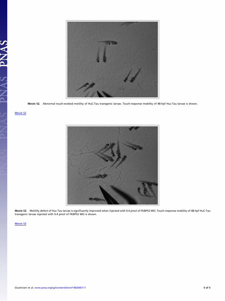
Movie S2. Abnormal touch-evoked motility of HuC-Tau transgenic larvae. Touch-response mobility of 48-hpf Huc-Tau larvae is shown.
Movie S2
Movie S3. Motility defect of Huc-Tau larvae is significantly improved when injected with 0.4 pmol of FKBP52 MO. Touch-response mobility of 48-hpf HuC-Tautransgenic larvae injected with 0.4 pmol of FKBP52 MO is shown.
Movie S3
Giustiniani et al. www.pnas.org/cgi/content/short/1402645111 5 of 5