publisher relating to the retraction of: oxidative …bartoli, danial h. platt, david fulton, ruth...
TRANSCRIPT

PUBLISHER’S NOTE
Publisher’s Note – Relating to the retraction of: Oxidative stressinactivates VEGF survival signaling in retinal endothelial cells viaPI 3-kinase tyrosine nitration. Azza B. El-Remessy, ManuelaBartoli, Danial H. Platt, David Fulton, Ruth B. Caldwell. J. Cell Sci.doi:10.1242/jcs.195966Michael WayEditor-in-Chief, Journal of Cell Science ( [email protected])
This Publisher’s Note concerns the retraction notice (J. Cell Sci. 2016 129, 3203 doi: 10.1242/jcs.195966) relating to the article ‘Oxidativestress inactivates VEGF survival signaling in retinal endothelial cells via PI 3-kinase tyrosine nitration’ by Azza B. El-Remessy, ManuelaBartoli, Danial H. Platt, David Fulton, Ruth B. Caldwell. J. Cell Sci. 2005 118, 243-252 (doi: 10.1242/jcs.01612).
Journal of Cell Science would like to clarify the following points:• Although involved in the investigation, the Charlie Norwood VA Medical Center did not exercise jurisdiction over the conclusions ofthe investigation of the paper named above.
• UGAwas presented with the professional opinions of select independent experts in the same field of research as Dr El-Remessy thatdisputed the findings of research misconduct, but University of Georgia stood by their initial decision to recommend retraction of thepaper named above.
• Dr El-Remessy does not agree with the findings of the investigation.
1856
© 2017. Published by The Company of Biologists Ltd | Journal of Cell Science (2017) 130, 1856 doi:10.1242/jcs.205336
Journal
ofCe
llScience
RETRACTION
Oxidative stress inactivates VEGF survival signaling in retinalendothelial cells via PI 3-kinase tyrosine nitrationAzza B. El-Remessy, Manuela Bartoli, Danial H. Platt, David Fulton and Ruth B. Caldwell
Retraction of: J. Cell Sci. 118, 243-252.
Journal of Cell Science was contacted by a reader who alerted us to potential band duplication and manipulation in Fig. 3B, Fig. 4A,B andFig. 5B in this paper. We contacted the corresponding author, Dr Azza El-Remessy, but she was unable to provide us with a satisfactoryexplanation. We therefore referred the matter to the institutes at which the research was performed by Dr El-Remessy, University of Georgiaand Augusta University (formerly Georgia Regents University), for investigation.
A joint investigatory committee between the two institutes, as well as the Charlie Norwood Veterans Affairs Medical Center, where Dr El-Remessy has additional appointments, concluded that Dr El-Remessy committed research misconduct by falsification or fabrication in theabove-named article. Specifically, evidence of direct and mirrored duplication or band substitution was detected in Fig. 3B, Fig. 4A,B andFig. 5B.
These findings undermine the integrity of the presented findings, and Journal of Cell Science is therefore following the recommendation ofthe committee to retract this article.
3203
© 2016. Published by The Company of Biologists Ltd | Journal of Cell Science (2016) 129, 3203 doi:10.1242/jcs.195966
Journal
ofCe
llScience

IntroductionThe initial stages of diabetic retinopathy are believed to leadto sight-threatening ischemic and proliferative retinopathywhen a critical number of retinal capillaries become non-perfused. It has been reported that retinal endothelial cells indiabetic patients and animals undergo accelerated death by aprocess consistent with apoptosis (Mizutani et al., 1996).Vascular endothelial growth factor (VEGF), an angiogeniccytokine secreted by a variety of cells, is a potent survivalfactor for endothelial cells in vivo and in vitro (Gerber et al.,1998a). Paradoxically, even though levels of VEGF and VEGFreceptor (VEGFR2) are substantially increased in diabeticretinas (Aiello et al., 1994; Duh and Aiello, 1999; El-Remessyet al., 2003b; Gardner et al., 2002; Joussen et al., 2002;Obrosova et al., 2001; Obrosova et al., 2003) and retinalendothelial cell death is also increased.
Vascular endothelial cells are an important target ofhyperglycemic damage (Nishikawa et al., 2000). It has beenreported that high glucose is associated with formation ofreactive oxygen species and that glucose-induced oxidativestress contributes to the development of hyperglycemia-induced vascular injury (Ceriello et al., 2001; Ceriello et al.,2002; Cosentino et al., 1997; Graier et al., 1999). We have
shown increased formation of nitric oxide, superoxide anionand their combination product peroxynitrite in retinalendothelial cells maintained in high glucose (El-Remessy et al.,2003a).
Peroxynitrite is a highly reactive oxidant that mediates avariety of biological processes including inhibition of keymetabolic enzymes, lipid peroxidation, nitration of the proteintyrosine residues and reduction of cellular antioxidant defensesby oxidation of thiol pools (Misko et al., 1998; Salgo et al.,1995). Recently, it has been shown that increased levels ofnitrotyrosine correlate with cell death in pancreas (Suarez-Pinzon et al., 1997) and in a model for oxygen-inducedretinopathy (Brooks et al., 2001). It has been reported that highglucose treatment stimulated endothelial cell death in isolatedhearts (Ceriello et al., 2002), in cultured endothelial cells(Morishita et al., 1997; Nakagami et al., 2001; Du et al., 1999;Zou et al., 2002) and in retinas of diabetic mice and patients(Kern et al., 2000; Mohr et al., 2002). Moreover, we haveshown that increased formation of peroxynitrite mediatesapoptosis in endothelial cells cultured in high oxygen (Gu etal., 2003). However, molecular mechanisms underlyingdiabetes-induced peroxynitrite formation and acceleration ofapoptosis in endothelial cells have not been elucidated. The
243
In diabetic retinopathy, endothelial cell apoptosis isparadoxically increased despite upregulation of the potentpro-survival factor VEGF. We tested the hypothesis thatelevated glucose levels disrupt VEGF’s pro-survivalfunction via peroxynitrite-mediated alteration of the Akt-1/p38 MAP kinase signaling pathway by studies of retinalendothelial cells in vitro. High glucose or exogenousperoxynitrite caused significant increases in apoptosis inthe presence or absence of VEGF. Treatment with aperoxynitrite decomposition catalyst blocked these effects,implying a causal role of peroxynitrite. Peroxynitrite orhigh glucose treatment also increased phosphorylation ofp38 MAP kinase, whereas phosphorylation of Akt-1 wassignificantly decreased in basal or VEGF-stimulatedconditions. High glucose- or peroxynitrite-treated cells alsoshowed significant increases in tyrosine nitration on the p85subunit of PI 3-kinase that blocked PI 3-kinase and Akt-1
kinase activity. Inhibiting peroxynitrite formation orblocking tyrosine nitration of p85 restored the activity ofPI 3-kinase and Akt-1 kinase, blocked phosphorylation ofp38 MAP kinase and normalized pro-survival function.Transfecting the cells with constitutively active Akt-1 orinhibiting activity of p38 MAP kinase completely maskedthe pro-apoptotic effects of high glucose and exogenousperoxynitrite, suggesting an interaction between the Akt-1and p38 MAP kinase pathways. In conclusion, high glucosetreatment blocks the pro-survival effect of VEGF andcauses accelerated endothelial cell apoptosis via the actionof peroxynitrite in causing tyrosine nitration of PI 3-kinase,inhibiting activity of Akt-1 kinase and increasing theactivity of p38 MAP kinase.
Key words: Peroxynitrite, High glucose, Apoptosis, Endothelialcells, p38 MAP kinase, Tyrosine nitration, Akt-1
Summary
Oxidative stress inactivates VEGF survival signalingin retinal endothelial cells via PI 3-kinase tyrosinenitrationAzza B. El-Remessy1,2,*, Manuela Bartoli1,3, Danial H. Platt1, David Fulton1,2 and Ruth B. Caldwell1,4,5
1Vascular Biology Center, 2Department of Pharmacology, 3Department of Pathology, 4Department of Cellular Biology and Anatomy and5Department of Ophthalmology, Medical College of Georgia, Augusta, GA 30912, USA*Author for correspondence (e-mail: [email protected])
Accepted 20 October 2004Journal of Cell Science 118, 243-252 Published by The Company of Biologists 2005doi:10.1242/jcs.01612
Research Article
Jour
nal o
f Cel
l Sci
ence
RETRACTED

244
paradox between increases in VEGF expression and retinal celldeath under diabetic condition has prompted us to study theeffects of high glucose-induced oxidative stress and exogenousperoxynitrite on cell death in retinal endothelial cells in orderto define the molecular mechanism by which peroxynitritealters the survival signaling pathway of VEGF under highglucose conditions.
Materials and MethodsCellsRetinal endothelial cells were isolated from bovine retinas byhomogenization and a series of filtration steps following anestablished protocol in our laboratory (Behzadian et al., 1998). Thecells (passages 6-8) were plated on gelatin-coated dishes or 4-wellchamber slides, grown to confluence and then treated for 5 days inconditions of normal glucose (NG, 5 mM glucose) or high glucose(HG, 25 mM glucose). Various osmotic control agents including L-glucose, a glucose stereoisomer (LG, 25 mM) and 3-methyl-O-glucose, a glucose analogue that is transported into the cells but notmetabolized (3mG, 25 mM) were examined. Cells were cultured inNG, HG, LG or 3mG serum-containing media for the initial 3 daysof treatment and then cells were washed with PBS twice and switchedto corresponding serum free media for 2 days. Serum starvation isknown to induce apoptosis in endothelial cells (Gratton et al., 2001).Cell culture reagents were purchased from Gibco-BRL (Grand Island,NY), with the exception of fetal bovine serum (FBS), which waspurchased from HyClone Laboratories, Inc (Logan, UT) and CS-Ccomplete medium, which was from Cell Systems Corporations(Kirkland, WA). Inhibitors including L-NAME, uric acid andsuperoxide dismutase-polyethylene glycol (SOD-PEG) were obtainedfrom Sigma-Aldrich (Saint Louis, MO). Inhibitors for peroxynitrite(FeTPPS) and for p38 MAP kinase (SB203580) were obtained fromCalbiochem (La Jolla, CA).
Peroxynitrite treatmentCells were switched to serum free medium, treated with 0.5 mMperoxynitrite and cultured for 16 hours. Peroxynitrite was purchasedfrom Upstate Biotechnology (Lake Placid, NY). Stockconcentrations of peroxynitrite were freshly prepared in 0.3 NNaOH. Peroxynitrite concentration was determined byspectrophotometry as described by Zou et al. (Zou et al., 2003).An equal amount of 0.3 N NaOH or decomposed peroxynitrite wasused for control experiments. Neither of these had an effect on anyof the parameters analyzed.
Caspase-3 activityCaspase-3 is an intracellular cysteine protease that exists as aproenzyme, becoming activated during the cascade of apoptosis. Theactivity of the enzyme was determined using a kit from R&D systems(Minneapolis, MN) according to the manufacturer’s recommendation.Briefly cells were lysed on ice for 10 minutes with lysis bufferprovided with the kit. To 50 µl of cell lysate, 50 µl of 2× reactionbuffer were added followed by 5 µl of caspase-3 fluorogenic substrate(DEVD-AFC). The mixture was incubated for 2 hours at 37°C andfluorescence was measured with a Cytofluor-4000 spectrophotometer(Foster City, CA) with an excitation of 400 nm and an emission of505 nm. The assay was done with non-induced cells (serum-supplemented cells) for comparative analysis.
Hoechst stainApoptotic cells in cultures treated as described above were detectedby nuclear staining with Hoechst 33258 (2.5 µg/ml in PBS). Hoechst
33258 was purchased from Molecular Probes (Eugene, OR). Cellswere reacted with the stain for 30 minutes at room temperature,washed twice with PBS, examined and photographed in a ZeissAxioskop microscope. At least 15 fields were counted for eachtreatment. Each field contained over 130 cells. Cells with condensedchromatin, shrunken, irregular or fragmented nuclei were consideredapoptotic. The number of apoptotic nuclei was calculated relative tothe total number of nuclei. Total cell density was also calculated foreach treatment condition as an indicator of potential differences incytotoxicity.
Western blotting analysisRetinal endothelial cells were harvested after various treatments andlysed in modified RIPA buffer (20 mM Tris, 2.5 mM EDTA, 1% TritonX-100, 1% deoxycholate, 0.1% SDS, 40 mM NaF, 10 mM Na4P2O7,and 1 mM PMSF) for 30 minutes on ice. Insoluble material wasremoved by centrifugation at 12,000 g at 4°C for 30 minutes.Antibodies for phospho-p38 MAP kinase, p38 MAP kinase, phospho-Akt-1, Akt-1, PARP and Akt-1 kinase kit were purchased from CellSignaling Technology, Inc (Beverly, MA). For analysis of cleavedpoly ADP-ribose polymerase (PARP), phospho-p38 MAP kinase, p38MAP kinase, phospho-Akt-1 or Akt-1, 50 µg of total protein wasboiled in 2× Laemmli sample buffer, separated on a 10-12% SDS-polyacrylamide gel by electrophoresis, transferred to nitrocelluloseand reacted with specific antibody. The primary antibody was detectedusing a horseradish peroxidase-conjugated goat anti-mouse or anti-rabbit antibody (Amersham Biosciences, Buckinghamshire, UK) andenhanced chemiluminescence. Intensity of immunoreactivity wasmeasured using densitometry.
ImmunoprecipitationRetinal endothelial cells were cultured in high glucose in the presenceor absence of peroxynitrite decomposition catalyst, or treated withperoxynitrite for the desired time. Cell lysates were prepared asdescribed above for immunoblotting. Antibodies for nitrotyrosine,p85 and p110 subunits of PI 3-kinase were purchased from UpstateBiotechnology (Lake Placid, NY). For PI 3-kinase tyrosine nitration,500 µg protein was incubated with p85. The precipitated proteins wereanalyzed by SDS-PAGE and blotted with nitrotyrosine antibody orp85 for equal loading. To study the effects on the interaction of theregulatory subunit with the catalytic subunit of PI 3-kinase, 500 µgprotein was incubated with p110. The precipitated proteins wereanalyzed by SDS-PAGE and blotted by p85 antibody or p110 antibodyfor equal loading.
Akt-1 kinase assayRetinal endothelial cells were cultured in high glucose in thepresence or absence of ROS inhibitors, or treated with peroxynitritefor the desired time. For immunoprecipitation, 500 µg protein wasincubated with immobilized Akt-1G1 (Cell Signaling Technology,Beverly, MA) monoclonal antibody-containing beads overnight at4°C with gentle rocking. The beads were washed twice with lysisbuffer and once with kinase buffer (25 mM Tris, 5 mM β-glycerolphosphate, 2 mM DTT, 0.1 mM Na3VO4, 10 mM MgCl2).Beads were further incubated in 40 µl kinase buffer supplementedwith 200 µM ATP and 1 µg GSK-3 fusion protein for 30 minutes at30°C. The reaction was terminated with 20 µl 3× SDS sample buffer.After boiling and centrifuging, the supernatant was separated on a12% SDS-PAGE gel, transferred to nitrocellulose membrane andwestern immunoblotting was carried out with phopho-GSK-3α/β (Ser21/9) antibody as the primary antibody. Horseradishperoxidase-conjugated goat anti-rabbit antibody and enhancedchemiluminescence were used to detect the primary antibody.Intensity of the immunoreactivity was measured using densitometry.
Journal of Cell Science 118 (1)
Jour
nal o
f Cel
l Sci
ence
RETRACTED

245Tyrosine nitration mediates cell apoptosis
Adenoviral constructsβ-Galactosidase and C-terminal HA-tagged constitutively active Akt(myr-Akt) were generated as described previously (Fulton et al.,1999). Retinal endothelial cells were infected with adenovirus[multiplicity of infection (m.o.i.) of 100] containing the β-galactosidase and myr-Akt. The virus was removed and cells were leftto recover for 12 hours in complete medium. These conditionsresulted in uniform expression of the transgenes in close to 95% ofthe cells (determined by infection with β-galactosidase, followed bystaining for β-galactosidase activity). Western blot analysis usingantibodies against phospho Akt-1 and GSK-3 (Cell SignalingTechnology, Inc. Beverly, MA) confirmed the over-expression andactivity of Akt-1 in cells transfected with myr-Akt but not in cellstransfected with β-galactosidase.
Statistical analysisThe results are expressed as mean ± s.e.m. Differences amongexperimental groups were evaluated by ANOVA and the significanceof differences between these groups was assessed by Fisher’s post-hoc least significant difference test. Significance was defined atP<0.05.
ResultsHigh glucose-induced peroxynitrite inactivates VEGFpro-survival signal and induces apoptosis of endothelialcellsOur previous studies in retinal endothelial cells showed thatcultures maintained in high glucose had significant increasesin superoxide anion, nitric oxide and peroxynitrite formation(El-Remessy et al., 2003a). In the present study, we examinedthe effects of high glucose-induced oxidative stress andexogenous peroxynitrite on VEGF pro-survival function. Asshown in Fig. 1, high glucose and peroxynitrite treatmentaccelerated serum starvation-induced endothelial cell deathas indicated by significant increases in caspase-3 activitycompared to normal glucose. Treatment with vehicle ordecomposed peroxynitrite had no effect on capsase-3 activity(data not shown). Caspase-3 is an intracellular cysteineprotease that exists as a pro-enzyme, becoming activatedduring the cascade of intracellular signaling events thatculminates in apoptosis. VEGF (40 ng/ml) inhibited serumstarvation-induced activation of caspase-3 in control culturesmaintained in normal glucose, but had no effect on culturestreated with either high glucose or exogenous peroxynitrite.Moreover, the specific peroxynitrite decomposition catalystFeTPPS (2.5 µM) restored the pro-survival effects of VEGF oncultures maintained in high glucose. This finding suggests thatthe effect of high glucose in increasing apoptosis in retinalendothelial cells may be due to the action of peroxynitrite inaltering the VEGF cell survival pathway.
Osmotic stress contributes to oxidative stress-inducedcell deathOur previous studies have shown that osmotic control agentsincluding the glucose stereoisomer (LG) and the glucoseanalog, 3-O-methyl glucose (3mG) also cause increases ofsuperoxide anion and peroxynitrite formation in retinalendothelial cells, though to a lesser extent than high glucose.This effect was found to be due to activation of aldosereductase, a rate-limiting step in the polyol pathway (El-
Remessy et al., 2003a). As shown in Table 1, caspase-3 activitywas increased in cells cultured in high osmolarity (5 mMglucose + 20 mM LG or 5 mM glucose + 20 mM 3mG)compared to cells cultured in normal glucose. The effects ofhigh glucose/osmotic stress were blocked by the specific aldosereductase inhibitor Zopolrestat (10 µM) and by theperoxynitrite scavengers uric acid (1 mM) and FeTPPS (2.5µM). The caspase-3 activity was also blocked by inhibitingNOS using L-NAME (0.5 mM) and significantly reducedby superoxide dismutase (SOD, 100 U/ml) confirming theinvolvement of peroxynitrite in high glucose and highosmolarity-induced cell death.
The oxidative stress-induced cell death was additionally
0
2
4
6
8
01
21
I+V+GHI+S-GHV+NPNPV+GHGHV+GNGNF
old
ofca
spas
e-3
acti
vity
GHGHNPNPGHGHGNGN
FGEV lm/gn04 +-+-+-+-
SPPTeF 5.2 µµµµM ++------
*
*
*
*
*†
Fig. 1. High glucose and peroxynitrite inactivate VEGF pro-survivaland accelerate endothelial cell death. Statistical analysis of caspase-3activity showing significant increases in apoptosis in cells treatedwith peroxynitrite (PN) or cultured in high glucose (HG) comparedto normal glucose (NG). Exogenous VEGF (40 ng/ml) protectedcells cultured in normal glucose from serum starvation-inducedapoptosis but did not rescue cells cultured in high glucose orperoxynitrite. FeTPPS (2.5 µM) a specific peroxynitritedecomposition catalyst restored the pro-survival effect of VEGF incells maintained in high glucose cultures. Similar results wereobtained in another three experiments. (*P<0.05 compared to NG,P<0.05 compared to HG.)
Table 1. Retinal endothelial cell caspase-3 activity as ameasure of apoptosis induced in high glucose and high
osmolarity is blocked by inhibiting the polyol pathway andby blocking peroxynitrite formation
Caspase-3 activity (fold)
Treatment NG HG 3mG LG
Untreated 4.6±0.45 8.1±0.7* 7.15±0.6* 7.6±0.5*Zoplorestat 3.6±0.26 4.9±0.29 4.7±0.27 4.6±0.34Uric acid 3.6±1.34 4.6±0.5 5.1±0.4 4.8±0.7FeTTPS 3.4±0.31 3.85±0.4 3.7±0.21 NDL-NAME 3.93±0.3 3.6±0.3 3.5±0.2 NDSOD 3.8±0.37 5.6±0.6† 5.9±0.5† 5.8±0.6†
NG, normal glucose (5 mM); HG, high glucose (25 mM); 3mG, 3-O-methyl glucose (5 mM glucose+20 mM 3mG); LG, L-glucose (5 mMglucose+20 mM L-glucose)
Zopolrestat (10 µM), an aldose reductase inhibitor; FeTPPS (2.5 µM) anduric acid (1 mM), peroxynitrite scavengers; L-NAME (0.5 mM), a NOSinhibitor; SOD (100 U/ml) superoxide dismutase.
*P<0.05 compared with NG; †P<0.05 compared with untreated HG, 3mGor LG.
Jour
nal o
f Cel
l Sci
ence
RETRACTED

246
assessed by nuclear staining with Hoechst 33258 whichshowed that the cultures maintained in medium with highglucose or high osmolarity (3mG or LG) had significantlyincreased numbers of cells with shrunken or irregular nucleicharacteristic of apoptosis (32%, 28% and 26% respectively,P<0.01, Table 2). Fig. 2A shows images of Hoechst nuclearstain of cells undergoing apoptosis. The increase in apoptoticcell profiles was significantly inhibited by treatment with uricacid, superoxide dismutase, or L-NAME providing furthersupport for the role of peroxynitrite in the apoptotic process.The oxidative stress-induced cell apoptosis was further
confirmed using western blot analysis of cleaved poly ADP-ribose polymerase (PARP). Detection of cleaved PARP (89kDa), a cleavage target of caspase-3, is accepted as a markerfor cell apoptosis. A representative western blot andnormalized PARP/actin expression are shown in Fig. 2B.Cultures maintained in high glucose, 3-O-methyl glucose orexogenous peroxynitrite showed significant increases in PARPexpression compared to cultures maintained in normal glucose.
High glucose-induced peroxynitrite inactivates VEGFpro-survival via alteration of Akt-1/P38 MAP kinaseThe above data indicate that peroxynitrite-mediated oxidative
Journal of Cell Science 118 (1)
Fig. 2. Oxidative stress-induced apoptosis in high glucose, highosmolarity and peroxynitrite. (A) Representative images of Hoechst33258 nuclear staining show apoptotic cells that were identified basedon their shrunken, irregular or fragmented nuclei. Quantification ofthese data is shown in Table 2. (B) A representative immunoblot, usingcell lysates (50 µg), for cleaved PARP and β-actin. Below: statisticalanalysis of quantification of cleaved PARP showing significantincreases in apoptosis in cells treated with high glucose (HG), 3-O-methyl glucose (3mG) or peroxynitrite in comparison to cells culturedin normal glucose (NG). (*P<0.05 vs NG.)
Table 2. High glucose- and high osmolarity-inducedapoptosis of retinal endothelial cells is blocked by
inhibiting peroxynitrite formation % Apoptotic cells
Treatment NG HG 3mG LG
Untreated 16.8±1.5 31.4±1.9* 28.1±2.5* 25.8±2.1*Uric acid 15.7±1.3 19.5±1.7 20.2±1.7 19.7±1.7L-NAME 14.6±1.3 20.1±1.9 19.65±1.8 17.6±1.5SOD 15.7±1.4 26.3±2.3† 22.9±2.1† 21.5±1.7†
Percentage of apoptotic cells were calculated by counting 10 fields, 200cells each in at least three independent cultures.
NG, normal glucose (5 mM); HG, high glucose (25 mM); 3mG, 3-O-methyl glucose (5 mM glucose+20 mM 3mG); LG, L-glucose (5 mMglucose+20 mM L-glucose)
Uric acid (1 mM) a peroxynitrite scavenger; L-NAME (0.5 mM), a NOSinhibitor; SOD (100 U/ml), superoxide dismutase.
*P<0.05 compared with NG; †P<0.05 compared with untreated HG, 3mGor LG.
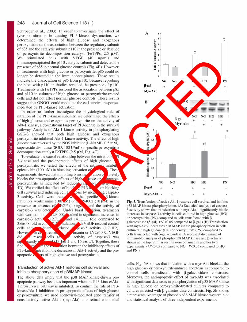
Fig. 3. High glucose and peroxynitrite inactivate VEGF pro-survivalvia alteration of Akt-1/P38 MAP kinase activation. (A) High glucose(HG) and peroxynitrite (PN) significantly decreased phosphorylationof Akt-1 compared to normal glucose (NG) even in the presence ofexogenous VEGF (40 mg/ml). (*P<0.05 compared to NG.) (B) Highglucose (HG) and peroxynitrite (PN) significantly increasedphosphorylation of p38 MAP kinase in basal or VEGF (40 mg/ml)-stimulated conditions. FeTPPS (2.5 µM) a specific peroxynitritedecomposition catalyst blocked the increases in p38 MAP kinasephosphorylation in both basal and VEGF stimulated conditions.Identical results were obtained in another two experiments. (*P<0.05compared to NG, †P<0.05 compared to HG.)
Jour
nal o
f Cel
l Sci
ence
RETRACTED

247Tyrosine nitration mediates cell apoptosis
stress accelerates retinal endothelial cell death in culturesmaintained in high glucose and inactivates VEGF pro-survivalfunction. Activation of the PI 3-kinase/Akt-1 pathway plays acritical role in the VEGF-survival pathway, whereas activationof p38 MAP kinase can induce cell death. We, therefore,examined whether the high glucose-induced impairment ofVEGF-mediated cell survival function could be the result ofalterations of the pro-survival Akt-1 pathway or the pro-apoptotic p38 MAP kinase pathway. Analysis of the VEGF-induced phosphorylation of Akt-1 showed that the pro-apoptotic effect of either high glucose-induced oxidative stressor exogenous peroxynitrite was associated with significantdecreases in phosphorylation of Akt-1 in basal and VEGF-stimulated conditions (Fig. 3A). Analysis of the VEGF-induced phosphorylation of P38 MAP kinase showed that the
pro-apoptotic effect of either high glucose orperoxynitrite was associated with significant increasesin phosphorylation of p38 MAP kinase in the presenceor absence of exogenous VEGF (Fig. 3B). Treatmentof cultures maintained in high glucose with the specificperoxynitrite decomposition catalyst FeTTPS blockedthe increases in p38 MAP kinase phosphorylation inboth basal and VEGF-stimulated conditions. Controlexperiments with decomposed peroxynitrite showedno significant difference compared to normal glucose(data not shown).
Tyrosine nitration inactivates PI 3-kinase/Akt-1 pro-survival pathwayPrevious work in vitro has shown that the p85 regulatorysubunit of PI 3-kinase is one target for peroxynitrite-inducedprotein nitration on tyrosine (Hellberg et al., 1998). Ourimmunoprecipitation studies with an antibody that recognizesthe p85 regulatory subunit of PI 3-kinase showed that culturesmaintained in high glucose or treated with peroxynitrite had asignificant increase in tyrosine nitration of p85 compared tocultures in normal glucose (Fig. 4A). These increases intyrosine nitration were blocked by the specific peroxynitritedecomposition catalyst FeTTPs (2.5 µM) and by the specificnitration inhibitor epicatechin (100 µM). Epicatechin, a dietaryflavanol, has been used to selectively block the nitrating effectof peroxynitrite on tyrosine residues (Schroeder et al., 2001;
0
2
4
6
8
01
21
Fol
dof
casp
ase-
3ac
tivi
ty
NPNPGHGHGNGN,nihcetacipE 001 µµµµM +-+-+-
**
D
Fig. 4. Tyrosine nitration inactivates PI 3-kinase/Akt-1 pro-survival pathway. (A) Immunoprecipitation with p85subunit of PI 3-kinase and western blot analysis using anti-nitrotyrosine antibody showed that cells treated with highglucose (HG) or peroxynitrite (PN) had significantly morenitration on the regulatory p85 subunit compared with cellscultured in normal glucose (NG). (*P<0.05 compared toNG.) This effect was blocked by the specific peroxynitritedecomposition catalyst FeTPPS (2.5 µM) and the specificnitration inhibitor epicatechin (100 µM).(B) Immunoprecipitation with p110 subunit of PI 3-kinaseand western blot analysis using p85 antibody showed thatunder high glucose (HG) or peroxynitrite (PN) treatments,the p85 subunit was hardly detected in theimmunopreicipitate of the catalytic p110 subunit comparedwith cells cultured in normal glucose (NG). Cells werestimulated with VEGF (40 ng/ml) in the presence orabsence of the peroxynitrite decomposition catalyst(FeTPPs). The association between p85 and p110 wasrestored by treatment with FeTPPs (2.5 µM). (C) Highglucose (HG) and peroxynitrite (PN) decreased Akt-1activity significantly compared to normal glucose (NG).Akt-1 kinase activity was restored by treatment of highglucose cultures with a specific peroxynitrite inhibitor (2.5µM), NOS inhibitor (L-NAME, 0.5 mM) and superoxidedismutase (SOD, 100 U/ml). A western blot of phosphoGSK-3, the substrate of Akt-1 kinase is shown at the top.(*P<0.05 compared to NG.) (D) Statistical analysis ofcaspase-3 activity showing significant increases in apoptosisin cells cultured in high glucose (HG) or treated withperoxynitrite (PN) compared to normal glucose (NG).These effects were blocked by the specific nitrationinhibitor epicatechin (100 µM). Similar results wereobtained in another three experiments. (*P<0.05 comparedto NG.)
Jour
nal o
f Cel
l Sci
ence
RETRACTED
248
Schroeder et al., 2003). In order to investigate the effect oftyrosine nitration in causing PI 3-kinase dysfunction, wedetermined the effects of high glucose and exogenousperoxynitrite on the association between the regulatory subunitof p85 and the catalytic subunit p110 in the presence or absenceof peroxynitrite decomposition catalyst (FeTPPs, 2.5 µM).We stimulated cells with VEGF (40 ng/ml) andimmunoprecipitated the p110 catalytic subunit and detected thepresence of p85 in normal glucose controls (Fig. 4B). However,in treatments with high glucose or peroxynitrite, p85 could nolonger be detected in the immunoprecipitates. These resultsindicate the dissociation of p85 from p110, because reprobingthe blots with p110 antibodies revealed the presence of p110.Treatments with FeTPPs restored the association between p85and p110 in cultures of high glucose or peroxynitrite-treatedcells and did not affect normal glucose controls. These resultssuggest that ONOO– could modulate the cell survival responsesmediated by PI 3-kinase activation.
In order to further investigate the physiological role ofnitration of the PI 3-kinase subunits, we determined the effectsof high glucose and exogenous peroxynitrite on the activity ofAkt-1 kinase, a downstream target of PI 3-kinase in the survivalpathway. Analysis of Akt-1 kinase activity in phosphorylatingGSK-3 showed that both high glucose and exogenousperoxynitrite inhibited Akt-1 kinase activity. The effect of highglucose was reversed by the NOS inhibitor (L-NAME, 0.5 mM),superoxide dismutase (SOD, 100 U/ml) or specific peroxynitritedecomposition catalyst FeTPPS (2.5 µM, Fig. 4C).
To evaluate the causal relationship between the nitration of PI3-kinase and the pro-apoptotic effects of high glucose andperoxynitrite, we tested the effects of the nitration inhibitorepicatechin (100 µM) in blocking activation of caspase-3. Theseexperiments showed that inhibiting tyrosine nitration completelyblocks the pro-apoptotic effects of high glucose or exogenousperoxynitrite as indicated by reduced caspase-3 activity (Fig.4D). We verified the effects of blocking PI 3-kinase on blockingcell survival and inducing cell apoptosis by measuring caspase-3 activity. Cells were treated with the specific PI 3-kinaseinhibitors wortmannin (100 nM) or LY294002 (10 µM) in thepresence or absence of VEGF (40 ng/ml) and the activity ofcaspase-3 was determined. Under basal condition, treatmentswith wortmanin or LY294002 resulted in significant increases incaspase-3 activity (12.7±1.3 and 14.1±1.1 fold compared to3.6±0.8 fold in control). Stimulation with VEGF rescued controlcells and significantly reduced caspase-2 activity (1.7±0.2).However, in cells treated with wortmanin or LY294002, VEGFdid not rescue cells and the activity of caspase-3 wassignificantly increased (13.1±1.1 and 16.9±1.7). Together, thesefindings confirm the correlation between the inhibitory effects ofPI 3-kinase nitration, the decreases in Akt-1 activity and the pro-apoptotic effects of high glucose and peroxynitrite.
Transfection of active Akt-1 restores cell survival andinhibits phosphorylation of p38MAP kinaseThe above data imply that the p38 MAP kinase-driven pro-apoptotic pathway becomes important when the PI 3-kinase/Akt-1 pro-survival pathway is inhibited. To confirm the role of PI 3-kinase/Akt-1 inhibition in pro-apoptotic effects of high glucoseor peroxynitrite, we used adenoviral-mediated gene transfer ofconstitutively active Akt-1 (myr-Akt) into retinal endothelial
cells. Fig. 5A shows that infection with a myr-Akt blocked thehigh glucose- or peroxynitrite-induced apoptosis as compared tocontrol cells transfected with β-galactosidase constructs.Moreover, the anti-apoptotic effect of myr-Akt was associatedwith significant decreases in phosphorylation of p38 MAP kinasein high glucose or peroxynitrite-treated cultures compared tocultures infected with β-galactosidase constructs. Fig. 5B showsa representative image of phospho p38 MAP kinase western blotand statistical analysis of three independent experiments.
Journal of Cell Science 118 (1)
0
1
2
3
4
5
6
7
8
LAG-B-NPrym-GHLAG-B-GHrym-B-GNLAG-B-GN
Fol
dof
casp
ase-
3ac
tivi
ty
NPGHGHGNGN
+-+-+
-+-+-
ββββ lag-
tkA-ryM
*
*
A
Fig. 5. Transfection of active Akt-1 restores cell survival and inhibitsp38 MAP kinase phosphorylation. (A) Statistical analysis of caspase-3 activity shows that transfection with myr-Akt-1 significantly blocksincreases in caspase-3 activity in cells cultured in high glucose (HG)or peroxynitrite (PN) compared to cells transfected with β-galactosidase (β-gal). (*P<0.05 compared to β-gal.) (B) Transfectionwith myr-Akt-1 decreases p38 MAP kinase phosphorylation in cellscultured in high glucose (HG) or peroxynitrite (PN) compared tocells transfected with β-galactosidase. A representative image ofimmunoblot analysis of phospho p38 MAP kinase and β-actin isshown at the top. Similar results were obtained in another twoexperiments. (*P<0.05 compared to NG, †P<0.05 compared to HGand PN.)
Jour
nal o
f Cel
l Sci
ence
RETRACTED

249Tyrosine nitration mediates cell apoptosis
Inhibition of p38 MAP kinase restores cellsurvival and Akt-1 phosphorylationTo confirm the role of p38 MAP kinase activationin the pro-apoptotic effects of high glucose orexogenous peroxynitrite, we treated the cellswith the specific p38 MAP kinase inhibitorSB203580 (25 µM) and assessed apoptosis bymeasurement of caspase-3 activity. The p38MAP kinase inhibitor did not affect cell deathin the normal glucose control but reducedsignificantly the apoptosis induced in highglucose-maintained or exogenous peroxynitrite-treated cultures (Fig. 6A). Moreover, inhibitionof p38 MAP kinase restored basal levels ofAkt-1 phosphorylation in high glucose orperoxynitrite-treated cultures, but had nosignificant effect on normal cultures (Fig. 6B).This result suggests that cross-talk between PI 3-kinase/Akt-1 and p38 MAP kinase signalingpathways contributes to the balance of pro- vsanti-apoptotic signaling pathway and hence thesurvival of endothelial cells.
DiscussionThe present study provides a novel mechanism of oxidativestress-induced apoptosis via tyrosine nitration-mediatedinhibition of PI-3kinase/Akt-1 and activation of p38 MAPkinase pathway. In this study, we demonstrate that tyrosinenitration of the PI 3-kinase p85 subunit inactivates the survivalsignaling pathway in retinal endothelial cells cultured in highglucose or exogenous peroxynitrite. These effects are blockedby the specific peroxynitrite decomposition catalyst FeTPPsand by the specific nitration inhibitor epicatechin. We showalso for the first time that the pro-apoptotic effect of highglucose or peroxynitrite is associated with imbalance betweenAkt-1 and p38 MAP kinase phosphorylation and that blockingp38 MAP kinase or over-expression of constitutively activeAkt-1 masks the pro-apoptotic effect of high glucose orperoxynitrite and restores survival function in retinalendothelial cells.
High glucose concentrations mimicking the diabeticsituation have been shown to trigger endothelial cell death(Baumgartner-Parzer et al., 1995; Du et al., 1999; Ido et al.,
2002; Mizutani et al., 1996; Mohr et al., 2002; Quagliaro et al.,2003) and to increase formation of reactive oxygen andnitrogen species (Cosentino et al., 1997; El-Remessy et al.,2003a; Quagliaro et al., 2003; Zou et al., 2002). Paradoxically,high glucose also stimulates formation of the endothelial cellsurvival factor VEGF (Behzadian et al., 2003; Kim et al., 2000;Williams et al., 1997). Thus, we tested the hypothesis that highglucose increases apoptosis in endothelial cells through theaction of peroxynitrite altering the VEGF cell survivalpathway. We examined the pro-survival effects of exogenousVEGF (40 ng/ml) on rescuing endothelial cells from serumwithdrawal-induced apoptosis under various conditions bydetermining caspase-3 activity. VEGF protected endothelialcells cultured in normal glucose but not those cultured in highglucose or peroxynitrite. The peroxynitrite decompositioncatalyst (FeTPPs) restored the pro-survival effect of VEGF incells cultured in high glucose, suggesting that high glucosealters pro-survival signaling of VEGF via peroxynitriteformation. Cell death by apoptosis was also confirmed bymorphological study of nuclear changes, using the nuclearstain Hoechst 33258 and western blot analysis of cleaved poly
0
2
4
6
8
01
BS-NPNPBS-GHS-GHBS-GNS-GN
Fol
dof
casp
ase-
3ac
tivi
ty
NPNPGHGHGNGN
+-+-+-52,085302BS µµµµM
*
*
†
AFig. 6. Inhibition of p38 MAP kinase with SB203580restores cell survival and Akt-1 phosphorylation.(A) Statistical analysis of caspase-3 activity showsthat inhibition of p38 MAP kinase significantlyreduced increases of caspase-3 activity in cellscultured in high glucose (HG) or peroxynitrite (PN)compared to cells cultured in normal glucose (NG).(*P<0.05 compared to NG, †P<0.05 compared toPN.) (B) Inhibition of p38 MAP kinase restores Akt-1 phosphorylation in cells cultured in high glucose(HG) or peroxynitrite (PN) compared to cellscultured in normal glucose (NG). A representativeimage of immunoblot analysis, using cell lysates (50µg), for phospho Akt-1 and β-actin is shown at thetop. Similar results were obtained in another twoexperiments. (*P<0.05 compared to NG.)
Jour
nal o
f Cel
l Sci
ence
RETRACTED

250
(ADP-ribose) polymerase (PARP). The pro-apoptotic effects ofhigh glucose and peroxynitrite were associated with activationof p38 MAP kinase in the presence or absence of exogenousVEGF. These results are in agreement with previous reports(Igarashi et al., 1999; Nakagami et al., 2001). Moreover, theperoxynitrite decomposition catalyst FeTPPS blocked theincreases in p38 MAP kinase phosphorylation in high glucosecultures under basal or VEGF-stimulated conditions, furthersupporting a causal role of peroxynitrite inactivating VEGFpro-survival signal and causing cell death.
The role of the PI 3-kinase/Akt-1 pathway in VEGF’s pro-survival function was confirmed by studies showing that thespecific inhibitors of PI 3-kinase, including wortmanin andLY294002, caused retinal endothelial cells to undergoapoptosis as indicated by significant increases in caspase-3activity in the presence or absence of exogenous VEGF. Theseresults are in agreement with several reports showing thatVEGF does not rescue endothelial cells from serum starvation-induced apoptosis in the presence of a specific inhibitor of PI3-kinase (Fujio and Walsh, 1999; Gerber et al., 1998b; Grattonet al., 2001; Suhara et al., 2001). Previous work in vitro hasshown that the p85 regulatory subunit of PI 3-kinase is a targetfor peroxynitrite-induced tyrosine nitration (Hellberg et al.,1998). Our data showed that cultures treated with high glucoseor exogenous peroxynitrite had significant increases in tyrosinenitration of the p85 subunit. This effect was accompanied bydramatic decreases in the association of the regulatory p85subunit from the catalytic p110 subunit, as well as bysignificant decreases in Akt-1 phosphorylation and Akt-1kinase activity, confirming an inhibitory effect of the nitrationof PI 3-kinase. Moreover, the specific peroxynitritedecomposition catalyst FeTPPs and the nitration inhibitorepicatechin blocked tyrosine nitration, restored p85/p110interaction of PI 3-kinase and Akt-1 kinase and survivalpromoting activity. These results confirm the relationshipbetween nitration of p85, the decreases in Akt-1 activity andthe pro-apoptotic effects of high glucose and peroxynitrite.Akt-1 kinase activity was restored also by inhibiting NOS andby SOD. Our findings lend further support to previous reportsof significant increases in tyrosine nitration in diabetic patients(Ceriello et al., 2001), in high glucose models (Ceriello et al.,2002; Zou et al., 2002) and in experimental diabetes (El-Remessy et al., 2003b; Turko et al., 2001). However, our study
is the first we know of to elucidate the molecular mechanismof inhibitory tyrosine nitration of p85 leading to inactivationof the pro-survival effects of VEGF/Akt-1in a physiologicalmodel of high glucose-induced oxidative stress.
Further work is needed to determine the effects of highglucose and peroxynitrite-induced tyrosine nitration on othersteps in the VEGF signal transduction pathway. Studies nowin progress in our lab indicate that high glucose andperoxynitrite also induce tyrosine nitrosylation of VEGFR2.Interestingly, the high glucose treatment also causes increasesin tyrosine phosphorylation of VEGFR2. This suggests thattyrosine nitration induces activation of VEGFR2, as has beenshown previously for PDGF and EGF (Klotz et al., 2000; vander Vliet et al., 1998). More work is needed to define thespecific mechanism of VEGFR2 activation by high glucose andto determine the molecular and physiological consequences.However, this observation suggests that the inhibitory effectsof high glucose and peroxynitrite on cell survival are specificto a direct action of the tyrosine nitration inhibiting the PI3K/Akt signaling pathway.
Parallel activation of survival and death pathways hasrecently been documented in response to VEGF, FGF andcytokines such as TNF-α (Cardier and Erickson-Miller, 2002;Gratton et al., 2001; Harfouche et al., 2003; Matsumoto et al.,2002). Thus, it is likely that high glucose and peroxynitrite-induced alterations in the Akt-1 and p38 MAP kinaseinteraction could alter cell responses to other cell survivalstimuli in addition to VEGF. While further study is needed todirectly test this hypothesis, our previous study has shown thatperoxynitrite blocks the effects of either basic FGF or serumin activating the PI 3-kinase/Akt cell survival pathway (Gu etal., 2003).
We investigated the interaction between Akt-1 and p38MAP kinase pathways under high glucose or exogenousperoxynitrite treatment. It is interesting that expression ofconstitutively active Akt-1 (myr-Akt-1) masked the pro-apoptotic effects of high glucose and exogenous peroxynitriteand restored cell survival function in treated cells. In addition,blocking of p38 MAP kinase also rescued retinal endothelialcells from accelerated cell death. The protective effects ofSB203580 were more prominent in high glucose cultures thanin peroxynitrite-treated cells, probably because of themagnitude of peroxynitrite-induced cell death. Our resultsare in agreement with previous reports in other vascularendothelial cells (Fujio and Walsh, 1999; Gratton et al., 2001;Harfouche et al., 2003; Matsumoto et al., 2002; Yue et al.,1999). Furthermore, the anti-apoptotic effect of myr-Akt-1 wasassociated with significant inhibition of p38 MAP kinasephosphorylation and the protective effect of SB203580 wasassociated with significant increases in Akt-1 phosphorylation.(see Figs 5 and 6). These findings demonstrate for the first timea cross talk between Akt-1 and the p38 MAP kinase signalingpathway in retinal endothelial cells. It has been reportedthat inhibition of p38 activation is mediated throughphosphorylation and inhibition of MEKK3 by Akt-1 in aorticendothelial cells (Gratton et al., 2001). Similar results werereported (Harfouche et al., 2003) in HUVEC in response toangiopoietin-1 suggesting that the negative influence of Akt-1on p38 MAP kinase is a general phenomenon in endothelialcells.
Of note, we and others have reported the effects of osmotic
Journal of Cell Science 118 (1)
011P
esaniK3IP
58P
FGEV FGEV
+ P
1-tkA
sisotpopA sisotpopA
OONO -
83P
+ P
lavivruSlleC lavivruSlleC
Fig. 7. A schematic representation of the proposed mechanism bywhich high glucose, via its effect on peroxynitrite, inactivates theVEGF/PI 3-kinase/Akt-1 pro-survival pathway and stimulates celldeath via activation of p38 MAP kinase pathway. Nitration of PI 3-kinase is proposed as a mechanism by which high glucose switchesoff the VEGF pro-survival pathway and triggers the pro-apoptoticpathway.
Jour
nal o
f Cel
l Sci
ence
RETRACTED
251Tyrosine nitration mediates cell apoptosis
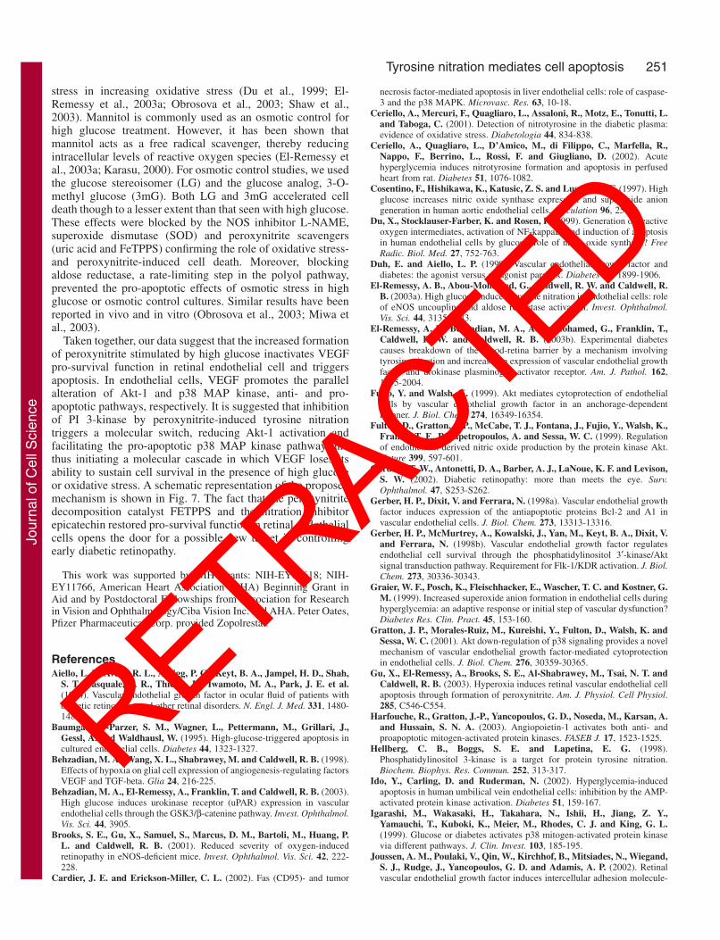
stress in increasing oxidative stress (Du et al., 1999; El-Remessy et al., 2003a; Obrosova et al., 2003; Shaw et al.,2003). Mannitol is commonly used as an osmotic control forhigh glucose treatment. However, it has been shown thatmannitol acts as a free radical scavenger, thereby reducingintracellular levels of reactive oxygen species (El-Remessy etal., 2003a; Karasu, 2000). For osmotic control studies, we usedthe glucose stereoisomer (LG) and the glucose analog, 3-O-methyl glucose (3mG). Both LG and 3mG accelerated celldeath though to a lesser extent than that seen with high glucose.These effects were blocked by the NOS inhibitor L-NAME,superoxide dismutase (SOD) and peroxynitrite scavengers(uric acid and FeTPPS) confirming the role of oxidative stress-and peroxynitrite-induced cell death. Moreover, blockingaldose reductase, a rate-limiting step in the polyol pathway,prevented the pro-apoptotic effects of osmotic stress in highglucose or osmotic control cultures. Similar results have beenreported in vivo and in vitro (Obrosova et al., 2003; Miwa etal., 2003).
Taken together, our data suggest that the increased formationof peroxynitrite stimulated by high glucose inactivates VEGFpro-survival function in retinal endothelial cell and triggersapoptosis. In endothelial cells, VEGF promotes the parallelalteration of Akt-1 and p38 MAP kinase, anti- and pro-apoptotic pathways, respectively. It is suggested that inhibitionof PI 3-kinase by peroxynitrite-induced tyrosine nitrationtriggers a molecular switch, reducing Akt-1 activation andfacilitating the pro-apoptotic p38 MAP kinase pathway andthus initiating a molecular cascade in which VEGF loses itsability to sustain cell survival in the presence of high glucoseor oxidative stress. A schematic representation of the proposedmechanism is shown in Fig. 7. The fact that the peroxynitritedecomposition catalyst FETPPS and the nitration inhibitorepicatechin restored pro-survival function in retinal endothelialcells opens the door for a possible new target in controllingearly diabetic retinopathy.
This work was supported by NIH Grants: NIH-EY04618; NIH-EY11766, American Heart Association (AHA) Beginning Grant inAid and by Postdoctoral Fellowships from Association for Researchin Vision and Ophthalmology/Ciba Vision Inc. and AHA. Peter Oates,Pfizer Pharmaceutical Corp. provided Zopolrestat.
ReferencesAiello, L. P., Avery, R. L., Arrigg, P. G., Keyt, B. A., Jampel, H. D., Shah,
S. T., Pasquale, L. R., Thieme, H., Iwamoto, M. A., Park, J. E. et al.(1994). Vascular endothelial growth factor in ocular fluid of patients withdiabetic retinopathy and other retinal disorders. N. Engl. J. Med. 331, 1480-1487.
Baumgartner-Parzer, S. M., Wagner, L., Pettermann, M., Grillari, J.,Gessl, A. and Waldhausl, W. (1995). High-glucose-triggered apoptosis incultured endothelial cells. Diabetes 44, 1323-1327.
Behzadian, M. A., Wang, X. L., Shabrawey, M. and Caldwell, R. B. (1998).Effects of hypoxia on glial cell expression of angiogenesis-regulating factorsVEGF and TGF-beta. Glia 24, 216-225.
Behzadian, M. A., El-Remessy, A., Franklin, T. and Caldwell, R. B. (2003).High glucose induces urokinase receptor (uPAR) expression in vascularendothelial cells through the GSK3/β-catenine pathway. Invest. Ophthalmol.Vis. Sci. 44, 3905.
Brooks, S. E., Gu, X., Samuel, S., Marcus, D. M., Bartoli, M., Huang, P.L. and Caldwell, R. B. (2001). Reduced severity of oxygen-inducedretinopathy in eNOS-deficient mice. Invest. Ophthalmol. Vis. Sci. 42, 222-228.
Cardier, J. E. and Erickson-Miller, C. L. (2002). Fas (CD95)- and tumor
necrosis factor-mediated apoptosis in liver endothelial cells: role of caspase-3 and the p38 MAPK. Microvasc. Res. 63, 10-18.
Ceriello, A., Mercuri, F., Quagliaro, L., Assaloni, R., Motz, E., Tonutti, L.and Taboga, C. (2001). Detection of nitrotyrosine in the diabetic plasma:evidence of oxidative stress. Diabetologia 44, 834-838.
Ceriello, A., Quagliaro, L., D’Amico, M., di Filippo, C., Marfella, R.,Nappo, F., Berrino, L., Rossi, F. and Giugliano, D. (2002). Acutehyperglycemia induces nitrotyrosine formation and apoptosis in perfusedheart from rat. Diabetes 51, 1076-1082.
Cosentino, F., Hishikawa, K., Katusic, Z. S. and Luscher, T. F. (1997). Highglucose increases nitric oxide synthase expression and superoxide aniongeneration in human aortic endothelial cells. Circulation 96, 25-28.
Du, X., Stocklauser-Farber, K. and Rosen, P. (1999). Generation of reactiveoxygen intermediates, activation of NF-kappaB, and induction of apoptosisin human endothelial cells by glucose: role of nitric oxide synthase? FreeRadic. Biol. Med. 27, 752-763.
Duh, E. and Aiello, L. P. (1999). Vascular endothelial growth factor anddiabetes: the agonist versus antagonist paradox. Diabetes 48, 1899-1906.
El-Remessy, A. B., Abou-Mohamed, G., Caldwell, R. W. and Caldwell, R.B. (2003a). High glucose-induced tyrosine nitration in endothelial cells: roleof eNOS uncoupling and aldose reductase activation. Invest. Ophthalmol.Vis. Sci. 44, 3135-3143.
El-Remessy, A. B., Behzadian, M. A., Abou-Mohamed, G., Franklin, T.,Caldwell, R. W. and Caldwell, R. B. (2003b). Experimental diabetescauses breakdown of the blood-retina barrier by a mechanism involvingtyrosine nitration and increases in expression of vascular endothelial growthfactor and urokinase plasminogen activator receptor. Am. J. Pathol. 162,1995-2004.
Fujio, Y. and Walsh, K. (1999). Akt mediates cytoprotection of endothelialcells by vascular endothelial growth factor in an anchorage-dependentmanner. J. Biol. Chem. 274, 16349-16354.
Fulton, D., Gratton, J. P., McCabe, T. J., Fontana, J., Fujio, Y., Walsh, K.,Franke, T. F., Papapetropoulos, A. and Sessa, W. C. (1999). Regulationof endothelium-derived nitric oxide production by the protein kinase Akt.Nature 399, 597-601.
Gardner, T. W., Antonetti, D. A., Barber, A. J., LaNoue, K. F. and Levison,S. W. (2002). Diabetic retinopathy: more than meets the eye. Surv.Ophthalmol. 47, S253-S262.
Gerber, H. P., Dixit, V. and Ferrara, N. (1998a). Vascular endothelial growthfactor induces expression of the antiapoptotic proteins Bcl-2 and A1 invascular endothelial cells. J. Biol. Chem. 273, 13313-13316.
Gerber, H. P., McMurtrey, A., Kowalski, J., Yan, M., Keyt, B. A., Dixit, V.and Ferrara, N. (1998b). Vascular endothelial growth factor regulatesendothelial cell survival through the phosphatidylinositol 3′-kinase/Aktsignal transduction pathway. Requirement for Flk-1/KDR activation. J. Biol.Chem. 273, 30336-30343.
Graier, W. F., Posch, K., Fleischhacker, E., Wascher, T. C. and Kostner, G.M. (1999). Increased superoxide anion formation in endothelial cells duringhyperglycemia: an adaptive response or initial step of vascular dysfunction?Diabetes Res. Clin. Pract. 45, 153-160.
Gratton, J. P., Morales-Ruiz, M., Kureishi, Y., Fulton, D., Walsh, K. andSessa, W. C. (2001). Akt down-regulation of p38 signaling provides a novelmechanism of vascular endothelial growth factor-mediated cytoprotectionin endothelial cells. J. Biol. Chem. 276, 30359-30365.
Gu, X., El-Remessy, A., Brooks, S. E., Al-Shabrawey, M., Tsai, N. T. andCaldwell, R. B. (2003). Hyperoxia induces retinal vascular endothelial cellapoptosis through formation of peroxynitrite. Am. J. Physiol. Cell Physiol.285, C546-C554.
Harfouche, R., Gratton, J.-P., Yancopoulos, G. D., Noseda, M., Karsan, A.and Hussain, S. N. A. (2003). Angiopoietin-1 activates both anti- andproapoptotic mitogen-activated protein kinases. FASEB J. 17, 1523-1525.
Hellberg, C. B., Boggs, S. E. and Lapetina, E. G. (1998).Phosphatidylinositol 3-kinase is a target for protein tyrosine nitration.Biochem. Biophys. Res. Commun. 252, 313-317.
Ido, Y., Carling, D. and Ruderman, N. (2002). Hyperglycemia-inducedapoptosis in human umbilical vein endothelial cells: inhibition by the AMP-activated protein kinase activation. Diabetes 51, 159-167.
Igarashi, M., Wakasaki, H., Takahara, N., Ishii, H., Jiang, Z. Y.,Yamauchi, T., Kuboki, K., Meier, M., Rhodes, C. J. and King, G. L.(1999). Glucose or diabetes activates p38 mitogen-activated protein kinasevia different pathways. J. Clin. Invest. 103, 185-195.
Joussen, A. M., Poulaki, V., Qin, W., Kirchhof, B., Mitsiades, N., Wiegand,S. J., Rudge, J., Yancopoulos, G. D. and Adamis, A. P. (2002). Retinalvascular endothelial growth factor induces intercellular adhesion molecule-
Jour
nal o
f Cel
l Sci
ence
RETRACTED

252
1 and endothelial nitric oxide synthase expression and initiates early diabeticretinal leukocyte adhesion in vivo. Am. J. Pathol. 160, 501-509.
Karasu, C. (2000). Time course of changes in endothelium-dependent and-independent relaxation of chronically diabetic aorta: role of reactive oxygenspecies. Eur. J. Pharmacol. 392, 163-173.
Kern, T. S., Tang, J., Mizutani, M., Kowluru, R. A., Nagaraj, R. H.,Romeo, G., Podesta, F. and Lorenzi, M. (2000). Response of capillary celldeath to aminoguanidine predicts the development of retinopathy:comparison of diabetes and galactosemia. Invest. Ophthalmol. Vis. Sci. 41,3972-3978.
Kim, N. H., Jung, H. H., Cha, D. R. and Choi, D. S. (2000). Expression ofvascular endothelial growth factor in response to high glucose in ratmesangial cells. J. Endocrinol. 165, 617-624.
Klotz, L. O., Schieke, S. M., Sies, H. and Holbrook, N. J. (2000).Peroxynitrite activates the phosphoinositide 3-kinase/Akt pathway in humanskin primary fibroblasts. Biochem. J. 352, 219-225.
Matsumoto, T., Turesson, I., Book, M., Gerwins, P. and Claesson-Welsh,L. (2002). p38 MAP kinase negatively regulates endothelial cell survival,proliferation, and differentiation in FGF-2-stimulated angiogenesis. J. CellBiol. 156, 149-160.
Misko, T. P., Highkin, M. K., Veenhuizen, A. W., Manning, P. T., Stern,M. K., Currie, M. G. and Salvemini, D. (1998). Characterization of thecytoprotective action of peroxynitrite decomposition catalysts. J. Biol.Chem. 273, 15646-15653.
Miwa, K., Nakamura, J., Hamada, Y., Naruse, K., Nakashima, E., Kato,K., Kasuya, Y., Yasuda, Y., Kamiya, H. and Hotta, N. (2003). The roleof polyol pathway in glucose-induced apoptosis of cultured retinal pericytes.Diabetes Res. Clin. Pract. 60, 1-9.
Mizutani, M., Kern, T. S. and Lorenzi, M. (1996). Accelerated death ofretinal microvascular cells in human and experimental diabetic retinopathy.J. Clin. Invest. 97, 2883-2890.
Mohr, S., Xi, X., Tang, J. and Kern, T. S. (2002). Caspase activation in retinasof diabetic and galactosemic mice and diabetic patients. Diabetes 51, 1172-1179.
Morishita, R., Higaki, J., Hayashi, S. I., Yo, Y., Aoki, M., Nakamura, S.,Moriguchi, A., Matsushita, H., Matsumoto, K., Nakamura, T. et al.(1997). Role of hepatocyte growth factor in endothelial regulation:prevention of high D-glucose-induced endothelial cell death byprostaglandins and phosphodiesterase type 3 inhibitor. Diabetologia 40,1053-1061.
Nakagami, H., Morishita, R., Yamamoto, K., Yoshimura, S. I., Taniyama,Y., Aoki, M., Matsubara, H., Kim, S., Kaneda, Y. and Ogihara, T.(2001). Phosphorylation of p38 mitogen-activated protein kinasedownstream of bax-caspase-3 pathway leads to cell death induced by highD-glucose in human endothelial cells. Diabetes 50, 1472-1481.
Nishikawa, T., Edelstein, D., Du, X. L., Yamagishi, S., Matsumura, T.,Kaneda, Y., Yorek, M. A., Beebe, D., Oates, P. J., Hammes, H. P. et al.(2000). Normalizing mitochondrial superoxide production blocks threepathways of hyperglycaemic damage. Nature 404, 787-790.
Obrosova, I. G., Minchenko, A. G., Marinescu, V., Fathallah, L., Kennedy,A., Stockert, C. M., Frank, R. N. and Stevens, M. J. (2001). Antioxidantsattenuate early up regulation of retinal vascular endothelial growth factor instreptozotocin-diabetic rats. Diabetologia 44, 1102-1110.
Obrosova, I. G., Minchenko, A. G., Vasupuram, R., White, L., Abatan, O.I., Kumagai, A. K., Frank, R. N. and Stevens, M. J. (2003). Aldose
reductase inhibitor fidarestat prevents retinal oxidative stress and vascularendothelial growth factor overexpression in streptozotocin-diabetic rats.Diabetes 52, 864-871.
Quagliaro, L., Piconi, L., Assaloni, R., Martinelli, L., Motz, E. andCeriello, A. (2003). Intermittent high glucose enhances apoptosis related tooxidative stress in human umbilical vein endothelial cells: the role of proteinkinase C and NAD(P)H-oxidase activation. Diabetes 52, 2795-2804.
Salgo, M. G., Squadrito, G. L. and Pryor, W. A. (1995). Peroxynitrite causesapoptosis in rat thymocytes. Biochem. Biophys. Res. Commun. 215, 1111-1118.
Schroeder, P., Klotz, L. O., Buchczyk, D. P., Sadik, C. D., Schewe, T. andSies, H. (2001). Epicatechin selectively prevents nitration but notoxidation reactions of peroxynitrite. Biochem. Biophys. Res. Commun.285, 782-787.
Schroeder, P., Klotz, L.-O. and Sies, H. (2003). Amphiphilic properties of(–)-epicatechin and their significance for protection of cells againstperoxynitrite. Biochem. Biophys. Res. Commun. 307, 69-73.
Shaw, S., Wang, X., Redd, H., Alexander, G. D., Isales, C. M. and Marrero,M. B. (2003). High glucose augments the angiotensin II-induced activationof JAK2 in vascular smooth muscle cells via the polyol pathway. J. Biol.Chem. 278, 30634-30641.
Suarez-Pinzon, W. L., Szabo, C. and Rabinovitch, A. (1997). Developmentof autoimmune diabetes in NOD mice is associated with the formation ofperoxynitrite in pancreatic islet beta-cells. Diabetes 46, 907-911.
Suhara, T., Mano, T., Oliveira, B. E. and Walsh, K. (2001).Phosphatidylinositol 3-kinase/Akt signaling controls endothelial cellsensitivity to Fas-mediated apoptosis via regulation of FLICE-inhibitoryprotein (FLIP). Circ. Res. 89, 13-19.
Turko, I. V., Marcondes, S. and Murad, F. (2001). Diabetes-associatednitration of tyrosine and inactivation of succinyl-CoA:3-oxoacid CoA-transferase. Am. J. Physiol. Heart Circ. Physiol. 281, H2289-H2294.
van der Vliet, A., Hristova, M., Cross, C. E., Eiserich, J. P. and Goldkorn,T. (1998). Peroxynitrite induces covalent dimerization of epidermal growthfactor receptors in A431 epidermoid carcinoma cells. J. Biol. Chem. 273,31860-31866.
Williams, B., Gallacher, B., Patel, H. and Orme, C. (1997). Glucose-inducedprotein kinase C activation regulates vascular permeability factor mRNAexpression and peptide production by human vascular smooth muscle cellsin vitro. Diabetes 46, 1497-1503.
Yue, T. L., Ni, J., Romanic, A. M., Gu, J. L., Keller, P., Wang, C., Kumar,S., Yu, G. L., Hart, T. K., Wang, X. et al. (1999). TL1, a novel tumornecrosis factor-like cytokine, induces apoptosis in endothelial cells.Involvement of activation of stress protein kinases (stress-activated proteinkinase and p38 mitogen-activated protein kinase) and caspase-3-likeprotease. J. Biol. Chem. 274, 1479-1486.
Zou, M. H., Shi, C. and Cohen, R. A. (2002). High glucose via peroxynitritecauses tyrosine nitration and inactivation of prostacyclin synthase that isassociated with thromboxane/prostaglandin H(2) receptor-mediatedapoptosis and adhesion molecule expression in cultured human aorticendothelial cells. Diabetes 51, 198-203.
Zou, M.-H., Hou, X.-Y., Shi, C.-M., Kirkpatick, S., Liu, F., Goldman, M.H. and Cohen, R. A. (2003). Activation of 5′-AMP-activated kinase ismediated through c-Src and phosphoinositide 3-kinase activity duringhypoxia-reoxygenation of bovine aortic endothelial cells: role ofperoxynitrite. J. Biol. Chem. 278, 34003-34010.
Journal of Cell Science 118 (1)
Jour
nal o
f Cel
l Sci
ence
RETRACTED