pulmonary abscess in children .. dr padmesh
TRANSCRIPT
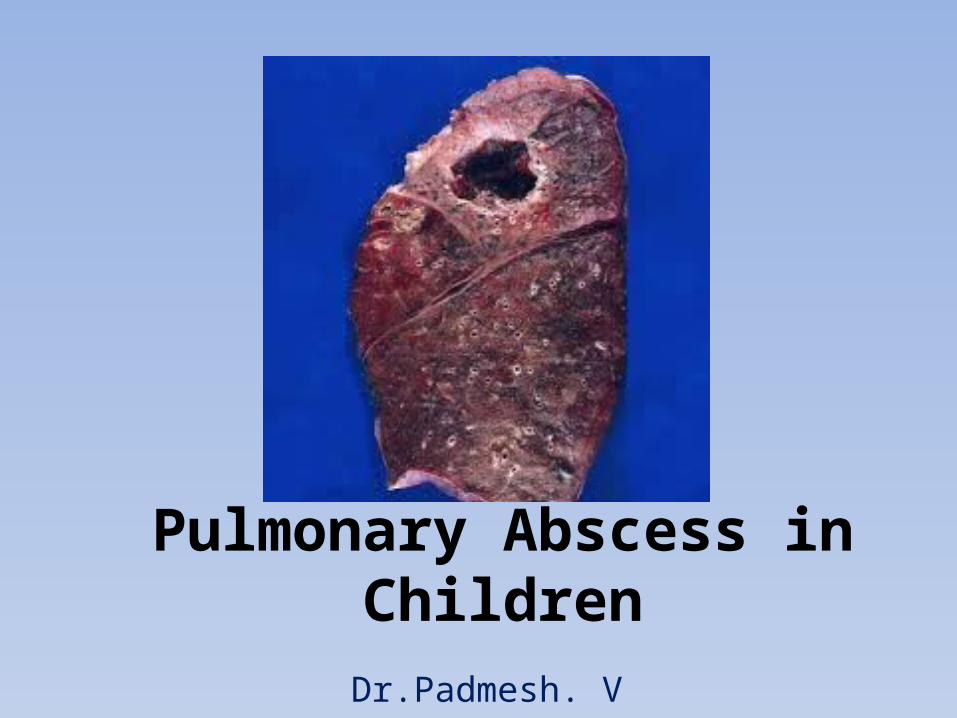
Pulmonary Abscess in Children
Dr.Padmesh. V
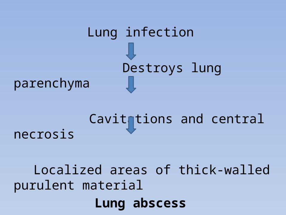
Lung infection
Destroys lung parenchyma
Cavitations and central necrosis
Localized areas of thick-walled purulent material Lung abscess

• Primary lung abscess:• Occur in previously healthy patients with no
underlying medical disorders.• Usually solitary.
• Secondary lung abscess:• Occur in patients with underlying or predisposing
conditions• May be multiple.

• PATHOLOGY AND PATHOGENESIS• Predisposing conditions:• Aspiration (of infected material or FB)• Pneumonia / Hematogenous seeding from other sites• Cystic fibrosis• Gastroesophageal reflux • Tracheoesophageal fistula• Immunodeficiencies • Postoperative complications of tonsillectomy and
adenoidectomy• Seizures• Neurologic and other conditions associated with
impaired mucociliary defense.

• PATHOLOGY AND PATHOGENESIS:
Aspiration of infected material or foreign body
Pneumonitis impairs drainage of fluid or aspirated material
Inflammatory vascular obstruction
Tissue necrosis, liquefaction
Abscess formation
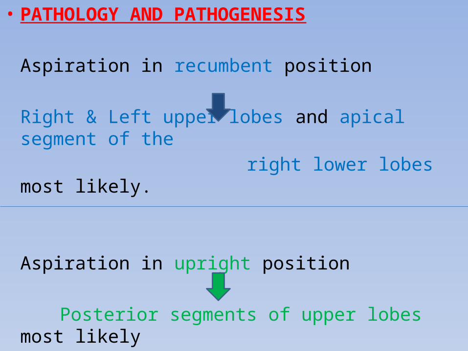
• PATHOLOGY AND PATHOGENESIS
Aspiration in recumbent position
Right & Left upper lobes and apical segment of the right lower lobes most likely.
Aspiration in upright position
Posterior segments of upper lobes most likely
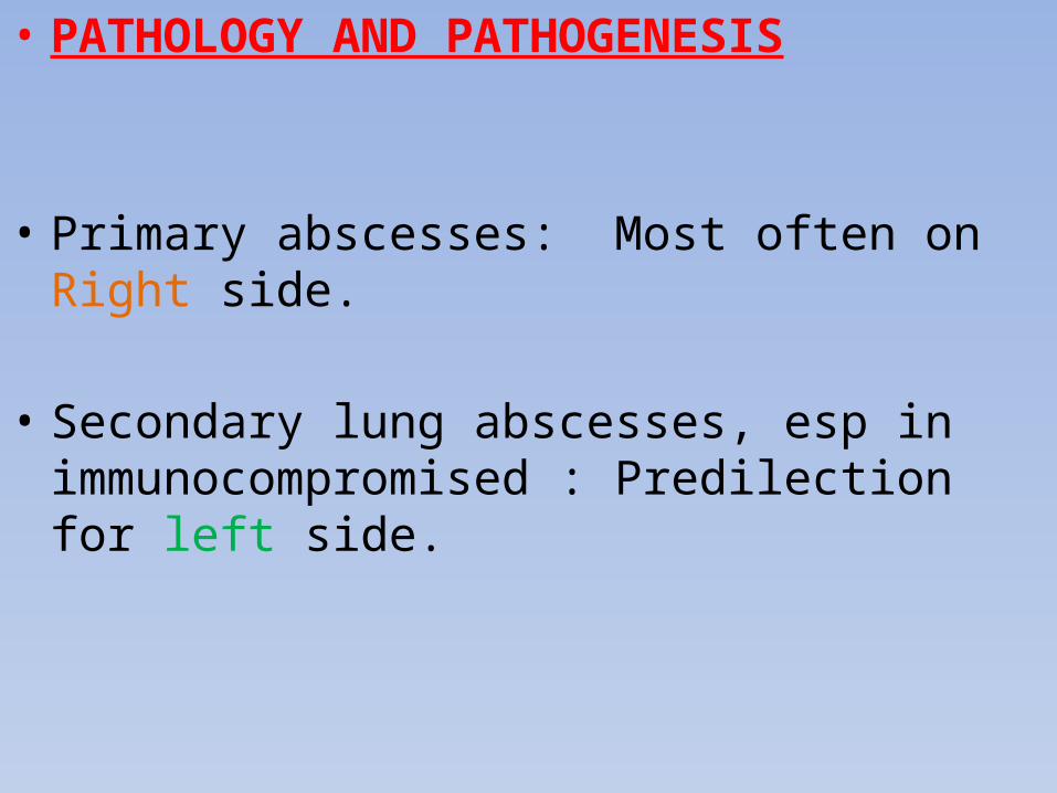
• PATHOLOGY AND PATHOGENESIS
• Primary abscesses: Most often on Right side.
• Secondary lung abscesses, esp in immunocompromised : Predilection for left side.
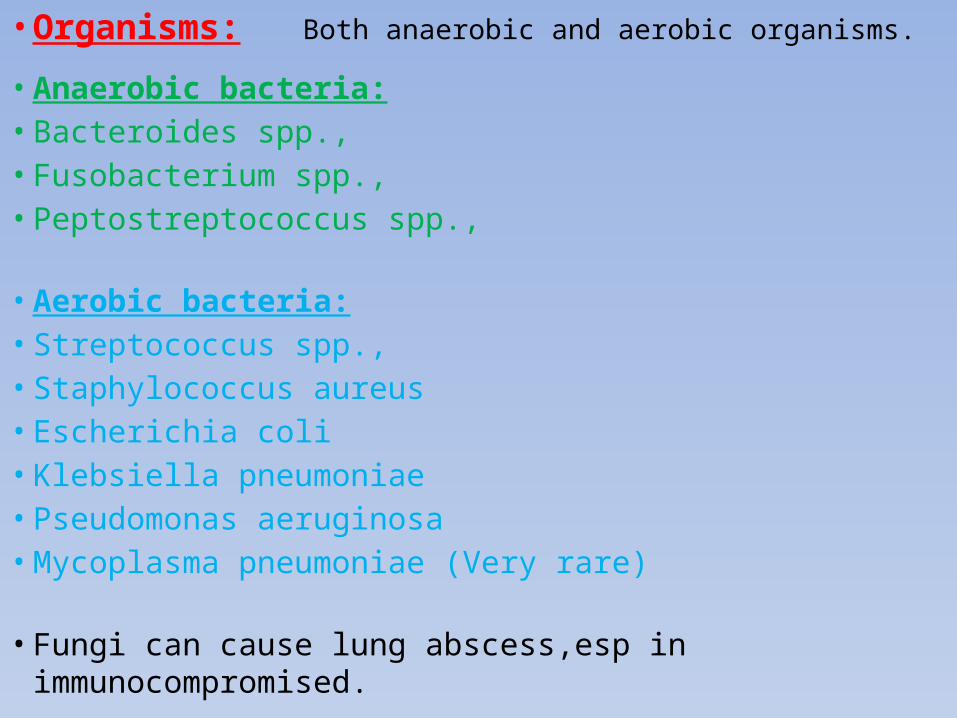
• Organisms: Both anaerobic and aerobic organisms.
• Anaerobic bacteria:• Bacteroides spp., • Fusobacterium spp.,• Peptostreptococcus spp.,
• Aerobic bacteria:• Streptococcus spp., • Staphylococcus aureus • Escherichia coli • Klebsiella pneumoniae• Pseudomonas aeruginosa• Mycoplasma pneumoniae (Very rare)
• Fungi can cause lung abscess,esp in immunocompromised.

• CLINICAL MANIFESTATIONS: Symptoms• Cough, • Fever, • Dyspnea, • Chest pain, • Vomiting,• Sputum production, • Weight loss, • Hemoptysis.
• CLINICAL MANIFESTATIONS: Signs• Tachypnea• Dyspnea• Retractions with accessory muscle use• Decreased breath sounds • Dullness to percussion in affected area • Crackles • Occasionally a prolonged expiratory phase on
auscultation
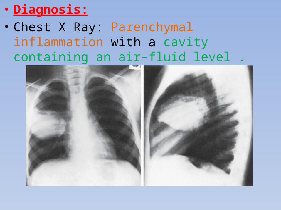
• Diagnosis:• Chest X Ray: Parenchymal inflammation with a
cavity containing an air–fluid level .

• Diagnosis:• Chest CT scan: Abscess is usually a thick-walled lesion with a low-density center progressing to an air–fluid level.

• Diagnosis:
• Gram stain of sputum: Early clue. • Sputum cultures: Yield mixed bacteria, therefore
not always reliable.
• Attempts to avoid contamination from oral flora include direct lung puncture, percutaneous (aided by CT guidance) or transtracheal aspiration, and bronchoalveolar lavage specimens obtained bronchoscopically.
• Diagnosis:
• Bronchoscopic aspiration should be avoided as it can be complicated by massive intrabronchial aspiration.
• To avoid invasive procedures in previously normal hosts, empiric therapy can be initiated in the absence of culturable material.

• Differential Diagnosis:
• Abscesses should be distinguished from pneumatoceles.
• Pneumatoceles often complicate severe bacterial pneumonias.
• Pneumatoceles: Thin- and Smooth-walled, localized air collections with or without air–fluid level.
• Pneumatoceles often resolve spontaneously with treatment of specific cause of the pneumonia.

• TREATMENT:
• Conservative management.• 2-3 wk course of parenteral antibiotics for
uncomplicated cases, followed by oral antibiotics to complete a Total of 4-6 wk.
• Aerobic and anaerobic coverage.
• Include penicillinase-resistant agent active against S.aureus and anaerobic coverage, typically clindamycin or ticarcillin/clavulanic acid.
• If Gram-negative bacteria are suspected or isolated, an aminoglycoside should be added.

• TREATMENT
• Early CT-guided percutaneous aspiration or drainage.
• Severely ill or those who fail to improve after 7-10 days of antibiotics Surgical intervention.
• Minimally invasive CT guided percutaneous aspiration.
• Thorascopic drainage.
• In rare complicated cases Thoracotomy with surgical drainage or lobectomy and/or decortication.

• PROGNOSIS
• Excellent.
• Presence of aerobic organisms may be a negative prognostic indicator, particularly in those with secondary lung abscesses.
• Most become asymptomatic within 7-10 days, although fever can persist for as long as 3 wk.
• Radiologic abnormalities usually resolve in 1-3 mo but can persist for years.

www.slideshare.net/Dr_Padmeshdnbpediatricstheory.blogspot.com/
oscepediatrics.blogspot.com/
The End!