pulmonary aspergillosis: a clinical review -...
TRANSCRIPT
REVIEW
Pulmonary aspergillosis: a clinical reviewM. Kousha, R. Tadi and A.O. Soubani
ABSTRACT: Aspergillus is a mould which may lead to a variety of infectious, allergic diseases
depending on the host’s immune status or pulmonary structure. Invasive pulmonary aspergillosis
occurs primarily in patients with severe immunodeficiency. The significance of this infection has
dramatically increased with growing numbers of patients with impaired immune state associated
with the management of malignancy, organ transplantation, autoimmune and inflammatory
conditions; critically ill patients and those with chronic obstructive pulmonary disease appear to
be at an increased risk. The introduction of new noninvasive tests, combined with more effective
and better-tolerated antifungal agents, has resulted in lower mortality rates associated with this
infection. Chronic necrotising aspergillosis is a locally invasive disease described in patients with
chronic lung disease or mild immunodeficiency. Aspergilloma is usually found in patients with
previously formed cavities in the lung, whereas allergic bronchopulmonary aspergillosis, a
hypersensitivity reaction to Aspergillus antigens, is generally seen in patients with atopy, asthma
or cystic fibrosis. This review provides an update on the evolving epidemiology and risk factors of
the major manifestations of Aspergillus lung disease and the clinical manifestations that should
prompt the clinician to consider these conditions. Current approaches for the diagnosis and
management of these syndromes are discussed.
KEYWORDS: Aspergillosis, diagnosis, management, pulmonary, risk factors
Aspergillus spp. are widespread in theenvironment and are commonly isolatedfrom both the outdoor environment (i.e.
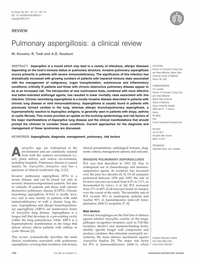
soil, plant debris) and indoor environment,including hospitals. Pulmonary disease is causedmainly by Aspergillus fumigatus and has aspectrum of clinical syndromes (fig. 1) [1].
Invasive pulmonary aspergillosis (IPA) is asevere disease, and can be found not only inseverely immunocompromised patients, but alsoin critically ill patients and those with chronicobstructive pulmonary disease (COPD). Chronicnecrotising aspergillosis (CNA) is locally inva-sive and is seen mainly in patients with mildimmunodeficiency or with a chronic lung dis-ease. Aspergilloma and allergic bronchopulmon-ary aspergillosis (ABPA) are noninvasive formsof Aspergillus lung disease. Aspergilloma is afungus ball that develops in a pre-existing cavitywithin the lung parenchyma, while ABPA is ahypersensitivity manifestation in the lungs thatalmost always affects patients with asthma orcystic fibrosis [2].
This review systematically describes the mainclinical syndromes associated with pulmonaryaspergillosis, covering their incidence, risk factors,
clinical presentations, radiological features, diag-nostic criteria, management options and outcome.
INVASIVE PULMONARY ASPERGILLOSISIPA was first described in 1953 [3]. Due towidespread use of chemotherapy and immuno-suppressive agents, its incidence has increasedover the past two decades [4, 5]. Of all autopsiesperformed between 1978 and 1992, the rate ofinvasive mycoses increased from 0.4% to 3.1%, asdocumented by GROLL et al. [6]. IPA increasedfrom 17% to 60% of all mycoses found on autopsyover the course of the study. The mortality rate ofIPA exceeds 50% in neutropenic patients andreaches 90% in haematopoietic stem-cell trans-plantation (HSCT) recipients [7, 8].
Risk factorsAlveolar macrophages are the first line of defenceagainst inhaled Aspergillus conidia. In the lungs,pathogen recognition receptors, such as Toll-likereceptors, dectin-1 and mannose-binding lectin,identify specific fungal wall components andproduce cytokines that stimulate neutrophil rec-ruitment, the main defence mechanism againstAspergillus hyphae [9]. The major risk factorfor IPA is immunodeficiency (table 1), which
AFFILIATION
Division of Pulmonary Critical Care
and Sleep Medicine, Wayne State
University School of Medicine,
Detroit, MI, USA.
CORRESPONDENCE
A.O. Soubani
Division of Pulmonary Critical Care
and Sleep Medicine
Wayne State University
School of Medicine
Harper University Hospital
3990 John R - 3 Hudson
Detroit
MI 48201
USA
E-mail: [email protected]
Received:
Jan 23 2011
Accepted after revision:
March 10 2011
PROVENANCE
Submitted article, peer reviewed.
European Respiratory Review
Print ISSN 0905-9180
Online ISSN 1600-0617
156 VOLUME 20 NUMBER 121 EUROPEAN RESPIRATORY REVIEW
Eur Respir Rev 2011; 20: 121, 156–174
DOI: 10.1183/09059180.00001011
Copyright�ERS 2011
includes neutropenia, HSCT and solid-organ transplantation,prolonged therapy with high-dose corticosteroids, haemato-logical malignancy, cytotoxic therapy, advanced AIDS andchronic granulomatous disease (CGD) [1, 10, 15].
The most important risk factor is neutropenia, especially whenthere is an absolute neutrophil count of ,500 cells?mm-3. Therisk of IPA correlates strongly with the duration and degree ofneutropenia. The risk in neutropenic patients is estimated toincrease by 1% per day for the first 3 weeks and then by 4% perday thereafter [10]. HSCT and solid-organ transplantation(especially lung transplantation) are also significant risk factors[11, 23] Several other factors predispose patients with trans-plantation to acquire IPA: multiple immune defects includ-ing prolonged neutropenia in the pre-engraftment phase ofHSCT; the use of multiple anti-rejection or anti-graft versushost disease (GVHD) therapy (such as corticosteroids andcyclosporine); parenteral nutrition; use of multiple antibiotics;and prolonged hospitalisation.
There has been a steady increase in the documented cases ofIPA following HSCT, where the risk is much higher followingallogeneic rather than autologous HSCT (incidences of 2.3–15%and 0.5–4%, respectively) [8, 12–14, 24]. In allogeneic HSCT, thehighest risk is in patients with severe GVHD (grade III–IV). Thetimeline of IPA in these patients follows a bimodal distribution,with a peak in the first month following HSCT, which isassociated with neutropenia. The second peak is during thetreatment of GVHD (median 78–112 days post-transplantation)[8, 13, 25]. Currently, the first peak is less significant because ofthe routine use of stem cells instead of bone marrow fortransplantation, nonmyeloablative regimens, the use of colony-stimulating factors during neutropenia and the widespread useof antifungal agents [13, 26]. The second peak has become moresignificant, especially with the higher incidence of GVHDassociated with unrelated allogeneic transplantation and treat-ment with intensive immunosuppressive therapy, includingcorticosteroids, cyclosporine A, anti-TNF agents, and otherT-cell depleting strategies [13, 14, 17, 27–29].
In a study by MARR et al. [14], the probability of IPA reachedapproximately 5% at 2 months, 9% at 6 months, 10% at 1 yearand 11.1% by the third year following allogeneic HSCT. Inaddition, there is evidence that cytomegalovirus (CMV)infection in these patients increases the risk of IPA [14, 30]
the hazard ratio for IPA in the setting of CMV disease increases13.3-fold (95% CI 4.7–37.7) [8].
Neutrophil dysfunction is another risk factor for IPA and isseen primarily in CGD. IPA is an important cause of mortalityin these patients [22].
IPA is relatively uncommon in patients with HIV infection,especially with the routine use of highly active anti-retroviraltherapy. The incidence of aspergillosis in a large cohort ofpatients with HIV infection was 3.5 cases per 1,000 person-yrs[18]. A low CD4 count (,100 cells?mm-3) is present in almostall cases of AIDS-associated aspergillosis, and half of HIV-infected patients with IPA have coexistent neutropenia or areon corticosteroid therapy. The rest of the cases appear to haveno particular risk factors other than advanced AIDS [19–21].There is increased incidence of tracheobronchial involvementin these patients in addition to the usual clinical picture of IPA[21, 31]. Since isolation of Aspergillus from respiratory secre-tions has poor predictive value for invasive IPA in AIDSpatients, a histopathological diagnosis is usually required toestablish the diagnosis [32]. Response to therapy in this patientgroup tends to be particularly poor [19–21, 32, 33]. Theprognosis of HIV-infected patients with IPA is generally poor,with median survival of 3 months following diagnosis [18].
There are increasing numbers of reports documenting IPA inimmunocompetent patients who do not have the classic riskfactors. Two at-risk groups stand out: patients with severeCOPD and critically ill patients. IPA is an emerging seriousinfection in patients with COPD. The majority of these patientshave advanced COPD and/or are on corticosteroid therapy.Patients with COPD have increased susceptibility to IPA forseveral reasons, including structural changes in lung architec-ture, prolonged use of corticosteroid therapy [16], frequenthospitalisation, broad-spectrum antibiotic treatment, invasiveprocedures, mucosal lesions and impaired mucociliary clear-ance, and comorbid illnesses such as diabetes mellitus,alcoholism and malnutrition. It is also possible that abnorm-alities or deficiencies in surfactant proteins, alveolar macro-phages and Toll-like receptors play a role in the pathogenesisof IPA in some patients with COPD [34–38].
It has been documented that chronic lung disease predisposesto colonisation of airways by Aspergillus spp., and it is possible,under certain circumstances, that this colonisation transforms
Inhalation ofAspergillus spores
Chronic lung disease or mild immunocompromised
host
Immunocompromisedhost
AsthmaCystic fibrosis
Atopy
Cavitarylung disease
Normalhost
Chronicnecrotising
aspergillosis
Invasivepulmonary
aspergillosis
Allergicbronchopulmonary
aspergillosisAspergillomaNo sequel
FIGURE 1. The spectrum of pulmonary aspergillosis.
M. KOUSHA ET AL. REVIEW: PULMONARY ASPERGILLOSIS
cEUROPEAN RESPIRATORY REVIEW VOLUME 20 NUMBER 121 157
to an invasive disease [39]. In a review of 65 cases of IPA inpatients with COPD who did not have the traditional riskfactors for IPA [40], 75% were on corticosteroid therapy for amedian 2.6 yrs prior to the diagnosis of IPA. The dose wasincreased in 34% of cases following hospitalisation (usually forthe treatment of acute exacerbation of COPD) and prior to thediagnosis of IPA. Several patients in the study had low forcedexpiratory volume in 1 s or were described to have severe orvery severe COPD. Only 43% of the cases had documentedcomorbidities including atypical mycobacteria, remote historyof Mycobacterium tuberculosis, polymyalgia rheumatica, asthma,cirrhosis, diabetes mellitus, pneumoconiosis, Dressler’s syn-drome and lung cancer. 59 (91%) patients died; the main causeof death was progressive respiratory failure. 31 (48%) patientswere reported to have received mechanical ventilation, and13 (20%) patients had evidence of disseminated IPA [40].Potential explanations of the high mortality of IPA in thesepatients, also reported in other studies [41–43], are the delayeddiagnosis secondary to low index of suspicion of this infec-tion in this patient population, older age, poor pulmonary re-serve and multiple comorbid illnesses. Since the clinical andradiological presentation of IPA in patients with COPD isnonspecific, to avoid delay in the diagnosis and management,a high index of suspicion is warranted. The isolation ofAspergillus spp. from a lower respiratory tract specimen shouldnot be routinely dismissed as colonisation [40, 41].
IPA is also becoming an important infectious disease inintensive care unit (ICU) patients without the classical riskfactors (neutropenia, leukaemia, HSCT), and the mortality isalso devastating in these apparently less immunocompromisedpatients. Aspergillus spp. are isolated from lower respiratorypatients, and in about half of these patients, this findingrepresents IPA [44–48]. In one retrospective study in a medicalICU, an incidence of invasive aspergillosis of 5.8% was foundwith pulmonary involvement in most cases. 70% of the caseswere found in patients without leukaemia or cancer and thedisease had a mortality rate exceeding 90% [47]. In anotherstudy of 172 critically ill patients who had positive sputumsamples for Aspergillus, 83 had invasive disease, and 60% ofthese patients had no classic risk factors for IPA [44]. Criticallyill patients are prone to developing disturbances in immuno-regulation during their stay in the ICU, which renders themmore vulnerable to fungal infections. Risk factors such as COPD,systemic corticosteroid therapy, non-haematological malig-nancy, chronic renal disease, liver failure, diabetes mellitus,near-drowning, HIV infection, autoimmune diseases, malnutri-tion and extensive burns have been described [44–47, 49].
The clinical signs and symptoms of IPA and its radiographicfeatures are often nonspecific in ICU patients. The finding ofAspergillus spp. in respiratory tract samples in these patientsshould not be routinely discarded as colonisation, even if thesepatients are immunocompetent [44]. Therefore, in order not tomiss a critical window of therapeutic opportunity, adaptedclinical diagnostic criteria should be used for this category andfurther diagnostic evaluation with early antifungal therapyshould be considered once IPA is suspected in critically illpatients [50, 51]. IPA in this patient population carries anattributable mortality of 18.9% after adjusting for confoundingfactors [47]. A report suggests that the isolation of Aspergillusfrom lower respiratory tract samples was associated with aworse ICU outcome, regardless of whether the findingrepresented IPA or colonisation [52].
Clinical presentationIn most cases, Aspergillus is introduced to the lower respiratorytract by inhalation of the infectious spores. Less commonly, IPAmay start in locations other than the lungs, such as sinuses, thegastrointestinal tract or the skin (via intravenous catheters,prolonged skin contact with adhesive tapes or burns) [53–56].
Symptoms are nonspecific and usually mimic bronchopneu-monia: fever unresponsive to antibiotics, cough, sputumproduction and dyspnoea. Patients may also present withpleuritic chest pain (due to vascular invasion leading tothromboses that cause small pulmonary infarcts) and haemop-tysis, which is usually mild, but can be severe. IPA is one of themost common causes of haemoptysis in neutropenic patients,and may be associated with cavitation that occurs withneutrophil recovery [57].
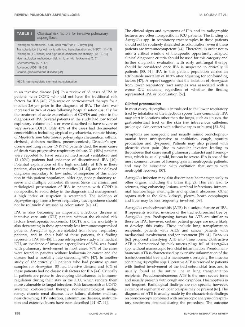
Aspergillus infection may also disseminate haematogenously toother organs, including the brain (fig. 2). This can lead toseizures, ring-enhancing lesions, cerebral infarctions, intracra-nial haemorrhage, meningitis and epidural abscesses. Otherorgans such as the skin, kidneys, pleura, heart, oesophagusand liver may be less frequently involved [58].
Aspergillus tracheobronchitis (ATB) is a unique feature of IPA.It represents isolated invasion of the tracheobronchial tree byAspergillus spp. Predisposing factors for ATB are similar tothose for IPA; however, certain patient groups are more likelyto develop this entity. These include lung transplantationrecipients, patients with AIDS and cancer patients withmediastinal involvement and/or treatment [59–61]. DENNING
[62] proposed classifying ATB into three forms. ObstructiveATB is characterised by thick mucus plugs full of Aspergillusspp. without macroscopic bronchial inflammation. Pseudomem-braneous ATB is characterised by extensive inflammation of thetracheobronchial tree and a membrane overlaying the mucosacontaining Aspergillus spp. Ulcerative ATB is reserved to patientswith limited involvement of the tracheobronchial tree, and isusually found at the suture line in lung transplantationrecipients. Pseudomembraneous ATB is the most severe formand usually presents with cough and dyspnoea. Haemoptysis isnot frequent. Radiological findings are not specific; however,evidence of segmental or lobar collapse may be present [61]. Thediagnosis of ATB is usually made by the characteristic findingson bronchoscopy combined with microscopic analysis of respira-tory specimens obtained during the procedure. The outcome
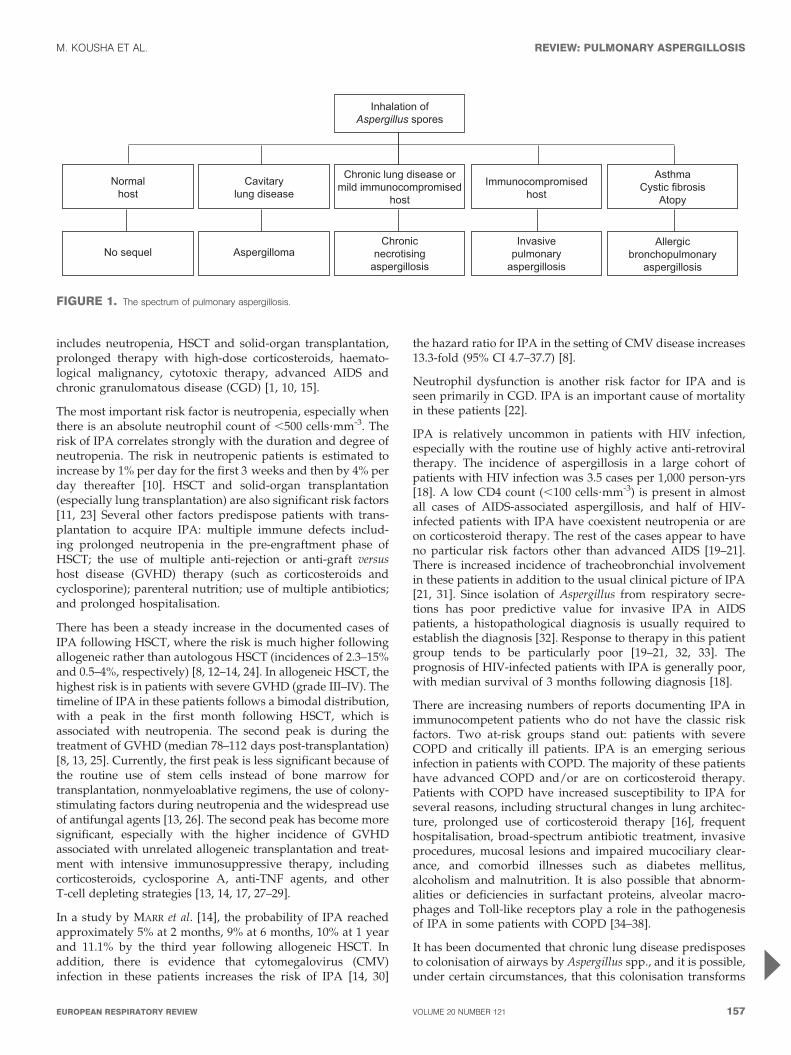
TABLE 1 Classical risk factors for invasive pulmonaryaspergillosis
Prolonged neutropenia (,500 cells?mm-3 for .10 days) [10]
Transplantation (highest risk is with lung transplantation and HSCT) [11–14]
Prolonged (.3 weeks) and high-dose corticosteroid therapy [10, 15, 16]
Haematological malignancy (risk is higher with leukaemia) [5, 7]
Chemotherapy [5, 7, 17]
Advanced AIDS [18–21]
Chronic granulomatous disease [22]
HSCT: haematopoietic stem-cell transplantation.
REVIEW: PULMONARY ASPERGILLOSIS M. KOUSHA ET AL.
158 VOLUME 20 NUMBER 121 EUROPEAN RESPIRATORY REVIEW
of ulcerative ATB is generally favourable with antifungaltherapy. On the other hand, the prognosis is poor in patientswith pseudomembranous and obstructive ATB, with mortalityreaching 78% [63]. The need for mechanical ventilation is apredictor of high mortality in these patients [63]. High index ofsuspicion of ATB, early diagnosis and prompt antifungaltherapy may be associated with improved outcome [61].
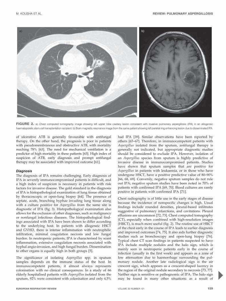
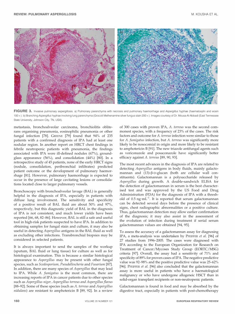
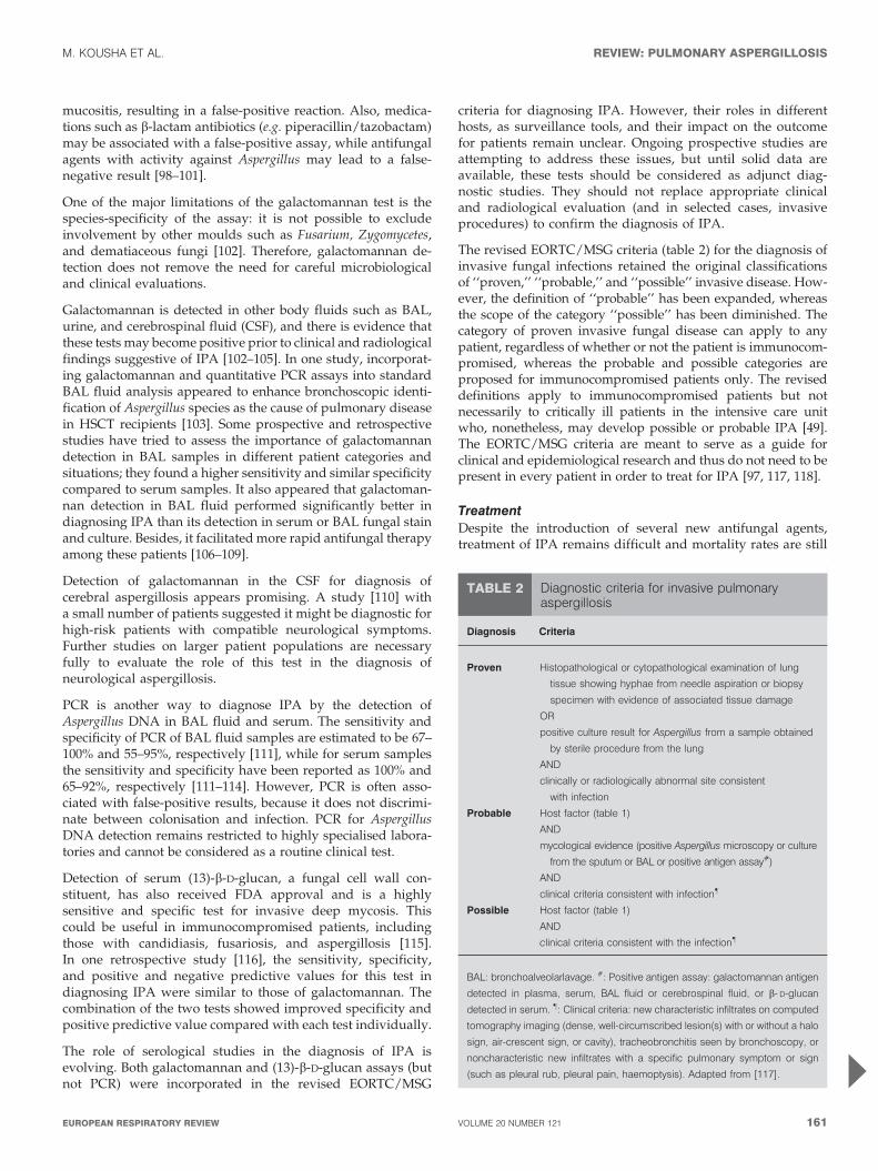
DiagnosisThe diagnosis of IPA remains challenging. Early diagnosis ofIPA in severely immunocompromised patients is difficult, anda high index of suspicion is necessary in patients with riskfactors for invasive disease. The gold standard in the diagnosisof IPA is histopathological examination of lung tissue obtainedby thoracoscopic or open-lung biopsy [64]. The presence ofseptate, acute, branching hyphae invading lung tissue alongwith a culture positive for Aspergillus from the same site isdiagnostic of IPA (fig. 3). Histopathological examination alsoallows for the exclusion of other diagnoses, such as malignancyor nonfungal infectious diseases. The histopathological find-ings associated with IPA have been shown to differ accordingto the underlying host. In patients with allogeneic HSCTand GVHD, there is intense inflammation with neutrophilicinfiltration, minimal coagulation necrosis and low fungalburden. In neutropenic patients, IPA is characterised by scantinflammation, extensive coagulation necrosis associated withhyphal angio-invasion, and high fungal burden. Disseminationto other organs is equally high in both groups [5].
The significance of isolating Aspergillus spp. in sputumsamples depends on the immune status of the host. Inimmunocompetent patients, it almost always representscolonisation with no clinical consequences. In a study of 66elderly hospitalised patients with Aspergillus isolated from thesputum, 92% were consistent with colonisation and only 4.5%
had IPA [39]. Similar observations have been reported byothers [65–67]. Therefore, in immunocompetent patients withAspergillus isolated from the sputum, antifungal therapy isgenerally not indicated, but appropriate diagnostic studiesshould be considered to exclude IPA. However, isolation ofan Aspergillus species from sputum is highly predictive ofinvasive disease in immunocompromised patients. Studieshave shown that sputum samples that are positive forAspergillus in patients with leukaemia, or in those who haveundergone HSCT, have a positive predictive value of 80–90%[66, 68, 69]. Conversly, negative sputum samples do not ruleout IPA; negative sputum studies have been noted in 70% ofpatients with confirmed IPA [69, 70]. Blood cultures are rarelypositive in patients with confirmed IPA [71].
Chest radiography is of little use in the early stages of diseasebecause the incidence of nonspecific changes is high. Usualfindings include rounded densities, pleural-based infiltratessuggestive of pulmonary infarctions, and cavitations. Pleuraleffusions are uncommon [72, 73]. Chest computed tomography(CT), especially when combined with high-resolution images(HRCT), is much more useful (fig. 2). The routine use of HRCTof the chest early in the course of IPA leads to earlier diagnosisand improved outcomes [74, 75]. It also aids further diagnosticstudies such as bronchoscopy and open-lung biopsy [76].Typical chest CT scan findings in patients suspected to haveIPA include multiple nodules and the halo sign, which ismainly seen in neutropenic patients early in the course ofinfection (usually in the first week) and appears as a zone oflow attenuation due to haemorrhage surrounding the pul-monary nodule. Another late radiological sign is the aircrescent sign, which appears as a crescent-shaped lucency inthe region of the original nodule secondary to necrosis [73, 77].Neither sign is sensitive or pathognomic of IPA. The halo signmay be found in many other situations: as a result of
a) b)
FIGURE 2. a) Chest computed tomography image showing left upper lobe cavitary lesion consistent with invasive pulmonary aspergillosis (IPA) in an allogeneic
haematopoietic stem-cell transplantation recipient. b) Brain magnetic resonance image from the same patient showing left parietal ring enhancing lesion due to disseminated IPA.
M. KOUSHA ET AL. REVIEW: PULMONARY ASPERGILLOSIS
cEUROPEAN RESPIRATORY REVIEW VOLUME 20 NUMBER 121 159
metastasis, bronchoalveolar carcinoma, bronchiolitis oblite-rans organising pneumonia, eosinophilic pneumonia or otherfungal infection [78]. GREENE [79] found that 94% of 235patients with a confirmed diagnosis of IPA had at least onenodular region. In another report on HRCT chest findings infebrile neutropenic patients with pneumonia, the findingsassociated with IPA were ill-defined nodules (67%), ground-glass appearance (56%), and consolidation (44%) [80]. In aretrospective study of 45 patients, none of the early HRCT signs(nodule, consolidation, peribronchial infiltrates) predictedpatient outcome or the development of pulmonary haemor-rhage [81]. However, pulmonary haemorrhage is expected tooccur in the presence of large cavitating lesions or consolida-tions located close to larger pulmonary vessels.
Bronchoscopy with bronchoalveolar lavage (BAL) is generallyhelpful in the diagnosis of IPA, especially in patients withdiffuse lung involvement. The sensitivity and specificityof a positive result of BAL fluid are about 50% and 97%,respectively, but this diagnostic yield of BAL in the diagnosisof IPA is not consistent, and much lower yields have beenreported [66, 68, 82–86]. However, BAL is still a safe and usefultool in high-risk patients suspected to have IPA. In addition toobtaining samples for fungal stain and culture, it may also beuseful in detecting Aspergillus antigens in the BAL fluid as wellas excluding other infections. Transbronchial biopsies may beconsidered in selected patients.
It is always important to send the samples of the workup(sputum, BAL fluid or lung tissue) for culture as well as forhistological examination. This is because a similar histologicalappearance to Aspergillus may be present with other fungalspecies, such as Scedosporium, Pseudallescheria and Fusarium [87].In addition, there are many species of Aspergillus that may leadto IPA. While A. fumigatus is the most common, there areincreasing reports of IPA in cancer patients due to other speciessuch as Aspergillus niger, Aspergillus terreus and Aspergillus flavus[88–92]. Some of these species (such as A. terreus and Aspergillusnidulans) are resistant to amphotericin B [89, 92]. In a review
of 300 cases with proven IPA, A. terreus was the second com-monest species, with a frequency of 23% of the cases. The riskfactors and outcome for A. terreus infection were similar to thosefor A. fumigatus infection, but A. terreus was significantly morelikely to be nosocomial in origin and more likely to be resistantto amphotericin B [91]. The new triazole antifungal agents suchas voriconazole and posaconazole have significantly betterefficacy against A. terreus [89, 90, 93].
The most recent advances in the diagnosis of IPA are related todetecting Aspergillus antigens in body fluids, mainly galacto-mannan and (13)-b-D-glucan (both are cellular wall con-stituents). Galactomannan is a polysaccharide released byAspergillus during growth. A double-sandwich ELISA forthe detection of galactomannan in serum is the best character-ised test and was approved by the US Food and DrugAdministration (FDA) for the diagnosis of IPA with a thresh-old of 0.5 ng?mL-1. It is reported that serum galactomannancan be detected several days before the presence of clinicalsigns, chest radiographic abnormalities or a positive culture.Thus, galactomannan detection may allow earlier confirmationof the diagnosis; it may also assist in the assessment ofthe evolution of infection during treatment if serial serumgalactomannan values are obtained [94, 95].
To assess the accuracy of a galactomannan assay for diagnosingIPA, a meta-analysis was undertaken by PFEIFFER et al. [96] of27 studies from 1996–2005. The cases were diagnosed withIPA according to the European Organization for Research onTreatment of Cancer/Mycoses Study Group (EORTC/MSG)criteria [97]. Overall, the assay had a sensitivity of 71% andspecificity of 89% for proven cases of IPA. The negative predictivevalue was 92–98% and the positive predictive value was 25–62%[96]. PFEIFFER et al. [96] also concluded that the galactomannanassay is more useful in patients who have a haematologicalmalignancy or who have undergone allogeneic HSCT than insolid-organ transplant recipients or non-neutropenic patients.
Galactomannan is found in food and may be absorbed by thedigestive tract, especially in patients with post-chemotherapy
a) b)
FIGURE 3. Invasive pulmonary aspergillosis. a) Pulmonary parenchyma with necrosis and pulmonary haemorrhage and Aspergillus hyphae (haematoxylin and eosin
1006). b) Branching Aspergillus hyphae involving lung parenchyma (Grocott Methenamine silver fungus stain 2006). Images courtesy of Dr. Mousa Al-Abbadi (East Tennessee
State University, Johnson City, TN, USA).
REVIEW: PULMONARY ASPERGILLOSIS M. KOUSHA ET AL.
160 VOLUME 20 NUMBER 121 EUROPEAN RESPIRATORY REVIEW
mucositis, resulting in a false-positive reaction. Also, medica-tions such as b-lactam antibiotics (e.g. piperacillin/tazobactam)may be associated with a false-positive assay, while antifungalagents with activity against Aspergillus may lead to a false-negative result [98–101].
One of the major limitations of the galactomannan test is thespecies-specificity of the assay: it is not possible to excludeinvolvement by other moulds such as Fusarium, Zygomycetes,and dematiaceous fungi [102]. Therefore, galactomannan de-tection does not remove the need for careful microbiologicaland clinical evaluations.
Galactomannan is detected in other body fluids such as BAL,urine, and cerebrospinal fluid (CSF), and there is evidence thatthese tests may become positive prior to clinical and radiologicalfindings suggestive of IPA [102–105]. In one study, incorporat-ing galactomannan and quantitative PCR assays into standardBAL fluid analysis appeared to enhance bronchoscopic identi-fication of Aspergillus species as the cause of pulmonary diseasein HSCT recipients [103]. Some prospective and retrospectivestudies have tried to assess the importance of galactomannandetection in BAL samples in different patient categories andsituations; they found a higher sensitivity and similar specificitycompared to serum samples. It also appeared that galactoman-nan detection in BAL fluid performed significantly better indiagnosing IPA than its detection in serum or BAL fungal stainand culture. Besides, it facilitated more rapid antifungal therapyamong these patients [106–109].
Detection of galactomannan in the CSF for diagnosis ofcerebral aspergillosis appears promising. A study [110] witha small number of patients suggested it might be diagnostic forhigh-risk patients with compatible neurological symptoms.Further studies on larger patient populations are necessaryfully to evaluate the role of this test in the diagnosis ofneurological aspergillosis.
PCR is another way to diagnose IPA by the detection ofAspergillus DNA in BAL fluid and serum. The sensitivity andspecificity of PCR of BAL fluid samples are estimated to be 67–100% and 55–95%, respectively [111], while for serum samplesthe sensitivity and specificity have been reported as 100% and65–92%, respectively [111–114]. However, PCR is often asso-ciated with false-positive results, because it does not discrimi-nate between colonisation and infection. PCR for AspergillusDNA detection remains restricted to highly specialised labora-tories and cannot be considered as a routine clinical test.
Detection of serum (13)-b-D-glucan, a fungal cell wall con-stituent, has also received FDA approval and is a highlysensitive and specific test for invasive deep mycosis. Thiscould be useful in immunocompromised patients, includingthose with candidiasis, fusariosis, and aspergillosis [115].In one retrospective study [116], the sensitivity, specificity,and positive and negative predictive values for this test indiagnosing IPA were similar to those of galactomannan. Thecombination of the two tests showed improved specificity andpositive predictive value compared with each test individually.
The role of serological studies in the diagnosis of IPA isevolving. Both galactomannan and (13)-b-D-glucan assays (butnot PCR) were incorporated in the revised EORTC/MSG
criteria for diagnosing IPA. However, their roles in differenthosts, as surveillance tools, and their impact on the outcomefor patients remain unclear. Ongoing prospective studies areattempting to address these issues, but until solid data areavailable, these tests should be considered as adjunct diag-nostic studies. They should not replace appropriate clinicaland radiological evaluation (and in selected cases, invasiveprocedures) to confirm the diagnosis of IPA.
The revised EORTC/MSG criteria (table 2) for the diagnosis ofinvasive fungal infections retained the original classificationsof ‘‘proven,’’ ‘‘probable,’’ and ‘‘possible’’ invasive disease. How-ever, the definition of ‘‘probable’’ has been expanded, whereasthe scope of the category ‘‘possible’’ has been diminished. Thecategory of proven invasive fungal disease can apply to anypatient, regardless of whether or not the patient is immunocom-promised, whereas the probable and possible categories areproposed for immunocompromised patients only. The reviseddefinitions apply to immunocompromised patients but notnecessarily to critically ill patients in the intensive care unitwho, nonetheless, may develop possible or probable IPA [49].The EORTC/MSG criteria are meant to serve as a guide forclinical and epidemiological research and thus do not need to bepresent in every patient in order to treat for IPA [97, 117, 118].
TreatmentDespite the introduction of several new antifungal agents,treatment of IPA remains difficult and mortality rates are still
TABLE 2 Diagnostic criteria for invasive pulmonaryaspergillosis
Diagnosis Criteria
Proven Histopathological or cytopathological examination of lung
tissue showing hyphae from needle aspiration or biopsy
specimen with evidence of associated tissue damage
OR
positive culture result for Aspergillus from a sample obtained
by sterile procedure from the lung
AND
clinically or radiologically abnormal site consistent
with infection
Probable Host factor (table 1)
AND
mycological evidence (positive Aspergillus microscopy or culture
from the sputum or BAL or positive antigen assay#)
AND
clinical criteria consistent with infection"
Possible Host factor (table 1)
AND
clinical criteria consistent with the infection"
BAL: bronchoalveolarlavage. #: Positive antigen assay: galactomannan antigen
detected in plasma, serum, BAL fluid or cerebrospinal fluid, or b- D-glucan
detected in serum. ": Clinical criteria: new characteristic infiltrates on computed
tomography imaging (dense, well-circumscribed lesion(s) with or without a halo
sign, air-crescent sign, or cavity), tracheobronchitis seen by bronchoscopy, or
noncharacteristic new infiltrates with a specific pulmonary symptom or sign
(such as pleural rub, pleural pain, haemoptysis). Adapted from [117].
M. KOUSHA ET AL. REVIEW: PULMONARY ASPERGILLOSIS
cEUROPEAN RESPIRATORY REVIEW VOLUME 20 NUMBER 121 161

high (table 3). Therapy should be considered as soon as there isa clinical suspicion of IPA, and while a workup is under way.Amphotericin B has been the first line of therapy for IPA formany years, with a recommended dose 1–1.5 mg?kg-1?day-1.However, it can cause serious side-effects, including nephro-toxicity, electrolyte disturbances and hypersensitivity. Toreduce these side-effects, newer lipid-based preparations ofamphotericin B (like liposomal amphotericin B and lipidcomplex amphotericin B) have been introduced, but higherdoses of the lipid formulations are needed for equivalentantifungal efficacy. A recent large randomised trial demon-strated no additional benefits of high-dose liposomal am-photericin B (10 mg?kg-1?day-1) compared with lower-doseliposomal amphotericin B regimens (3 mg?kg-1?day-1), andoutcomes were generally good with the lower dose, suggestingutility of liposomal amphotericin B in the low doses, andtherapeutic risk associated with excessive toxicities at thehigher doses [124].
A new broad-spectrum triazole, voriconazole, has beenapproved as the initial treatment of invasive aspergillosis andis currently considered the treatment of choice in many patientswith IPA [119–121]. In a large prospective, randomised, multi-centre trial, voriconazole was compared to amphotericin B asthe primary therapy for IPA [122]. Patients receiving voricona-zole had a higher favourable response rate at week 12 (53%versus 32% in patients receiving amphotericin B) and a higher12-week survival (71% versus 58%). Voriconazole is available inboth intravenous and oral formulations. The recommendeddose is 6 mg?kg-1 twice daily intravenously on day 1, followedby 4 mg?kg-1?day-1. After 7 days, switching to 200 mg p.o. twicedaily may be considered. Voriconazole has a milder side-effectprofile and is much better tolerated than amphotericin B. Themost frequent adverse effect is visual disturbances, described asblurred vision, photophobia and altered colour perception.Liver function test abnormalities and skin reactions are lesscommon side-effects. However, voriconazole is associated witha significant number of drug–drug interactions, such as withcyclosporine, warfarin, terfenadine, carbamazepine, quinidine,rifampin, statins and sulfonylureas [119]. Since there is inter-individual and intra-individual variability in voriconazoleplasma levels, therapeutic drug monitoring for voriconazoleshould be considered in cases of refractory fungal infection or
concerns about drug toxicity [147]. Another broad-spectrumtriazole, posaconazole, is effective and safe as salvage therapy inpatients with IPA refractory to standard antifungal therapy[22, 93, 125].
Echinocandin derivatives such as caspofungin, micafungin andanidulafungin are also effective agents in the treatment of IPArefractory to standard treatment, or if the patient cannot toleratefirst-line agents [126, 127]. While polyenes and azoles target thefungal cell membrane, echinocandins inhibit the (13)-b-D-glucanconstituent of the fungal cell wall. Therefore, a combinationantifungal therapy could be a strategy to treat refractory IPA[148, 149]. There are in vitro and limited clinical studies (casereports and retrospective case series) that suggest a benefit fromcombining antifungal agents as salvage therapy in refractoryIPA [149–152]. The combination of caspofungin and liposomalamphotericin B as a salvage therapy showed an overall responserate of 42%, although in patients with documented progressiveIPA, the response rate was only 18% [149]. A survival advantageof voriconazole plus caspofungin compared with voriconazolealone was reported in one retrospective analysis of salvagetherapy for IPA [151]. This combination was also compared withliposomal amphotericin B as primary therapy for IPA in solid-organ transplant recipients in a prospective, multicentre,observational study [153]. The combination was associated withimproved survival in subsets of recipients with renal failure orA. fumigatus. Conversely, another report showed no differencein the response rate between patients who received micafunginalone or those who received it in combination with otherantifungal agents as primary or salvage therapy for acute IPA[154]. Combination therapy of an echinocandin with either alipid formulation of amphotericin B or triazole agent appearspromising and should be considered in critically ill patients[155], but cannot be recommended for the routine treatment ofprimary IPA. Controlled randomised prospective studies areneeded to document the value of this approach. Becausegalactomannan is covalently bound to (13)-b-D-glucan in thefungal cell wall, an initial increase in circulating galactomannanmight be expected in patients treated with echinocandins, whichinhibit the (13)-b-D-glucan constituent [156].
According to the recent statement of the American ThoracicSociety for treating fungal infections in adults [123], theduration of IPA therapy should be individualised to the
TABLE 3 Treatment recommendations for pulmonary aspergillosis
Disease Primary treatment Other treatments
Invasive pulmonary aspergillosis Voriconazole [119–123] Alternative therapy: liposomal amphotericin B [124]
Continuation therapy: voriconazole or itraconazole [122, 123]
Salvage therapy: echinocandin or posaconazole [125–127]
Chronic necrotising aspergillosis Voriconazole [120, 123] Alternative therapy: itraconazole [128, 129]
Severe cases: intravenous voriconazole or liposomal
amphotericin B [123, 128, 130]
Consider surgical resection [130]
Aspergilloma Observation [123] Bronchial artery embolisation [131]
Surgical resection [132–135]
Consider itraconazole [136–138]
Allergic bronchopulmonary aspergillosis Corticosteroids [139–142] Itraconazole or voriconazole as steroid-sparing agents [143–146]
REVIEW: PULMONARY ASPERGILLOSIS M. KOUSHA ET AL.
162 VOLUME 20 NUMBER 121 EUROPEAN RESPIRATORY REVIEW
patient’s clinical and radiological response. The treatment isoften prolonged, lasting several months to .1 yr. Prerequisitesfor discontinuing treatment include clinical and radiographicresolution, microbiological clearance and reversal of immuno-suppression. Reinstating therapy in patients who haveresponded should be considered if immunosuppression isresumed, or if the patient requires additional cytotoxic therapyor another HSCT.
Surgical resection has generally a limited role in the manage-ment of patients with IPA, but it becomes important in caseswith invasion of bone, burn wounds, epidural abscesses andvitreal disease [123]. It should also be considered in cases ofmassive haemoptysis, pulmonary lesions close to the greatblood vessels or pericardium, or residual localised pulmonarylesions in patients with continuing immunosuppression orthose who are expected to have immunosuppressive therapyin the future. Several reports have shown the relative efficacyand safety of surgical intervention, in addition to antifungaltherapy, in these situations [75, 157–162].
Immunomodulatory therapy could be used to decrease thedegree of immunosuppression and as an adjunct to antifungaltherapy for the treatment of IPA. This includes colony-stimulating factors, like granulocyte colony-stimulating factor(G-CSF) and granulocyte-macrophage colony-stimulating fac-tor (GM-CSF), and interferon-c. Colony-stimulating factorsstimulate the bone marrow to produce more neutrophils, andhave been shown to augment the phagocytic activity ofneutrophils against fungi, including Aspergillus spp. [163–165]. There is a theoretical advantage to adding these agents tothe treatment of neutropenic patients suspected to have IPA. Inone randomised study in patients receiving chemotherapy foracute myelogenous leukaemia, prophylaxis with GM-CSF ledto a lower frequency of fatal fungal infections compared withplacebo (1.9% versus 19%, respectively) and reduced over-all mortality [166]. It is recommended to consider colony-stimulating factors in neutropenic patients with seriousinfections, but there are no definitive studies that show benefitin patients with IPA [167]. Interferon-c is another cytokine thathas been shown in vitro and in animal models to augmentimmunity by increasing neutrophil and monocyte activityagainst Aspergillus [164, 168, 169]. It has been used to decreasethe risk of Aspergillus infection in patients with CGD [170].Evidence on the value of adding interferon-c as an adjuncttreatment of IPA is limited to case reports and small reports,and there are no guidelines on its role in the treatment of IPA[171]. There was a concern about the use of interferon-c inallogeneic HSCT recipients, since it may worsen GVHD;however, in a recent trial, GVHD actually improved duringthis therapy [172].
Granulocyte transfusion is another potential supportive ther-apy, especially for patients with prolonged neutropenia andlife-threatening infections refractory to conventional therapy. Ithas been shown that it is safe for potential donors to donateneutrophils by granulocytophoresis, but there are no rando-mised studies that prove the benefit of adjuvant granulocytetransfusion in the treatment of IPA [173]. It is also important inpatients with IPA, whenever possible, to decrease the dose ofsystemic corticosteroids and immunosuppressive agents.
The management of IPA is difficult, and an important approachto this problem is prophylaxis in patients at increased risk forIPA. Avoiding the hospitalisation of patients in areas wherethere is construction and the use of high-efficiency particulate air(HEPA) filtration, with or without laminar air flow ventilation,have both proven useful [174]. A meta-analysis suggested thatitraconazole was effective in preventing fungal infections inneutropenic patients [175]. Recent studies confirmed the efficacyof posaconazole as IPA prophylaxis in patients with acutemyelogenous leukaemia, myelodysplastic syndrome or HSCT[176–178]. Currently, chemoprophylaxis trials using other anti-fungal agents (such as voriconazole, caspofungin, micafungin,and inhaled amphotericin B formulation) are under way in high-risk patients [123].
CHRONIC NECROTISING ASPERGILLOSISCNA, also called semi-invasive or subacute invasive aspergil-losis, was first described by GEFTER et al. [179] and BINDER et al.[130] in 1981. It is an indolent, cavitary and infectious process ofthe lung parenchyma secondary to local invasion by Aspergillusspecies, usually A. fumigatus [128]. In contrast to IPA, CNA runsa slowly progressive course over weeks to months, and vascularinvasion or dissemination to other organs is unusual. Thissyndrome is rare, and the available literature is based on casereports and small case series [128, 130, 179].
Risk factorsCNA usually affects middle-aged and elderly patients withaltered local defences, associated with underlying chronic lungdiseases such as COPD, previous pulmonary tuberculosis,thoracic surgery, radiation therapy, pneumoconiosis, cysticfibrosis, lung infarction or sarcoidosis [180]. It may also occurin patients who are mildly immunocompromised due todiabetes mellitus, alcoholism, chronic liver disease, low-dosecorticosteroid therapy, malnutrition, or connective tissuediseases such as rheumatoid arthritis and ankylosing spondy-litis [130]. Mannose-binding lectin polymorphism may play arole in the pathogenesis of CNA [181]. It may be difficult todistinguish CNA from aspergilloma, especially if a previouschest radiograph is not available [182]. However, in CNA thereis local invasion of the lung tissue and a pre-existing cavity isnot needed, although a cavity with a fungal ball may developin the lung as a secondary phenomenon due to destruction bythe fungus. In a report of aspergillomas in AIDS patients,progression over time was seen, with considerable morbidityand some mortality [182]. This probably reflects the possibilitythat an aspergilloma may invade the cavity wall, causing localparenchyma destruction, as seen in patients with CNA [128].Due to this overlap between the CNA and aspergilloma, someauthors put them in one group called chronic cavitarypulmonary aspergillosis which is primarily a non- or semi-invasive disease that is seen mainly in nonimmunocompro-mised patients with chronic lung diseases [183].
Clinical presentation and diagnosisPatients frequently complain of constitutional symptoms suchas fever, malaise, fatigue, and weight loss of 1–6 months’duration, in addition to chronic productive cough andhaemoptysis, which varies from mild to severe [182]. Occa-sionally, CNA may be asymptomatic.
M. KOUSHA ET AL. REVIEW: PULMONARY ASPERGILLOSIS
cEUROPEAN RESPIRATORY REVIEW VOLUME 20 NUMBER 121 163

Imaging studies, such as chest radiograph and chest CT scan,usually show consolidation, pleural thickening and cavitarylesions in the upper lung lobes. Aspergilloma may be seen innearly 50% of patients [130]. The adjacent pleural thickeningmay progress to form a broncho–pleural fistula, so it isconsidered an early indication of a locally invasive process[179, 184]. Characteristically, these radiological findings tend toprogress over weeks to months [184].
The vast majority of patients with CNA have positive serumimmunoglobuling (Ig)G antibodies to A. fumigatus, but thisvaries over time and may be negative at some points in thecourse of CNA [182]. Immediate skin reactivity for Aspergillusantigens is another helpful, but not diagnostic, test. Culture ofsputum and bronchoscopy samples is usually positive forAspergillus spp. [182].
Confirmation of the diagnosis requires a histological demon-stration of tissue invasion by the fungus and the growth ofAspergillus species on culture. Pathologically, CNA is char-acterised by necrosis of lung tissue, acute or chronicinflammation of the cavity wall and presence of hyphaeconsistent with Aspergillus species [185]. The yield of trans-bronchial biopsy specimens or percutaneous aspirates isrelatively poor, and a thoracoscopic or open-lung biopsy israrely performed in these patients. As a result, confirmation ofthe diagnosis is commonly delayed, which may contributeto the morbidity and mortality associated with CNA. Thecombination of characteristic clinical and radiological findingsand either serological results positive for Aspergillus or theisolation of Aspergillus from respiratory samples is highlyindicative of CNA [186]. DENNING et al. [186] have proposedcriteria for diagnosis of chronic pulmonary aspergillosis,including CNA (table 4).
TreatmentThe mainstay of treatment for CNA is the antifungal therapy(table 3). Amphotericin B was initially used in doses of 0.5–1 mg-1?kg-1?day-1 (4–5 mg?kg-1?day-1 for the lipid formulation)with favourable results [128, 130]. Itraconazole later became aneffective alternative to the relatively toxic amphotericin B [128,129]. More recently, voriconazole has emerged as a primarytherapy for CNA. In a recent prospective study, wherevoriconazole 200 mg was given twice daily for a period of 4–24 weeks as primary or salvage therapy for 39 patients withCNA [120], a complete or partial response was seen in 43% of
patients, and improvement or stability was seen in 80%. Therecent statement of the American Thoracic Society favoursgiving either voriconazole or itraconazole for mild to moderatedisease until resolution or stabilisation of the clinical andradiographic manifestations, while in patients with severedisease, initial therapy with intravenous amphotericin B orintravenous voriconazole should be considered [123].
Evaluation of the response to treatment is best done byfollowing clinical, radiological, serological and microbiologicalparameters [182]. Useful parameters of response includeweight gain and enhanced energy level, improved pulmonarysymptoms, falling inflammatory markers and total serum IgElevel, improvement in paracavitary infiltrates, and eventually areduction in cavity size [182].
Surgical resection has a minor role in the treatment of CNA,being reserved for healthy young patients with focal diseaseand good pulmonary reserves, patients not tolerating anti-fungal therapy and patients with residual localised but activedisease despite adequate antifungal therapy. BINDER et al. [130]reported that 90% of patients who underwent surgicalresection had good responses, but surgery was associatedwith significant post-operative complications.
The reported mortality of CNA varies widely and may belimited by incomplete follow-up [128]. Mortality was 39% inAmerican reports, but less than 10% in European reports usingitraconazole [128].
ASPERGILLOMAAspergilloma is the most common and best-recognised form ofpulmonary involvement by Aspergillus species, and it usuallydevelops in a pre-existing cavity in the lung. The aspergilloma(fungus ball) is composed of fungal hyphae, inflammatory cells,fibrin, mucus, and tissue debris. The most common species ofAspergillus recovered from such lesions is A. fumigatus; however,other fungi, such as Zygomycetes and Fusarium, may cause theformation of a fungal ball. Many cavitary lung diseases arecomplicated by aspergilloma, including tuberculosis, sarcoi-dosis, bronchiectasis, bronchial cysts and bullae, ankylosingspondylitis, neoplasm, and pulmonary infection [187, 188]. Ofthese, tuberculosis is the most common [189]. In a study of 544patients with pulmonary cavities secondary to tuberculosis, 11%had radiological evidence of aspergilloma [190]. Less frequently,aspergilloma has been described in cavities caused by otherfungal infections [191, 192]. It is thought that inadequate
TABLE 4 Diagnostic criteria for chronic necrotising aspergillosis
Diagnostic criteria Characteristics
Clinical Chronic (.1 month) pulmonary or systemic symptoms, including at least one of: weight loss, productive cough or haemoptysis
No overt immunocompromising conditions (e.g. haematological malignancy, neutropenia, organ transplantation)
Radiological Cavitary pulmonary lesion with evidence of paracavitary infiltrate
New cavity formation, or expansion of cavity size over time
Laboratory Elevated levels of inflammatory markers (C-reactive protein, plasma viscosity or erythrocyte sedimentation rate). Isolation of Aspergillus spp. from
pulmonary or pleural cavity, or positive serum Aspergillus precipitin test. Exclusion of other pulmonary pathogens, by results of appropriate
cultures and serological tests, that are associated with similar disease presentation, including mycobacteria and endemic fungi
Adapted from [186].
REVIEW: PULMONARY ASPERGILLOSIS M. KOUSHA ET AL.
164 VOLUME 20 NUMBER 121 EUROPEAN RESPIRATORY REVIEW
drainage can facilitate the growth of Aspergillus on the walls ofthese cavities.
The fungus ball may move within the cavity, but it does notusually invade the surrounding lung parenchyma or bloodvessels, although exceptions have been noted [193, 194]. Thelesion remains stable in the majority of cases, but it maydecrease in size or resolve spontaneously without treatment in10% of cases [195]. Rarely, the aspergilloma may increase in size.
Clinical presentationMost patients with aspergilloma are asymptomatic. Whensymptoms are present, most patients experience mild haem-optysis, but severe and life-threatening haemoptysis mayoccur, particularly in patients with underlying tuberculosis[196]. The mortality rate from haemoptysis related to asper-gilloma ranges between 2–14% [197–201]. The source ofbleeding is usually the bronchial blood vessels, and it maybe caused by local invasion of blood vessels lining the cavity,endotoxins released from the fungus, or mechanical irritationof the exposed vasculature inside the cavity by the movingfungus ball [193, 202, 203]. Less commonly, patients maydevelop cough, dyspnoea that is probably more related to theunderlying lung disease and fever that could be secondary tothe underlying disease or bacterial superinfection.
Risk factors for poor prognosis of aspergilloma include theseverity of the underlying lung disease, increase in size ornumber of lesions as seen on chest radiographs, immunosup-pression (including corticosteroid therapy and HIV infection),increasing Aspergillus-specific IgG titres, recurrent large vol-ume haemoptysis and underlying sarcoidosis [204].
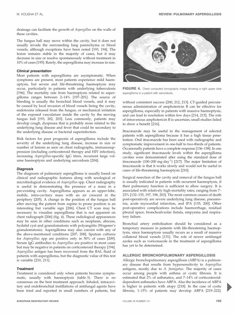
DiagnosisThe diagnosis of pulmonary aspergilloma is usually based onclinical and radiographic features along with serological ormicrobiological evidence of Aspergillus spp. Chest radiographyis useful in demonstrating the presence of a mass in apre-existing cavity. Aspergilloma appears as an upper-lobe,mobile, intra-cavitary mass with an air crescent in theperiphery [205]. A change in the position of the fungus ballafter moving the patient from supine to prone position is aninteresting but variable sign [206]. Chest CT scan may benecessary to visualise aspergilloma that is not apparent onchest radiograph [206] (fig. 4). These radiological appearancesmay be seen in other conditions such as neoplasm, abscess,hydatid cyst and granulomatosis with polyangiitis (Wegener’sgranulomatosis). Aspergilloma may also coexist with any ofthe above-mentioned conditions [207, 208]. Sputum culturesfor Aspergillus spp are positive only in 50% of cases [209].Serum IgG antibodies to Aspergillus are positive in most casesbut may be negative in patients on corticosteroid therapy [194].Aspergillus antigen has been recovered from the BAL fluid ofpatients with aspergilloma, but the diagnostic value of this testis variable [210, 211].
TreatmentTreatment is considered only when patients become sympto-matic, usually with haemoptysis (table 3). There is noconsensus on the best treatment approach. Inhaled, intracavi-tary and endobronchial instillations of antifungal agents havebeen tried and reported in small numbers of patients, but
without consistent success [200, 212, 213]. CT-guided percuta-neous administration of amphotericin B can be effective foraspergilloma, especially in patients with massive haemoptysis,and can lead to resolution within few days [214, 215]. The roleof intravenous amphotericin B is uncertain; small studies failedto show a benefit [216].
Itraconazole may be useful in the management of selectedpatients with aspergilloma because it has a high tissue pene-tration. Oral itraconazole has been used with radiographic andsymptomatic improvement in one-half to two-thirds of patients.Occasionally patients have a complete response [136–138]. In onestudy, significant itraconazole levels within the aspergillomacavities were demonstrated after using the standard dose ofitraconazole (100–200 mg?day-1) [217]. The major limitation ofitraconazole is that it works slowly and would not be useful incases of life-threatening haemoptysis [210].
Surgical resection of the cavity and removal of the fungus ballis usually indicated in patients with recurrent haemoptysis, iftheir pulmonary function is sufficient to allow surgery. It isassociated with relatively high mortality rates, ranging from 7–23% [132–135, 197, 198, 203]. The most common causes of deathpost-operatively are severe underlying lung disease, pneumo-nia, acute myocardial infarction, and IPA [135, 200]. Otherpost-operative complications include haemorrhage, residualpleural space, bronchoalveolar fistula, empyema and respira-tory failure.
Bronchial artery embolisation should be considered as atemporary measure in patients with life-threatening haemop-tysis, since haemoptysis usually recurs as a result of massivecollateral blood vessels [131]. The role of newer antifungalazoles such as voriconazole in the treatment of aspergillomahas yet to be determined.
ALLERGIC BRONCHOPULMONARY ASPERGILLOSISAllergic bronchopulmonary aspergillosis (ABPA) is a pulmon-ary disease that results from hypersensitivity to Aspergillusantigens, mostly due to A. fumigatus. The majority of casesoccur among people with asthma or cystic fibrosis. It isestimated that 2% of asthmatics, and 7–14% of corticosteroid-dependent asthmatics have ABPA. Also the incidence of ABPAis higher in patients with atopy [218]. In the case of cysticfibrosis, 1–15% of patients may develop ABPA [219–222].
FIGURE 4. Chest computed tomography image showing a right upper lobe
aspergilloma in a patient with sarcoidosis.
M. KOUSHA ET AL. REVIEW: PULMONARY ASPERGILLOSIS
cEUROPEAN RESPIRATORY REVIEW VOLUME 20 NUMBER 121 165
Sensitisation to Aspergillus antigens is an important phenom-enon in asthmatics, especially those with atopy. In a meta-analysis of 21 studies, the prevalence of sensitisation toAspergillus antigens in selected patients with asthma was 28%[223]. The prevalences of ABPA in patients with asthma andthose with Aspergillus hypersensitivity were 12.9% and 40%,respectively. In addition to increasing the risk of ABPA,sensitisation to Aspergillus antigens appears to increase theseverity of asthma. In a study of 105 patients with asthma,28.5% of patients were sensitised to Aspergillus antigens [224].About one-third of this group had ABPA. Patients withsensitisation to Aspergillus antigens had significantly moresevere airflow obstruction and more prescriptions for oralcorticosteroids. Similar findings were reported in anotherstudy from Cleveland (OH, USA) and London (UK) [225].These observations suggest that it is crucial to screen asthmaticpatients for sensitisation to Aspergillus antigens and to monitorthese patients more closely and exclude the presence of ABPA.Among patients with cystic fibrosis, ABPA is more commonlyseen in those who are males, have a history of asthma or atopy,have lower lung function or have Pseudomonas in sputumcultures [220].
The pathogenesis of ABPA is not completely understood.There does not appear to be a correlation between Aspergillusload in the environment and the development of ABPA [226].Many immune responses appear to be involved, includingAspergillus-specific IgE-mediated type I hypersensitivity reac-tions, specific IgG-mediated type III hypersensitivity reactions,and abnormal T-lymphocyte responses [227–230].
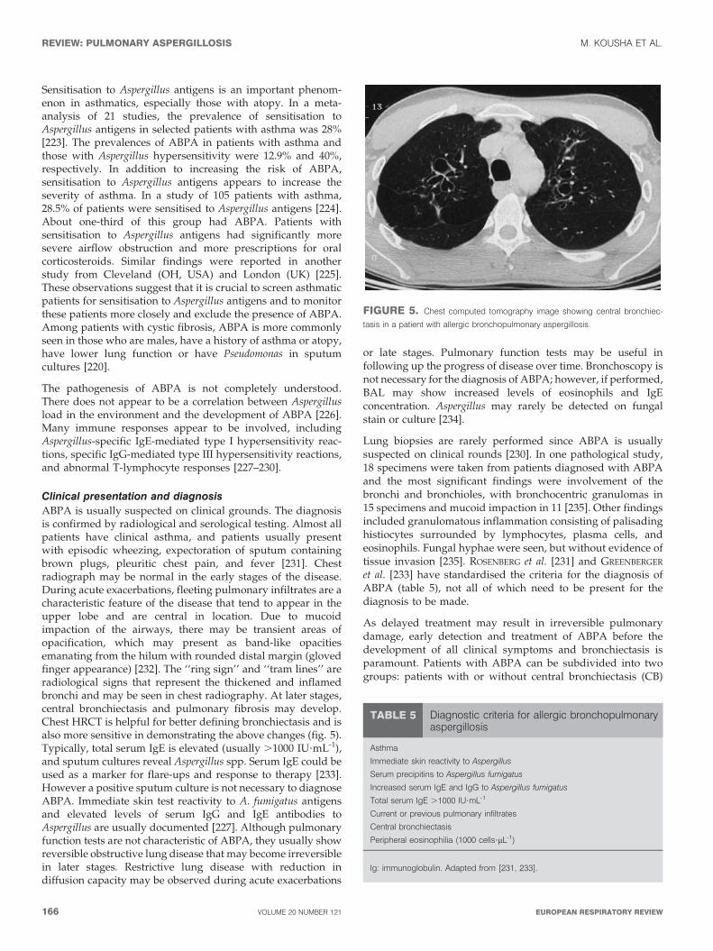
Clinical presentation and diagnosisABPA is usually suspected on clinical grounds. The diagnosisis confirmed by radiological and serological testing. Almost allpatients have clinical asthma, and patients usually presentwith episodic wheezing, expectoration of sputum containingbrown plugs, pleuritic chest pain, and fever [231]. Chestradiograph may be normal in the early stages of the disease.During acute exacerbations, fleeting pulmonary infiltrates are acharacteristic feature of the disease that tend to appear in theupper lobe and are central in location. Due to mucoidimpaction of the airways, there may be transient areas ofopacification, which may present as band-like opacitiesemanating from the hilum with rounded distal margin (glovedfinger appearance) [232]. The ‘‘ring sign’’ and ‘‘tram lines’’ areradiological signs that represent the thickened and inflamedbronchi and may be seen in chest radiography. At later stages,central bronchiectasis and pulmonary fibrosis may develop.Chest HRCT is helpful for better defining bronchiectasis and isalso more sensitive in demonstrating the above changes (fig. 5).Typically, total serum IgE is elevated (usually .1000 IU?mL-1),and sputum cultures reveal Aspergillus spp. Serum IgE could beused as a marker for flare-ups and response to therapy [233].However a positive sputum culture is not necessary to diagnoseABPA. Immediate skin test reactivity to A. fumigatus antigensand elevated levels of serum IgG and IgE antibodies toAspergillus are usually documented [227]. Although pulmonaryfunction tests are not characteristic of ABPA, they usually showreversible obstructive lung disease that may become irreversiblein later stages. Restrictive lung disease with reduction indiffusion capacity may be observed during acute exacerbations
or late stages. Pulmonary function tests may be useful infollowing up the progress of disease over time. Bronchoscopy isnot necessary for the diagnosis of ABPA; however, if performed,BAL may show increased levels of eosinophils and IgEconcentration. Aspergillus may rarely be detected on fungalstain or culture [234].
Lung biopsies are rarely performed since ABPA is usuallysuspected on clinical rounds [230]. In one pathological study,18 specimens were taken from patients diagnosed with ABPAand the most significant findings were involvement of thebronchi and bronchioles, with bronchocentric granulomas in15 specimens and mucoid impaction in 11 [235]. Other findingsincluded granulomatous inflammation consisting of palisadinghistiocytes surrounded by lymphocytes, plasma cells, andeosinophils. Fungal hyphae were seen, but without evidence oftissue invasion [235]. ROSENBERG et al. [231] and GREENBERGER
et al. [233] have standardised the criteria for the diagnosis ofABPA (table 5), not all of which need to be present for thediagnosis to be made.
As delayed treatment may result in irreversible pulmonarydamage, early detection and treatment of ABPA before thedevelopment of all clinical symptoms and bronchiectasis isparamount. Patients with ABPA can be subdivided into twogroups: patients with or without central bronchiectasis (CB)
FIGURE 5. Chest computed tomography image showing central bronchiec-
tasis in a patient with allergic bronchopulmonary aspergillosis.
TABLE 5 Diagnostic criteria for allergic bronchopulmonaryaspergillosis
Asthma
Immediate skin reactivity to Aspergillus
Serum precipitins to Aspergillus fumigatus
Increased serum IgE and IgG to Aspergillus fumigatus
Total serum IgE .1000 IU?mL-1
Current or previous pulmonary infiltrates
Central bronchiectasis
Peripheral eosinophilia (1000 cells?mL-1)
lg: immunoglobulin. Adapted from [231, 233].
REVIEW: PULMONARY ASPERGILLOSIS M. KOUSHA ET AL.
166 VOLUME 20 NUMBER 121 EUROPEAN RESPIRATORY REVIEW

(ABPA-CB and ABPA-seropositive, respectively) [1]. Theminimum essential criteria to diagnose patients with ABPA-CB include asthma, immediate skin reactivity to Aspergillusantigens, serum IgE level .1,000 ng?mL-1 and central bronch-iectasis. The minimum criteria to diagnose ABPA-seropositivepatients include asthma, immediate skin reactivity toAspergillus antigens, serum IgE .1,000 ng?mL-1, history ofpulmonary infiltrates and elevated levels of serum IgE or IgGantibodies to A. fumigatus [236].
PATTERSON et al. [237] have also subdivided ABPA into fivestages on the basis of clinical course, which helps to guide themanagement of the disease [237]. These stages do not need tooccur in order. The first four are potentially reversible with nolong-term sequel. Stage I, the acute stage, is the initial acutepresentation with asthma, elevated IgE level, peripheraleosinophilia, pulmonary infiltrates, and IgE and IgG anti-bodies to A. fumigatus. In practice, patients are seldomidentified in this stage. In stage II, the remission stage, theIgE falls but usually remains elevated, eosinophilia is absent,and the chest radiograph is clear. Serum IgG antibodies toAspergillus antigen may be slightly elevated. Stage III, theexacerbation stage, is the recurrence of the same findings as instage I in patients known to have ABPA. IgE rises to at leastdouble the baseline level. Stage IV, the corticosteroid-depen-dent stage, occurs in patients who have asthma dependent onchronic use of high-dose corticosteroid therapy. Exacerbationsare marked by worsening asthma, radiographic changes and apotential increase in IgE levels. Frequently, the chest CT scanwill show central bronchiectasis. Unfortunately, most patientsare diagnosed at this stage [238]. In stage V, the fibrotic stage,bronchiectasis and fibrosis develop usually leading to irrever-sible lung disease. Patients in this stage may present withdyspnoea, cyanosis, rales, and cor pulmonale. Clubbing maybe present. The serum IgE level and eosinophil count might below or high. Fortunately, few patients progress to this stage.
TreatmentTreatment of ABPA aims to treat acute exacerbations of thedisease and limit progressive lung disease and bronchiectasis.Oral corticosteroids are the main treatment for ABPA (table 3).They suppress the hypersensitivity and inflammatory responseprovoked by A. fumigatus rather than eradicating the organism.Treatment with corticosteroids leads to the relief of broncho-spasm, the resolution of radiographic infiltrates and areduction in serum total IgE and peripheral eosinophilia[139, 140]. 2 weeks of daily therapy of oral prednisone(0.5 mg?kg-1?day-1), followed by gradual tapering, has beenrecommended for new ABPA-related infiltrates [141, 142]. Theduration of therapy should be individualised according to thepatient’s clinical condition. However, most patients requireprolonged low-dose corticosteroid therapy to control theirsymptoms and decrease the rate of relapse [141, 142]. Totalserum IgE serves as a marker of ABPA disease activity. Itshould be checked 6–8 weeks after the initiation of therapy andthen every 8 weeks for 1 year after that to determine a baselinerange for each individual patient [239]. Inhaled corticosteroidsmay help to control symptoms of asthma, but small studieshave failed to demonstrate the efficacy of inhaled corticoster-oids in preventing the progression of lung damage in patientswith ABPA [240, 241].
Several studies have been done on the utility of the antifungalagent itraconazole in the management of patients with ABPA.It has been effective in improving symptoms, facilitatingweaning from corticosteroids, decreasing Aspergillus titresand improving radiographic abnormalities and pulmonaryfunction [210]. A randomised, double-blind, placebo-con-trolled trial of itraconazole 200 mg twice daily for 16 weeksfor patients with ABPA already receiving corticosteroids wasrecently conducted by STEVENS et al. [143]. 46% of patientstreated with itraconazole achieved significant response,defined as a reduction of at least 50% in the corticosteroiddose, decrease of at least 25% in the serum IgE concentration,and one of the following: a 25% improvement in exercisetolerance or pulmonary function test results or partial orcomplete resolution of pulmonary infiltrates. Of note, how-ever, itraconazole may augment the activity of corticosteroidsvia inhibition of their metabolism, which may lead to abnormaladrenocorticotropic hormone stimulation and adrenal insuffi-ciency [242]. Recently, voriconazole has also been tried in thetreatment of ABPA and showed a favourable therapeuticresponse in the few case reports available [144–146]. In onestudy of small number of children with cystic fibrosis andABPA, voriconazole treatment demonstrated significant clin-ical and serological improvements [243]. Randomised trials areneeded to assess the efficacy of voriconazole in the manage-ment of ABPA. Few case reports have described the beneficialuse of the anti-IgE monoclonal antibody (omalizumab) inpatients with ABPA. They have shown rapid improvement ofthe respiratory symptoms and lung function [244, 245].
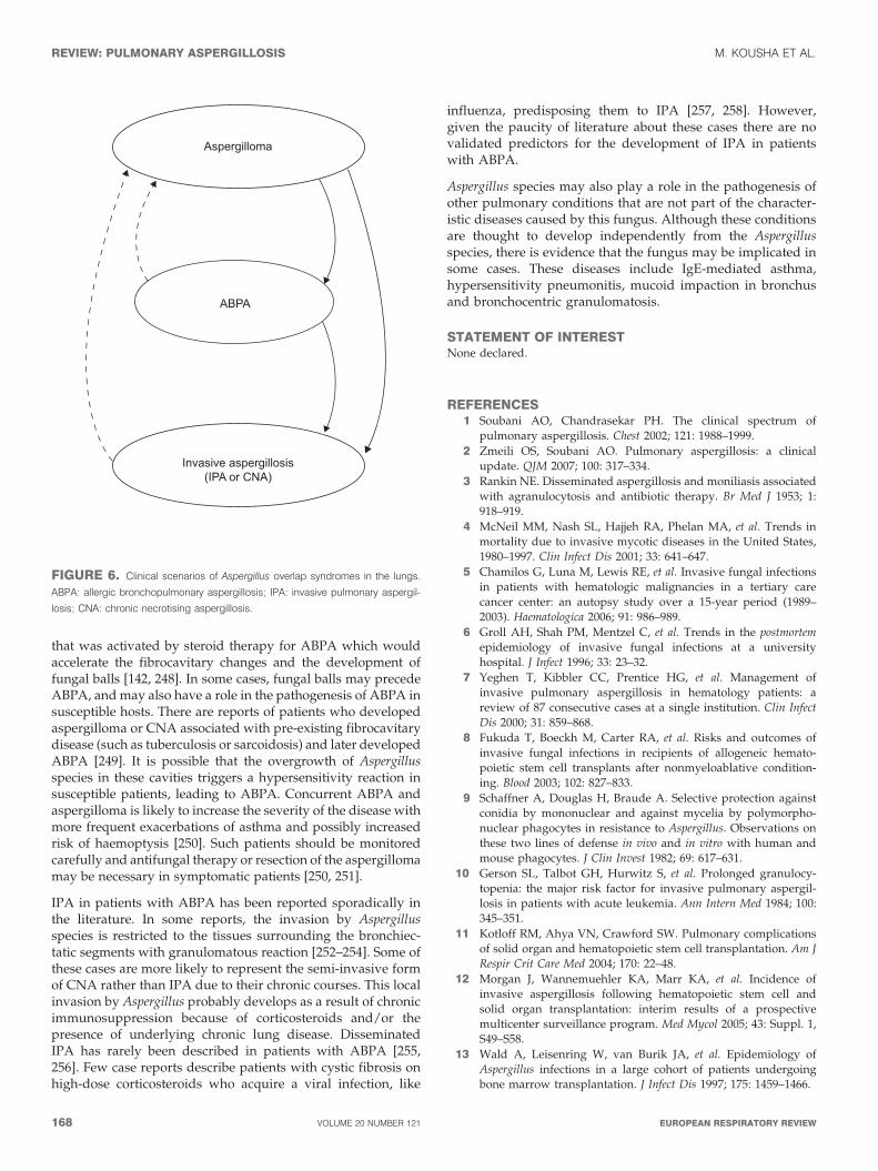
PULMONARY ASPERGILLUS OVERLAP SYNDROMESThe above-mentioned Aspergillus syndromes may co-exist (e.g.fungal balls in patients with ABPA) or may progress from oneentity to another (e.g. IPA in a patient with ABPA) (fig. 6).These Aspergillus overlap syndromes have been reported incase reports or small case series. The proposed mechanisms forthe development of Aspergillus overlap syndromes includecoincidence, the presence of severe underlying lung disease(e.g. a patient with aspergilloma who develops CNA),corticosteroid therapy (IPA in a patient with aspergilloma orABPA), or Aspergillus fungal load. It is also possible thatgenetic factors may predispose patients to progress from oneform of aspergillosis to another. For example, CFTR genemutation may lead to ABPA, and mannose-binding lectin genemutations may result in CNA or IPA. Viral illnesses (in apatient with ABPA or aspergilloma) have been rarely reportedas a risk factor for IPA [183].
The emergence of fungal balls in patients with ABPA has beenreported infrequently in the literature. It may occur as an earlyevent as well as a late phenomenon. In early cases, thebronchiectatic areas affected by ABPA may enlarge to formcavities that colonise with Aspergillus spp., creating fungal ballsthat may present as haemoptysis and/or a cavitary mass [142,246]. The knowledge that aspergillomas may develop in patientswith ABPA helps avoid unnecessary invasive procedures to ruleout alternative aetiologies, such malignancy or tuberculosis.Fungal balls may also be a late finding in patients with fibrosisand cavitation associated with long-standing or poorly treatedABPA. In some of these patients, this aspergilloma could be dueto concomitant fibrocavitary disease [247] (such as tuberculosis)
M. KOUSHA ET AL. REVIEW: PULMONARY ASPERGILLOSIS
cEUROPEAN RESPIRATORY REVIEW VOLUME 20 NUMBER 121 167
that was activated by steroid therapy for ABPA which wouldaccelerate the fibrocavitary changes and the development offungal balls [142, 248]. In some cases, fungal balls may precedeABPA, and may also have a role in the pathogenesis of ABPA insusceptible hosts. There are reports of patients who developedaspergilloma or CNA associated with pre-existing fibrocavitarydisease (such as tuberculosis or sarcoidosis) and later developedABPA [249]. It is possible that the overgrowth of Aspergillusspecies in these cavities triggers a hypersensitivity reaction insusceptible patients, leading to ABPA. Concurrent ABPA andaspergilloma is likely to increase the severity of the disease withmore frequent exacerbations of asthma and possibly increasedrisk of haemoptysis [250]. Such patients should be monitoredcarefully and antifungal therapy or resection of the aspergillomamay be necessary in symptomatic patients [250, 251].
IPA in patients with ABPA has been reported sporadically inthe literature. In some reports, the invasion by Aspergillusspecies is restricted to the tissues surrounding the bronchiec-tatic segments with granulomatous reaction [252–254]. Some ofthese cases are more likely to represent the semi-invasive formof CNA rather than IPA due to their chronic courses. This localinvasion by Aspergillus probably develops as a result of chronicimmunosuppression because of corticosteroids and/or thepresence of underlying chronic lung disease. DisseminatedIPA has rarely been described in patients with ABPA [255,256]. Few case reports describe patients with cystic fibrosis onhigh-dose corticosteroids who acquire a viral infection, like
influenza, predisposing them to IPA [257, 258]. However,given the paucity of literature about these cases there are novalidated predictors for the development of IPA in patientswith ABPA.
Aspergillus species may also play a role in the pathogenesis ofother pulmonary conditions that are not part of the character-istic diseases caused by this fungus. Although these conditionsare thought to develop independently from the Aspergillusspecies, there is evidence that the fungus may be implicated insome cases. These diseases include IgE-mediated asthma,hypersensitivity pneumonitis, mucoid impaction in bronchusand bronchocentric granulomatosis.
STATEMENT OF INTERESTNone declared.
REFERENCES1 Soubani AO, Chandrasekar PH. The clinical spectrum of
pulmonary aspergillosis. Chest 2002; 121: 1988–1999.
2 Zmeili OS, Soubani AO. Pulmonary aspergillosis: a clinical
update. QJM 2007; 100: 317–334.
3 Rankin NE. Disseminated aspergillosis and moniliasis associated
with agranulocytosis and antibiotic therapy. Br Med J 1953; 1:
918–919.
4 McNeil MM, Nash SL, Hajjeh RA, Phelan MA, et al. Trends in
mortality due to invasive mycotic diseases in the United States,
1980–1997. Clin Infect Dis 2001; 33: 641–647.
5 Chamilos G, Luna M, Lewis RE, et al. Invasive fungal infections
in patients with hematologic malignancies in a tertiary care
cancer center: an autopsy study over a 15-year period (1989–
2003). Haematologica 2006; 91: 986–989.
6 Groll AH, Shah PM, Mentzel C, et al. Trends in the postmortem
epidemiology of invasive fungal infections at a university
hospital. J Infect 1996; 33: 23–32.
7 Yeghen T, Kibbler CC, Prentice HG, et al. Management of
invasive pulmonary aspergillosis in hematology patients: a
review of 87 consecutive cases at a single institution. Clin Infect
Dis 2000; 31: 859–868.
8 Fukuda T, Boeckh M, Carter RA, et al. Risks and outcomes of
invasive fungal infections in recipients of allogeneic hemato-
poietic stem cell transplants after nonmyeloablative condition-
ing. Blood 2003; 102: 827–833.
9 Schaffner A, Douglas H, Braude A. Selective protection against
conidia by mononuclear and against mycelia by polymorpho-
nuclear phagocytes in resistance to Aspergillus. Observations on
these two lines of defense in vivo and in vitro with human and
mouse phagocytes. J Clin Invest 1982; 69: 617–631.
10 Gerson SL, Talbot GH, Hurwitz S, et al. Prolonged granulocy-
topenia: the major risk factor for invasive pulmonary aspergil-
losis in patients with acute leukemia. Ann Intern Med 1984; 100:
345–351.
11 Kotloff RM, Ahya VN, Crawford SW. Pulmonary complications
of solid organ and hematopoietic stem cell transplantation. Am J
Respir Crit Care Med 2004; 170: 22–48.
12 Morgan J, Wannemuehler KA, Marr KA, et al. Incidence of
invasive aspergillosis following hematopoietic stem cell and
solid organ transplantation: interim results of a prospective
multicenter surveillance program. Med Mycol 2005; 43: Suppl. 1,
S49–S58.
13 Wald A, Leisenring W, van Burik JA, et al. Epidemiology of
Aspergillus infections in a large cohort of patients undergoing
bone marrow transplantation. J Infect Dis 1997; 175: 1459–1466.
ABPA
Aspergilloma
Invasive aspergillosis(IPA or CNA)
FIGURE 6. Clinical scenarios of Aspergillus overlap syndromes in the lungs.
ABPA: allergic bronchopulmonary aspergillosis; IPA: invasive pulmonary aspergil-
losis; CNA: chronic necrotising aspergillosis.
REVIEW: PULMONARY ASPERGILLOSIS M. KOUSHA ET AL.
168 VOLUME 20 NUMBER 121 EUROPEAN RESPIRATORY REVIEW

14 Marr KA, Carter RA, Boeckh M, et al. Invasive aspergillosis in
allogeneic stem cell transplant recipients: changes in epidemiol-
ogy and risk factors. Blood 2002; 100: 4358–4366.
15 Segal BH, Walsh TJ. Current approaches to diagnosis andtreatment of invasive aspergillosis. Am J Respir Crit Care Med
2006; 173: 707–717.
16 Lionakis MS, Kontoyiannis DP. Glucocorticoids and invasive
fungal infections. Lancet 2003; 362: 1828–1838.
17 Warris A, Bjorneklett A, Gaustad P. Invasive pulmonaryaspergillosis associated with infliximab therapy. N Engl J Med
2001; 344: 1099–1100.
18 Holding KJ, Dworkin MS, Wan PC, et al. Aspergillosis among
people infected with human immunodeficiency virus: incidence
and survival. Adult and adolescent spectrum of HIV diseaseproject. Clin Infect Dis 2000; 31: 1253–1257.
19 Denning DW, Follansbee SE, Scolaro M, et al. Pulmonary
aspergillosis in the acquired immunodeficiency syndrome. N Engl
J Med 1991; 324: 654–662.
20 Lortholary O, Meyohas MC, Dupont B, et al. Invasive aspergil-losis in patients with acquired immunodeficiency syndrome:
report of 33 cases. French cooperative study group on asper-
gillosis in AIDS. Am J Med 1993; 95: 177–187.
21 Mylonakis E, Barlam TF, Flanigan T, et al. Pulmonary aspergil-
losis and invasive disease in AIDS: review of 342 cases. Chest
1998; 114: 251–262.
22 Segal BH, Barnhart LA, Anderson VL, et al. Posaconazole as
salvage therapy in patients with chronic granulomatous disease
and invasive filamentous fungal infection. Clin Infect Dis 2005;
40: 1684–1688.
23 Soubani AO, Miller KB, Hassoun PM. Pulmonary complications
of bone marrow transplantation. Chest 1996; 109: 1066–1077.
24 Marr KA, Carter RA, Crippa F, et al. Epidemiology and outcome
of mould infections in hematopoietic stem cell transplant
recipients. Clin Infect Dis 2002; 34: 909–917.
25 Baddley JW, Stroud TP, Salzman D, et al. Invasive mold
infections in allogeneic bone marrow transplant recipients. Clin
Infect Dis 2001; 32: 1319–1324.
26 Junghanss C, Marr KA, Carter RA, et al. Incidence and outcome
of bacterial and fungal infections following nonmyeloablativecompared with myeloablative allogeneic hematopoietic stem cell
transplantation: a matched control study. Biol Blood Marrow
Transplant 2002; 8: 512–520.
27 Cordonnier C, Ribaud P, Herbrecht R, et al. Prognostic factors for
death due to invasive aspergillosis after hematopoietic stem cell
transplantation: a 1-year retrospective study of consecutivepatients at French transplantation centers. Clin Infect Dis 2006;
42: 955–963.
28 Ribaud P, Chastang C, Latge JP, et al. Survival and prognostic
factors of invasive aspergillosis after allogeneic bone marrow
transplantation. Clin Infect Dis 1999; 28: 322–330.
29 van Burik JA, Carter SL, Freifeld AG, et al. Higher risk of
cytomegalovirus and Aspergillus infections in recipients of T
cell-depleted unrelated bone marrow: analysis of infectious
complications in patients treated with T cell depletion versus
immunosuppressive therapy to prevent graft-versus-host disease.Biol Blood Marrow Transplant 2007; 13: 1487–1498.
30 Nichols WG, Corey L, Gooley T, et al. High risk of death due to
bacterial and fungal infection among cytomegalovirus (CMV)-
seronegative recipients of stem cell transplants from seropositive
donors: evidence for indirect effects of primary CMV infection.J Infect Dis 2002; 185: 273–282.
31 Kemper CA, Hostetler JS, Follansbee SE, et al. Ulcerative and
plaque-like tracheobronchitis due to infection with Aspergillus in
patients with AIDS. Clin Infect Dis 1993; 17: 344–352.
32 Wallace JM, Lim R, Browdy BL, et al. Risk factors and outcomesassociated with identification of Aspergillus in respiratory specimens
from persons with HIV disease. Pulmonary complications of HIV
infection study group. Chest 1998; 114: 131–137.
33 Libanore M, Prini E, Mazzetti M, et al. Invasive aspergillosis inItalian AIDS patients. Infection 2002; 30: 341–345.
34 Hawgood S, Ochs M, Jung A, et al. Sequential targeted deficiency
of SP-A and -D leads to progressive alveolar lipoproteinosisand emphysema. Am J Physiol Lung Cell Mol Physiol 2002; 283:
L1002–L1010.
35 Clark H, Reid K. The potential of recombinant surfactant protein
D therapy to reduce inflammation in neonatal chronic lung
disease, cystic fibrosis, and emphysema. Arch Dis Child 2003; 88:981–984.
36 Wert SE, Yoshida M, LeVine AM, et al. Increased metalloprotei-
nase activity, oxidant production, and emphysema in surfactantprotein D gene-inactivated mice. Proc Natl Acad Sci USA 2000; 97:
5972–5977.
37 Bochud PY, Chien JW, Marr KA, et al. Toll-like receptor 4polymorphisms and aspergillosis in stem-cell transplantation.
N Engl J Med 2008; 359: 1766–1777.
38 Sarir H, Mortaz E, Karimi K, et al. Cigarette smoke regulates theexpression of TLR4 and IL-8 production by human macro-
phages. J Inflamm (Lond) 2009; 6: 12.
39 Soubani AO, Khanchandani G, Ahmed HP. Clinical significanceof lower respiratory tract aspergillus culture in elderly hospita-
lized patients. Eur J Clin Microbiol Infect Dis 2004; 23: 491–494.
40 Samarakoon P, Soubani AO. Invasive pulmonary aspergillosis in
patients with COPD, a report of five cases and systematic review
of the literature. Chron Respir Dis 2008; 5: 19–27.
41 Ader F, Nseir S, Le Berre R, et al. Invasive pulmonary
aspergillosis in chronic obstructive pulmonary disease: an
emerging fungal pathogen. Clin Microbiol Infect 2005; 11: 427–429.
42 Carrascosa Porras M, Herreras Martinez R, Corral Mones J, et al.
Fatal Aspergillus myocarditis following short-term corticosteroid
therapy for chronic obstructive pulmonary disease. Scand J Infect
Dis 2002; 34: 224–227.
43 Bulpa PA, Dive AM, Garrino MG, et al. Chronic obstructive
pulmonary disease patients with invasive pulmonary aspergillo-sis: benefits of intensive care? Intensive Care Med 2001; 27: 59–67.
44 Vandewoude KH, Blot SI, Depuydt P, et al. Clinical relevance of
Aspergillus isolation from respiratory tract samples in critically illpatients. Crit Care 2006; 10: R31.
45 Dimopoulos G, Piagnerelli M, Berre J, et al. Disseminatedaspergillosis in intensive care unit patients: an autopsy study.
J Chemother 2003; 15: 71–75.
46 Vandewoude KH, Blot SI, Benoit D, et al. Invasive aspergillosis incritically ill patients: attributable mortality and excesses in length
of ICU stay and ventilator dependence. J Hosp Infect 2004; 56:
269–276.
47 Meersseman W, Vandecasteele SJ, Wilmer A, et al. Invasive
aspergillosis in critically ill patients without malignancy. Am J
Respir Crit Care Med 2004; 170: 621–625.
48 Janssen JJ, Strack van Schijndel RJ, van der Poest Clement EH,
et al. Outcome of ICU treatment in invasive aspergillosis.
Intensive Care Med 1996; 22: 1315–1322.
49 Meersseman W, Lagrou K, Maertens J, et al. Invasive aspergil-
losis in the intensive care unit. Clin Infect Dis 2007; 45: 205–216.
50 Garnacho-Montero J, Amaya-Villar R, Ortiz-Leyba C, et al.
Isolation of Aspergillus spp. from the respiratory tract in critically
ill patients: risk factors, clinical presentation and outcome. Crit
Care 2005; 9: R191–R199.
51 Garnacho-Montero J, Amaya-Villar R. A validated clinical
approach for the management of aspergillosis in critically illpatients: Ready, steady, go! Crit Care 2006; 10: 132.
52 Khasawneh F, Mohamad T, Moughrabieh MK, et al. Isolation of
Aspergillus in critically ill patients: a potential marker of pooroutcome. J Crit Care 2006; 21: 322–327.
M. KOUSHA ET AL. REVIEW: PULMONARY ASPERGILLOSIS
cEUROPEAN RESPIRATORY REVIEW VOLUME 20 NUMBER 121 169
53 Iwen PC, Rupp ME, Langnas AN, et al. Invasive pulmonaryaspergillosis due to Aspergillus terreus: 12-year experience andreview of the literature. Clin Infect Dis 1998; 26: 1092–1097.
54 Young RC, Bennett JE, Vogel CL, et al. Aspergillosis. Thespectrum of the disease in 98 patients. Medicine (Baltimore) 1970;49: 147–173.
55 Prystowsky SD, Vogelstein B, Ettinger DS, et al. Invasiveaspergillosis. N Engl J Med 1976; 295: 655–658.
56 Allo MD, Miller J, Townsend T, et al. Primary cutaneousaspergillosis associated with Hickman intravenous catheters.N Engl J Med 1987; 317: 1105–1108.
57 Albelda SM, Talbot GH, Gerson SL, et al. Pulmonary cavitationand massive hemoptysis in invasive pulmonary aspergillosis.Influence of bone marrow recovery in patients with acuteleukemia. Am Rev Respir Dis 1985; 131: 115–120.
58 Denning DW. Invasive aspergillosis. Clin Infect Dis 1998; 26:781–803.
59 Judson MA, Sahn SA. Endobronchial lesions in HIV-infectedindividuals. Chest 1994; 105: 1314–1323.
60 Nathan SD, Shorr AF, Schmidt ME, et al. Aspergillus andendobronchial abnormalities in lung transplant recipients.Chest 2000; 118: 403–407.
61 Wu N, Huang Y, Li Q, et al. Isolated invasive Aspergillustracheobronchitis: a clinical study of 19 cases. Clin Microbiol Infect
2010; 16: 689–695.
62 Denning DW. Commentary: unusual manifestations of aspergil-losis. Thorax 1995; 50: 812–813.
63 Tasci S, Glasmacher A, Lentini S, et al. Pseudomembranous andobstructive Aspergillus tracheobronchitis – optimal diagnosticstrategy and outcome. Mycoses 2006; 49: 37–42.
64 Ruhnke M, Bohme A, Buchheidt D, et al. Diagnosis of invasivefungal infections in hematology and oncology–guidelines of theinfectious diseases working party (AGIHO) of the Germansociety of Hematology and Oncology (DGHO). Ann Hematol
2003; 82: Suppl. 2, S141–S148.
65 Nalesnik MA, Myerowitz RL, Jenkins R, et al. Significance ofAspergillus species isolated from respiratory secretions in thediagnosis of invasive pulmonary aspergillosis. J Clin Microbiol
1980; 11: 370–376.
66 Horvath JA, Dummer S. The use of respiratory-tract cultures inthe diagnosis of invasive pulmonary aspergillosis. Am J Med
1996; 100: 171–178.
67 Treger TR, Visscher DW, Bartlett MS, et al. Diagnosis ofpulmonary infection caused by Aspergillus: usefulness ofrespiratory cultures. J Infect Dis 1985; 152: 572–576.
68 Soubani AO, Qureshi MA. Invasive pulmonary aspergillosisfollowing bone marrow transplantation: risk factors and diag-nostic aspect. Haematologia (Budap) 2002; 32: 427–437.
69 Yu VL, Muder RR, Poorsattar A. Significance of isolation ofAspergillus from the respiratory tract in diagnosis of invasivepulmonary aspergillosis. Results from a three-year prospectivestudy. Am J Med 1986; 81: 249–254.
70 Tang CM, Cohen J. Diagnosing fungal infections in immuno-compromised hosts. J Clin Pathol 1992; 45: 1–5.
71 Duthie R, Denning DW. Aspergillus fungemia: report of two casesand review. Clin Infect Dis 1995; 20: 598–605.
72 Libshitz HI, Pagani JJ. Aspergillosis and mucormycosis: twotypes of opportunistic fungal pneumonia. Radiology 1981; 140:301–306.
73 Kuhlman JE, Fishman EK, Siegelman SS. Invasive pulmonaryaspergillosis in acute leukemia: characteristic findings on CT, theCT halo sign, and the role of CT in early diagnosis. Radiology
1985; 157: 611–614.
74 Caillot D, Casasnovas O, Bernard A, et al. Improved manage-ment of invasive pulmonary aspergillosis in neutropenic patientsusing early thoracic computed tomographic scan and surgery.J Clin Oncol 1997; 15: 139–147.
75 Caillot D, Mannone L, Cuisenier B, et al. Role of early diagnosisand aggressive surgery in the management of invasive pulmon-ary aspergillosis in neutropenic patients. Clin Microbiol Infect
2001; 7: Suppl. 2, 54–61.
76 Kuhlman JE, Fishman EK, Burch PA, et al. Invasive pulmonaryaspergillosis in acute leukemia. The contribution of CT to earlydiagnosis and aggressive management. Chest 1987; 92: 95–99.
77 Curtis AM, Smith GJ, Ravin CE. Air crescent sign of invasiveaspergillosis. Radiology 1979; 133: 17–21.
78 Gaeta M, Blandino A, Scribano E, et al. Computed tomographyhalo sign in pulmonary nodules: frequency and diagnostic value.J Thorac Imaging 1999; 14: 109–113.
79 Greene R. The radiological spectrum of pulmonary aspergillosis.Med Mycol 2005; 43: Suppl. 1, S147–S154.
80 Heussel CP, Kauczor HU, Heussel GE, et al. Pneumonia in febrileneutropenic patients and in bone marrow and blood stem-celltransplant recipients: use of high-resolution computed tomo-graphy. J Clin Oncol 1999; 17: 796–805.
81 Horger M, Hebart H, Einsele H, et al. Initial CT manifestations ofinvasive pulmonary aspergillosis in 45 non-HIV immunocom-promised patients: association with patient outcome?Eur J Radiol
2005; 55: 437–444.
82 Albelda SM, Talbot GH, Gerson SL, et al. Role of fiberopticbronchoscopy in the diagnosis of invasive pulmonary asper-gillosis in patients with acute leukemia. Am J Med 1984; 76:1027–1034.
83 Kahn FW, Jones JM, England DM. The role of bronchoalveolarlavage in the diagnosis of invasive pulmonary aspergillosis. Am J
Clin Pathol 1986; 86: 518–523.
84 Levy H, Horak DA, Tegtmeier BR, et al. The value ofbronchoalveolar lavage and bronchial washings in the diagnosisof invasive pulmonary aspergillosis. Respir Med 1992; 86: 243–248.
85 Reichenberger F, Habicht J, Matt P, et al. Diagnostic yield ofbronchoscopy in histologically proven invasive pulmonaryaspergillosis. Bone Marrow Transplant 1999; 24: 1195–1199.
86 Maschmeyer G, Beinert T, Buchheidt D, et al. Diagnosis andantimicrobial therapy of pulmonary infiltrates in febrile neu-tropenic patients–guidelines of the infectious diseases workingparty (AGIHO) of the German Society of Hematology andOncology (DGHO). Ann Hematol 2003; 82: Suppl. 2, S118–S126.
87 Panackal AA, Marr KA. Scedosporium/Pseudallescheria infections.Semin Respir Crit Care Med 2004; 25: 171–181.
88 Walsh TJ, Groll AH. Overview: Non-fumigatus species ofAspergillus: perspectives on emerging pathogens in immuno-compromised hosts. Curr Opin Investig Drugs 2001; 2: 1366–1367.
89 Lass-Florl C, Griff K, Mayr A, et al. Epidemiology and outcomeof infections due to Aspergillus terreus: 10-year single centreexperience. Br J Haematol 2005; 131: 201–207.
90 Steinbach WJ, Benjamin DK Jr, Kontoyiannis DP, et al. Infectionsdue to Aspergillus terreus: a multicenter retrospective analysis of83 cases. Clin Infect Dis 2004; 39: 192–198.
91 Hachem RY, Kontoyiannis DP, Boktour MR, et al. Aspergillus
terreus: an emerging amphotericin B-resistant opportunistic moldin patients with hematologic malignancies. Cancer 2004; 101:1594–1600.
92 Kontoyiannis DP, Lewis RE, May GS, et al. Aspergillus nidulans isfrequently resistant to amphotericin B. Mycoses 2002; 45: 406–407.
93 Walsh TJ, Raad I, Patterson TF, et al. Treatment of invasiveaspergillosis with posaconazole in patients who are refractory toor intolerant of conventional therapy: an externally controlledtrial. Clin Infect Dis 2007; 44: 2–12.
94 Boutboul F, Alberti C, Leblanc T, et al. Invasive aspergillosis inallogeneic stem cell transplant recipients: increasing antigenemiais associated with progressive disease. Clin Infect Dis 2002; 34:939–943.
95 Marr KA, Balajee SA, McLaughlin L, et al. Detection ofgalactomannan antigenemia by enzyme immunoassay for the
REVIEW: PULMONARY ASPERGILLOSIS M. KOUSHA ET AL.
170 VOLUME 20 NUMBER 121 EUROPEAN RESPIRATORY REVIEW
diagnosis of invasive aspergillosis: variables that affect perfor-mance. J Infect Dis 2004; 190: 641–649.
96 Pfeiffer CD, Fine JP, Safdar N. Diagnosis of invasive aspergillosisusing a galactomannan assay: a meta-analysis. Clin Infect Dis
2006; 42: 1417–1427.
97 Ascioglu S, Rex JH, de Pauw B, et al. Defining opportunisticinvasive fungal infections in immunocompromised patients withcancer and hematopoietic stem cell transplants: an internationalconsensus. Clin Infect Dis 2002; 34: 7–14.
98 Herbrecht R, Letscher-Bru V, Oprea C, et al. Aspergillus
galactomannan detection in the diagnosis of invasive aspergil-losis in cancer patients. J Clin Oncol 2002; 20: 1898–1906.
99 Singh N, Obman A, Husain S, et al. Reactivity of plateliaAspergillus galactomannan antigen with piperacillin-tazobactam:clinical implications based on achievable concentrations inserum. Antimicrob Agents Chemother 2004; 48: 1989–1992.
100 Marr KA, Laverdiere M, Gugel A, et al. Antifungal therapydecreases sensitivity of the Aspergillus galactomannan enzymeimmunoassay. Clin Infect Dis 2005; 40: 1762–1769.
101 Ansorg R, van den Boom R, Rath PM. Detection of Aspergillus
galactomannan antigen in foods and antibiotics. Mycoses 1997;40: 353–357.
102 Busca A, Locatelli F, Barbui A, et al. Usefulness of sequentialAspergillus galactomannan antigen detection combined withearly radiologic evaluation for diagnosis of invasive pulmonaryaspergillosis in patients undergoing allogeneic stem cell trans-plantation. Transplant Proc 2006; 38: 1610–1613.
103 Musher B, Fredricks D, Leisenring W, et al. Aspergillusgalactomannan enzyme immunoassay and quantitative PCRfor diagnosis of invasive aspergillosis with bronchoalveolarlavage fluid. J Clin Microbiol 2004; 42: 5517–5522.
104 Klont RR, Mennink-Kersten MA, Verweij PE. Utility ofAspergillus antigen detection in specimens other than serumspecimens. Clin Infect Dis 2004; 39: 1467–1474.
105 Salonen J, Lehtonen OP, Terasjarvi MR, et al. Aspergillus antigenin serum, urine and bronchoalveolar lavage specimens ofneutropenic patients in relation to clinical outcome. Scand J
Infect Dis 2000; 32: 485–490.
106 Danpornprasert P, Foongladda S, Tscheikuna J. Impact ofbronchoalveolar lavage galactomannan on the outcome ofpatients at risk for invasive pulmonary aspergillosis. J MedAssoc Thai 2010; 93: Suppl. 1, S86–S93.
107 Hsu LY, Ding Y, Phua J, et al. Galactomannan testing ofbronchoalveolar lavage fluid is useful for diagnosis of invasivepulmonary aspergillosis in hematology patients. BMC Infect Dis
2010; 10: 44.
108 Meersseman W, Lagrou K, Maertens J, et al. Galactomannan inbronchoalveolar lavage fluid: a tool for diagnosing aspergillosisin intensive care unit patients. Am J Respir Crit Care Med 2008;177: 27–34.
109 Clancy CJ, Jaber RA, Leather HL, et al. Bronchoalveolar lavagegalactomannan in diagnosis of invasive pulmonary aspergillosisamong solid-organ transplant recipients. J Clin Microbiol 2007; 45:1759–1765.
110 Viscoli C, Machetti M, Gazzola P, et al. Aspergillus galactoman-nan antigen in the cerebrospinal fluid of bone marrow transplantrecipients with probable cerebral aspergillosis. J Clin Microbiol2002; 40: 1496–1499.
111 Hizel K, Kokturk N, Kalkanci A, et al. Polymerase chain reaction inthe diagnosis of invasive aspergillosis. Mycoses 2004; 47: 338–342.
112 Buchheidt D, Baust C, Skladny H, et al. Detection of Aspergillus
species in blood and bronchoalveolar lavage samples fromimmunocompromised patients by means of 2-step polymerasechain reaction: clinical results. Clin Infect Dis 2001; 33: 428–435.
113 Loeffler J, Kloepfer K, Hebart H, et al. Polymerase chain reactiondetection of Aspergillus DNA in experimental models of invasiveaspergillosis. J Infect Dis 2002; 185: 1203–1206.
114 Halliday C, Hoile R, Sorrell T, et al. Role of prospective screeningof blood for invasive aspergillosis by polymerase chain reactionin febrile neutropenic recipients of haematopoietic stem celltransplants and patients with acute leukaemia. Br J Haematol
2006; 132: 478–486.
115 Obayashi T, Yoshida M, Mori T, et al. Plasma (1–.3)-beta-D-glucan measurement in diagnosis of invasive deep mycosis andfungal febrile episodes. Lancet 1995; 345: 17–20.
116 Pazos C, Ponton J, Del Palacio A. Contribution of (1-.3)-beta-D-glucan chromogenic assay to diagnosis and therapeutic monitor-ing of invasive aspergillosis in neutropenic adult patients: acomparison with serial screening for circulating galactomannan.J Clin Microbiol 2005; 43: 299–305.
117 De Pauw B, Walsh TJ, Donnelly JP, et al. Revised definitions ofinvasive fungal disease from the European Organization forResearch and Treatment of Cancer/invasive fungal infectionscooperative group and the National Institute of Allergy andInfectious Diseases mycoses study group (EORTC/MSG) con-sensus group. Clin Infect Dis 2008; 46: 1813–1821.
118 de Pauw BE, Patterson TF. Should the consensus guidelines’specific criteria for the diagnosis of invasive fungal infection bechanged? Clin Infect Dis 2005; 41: Suppl. 6, S377–S380.
119 Johnson LB, Kauffman CA. Voriconazole: a new triazoleantifungal agent. Clin Infect Dis 2003; 36: 630–637.
120 Sambatakou H, Dupont B, Lode H, et al. Voriconazole treatmentfor subacute invasive and chronic pulmonary aspergillosis. Am J
Med 2006; 119: 527 e517–524.
121 Ghannoum MA, Kuhn DM. Voriconazole – better chances forpatients with invasive mycoses. Eur J Med Res 2002; 7: 242–256.
122 Herbrecht R, Denning DW, Patterson TF, et al. Voriconazoleversus amphotericin B for primary therapy of invasive aspergil-losis. N Engl J Med 2002; 347: 408–415.
123 Limper AH, Knox KS, Sarosi GA, et al. An official AmericanThoracic Society statement: treatment of fungal infections inadult pulmonary and critical care patients. Am J Respir Crit Care
Med 2011; 183: 96–128.
124 Cornely OA, Maertens J, Bresnik M, et al. Liposomal amphoter-icin B as initial therapy for invasive mold infection: arandomized trial comparing a high-loading dose regimen withstandard dosing (AmBiLoad trial). Clin Infect Dis 2007; 44:1289–1297.
125 Pitisuttithum P, Negroni R, Graybill JR, et al. Activity ofposaconazole in the treatment of central nervous system fungalinfections. J Antimicrob Chemother 2005; 56: 745–755.
126 Spanakis EK, Aperis G, Mylonakis E et al. New agents for thetreatment of fungal infections: clinical efficacy and gaps incoverage. Clin Infect Dis 2006; 43: 1060–1068.
127 Cohen-Wolkowiez M, Benjamin DK Jr, Steinbach WJ, et al.
Anidulafungin: a new echinocandin for the treatment of fungalinfections. Drugs Today (Barc) 2006; 42: 533–544.
128 Saraceno JL, Phelps DT, Ferro TJ, et al. Chronic necrotizingpulmonary aspergillosis: approach to management. Chest 1997;112: 541–548.
129 Caras WE, Pluss JL. Chronic necrotizing pulmonary aspergillo-sis: pathologic outcome after itraconazole therapy. Mayo Clin
Proc 1996; 71: 25–30.
130 Binder RE, Faling LJ, Pugatch RD, et al. Chronic necrotizingpulmonary aspergillosis: a discrete clinical entity. Medicine
(Baltimore) 1982; 61: 109–124.
131 Uflacker R, Kaemmerer A, Picon PD, et al. Bronchial arteryembolization in the management of hemoptysis: technicalaspects and long-term results. Radiology 1985; 157: 637–644.
132 Soltanzadeh H, Wychulis AR, Sadr F, et al. Surgical treatment ofpulmonary aspergilloma. Ann Surg 1977; 186: 13–16.
133 Massard G, Roeslin N, Wihlm JM, et al. Pleuropulmonaryaspergilloma: clinical spectrum and results of surgical treatment.Ann Thorac Surg 1992; 54: 1159–1164.
M. KOUSHA ET AL. REVIEW: PULMONARY ASPERGILLOSIS
cEUROPEAN RESPIRATORY REVIEW VOLUME 20 NUMBER 121 171
134 Kilman JW, Ahn C, Andrews NC, et al. Surgery for pulmonary
aspergillosis. J Thorac Cardiovasc Surg 1969; 57: 642–647.
135 Chen JC, Chang YL, Luh SP, et al. Surgical treatment forpulmonary aspergilloma: a 28 year experience. Thorax 1997; 52:
810–813.
136 Campbell JH, Winter JH, Richardson MD, et al. Treatment ofpulmonary aspergilloma with itraconazole. Thorax 1991; 46:
839–841.
137 Dupont B. Itraconazole therapy in aspergillosis: study in 49
patients. J Am Acad Dermatol 1990; 23: 607–614.
138 Impens N, De Greve J, De Beule K, et al. Oral treatment withitraconazole of aspergilloma in cavitary lung cancer. Eur Respir J
1990; 3: 837–839.
139 Rosenberg M, Patterson R, Roberts M, et al. The assessment ofimmunologic and clinical changes occurring during corticoster-
oid therapy for allergic bronchopulmonary aspergillosis. Am J
Med 1978; 64: 599–606.
140 Wang JL, Patterson R, Roberts M, et al. The management of
allergic bronchopulmonary aspergillosis. Am Rev Respir Dis 1979;
120: 87–92.
141 Capewell S, Chapman BJ, Alexander F, et al. Corticosteroid
treatment and prognosis in pulmonary eosinophilia. Thorax 1989;
44: 925–929.
142 Safirstein BH, D’Souza MF, Simon G, et al. Five-year follow-up of
allergic bronchopulmonary aspergillosis. Am Rev Respir Dis 1973;108: 450–459.
143 Stevens DA, Schwartz HJ, Lee JY, et al. A randomized trial of
itraconazole in allergic bronchopulmonary aspergillosis. N Engl J
Med 2000; 342: 756–762.
144 Mulliez P, Croxo C, Roy-Saint Georges F, et al. Allergic broncho-
pulmonary aspergillosis treated with voriconazole. Rev Mal
Respir 2006; 23: 93–94.
145 Bandres Gimeno R, Munoz Martinez MJ. Prolonged therapeutic
response to voriconazole in a case of allergic bronchopulmonaryaspergillosis. Arch Bronconeumol 2007; 43: 49–51.
146 Erwin GE, Fitzgerald JE. Case report: allergic bronchopulmonary
aspergillosis and allergic fungal sinusitis successfully treatedwith voriconazole. J Asthma 2007; 44: 891–895.
147 Pascual A, Calandra T, Bolay S, et al. Voriconazole therapeutic
drug monitoring in patients with invasive mycoses improvesefficacy and safety outcomes. Clin Infect Dis 2008; 46: 201–211.
148 Aliff TB, Maslak PG, Jurcic JG, et al. Refractory Aspergillus
pneumonia in patients with acute leukemia: successful therapywith combination caspofungin and liposomal amphotericin.
Cancer 2003; 97: 1025–1032.
149 Kontoyiannis DP, Hachem R, Lewis RE, et al. Efficacy andtoxicity of caspofungin in combination with liposomal ampho-
tericin B as primary or salvage treatment of invasive aspergil-losis in patients with hematologic malignancies. Cancer 2003; 98:
292–299.
150 Graybill JR, Bocanegra R, Gonzalez GM, et al. Combinationantifungal therapy of murine aspergillosis: liposomal amphoter-
icin B and micafungin. J Antimicrob Chemother 2003; 52: 656–662.
151 Marr KA, Boeckh M, Carter RA, et al. Combination antifungaltherapy for invasive aspergillosis. Clin Infect Dis 2004; 39: 797–802.
152 Perea S, Gonzalez G, Fothergill AW, et al. In vitro interaction of
caspofungin acetate with voriconazole against clinical isolates ofAspergillus spp. Antimicrob Agents Chemother 2002; 46: 3039–3041.
153 Singh N, Limaye AP, Forrest G, et al. Combination of voriconazole
and caspofungin as primary therapy for invasive aspergillosis insolid organ transplant recipients: a prospective, multicenter,
observational study. Transplantation 2006; 81: 320–326.
154 Denning DW, Marr KA, Lau WM, et al. Micafungin (fk463), aloneor in combination with other systemic antifungal agents, for
the treatment of acute invasive aspergillosis. J Infect 2006; 53:337–349.
155 Fluckiger U, Marchetti O, Bille J, et al. Treatment options of
invasive fungal infections in adults. Swiss Med Wkly 2006; 136:
447–463.
156 Klont RR, Mennink-Kersten MA, Ruegebrink D, et al.
Paradoxical increase in circulating Aspergillus antigen during
treatment with caspofungin in a patient with pulmonary
aspergillosis. Clin Infect Dis 2006; 43: e23–e25.
157 Moreau P, Zahar JR, Milpied N, et al. Localized invasive
pulmonary aspergillosis in patients with neutropenia. Effec-
tiveness of surgical resection. Cancer 1993; 72: 3223–3226.
158 Reichenberger F, Habicht J, Kaim A, et al. Lung resection for
invasive pulmonary aspergillosis in neutropenic patients with
hematologic diseases. Am J Respir Crit Care Med 1998; 158:
885–890.
159 Habicht JM, Reichenberger F, Gratwohl A, et al. Surgical aspectsof resection for suspected invasive pulmonary fungal infection in
neutropenic patients. Ann Thorac Surg 1999; 68: 321–325.
160 Pidhorecky I, Urschel J, Anderson T. Resection of invasive
pulmonary aspergillosis in immunocompromised patients. Ann
Surg Oncol 2000; 7: 312–317.
161 Matt P, Bernet F, Habicht J, et al. Predicting outcome after lung
resection for invasive pulmonary aspergillosis in patients with
neutropenia. Chest 2004; 126: 1783–1788.
162 Baron O, Guillaume B, Moreau P, et al. Aggressive surgical
management in localized pulmonary mycotic and nonmycotic
infections for neutropenic patients with acute leukemia: report of
eighteen cases. J Thorac Cardiovasc Surg 1998; 115: 63–68.
163 Giles FJ. Monocyte-macrophages, granulocyte-macrophage colony-
stimulating factor, and prolonged survival among patients with
acute myeloid leukemia and stem cell transplants. Clin Infect Dis
1998; 26: 1282–1289.
164 Roilides E, Holmes A, Blake C, et al. Antifungal activity of
elutriated human monocytes against Aspergillus fumigatus
hyphae: enhancement by granulocyte-macrophage colony-
stimulating factor and interferon-c. J Infect Dis 1994; 170: 894–899.
165 Roilides E, Sein T, Holmes A, et al. Effects of macrophage colony-
stimulating factor on antifungal activity of mononuclear phagocytes
against Aspergillus fumigatus. J Infect Dis 1995; 172: 1028–1034.
166 Rowe JM, Andersen JW, Mazza JJ, et al. A randomized placebo-controlled phase III study of granulocyte-macrophage colony-
stimulating factor in adult patients (.55 to 70 years of age) with
acute myelogenous leukemia: a study of the eastern cooperative
oncology group (e1490). Blood 1995; 86: 457–462.
167 Ozer H, Armitage JO, Bennett CL, et al. 2000 update of
recommendations for the use of hematopoietic colony-stimu-
lating factors: evidence-based, clinical practice guidelines.
American Society of Clinical Oncology growth factors expert
panel. J Clin Oncol 2000; 18: 3558–3585.
168 Roilides E, Uhlig K, Venzon D, et al. Prevention of corticosteroid-
induced suppression of human polymorphonuclear leukocyte-
induced damage of Aspergillus fumigatus hyphae by granulocyte
colony-stimulating factor and gamma interferon. Infect Immun
1993; 61: 4870–4877.
169 Nagai H, Guo J, Choi H, et al. Interferon-c and tumour necrosis
factor-a protect mice from invasive aspergillosis. J Infect Dis 1995;
172: 1554–1560.
170 A controlled trial of interferon gamma to prevent infection inchronic granulomatous disease. The international chronic gran-
ulomatous disease cooperative study group. N Engl J Med 1991;
324: 509–516.
171 Safdar A. Strategies to enhance immune function in hemato-
poietic transplantation recipients who have fungal infections.
Bone Marrow Transplant 2006; 38: 327–337.
172 Safdar A, Rodriguez G, Ohmagari N, et al. The safety of
interferon-c-1b therapy for invasive fungal infections after
hematopoietic stem cell transplantation. Cancer 2005; 103: 731–739.
REVIEW: PULMONARY ASPERGILLOSIS M. KOUSHA ET AL.
172 VOLUME 20 NUMBER 121 EUROPEAN RESPIRATORY REVIEW
173 Price TH, Bowden RA, Boeckh M, et al. Phase I/II trial of
neutrophil transfusions from donors stimulated with G-CSFand dexamethasone for treatment of patients with infections
in hematopoietic stem cell transplantation. Blood 2000; 95:3302–3309.
174 Sherertz RJ, Belani A, Kramer BS, et al. Impact of air filtration onnosocomial aspergillus infections. Unique risk of bone marrow
transplant recipients. Am J Med 1987; 83: 709–718.
175 Mattiuzzi GN, Kantarjian H, O’Brien S, et al. Intravenousitraconazole for prophylaxis of systemic fungal infections in
patients with acute myelogenous leukemia and high-riskmyelodysplastic syndrome undergoing induction chemother-
apy. Cancer 2004; 100: 568–573.
176 Ullmann AJ, Cornely OA. Antifungal prophylaxis for invasive
mycoses in high risk patients. Curr Opin Infect Dis 2006; 19:571–576.
177 Ullmann AJ, Lipton JH, Vesole DH, et al. Posaconazole or
fluconazole for prophylaxis in severe graft-versus-host disease.N Engl J Med 2007; 356: 335–347.
178 Cornely OA, Maertens J, Winston DJ, et al. Posaconazole vs.
fluconazole or itraconazole prophylaxis in patients with neu-
tropenia. N Engl J Med 2007; 356: 348–359.
179 Gefter WB, Weingrad TR, Epstein DM, et al. ‘‘Semi-invasive’’pulmonary aspergillosis: a new look at the spectrum of
aspergillus infections of the lung. Radiology 1981; 140: 313–321.
180 Grahame-Clarke CN, Roberts CM, Empey DW. Chronic necro-
tizing pulmonary aspergillosis and pulmonary phycomycosis incystic fibrosis. Respir Med 1994; 88: 465–468.
181 Crosdale DJ, Poulton KV, Ollier WE, et al. Mannose-bindinglectin gene polymorphisms as a susceptibility factor for chronic
necrotizing pulmonary aspergillosis. J Infect Dis 2001; 184: 653–656.
182 Denning DW. Chronic forms of pulmonary aspergillosis. Clin
Microbiol Infect 2001; 7: Suppl. 2, 25–31.
183 Soubani AO. Aspergillus overlap syndromes. In: Pasqualotto A,ed. Aspergillosis: from Diagnosis to Prevention. Dordrecht,
Springer, 2010: pp. 817–831.
184 Kim SY, Lee KS, Han J, et al. Semiinvasive pulmonary asper-gillosis: CT and pathologic findings in six patients. AJR Am J
Roentgenol 2000; 174: 795–798.
185 Parra I, Remacha A, Rezusta A, et al. Chronic necrotizing
pulmonary aspergillosis. Med Mycol 2004; 42: 369–371.
186 Denning DW, Riniotis K, Dobrashian R, et al. Chronic cavitaryand fibrosing pulmonary and pleural aspergillosis: case series,
proposed nomenclature change, and review. Clin Infect Dis 2003;37: Suppl. 3, S265–S280.
187 Kauffman CA. Quandary about treatment of aspergillomaspersists. Lancet 1996; 347: 1640.
188 Zizzo G, Castriota-Scanderbeg A, Zarrelli N, et al. Pulmonary
aspergillosis complicating ankylosing spondylitis. Radiol Med
1996; 91: 817–818.
189 Kawamura S, Maesaki S, Tomono K, et al. Clinical evaluation of61 patients with pulmonary aspergilloma. Intern Med 2000; 39:
209–212.
190 Aspergilloma and residual tuberculous cavities – the results of aresurvey. Tubercle 1970; 51: 227–245.
191 Rosenheim SH, Schwarz J. Cavitary pulmonary cryptococcosiscomplicated by aspergilloma. Am Rev Respir Dis 1975; 111: 549–553.
192 Sarosi GA, Silberfarb PM, Saliba NA, et al. Aspergillomas
occurring in blastomycotic cavities. Am Rev Respir Dis 1971;104: 581–584.
193 Tomee JF, van der Werf TS, Latge JP, et al. Serologic monitoringof disease and treatment in a patient with pulmonary aspergil-
loma. Am J Respir Crit Care Med 1995; 151: 199–204.
194 Rafferty P, Biggs BA, Crompton GK, et al. What happens topatients with pulmonary aspergilloma? Analysis of 23 cases.
Thorax 1983; 38: 579–583.
195 Gefter WB. The spectrum of pulmonary aspergillosis. J Thorac
Imaging 1992; 7: 56–74.196 Faulkner SL, Vernon R, Brown PP, et al. Hemoptysis and
pulmonary aspergilloma: operative versus nonoperative treat-ment. Ann Thorac Surg 1978; 25: 389–392.
197 Garvey J, Crastnopol P, Weisz D, et al. The surgical treatment ofpulmonary aspergillomas. J Thorac Cardiovasc Surg 1977; 74:542–547.
198 Daly RC, Pairolero PC, Piehler JM, et al. Pulmonary aspergil-loma. Results of surgical treatment. J Thorac Cardiovasc Surg 1986;92: 981–988.
199 Karas A, Hankins JR, Attar S, et al. Pulmonary aspergillosis: ananalysis of 41 patients. Ann Thorac Surg 1976; 22: 1–7.
200 Jewkes J, Kay PH, Paneth M, et al. Pulmonary aspergilloma:analysis of prognosis in relation to haemoptysis and survey oftreatment. Thorax 1983; 38: 572–578.
201 Glimp RA, Bayer AS. Pulmonary aspergilloma. Diagnostic andtherapeutic considerations. Arch Intern Med 1983; 143: 303–308.
202 Addrizzo-Harris DJ, Harkin TJ, McGuinness G, et al. Pulmonaryaspergilloma and AIDS. A comparison of HIV-infected and HIV-negative individuals. Chest 1997; 111: 612–618.
203 Aslam PA, Eastridge CE, Hughes FA Jr. Aspergillosis of thelung–an eighteen-year experience. Chest 1971; 59: 28–32.
204 Stevens DA, Kan VL, Judson MA, et al. Practice guidelines fordiseases caused by Aspergillus. Infectious Diseases Society ofAmerica. Clin Infect Dis 2000; 30: 696–709.
205 Tuncel E. Pulmonary air meniscus sign. Respiration 1984; 46:139–144.
206 Roberts CM, Citron KM, Strickland B. Intrathoracic aspergil-loma: role of CT in diagnosis and treatment. Radiology 1987; 165:123–128.
207 Bandoh S, Fujita J, Fukunaga Y, et al. Cavitary lung cancer withan aspergilloma-like shadow. Lung Cancer 1999; 26: 195–198.
208 Le Thi Huong D, Wechsler B, Chamuzeau JP, et al. Pulmonaryaspergilloma complicating Wegener’s granulomatosis. Scand JRheumatol 1995; 24: 260.
209 McCarthy DS, Pepys J. Pulmonary aspergilloma–clinical immu-nology. Clin Allergy 1973; 3: 57–70.
210 Judson MA. Noninvasive Aspergillus pulmonary disease. SeminRespir Crit Care Med 2004; 25: 203–219.
211 Andrews CP, Weiner MH. Aspergillus antigen detection inbronchoalveolar lavage fluid from patients with invasiveaspergillosis and aspergillomas. Am J Med 1982; 73: 372–380.
212 Yamada H, Kohno S, Koga H, et al. Topical treatment ofpulmonary aspergilloma by antifungals. Relationship betweenduration of the disease and efficacy of therapy. Chest 1993; 103:1421–1425.
213 Munk PL, Vellet AD, Rankin RN, et al. Intracavitary aspergil-loma: transthoracic percutaneous injection of amphotericingelatin solution. Radiology 1993; 188: 821–823.
214 Lee KS, Kim HT, Kim YH, et al. Treatment of hemoptysis inpatients with cavitary aspergilloma of the lung: value ofpercutaneous instillation of amphotericin B. AJR Am JRoentgenol 1993; 161: 727–731.
215 Klein JS, Fang K, Chang MC. Percutaneous transcathetertreatment of an intracavitary aspergilloma. Cardiovasc Intervent
Radiol 1993; 16: 321–324.
216 Hammerman KJ, Sarosi GA, Tosh FE. Amphotericin B in thetreatment of saprophytic forms of pulmonary aspergillosis. Am
Rev Respir Dis 1974; 109: 57–62.
217 Tsubura E. Multicenter clinical trial of itraconazole in thetreatment of pulmonary aspergilloma. Pulmonary aspergillomastudy group. Kekkaku 1997; 72: 557–564.
218 Patterson K, Strek ME. Allergic bronchopulmonary aspergillosis.Proc Am Thorac Soc 2010; 7: 237–244.
219 Agarwal R. Allergic bronchopulmonary aspergillosis. Chest 2009;135: 805–826.
M. KOUSHA ET AL. REVIEW: PULMONARY ASPERGILLOSIS
cEUROPEAN RESPIRATORY REVIEW VOLUME 20 NUMBER 121 173
220 Stevens DA, Moss RB, Kurup VP, et al. Allergic bronchopul-monary aspergillosis in cystic fibrosis–state of the art: CysticFibrosis Foundation consensus conference. Clin Infect Dis 2003;37: Suppl. 3, S225–S264.
221 Basich JE, Graves TS, Baz MN, et al. Allergic bronchopulmonaryaspergillosis in corticosteroid-dependent asthmatics. J Allergy
Clin Immunol 1981; 68: 98–102.
222 Mroueh S, Spock A. Allergic bronchopulmonary aspergillosis in
patients with cystic fibrosis. Chest 1994; 105: 32–36.
223 Agarwal R, Aggarwal AN, Gupta D, et al. Aspergillus hypersen-sitivity and allergic bronchopulmonary aspergillosis in patientswith bronchial asthma: systematic review and meta-analysis. Int
J Tuberc Lung Dis 2009; 13: 936–944.
224 Maurya V, Gugnani HC, Sarma PU, et al. Sensitization toAspergillus antigens and occurrence of allergic bronchopul-monary aspergillosis in patients with asthma. Chest 2005; 127:1252–1259.
225 Schwartz HJ, Citron KM, Chester EH, et al. A comparison of theprevalence of sensitization to Aspergillus antigens amongasthmatics in Cleveland and London. J Allergy Clin Immunol.
1978; 62: 9–14.
226 Tillie-Leblond I, Tonnel AB. Allergic bronchopulmonary asper-gillosis. Allergy 2005; 60: 1004–1013.
227 Wang JL, Patterson R, Rosenberg M, et al. Serum IgE and IgGantibody activity against Aspergillus fumigatus as a diagnostic aidin allergic bronchopulmonary aspergillosis. Am Rev Respir Dis
1978; 117: 917–927.
228 Cockrill BA, Hales CA. Allergic bronchopulmonary aspergillo-sis. Annu Rev Med 1999; 50: 303–316.
229 Knutsen AP, Slavin RG. In vitro T cell responses in patients withcystic fibrosis and allergic bronchopulmonary aspergillosis. J Lab
Clin Med 1989; 113: 428–435.
230 Chauhan B, Santiago L, Kirschmann DA, et al. The association ofHLA-DR alleles and T cell activation with allergic bronchopul-monary aspergillosis. J Immunol 1997; 159: 4072–4076.
231 Rosenberg M, Patterson R, Mintzer R, et al. Clinical andimmunologic criteria for the diagnosis of allergic bronchopul-monary aspergillosis. Ann Intern Med 1977; 86: 405–414.
232 Mintzer RA, Rogers LF, Kruglik GD, et al. The spectrum ofradiologic findings in allergic bronchopulmonary aspergillosis.Radiology 1978; 127: 301–307.
233 Greenberger PA, Patterson R. Diagnosis and management ofallergic bronchopulmonary aspergillosis. Ann Allergy 1986; 56:444–448.
234 Riscili BP, Wood KL. Noninvasive pulmonary aspergillusinfections. Clin Chest Med 2009; 30: 315–335.
235 Bosken CH, Myers JL, Greenberger PA, et al. Pathologic featuresof allergic bronchopulmonary aspergillosis. Am J Surg Pathol
1988; 12: 216–222.
236 Greenberger PA. Immunologic aspects of lung diseases andcystic fibrosis. JAMA 1997; 278: 1924–1930.
237 Patterson R, Greenberger PA, Radin RC, et al. Allergic
bronchopulmonary aspergillosis: staging as an aid to manage-ment. Ann Intern Med 1982; 96: 286–291.
238 Patterson R, Greenberger PA, Halwig JM, et al. Allergic broncho-pulmonary aspergillosis. Natural history and classification of early
disease by serologic and roentgenographic studies. Arch Intern Med1986; 146: 916–918.
239 Mikita CP, Mikita JA. Allergic bronchopulmonary aspergillosis.Allergy Asthma Proc 2006; 27: 82–84.
240 Hilton AM, Chatterjee SS. Bronchopulmonary aspergillosis–treatment with beclomethasone dipropionate. Postgrad Med J
1975; 51: Suppl. 4, 98–103.241 Seaton A, Seaton RA, Wightman AJ. Management of allergic
bronchopulmonary aspergillosis without maintenance oral cortico-steroids: a fifteen-year follow-up. QJM 1994; 87: 529–537.
242 Skov M, Main KM, Sillesen IB, et al. Iatrogenic adrenalinsufficiency as a side-effect of combined treatment of itracona-zole and budesonide. Eur Respir J 2002; 20: 127–133.
243 Hilliard T, Edwards S, Buchdahl R, et al. Voriconazole therapy inchildren with cystic fibrosis. J Cyst Fibros 2005; 4: 215–220.
244 van der Ent CK, Hoekstra H, Rijkers GT. Successful treatment ofallergic bronchopulmonary aspergillosis with recombinant anti-IgE antibody. Thorax 2007; 62: 276–277.
245 Lebecque P, Leonard A, De Boeck K, et al. Early referral to cysticfibrosis specialist centre impacts on respiratory outcome. J CystFibros 2009; 8: 26–30.
246 Israel RH, Poe RH, Bomba PA, et al. The rapid development of anaspergilloma secondary to allergic bronchopulmonary aspergil-losis. Am J Med Sci 1980; 280: 41–44.
247 McCarthy DS, Simon G, Hargreave FE. The radiologicalappearances in allergic broncho-pulmonary aspergillosis. Clin
Radiol 1970; 21: 366–375.248 Shah A. Allergic bronchopulmonary aspergillosis: an Indian
perspective. Curr Opin Pulm Med 2007; 13: 72–80.249 Ein ME, Wallace RJ Jr, Williams TW Jr. Allergic bronchopul-
monary aspergillosis-like syndrome consequent to aspergilloma.Am Rev Respir Dis 1979; 119: 811–820.
250 Shah A. Concurrent allergic bronchopulmonary aspergillosisand aspergilloma: is it a more severe form of the disease? Eur
Respir Rev 2010; 19: 261–263.251 Montani D, Zendah I, Achouh L, et al. Association of pulmonary
aspergilloma and allergic bronchopulmonary aspergillosis. Eur
Respir Rev 2010; 19: 349–351.252 Riley DJ, Mackenzie JW, Uhlman WE, et al. Allergic broncho-
pulmonary aspergillosis: evidence of limited tissue invasion. Am
Rev Respir Dis 1975; 111: 232–236.253 Henderson AH. Allergic aspergillosis: review of 32 cases. Thorax
1968; 23: 501–512.254 Ganassini A, Cazzadori A. Invasive pulmonary aspergillosis
complicating allergic bronchopulmonary aspergillosis. Respir
Med 1995; 89: 143–145.255 Slavin RG, Bedrossian CW, Hutcheson PS, et al. A pathologic
study of allergic bronchopulmonary aspergillosis. J Allergy Clin
Immunol 1988; 81: 718–725.256 Costabel U, Billmann P, Schaefer HE, et al. Invasive and
granulomatous pulmonary aspergillosis with cerebral dissemi-nation. Mycoses 1988; 31: Suppl. 1, 29–38.
257 Anderson CJ, Craig S, Bardana EJ Jr. Allergic bronchopulmonaryaspergillosis and bilateral fungal balls terminating in dissemi-nated aspergillosis. J Allergy Clin Immunol 1980; 65: 140–144.
258 Brown K, Rosenthal M, Bush A. Fatal invasive aspergillosis in anadolescent with cystic fibrosis. Pediatr Pulmonol 1999; 27: 130–133.
REVIEW: PULMONARY ASPERGILLOSIS M. KOUSHA ET AL.
174 VOLUME 20 NUMBER 121 EUROPEAN RESPIRATORY REVIEW