pulmonology - mycmemedia.mycme.com/documents/219/2015_pulmonology… · · 2016-03-21rutgers, the...
TRANSCRIPT

Rutgers, The State University of New Jersey
Matthew A. McQuillan, M.S., PA-C
Associate Professor & Associate Program Director
PANCE/PANRE Review Course June 2-5, 2015
Pulmonology
PANCE/PANRE Review Course
Part I:
Infectious Disorders
• Influenza
• Acute Bronchitis
• Pneumonia
• Tuberculosis
• Epiglottitis
• Pertussis
PANCE/PANRE Review Course
Influenza

PANCE/PANRE Review Course
Influenza
PANCE/PANRE Review Course
Influenza
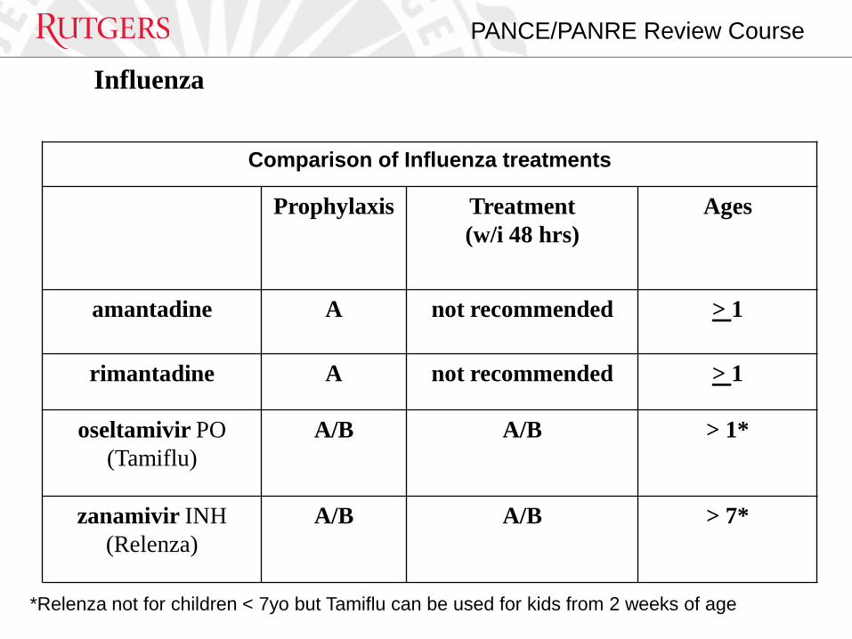
Comparison of Influenza treatments
Prophylaxis Treatment
(w/i 48 hrs)
Ages
amantadine A not recommended > 1
rimantadine A not recommended > 1
oseltamivir PO
(Tamiflu)
A/B A/B > 1*
zanamivir INH
(Relenza)
A/B A/B > 7*
*Relenza not for children < 7yo but Tamiflu can be used for kids from 2 weeks of age

PANCE/PANRE Review Course
Influenza
PANCE/PANRE Review Course
Influenza

PANCE/PANRE Review Course
Acute Bronchitis (aka tracheobronchitis)

PANCE/PANRE Review Course
Acute Bronchitis (aka tracheobronchitis)
PANCE/PANRE Review Course
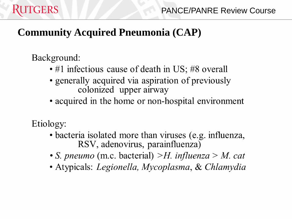
Community Acquired Pneumonia (CAP)

PANCE/PANRE Review Course Community Acquired Pneumonia (CAP)

PANCE/PANRE Review Course
Community Acquired Pneumonia (CAP)

PANCE/PANRE Review Course
A very prominent pneumonia of the middle lobe of the right lung
Source: http://en.wikipedia.org/wiki/File:PneumonisWedge09.JPG

PANCE/PANRE Review Course
Community Acquired Pneumonia (CAP)
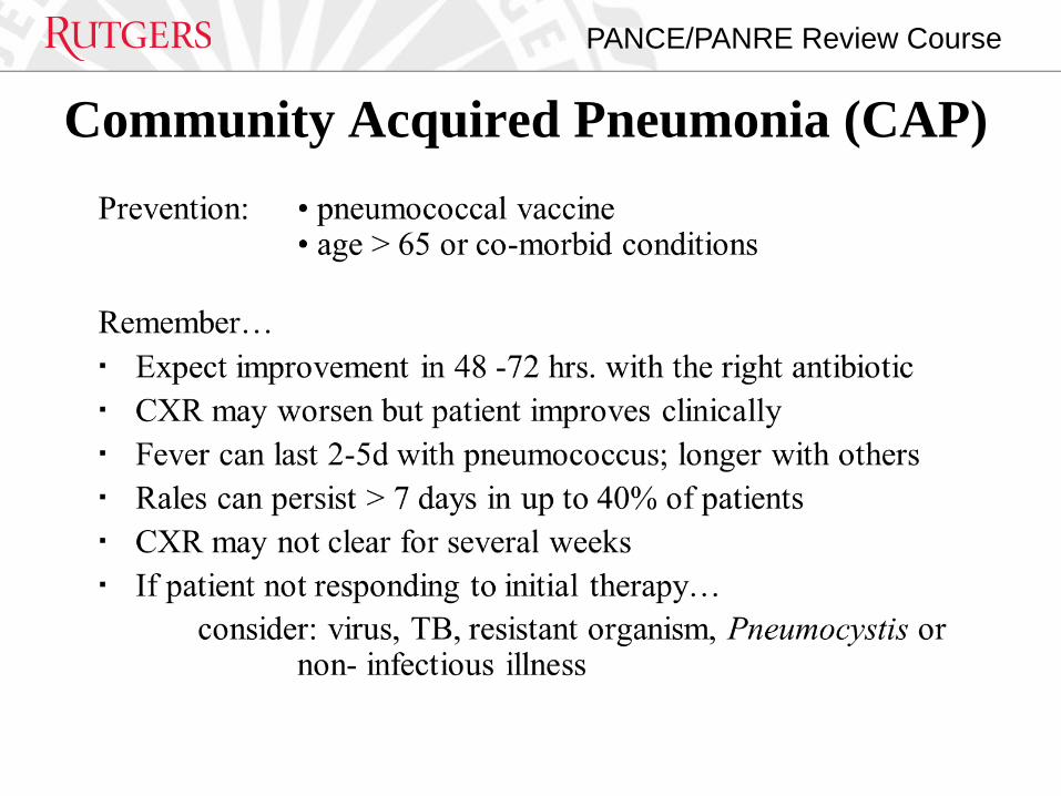
PANCE/PANRE Review Course
Community Acquired Pneumonia (CAP)
PANCE/PANRE Review Course
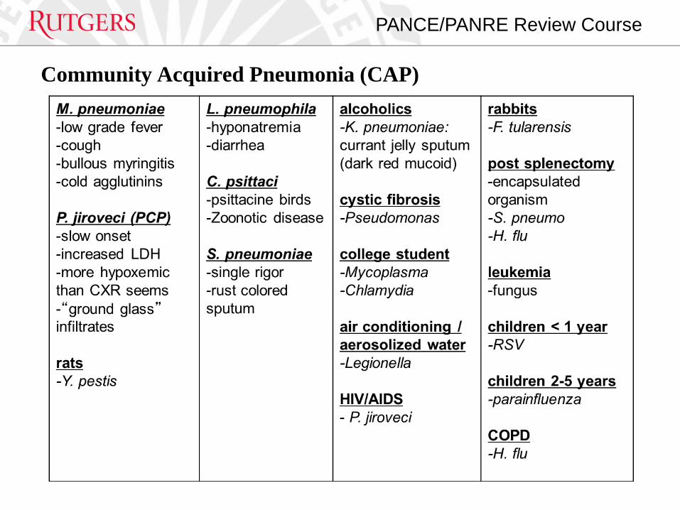
Community Acquired Pneumonia (CAP)
PANCE/PANRE Review Course
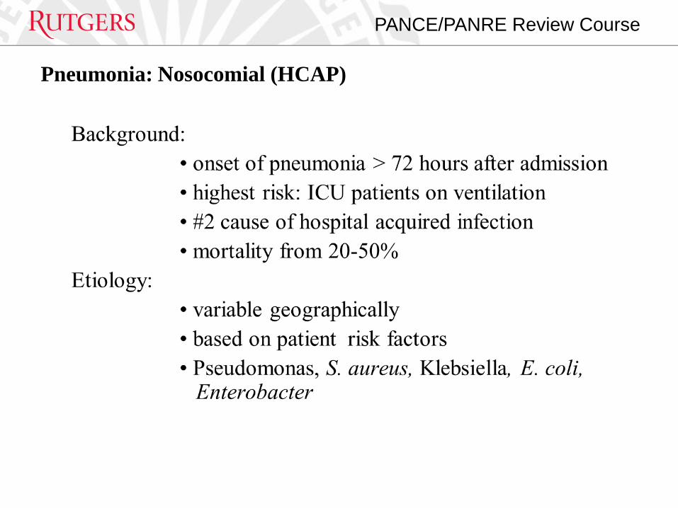
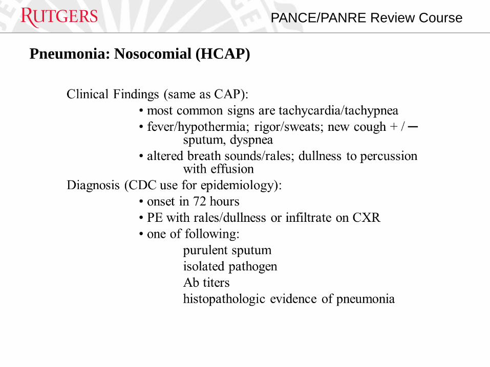
Pneumonia: Nosocomial (HCAP)
PANCE/PANRE Review Course
Pneumonia: Nosocomial (HCAP)
PANCE/PANRE Review Course
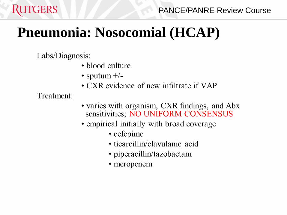
Pneumonia: Nosocomial (HCAP)

PANCE/PANRE Review Course
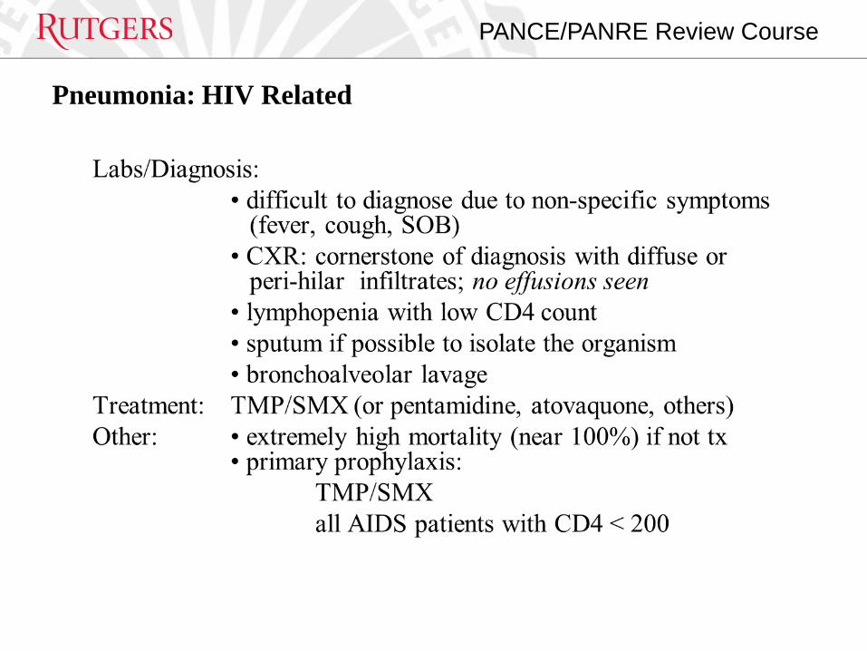
Pneumonia: HIV Related
PANCE/PANRE Review Course
Pneumonia: HIV Related

PANCE/PANRE Review Course
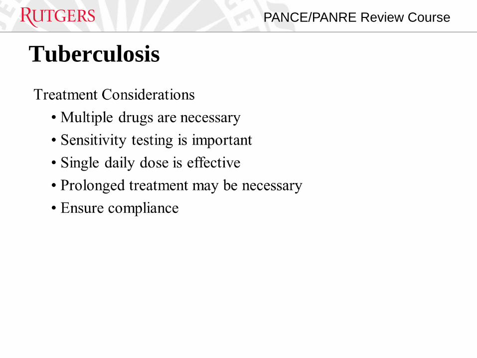
Tuberculosis

PANCE/PANRE Review Course
Tuberculosis

PANCE/PANRE Review Course
Tuberculosis

PANCE/PANRE Review Course
Tuberculosis

PANCE/PANRE Review Course
An AP CXR of a patient with advanced bilateral pulmonary tuberculosis. It reveals the presence of bilateral
pulmonary infiltrate (white triangles), and “caving formation“ (black arrows) present in the right apical region.
Source: http://phil.cdc.gov/phil/home.asp ID#: 2543 US Department of Health and Human Services

PANCE/PANRE Review Course
Tuberculosis
PANCE/PANRE Review Course
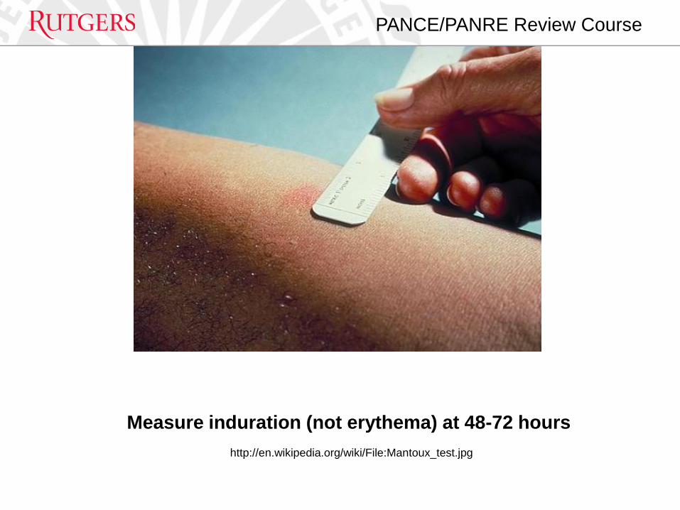
Measure induration (not erythema) at 48-72 hours
http://en.wikipedia.org/wiki/File:Mantoux_test.jpg

PANCE/PANRE Review Course
Tuberculosis

PANCE/PANRE Review Course
Tuberculosis
PANCE/PANRE Review Course
Tuberculosis

PANCE/PANRE Review Course
Epiglottitis (supraglottitis)
PANCE/PANRE Review Course
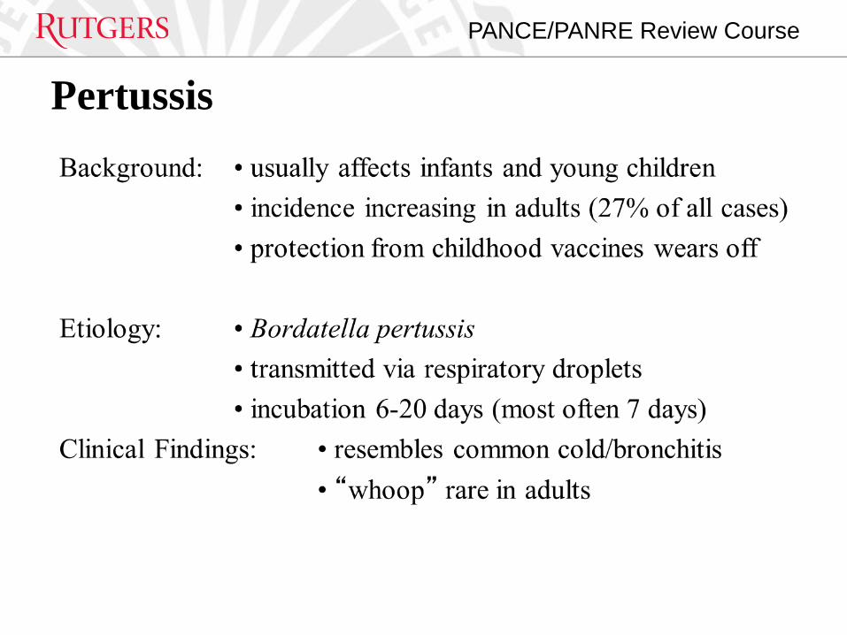
Pertussis

PANCE/PANRE Review Course
Pertussis
Labs: • PCR is current diagnostic standard
• more sensitive then culture
Treatment: • antibiotics to eradicate organism but does not
alter course of illness
• erythromycin, azithromycin, clarithromycin or
TMP-SMX
Prevention: • vaccination with Tdap (instead of Td)
Prophylaxis: • same as treatment when given within 3 weeks of
onset of cough in index case

PROPERTIES
On passing, 'Finish' button: Goes to Next Slide
On failing, 'Finish' button: Goes to Next Slide
Allow user to leave quiz: At any time
User may view slides after quiz: At any time
User may attempt quiz: Just Once
PANCE/PANRE Review Course
Part II:
Neoplastic Diseases
• Pulmonary Nodules
• Bronchogenic Carcinoma
• Carcinoid Tumors
• Metastatic (Secondary) Tumors
PANCE/PANRE Review Course
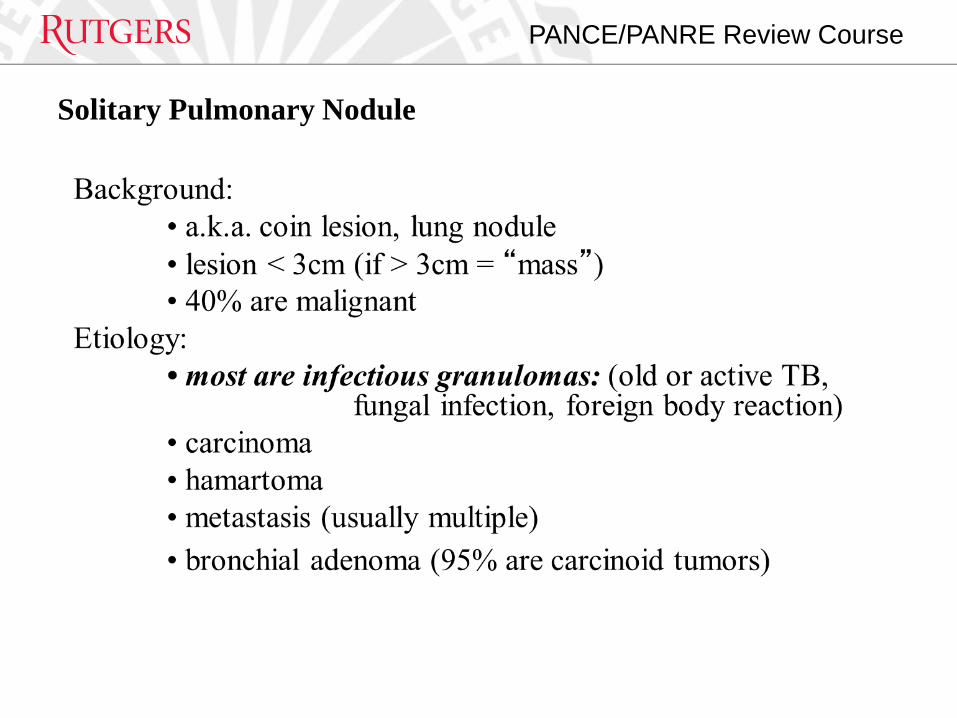
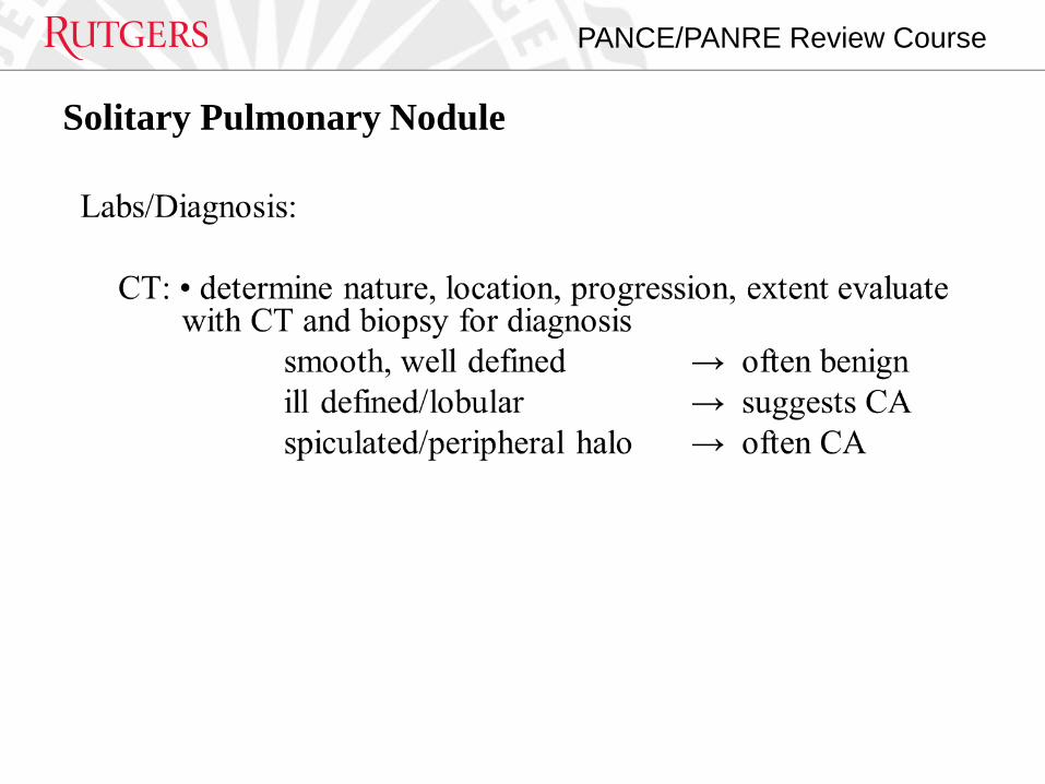
Solitary Pulmonary Nodule

PANCE/PANRE Review Course
Solitary Pulmonary Nodule
PANCE/PANRE Review Course
Solitary Pulmonary Nodule
PANCE/PANRE Review Course
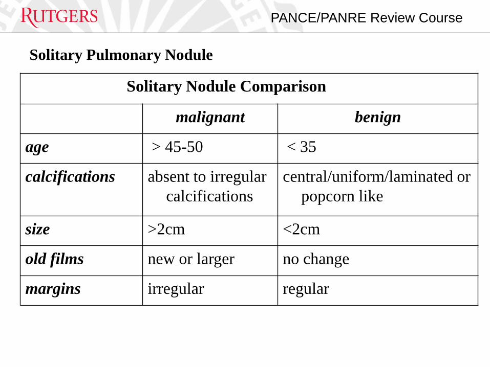
Solitary Nodule Comparison
malignant benign
age > 45-50 < 35
calcifications absent to irregular
calcifications
central/uniform/laminated or
popcorn like
size >2cm <2cm
old films new or larger no change
margins irregular regular
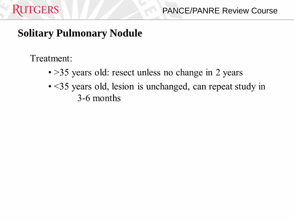
Solitary Pulmonary Nodule
PANCE/PANRE Review Course
Solitary Pulmonary Nodule
PANCE/PANRE Review Course
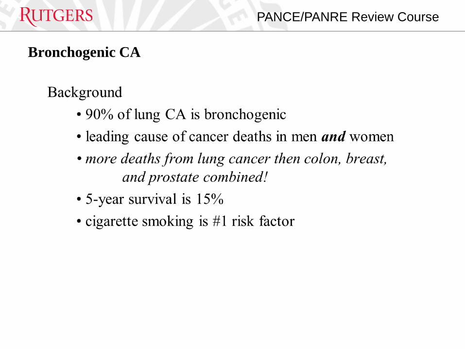
Bronchogenic CA

PANCE/PANRE Review Course
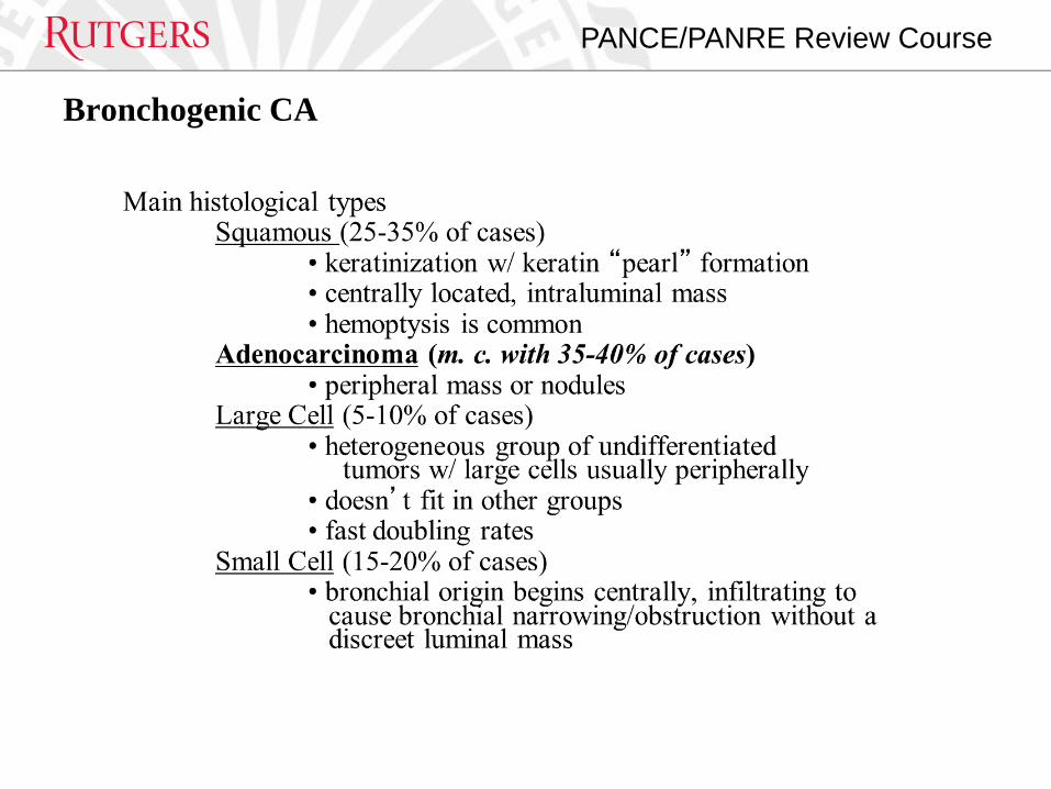
Bronchogenic CA
Classification Scheme
• SCLC: early mets & aggressive clinical course
assumes micro metastases at presentation
• NSCLC: (adeno, squamous, large cell)
slower spreading
more amenable to treatment (i.e., surgery)
Clinical Findings
• often presents in 50s-70s
• cough, dyspnea, hemoptysis, anorexia, weight loss
PANCE/PANRE Review Course
Bronchogenic CA

PANCE/PANRE Review Course
Bronchogenic CA
PANCE/PANRE Review Course
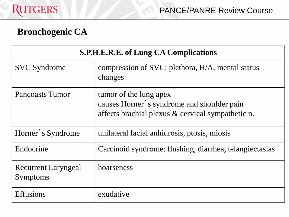
S.P.H.E.R.E. of Lung CA Complications
SVC Syndrome compression of SVC: plethora, H/A, mental status
changes
Pancoasts Tumor tumor of the lung apex
causes Horner’s syndrome and shoulder pain
affects brachial plexus & cervical sympathetic n.
Horner’s Syndrome unilateral facial anhidrosis, ptosis, miosis
Endocrine Carcinoid syndrome: flushing, diarrhea, telangiectasias
Recurrent Laryngeal
Symptoms
hoarseness
Effusions exudative
Bronchogenic CA
PANCE/PANRE Review Course
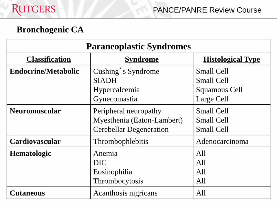
Paraneoplastic Syndromes
Classification Syndrome Histological Type
Endocrine/Metabolic Cushing’s Syndrome
SIADH
Hypercalcemia
Gynecomastia
Small Cell
Small Cell
Squamous Cell
Large Cell
Neuromuscular Peripheral neuropathy
Myesthenia (Eaton-Lambert)
Cerebellar Degeneration
Small Cell
Small Cell
Small Cell
Cardiovascular Thrombophlebitis Adenocarcinoma
Hematologic Anemia
DIC
Eosinophilia
Thrombocytosis
All
All
All
All
Cutaneous Acanthosis nigricans All
Bronchogenic CA

PANCE/PANRE Review Course
Carcinoid Tumor

PANCE/PANRE Review Course
Carcinoid Tumor

PANCE/PANRE Review Course
Mesothelioma
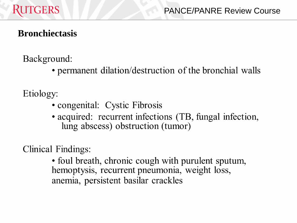
Background:
• primary tumors from pleural lining (80%) or peritoneum (20%)
Etiology: history of asbestos exposure
Clinical Findings:
• insidious onset of SOB, non-pleuritic chest pain,
weight loss; dullness to percussion,
decreased breath sounds, digital clubbing

PANCE/PANRE Review Course
Mesothelioma
Labs/Diagnosis:
• pleural fluid is exudative and hemorrhagic
• CXR reveals nodular, irregular, unilateral pleural thickening, and effusion
• video assisted thoracic surgery (VATS): biopsy
Treatment:
• none that are effective
• some do chemo/radiation
Other: five year survival is less than 5%

PANCE/PANRE Review Course
Malignant mesothelioma marked by yellow arrows
Source: http://commons.wikimedia.org/wiki/File:Tumor_Mesothelioma2_legend.jpg

PANCE/PANRE Review Course
Secondary Lung Cancer
• represents extra-pulmonary metastases
• most frequently primary’s that metastasize to lung:
breast, liver, colon
• almost any CA can spread to lungs
• imaging reveals multiple nodules/masses
• diagnose and treat the primary tumor

PROPERTIES
On passing, 'Finish' button: Goes to Next Slide
On failing, 'Finish' button: Goes to Next Slide
Allow user to leave quiz: At any time
User may view slides after quiz: At any time
User may attempt quiz: Just Once

PANCE/PANRE Review Course
Part III:
Obstructive Pulmonary Disease
• Asthma
• Bronchiectasis
• Chronic Bronchitis
• Emphysema

PANCE/PANRE Review Course
Obstructive Pulmonary Disease
FEV/FVC
Normal / ↑ TLC
PANCE/PANRE Review Course
Asthma
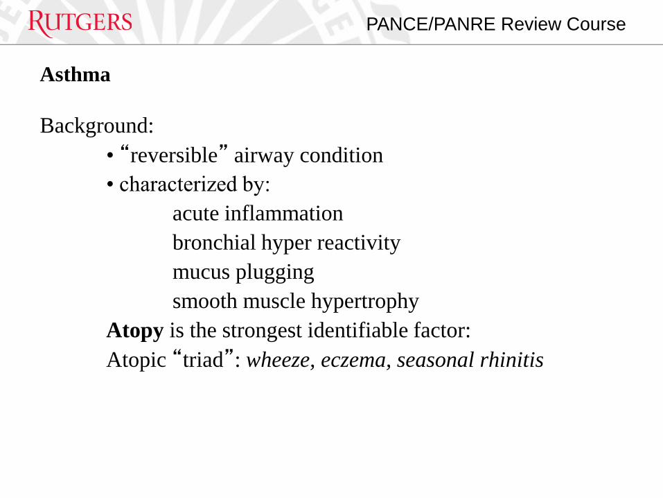
Background:
• “reversible” airway condition
• characterized by:
acute inflammation
bronchial hyper reactivity
mucus plugging
smooth muscle hypertrophy
Atopy is the strongest identifiable factor:
Atopic “triad”: wheeze, eczema, seasonal rhinitis

PANCE/PANRE Review Course
Asthma
Etiology:
Precipitants: allergens (esp. dust and dust mites), exercise, URI, post nasal drip, GERD, meds (beta blocker, ACEI, ASA, NSAIDS), stress, cold air
Clinical Findings:
• episodic/chronic symptoms of airway obstruction
• breathlessness, cough, wheeze
• 1/3 of children have no wheeze
• prolonged expiration/diffuse wheeze

PANCE/PANRE Review Course
Asthma
PANCE/PANRE Review Course
Asthma
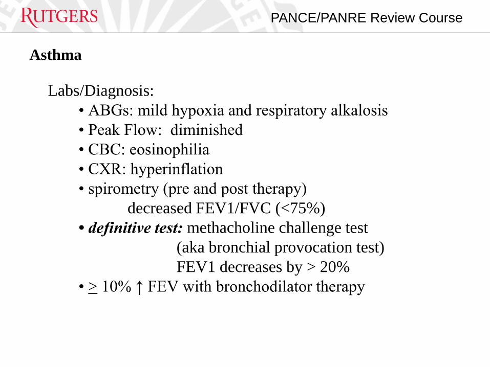
Labs/Diagnosis:
• ABGs: mild hypoxia and respiratory alkalosis
• Peak Flow: diminished
• CBC: eosinophilia
• CXR: hyperinflation
• spirometry (pre and post therapy)
decreased FEV1/FVC (<75%)
• definitive test: methacholine challenge test
(aka bronchial provocation test)
FEV1 decreases by > 20%
• > 10% ↑ FEV with bronchodilator therapy

PANCE/PANRE Review Course
Asthma
Treatment: • General remove irritants education on peak flow measurements desensitization oxygen • Pharmacological Quick relief meds INH beta 2 agonists (e.g. albuterol) glucocorticoids (e.g. prednisone) anticholinergics (e.g. ipratropium)
PANCE/PANRE Review Course
Asthma

PANCE/PANRE Review Course
National Asthma Education and Prevention Program, Expert Panel Report 3:
Guidelines for the Diagnosis and Management of Asthma, NIH Pub No 08-4051, 2007
PANCE/PANRE Review Course
Bronchiectasis
PANCE/PANRE Review Course
Bronchiectasis

PANCE/PANRE Review Course
Bronchiectasis
Treatment:
• ambulatory oxygen
• aggressive antibiotics (10-14 days):
guided by sputum cultures or
empiric therapy
amoxicillin
amox-clavulante (Augmentin)
TMP/SMX (Bactrim)
ciprofloxacin
• INH bronchodilators for maintenance and acute exacerbations
• lung transplantation

PANCE/PANRE Review Course
Lung CT with thin slices (1 mm) showing bronchiectasis in the lower lung lobes of a subject with
type ZZ alpha-1-antitrypsin deficiency. There are no signs of emphysema.
Source: Fregonese L, Stolk J. Hereditary alpha-1-antitrypsin deficiency and its clinical consequences. Orphanet J Rare Dis. 3, 1, 16.
2008. doi:10.1186/1750-1172-3-16. PMID 18565211.
PANCE/PANRE Review Course
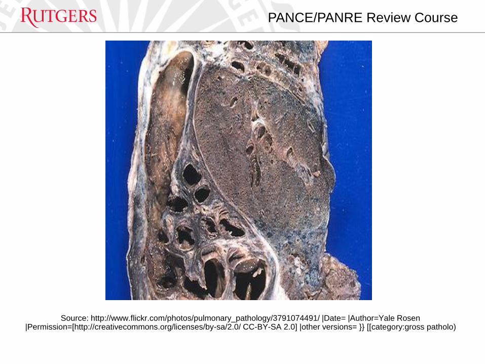
Source: http://www.flickr.com/photos/pulmonary_pathology/3791074491/ |Date= |Author=Yale Rosen |Permission=[http://creativecommons.org/licenses/by-sa/2.0/ CC-BY-SA 2.0] |other versions= }} [[category:gross patholo)

PANCE/PANRE Review Course
COPD: Chronic Bronchitis/Emphysema

PANCE/PANRE Review Course
COPD: Chronic Bronchitis/Emphysema
Etiology • smoking/exposure to tobacco (80%) • environmental pollutants • recurrent URI’s • eosinophilia • bronchial hyper responsiveness Labs/Diagnosis: • PFT: normal early in the disease decreased FEV1/FVC occur later increased RV and TLC confirmed by biopsy ↑ Reid index: gland layer is > 50% of total
bronchial wall
PANCE/PANRE Review Course
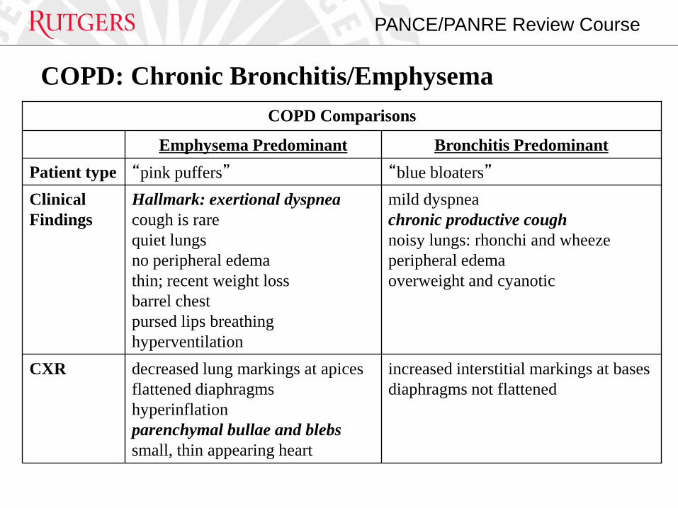
COPD Comparisons
Emphysema Predominant Bronchitis Predominant
Patient type “pink puffers” “blue bloaters”
Clinical
Findings
Hallmark: exertional dyspnea
cough is rare
quiet lungs
no peripheral edema
thin; recent weight loss
barrel chest
pursed lips breathing
hyperventilation
mild dyspnea
chronic productive cough
noisy lungs: rhonchi and wheeze
peripheral edema
overweight and cyanotic
CXR decreased lung markings at apices
flattened diaphragms
hyperinflation
parenchymal bullae and blebs
small, thin appearing heart
increased interstitial markings at bases
diaphragms not flattened
COPD: Chronic Bronchitis/Emphysema

PANCE/PANRE Review Course
CXR of patient with severe emphysema
Source: http://commons.wikimedia.org/wiki/File:Emphysema2008.jpg

PANCE/PANRE Review Course
COPD: Chronic Bronchitis/Emphysema

PROPERTIES
On passing, 'Finish' button: Goes to Next Slide
On failing, 'Finish' button: Goes to Next Slide
Allow user to leave quiz: At any time
User may view slides after quiz: At any time
User may attempt quiz: Just Once

PANCE/PANRE Review Course
Part IV:
Restrictive Pulmonary Diseases
• Idiopathic Pulmonary Fibrosis
• Pneumoconioses
• Sarcoidosis
PANCE/PANRE Review Course
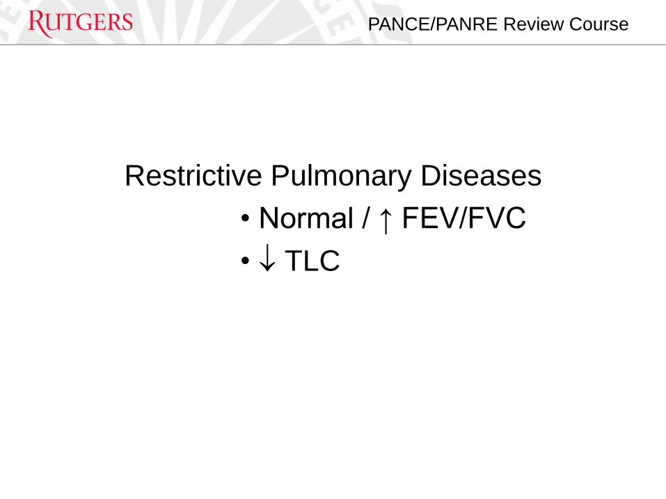
Restrictive Pulmonary Diseases
• Normal / ↑ FEV/FVC
• TLC

PANCE/PANRE Review Course Idiopathic Pulmonary Fibrosis

PANCE/PANRE Review Course
American Thoracic Society/European Respiratory Society International Multidisciplinary Consensus Classification of the Idiopathic Interstitial
Pneumonias. (2013). Am Respir Crit Care Med. 188 (6): 733–748. PMID 24032382.

PANCE/PANRE Review Course
Idiopathic Pulmonary Fibrosis
Labs/Diagnosis:
CXR/HRCT:
• low lung volumes
• patchy, diffuse fibrosis
• pleural honeycombing
• biopsy helps to exclude other causes
Treatment:
• controversial
• corticosteroids
• interferon
PANCE/PANRE Review Course Pneumoconioses

PANCE/PANRE Review Course
Comparison of Pneumoconioses
Disease Occupation Diagnosis Complications
asbestosis insulation,
demolition,
construction,
BX: asbestos bodies
CXR: linear opacities at
bases and pleural plaques
increased risk of lung CA
and mesothelioma, esp. if
smoker
CWP coal miner CXR: nodular opacities at
upper lung fields
progressive massive
fibrosis
silicosis miners,
sand blasters,
quarry workers,
stone workers,
CXR: nodular opacities at
upper lung fields
increased risk of TB;
progressive massive
fibrosis
berryliosis high technology fields:
aerospace, nuclear power,
ceramics, foundries,
tool & die manufacturing
CXR: diffuse infiltrates and
hilar adenopathy
needs chronic steroids
Pneumoconioses

PANCE/PANRE Review Course Sarcoidosis
PANCE/PANRE Review Course Sarcoidosis
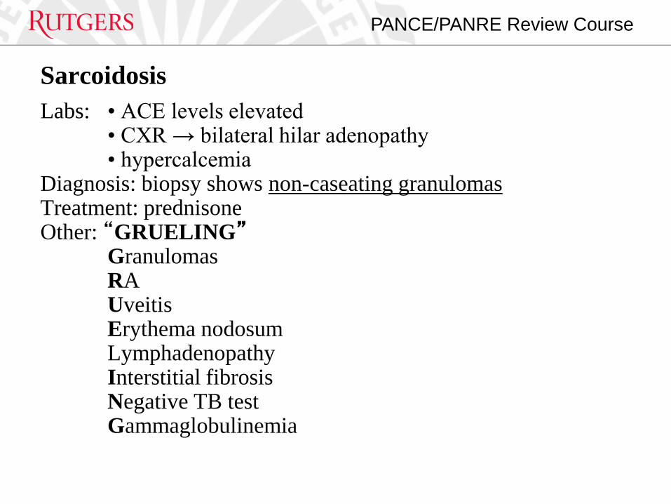
Labs: • ACE levels elevated • CXR → bilateral hilar adenopathy • hypercalcemia Diagnosis: biopsy shows non-caseating granulomas Treatment: prednisone Other: “GRUELING” Granulomas RA Uveitis Erythema nodosum Lymphadenopathy Interstitial fibrosis Negative TB test Gammaglobulinemia

PROPERTIES
On passing, 'Finish' button: Goes to Next Slide
On failing, 'Finish' button: Goes to Next Slide
Allow user to leave quiz: At any time
User may view slides after quiz: At any time
User may attempt quiz: Just Once
PANCE/PANRE Review Course
Part V:
Pleural Diseases
• Pleural Effusion
• Pneumothorax
PANCE/PANRE Review Course Pleural Effusions
Background
• abnormal fluid collection in the pleural space
• 25% of effusions are associated with malignancy
Important to distinguish transudate from exudate

PANCE/PANRE Review Course

PANCE/PANRE Review Course
Pleural Effusions
Etiology: 5 types of effusions
• exudates: “leaky capillaries”
these three cause 80%
para-pneumonic
malignancy
PE (but 20% as transudate)
infection (TB), malignancy, trauma
• transudates: “intact capillaries”
CHF (90%), atelectasis, renal/liver ds. (cirrhosis)
• empyemas: direct infection of an exudate
• hemothorax: trauma
• chylothorax: TB
PANCE/PANRE Review Course Pleural Effusions

PANCE/PANRE Review Course Pleural Effusions
Labs:
imaging helps define extent of effusion
• lateral decubitus (free flowing vs. loculated fluid)
• upright (blunting of costophrenic sulcus)
• CT scan for small effusions
Diagnosis:
thoracentesis is the gold standard
• send for protein, LDH, pH, total & cell counts, glucose
cytology?
• Gram stain with C & S?

PANCE/PANRE Review Course
Massive Left-Sided Pleural Effusion
Source: http://en.wikipedia.org/wiki/Image:Left-sided_Pleural_Effusion.jpg
PANCE/PANRE Review Course Pleural Effusions

PANCE/PANRE Review Course Pleural Effusions

PANCE/PANRE Review Course
Pneumothorax
PANCE/PANRE Review Course
Pneumothorax
PANCE/PANRE Review Course
Pneumothorax

PANCE/PANRE Review Course
Bilateral pneumothorax (larger arrows)
Source: Le Guen et al. Critical Care 2007 11:R94 doi:10.1186/cc6109 http://ccforum.com/content/11/5/R94

PANCE/PANRE Review Course
Left tension pneumothorax
Source: http://clinicalcases.blogspot.com/2004/02/tension-pneumothorax.html

PROPERTIES
On passing, 'Finish' button: Goes to Next Slide
On failing, 'Finish' button: Goes to Next Slide
Allow user to leave quiz: At any time
User may view slides after quiz: At any time
User may attempt quiz: Just Once
PANCE/PANRE Review Course
Part VI:
Pulmonary Circulation
• Pulmonary Thromboembolism
• Pulmonary Hypertension
• Cor Pulmonale
• ARDS
PANCE/PANRE Review Course
Rudolf Virchow by Hugo Vogel, 1861
Source: http://www.kunsttexte.de/download/bwt/werner.pdf

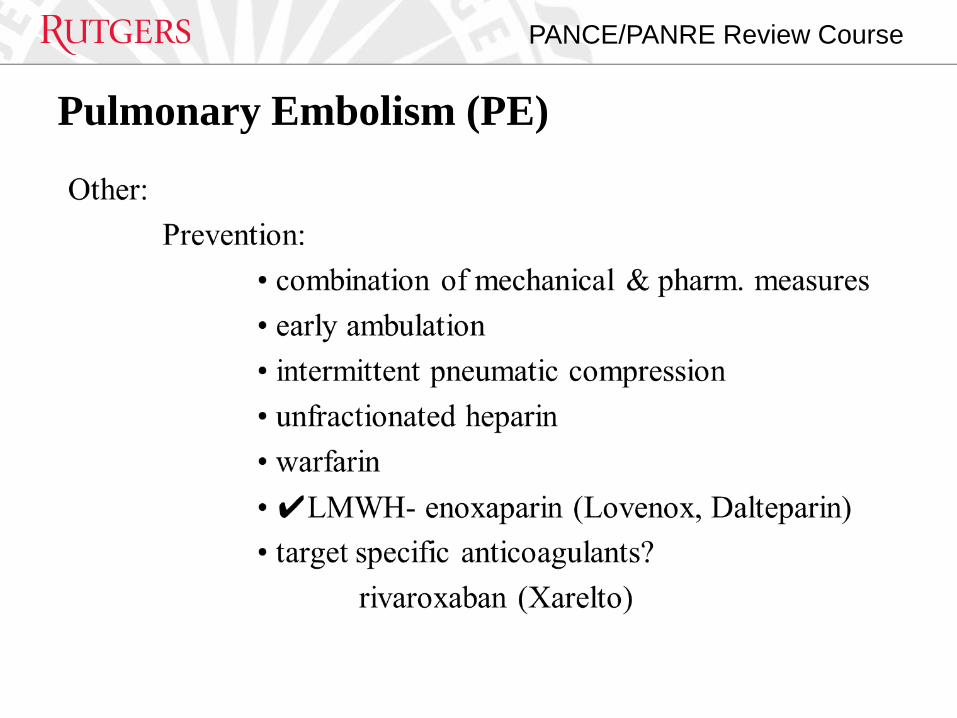
PANCE/PANRE Review Course Pulmonary Embolism (PE)
PANCE/PANRE Review Course Pulmonary Embolism (PE)
Etiology: most are from thrombus
• 95% deep calf veins that propagate proximal to popliteal / ileofemoral veins
• risk of PE greater with proximal thrombus
• others:?
air → central lines
amniotic fluid → active labor
fat → long bone (femur) fx
• negative workup in 25-50% patients for VTE
PANCE/PANRE Review Course
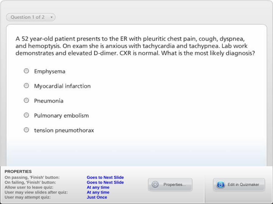
Pulmonary Embolism (PE)
Clinical Findings:
• Homans’ sign: low sensitivity/specificity
calf pain with passive, forcible, dorsiflexion of foot with knee flexed
• variable, signs and symptoms are non-specific but <3% chance of PE in absence of dyspnea with tachypnea or pleuritic chest pain
• most emboli are clinically silent
• most common symptom: dyspnea (sudden; 85% with RR>16) / pain on inspiration
• (*consider PE in any hospitalized pt. w/ acute SOB)
• most common sign: tachycardia (60% with P > 100)

PANCE/PANRE Review Course
Pulmonary Embolism (PE)
PANCE/PANRE Review Course
Pulmonary Embolism (PE)
CXR (abnormalities may be subtle / absent)
• m.c. abnormality is atelectasis at bases
• Westermark’s Sign: focal oligemia (vasoconstriction) in the embolized zone
• Hampton’s Hump: (classic finding)
wedge shaped infarct
VQ scans:
• “normal” practically rules out PE
• “abnormal” is non specific
• categorized along with CXR
normal/very low
low
intermediate
high probability

PANCE/PANRE Review Course
Chest Spiral CT (with and without contrast agent) showing multiple filling defects of principal branches,
due to acute and chronic pulmonary embolism
Source: Cardiovascular Ultrasound 2007, 5:26. doi:10.1186/1476-7120-5-26

PANCE/PANRE Review Course Pulmonary Embolism (PE)

PANCE/PANRE Review Course Pulmonary Embolism (PE)
PANCE/PANRE Review Course Pulmonary Embolism (PE)
PANCE/PANRE Review Course
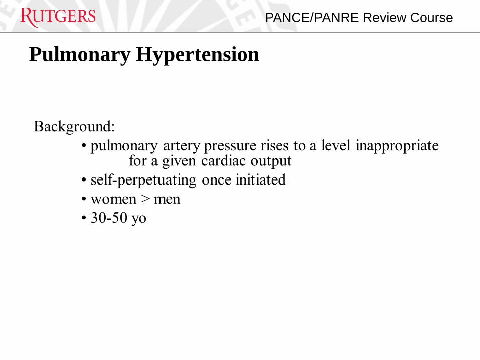
Pulmonary Hypertension

PANCE/PANRE Review Course
Pulmonary Hypertension

PANCE/PANRE Review Course
Pulmonary Hypertension

PANCE/PANRE Review Course
Pulmonary Hypertension
Diagnosis: Multifactorial
Work-up:
• CXR/CT: increased vasculature
• PFTs: underlying airflow obstruction or restricted lung volumes
• ECHO: RVH, estimated pulmonary artery pressure
• catheterization to determine degree of HTN
• others (VQ scan, serology: conn tissue disorders, etc.)

PANCE/PANRE Review Course
Pulmonary Hypertension
PANCE/PANRE Review Course
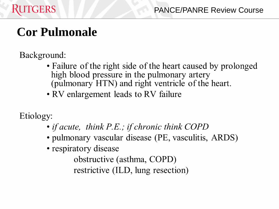
Cor Pulmonale
PANCE/PANRE Review Course
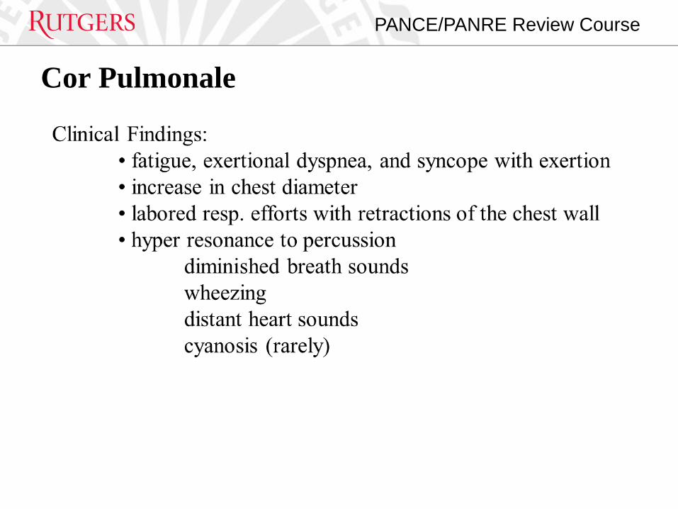
Cor Pulmonale

PANCE/PANRE Review Course
Cor Pulmonale
Labs/Diagnosis:
• CXR
• EKG: RAD > 30°; flat, inverted T waves in RV precordial leads
Treatment:
• oxygen
• decrease pulm. Vasc. resistance and pulmonary HTN
• treat underlying disorder

PANCE/PANRE Review Course
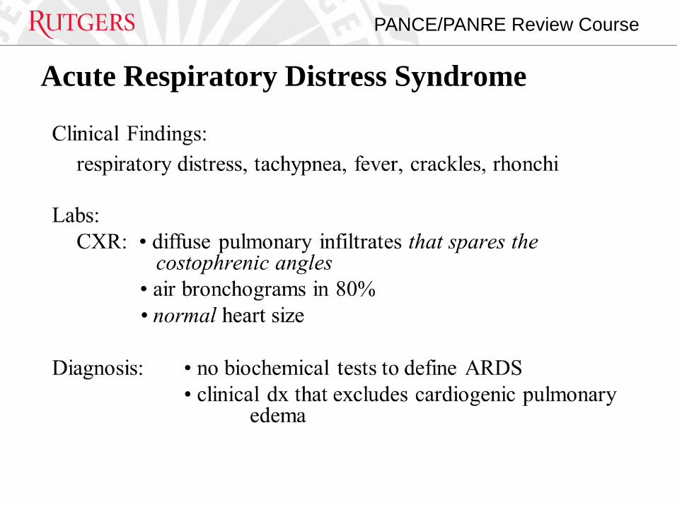
Acute Respiratory Distress Syndrome
Clinical Definition: • acute (12-18hours) hypoxemic respiratory failure after a
systemic or pulmonary insult without heart failure Physiological Definition • bilateral diffuse pulmonary infiltrates • normal PCWP (<18 mmHg) • PaO2/FiO2 < 200 Etiology: • most common (one-third of patients): sepsis • others: toxic inhalation, near drowning, aspiration, etc.
PANCE/PANRE Review Course
Acute Respiratory Distress Syndrome

PANCE/PANRE Review Course
Acute Respiratory Distress Syndrome
PROPERTIES
On passing, 'Finish' button: Goes to Next Slide
On failing, 'Finish' button: Goes to Next Slide
Allow user to leave quiz: At any time
User may view slides after quiz: At any time
User may attempt quiz: Just Once

Rutgers, The State University of New Jersey
Good luck!