purification and characterization of a cytochrome c with
TRANSCRIPT

RESEARCH ARTICLE Open Access
Purification and characterization of acytochrome c with novel caspase-3activation activity from the pathogenicfungus Rhizopus arrhizusManoj Saxena1, Rohit Kumar Sharma1, Josell Ramirez-Paz2, Arthur D. Tinoco2 and Kai Griebenow2*
Abstract
Background: Members of Rhizopus species are the most common cause of mucormycosis, a rare but often fatalfungal infection. Host induced pathogen apoptosis and pathogen induced host cell apoptosis are often involved infungal infections. In many organisms, the release of mitochondrial cytochrome c can trigger apoptosis by activatingcaspase proteases, but the role of fungal cytochrome c in apoptosis remains unknown.
Results: DNA sequence encoding Rhizopus arrhizus cytochrome c was cloned and expressed in E. coli. Both nativeand recombinant cytochrome c were purified using ion exchange followed by gel filtration chromatography. Theidentities of purified proteins were confirmed by MALDI-MS and UV-Visible spectroscopy. For the first time, wedemonstrated that Rhizopus arrhizus cytochrome c could activate human capspase-3 in HeLa cell extracts. We alsofound that Rhizopus arrhizus cytochrome c has redox potential, peroxidase activity, and spectral properties similar tohuman and horse cytochrome c proteins.
Conclusions: Rhizopus arrhizus cytochrome c can activate human caspase-3 in HeLa cell extracts and it possessessimilar physical and spectral properties as human and horse cytochrome c. This protein was found to have apreviously unknown potential to activate human caspase-3, an important step in the apoptosis cascade.
BackgroundMucormycosis are rare but often life-threatening infec-tions seen in immunocompromised, diabetic, and organtransplant patients [1]. These infections are difficult totreat and with an increase in the number of diabetic pa-tients and organ transplants, in the future such infectionsare likely to increase [1, 2]. Rhizopus spp. accounts for themajority of the mucormycosis infections [1]. Results in re-cent decades suggest that apoptosis may play an importantrole in both establishment and clearance of fungal infec-tions [3]. In response to fungal infections, the oxidativeburst by host immune cells could help in infection clear-ance by triggering apoptosis in the fungus [4, 5]. Also,pathogen-induced apoptosis in host immune cells couldhelp the pathogen to establish the infection [6]. Thus, the
success of an infection partly depends on the outcome ofsuch host pathogen interactions. It has been shown that ex-tracts from the filamentous fungus could induce apoptosisin human cells [6, 7], but the identity of the protein/factorresponsible for the induction of apoptosis remains un-known. In recent decades, it has also been established thatin many organisms cyt c plays an important role in apop-tosis [8, 9]. During apoptosis, cyt c is released from themitochondria into the cytoplasm and binds to apoptosisprotease-activating factor-1 (Apaf-1) thus triggering a cas-pase activation cascade [8]. For example, in humans, cyt ccan activate caspase-3 in cell-free activation assay [9]. Thefirst evidence of the existence of apoptosis in fungi camefrom the study by Medeo et al. of a Saccharomyces cerevi-siae mutant that showed signs of apoptosis [10]. Since then,many homologous mammalian apoptotic proteins were dis-covered in budding yeast [11]. In an important finding,using deletion mutants which were unable to produce func-tional cyt c, Silva et al. presented the first evidence that cyt
* Correspondence: [email protected] of Chemistry, University of Puerto Rico, Rio Piedras Campus,P.O. Box 70377, San Juan, PR 00936-837, USAFull list of author information is available at the end of the article
© 2015 Saxena et al. Open Access This article is distributed under the terms of the Creative Commons Attribution 4.0International License (http://creativecommons.org/licenses/by/4.0/), which permits unrestricted use, distribution, andreproduction in any medium, provided you give appropriate credit to the original author(s) and the source, provide a link tothe Creative Commons license, and indicate if changes were made. The Creative Commons Public Domain Dedication waiver(http://creativecommons.org/publicdomain/zero/1.0/) applies to the data made available in this article, unless otherwise stated.
Saxena et al. BMC Biochemistry (2015) 16:21 DOI 10.1186/s12858-015-0050-9

c is involved in hyperosmotic stress induced yeast apoptosis[12]. However, the nature of the role cyt c plays in apoptosisof filamentous fungi like R. arrhizus remains unknown.To characterize R. arrhizus cyt c, we cloned the gene
for cyt c from R. arrhizus in E. coli. The purified cyt cwas compared with two mammalian cyt c (horse andhuman). We selected R. arrhizus cyt c as a candidate forthis study mainly for two reasons. First, this fungal cyt cis distantly related to mammalian cyt c and its biochem-ical properties are largely unknown.Secondly, R. arrhizus is a pathogenic fungus and any
information gained on the pro-apoptotic activity of cyt cmay also contribute to the identification of better thera-peutic targets. We found that cyt c of R. arrhizus hassimilar biochemical properties to mammalian cyt c.
Results and discussionA 19-fold purification was achieved for recombinant R.arrhizus cyt c using a three step process involving ammo-nium sulphate precipitation, cation exchange and gel fil-tration chromatography (Fig. 1, Table 1). The purifiedrecombinant cyt c protein eluted from gel filtration col-umn with absorption ratio 410/280 nm of >4.0 (Fig. 1b,Table 1). Purity was also checked by SDS electrophoresis.The eluted cyt c produced one band, which had an appar-ent MW of ca. 14 kDa (Fig. 1c). Since this is the first re-port on purification of recombinant R. arrhizus cyt c, wecompared our purification results with those reported byothers using similar plasmids. We obtained ~9 mg of cyt cwith a 410/280 ratio of >4 from 1 L of E. coli culture. Ouryield was similar to the reported yield for human cyt c of>8 mg L−1 [13]. Patel et al. reported a yield of ~15 mg L−1
for horse cyt c [14]. However, in that work the authorsonly performed a two-step purification (ammoniumsulphate precipitation and a cation exchange column)[14]. Our yield was only marginally lower if we compareour yield after the second purification step (Table 1).In the case of the native R. arrhizus cyt c (protein iso-
lated form commercial preparation of lipase) the final pro-tein eluted from a Superdex 75 column as a single peak(Fig. 2a), but the 410/280 nm ratio was only 0.65. Effortsto further purify the native protein using an additionalanion exchange column to trap impurities did not resultin any improvement.This concurs with a previous study that reported diffi-
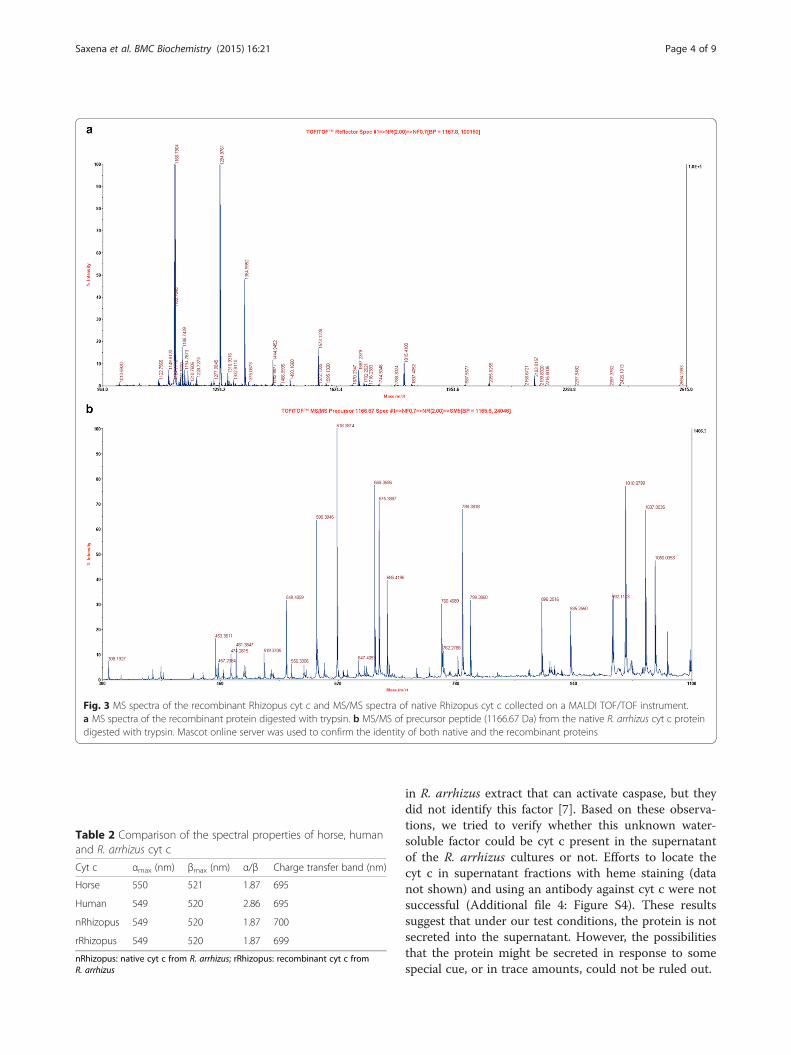
culties in purifying this protein from R. oryzae (syn. toR. arrhizus) to high purity [15, 16]. The relative molecu-lar weight determined for the native R. arrhizus cyt c bySDS-PAGE was 12.55 ± 1.27 kDa (Fig. 2b) in agreementwith expectations for its molecular weight. Identity ofboth, the native and the recombinant protein bands wasunequivocally confirmed by MALDI tandem mass spec-troscopy (Fig. 3 and Additional file 1: Figure S1).The UV–vis spectra of the oxidized native R. arrhizus
cyt c show peaks at 409 and 529 nm and in the reducedstate, α and β peaks are prominent at 549 and 520 nm,respectively (Fig. 2c). Similar values were seen for therecombinant R. arrhizus cyt c. These values are close toother type-c cytochromes, thus suggesting a similar hemeenvironment (Table 2).The presence of an absorption maximum at ~700 nm
in both recombinant and the native R. arrhizus ferricytochrome (Additional file 2: Figure S2A & B) suggeststhat, like in other c-type cytochromes, methionine is one
a c b
20
97
66
45
30
14
1 2 3
Fig. 1 a Elution profile of recombinant R. arrhizus cyt c on a Hi-Trap SP Sepharose column (5 ml). The bound protein was eluted with an increasingNaCl gradient over 7 column volumes. Peak fractions containing cyt c protein is indicated by *. b Elution profile of the pooled HiTrap SP Sepharosepeak fractions loaded onto a Superdex 200 GL column. Cyt c elution was followed using 410 nm absorption. Cyt c protein elutes as two unresolvedpeaks (in inset peak 1 and 2). c Coomassie stained 15 % SDS-PAGE gel loaded (lane 1 and 2) with the peak 1 and 2 from the Superdex 200 GL column,lane 3 =marker proteins. Cyt c band moves close to the 14 kDa marker band
Saxena et al. BMC Biochemistry (2015) 16:21 Page 2 of 9

of the axial heme ligands. Additionally, sequence align-ment shows that residues corresponding to Met-80 andHis-18 are conserved in 14 fungal cyt c [17], as well asin human and horse cyt c.The redox potential of native and recombinant R. arrhizus
cyt c was found to be similar to that of human and horsecyt c, respectively, with no statistically significant difference(Fig. 4 and Additional file 3: Figure S3). The redox potentialmeasurements for human and horse cyt c with 263.43 and268.64 mV, respectively, were in close agreement with earlierreported values validating our technique [18, 19]. The redoxpotential measurements of native and recombinant R. arrhi-zus cyt c were 266.90 and 270.04 mV and statistically thesame. These results further support structural similarity be-tween the recombinant and native R. arrhizus cyt c proteins.A major portion of cyt c remains loosely associated with
the inner mitochondrial membrane via ionic interactionswith the negatively charged mitochondrion-specificphospholipid, cardiolipin. Kagan et al. had shown that car-diolipin specific peroxidase activity of cardiolipin bound cytc may play an important role in triggering apoptosis by aid-ing in the release of pro-apoptotic proteins, including cyt c,from the matrix of the mitochondria [20]. Furthermore,
peroxidase activity assay provides an indirect measure ofheme accessibility and we therefore compared the peroxid-ase activities of mammalian and R. arrhizus cyt c.The peroxidase activity of native R. arrhizus cyt c was
found to be low compared to other known peroxidases,which is a general characteristic of type-c cytochromes.Recombinant cyt c showed a higher Kcat and Vmax com-pared to native R. arrhizus cyt c. These variations aremost likely due to the higher purity of the recombinantprotein compared to native one (Table 2, Table 3). Also,a high peroxidase activity could be related to partialdenaturation and thus a more accessible heme in thenon-recombinant cyt c. A partial denaturation can involvethe breaking of the methionine coordination that leavesthe heme more accessible for catalysis [21]. However, thisis ruled out by the UV-spectra of the recombinant cyt cbecause they show the presence of the charge transferband thus indicating that methionine coordination isintact (Additional file 2: Figure S2).Native R. arrhizus cyt c showed similar activity as horse
cyt c while it was higher than human cyt c (Table 3). Onthe other hand, judging from KM values the affinity of R.arrhizus cyt c seems to be lower than human cyt c, andvery similar compared to horse cyt c (Table 3). The KM
value measured for horse cyt c was in close agreementwith Kim et al. [22] but varied from the one reported byRandi et al. [23]. It is important to note that we used non-linear regression to analyze the kinetics while the twocited studies used Lineweaver-Burk plots, a method con-sidered less reliable [24].We noted the presence of the cyt c in good amounts
in a commercial preparation of a secreted lipase of R.arrhizus (Fig. 2). Interestingly, in an earlier study, theauthors have indicated the presence of a “soluble factor”
Table 1 Purification of recombinant R. arrhizus cyt c
Purification step Total Cyt c(mg)
Purity(OD410/280)
Recovery(%)
Foldpurification
Lysate 43.9 ± 1.2 0.223 ± 0.005 100 1.00
Ammonium Sulphate(Precipitation)
30.8 ± 0.7 0.306 ± 0.001 70.2 1.37
Dialysis 20.7 ± 0.2 0.440 ± 0.001 47.2 1.97
Cation exchange 12.4 ± 0.3 3.91 ± 0.10 28.2 17.50
Gel filtration 9.12 ± 0.13 4.26 ± 0.03 19.7 19.10
20
97
66
45
30
14
1 2b ca
Fig. 2 a Elution profile of native R. arrhizus cyt c on a Superdex 75 column. b 15 % SDS PAGE gel showing the peak fraction (lane-1), Molecularweight marker (lane-2). Band marked with an arrow indicate the position of protein band identified as cyt c. c Absorption spectra of the oxidized (—)and reduced form (_) of the native R. arrhizus cyt c. The oxidized form shows a peak in the Soret region at 409 nm while in the reducedform the Soret peak shifts to 415 nm
Saxena et al. BMC Biochemistry (2015) 16:21 Page 3 of 9
in R. arrhizus extract that can activate caspase, but theydid not identify this factor [7]. Based on these observa-tions, we tried to verify whether this unknown water-soluble factor could be cyt c present in the supernatantof the R. arrhizus cultures or not. Efforts to locate thecyt c in supernatant fractions with heme staining (datanot shown) and using an antibody against cyt c were notsuccessful (Additional file 4: Figure S4). These resultssuggest that under our test conditions, the protein is notsecreted into the supernatant. However, the possibilitiesthat the protein might be secreted in response to somespecial cue, or in trace amounts, could not be ruled out.
Fig. 3 MS spectra of the recombinant Rhizopus cyt c and MS/MS spectra of native Rhizopus cyt c collected on a MALDI TOF/TOF instrument.a MS spectra of the recombinant protein digested with trypsin. b MS/MS of precursor peptide (1166.67 Da) from the native R. arrhizus cyt c proteindigested with trypsin. Mascot online server was used to confirm the identity of both native and the recombinant proteins
Table 2 Comparison of the spectral properties of horse, humanand R. arrhizus cyt c
Cyt c αmax (nm) βmax (nm) α/β Charge transfer band (nm)
Horse 550 521 1.87 695
Human 549 520 2.86 695
nRhizopus 549 520 1.87 700
rRhizopus 549 520 1.87 699
nRhizopus: native cyt c from R. arrhizus; rRhizopus: recombinant cyt c fromR. arrhizus
Saxena et al. BMC Biochemistry (2015) 16:21 Page 4 of 9

In the cell free caspase-3 activation assay, compared toall cyt c tested, the recombinant R. arrhizus cyt c showedthe lowest activity. Its activity was similar to the negativecontrol, a cell lysate with asparaginase-II (Fig. 5). The na-tive R. arrhizus cyt c showed a statistically significant (P <0.05) higher caspase-3 signal but under the same condi-tions no statistically significant difference was observed inthe signals of recombinant R. arrhizus cyt c and the nega-tive asparaginase-II control (Fig. 5).In our in vitro caspase-3 assay, we did not observe any
caspase-3 activity in the aqueous extracts prepared fromthe R. arrhizus culture (Additional file 5: Figure S5). Thisresult indicate that the source of previously reportedactivity in the aqueous extract is unlikely to be cyt c [7].Compared to recombinant cyt c, higher caspase-3 acti-
vation by native R. arrhizus cyt c could be due to thepresence of other unidentified factor/s. Identity of thesefactors is a matter of further investigation and beyond thescope of the present study.Additionally, the activation differences between native
R. arrhizus and the horse cyt c could be due to the
variations in distribution of positive patches on the R.arrhizus cyt c surface, which are known to be criticalfor cyt c/Apaf-1 interaction. Cyt c and Apaf-1 interactthrough an extensive region. Many lysine residues (7, 8,25, 39 and 72) spread across the surface of cyt c areknown to be important for this interaction. Disruptionof these residues by site directed mutagenesis has beenshown to abolish or lower the ability of horse and hu-man cytochrome to activate caspases [25, 26]. In thecase of native R. arrhizus, the reduction in caspase-3activation was seen when compared to horse cyt c. Se-quence comparison with horse cyt c revealed that inthe native R. arrhizus cyt c two of these lysine residues(7 and 25) are occupied by alanine (Fig. 6). We proposethat these changes to hydrophobic non-polar groups(Ala) could decrease cyt c affinity for Apaf-1. The lowercaspase-3 activation by the native R. arrhizus cyt c is apotential reflection of this effect. Indeed, these twolysine residues (7 and 25) were demonstrated to beessential for Apaf-1 binding, since a 10–100 fold dropin caspase activity was observed by Yu et al. when theseamino acids were mutated in combination with otherApaf-1 interacting residues in horse cyt c [25].In Saccharomyces cerevisiae cyt c, a tri-methylation at
lysine 72 was attributed for being responsible for its lowercaspase activation potential [26]. We reasoned that itis possible that a similar tri-methylation on the K72 in
Fig. 4 Peroxidase activity of native R. arrhizus cyt c as a function ofsubstrate (H2O2) concentration. Shown here is the amount of oxidizedABTS (y-axis) formed per minute at different H2O2 concentrations(X-axis). Error bars represent the standard deviation
Table 3 Kinetic parameters for the peroxidase activity of horse,human and R. arrhizus cyt c
Cyt c KM (M) Vmax (mM min−1) Kcat (min−1)
Horse 23.0 ± 6.5 0.0541 ± 0.0091 22.0 ± 4.0
Human 3.74 ± 0.80 0.0190 ± 0.0020 7.60 ± 0.62
nRhizopus 20.1 ± 8.3 0.0434 ± 0.0093 29.0 ± 6.2
rRhizopus 13.12 ± 5.05 0.0892 ± 0.0203 59.47 ± 13.55
Results are the means ± S.D. of three readingsnRhizopus: native cyt c from R. arrhizus; rRhizopus: recombinant cyt c fromR. arrhizus
Fig. 5 Comparison of cell-free caspase-3 activation by horse, humanand R. arrhizus cyt c. Caspase-3 activation was followed at 405 nm andnormalized to the horse cyt c signal. rRA represents the recombinantand nRA the native cyt c from R. arrhizus, while AspII represents thenegative control contains asparaginase-II (no cyt c). Each columnrepresents the mean of independent measurements, with errorbars representing the standard deviation. The initial incubationto activate caspase was performed with samples at 10 μM finalconcentration. The native cyt c signal (*) was significantly different fromthe others (P < 0.05)
Saxena et al. BMC Biochemistry (2015) 16:21 Page 5 of 9

R. arrhizus cyt c could also partly account for its lowercaspase activity as seen in our assays (Fig. 5). However,our MALDI data does not support the presence of Lys-72methylation (Additional file 6: Figure S6).We also found four additional lysine substitutions in R.
arrhizus cyt c (K5A, K22E, K88A & K100E) that have notbeen studied before, and could potentially contribute toits reduced caspase-3 activation (Fig. 6). It will be interest-ing to see if an R. arrhizus mutant with A7K and A25Kmutations shows increased caspase-3 activation to thelevel similar to that of horse cyt c, since this would help toelucidate the role of other lysine substitutions seen in R.arrhizus cyt c.
ConclusionsWe have reported here a comparative characterizationof R. arrhizus cyt c. To the best of our knowledge, thisis the first report on recombinant purification and bio-chemical characterization of the R. arrhizus cyt c.The mitochondrial proteins are attractive targets for
new antifungal drugs [27]. A better understanding of therole of specific sequences in cyt c’s ability to induce apop-tosis and its differences with mammalian cyt c could leadto the identification of methods for exclusive targeting ofthis fungal pathogen. It will be interesting to see if the
amino acid differences in cyt c of R. arrhizus could pro-vide it with sufficient specificity to induce apoptosis exclu-sively in fungal cells. If this turns out to be the case,designing of fungi specific apoptosis inducing drugs maybe possible. Although results of targeting human cancercells by actively inducing apoptosis using mammalian cytc delivery have been promising [28, 29], replicating themin the fungal cells could be very challenging due to thepresence of their cell wall. Our results show the ability ofR. arrhizus cyt c to activate caspase-3. This finding indi-cates that this pathogen could potentially use a similarmechanism in vivo to establish infections. To explore thepossibility of existence of such mechanism in vivo, a studyof clinical isolates and samples from infected patientswould be helpful. At present such studies are impeded bythe lack of our knowledge about the role of R. arrhizus cytc in apoptosis. This study will help catalyze explorationinto the possible involvement of cyt c in establishingmucormycosis infections.
MethodsGene cloning and protein purificationRecombinant R. arrhizus cyt c was expressed from a con-struct made by modifying the pBTR1 plasmid [13]. pBTR1was a gift from Gary Pielak (Addgene plasmid # 22468). It
Fig. 6 a Sequence alignment of R. arrhizus cyt c with human, horse, and budding yeast cyt c. The shaded areas are the residues (in horse) essentiallyinvolved in Apaf-1 binding. Residues in R. arrhizus showing charge reversal/neutralization (with respect to horse Lys) substitutions are shown in green.The numbering position of residues is with respect to horse cyt c. b Protein models of R. arrhizus and horse cyt c showing positions of 3 importantlysine residues involved in Apaf-1 binding. From left to right: R. arrhizus model (generated using Swiss-model homology modeling server) and horsecyt c (PDB ID-1HRC). In R. arrhizus, two residues important for Apaf-1 binding, lysine 7 and 25 (red) are substituted by alanine. Anotherimportant lysine residue (blue) is conserved in both R. arrhizus and horse cyt c. Both images were generated using PyMOL
Saxena et al. BMC Biochemistry (2015) 16:21 Page 6 of 9

contained human cyt c gene and a heme lyase gene fromSaccharomyces cerevisiae. The lyase gene is essential forheme incorporation into cyt c protein. In the modifiedplasmid (pBRA), human cyt c was replaced with the geneencoding R. arrhizus cyt c. The sequence coding for R.arrhizus cyt c was commercially synthesized (GenScript)and ligated into pBTR1 vector (without the human cyt cgene) using Gibson Assembly (NEB). The primers utilizedand steps performed to obtain pBRA are provided in theAdditional file (primer and vector details are provided inAdditional file 7: Figure S7). pBRA was transformed intoBL21(DE3)T1R cells (Sigma). The bacterial cultures weregrown in TB media (12 gmL−1 tryptone, 24 gmL−1 yeastextract, 8 mL glycerol, 2.3 gL−1 KH2PO4 and 12.5 gL−1
K2HPO4) containing ampicillin (100 mgL−1) at 37 °Cunder constant shaking at 220 rpm. A 5 mL overnight cul-ture grown from a single colony was used to inoculate a500 mL culture. The culture was harvested after 18 h ofgrowth at 37 °C by centrifuging at 7000 g at 4 °C. The pel-let obtained was incubated with lysozyme 3 gL−1 and3 mg of DNase 1 in lysis buffer (50 mM Tris-Cl pH 6.8and 1 mM EDTA) for 8 h at 4 °C with stirring. The lysismixture was sonicated on ice for 5 min. Lysed cells werecentrifuged at 7000 g on a Sorvall RC 6 plus (ThermoScientific) at 4 °C. Ammonium sulfate was added slowly tothe supernatant at 4 °C under constant stirring to a finalconcentration of 350 gL−1. The resulting precipitate wascentrifuged at 7000 g for 30 min at 4 °C. The supernatantcontaining cyt c was dialyzed using a 3.5 kDa dialysismembrane overnight against 20 mM sodium phosphatebuffer at pH 6.8. Dialyzed protein was loaded on a HiTrapSP Sepharose column (GE Healthcare) equilibrated withbuffer A (40 mM sodium phosphate buffer at pH 6.8).Bound protein was eluted with elution buffer B (40 mMsodium phosphate at pH 6.8 and 1 M NaCl) using a lineargradient from 0 % B to 100 % B over a 7 column volume.Peak fractions with an absorbance ratio of 410/280 nm >2.5 were pooled and loaded on a Superdex 200 gel filtra-tion column (GE Healthcare). Protein elution from theSuperdex 200 column was followed by absorption at 410and 280 nm. Peak fractions were analyzed by loading ontoa 15 % SDS-PAGE. Native R. arrhizus cyt c was purifiedusing lipase of R. arrhizus obtained from Sigma using asimilar scheme as that of the recombinant protein.Human and horse cyt c were used as controls in all
the assays performed unless otherwise stated. Humancyt c was expressed in E. coli using plasmid pBRT1 andpurification was done as described by Olteanu et al. [13].Horse heart cyt c was purchased from Sigma and usedwithout further purification.
UV–vis spectroscopyAll spectra were measured using a UV–Vis NanoDrop2000/2000c (Thermo Scientific) spectrophotometer except
for the charge transfer band (CTB). The CTB region wasmeasured on a UV-2450 Shimadzu spectrophotometer,with a slit width of 0.5 nm. The peak location was deter-mined by using the picking algorithm of the UV-Probe2.33 software (Shimadzu).
Trypsin digestion and mass spectrometryIn-gel trypsin digestion was performed using 500–1000 ngof both native and recombinant R. arrhizus cyt c as de-scribed [30]. In brief, the gel-eluted peptides resultingfrom trypsin digestion were first vacuum dried and laterpurified on a C18 reverse phase tip column (Millipore)using the instructions supplied by the manufacturer.Trypsin-digested peptides were eluted in a total volume of5 μl of acetonitrile and 0.1 % trifluoro acetic acid solution(50:50 v/v). The purified peptides were directly spottedonto the MALDI plate following the dried droplet method[31]. The matrix solution used contained 5 mg/mL α-cyano-4-hydrooxycinnamic acid (HCCA, Sigma) in aceto-nitrile and 0.1 % trifluoro acetic acid (50:50 v/v). Tandemmass spectrometric analysis was performed using anABSCIEX 4800 Plus MALDI TOF/TOF™ Analyzer in Top6 mode. The spectra were collected in positive ion re-flector mode with 500–1000 laser shots per spectrum.Protein identification was performed using the Mascotserver (Matrix Science, http://www.matrixscience.com/search_form_select.html).
Redox potentialThe redox potential of R. arrhizus cyt c was determined asdescribed [18] with some minor modifications. Briefly,100 mM stock solutions of potassium ferrocyanide (fer-roCN), potassium ferricyanide (ferriCN), and (+)-sodiumL-ascorbate were prepared in 100 mM potassium phos-phate buffer at pH 7.0. Prior to use, all solutions were vac-uum degasified and purged with nitrogen for 2–3 min.The absorbance of samples was recorded at 550 nm usinga NanoDrop 2000/2000c spectrophotometer (ThermoScientific). A plot of log([ferroCN]/[ferriCN]) vs. log([fer-rocyt c]/[ferricyt c]) was drawn using Prism 5 (GraphPadSoftware).
Peroxidase assayCyt c peroxidase activity was measured as described [22].Briefly, each reaction was performed in a total reactionvolume of 150 μL with 50 μM 2,2′-azino-bis(3-ethylben-zothiazoline-6-sulphonic acid) (ABTS), 2.5 μM for humanand horse cyt c, and 1.5 μM for native R. arrhizus cyt c.The concentration of cyt c was determined using theabsorption coefficient of 29.5 mM−1 cm−1 at 550 nm [32].Each reaction was followed for 40 s by recording theabsorption of oxidized ABTS at 405 nm. The concentra-tion of oxidized ABTS was calculated using the extinction
Saxena et al. BMC Biochemistry (2015) 16:21 Page 7 of 9

coefficient of 36.8 mM−1 cm−1 at 405 nm. For each reac-tion with varying H2O2 concentrations (ranging from 1.0to 28 mM), initial rates were measured using the linearpart of the absorption curve. All absorption readings weremade using a Shimadzu UV-2450 spectrophotometerusing a cell of 1 cm path length at 25 °C. The Michaelis-Menten constants KM, Kcat and Vmax were determined bynonlinear regression curve fitting using Prism 5 (Graph-Pad Software).
Cell free caspase-3 activation assayHeLa cells were grown to 90 % confluency, harvested,and disrupted as described [29]. Prior to disruption,approximately a total of 2 × 10⁷ cells were suspended in2 mL of lysis buffer consisting of 20 mM HEPES atpH 7.5, 10 mM KCl, 1.5 mM MgCl2, 1 mM Na-EDTA,1 mM Na-EGTA, 1 mM DTT, 250 mM sucrose, and0.1 % v/v protease inhibitor cocktail (2 mM AEBSF,0.3 μM aprotinin, 130 μM bestatin, 14 mM E-64, 1 mMleupeptin, 1 mM EDTA). The HeLa cell extract (lys-ate) was stored at −80 °C for at least 5 days beforeuse. Total protein content in the lysate was deter-mined by the Bradford method. Each cyt c samplewas incubated with the lysate in a total volume of50 μL at 37 °C for 1 h using a Mastercycler (Eppen-dorf ). This incubation mixture consisted of 1 mMdATP, 4 mg/mL total protein from lysate, and 10 μMcyt c; cyt c concentration was determined using theabsorption coefficient 29.5 mM−1 cm−1 at 550 nm[32]. Immediately thereafter, caspase-3 activation wasperformed as per the manufacturer’s protocol (CaspACE™Colorimetric Assay System, G7220). Briefly, 20 μL fromthe incubation mixture was added to 78 μL of a mixturecontaining 128.2 mM HEPES at pH 7.5, 12.82 %w/v su-crose, 0.1282 %w/v CHAPS, 2.56 % v/v DMSO, and12.8 mM DTT. Afterwards, 2 μL of 10 mM caspase-3 sub-strate (Ac-DEVD-pNA) was added. The plate was incu-bated overnight, and the absorbance measured at 405 nmusing a microplate reader (BioTek-Synergy H1 HybridReader). All measurements were performed in triplicate.The activation by horse cyt c was considered 100 %, andother results were normalized relative to horse cyt c. Inthese assays, a negative control that contained HeLa celllysate with asparaginase-II, an unrelated non-apoptoticprotein, was used. Asparaginase-II was purified from E.coli as described earlier [33]. Additionally, an aqueous ex-tract of R. arrhizus was also made and tested for caspase-3activation (Additional file 5: Figure S6).
Availability of supporting dataThe data supporting the results of this article is includedwithin the article in seven additional files.
Additional files
Additional file 1: Figure S1. Mascot search results of the selectedprecursors of the recombinant and the native R. arrhizus cyt c peptides.(DOCX 18 kb)
Additional file 2: Figure S2. Absorbance versus wavelength graphshowing the charge transfer band of Rhizopus cyt c of both native andrecombinant purified protein. (DOCX 80 kb)
Additional file 3: Figure S3. Graph log of the ferro-/ferrocytochrome c(R. arrhizus) reaction quotients vs. log of the ferro/ferricyanide reactionquotients, used for redox potential calculation and the equation used forcalculation. (DOCX 96 kb)
Additional file 4: Figure S4. Western blot analysis to test the presenceof cyt c in culture supernatants of R. arrhizus using horse cyt c monoclonalantibody. (DOCX 166 kb)
Additional file 5: Figure S5. Caspase-3 activation assay showingcomparison of activity of different cyt c and aqueous extract from R. arrhizusculture. (DOCX 106 kb)
Additional file 6: Figure S6. MS/MS spectra of the recombinantRhizopus cyt c for the peptide corresponding to K72 of yeast, to showabsence of trimethylation. (DOCX 51 kb)
Additional file 7: Figure S7. Vector map and the primers used incloning. (DOCX 146 kb)
AbbreviationsABTS: 2,2′-azino-bis(3-ethylbenzothiazoline-6-sulphonic acid); Apaf-1: Apoptotic protease activating factor 1; Cyt c: Cytochrome c;DTT: Dithiothreitol; MALDI MS: Matrix-assisted laser desorption/ionizationmass spectroscopy; SDS-PAGE: Sodium dodecyl sulfate-polyacrylamide gelelectrophoresis.
Competing interestsThe authors declare that they have no competing interests.
Authors’ contributionsMS and RKS carried out the purification and performed redox potential andperoxidase activity measurements. MS conducted MALDI MS measurementsand drafted the manuscript with inputs from all authors. JRP conductedcloning, caspase-3 assay and helped in peroxidase assay and recombinantR. arrhizus cyt c purification. AT participated in the design of the study andMALDI MS analysis. KG conceived of the study, and participated in its designand coordination and helped to draft the manuscript. All authors read andapproved the final manuscript.
AcknowledgementsThis work was supported in part by the Institute for FunctionalNanomaterials (NSF Cooperative Agreement 1002410). A. D. Tinoco wasfunded by the NIH SC1 Grant 1SC1CA190504-01.
Author details1Department of Environmental Sciences, University of Puerto Rico, RioPiedras Campus, P.O. Box 70377, San Juan, PR 00936-837, USA. 2Departmentof Chemistry, University of Puerto Rico, Rio Piedras Campus, P.O. Box 70377,San Juan, PR 00936-837, USA.
Received: 7 May 2015 Accepted: 26 August 2015
References1. Petrikkos G, Skiada A, Lortholary O, Roilides E, Walsh TJ, Kontoyiannis DP.
Epidemiology and clinical manifestations of mucormycosis. Clin Infect Dis.2012;54 Suppl 1:S23–34.
2. Mane RS, Watve JK, Mohite AA, Patil BC. Rhinocerebral mucormycosis: adeadly disease on the rise. Indian J Otolaryngol. 2007;59:112–5.
3. Shirazi F, Kontoyiannis DP. Mitochondrial respiratory pathways inhibition inRhizopus oryzae potentiates activity of posaconazole and itraconazole viaapoptosis. Plos One. 2013;8:1–11.
Saxena et al. BMC Biochemistry (2015) 16:21 Page 8 of 9

4. Pagano L, Valentini GC, Fianchi L, Caira M. The role of neutrophils in thedevelopment and outcome of zygomycosis in haematological patients. ClinMicrobiol Infec. 2009;15:33–6.
5. Lamaignere CG, Simitsopoulou M, Roilides E, Maloukou A, MWinn R, WalshTJ. Interferon-γ and granulocyte-macrophage colony-stimulating factoraugment the activity of polymorphonuclear leukocytes against medicallyimportant zygomycetes. J Infect Dis. 2005;191:1180–7.
6. Brust D, Hamann A, Osiewacz HD. Apoptosis in fungal development andageing. In Physiology and Genetics. 2009;15:63–78.
7. Suzuki T, Ushikoshi S, Morita H, Fukuoka H. Aqueous extracts of Rhizopusoryzae induce apoptosis in human promyelocytic leukemia cell line HL-60.J Health Sci. 2007;53:760–5.
8. Kluck RM, Bossy-Wetzel E, Green DR, Newmeyer DD. The release ofcytochrome c from mitochondria: a primary site for Bcl-2 regulation ofapoptosis. Science. 1997;275:1132–6.
9. Liu X, Kim CN, Yang J, Jemmerson R, Wang X. Induction of apoptoticprogram in cell-free extracts: requirement for dATP and cytochrome c. Cell.1996;86:47–57.
10. Madeo F, Fröhlich E, Fröhlich KU. A yeast mutant showing diagnosticmarkers of early and late apoptosis. J Cell Biol. 1997;139:729–34.
11. Carmona-Gutierrez D, Eisenberg T, Büttner S, Meisinger C, Kroemer G,Madeo F. Apoptosis in yeast: triggers, pathways, subroutines. Cell DeathDiffer. 2010;17:763–73.
12. Silva RD, Sotoca R, Johansson B, Ludovico P, Sansonetty F, Silva MT, et al.Hyperosmotic stress induces metacaspase‐and mitochondria‐dependentapoptosis in Saccharomyces cerevisiae. Mol Microbial. 2005;58:824–34.
13. Olteanu A, Patel CN, Dedmon MM, Kennedy S, Linhoff MW, Minder CM, etal. Stability and apoptotic activity of recombinant human cytochrome c.Biochem Biophys Res Commun. 2003;312:733–40.
14. Patel CN, Lind MC, Pielak GJ. Characterization of horse cytochrome cexpressed in Escherichia coli. Protein Expres Purif. 2001;22:220–4.
15. Obayashi A, Yorifuji H, Yamagata T, Ijichi T, Kanie M. Respiration in organicacid-forming molds. Part I. Purification of cytochrome c, coenzyme Q9 andl-lactic dehydrogenase from lactate forming Rhizopus oryzae. Agric BiolChem. 1966;30:717–24.
16. Dolatabadi S, Hoog GS, Meis JF, Walther G. Species boundaries andnomenclature of Rhizopus arrhizus (syn. R. oryzae). Mycoses. 2014;57 Suppl3:108–27.
17. Janbon G, Rustchenko EP, Klug S, Scherer S, Sherman F. Phylogeneticrelationships of fungal cytochromes c. Yeast. 1997;13:985–90.
18. Craig DB, Nichols ER. Spectroscopic measurement of the redox potential ofcytochrome c for the undergraduate biochemistry laboratory. J Chem Educ.2006;83:1325–6.
19. Eddowes MJ, Hill HAO. Electrochemistry of horse heart cytochrome c. J AmChem Soc. 1979;101:4461–4.
20. Kagan VE, Tyurin VA, Jiang J, Tyurina YY, Ritov VB, Amoscato AA, et al.Cytochrome c acts as a cardiolipin oxygenase required for release ofproapoptotic factors. Nat Chem Biol. 2005;1:223–32.
21. Diederix REM, Ubbink M, Canters GW. Peroxidase activity as a tool forstudying the folding of c-type (50:50 v/v) cytochromes. Biochemistry.2002;41:13067–77.
22. Kim NH, Jeong MS, Choi SY, Kang JH. Peroxidase activity of cytochrome c.Bull Korean Chem Soc. 2004;25:1889–92.
23. Radi R, Thomson L, Rubbo H, Prodanov E. Cytochrome c -catalyzedoxidation of organic molecules by hydrogen peroxide. Arch BiochemBiophys. 1991;288:112–7.
24. Schnell S, Maini PK. A century of enzyme kinetics: reliability of the KM andvmax estimates. Comm Theoret Biol. 2003;8:169–87.
25. Yu T, Wang X, Purring-Koch C, Wei Y, McLendon GL. A mutational epitopefor cytochrome C binding to the apoptosis protease activation factor-1.J Biol Chem. 2001;276:13034–8.
26. Kluck RM, Ellerby LM, Ellerby HM, Naiem S, Yaffe MP, Margoliash E, et al.Determinants of cytochrome c pro-apoptotic activity, the role of lysine 72trimethylation. J Biol Chem. 2000;275:16127–33.
27. Shingu-Vazquez M, Traven A. Mitochondria and fungal pathogenesis: drugtolerance, virulence, and potential for antifungal therapy. Eukaryot Cell.2011;10:1376–83.
28. Zhivotovsky B, Orrenius S, Brustugun OT, Døskeland SO. Injectedcytochrome c induces apoptosis. Nature. 1998;391:449–50.
29. Méndez J, Morales-Cruz M, Delgado Y, Figueroa CM, Orellano EA, Morales M,et al. Deliver of chemically glycosylated cytochrome c immobilized in
mesoporous silica nanoparticles induces apoptosis in HeLa cancer cells. MolPharm. 2014;11:102–11.
30. Shevchenko A, Tomas H, Havlis J, Olsen JV, Mann M. In-gel digestion formass spectrometric characterization of proteins and proteomes. Nat Protoc.2007;1:2856–60.
31. Kussmann M, Nordhoff E, Rahbek-Nielsen H, Haebel S, Rossel-Larsen M,Jakobsen L, et al. Matrix-assisted laser desorption/ionization massspectrometry sample preparation techniques designed for various peptideand protein analytes. J of Mass Spectrom. 1997;32:593–601.
32. Van Gelder BF, Slater EC. The extinction coefficient of cytochrome c.Biochim Biophys Acta. 1962;58:1593–5.
33. Khushoo A, Pal Y, Singh BN, Mukherjee KJ. Extracellular expression andsingle step purification of recombinant Escherichia coli L-asparaginase II.Prot Expr Purif. 2004;38:29–36.
Submit your next manuscript to BioMed Centraland take full advantage of:
• Convenient online submission
• Thorough peer review
• No space constraints or color figure charges
• Immediate publication on acceptance
• Inclusion in PubMed, CAS, Scopus and Google Scholar
• Research which is freely available for redistribution
Submit your manuscript at www.biomedcentral.com/submit
Saxena et al. BMC Biochemistry (2015) 16:21 Page 9 of 9