pyomyositis of obturator muscles: unusual late ... · pyomyositis of obturator muscles: ......
TRANSCRIPT

Editorial
Case Report Journal of Orthopaedic Case Reports 2013 July-Sep;3(3): Page 7-10
Pyomyositis of Obturator Muscles:
Unusual Late Presentation1Prasad Channappa Soraganvi , Ramakanth R1
Abstract
Introduction:
Case Report:
Conclusion:
Keywords:
Pyomyositis of obturator muscles is rare condition. Late presentation with deformities of hip
misleads the clinician. Late presentation (6 weeks) of this condition has not been reported earlier. This report
highlights this unusual presentation of Pyomyositis of the obturator muscles.
We are reporting a 14year old female patient presented with limp and pain in hip since 6 weeks.
Her hip radiographs were unremarkable. Patient was admitted and MRI done. MRI findings were consistent
with obturator pyomyositis. Diagnosis of pyomyositis confirmed by MRI and we performed percutaneous
aspiration and drained about 25ml of purulent material mixed with blood. The culture grew Staphylococcus
aureus. Patient received intravenous antibiotic for 1week and oral antibiotic for 2weeks. Patient was
immobilized in fixed skin traction in Thomas splint for 5days, later gentle mobilization was started. Her
condition improved dramatically after aspiration. A follow up MRI done at 3 weeks following aspiration
revealed a significant reduction in intramuscular collection of obturator internus and obturator externus.
Three weeks following aspiration patient was relieved of the pain and was able to walk normally. At 6 months
follow up visit patient was asymptomatic.
Late presentation of obturator pyomyositis is rare. We emphasise on careful examination and
need for early imaging for diagnosis. Percutaneous drainage results in successful treatment.
Pyomyositis, septic arthritis, infection, obturator muscle.
Copyright © 2013 by Journal of Orthpaedic Case ReportsJournal of Orthopaedic Case Reports | pISSN 2250-0685 | eISSN | Available on www.jocr.co.in | doi:
This is an Open Access article distributed under the terms of the Creative Commons Attribution Non-Commercial License (http://creativecommons.org/licenses/by-nc/3.0) which permits unrestricted non-commercial use, distribution, and reproduction in any medium, provided the original work is properly cited.
2321-3817 10.13107/jocr.2250-0685.106
What to Learn from this Article?Presentation of Pyomyositis of Obturator muscles and how to diagnose it.
Management of Obturator Pyomyositis and expected results.
1PESIMSR
Medical college, Kuppam, Andhra Pradesh, India.
Address of Correspondence
Dr Prasad Channappa Soraganvi
Medical college, Kuppam, Andhra Pradesh, India.
Email: [email protected]
Dr. Prasad Soraganvi Dr. Ramakanth R
Introduction
Case Report
Pyomyositis of the obturator muscles is an uncommon entity often mistaken for septic arthritis of the hip.[1] Lack of familiarity with this entity frequently leads to a delayed diagnosis or misdiagnosis. Often in a developing countries like India patients presents late and were previously treated empirically without a firm diagnosis. To date all the cases of Primary Pyomyositis of the obturator muscles have been reported at early presentation (three weeks) with fever, sepsis, hip pain, inability to bear weight and painful limitation of movements. We report the case which is presented late (six weeks) with confusing array of findings. The correct diagnosis can be difficult unless the pyomyositis is kept as a differential diagnosis.
A 14 year old girl presented with pain in the left hip, pyrexia and difficulty in walking since six weeks. Initially hip pain was dull aching and exacerbated on walking. Over next six days dull aching pain progressively increased to more severe pain and patient developed difficulty in walking. 7
Author’s Photo Gallery
www.jocr.co.inSoraganvi PC & Ramakant R
8
Journal of Orthopaedic Case Reports | Volume 3 | Issue 3 | July - Sep 2013 | Page 7-10
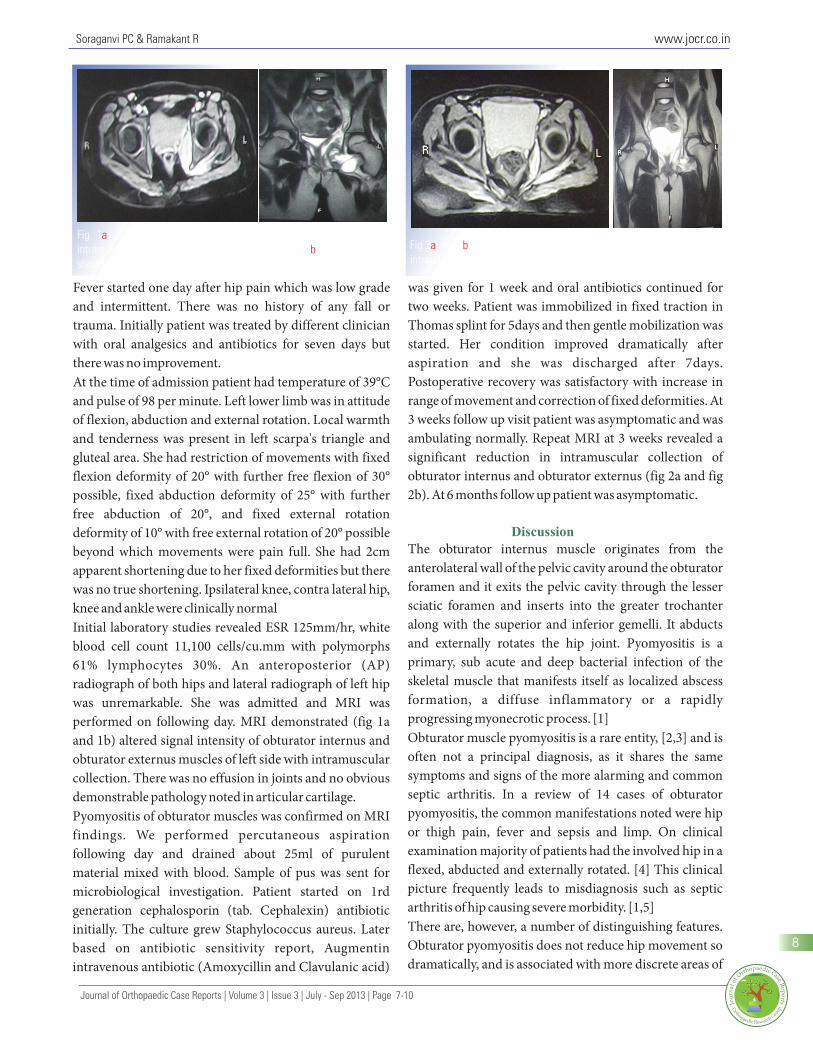
Fever started one day after hip pain which was low grade was given for 1 week and oral antibiotics continued for and intermittent. There was no history of any fall or two weeks. Patient was immobilized in fixed traction in trauma. Initially patient was treated by different clinician Thomas splint for 5days and then gentle mobilization was with oral analgesics and antibiotics for seven days but started. Her condition improved dramatically after there was no improvement. aspiration and she was discharged after 7days. At the time of admission patient had temperature of 39°C Postoperative recovery was satisfactory with increase in and pulse of 98 per minute. Left lower limb was in attitude range of movement and correction of fixed deformities. At of flexion, abduction and external rotation. Local warmth 3 weeks follow up visit patient was asymptomatic and was and tenderness was present in left scarpa's triangle and ambulating normally. Repeat MRI at 3 weeks revealed a gluteal area. She had restriction of movements with fixed significant reduction in intramuscular collection of flexion deformity of 20° with further free flexion of 30° obturator internus and obturator externus (fig 2a and fig possible, fixed abduction deformity of 25° with further 2b). At 6 months follow up patient was asymptomatic.free abduction of 20°, and fixed external rotation deformity of 10° with free external rotation of 20° possible
The obturator internus muscle originates from the beyond which movements were pain full. She had 2cm anterolateral wall of the pelvic cavity around the obturator apparent shortening due to her fixed deformities but there foramen and it exits the pelvic cavity through the lesser was no true shortening. Ipsilateral knee, contra lateral hip, sciatic foramen and inserts into the greater trochanter knee and ankle were clinically normalalong with the superior and inferior gemelli. It abducts Initial laboratory studies revealed ESR 125mm/hr, white and externally rotates the hip joint. Pyomyositis is a blood cell count 11,100 cells/cu.mm with polymorphs primary, sub acute and deep bacterial infection of the 61% lymphocytes 30%. An anteroposterior (AP) skeletal muscle that manifests itself as localized abscess radiograph of both hips and lateral radiograph of left hip formation, a diffuse inflammatory or a rapidly was unremarkable. She was admitted and MRI was progressing myonecrotic process. [1]performed on following day. MRI demonstrated (fig 1a Obturator muscle pyomyositis is a rare entity, [2,3] and is and 1b) altered signal intensity of obturator internus and often not a principal diagnosis, as it shares the same obturator externus muscles of left side with intramuscular symptoms and signs of the more alarming and common collection. There was no effusion in joints and no obvious septic arthritis. In a review of 14 cases of obturator demonstrable pathology noted in articular cartilage. pyomyositis, the common manifestations noted were hip Pyomyositis of obturator muscles was confirmed on MRI or thigh pain, fever and sepsis and limp. On clinical findings. We performed percutaneous aspiration examination majority of patients had the involved hip in a following day and drained about 25ml of purulent flexed, abducted and externally rotated. [4] This clinical material mixed with blood. Sample of pus was sent for picture frequently leads to misdiagnosis such as septic microbiological investigation. Patient started on 1rd arthritis of hip causing severe morbidity. [1,5] generation cephalosporin (tab. Cephalexin) antibiotic There are, however, a number of distinguishing features. initially. The culture grew Staphylococcus aureus. Later Obturator pyomyositis does not reduce hip movement so based on antibiotic sensitivity report, Augmentin dramatically, and is associated with more discrete areas of intravenous antibiotic (Amoxycillin and Clavulanic acid)
Discussion
Fig 1 : Axial section at the level of lesser sciatic notch shows intramuscular bright signal in obturator internus. Fig 1 : Coronal section shows signal intensification of obturator externus muscle.
ab Fig 2 and 2 : Repeat MRI after 3 weeks shows significant reduction in
intramuscular collectiona b

www.jocr.co.in
9
tenderness, including pain on rectal examination when required. [12] We performed percutaneous aspiration for the obturator internus is involved. There may be pain our case, as the presentation was late and initial antibiotic radiating to the leg if the sciatic nerve is involved, [3,6] treatment was given for short duration.and in females, there may be oedema of the ipsilateral In a review of 22 cases as reported by R.J.King et al. [12] labia.[2,7] Although these distinguishing features may shows all reported cases of pyomyositis of obturator suggest diagnosis at early presentation, but in late muscles had presented within three weeks of initial presentation like our case these may not be helpful. symptoms, however this is first reported case which Local trauma is a recognised initiating factor for presented at six weeks with limp and fixed deformities of pyomyositis.[3] It is documented in between 21% and hip. Most of the cases reported were within one weeks of 66% of cases. [8,9] Local trauma to the muscle that results initial symptom and very few at 3 weeks. The case in inflammation or haematoma is thought to be presented here highlights unusual late presentation (at 6 important in addition to concurrent episode of weeks) of obturator pyomyositis which itself is rare. This bacteraemia.[10,11] Our patient did not give any history patient presented with difficulty in walking and fixed of trauma preceding initial symptoms. deformities of hip. Pain and fever was present but it was of The causative organism is usually Staph. aureus mild grade. Most of reported cases had pain and fever with (81%),[12] the rest were Streptococcus pyogenes and toxic symptoms as presenting complaint which was absent Neisseria gonorrhoea. [13] Others have documented in the case reported here. This clinical picture created similar bacteriological profiles for pyomyositis in more difficulty in diagnosis, as tuberculosis of hip is general.[8,9] Involvement of the obturator muscles has common in India and presents with similar features which also been reported with N. gonorrhoea in women but it made us to misdiagnose this case as tuberculosis of hip seems likely that these were secondary to local pelvic initially. In our patient MRI done following day of spread.[3] As these organisms commonly cause admission and MRI findings were consistent with bacteraemia in children, any initial empirical antibiotic pyomyositis of obturator muscle. Percutaneous aspiration therapy being considered should cover the above- with antibiotic cover leads complete resolution.mentioned organisms. [2,3]In a majority of patients, treatment with appropriate
Late presentation of obturator pyomyositis is rare; antibiotics alone is sufficient.[14] Surgical drainage is diagnosis depends on careful examination and early use of rarely indicated if the patient fails to respond to medical magnetic resonance imaging. Percutaneous drainage therapy with unusual organism.[14] Antibiotics of choice results in successful treatment.should be efficient against S. aureus. The duration of
antibiotics has not been established and can vary from 2 to 6 weeks, depending on the clinical severity and response to antibiotics. [3,15] Our patient received empirical oral antibiotic initially for 1 week, which was for short duration and infection was still persisting. Hence our patient manifested at 6 weeks pain, low grade fever and fixed deformities.However, when medical management is unsuccessful, as demonstrated by persistence of fever pain for more than 5 to 7 days (particularly in cases where blood cultures are sterile), percutaneous guided drainage is indicated. [2] Surgical drainage should be reserved for cases of obturator abscess complicated by osteomyelitis because the anatomy at the site of the obturator muscle renders the percutaneous drainage hazardous. [2] In a review of 22 cases by R.J.King et al. 54% of cases the infection resolved with intravenous antibiotic treatment alone. In five patients (23%) percutaneous aspiration was performed, and in another five (23%), incision and drainage were
Conclusion
References
1.Orlicek SL, Abramson JS, Woods CR,Givner LB. Obturator internus muscle abscess in children. J Pediatr Orthop. 2001;21:744-8.
2.Viani RM, Bromberg K, Bradley JS. Obturator internus muscle abscess in children: report of seven cases and review. Clin Infect Dis. 1999;28:117-22.
3.Birkbeck D, Watson JT. Obturator internus pyomyositis. A case report. Clin Orthop Relat Res. 1995;316:221-6.
4. Wong RKF,1 Ng BWK,1 Greg A2. A Rare Condition Mimicking
Clinical Message
Late presentation of obturator pyomyositis is
diagnostic challenge to the clinician as the patients
present with unusual pain around hip joint with
pyrexia and fixed deformities. Careful history, clinical
examination and MRI needed for diagnosis and can be
successfully treated by percutaneous drainage
Soraganvi PC & Ramakant R
Journal of Orthopaedic Case Reports | Volume 3 | Issue 3 | July - Sep 2013 | Page 7-10

www.jocr.co.in
Septic Hip in Children -Case Report of a Child with Obturator J Surg 1979;137:255-9.Internus Muscle Pyomyositis . Hong Kong J Orthop 10. Schlech WF, Moulton P, Kaiser AB. Pyomyositis: tropical disease Surg.2006;10(1):39-41. in a . temperate climate. Am J Med 1981;71:900–2.
5. Nikolopoulos DD, Apostolopoulos A, Polyzois I, Liarokapis 11.Echeverria P, Vaughn C. ''Tropical pyomyositis.'' A diagnostic S,Michos I. Obturator internus pyomyositis in a young adult: a case problem S. aureus- in temperate climates. Am J Dis Child report and review of the literature. Cases J. 2009;2:8588. 1975;129:856–7.
6. Chatwani A, Shapiro T, Mitra A, Levtoaff A, Reece EA. 12. R. J. King, D. Laugharne, R. W. Kerslake, B. J. Holdsworth. Postpartum paravaginal hematoma and lower-extremity Primary obturator pyomyositis: a diagnostic challenge. J Bone Joint infection. Am J Obstet Gynecol 1992;166:598-600. Surg [Br] 2003;85-B:895-8.
7. Guis-Sabatier S, Pieri-Balandraud N, Garnier-Soumet P, et al. 13.Grose C. Staphylococcal pyomyositis in south Texas. J Pediatr. Pubic pain in athletes: a case due to an abscess in the obturator 1978:93:457-8.muscle. Rhum Engl Ed 1999;66:58-60.
14.Bansal M, Bhaliak V, Bruce CE. Obturator internus muscle 8. Chacha PB. Muscle abscesses in children. Clin Orthop 1970;70: abscess in a child: a case report. J Pediatr Orthop B. 2008;17:223-4..
174-80.15.Christin L,Sarosi GA. Pyomyositis in North America: case reports
9. Chiedozi LC. Pyomyositis: review of 205 cases in 112 patients. Am and review. Clin Infect Dis. 1992;15:668-77.
10
Conflict of Interest: Nil Source of Support: None
How to Cite this Article:
Soraganvi PC, Ramakanth R. Pyomyositis of obturator muscle: unusual late presentation.
Journal of Orthopaedic Case Reports 2013 July-Sep;3(3):7-10
Soraganvi PC & Ramakant R
Journal of Orthopaedic Case Reports | Volume 3 | Issue 3 | July - Sep 2013 | Page 7-10