q&m - the cichlid fishes of lake malawi, africai fishes in the british museunl (natural history)...
TRANSCRIPT

q&m - &.
di E HUTCHICSWM :jj.ckc , L% .U"NIVFRSIW
[&iy/i%&dfrm hw. ZOOL. Ssc. Lorn. Vo1.127, Part 3, pp. 293-344.3
1956.1. *
-
'.. : .<
A REPORT ON THE PARASITIC COPEPODA AND :I:;
B~A(ANCHIURA OF THE FISHES OF LARE NYAW . 7.i-
BY
G, E"R
. 7, , - <
. . < .
i

/
A REPORT ON THE Z'ARASITlCI COPEPOD.4 AND BRANCHIURA OF THE FISHES OF LAKE NYASA
G . FRYER Northern R1~ode~~ia.- flya9ala)~cJ Joint Fisheries Renearch Orgnnisation
[Comml~~~ic:atc~d by Dr . J . P . Hardir>g.--Acceptfled 14th February 1966.1
(With 79 figures in the text)
CONTENTS
Introduction . . . . . . . . . . . . Systematic and biological notes . . . . . .
COPEPODA Family Ergasilidae . . . . . .
Ergasilus macrodactyh~~s (Sara) . . Trigasikts nzinuttha gen . ct sp . n . . .
Family Dichelesthiidae . . . . . . Larrt.p~.oglena nyctsnr sp . 11 . . . . . Lwt~proglena clnriae sp . n . . . . .
Family Lernaeidae . . . . . . Lemma bagri Harding . . . . Lernaea lophiara Harding . . . . Lernaea hardingi nom . n . . . . . Lernnea tilapiae Harding . . . . Lernaea palati Harding . . . . Lernnea bnrnimiann (Hartmann) . . Lernaea sp . . . . . . . . . Other species of Lemaecz in L . Nyasa
Family incertae sedis . . . . . . Afrolernuea longicollia gen . et sp . 11 .
BRANCHIURA Frtmily Argulidao . . . . . .
Argulti. s clfi.icunus Thiele . . . . Arq?~lt6n jollymani sp . n . . . . . Dolops rannrum (Stuhllnann) . . Chonopeltis ilaermis Thirlo . . . .
Geographical distribution . . . . . . . . The species flock of Lernaea in Lalie Nyrtsa . . Economic co~lsiderations . . . . . . . . Acknowledg~nrnts . . . . . . . . . . Summary . . . . . . . . . . . . R. cfcrrn.!r.s . . . . . . . . . . . .
Page . . . . 293 . . . . 294
INTRODUCTION
During the course of biological work in Lake Nyasa extending over a period of about two years the writer has had the opportunity of making a study of the parasitic Copepoda and Branchinra of the fishes of this ancient lake . The results of this work are describecl in the preiient. paper.

294 C . FRYER
The earliest reference to parasitic C'rustacea in Lake Nyasa is that of Thiele ( I !)o( 1) 1r711o gave brief diagnoses of two branchinrans, Avgt t l us c4frirrlz us and Chonopeltis inermis, both being described as new to science. L. Nyasa was only one of several localities from which Thiele had received the fornler species which is now known to be of widespread distribution in the African ('ontinent, but Cho)lopeltis inermis, which he described from a single female specime~~, was for long thonght to represent a genus endemic to L. Nyasa and nearly t,hirty years were to elapse before any species of the genus was discovered elsewhere in Africa. Thiele (1904) later amplified his description of these species and published several illustrations.
During the course of the Third Tanganyika Expedition a brief visit was made to Lake Nyasa and the branchiuran Dolops ,.anarum (Stuhlmann) was added to the list of species known to exist there (C~xnnington 1913), whilst Sars (1909) who worked through the plankton samples collected by Cunnington, described two ergasil~tl copepotls from the lake . one, described from immature specimens,
I he named Ergasiloides brevimanus ; the other he characterised merely as Ergasilus sp.
Harding (1950) published an important systematic paper on the copepod genus Lernaea and described no fewer than six species from L. Nyasa as well as a for111 which, because of lack of material, he left tunnamed as it was not clear whether it merited specific rank or came within the range of variation of another species. Most of his specimens were obtained from the large collection of Nyasa~i fishes in the British Museunl (Natural History) but in one case he was able to add brief notes on the appearance of living specimens from data supplied to him by Miss R . H. Low-e who encountered two species of hernaea during the course of fishery work in the lake. I
A large proportion of the work described in the present paper was carried out on specimens obtained in the vicinity of Nkata Bay which is situated on the western side of the lake approximately 150 miles from its nortllerll extremity, but a considerable amount of material, collected both by the writer and his colleagues, has been examined from other parts of the lake and adjacent waters.
SYSTEMATIC AND BIOLOGICAI, NOTES
The following species have been found during the course of the present work. COPEPODA
Family EI-gasilidae Eryasilus macrodactylus (Sars) Trigasilus minutus gen. et sp. n.
Family Dichele~t~hiidae Lamyroglena nyasac! sp. n. I La~nproglenn cltrj,ircr sp. n.
Family Lernaeidae Lernaea bagri Harding Lernaea lophinra Harding Lernaea hardingi nom. 11. Lernaea tilapiae Harding

PARASITIC COPEPODA OF THE FISHES OF LAKE NYASA 295
Family Lernaeidae Lernaea palati Harding Lernaea barnimiana (Hartmann) Lernuea sp.
Family incertae sedis Afrolernaea longicollis gen. et sp. n.
BRANCHIURA
Family Argulidae Argulus africanus Thiele Argulus jollyrr~ani sp. n. Dolops ranarurn (Stuhlmann) Chonopeltis inernzis Thiele
Each species is discussed in turn in the following account.
COPEPODA
I ' Family ERGASILIDAE
i The present investigations have revealed two representatives of the family
I Ergasilidae in L. Nyasa. One of these, which I refer to Ergasilus macrodactylus (Sars) (Syn. Ergasiloides rnacroductylus Sars) has, by its discovery in the adult stage, enabled a number of systematic tangles dating back to the early years of the present century to be unravelled. In order to fully comprehend the nomenclatural complexities involved it is necessary to state briefly the various items of information which have led to our present state of knowledge of the African Ergasilidae, and the vicissitudes of the nomenclature of the ergasilid genera as a whole.
Sars (1909) in his paper on the copepods of the African lakes, collected by Cunnington, described three new ergasilids all of which he placed in a new genus Ergasiloides, chiefly distinguished from Ergasilus by the possession by the female of only a single abdominal somite. (Three such soinites are found in Ergasilus.) However, it is obvious that all Sars' species are based on descriptions of im- mature forms and it is curious that he did not realise that the mature female may have possessed more abdominal somites than the larval forms which he had before him.
Gurney (1928) received material from L. Tanganyika containing immature specimens which he ascribed to two of the species described by Sars. However, these differed from Sars' examples in having an additional abdominal somite. In all cases save one, in which these two somites were distinctly separated, the two were only partially free and it is probable that Sars failed to note this incipient separation in his material which apparently contained examples of the same stage of development as those examined by Gurney. The information gained from Gurney's material did not invalidate the genus Ergasiloides Sars, but rendered necessary a slight modification in its definition which Gurney implied but did not make.
All these specimens belonged to immature, free-living stages of the species concerned and it was doubtful whether, on the discovery of adult individuals, it wonld be found possible to maintain the genus Ergasiloidcs. However, Capart

2 9 6 O. FRYER
(1944) obtained adult females of an ergasilid from L. Tanganyika which he assigned to Ergasiloides megacheir Sars, and as these specimens had an abdomen composed of only two somites* they validated for the first time the genus Ergasiloides Sars.
On the other hand, my specimens from L. Nyasa (with the exception of those which obviously belong to a new genus) are all assignable to the form described by Sars from immature individuals as Ergasiloides rnacrodoctylus, bnt the adults show without doubt that Sars' specimens were placed in tfhe genus Ergasiloides on the basis of characteristics, wrongly assumed to be those of the adult, which are present only in the larval stages, and that the correct systematic position of the species is in the genus Ergasilus. The species should therefore be referred to as Ergasilus macrodactylus (Sars).
A further complication in the nomenclature arises from the fact that Yamaguti (1939), apparently unaware of the erection of the genus Ergasiloides by Sars, bestowed the same name on a genus which he created for the reception of some ergasilicls from Japan. However, the discovery by Capart of adults exhibiting the features of Ergasiloides Sars established beyond all doubt the validity of that genus, so the name Ergasiloides Yamaguti must be suppressed and a new name substituted in its place. Por this I propose Yamagutia. The three genera concerned now become : Ergasilus Nordmann. 1832. Type species : E. sieboldi, selected by Gurney 1933. Ergasiloides Sars. 1909. Type species : E. megacheir, here select'ed. Yamngutia nom. n. Type species : Ergasiloides bora Yamaguti (syn.
Ergasiloides Yamaguti, 1939, Non Sars 1909). A description of the adult of Ergasilus nzacrodactylus follows.
E R f f A S I L U S M A C R O D A C T Y L U S (Ears) (Figs. 1-7) Adult female
Length 0.97-1.0 mm. Cephalothorax often apparently fused with somite of leg 1 but sometimes clearly separated (Young adults). Cephalothorax con- siderably longer t,han wide, bluntly rounded anteriorly, somewhat bulged laterally, and shallowly indented in posterior third. Head sculpture as des- cribed by Sars for immature stages, consisting of an anterior circular marking and a more posterior, somewhat ovoid marking. Between the two is the dorsal chitinous frame which takes t,he form of an inverted T. Eye well developed.
Thoracic somites 2 to 5 distinct, evenly rounded at lateral margins. Soillit,e 5 narrow bnt quite distinct.
* The use of ihe term " urosome " has led to considerable confusion in descriptions. Sars wed the term to mean the abdomen plus the telson, and Gurney took the term as being equivalent to abdomen, which ~t is not, and therefore involved himself in inaccuracies when referring to the number of abdo?linal somites. Capart also confused the two terms for in the description of Ergccsilus sarsi he says " urosome compos6 de quatre segmeiits " using the term urosome to c~nbrace both telson and abdomen, or, in other words, stating that the abdomen is composed of three somites : yet when describing Ergasiloides megacheir he says " urosome compos6 de deux segments," meaning in this case that the abdomen alone consists of two somites as his illustration
i B
shows. I t seems advisable, therefore, to drop the use of the term urosome completely and refer to the posterior tagma of the body rts the abdomen plus the telson.
I

PARASITIC COPEPO1)A OF THE FISHES OF L.4KE NYASA 297

298 O. 'FRYER
Abdomen of t,hree son~it~es. Geiiit~al somite bulged laterally in ant,eriur t)or.t,ion. Frlrcal rami simple, a litt'le longer tml1a'u wide. So~uewhat widener1 ~o"~eriorly 2nd aimed with 4 unjointed setae. lilllerrnost furcal spta itbollt 5 t'irnes as loilg as next longest which is directed obliquely outwards. J,ollgest seta soiliewliat swolleii distal to t'he base.
Antennule of (i segments. Ailtelllla prehensile : long and slendej., of fi)rm show~l ill Fig. 4.
Legs 1--4 of struct rue tj-pica1 fhl. gcbl~us, havil~g f'ollowilig ;t~.r.;r11gel11ckl1t of' spines and setae.':'
P.1. Exoprod. Endopotl.
P.2. Exopod. Endopod.
P.3. Exopod. Endopod.
F.4. 15xoj)od. Elldopod.
I -I 2 .i 0 I 2- .i ( I - I 0-0
U -2 I 4 0-2 1 -4. 0 - 2 I -4 )-.5 I I-- 1 1-3
Leg 5 siinple, eylil~drical ; arined wit'll 2 ht:rillinal s e t ~ e of' wliiull loilgest is :ilmost, as long as leg 11,~aring i t) and more tllan 2.5 times as long as sho1.test.
Egg sac;; long, l,e;tcliing beyond entl of longest fi:i.ca'l seta. ( '(dour white, but wit11 Iwge blot'clies of l~urple pigmcnt \.rrltl.all~- i t I
~epl~alot~horns 1,rgioll.
rl rllrlf rnulr, 1,eugth 0.6iri-O.S-k;i 111111, 1Zc)dj' cy~Io1jifiwiil. 1 1 o - i i i s !:enit';l'l sc)~nite much swolleii L!~.IC to ~)r~; -~ci ice of'
s ~ : N , I ~ ~ : L ~ ~ o J ! J ~ ~ ~ I ' ~ s . ?Olllit~cs 2 t80 4 sln;dl, \videla t8lla11 long, :tl)pi.oxiiliat~ly eclua,l ill siztx i ~ l t l inucll sniallel. t'llail genital ~ornit~e. Total leiigth of sonlites 2 t'o 2- l r ss t'lia11 Ic-iigt'll of gcilit'al soii~ite. Furcal rami and a'rmature sirnilnr to t'liosc Of fPlllalI'.
_4lltenil11le 01'5 st~ginei~ts. Xilteilllit l?i~elleilsil~, less c1o11ga,t8e. nl~d st80ut8er t'haiz ir1 fernale : liavirlg
f'oi,~n sll(o14 11 i l l Fig. 6. bI:tsilli~~cds clll;!i~jiecl. p~,c.l~c~!sile : Ila1.ii1g f'otvn sl~o\i 11 i r l Fig. 7. I-cgs 1 - .1- :;jr!ijlnr to t'llost. of feinalc. \,lit csopocl. of Icg 1 llns 3 svgirlents.
..\i.~.a~lge~ucllt of' spiiit,s alltl si.ta,e of legs I--:hs in female. Ar:.n~iigemeilt of spiiles and setnc of 1
I ~ s o ~ ~ o ' l . 0-1 (1-2 1-4 1~lltlo~""l. 0-1 ( ) - ~ ~ 2 1 -:;

PARASITIC COPEPODA O F THE FISFIE8 OF LAKE NYASA 299
r TRiGASILUS MINUTUS gen. et sp. n. (Figs. 8-10) Adult ji~~trlale
1,ength (holotype) 0.344 mm. (paratype) 0.442 mni. Body cyclopiform, '
d o r ~ ~ - \ ~ ~ l l t l ' d l y flnttc~led ; inuch foreshortelled posteriorly. Pil-st leg-bearing 1 Wit11 the exvc-ljtloli of one bcloilgil~g to n new genus with uyhich collfusioii could not arise
I
Occurrence Adult females are very common on the gills of cichlid fishes belonging to the
genera Haplochromis, Tilapia, Lethrinops and Pseudofl.opheus and have also been found on the gills of the cliaracin Alestes imberi Peters. The species is not strictly confined to tlie lake for i t has been found on fishes in an inflowing river fifteen miles upstream. The few adult males seen have been found in inshore plankton hauls. A few adult females have also been obtained in a plankton sample. The oiily record in the available literature of a fully developed female ergasilid having been found away from its host is that of Wilson (1911), who records the occurrence of adults of both sexes of E, chautauquaensis Fellows, in the plankton of North American lakes.
Rerr~arlcs / '
Sars (190!)) (lid not record this species from L. fiyasa though he refers to having seen a mounted specimen of a species of Eryasilus, to which he gave no name, which had been obtained from this lake. On the other hand lle records Ergasiloides hrevimanus from L. Nyasa, though on the basis of only a single inlmatwe specimen. At the stage of development on which Sars based his descriptioil of E. brevirnarlus i t is exceediilgly difficult to assign a given specimen to a specific category and too much reliance cannot be placed on Sars' record. Indeed Gurney (1928) suggested that E. brevimanus was no more than an earlier developmeiltal stage of Ergasiloides megachei~, itself described by Sars from ilriniature specimens. Harding (1942), however, who eilcouiltered a single immature bretii/,~anus-like specinlei1 in material collected in L. Tanganyika doubted the truth of this suggestion. Immature free-living stages of an ergasi- lid are sometimes encountered in inshore waters of L. Nyasa which, because of tlie sculpturing of the head and the fact that only one adult species has been fo~uitl,* I take to represent immature stages of E . mac~odactylus. These very much reseiltblc " E. h~c~oimanus " and had only these immature stages been available I should have been very reluctant to give thein a name.
Tlle sinlilarity of the ndults to the immature stage described as Eryrcsiloides macrodactylus by Sars, particularly in the form of the antennule, is so great as to make i t virtually impossible to assign them to any other species. While some advantages might have accrued from describing the Nyasan specimens as a new species which could, if necessary, bc merged in the synonymy of E . macro- dactylus when adults were found i11 1,. Tanganyika, on the whole it has seemed inadvisable to do so.
E. macrodactyl~is appears to be most closely related to E. sarsi Capart, which is known from L. Alweru and the Iiatanga.
Infestatioils of' up to as many as two dozen specimeils on a single set of gills
I are not nncoinmun at Nkata. Bay, but tlie pavasite seems to do little serious damage to its host.

300 G. FRYER
thoracic sornite fused with head to form an enormous pentagonal cephalothorax, comprising some 60 per cent of total length. Head shield in form of an isosceles triangle with bluntly rounded corners. Ocellus very large, roughly rectangular in shape, deep purple in colour.
Thoracic somites 2 to 4 gradually diminishing in size, the largest being much smaller than somite 1. Thoracic somite 5 very narrow, only incipiently sepa- rated from genital somite.
Figs. 8-10.-Trigasilus minutus gen. et sp. n. 8. Adult 9 (dorsal). 9. Antenna. 10. Leg 5.
Abdomen very short, composed of 3 somites. Genital sornite not or but slightly swollen, but broader than long. Abdominal somites 2 and 3 very short. Furcal rami short, each about as long as wide and bearing 4 terminal setae. Innermost terminal seta the longest, somewhat swollen at the base. Outermost
I seta directed outwards a t an angle of about 45".
Antennule fairly well developed, about 63 p in length in holotype and com- po*ecl of 5 segmeilts. Anterior border well supplied with setae. Terminal I
segment with l o ~ ~ g teriniilal setae, the two longest being about 80 per cent ' length of a~lteiiiiulc. Ailterliiae stout aiicl p~t.llensile, of 3 segments, most
distal braring 3 1.c.curved and subequal spinc~s, allortest being about half length of' i ~ p p c ~ ~ ~'l~tgc.

PARASITIC COPEPOUA OF THE FISHES OP LAKE NYAYA 301
Mouthparts not distinguished with sufficient clarity to merit detailed description, but very similar to those of species of Ergasilus. Mandible and palp of Eryasilus type. Maxillules not clearly distinguished but with one, possibly two, posteriorly directed spinules as in Ergasilus. Maxillae of type found in Ergasilus but no tuft of " setae " distinguished at tip. Maxillipeds absent.
Legs 1 to 4 biramous. Exopod. of leg 4 of 2 segments, otherwise all rami 3-segmented. Arrangement of spines and setae of exopod. segments as follows.
Leg 5 a minute plate bearing 2 terminal setae and a slightly subterminal seta on posterior margin.
Egg sacs long, 321 p in length in holotype; almost as long as animal itself. About 30 or 32 eggs per sac arranged as in Ergasilus. Colourless except for small flecks of purple pigment dorsally in cephalothorax. Eggs whitish.
Occurrence A single specimen has been taken from the posterior wall of the gill chamber
of a cichlid fish Petrotilapia tridentiger Trewavas and another has been found on the cichlid Pseudotropheus tropheops Regan, being located where thegill chamber unites with the roof of the mouth. Both fishes were collected on a rocky shore of the lake a t Nkata Bay.
Remarks and systematic position A specimen examined alive was unable to swim when removed from its
host, but wriggled violently. While certain aspects of its anatomy could be described in greater detail
if more material was available for dissection, sufficient illformation has been gained to merit the erection of a new genus and species.
T. minutus is obviously a member of the family Ergasilidae and is closely related to Ergasilus and allied genera, yet differs markedly in the structure of the distal portioil of the antennae, each of which terminates in 3 spines as opposed to a single spine in all other members of the group which TVilson (191 1) designated as the subfamily Ergasilinae. Otherwise, it stands very close to Ergasilus and allied genera.
Wilson (191 1) divided the Ergasilidae into three subfamilies, viz., the Ergasilinae, the Bomolochinae and the Taeniacanthinae. While members of the two latter subfamilies have antennae terminating in three claws as is the case in l'rigasilus there is no doubt that the affinities of the latter are with the Ergasilinae and not with the Boi~iolouhiiiae or Taeniacallthiuae. Furthermore there is no possibility of regarding it as a genus bridging the gap between the Ergasilinae and either of the other two subfamilies for, althougll terminating in three spiilcs, the structure of the anlenna is, like so many other morphological characteristics, essentially like that of the Ergasilinae and differs considerably

from its homologuein the Bomolochinae and Taeniacanthinae. Trigasilus is rather to be regarded as a specialized member of the ergasiliile stock whose antennae have become specially modified as an adaptation to attachment, not to the gill filaments of fishes, but t'o the menibraiious wall of the gill chambers.
As it is so closely allied to the previously described members of the Ergasil- inae, it is pointless to erect a new subfamily for its reception. Such a procedure would only obscure obvious relationships. It therefore seems justifiable to widen the limits of the definition of the Ergasilinae, of which subfamily it can be regarded as a somewhat " aberrant " member.
.A remarkable feature of Trigasilus minutus is its small size, its length being little more than half that of even the smallest ergasilids hitherto described. Indeed, so far as a search of available literature reveals, it may well be the smallest of all parasit'ic copepods which, on the whole, exhibit trends towards an increase in bodily dimensions over t'heir free swinlmiilg relatives. While a sedentary life reduces the respiratory demands of t'issues and permit's an increase in the volume : surface area iat'io, esploitatioil of such a possibility by a parasite living in t,he coi~fiiled space of a gill chamber which, however, is well oxygeilat'ed, is likely to be disadvantageous, aiid a reverse trend, namely towards a dilniilut'iorl in size, probably offers certain advantages. I t is interesting in this connect'ioil to note that Thersitina !/astc.i,ostei Pagenstecher, t'he other ergasilid with which it shares the habit of at~t~acl~i~ig itself to the walls of the gill chamber, is also very small, an adult female having a length of only about 0.6 mm.
Tlle family definit,ion requires no modification as a result of the discovery of l'~iyasil?~.s but the sllbfamily call be retlefiiled as follows.
Subfamily Ergasilinae :-Small species! f'reclnently less than 1 min. in length. Body cyclopiform. Margins of cephalot'horax not forming walls of a silckitig disc. Antennnles small, basal segn~ellts neither enlarged nor flatt'ened, and armed with slcxder setae.
Antennae modified to form st'rong prehensile structures terininatiug in a single claw, or, in Triyasilus, in three claws. Mol~th located near centre of ventral surface of cephalothorax and projecting somewhat. Swimming legs well developed and biramous.
Fifth legs, simple and composed of one segment. ! The genus l'i~ii~rc~silus call be defined as follorvs.
TRIGASILUS gen. 11.
Alirlutc for~nr. Bociy cyclopiform, nlllcll ilarrowetl 1)osteriorly. First Icg- bearing thoracic somite filsetl to head. Abdonlell of tl~rec soinites. Furcal rarni short
Biltennules of 5 segments. Ailtellllae prehensile, of 3 segments and termin- ating in three chitinous hooks, Other appendages as in Ergasilus and allied genera. Egg sacs as in Erycxsilus. Adult female parasitic in gill chamber of freshwater fishes (hitherto of family C'icllliciae). Male nllki~own. , Di,qt.rihuiio.n. Africa (L. Nyasa) . .
7 ' , t j ~ ~ ~ s ~ ~ ( : c i c ~ s . l'i~lyrrsil us n c i t i rr./rrs.

P.4RASITIV COPRPODA OF THE FISHES OF LAKE NYASA 30.7
Family DICHELESTHIIDAE Two members of t'his family, assignable to the genus La~nproylena, and both
apparently new to science have been found in t,he lake. These are described below.
LAMPROGLENA NYASAE sp. n. (Pigs. 1 1-2,l) Adult female
Length about 3.6 mm. Body '' grub-like " in general appearance. First leg-bearing thoracic somite incorporated in cephalothorax, thoracic
somite 2 being first free somite. Cephalothorax comprising about 20 per cent of total lengt'h. Media,n eye small but distinct ; red in life. Thoracic somites 3 & 4 somewhat swollen and much larger than somit,e 2. No thoracic somite 5.
Abdomen of 3 distinct somites. Genital somite mucll expanded posteriorly, with genit,al apert,ures sit,nat.ed dorso-laterally in posterior part of somite. Each aperture approsinlately oval in form and with a rim of thickened chitin. Telson elongate, approximately as long as abdominal somit,e 3. Furcal rami short, sub-conical, each with an out'er " branch " situated somewhat ventrally. Terminal setae represented by 3 very short ~truct~ures with swollen tips. Dorsal seta unmodified and reaching a little beyorid end of ramus. A small triangular anal operclllum with broadly rounded apex present.
Ant'ennule short', flat'tened, and redilced t'o 2 segments. Basal segment about T times as long as distal anti bearing nllmerous moderately long setae O I I
its preaxial margin. Distal segment bearing a single preaxial seta and a tuft of short terminal setae. Antenna sh'ort, about one t,hird length of anteiinule and two-segmented. Basal segment approxinlately twice as long as distal. I11 life, antenna is obscured by antellnule and is backwardly directed. Oral regioii surrounded by a wide-walled, horseshoe-shaped, sucker-like structure. " Maxilla " long and st,rap-like wit,h t'wo chitinized teeth distally. Tips of opposing appendages overlapping slightly. Maxilliped stout and strongly reflexed. Distal segment with 3 stout, heavily chitinised, recurved spines. A distinct swelling between maxilliped and first thoracic leg.
Thoracic legs 1 t'o 4 of similar structure ; biramous, and retaining slight evidence of former segmentation, though in no case is distinct segmentation discernable. Setatioli of all legs much reduced.
Eggs maturing in oviduct's virtually confined to thoracic somites 3 & 4. Egg sacs consisting of long uliiseriate chains of eggs ; up t'o about 30 eggs per chain.
Colour dirty white, sometimes tinged with brown in cephalothorax and thorax.
Occurrence Fairly common on the gill filaments of cichlid fishes belonging to the genera
Ha11lochromis, Docimodus, Lethrin,ops, Tilapia, Petrotilapia, Pseudotropheus and possibly others. Most of the host species are inshore living rather than pelagic in habit.
Hen~,nrX.s lnfestations of this species are seldom heavy and the presence of half a
dozen specimens oil each set of gills is except'io1l;~l. Its effects on the host are 1i1.1t ve1.y noticeable. ,411 iilfectctl gill filn~ne~rl o f i c : ~ l exl~ibit~s a st8ructul3eJess

Figs. 11-21 .-Lniizproglam nyasae sp. 11.
11. Adult 9 in situ on gill of host (lalcral aspect). 1-0. ildult $2 (dorsal). 13. A~~te r io r region of body (volll rill). 14. T i l ~ of' abdomerl nnrl furcnl rnmi (vmnt'ral). 18. Iiight al~tennule splnyed out. (ventral). 16. Tip of " m:txilllt ". 1'7. Till of inasillipetl. 1s. Jtiglrt leg 1 . (vontxal). I!). Coni1,nI sorriiio wit11 sl)crn~iltc)phores ntlnclletl. 20. l i , igl~t f'urc.al I.il,mus (ventral). 21. Op~rculuni.
Legend : Al-anto~mule, A2-antenna, M-" maxilla ". Mxp-innsillil)ccl, 0.R.- oral region. S-spermatophores.

1 PARASITIC COPEPODA OF THE FIBRES OF LSRE NYASA 306
proliferation of tififille above the point of attachment of the parasite (Fig. 11.). This seems to intlic;~tr that aftel- macrrating the gill and causing this prolifer- ation the parasite gradll~lly edges its way down the filament. Otherwise body movements appear to be restricted to flexing movements of the trunk.
The gut wall contains numerous green objects apparently analagous with the black discs described in L. clariae (q.v.)
No " nrcklace " of sessile protozoans such as is frequently seen on many parasitic copepods, including members of the genus Lamproglena, has been observed on this species which invariably presents a very clean appearance.
No male specimens have been found, but from the size of t,he spermatophores which are sometimes to be seen struck to the genital somite of the female it can be deduced that they are very small creatures, probably less than 1 mm. in length.
The nauplii hatched from eggs offered no outstanding peculiarities. All
I attempts at rearing them proved unsuccessful.
LAMPROGLENA CLARIAE sp. n. (Figs. 22-33) Adult female
Length to :&bout 9 mm. Body elongate, very indistinctly segmented, and with a somewhat swollen cephalothorax. First leg-bearing thoracic somite not incorporated into cephalothorax. Cepha!othorax indistinctly separated from thorax and comprising only 7 to 8 per cent of total body length. Median eye small but distinct ; red in life. Thoracic somites all confluent. Region com- prised by somites 1 & 2 narrow and forming fairly distinct " neck " region. Former line of juncture between somites 3 & 4 indicated by distinct indenta- tion. Thoracic somite 5 indistinguishable.
Abdomen elongate and of 3 somites. Genital somite short, somewhat swollen dorsally ; demarcated from thorax and from abdominal somite 2 by deep dorsal grooves but not by joints. Genital pores situated dorso-laterally, broadly oval in outline and with a rim of thickened chitin. Abdominal somites 2 & 3 elongate, separated by a partial but still distinct joint.
Furcal rami short, simple, papillate, and bearing terminally 2 very short remnants of terminal setae.
Antennule short, reduced to 2 segments. Basal segment a little more than three times as long as distal and bearing a number of much reduced setae along its preaxial margin. Terminal segment bearing 4 much reduced terminal setae and a small preaxial seta.
Antennule short, of two segments ; distal segment reflexed on basal, less than half its length and bearing 2 small papillae anti 3 much reduced setae.
" Maxilla " short, very wide proximally, and bearing two short chitinized teeth distally.
Maxilliped short and stout without evident sign of segmentation : armed distally with 3 recurved chitinous teeth, the two largest approximately equal in size and considerably larger than the smallest.
Thoracic legs 1 to 4 much reduced, all biramous and showing no sign of segmentation. Setation of all legs much reduced.

G . FRYER
Figs. 22-20.-La,npro:ll~r1.(~ c l a ~ i a e sp. 11.
22. Adult 9 (lateral aspect). 23 . Antennule. 24. Antenna. 25. Maxilla. 26. RIaxilllped. 27. Leg 2. 28. Genita.1 nomite u . r d genital pore (Intel-al). 29. Furcal ramus (lateral).
Legend : G .P.--Qenit:~l pore.
Eggs produced in long uniseriate chains which extend well beyond end of body ; 50 or more per chain.
Body almost colourless or very lightly tinged wit'h brown ; eggs in oviduct conspicuous and yellow ; gut usually red due to jnclnded blood. Eggs in egg strings orange-yellow.
Occurrence Common on the gills of many, probably all, the Nyasan species of Clarias.
A single specimen has been found on a speci~nell of C'larias w~o~ssamhicus Peters taken from the Ranga River, a tributary of t,he TJuweya. fourteen iniles from its point of entry into the lake.
Remarks A dozen or more specimens may sometimes be found attached to a single set
of gills. The adult female completely buries its head in the gill tissues,
PARASITIC COPEPODA OF THE FISHES OF LAKE NYASA 307
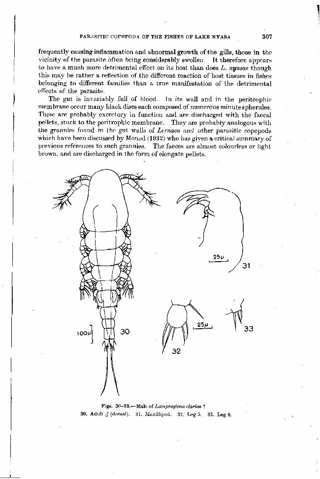
frequently causing inflammation and abnormal growth of t'he gills, those in the vicinity of the parasite often being considerably swollen. I t therefore appears to have a much more detrimental effect on its host than does L. nyasae though this may be rather a reflection of the different reaction of host tissues in fishes belonging to different families than a true manifestatioil of the detrimental
I effects of the parasite.
The gut is invariably full of blood. In its wall and in the peritrophic membrane occur many black discs each composed of numerous minute spherules.
I These are probably excretory in function and are discharged with the faecal pellets, stuck to the peritrophic membrane. They are probably analogous with
I I the granules fonud in the gut walls of Lernaen ant1 other parasitic copepods
which have been discussed by Monod (1932) who has given a critical summary of previous references to such granules. The faeces are almost colourless or light brown, and are discharged in the form of elongate pellets.
Figs. 30-33.-Male of Lamp~oglena claKae ?
30. Adult 3 (dorsal). 31. hIaxilliped. 32. Leg 5. 33. Leg 6.

308 G. FRYER
Because of its obviously considerable consuinption of blood and because of the structural damage which it does to the gill filaments this species must be considered as more than a mere nuisance. Any ill effects on the respiratory exchange of the host are, however, likely to be more than counteracted by the presence of accessory respiratory orgams which are a characteristic of the genus Clarias.
A " necldace " of sessile protozoans such as is of frequent occurrence on species of Lernaea (see below) is often present around the anterior part of the body.
The male of Lamproglena clariae ? (Figs 30-33).
Three adult male copepods and a male in what appears to be the last copepodid stage have been obtained, always in close association with attached females of L. clariae. This would appear to be more than a chance association and is strongly indicative of the fact that !hese males belong to L. clariae, but their similarity to the males of Lernaea is so striking as to invite caution in identifying them. These males can be described as follows.
Adult male Length 0.897 to 0.94 mm. Body cyclopiform. Thoracic somites 2 to 5
distinct. Abdomen of 3 distinct sornites. Furcal rami short, less than twice as long as broad ; each with one long, and
two extremely short terminal setae. Outer terminal seta longer than inner. Rami with a short dorsal and a short lateral seta.
Antennule of 5 segments. Antenna fairly long, of 3 segments. Two of the terminal spines modified as hooks.
Maxillae well developed, similar to those of male of Lernaea. Maxillipeds similar to those of male of Lernaea : structure as in Fig. 31. Legs 1 to 4 with both rami 3-segmented and with well developed setae. Spine formula 2.3.3.3. Cosopodite of all swimming legs with seta at inner margin, and basipodite with seta a t outer margin. Leg 5 of 2 segments. Segment 2 almost as wide aslong and with 5 spines and setae. Leg 6 a minute plate bearing 3 setae.
I n addition to the three adult males a copepodid, apparently in the stage before the final moult, was found. This specimen had a length of 777 p and was very similar to the adults but the rami of the swimming legs were only 2-seg- mented. Only 3 setae could be detected on the terminal segment of leg 6. The furcal rami had the same form as had those of the adult but the longest terminal seta was considerably more swollen at the base.
I In all cases the males were found at>t,ached to a gill in close proximity to a
female. In one case the female in question was not fully grown and had no egg sacs, but in two cases full-grown females with egg strings were involved.
The only male of any species ofLnnzproglena of which I have seen a description is that of L. angusta Wilson, found by Monod (1932). This differs only in detail from the copepodid stage described above and was almost certainly immature.
I I
The similarity of these LWO specimens, incidentally, is further evidence that the males here described do indeed belong to Lamproglena.
The most striking featnre of these illales is their remarkable resemhlailce to those of Lernaea, a i d it is this fact, together with the knowledge that males of
I I

PARASITIC COPEPODA OF THE PISHES OF LAKE NYASA 309
some species of Lernaea settle on intermediate hosts, that invites caution in stating their identity. If they are indeed males of Ln7r~proglena then a very close relationship between the families Dichelesthiidae and Lernaeidae is indicated.
Development Numerous nauplii have been obtained. These are active creatures having,
a t the time of hatching, a length of about 330 p. They are rather fat, being well provided with oil droplets, so much so that i t is a matter of some difficulty to
I trap a living specimen beneath a coverglass without rupturing the integument. Under admittedly primitive conditions, attempts to rear these nauplii have not met with much success, none being reared through all the naupliar stages.
I n addition to the males obtained from the gills of Clarias spp. a female copepodid very similar to the males described above, and therefore very similar to the free-living females of Lernaea, was also obtained. It seems possible that this female, which had a length of 1.08 mm. represents a stage in the development of L. clariae. This specimen was in the process of moulting, showing that a t least two copepodid stages are passed through.
While our knowledge of the life cycle of this species is still far from being complete i t call be stated as fairly certain that no chalimus stage is involved in the development of Lamproglena. Heegard (1947) predicted that such a stage would be found to exist and those authors who ranged the Dichelesthiidae alongside the Caligidae also presumably anticipated the discovery of such a stage. (In the development of the Caligidae there is only one copepodid stage which is followed by chalimus stages with a frontal filament.)
Such chalimus stages, if they existed in the life cycle of Lamproglena, would be much more conspicuous than the minute males which have been seen and could scarcely have escaped detection during the searches in which males were found. Further, if the female copepodid seen was indeed a Lamproglena then, because i t was moulting, more than one copepodid stage is involved.
It might also be noted that no evidence of a chalimus stage of L. nyasae has been obtained although a large number of adults has been seen.
Family LERNAEIDAE This is the best represented of all the families of the parasitic Copepoda of
L. Nyasa, no fewer than eight and possibily nine species having been recorded to date. Notes on the species found during the recent investigations follow.
LERXilE-4 BAURI Harding This species was described by Harding (1950) from preserved material
sent to him by Miss R. H. Lowe who (Lowe 1952) notes its occurrence in L. Nyasa. There is nothing to add to Harding's description of the mature adult female
save a note on the colour of the living specimens. The body is almost colour- less, though the alimentary canal is sometimes red owing to the presence of blood. The presence of spherules similar to those described as occurring in the gut wall and peritrophic membrane of Lamproglena nyasae, but dark green in colour, also give colour to the body. Eggs in the oviduct appear yellowish or greenish according to the state of development. While in the egg sacs they are olive green.

310 C. FRYER
Stages of the adult female younger than those available to Harding have been found. These are very slender and the dorsal anchor arms are straight and not curved as in older individuals. The pregenital prominence does not begin to develop until a length of about 7 mm. has been attained.
Male On one occasion a scraping from a slimy, blood smeared area in the mouth of
a Bagrus meridionalis Giinther produced a slender immature female some 7 mm. in length and an adult male. In some species at least of Lernaea the copepodid stages of bot,h male and female may seek the gills of an intermediate host and, according to Wilson (191 7) cit'ed by Gurney (1933) the male does not leave the intermediate host. In L. bagri the association of a male with a juvenile female in a situation other than the gills can be taken as almost certain proof that mating takes place on the definitive host and that if an intermediate host is involved the male leaves it.
The adult male had a length of 1.2 mm. and had a structure so very similar to that of the free living female of L. cyprinacea L. described and figured by Gurney (1933), with of course the usual differences associated with the genital somite of the male, that a detailed description is unnecessary. This specimen was almost colourless save for its large red eye. I t was an active swimmer.
Adult females of this species occur commonly on Bagrus rneridionalis to which host it appears to be strictly confined. Lowe's (1952) rather ambiguous statement that " Argulus nfricanus and a copepod Lernaea bagri Harding were externally parasitic on Chr ias and Bagrus " is rather misleading as it may lead one to suppose that L. bagri occurs on Clarias, from which genus it has never been recorded. I t is present on B. meridionalis at all times of the year and, in the northern part of the lake at least, there is no evidence of seasonal periodi- city. This conflicts with Lowe's notes, recorded by Harding (p. 5) that it was absent from Bagrus in the southern part of the lake in November.
Specimens usually occur in the buccal cavity of the host, on the gill arches, or under the opercula, but are sometimes found attached elsewhere, e.g. under the chin, on the flanks, and on the tail. On one occasion a specimen was found embedded in the eyeball.
Some data on the numerical abundance of L. bagri on its host have been obtained from a random sample of 399 specimens of Bagrus meridionalis examined between March and July 1954 and a further random sample of 16 1 specimens examined between June and August 1955. Of the combined total of 560 fishes 227 were free from the parasite and 333 were infected. The degree of infestation is shown in Fig. 34,4.
Bearing in mind the dispersal mechanism of the parasite one might reason- ably expect its numerical distribution to be random, as there would seem to be little likelihood of re-infection of the host by the progeny of parasites already established. If this were so the chances of infection or reinfection would be the same throughout the whole population and the classes with 0, 1, 2 . . . . parasites per fish should follow a Poisson distribution.
Figure 34 B shows both the Poisson distribution with a mean of 2.078 parasites per fish, and the actual distribution. The disparity is large and highly significant , there being more fishes with larger numbers of parasites than would

PARASITIC COPEPODA O F THE FISHES O F LAKE NYASA
1 2 3 4 5 6 7 8 9 1 0 1 1 1 2 1 3 1 4 NUMBER O F PARASITES PER FISH
Figs. 34 A & B.-34 A. Degree of infe~tation of random sample of Bagrus midioraalis Giinther b y L e m a bagri Harding. 34 B. Expected (Poisson) distribution if infestation was random (open circles), and actual distribution (dots).
be the case if the distribution was entirely random. This fact might be explained by assuming either that once a fish is infected the more likely is re-infection or that occasionally swarms of settling stage larvae are encountered by the fish and infection can then be initially relatively high, or perhaps individual fish are particularly susceptible either because they are in poor condition or for some quite incidental difference in their make-up. For the moment however all that can be said is that infection is not random.
A fact which may not be unconnected with this phenomenon is that quite frequently two, or sometimes even more, parasites have been found embedded in a fish in very close proximity to one another.
The heavily parasitized fishes, particularly the one carrying fifty-five specimens of L. bagri, were in very poor condition. The last mentioned indivi- dual also bore several specimens of the branchiuran ArguEus africanus.
L. bagri often causes a considerable amount of irritation to its host in the area in which the anchor is embedded. Here a raw area about 5 mm. in dia- meter often develops and suppuration is frequent. From the ease with which the anchor can often be removed from festering areas it appears possible that a stage is reached at which the anchor arms are embedded only in a mass of soft degenerating host tissue from which they can be easily dislodged or may even

312 G . F R Y E R
drop out. This is particularly likely on the flanks and belly from which they can probably be rubbed off and may explain to some extent the prevalence of the parasite within the buccal cavity.
The host reacts to those parasites which settle on ornear the branchial arches by producing copious amounts of mucus in which the parasite is sometimes completely enveloped.
A " necklace " of colonial protozoans is often present around the anterior end of the body of L. bagri near the point of entry of the parasite into the host. Such a " necklace " has been mentioned and figured for other species recorded elsewhere, e.g. Cunnington (1914), Capart (1044). Such a " necklace " some- times develops even before the body has completed its process of elongation. The nutrition of these protozoans seems to be largely dependent on the extrava- sated blood and other fluids exuded from the wound of the fish caused by the parasite.
LERNAEA LOPHIARA Harding
This species was previously known only from preserved material. A satisfactory description and good figures have been given by Harding (1950) but a note on the colour of living specimens may be added here. The cuticle is almost colourless but the gut and developing eggs impart a grey-green tinge to the body. The gut is dark due to the presence of black spherules in its walls while the developing eggs are grey-green in colour, as are those in the egg sacs.
L. lophiara is of quite common occurrence on the dorsal fins of cichlid fishes belonging to several species distributed through a number of genera and the most that can be said about its host preferences is that it appears to be fairly strictly confined to members of the family Cichlidae. It does, however, appear to be rather more common on fishes living just off the shore than on those living close inshore. This may be a manifestation of the ecological preferences of the larval stages or a reflection of the fact that most of the inshore cichlid fishes are small in size and therefbre offer less scope for their attachment.
By far the commonest site of attachment is the dorsal fin, as many as thirteen specimens having been seen there, though such a degree of infestation is unusual, at least in the vicinity of Nkata Bay. Rarely, specimens are found attached to the tail fin and there is no doubt that this species is essentially a fin parasite. On a few occasions, however, specimens have been found attached elsewhere, namely on the operculum and in the flesh just below the dorsal fin. It is highly significant that the few specimens dissected out from such situations had typical anchors as had the specimens taken from atypical loci by Harding (1950). In no case did they show any tendency to have enlarged anchor arms or to have the greater bodily dimensions exhibited by the next species to be discussed.
On two occasions specimens tentatively assigned to this species have been obtained from non-cichlid fishes the host in each case belonging to the family Cyprinidae. One was obtained from the belly of Labeo cylindricus Peters, the other from a similar site on a specimen of Varicorhinus nyasensis Worthington. The former specimen had more swollen dorsal arms and rather more pointed ventral arms than is usual in L. lophiara, while in the latter both

PARASITIC COPEPODA OF T H E FISHES OF LAKE NYASA 313
dorsal and ventral arms were rather atypically pointed. These differences are certainly insufficient to cause one to think that these specimens belong to an undescribed species, and the atypical structure of the anchor may be due to the fact that they were attached in an unusual position on an atypical host.
A settling stage (female) of what can scarcely be assigned to any species other than L. lophiara was obtained in May 1954 from the dorsal fin of a speci- men of Haplochro~nis quadrimaculatus Regan. This was so very similar to the corresponding stage of L. cyprinacea figured by Gurney (1933) that a description and figures are unnecessary.
The effect of L. lophiara on its host, a t least a t the rates of infestation encountered in L. Nyasa, is probably negligible.
LERNAEA HARDINGI nom. n. Lernaea sp. cf. lophiara Harding 1950
Harding (1950), in his study of certain species of Lernaea separated from his material of L. lophiara two specimens which were larger in size and had much longer anchor arms than those individuals which he was able to assign with certainty to the former species. Furthermore, these individuals were attached not to the fins but to other parts of the body of the host. Although Harding had some evidence that the anchor arms of L. lophiara remained fairly constant in structure irrespective of the location of the parasite and that his large, long- armed specimens therefore probably belonged to a different species, he refrained from giving them a specific name until further evidence came to light. The finding of six specimens which exhibited the features of his long-armed L. lophiara-like parasite and the finding of several typical specimens of L. lophiara attached to parts of the body of the host other than the fins has confirmed the possibility that two species are involved. For the species referred to by Harding as L. sp. cf. lophiara I now propose the name Lernaea hardinqi nom. n.
Six specimens of this species have been obtained, two being attached to the f i s t branchial arch of a specimen of Haplochromis serenus Trewavas the others being attached to the flanks of two specimens of Rharnphochromis sp. three on one fish and one on the other. The lengths of these specimens were 10.5, 12-0, 16.9, 17.0, and 17.7 mm., one being too much bent to measure accurately. The longest individual of the many specimens of L. lophiara seen had a length of 11.0 mm.
I n each case the anchor arms resembled those figures for this species by Harding .
While the abdomen of only one of my specimens is as conical as that of the specimens figured by Harding, there is nevertheless a noticeable difference between the abdomen of this species and that of L. lophiara. The abdomen of L. lophiara is, in five of the available specimens, distinctly tripartite ; slight constrictions indicating a former segmentation. In the sixth it is deformed. Harding's figures reveal a similar condition in his material. Such a tripartite abdomen is not found in L. lophiara, the general contour of whose abdomen is more rounded than that of L. hardingi.
In life, specimens are dark green in colour, with similarly coloured egg sacs. The fish bearing three specimens was collected near Salima in the southern
part of the lake, the others at Nkata Bay.

314 a. FRYER
LERNAEA TILAPIAE Harding
This species appears to be confined to the genus Tilapia. As representa- tives of this genus are much less common in the Nkata Bay area than in the S.E. arm of the lake, whence the original specimens were collected by Miss R. H. i Lowe, relatively few specimens have been seen. I t has, however, been taken, always in the ,mouth, from specimens of the squamipinnis-saka group of Tilapia a t Kkata Bay, the specimens, with two exceptions, being typical in structure.
During a visit to the S.E. arm of the lake it was found to be quite common on the relatively small number of Tilapia examined, no fewer than nine specimens being obtained from the roof of the mouth of one large T . squarnipinnis (Giinther). Mr. C. Yiannakis, who has handled thousands of Tilapia, informed me that it is very common on these fishes in this area.
One of the atypical specimens referred to above was abnormal only in that it exhibited excessive growth of one of the anchor arms. This arm had a length of 8.5 mm. while the body length of the parasite was only 10.5 mm. In the other, one of the anchor arms was bifurcated, having a short inner ramus.
The effects of this parasite on the host are not known, but the exceedingly long arms embedded in the roof of the mouth must cause irritation. The species of Tilapia which serves as hosts are mouth breeders so, particularly when several parasites are present, the brooding of eggs and young may be hindered.
LERNAEA PALATI Harding
This species was described by Harding (1950) from a single specimen found in the roof of the mouth of a specimen of Haplochromis chrysonotus (Boulenger) in the British Museum's collection of fishes. A few more specimens have now been obtained. Most of these have been found by Mr. T. D. lles during the course of an examination of large numbers of Haplochrornis spp. of the " Utaka " group, to which L. palati appears to be confined. In fact the indications are that it may be confined to H . crysonotus and another closely related and as yet undescribed species. These, like the rest of the " Utaka " group, are open 1 water plankton-feeding fishes but both tend to occur rather closer inshore than most of their near relatives.
Harding's description of the holotype applies fairly well to most of the specimens seen, particularly in that the thorax is bulged and indented in the region of legs 3 & 4. The anchor arms, however, by no means always exhibit the kinks near the end which are so characteristic of the holotype. Further, while the abdomen usually shows no sign of segmentation, as is the case in the holotype, two kinks indicative of former segmentation are occasion- d y to be seen. The egg sacs of the holotype are perhaps not fully developed as they are only about 1.5 mm. long whereas they are about 3 mm. long in one of the recently acquired specimens. There is no trace of furcal rami in most of the specimens examined.
In life specimens are usually rather dark green in colour. In addition to the specimens which obviously belong to this species one
specimen has been seen whose anchor arms differ rather markedly, being much shorter than is usual, and swollen at the ends. This specimen, however, was I

PARASITIC COPEPODA OF THE FISHES OF LAKE NYASA 315
embedded in the branchial arch of its host, an atypical site, so the abnormal anchor, which was unfortunately mutilated in removal, may well have been the result of the confined space and cartilaginous nature of its site of attachment.
This parasite does not appear to be very common and never more than two specimens have been seen on a single host. .As its site of attachment is in the mouth it may occasionally interfere with the brooding of young by its host. On the whole, however, its economic importance appears to be negligible.
LERNAEA BARNIMIANA (Hartmann) This, the only non-endemic Lernaea known to occur in L. Nyasa, was
recorded from the lake for the first time during the course of the present survey. Six specimens have been found on two specimens of the endemic Barbus eurystomus (Keilhack), four on one fish, two on the other. In all cases the anchor was embedded in the flank of the host.
As Capart (1944) and Harding (1950) have shown, the anchor of this species is very variable in form. The Nyasan specimens were no exception, no two specimens having identical anchors. As the variation of the Nyasan material falls within the range of that indicated by the descriptions and numerous illustrations of both Capart and Harding it seems unnecessary to give figures of the anchor of the specimens obtained which are now available for reference in the British Museum (Natural History).
Figs. 35-41 .-Lernaea sp. 35. Anterior end of body showing anchor arms (dorsal). 36. The same (ventral). 37. Posterior
end of body showing egg sac. 38. Furcel rerni. 39. Posterior end of body showing pregenital prominences (ventro-lateral). 40. The same (ventral). 41. The same (dorsal).

316 a. FRYER
According to Harding (1950), Hartmann (1870) gives the range of length of this species as being from 10-14 mm. The largest specimen available either to Harding or Capart had a length of 12 mm. and Harding suggests that Hartmann may have included the anterior arms of the anchor in his measurements of the length. However, three of the six specimens collected in L. Nyasa had a length of 13 mm. or more, the longest being 14 mm. exclusive of the anchor arms, allowance being made for a bend. The smallest had a length of 10.6 mm.
The specimens obtained were almost colourless.
LERNAEA sp. (Figs. 35-41)
In addition to the material assignable to the species listed above a single specimen of a Lernaea which may prove to belong to an as yet undescribed species was found on the flank of a specimen of Barbus johnstonii Bouleriger collected a t Nkata Bay in Feb. 1954.
Unfortunately this specimen was damaged by a fungus after preservation and the structure and position of the posterior appendages could not be made out. Further, one of the anchor arms is bifurcated, a fact which may be due to its encountering some hard object duririg its growth and not therefore a specific characteristic. It is not possible therefore to describe this specimen as new. Illustrations and a few anatomical notes are given, however, to assist those who may encounter further specimens in the future.
The specimeris had a length of 9.7 mm. arid an almost straight body. Only legs 1 & 2 were visible, and between them the body exhibited no torsion. These lay at distances of about 6 to 7 and 19 to 20 per cent of the way along the length of the body. The arrangement of the spines and setae of leg 2, which appears to be the only leg to vary in the genus, was the same as that of L. cyprinacea and most other species. The entire anchor was tilted through about 45" so that the ventral arms were directed obliquely forwards and the dorsal arms obliquely backwards. The structure of the anchor and of the abdomen can be readily ascertained from the illustrations. Only a single egg sac, apparently just extruded was present in this specimen.
LERNAEA SP. ( M A L E )
It is worth placing on record that two adult males of a Lernaea, which in our present state of knowledge it is impossible to assign to any particular species, have been found swimming freely in the lake. These specimens, typical males, were found in an inshore planktonsample taken a t dusk just below the surface in August 1954 near the southernmost extremity of the lake. The finding of these specimens indicates that the settling of the male of a t least one Nyasan species may riot take place until the adult state is achieved.
OTHER SPECIES OF LERNAEA IN LAKE NYASA
Two species of Lernaea described by Harding (1950), from material collected in L. Nyasa have not been found during the course of the present survey. These are L. barilii and L. tuberosa. The former was recorded from Barilius microlepis Giinther, a fish of which relatively few specimens have been examined during the present study. The fact that it has not been found therefore does not necessarily mean that it is particularly rare.

PARASITIC COPEPODA OF THE FISHES OF LAKE NYASA 317
The same cannot be said of L. tuberosa for its host, the small, sardine-like Engraulicypris sardella (Giinther) is an easily examined fish which is often caught in large numbers, and of which hundreds of specimens collected in the Nkata Bay area during 1954 were handled without revealing a single parasitic
Figs. 4245 . -Afro lemaea longicollia gen. et sp. n. 42. Adult 9. 43. Head region (clorsal). 44. Head region (ventral). 45. Genital apertures.
copepod. It would appear, therefore, that L. tuberosa is either very rare or, less likely, has a re~t~ricted area of distribution. It seems even less likely that the failure to find it can be attributed to seasonal influences.

(3. FRYER
Family INCERTAE SEDIS
AFROLERNAEA LONGICOLLIS gen. et sp. n. (Figs. 42-51)
Adult female Length about 15 mm. Body long and slender, consisting of a small " head ",
an extremely elongate and slender " neck " region, and a swollen posterior sac, comprising some 4, 78 & 18 per cent of total length respectively. Segmenta- tion absent except perhaps for anterior portion of " head ".
" Head " almost spherical with an anterior prolongation. Ventral surface with chitinous ribs anteriorly. Median eye spot distinct, consisting of two opposed crescentic areas of pigment.
" Neck " extremely slender, circular in section, and largely occupied by alimentary canal.
Posterior sac 3 or 4 times diameter of neck and tapering gradually to posterior end. Posterior extremity of body bluntly rounded and with a minute anal incision. No furcal rami present. Genital pores located a little posterior to middle of posterior sac ; each approximately oval in outline, with a thickened rim of chitin, and each surrounded by a rectangular plate of chitin.
No antennules or antennae present. " Maxillae " located at anterior end of " head ". Each with massive swollen
base and each terminating in a stout, recurved, terminal claw. Posterior to chitinous framework, head gives rise on ventral surface to two
pairs of stout heavily chitinized, outwardly-directed, sharp-pointed, hooks. Anterior pair of hooks projecting beyond lateral margin of head for a distance not quite equal to half width of head. Posterior pair located near point of juncture of " head " and " neck ". Similar to anterior pair but smaller and confluent at their bases.
No trace of thoracic appendages. Eggs in ovaries largely confined to region of posterior sac anterior to genital
pores, i.e. to thoracic region. Eggs produced in uniseriate strings projecting considerably beyond posterior end of body.
Colour dirty yellow. Cephalic hooks a fairly bright green. Gut usually red due to contained blood.
0ccurre.nce Exceptionally common on the gills of Mormyrus longirostris Peters, and
recorded on two occasions on the gills of Mormyrops de1icios.u~ (Leach).
Development The eggs hatch as typical nauplii (Fig. 46). The structure of a stage I
nauplius corresponds essentially to that of the free living Cyclopoida and is sufficiently evident from the figure to render a description unrecessary. Its length is about 230 p. From the moment they burst through the egg membrane the nauplii are active swimmers. Nauplius stage I1 is very similar to stage I, but is slightly larger : length 246-263 p. The exact number of naupliar stages has not been determined.
In small tubes the naupliar stages were passed through in ten or eleven days at a mean temperature of about 68°F. As these experiments were carried out

PARASITIC COPEPODA OF THE FISHES OF IAKE NYASA 319
Figs. 46-51.-Af~olemaea longicollis gen, et sp. n.
Naupliua (ventral). 47. Maxillule (P) of copepodid stage I. 48. Female in last free stage (dorsal). 49. Head region of last free stage (ventral). 50. Leg I of last free stage. 51. Leg 5 of same.
Legend. Mx2-" Maxilla ". Map.-Medliped.

320 Q. FRYER
a t the coldest time of the year under conditions which were probably f&r from ideal it is probable that in nature the naupliar stages are passed through in less than ten days.
After passing through the naupliar stages the larvae metamorphose into the first copepodid stage. This can be described as follows. Length 422 p. Body cyclopiform arid of general form seen in later stage figured in Fig. 48 but with only one abdominal somite. Furcal rami similar to those of later stages (q.v.) and with 4 terminal setae of which innermost is longest.
Eye large, well developed and of form seen in later stages. Antennule of 3 segments. Basal segment with 5 sensory setae and one
shorter seta. Antenna of 3 segments, with 2 minute terminal hooks and 1 minute subterminal hook. Basal segment with an exopodite of one segment indistinctly separated from segment bearing it. Exopodite with 3 terminal or subterminal setae and 1 lateral seta.
Between antenna and " maxilla " is a. minute biramous but unsegmented appendage discussed below and illustrated in Fig. 47.
" Maxilla " similar to that seen in later stages (q.v.). Maxfliped similar to that seen in later stages and with 5 terminal hooks. Legs 1 & 2 very similar, each consisting of a one-segmented endopodite and a one-segmented exopodite. Leg 3 represented only by 2 setae at postero-lateral corner of its segment of origin. Leg 4 represented only by a single short stumpy seta.
Practically colourless. In this stage the larva is an active swimmer but the specimens cultured spent
a large proportion of their time lying on the bottom of the tube. Specimens in culture remained at this stage for as long as six days then diedwithout moulting. Their death may have been due to starvation. There is no doubt that copepo- dids of this species take in food as fine detritus was observed in the gut of some of these stage I copepodids.
The tiny appendage noted between the antenna and " maxilla " in the stage I copepodids appears to represent a transient maxillule, and as such would validate the use of the term maxilla for the succeeding appendage. However, as no mandible could be made out in these tiny specimens a slight element of doubt as to its true identity remains.
Later copepodid stages have been obtained from the gills of M m y r u s longirostris. Their general similarity to the stage I copepodids reared from nauplii leaves their identity in no doubt. One of these later stages, a female, is described below.
Length 875 p. Body cyclopiform (Fig. 48). Cephalothorax subtriangular ; very narrow anteriorly.
Abdomen of 3 somites. Genital somite a little larger than the succeeding somites and with rudiments of genital apertures dorsally. Furcal rami about twice as long as wide ; each with one lateral and 4 terminal setae. Ratio of lengths of terminal setae (inner to outer) in specimen measured 35 : 100 : 81 : 13.
Eye large, consisting of two opposed and well defined pigment cups. Antennule of 3 segments of which basal is considerably longer than the two
distal segments combined, and has several long, conspicuous setae on preaxial margin. Antenna slender ; of 3 segments. Terminal segment with 5 minute

PARASITIC COPEPODA OF TIIE FISHES OF LAKE NYASA 321
hooks on ventral surface near distal extremity. Maxilla with massive basal segment, visible in dorsal view, and terminating in a long stout, slightly curved, chitinous spine. Maxilliped well developed, terminal portion bent inwards almost a t right angles to base and bearing 5 well developed terminal chitinous spines.
Legs 1 to 4 with all rami 2-segmented. Distal segments of legs 1 to 4 bearing following numbers of long setae. Exopod. 5.5.5.5. Endopod.
I
5.5.4.4. Basal segments of both exopod. and endopod. of all legs bear a single seta on inner margin. Leg 5, a simple plate bearing distally 3 setae of which most posterior is slightly the longer.
When detached from the gills this specimen was able to swim quite actively. At this stage the gut is a simple tube with no definite swelling to form a " stomach ". Food is moved backwards and forwards by contraction of muscles in the anterior region of the gut. No peristaltic waves were seen. No anal drinking was observed.
The specimen was just about to moult. I t is certain therefore that the larva settles on its definitive host before the last copepodid stage is reached, and the possibility that an intermediate host is involved is remote. It seems likely that the stage described is the fourth copepodid stage.
Remarks While the body of the adult female is usually quite devoid of segmentation,
faint lines have been seen in the chitinous integument of some specimens just anterior and just posterior to the genital pores, these possibly indicating former joints. While the position of the genital pores enables one to allude with certainty to the parts of the body which are thoracic and abdominal in origin the dil-ision between head and thorax is much less clear and one can do no more than speak of " head " and " neck " regions.
Some idea of the percentage of infected individuals of M o m y r u s longirostris can be gained by recording that of random samples taken a t Nkata Bay be- tween March and July 1954 and between June and August 1955, 145 out of 154 fishes were infected, i.e. 93.8 per cent of the fishes were parasitized. On the other hand only two specimens of Mormyrops deliciosus out of a considerable number examined were found to be parasitized, and each of these bore only a single specimen.
Infestations of up to about forty parasites may occur on a single fish. There is a distinct tendency for the parasites to occur on the more posterior gill arches, possibly because the anterior gills are compressed by the operculum. The head and anchoring hooks are buried in the gill arch itself and the long " neck " lies parallel to the gill filament, only the posterior part of the body and the egg sacs protruding beyond its tip. Not infrequently the gill becomes red and inflamed, and sometimes a proliferation of tissues occurs which completely encircles the " neck " of the parasite. This process must take a considerable amount of time and is probably indicative of the fact that the parasite lives in the adult state for several months. It also indicates that the adult does not moult as is believed to be the case throughout the Copepoda.
322 Q. FRYER
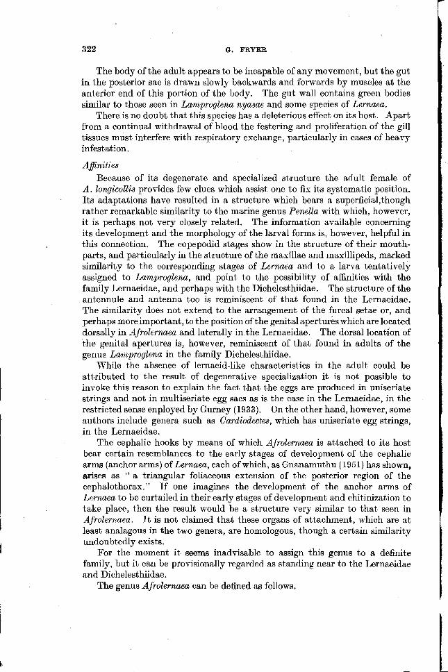
The body of the adult appears to be incapable of any movement, but the gut in the posterior sac is drawn slowly backwards and forwards by muscles at the anterior end of this portion of the body. The gut wall contains green bodies similar to those seen in Lamproglenu nyasae and some species of Lernaea.
There is no doubt that this species has a deleterious effect on its host. Apart from a continual withdrawal of blood the festering and proliferation of the gill tissues must interfere with respiratory exchange, particularly in cases of heavy infestation.
Afinif ies Because of its degenerate and specialized structure the adult female of
A. longicollis provides few clues which assist one to fix its systematic position. Its adaptations have resulted in a structure which bears a superficial,though rather remarkable similarity to the marine genus Penella with which, however, i t is perhaps not very closely related. The information available concerning its development and the morphology of the larval forms is, however, helpful in this connection. The copepodid stages show in the structure of their mouth- parts, and particularly in the structure of the masillae and maxillipeds, marked similarity to the corresponding stages of Lernaea and to a larva tentatively assigned to Lamproglenu, and point to the possibility of affinities with the family Lernaeidae, and perhaps with the Dichelesthiidae. The structure of the antennule and antenna too is reminiscent of that found in the Lernaeidae. The similarity does not extend to the arrangement of the furcal setae or, and perhaps moreimportant, to the position of the genital apertures which are located dorsally in Afrolernaeu and laterally in the Lernaeidae. The dorsal location of the genital apertures is, however, reminiscent of that found in adults of the genus Lamproglena in the family Dichelesthiidae.
While the absence of lernaeid-like characteristics in the adult could be attributed to the result of degenerative specialization it is not possible to invoke this reason to explain the fact that the eggs are produced in uniseriate strings and not in multiseriate egg sacs as is the case in the Lernaeidae, in the restricted sense enployed by Gurney (1933). On the other hand, however, some authors include genera such as Cardiodectes, which has uniseriate egg strings, in the Lernaeidae.
The cephalic hooks by means of which Afrolernaea is attached to its host bear certain resemblances to the early stages of development of the cephalic arms (anchor arms) of Lernaea, each of which, as Gnanamuthu (1951) has shown, arises as " a triangular foliaceous extension of the posterior region of the cephalothorax." If one imagines the development of the anchor arms of Lernaea to be curtailed in their early stages of development and chitinization to take place, then the result would be a structure very similar to that seen in Afrolernaea. It is not claimed that these organs of attachment, which are at least analagous in the two genera, are homologous, though a certain similarity undoubtedly esists.
For the moment it seems inadvisable to assign this genus to a definite family, but i t can be provisionally regarded as standing near to the Lernaeidae and Dichelesthiidae.
The genus Afrolernuea can be defined as follows.

I PARASITIC COPEPODA OF THE FISHES OF LAKE NYASA 323
AFROLERNAEA gen. n. Degenerate parasitic copepods of the gills of freshwater fishes. Adult
female with extremely elongate body not clearly divided into tagmata and without furcal rami. Maxillae present but no trace of antennules, antennae, or thoracic appendages. Attachment organs consisting of two pairs of short chitinised cephalic hooks. Eggs produced in long uniseriate rows. Develop- ment involving several free-living naupliar and copepodid stages and apparently not requiring intermediate host. Copepodids with Lernaea-like mouthparts, well developed furcal rami, and particularly large eye. Settling stage a t least one moult prior to last copepodid stage. Adult male unknown, but presumably similar to last female copepodid.
Distribution Africa (Lake Nyasa).
Type species. Afrolernaea longicollis.
Branchiura Family ARGULIDAE
ARGULUS AFRICANUS Tlliele (Figs. 52-53) This is the best known of all the African argulids and has been recorded from
I
many parts of the continent. It was already known to occur in L. Nyasa which
Figs. 52-53.-Argz~lus nfriraraus Thiele. 52. Part of a batch of eggs. 53. Diagrapmatic sketch illustrating how egg splits to reloase young.
Legencl. E.-Egg membrane. C.-Cement. F.C.-Film of cement. S.-Slit. F.-Flap.
is indeed one of the localities from which Thiele first received specimens. I n the Nkata Bay area it is very common on various parts of the body of Bagrus merjdionalis Giinther and on the lacustrine species of Clarias, but shows a distinct preference for the head region which in these fishes is encased in a massive bony casque covered with smooth skin. Specimens also occurred on
324 Q. FRYER
two of the three eels (Anguilla nebulosa labiata Peters) which have been seen alive. All these fishes are smooth skinned. More than fifty specimens can sometimes be found on a single fish.
By contrast it is very unusual to find specimens on scaly fishes. A single specimen has been found on Mormyrus longirostris Peters and a very few speci- mens have been seen on cichlid fishes. In L. Nyasa at least it has, therefore, a definite preference for smooth-skinned fishes from which nutriment can doubtless be more easily obtained than from those protected by scales.
The length of the largest female seen was about 12 mm. Although so often encountered in Africa little has been recorded concerning
the biology of this species so the following information is therefore of interest. Argulus africanus is a very actrive swimmer but when dislodged from its
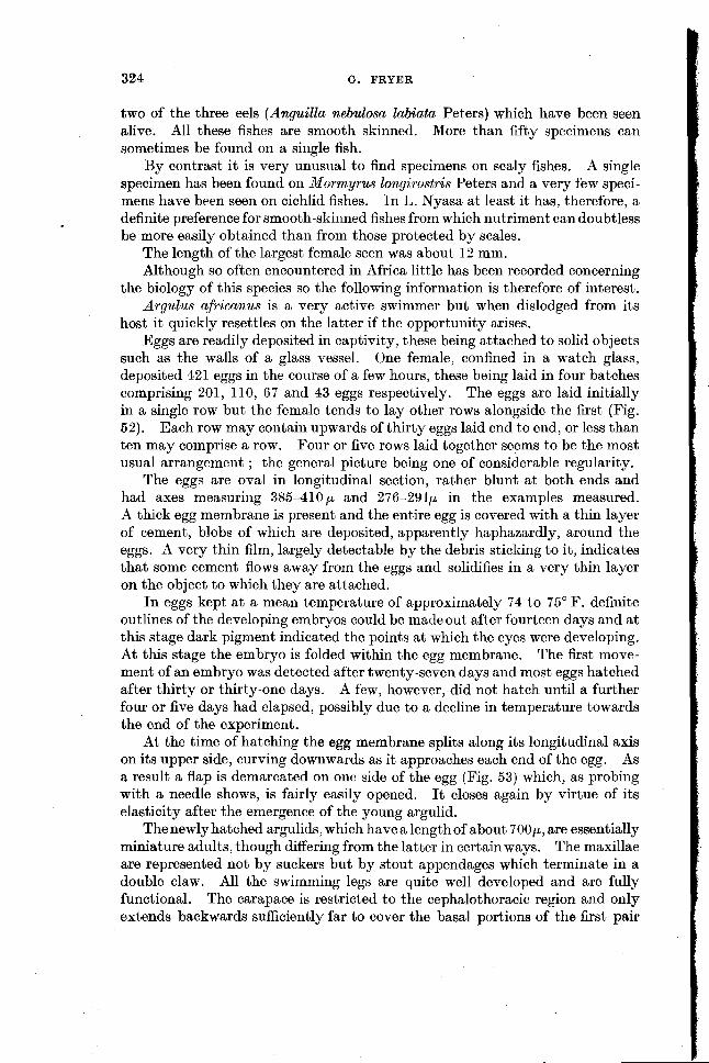
host it quickly resettles on the latter if the opportunity arises. Eggs are readily deposited in captivity, these being attached to solid objects
such as the walls of a glass vessel. One female, confined in a watch glass, deposited 421 eggs in the course of a few hours, these being laid in four batches comprising 201, 110, 67 and 43 eggs respectively. The eggs are laid initially in a single row but the female tends to lay other rows alongside the first (Fig. 52). Each row may contain upwards of thirty eggs laid end to end, or less than ten may comprise a row. Four or five rows laid together seems to be the most usual arrangement ; the general picture being one of considerable regularity.
The eggs are oval in longitudinal section, rather blunt at both ends and had axes measuring 385-410p and 276-291p in the examples measured. A thick egg membrane is present and the entire egg is covered with a thin layer of cement, blobs of which are deposited, apparently haphazardly, around the eggs. A very thin film, largely detectable by the debris sticking to it, indicates that some cement flows away from the eggs and solidifies in a very thin layer on the object to which they are attached.
In eggs kept at a mean temperature of approximately 74 to 75" F. definite outlines of the developing embryos could be made out after fourteen days and at this stage dark pigment indicated the points at which the eyes were developing. At this stage the embryo is folded within the egg membrane. The first move- ment of an embryo was detected after twenty-seven days and most eggs hatched after thirty or thirty-one days. A few, however, did not hatch until a further four or five days had elapsed, possibly due to a decline in temperature towards the end of the experiment.
At the time of hatching the egg membrane splits along its longitudinal axis on its upper side, curving downwards as it approaches each end of the egg. As a result a flap is demarcated on one side of the egg (Fig. 53) which, as probing with a needle shows, is fairly easily opened. It closes again by virtue of its elasticity after the emergence of the young argulid.
Thenewly hatched argulids, which have a length of about 7OOp, are essentially miniature adults, though differing from the latter in certainways. The maxillae are represented not by suckers but by stout appendages which terminate in a double claw. All the swimming legs are quite well developed and are fully functional. The carapace is restricted to the cephalothoracic region and only extends backwards sufficiently far to cover the basal portions of the first pair

PARASITIC COPEPODA OF THE FISHES OF IAKE NYASA 325
of thoracic swimming legs. The abdomen is very short and present only in
i rudimentary form. A " poison spine " is already present.
The newly hatched animals exhibit no special naupliar natatory organs such as are to be seen in A. folzaceus L., A. americanus Wilson, and certain other
I ~pecies. (See Wilson (1904) and Calman (1909) for figures of such larvae). Wilson (1904) suggests that those species whose embryonic development is prolonged hatch as juvenile adults and that those whose embryonic develop- ment is brief hatch as larvae. This seems to be a reasonable thing to expect 1 but it is doubtful if this correlation can be maintained. Thus, according to Wilson (1904) eggs of A. americanus kept a t about 72°F. hatched after seventeen
I or eighteen days, while eggs of A.*foliaceus took twice as long to hatch, or about the same length of time as eggs of A. africanus kept at similar temperatures and which give rise to a quite different form on hatching. Temperature is obviously
Figs. 64-65.-Arplus jo l lymni sp. n. 54. Adult (Holotype), (dorsal). 55. Adult 2 (dorsal).
of vital importance in this respect, for Meehan (1940) cites cases of the eggs of A. foliaceus remaining as such throughout an entire temperate zone winter and hatching in the following spring, and also cites Tokioka (1936) who hatched eggs of A. japonicus Thiele in twelve days at 30°C. while a t 15°C. the time taken was sixty days.
ARGULUS JOLLYMANI sp. n. (Figs. 54-60) Adult female
Length to 10.5 mm. Carapace broadly elliptical; about 1.1 to 1.2 times as long as wide. Cephalic portion distinctly separated from thoracic portion and produced into lateral lobes which are tucked under anterior part of latter.
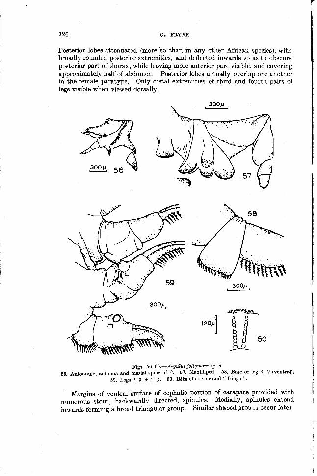
326 a. FRYER
Posterior lobes attenuated (more so than in any other African species), with broadly rounded posterior extremities, and deflected inwards so as to obscure posterior part of thorax, while leaving more anterior part visible, and covering approximately half of abdomen. Posterior lobes actually overlap one another in the female paratype. Only distal extremities of third and fourth pairs of legs visible when viewed dorsally.
Figs. 56-60.-Argulwe jollymani sp. n. 56. Antennule, antenna and mesial spine of 0. 57. MaxiUiped. 58. Bese of leg 4, 0 (ventral).
59. Legs 2, 3, & 4, $. 60. Ribs of sucker and " fringe ".
Margins of ventral surface of cephalic portion of carapace provided with numerous stout, backwardly directed, spinules. Medially, spinules extend inwards forming a broad triangular group. Similar shaped groups occur later-
I I

PARASITIC COPEPODA OF THE FISHES OF LAKE NYASA 327
ally on each side posterior to base of antennules and antennae. Margins of thoracic portion of carapace with similar spinules forming broad band anteriorly, rapidly narrowing posteriorly, and terminating at about level of first swimming leg. Isolated spinules occur as far back as level of second swimming leg.
Respiratory surface consisting of small anterior ovoid area and much larger elongate posterior area.
Abdomen very slightly wider than long and comprising about 21 per cent of total length ; its lobes kidney-shaped, tapering posteriorly, and broadly rounded at posterior extremities. Anal incision narrow and shallow. Purcal rami minute ; situated at base of anal incision.
Eyes fairly small, separated by a distance of approximately one quarter of maximum width of carapace. Median ocellus small, situated in relation to eyes so as to form with latter the points of an equilateral triangle. Basal seg- ment of antennule bearing at its posterior corner a stout, fairly sharp and heavily chitinised spine directed obliquely inwards and backwards. Second segment with distinct recurved and hook-like anterior and lateral spines, and a small but well chitinized posterior spine. Flagellum extending a little beyond distal extremity of lateral spine. Antenna closely associated with antennule. Basal
1 segment with moderately stout, heavily chitinized, backwardly-directed spine. Tip of antenna extending beyond tip of antennule.
Mesial spine at base of antenna stout, heavily chitinized, and fairly sharply pointed. " Poison spine " in position typical for genus.
Suckers well developed, with diameter approximately one quarter of maxi- mum width of carapace and situated close together. Margins of suckers " frilly ". Supporting rods of sucker membrane consisting of 10 to 11 small, simple contiguous pieces.
Maxillipeds typical in general form. Basal segment bearing a blunt, broad, flattened, and heavily chitinized lamina on posterior ventral margin. Second segment bearing three similar laminae on posterior ventral margin. A similar chitinous lamina arises posterior to second segment of maxilliped.
Swimming legs typical in general form. Legs 1 & 2 with flagellum dorsally. Coxopodite of leg 4 wedge-shaped and not produced laterally.
Adult male Length to 8.6 mm. Carapace broadly elliptical, 1.2 to 1.3 times as long as
wide. Posterior lobes reaching about half way along abdomen and overlapping slightly. (In one of the two male paratypes the right lobe overlaps the left ; in the other the left lobe overlaps the right.)
Abdomen about 23 to 26 per cent of total length and about 1.1 times as long as wide.
Cephalic appendages as in female. Leg 1, as in female. Legs 2, 3, & 4 modified by presence of accessory sexual
organs and taking form shown in Pig. 59. I Precoxa of leg 2 with chitinized cylindrical, backwardly directed process
and, external to it, a flattened chitinous peg. Colour of two specimens preserved in methylated spirits chestnut brown.
. One living specimen was distinctly green and two were virtually colourless but became greenish when preserved in formalin.

328 Q. FRYER
Two specimens, a male and a female, of this species taken from an unknown host in the southern part of L. Nyasa were given to me by Mr W. H. Jollyman after whom I take pleasure in naming the species. Two more specimens, one of each sex, were found a t Nkata Bay on openwater representatives of the genus Haplochromis and a fifth specimen was found at Ruarwe on a specimen of H. fenestratus Trewavas collected on a rocky shore. It thus seems to have both different host and different habitat preferences to A. africanus.
This species is very similar to A . arnbloplites Wilson, (Wilson, 1920) recorded from the Dungu River, one of the north-eastern tributaries of the Congo. The females in particular of the two species are very similar but can be separated by the fact that A . jollyrnani has large suckers, which are spaced fairly close together and whose membranes are supported by rods composed of 10 or 11 very short segments, while A . ambloplites has small, widely separated suckers supported by rods composed of 4 relatively long segments. The arma- ture of the maxilliped is also a little different in the two species, the laminae of the second segment being more widely separated in A . jollymani than in A . ambloplites. These differences seem to exceed those which might be ex- pected as a result of age differences or geographical variation.
The male of Argulus jollymani differs so much from that described as the male of A . amnb~oplites, which differs markedly in certain ways from its female, that one is tempted to suggest that Wilson's male and female are not con- specific. The structure of the carapace of the male and female described by Wilson are considerably different ; much more different for instance than in the male and female of the species here described ; and there are also several other surprising differences between the sexes. I t must be admitted, however,that the antennules, antennae and maxillipeds of the male and female must have been very similar for Wilson to associate the two, though he only figures these appendages for one sex.
As A . ambloplites is not included in the review by Aleehan (1940) of the species of Argulus in the collection of the United States National Museum while A . recticulatus Wilson, an African species described by Wilson at the same time as he described A . ambloplites, is mentioned, it would appear that the whereabouts of Wilson's material of the latter species is not known, and until such times as it or further specimens of A . arnbloplites come to light the association of his male and female must be regarded as somewhat suspect.
The male and female of the species here described are obviously conspecific and as the female differs in certain ways from that of A . a?t~bloplites the description of the material as new is justifiable.
DOLOPS RANARUM (Stuhlmann) This species, the only known African representative of the genus Dolops,
is widely distributed in Africa and was recorded from L. Nyasa by Cunnington (1913) who had specimens given to him by Capt. Rhoades. Several specimens have been seen during the recent survey, all having been taken from members of the genus Clarias.

PARASITIC COPEPODA OF THE FISHES OF LAKE NYASA 329
The ecological distribution of the hosts is of considerable interest. With one exception they have been collected in swampy situations. D. ranarum has been found on Clarias mossambicus Peters, in the R. Banga (a tributary of the Luweya) some fifteen miles from its point of entry into L. Nyasa and on C. mossambicus in a small swampy lagoon at the mouth of a stream where it enters L. Nj~asa a t Nkata Bay. Mr D: Harding has also collected several specimens from C. mossambicus in Bana Lagoon, a shallow, swampy body of water lying between Nkata Bay and Kota Kota, and in communication with the lake. A free swimming juvenile has also been collected in the R. Banga No specimens of Argulus africanus were seen on these or any other specimens of Clarias obtained from rivers. The exception mentioned above is a single speci- men obtained from the so far unique specimen of an as yet undescribed species of Clarias collected in 50 to 60 metres of water just north of Nkata Bay.
These data, scanty as they are, can possibly be interpreted as indicating that the conditions demanded by the free-living stages of D. ranarum and A. afri- canus, or the situations in which the adult female lays its eggs, are quite different. This suggestion might be borne in mind by those encountering these species elsewhere in Africa. It might also give a clue as to the real habitat of the new Clarias mentioned above. It is only fair to add, however, that, in spite of the clear cut differences found in the Nyasan region, Cunnington (1913) records finding the two species associated on a single host in both Lakes Tanganyika and Victoria, and the widespread distribution of both species indicates that they can tolerate a fairly wide range of environmental conditions.
The blood of all the specimens of D. ranarum seen alive had a red tinge. This may indicate the presence of haemoglobin, a respiratory pigment, the
;a( possession of which would doubtless be of value to this species which occurs in conditions where low oxygen tensions are probably frequent. 'i
L
CHONOPELTIS INERMIS Thiele (Pigs. 61-79)
The holotype of this species was recorded from L. Nya,sa by Thiele as long ago as 1900 and in 1904 he gave a good description with accurate figures of the single small female available to him. This specimen had apparently shrunk, 1 in preservation as is particularly apparent from the appearance of the suckers in Thiele's Pig. 11 1, but he nevertheless gave a good idea of its general form and accurate illustrations of some of its appendages.
Although never recorded from L. Nyasa since the time of Thiele's discovery, C. inermis is not uncommon there and many specimens have been found in the mouths of fishes of the genus Clarias. Several, and probably all, the Nyasan species of Clarias serve as hosts but it has not been encountered on any other genus. Curiously, Thiele states that his specimen was found on a specimen of " Chromis " sp. a member of the family Cichlidae. Specimens have also been found on Clarias mossambicus Peters in the It. Banga (Nkata Bay district), an affluent of L. Nyasa, some fifteen miles upstream. In the lake quite heavy infestations are sometimes encountered, and on one occasion no fewer than forty-three specimens were found in the mouth of a single host.
Thiele's descriptions and figures of the female are good, but can be amplified and corrected from the material now available. Females attain a total length

33 0 G . FRYER
of a t least 12 mm. According to Thiele the posterior lobes of the carapace extend sufficiently far backwards to cover the basipodite of leg 2, and from his figures obviously did so in his specimen. In my material, even in specimens smaller than Thiele's, it overlaps the basipodite of the first pair of legs only. The condition of Thiele's specimen may be due to shrinkage after preservation. Figures 61 & 62 give a better impression of the appearance of C. inermis in life than do Thiele's illustrations.
The ends of the chitinous supporting strips of the anterior part of the cephalothorax are not usually arranged " in einer nach hinten konkaven Bogenlinie " as they are in the holotype.
There is definitely no flagellum on the first thoracic appendage, as Thiele (1904) correctly states after indicating in his preliminary diagnosis (1900) that such a structure is present.
Other structural features of the female are apparent from the illustrations and are described by Thiele.
The male of C. inermis was previously unknown for, although Brian (1940) figures the male of a Chonopeltis from the Congo which he calls C. inermis var. schoutedeni, it is shown below that this is a distinct species. Many males have now been found. In general form they resemble the females but have propor- tionately longer abdominal lobes (comprising about 39 per cent of the total length as opposed to about 31 or 32 per cent in the female) and have thoracic legs 2, 3, & 4, modified to facilitate the mating process. The maximum size of any of the males seen was 8.5 mm.
The structural modifications of legs 2, 3, & 4, are most readily understood by reference to Figs 63-67 &Fig. 72. These modifications parallel those found in certain species of Argulus and can be assumed to function in a similar manner though copulation has not been observed.
The rediscovery of C. inermis in its type locality has served to make clear the systematic status of the Chonopeltis which occurs in the Congo system. Monod (1928) recorded the finding of two female specimens of a Chonopeltis in material from the Congo which he named as C. inermis, and his figures indicate that his specimens should be referred to this species. All subsequent finds from the Congo system, however, have been of a form which, while similar to C. inermis, would appear to be distinct. This form was described by Brian (1940) as C. inermis var. schoutedeni, and later Dartevelle (1951), who had collected the specimens described by Brian, announced its presence in tributaries of the Congo and suggested that it was specifically distinct from C. i n emi s . Examination of material of C. inermis from L. Nyasa and discovery of the male leaves no doubt that, as Dartevelle suggested, i t is specifically distinct from Brian's form from the Congo which should henceforth be known as C. schoutedeni Brian.
Both Brian and Dal-tevelle point to an elongation of the posterior portion of the body of C. schoutedeni as an important difference between it and C. inermis but this suggestion was made as a result of comparison with Thiele's figures which were made from a badly preserved specimen. The thoracic appendages of the two species, however, differ considerably in both sexes. Both Brian and Dartevelle remark on the reduced size of the fourth pair of legs of C. schoutedeni

Lce his :ns 1 ~ . In. i fe
10) ar. ,ve or- tal cic ize
3 8 1
m. Ctis rea :nt lile ,ed ' I), 1 C 8
1ct tsa ; is rth
of nis res sea ,nd eni
PARASITIC COPEPODA OF TFIE FISHES OF LAKE NYASA 33 1
Figs. 61-66.-Choraopeltis inermis Thiele.
61. Adult 9 (dorsal). 62. Adult 9 (ventral). 63. Left leg 3 $. (dorsal). 64. Left leg 3 $. (ventral). 65. Right leg 4 $. (dorsal). 66. Right leg 4 6 (ventral).

as compared with those of C. inerrnis and examination of abundant material of C. inermis shows this to be a ready means of separating the two species. Acomparison of the structure of the three posterior pairs of legs in the male of the two species which is now possible as a result of the finding of males of C. inermis shows that these appendages are vastly different in the two species as can best be seen by comparing the figures of C. schoutedeni given by Brian and reproduced by Dartevelle with those given here of C. inermis.
Figs. 67-72.-Chonopeltis i ~ ~ r n i s Theile.
67. Legs 2, 3, & 4 and abdomen $ (dorsal). 68. Antenna. 69. Part of sucking face of sucker margin showing chitinous supports. 70. Chitinous supports. 71 . Furcal ramus (dorsal). 72. Basal portion of right leg 2 ,3 , showing projection.
' A rather remarkable feature of C schoutedeni appears to be a striking varia- tion of structure with age which effects particularly the relative length of the abdominal lobes. Such striking variations have not been observed in C. inermis.
A representative of the genus Chonopeltis has recently been found in the Okawango River in the south west of Africa by Barnard (1955) who calls it C. inermis. However, the three specimens found were all much too small to identify with certainty and until more material becomes available the identity of this species must remain in doubt. For the moment therefore, all that can be said ofthe distributionof the genus in Africa is that it is represented in L.Nyasa by C. inerrnis and in the Congo system by C. schoutedeni, while an as yet unidentified species occurs in the south west of the continent.
As many specimens of C. inermis have been observed alive it is possible to give some account of its behaviour and general biology.
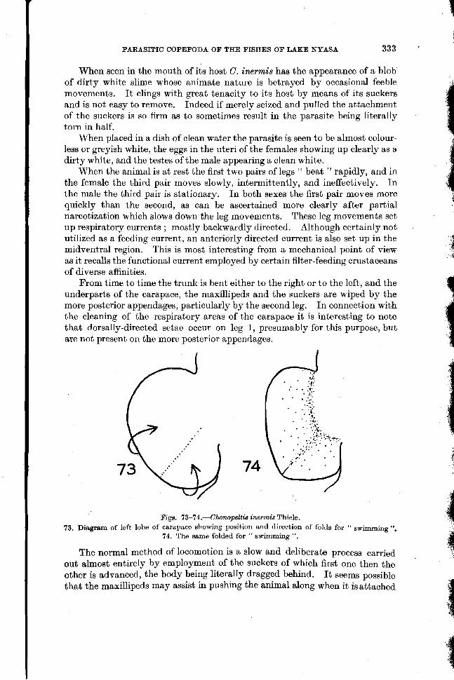
PARASITIC COPEPODA OF THE FISHES OF LAKE NYASA 333 .
When seen in the mouth of its host C. inermis has the appearance of a blob of dirty white slime whose animate nature is betrayed by occasional feeble movements. I t clings with great tenacity to its host by means of its suckers and is not easy to remove. Indeed if merely seized and pulled the attachment of the suckers is so firm as to sometimes result in the parasite being literally torn in half.
When placed in a dish of clean water the parasite is seen to be almost colour- less or greyish white, the eggs in the uteri of the females showing up clearly as a dirty white, and the testes of the male appearing a clean white.
When the animal is at rest the first two pairs of legs " beat " rapidly, and in the female the third pair moves slowly, intermittently, and ineffectively. In the male the third pair is stationary. In both sexes the first pair moves more
i quickly than the second, as can be ascertained more clearly after partial narcotization which slows down the leg movements. These leg nlovements set up respiratory currents ; mostly backwardly directed. Although certainly not utilized as a feeding current, an anteriorly directed current is also set up in the midventral region. This is most interesting from a mechanical point of view as it recalls the functional current employed by certain filter-feeding crustaceans of diverse affinities.
From time to time the trunk is bent either to the right or to the left, and the underparts of the carapace, the maxillipeds and the suckers are wiped by the more posterior appendages, particularly by the second leg. In connection with the cleaning of the respiratory areas of the carapace it is interesting to note that dorsally-directed setae occur on leg 1, presumably for this purpose, but are not present on the more posterior appendages.
Figs. 73-74.-Chonopeltis ine~mis Thiele. 73. Diagram of left lobe of carapace showing position and direction of folds for " swimming
74. The same folded for " swimming ". The normal method of locomotion is a $ 1 0 ~ and deliberate process carried
out almost entirely by employment of the suckers of which first one then the other is advanced, the body being literally dragged behind. I t seems possible that the maxillipeds may assist in pushing the animal along when it is attached

334 (f. FRYER
to its host, but on a smooth surface these are not used. When necessary it moves backwards by reversing the movements of its suckers. The ventral surface of the body and of the thoracic appendages is roughened by the presence of numerous minute papillae which probably help in the adhesion of the parasite to its host. C. inermis can " walk '' on the underside of the surface film by means of its suckers, though it is improbable that this faculty is made use of in nature.
Abortive swimming movements sometimes take place. The suckers are released from their hold, the posterior lateral corners of the carapace are raised (Figs. 73 & 74) and the maxillipeds are used as oars, being assisted by the first and second pairs of legs and, in the female, by feeble and ineffective move- ments of the third pair. In spite of the effort involved the forward movement is exceedingly slow and the animal, which is not lifted clear of the bottom, cannot be said to actually swim. Such movements are, in all probability, functional relics of the ancestral mode of locomotion employed by the Argulus-like ancestor from which this form was probably derived. The greatly reduced setation of the thoracic appendages (as compared with those of Argulus) is obviously correlated with the loss of the ability to swim.
When removed from the host the parasites always died within two or three days.
A number of eggs have been deposited in captivity. These were attached to the bottoms of the vessels in which the parasites were contained. Most were laid in scattered, rather irregularly arranged groups whose general nature is shown in Fig. 75. A few single eggs were also laid. The largest number depo- sited by a single female was 136, these being arranged in groups of 2, 3, 4(2), 5, 6, 7, 10, 14, 25 & 56. In the case of the group of 56 there was a slight tendency for the eggs to be arranged in rows.
Newly deposited eggs are dirty white in colour. Each is oval in longitudinal section, and the examples measured had axes measuring some 240-270 p and 172-179 p. Each is surrounded by a distinct egg membrane outside which is deposited a very thick layer of gelatinous material which is presumably quite soft a t the time of laying but which quickly hardens into an almost colourless thick walled capsule which can be subjected to considerable pressure by a needle without rupturing. The outer surface of this capsule is somewhat rough and irregular, but no definite sculpturing is present. I t appears that a t the time of laying the egg capsule is covered with a sticky fluid layer some of which runs off and forms a very thin adhesive layer over the substratum. Its stickiness is indicated by the presence of particles of debris stuck to this film and to the egg capsules. In a t least the case of eggs deposited singly, which could easily be examined from the side, the long axis is inclined at about 45" to the horizontal.
The following observations on the development within the eggs were made at a mean average temperature of 74 to 75" F. After fourteen days the contents of the egg were distinctly aggregated into several masses of cells and a slight recession from the egg membrane could be seen. After sixteen days this reces sion was very well marked but no definite form could be made out in the embryo as seen through the egg investment. Eye spots were detectable after seventeen days, and the darkly pigmented rudiments of both the two lateral compound

PARASITIC COPEPODA OF THE FISHES OF LAKE NYASA 335
Figs. 76-79.-4honopelti8 iraermis Thiele.
75. Cluster of eggs. 76. Single egg seen from side. 77. Newly hatched larva (ventral). 78. Tip of larval maxilla showing details of chelae. 79. Larval mandible.

eyes and of the median ocellus, all of which were about equal in size at this stage, could be clearly sten after twenty days. Movem.ents of the embryo were first observed after twenty-one days and the first larva to emerge did so twenty-two days after deposition of the egg. Other eggs hatched after twenty- three to twenty-five days development.
Actual emergence of the larva from the egg was not witnessed but it takes place via a longitudinal and almost straight slit running along almost the entire length of the upper surface of the egg. Because of the elasticity of the thick outer layer the slit completely closes after the emergence of the larva. One specimen was seen partly out of the egg capsule but trapped by the elasticity of the latter's walls. This specimen was dead, and, considering the strength of the walls of the egg capsule and the feebleness of the larva, it is surprising that such fatalities were not more numerous.
The newly hatched larva of C. inermis is a truly remarkable creature and differs considerably from any hitherto described juvenile form of a branchiuran. I ts structure, which is shown in Figs. 77-79, can be briefly described as follows.
Length about 410 p. Cephalothorax only indistinctly separated from thorax. Thorax elongate,
narrowing posteriorly. Abdomen very small and not distinctly separated from thorax. Cephalothorax covered dorsally with a flat, almost circular carapace, somewhat concavely flattened anteriorly and deeply indented posteriorly. Carapace covering basal portions of the longer cephalothoracic appendages and only partially covering rudiments of first pair of thoracic legs. Ventral surface of carapace with two respiratory areas on each side. Posterior pair much larger than anterior pair, oval, slightly kidney-shaped, and extending from near posterior margin of carapace to posterior margin of base of maxillae. Anterior pair minute, oval in form, and located close to anterior margin of posterior pair. Circular rudiments of compound eyes present, widely separated and located outside base of antennae. Median ocellus posterior to eyes and anterior to mandibles.
Abdomen rudimentary, very short, broader than long, and bearing a pair of rounded, knob-like furcal rami each bearing 4 short setae at postero-lateral corner.
Antennules represented by a pair of minute chitinous spinules between antennae. Antennae elongate, projecting far beyond carapace margin and composed of 3 distinct segments of which basal shows two more incipient divisions. Distal segment bearing a t its tip a stout, weakly curved, chitinous hook.
Distinct " oral mound " present. Mandibles well developed with mastica- tory portion consisting of stout, recurved, chitinous shaft strongly serrated on inner margin. Maxillae enormously developed into rowing organs. Each with turgid unsegmented basal portion with concertina-like walls, and each termina- ting in an elongate chitinized chelate structure. Tips of each arm of chela serrated, anterior arm finely, posterior coarsely. Posterior arm slides in groove of' anterior. Each maxilla with a recurved spine on posterior margin near base. Maxilliped large, cylindrical, and clearly divided into 4 segments of which basal is longest and stoutest. Tip of distal segment bears 2 chitinous hooks and a shorter lobe. Legs 1 to 4 all rudimentary and all unsegmented, but all showing

PARASITIC COPEPODA OF THE FISHES OF LAKE NYASA 337
very rudimentary beginnings of exopodite formation distally. No setae present but minute knobs present distally, 4 on leg 1, 2 on legs 2 & 3, and 1 on leg 4.
Alimentary canal present as a simple tube. No heart and no definite circulation of blood. Practically colourless. The larva is remarkable not only because of its structure, which is discussed
below, but on account of its helplessness. I t is, at least in the early stages observed, quite incapable of swimming or even of crawling effectively. A casual glance would lead one to suppose that locomotion is effected by slow rowing movements of the enormous maxillae but careful observations reveal that there is a definite, albeit ineffective, method of crawling, The appendages concerned are the antennae and maxillipeds. The appendages are stretched forwards, the terminal hook or hooks as the case may be make contact with the substratum and the body is pulled forwards by the appendage concerned. The members of a pair of appendages operate alternatively, but the co-ordination of antennae and maxillipeds is not particularly good. The very noticeable rowing move- ments of the maxillae actually have little influence on the movements of the larva. Progress is very slow and aimless. Sometimes larvae " capsize " and only right themselves with difficulty.
A tendency for the larvae to form small aggregations, was noted. No phototactic response seemed to be involved.
Larvae lived for as long as eight days during which period they showed no inclination to change their locoinotory mechanism. Death may have been due to an inability to supply the correct food, or due to rather sudden nocturnal drops in temperature (from the lower 80's to the lower 60's) during the period when the larvae were-being studied. No moulted skins were observed during this period but some individuals may have moulted as they exhibited certain minute differences to the larva described above, viz. a slightly longer and more distinctly bifurcated antennule, a slight development of the swimming legs, and slightly longer and stronger furcal setae.
At first glance the larva with its three enlarged anterior appendages and its rudimentary posterior appendages has a superficial resemblance to a somewhat modified post-nauplius. This apparent similarity does not bear close inspection, however, as the enlarged appendages are certainly not homologous with naupliar appendages, though they are analogous in function, being essentially locomotory.
The larva is, as i t were, intermediate in structure between the two types of newly hatched individuals found in the genus Argulus and which can be designated as the " juvenile adult " type and the naupliar type. It resembles the former in having no natatory palp on the mandible and no natatory setae on the antennae, but differs from it most markedly in having only very rudi- mentary thoracic appendages. In the latter respect it resembles the naupliar type, from which, however, it differs greatly in the absence of anterior natatory appendages. In other words it possesses neither the swimming organs of the naupliar type nor of the juvenile adult type. It also differs from both in the almost complete absence of antennules, and in the form of the maxillae. The maxillae are more enlarged than is the case in either of the two types of young

338 a. FRYER
Argulus and, although terminating in paired, serrated spines (chelae) as is the case in all described young forms of Argulus, the structure of the spines is different.
Summarizing, therefore, it can be said that, whilst showing certain obviously specialized features, the larva of Chonopeltis is more degenerate and certainly more helpless, than any described juvenile form of Argulus.
The structure of the maxillae is highly specialized. In life the two distal spines slide slowly one within the other but the functional significance of this remains obscure. Possibly these structures assist in the levering of the larva from the egg, and they may also serve as temporary attachment organs before the development of suckers.
No older stages of the larva have been encountered either living free or in the mouths of fishes, but it so happens that the three specimens of a Chonopeltis recorded by Barnard (1955) from the Okawango River were immature, and that these show the transformation of the chelate maxillae into sucking discs. A sketch of this process is given by Barnard. In this respect, therefore, Chonopeltis resembles all the species of Argulus whose development is known. Barnard's specimens were attached to different parts of the body, but not within the mouth, of a specimen of Synodontis melanostictus.
Having now a knowledge of the larval form of Chonopeltis we are able to study the relationship between this genus and Dipteropeltis and the relationships of both of them to Argulus. Calman (1912) homologized the unarmed preoral papilla of Dipteropeltis with the preoral " poison spine " or " sting " of Argulus, a suggestion which is probably correct. No trace of this structure exists in the adult or, perhaps more significantly, in the larva, of Chonopeltis. This evidence taken alone would tend to suggest that Chmpel t i s diverged from the main branchiuran stock before the evolution of a " poison spine " had occurred rather than that it lost this structure during the course of evolution. Other evidence, however, indicates that this interpretation may be incorrect, that Chonopeltis and Dipteropeltis are fairly closely related, and that both are rather degenerate argulids in which a reduction of structures has taken place and has been continued further in Chonopeltis than in Dipteropeltis. In this case the loss of a " poison spine " would accord well with other evidence and indicate that Chonopeltis is a degenerate and specialized rather than a necessarily primitive form. Both it and Dipteropeltis can probably be regarded as degenerate and specialized descendants of an Argulus-like ancestor from which they have been derived by an elongation of both thorax and abdomen ; reduction of the com- pound eyes ; reduction of the antennules, which have disappeared completely in the adult of Chonopeltis but of which a trace remains in the larva ; reduction of the pre-oral " poison spine " which is represented by an unarmed pre-oral papilla in Dipteropeltis and which has disappeared completely in Chonopeltis ; and by a reduction of the thoracic appendages, which again has been carried slightly further in Chonopeltis than in Dipteropeltis. Such observations as have been made on the abortive attempts a t swimming made by Chonopeltis support this postulated ancestry. Chonopelbis and Dipteropeltis have diverged in the rather superficial, though striking, characteristic of the form of the carapace, and Dipteropeltis has lost the supporting rods of the suckers.

PARASITIC COPEPODA OF THE FISHES OF LAKE NYASA 339
At the moment the means whereby Chonopeltis is transferred from host to host is quite unknown. The adult certainly gives no indication of ever leaving its host and it is possible that the eggs are attached to the latter. If so it seems that the emerging larvae will be dislodged and scattered. Indeed if they are not it is hardly possible to visualize any means by which the animals are dispersed. It remains to be seen if a more mobile stage exists between the early larvae and the adult, but it is difficult to conceive which appendages could be used for active propulsion. The fact that as many as forty-three individuals of C. inerrnis have been found in the mouth of a single fish speaks for the efficacy of the distributive process whatever it may be.
The effect of this parasite on its host is probably small. No blood has ever been seen in the gut and the food probably consists of the slime produced by the skin of its host.
GEOGRAPHICAL DISTRIBUTION
A considerable proportion of the parasitic crustaceans recorded from L. Nyasa have not yet been found in any other body of water and, although the detailed exploration of the fresh-waters of Africa can only be claimed to be in its infancy, it seems probable that some of them, particularly the species of Lernaea, which constitute a species flock, will indeed prove to be endemic to the lake. Consideration of the known distribution of some of the non-endemic species, together with their ecology, shows that these parasites can contribute something towards the explanation of the present day distribution and past movements of certain elements of the African freshwater fauna.
The branchiuran Argulus africanus can be considered first. This species is widely distributed in East and Central Africa and extends northwards into the R. Nile. I t so happens that the hosts preferred by this species-presumably on account of their smooth skins-include just those fishes which are most likely to distribute it. Thus the species of Clarias, of which L. Nyasa and adjoining waters possess several non-endemic species, are fishes capable of tolerating low oxygen concentrations, can live for relatively long periods with little water, and have already been cited (Worthington, 1933) as forms likely to move rather easily across certain African watersheds in times of temporary flooding. The migration of eels too is a well-known phenomenon and it is easy to see how argulids could be transported for long distances, even beyond barriers such as ,
waterfalls and rapids, by these fishes to which such barriers present few difficulties.
It is illuminating to note also that in other lakes A. africanus has often been found on species of Clarias and Bagrus, and in the R. Nile has been found on Heterobranchus bidorsalis Geoffr., a smooth skinned fish related to Clarias and which is similarly equipped for crossing swampy watersheds, as is to be seen from its occurrence in rivers of both the Nile and Niger systems. Another species of Heterobranchus, H. ZongiJilis Cuv. et Val., closely related to H. bidor- salis and a potential host for A. africanus, occurs in the Nile, Niger, Congo and Zambesi systems. I t seems obvious, therefore, that the host preferences of A . africanus, have been responsible for its wide distribution in Africa.

340 a. FRYER
A similar explanation of the equally wide distribution of Dolops ranarum, which also exhibits a preference for smooth skinned hosts such as Clarias and Heterobranchus, can be reasonably put forward.
The distribution of the copepod Lernaea barnimiana presents a somewhat different but equally interesting picture. This species has now been recorded from the R. Nile (Hartmann, 1865, 1870; Cunnington, 1914*), Lakes Edward (Capart, 1944 ; Harding, 1950), Mweru (Capart, 1944), andNyasa (present records). Brian (1940 a ) recorded L. temnocepl~ala from L. Ararobi some 100 km. south of Addis Ababa and Capart believes that this record refers to L. barnimiana. Leaving the latter record for a moment and considering the others it is seen that the distribution of the parasite, which can only have been distributed across some of the barriers between the various lakes and rivers in which it occurs by movement of its hosts, serves as a clear indication of past connections between certain water bodies. Worthington (1933) in seeking to explain the affinities of the Nyasan-Zambesi fish fauna with that of the Bangweulu-Luapula (Congo system) fauna, pointed out the possibility of inter-connections by swampy watersheds between the Kafue (Zambesi system) and Luapula (Congo system) Rivers, and also between the rivers entering the north west end of L. Nyasa and the Chambesi River (Congo system). He was also able to quote an actual instance, observed by Capt. Pitman, of a connection, in times of heavy rain, between the two systems, namely between the Loangwa and Luapula Rivers. This would explain how fishes bearing L . barnimiana could have moved between Lakes Mweru and Nyasa. From L. Mweru there exists a t the present time a direct route to L. Tanganyika via the Luvua and Lukuga Rivers.? From L. Tanganyika the tributaries of the Malagarasi River system run to within a few miles of L. Victoria, this narrow discontinuity being the only real barrier today to the continuous distribution of the parasite from L. Nyasa to the Nile. L. Victoria is linked by a curious series of river systems with Lakes Edward and Albert, which are themselves connected by a river, and which overflow into the R. Nile.
The distribution of L. barnimiana thus shows clearly that in the past there have been no barriers between the R. Nile and L. Nyasa unsurmountable at certain times by at least some fishes.
If, as seems probable, Brian's record of L. temnocephala from L. Ararobi refers to this species, then its presence there is explicable as a result of coloniza- tion via a previous connection with the headwaters of a tributary of the Blue Nile which today extends to within a few miles of the lake.
Although adults remain to be discovered in L. Tanganyika it appears that Ergasilus macrodactylus occurs both in this lake and in L. Nyasa, thus affording another instance of past movements between the two of fishes which serve as hosts.
* as L. temnocephala.
t Although the Lukuga connection is relatively recent by Tanganyikan standards, it is almost certain that the vdley now occupied by the river was a continuation of the westwardly draining Melagarasi Rivor before the subsidence of the Tanganyikan trough took place.

PARASITJC COPEPODA OF THE FISHES OF LAKE NYASA 341
THE SPECIES FLOCK OF LERNAEA IN L. NYASA.
With the exception of L. barnimiam all the species of Lermea recorded from L. Nyasa are, so far as is known, endemic, and there is little doubt that they constitute a species flock.
It is obvious that the stage was set for the evolution of a group of fish para- sites in L. Nyasa, just as i t was set in L. Tanganyika, by an isolated body of water containing a large number of fishes which were themselves undergoing speciation. It is interesting to note that while in L. Tanganyika these possi- bilities have been exploited by branchiurans of the genus Argulus, in L. Nyasa they have been exploited by copepods of the genus Lermea.
While it is both difficult and dangerous to speculate on the affinities of the Nyasan species of Lermea it is possible to point with reasonable certainty to certain relationships. First, however, the problem of the ancestral species must be considered. L. barnimiana as it exists today in the lake is possibly a fairly recent immigrant, but its ancestors may well have invaded L. Nyasa in the early years of its existence. From such a form it seems possible that the Nyasan species have been derived. The great variability of the present-day L. barnimiana and the fact that it is not restricted to one host genus, or even to a single family, gives a clue as to how this evolution could have commenced.
L. barilii is similar in many respects to L. barnimiam, and could have been derived from such a form by an elongation of the ventral cephalic arms and a shortening of the dorsal branch of the dorsal arms. The position along the body of the non-functional swimming legs is practically the same in the two species.
I t is less obvious to see how any of the other species could have been derived from a L . barnimiana-like ancestor, but a continuation of the trends seen in L. barilii would lead to forms with straight anchor arms such as comprise the rest of the Nyasan species. The affinities of some of these species are obscure, but it seems certain that L. filapiae, L. lophiara and L. l~ardingi areclosely related species whose evolution parallels that of the cichlids fishes which they parasitize. L. tilapiae appears to have become established as a parasite of the genus Tilapia and has specialized in the elongation of the ailchor arms, whilst the other two species have not yet become restricted to a single genus, and one of them, L. lophiara, shows a trend towards a reduction in the Length of the anchor arms. L. lophiara and L. hardingi are certainly the two most closely related species of the genus and their evolution can be correlated with the rapid evolution of the cichlid fishes which they parasitize. The shortening of the anchor arms of L. lophiara is obviously correlated with the habit of attaching itself' to the fins of cichlid fishes which was presumably acquired as this site of attachment became ever more readily available as the group concerned evolved and colonized an ever increasing number of ecological niches. Besides the re- duction in size of the anchor a reduction in bodily dimensions was probably of selective advantage to a species which is supported by the relatively fragile fin membrane of some of the smaller cichlids, and explains the significance of the smaller size of L. lophiara than L. hardingi.
The suggestion that L. lophiara and L. hardin6 are recently evolved species is supported by what, in the absence of knowledge of the fishes concerned,

342 G . FRYER
would otherwise be their distressingly wide host preferences. (The other species of the lake a t most seer11 to parasitize different species of a single genus.) This wide range of hosts is explained by the fact that the genera and species concerned represent the results of the recent spate of speciation which fish biologists and sgstelilatists acknowledge to have taken place in the Cichlidae of the lake, and with which the evolution of these parasites appears to be to some extent keeping pace.
The suggestion that a L. barnimiana-like form was possibly ancestral to the Nyasail species of Lernaea might be objected to on the grounds that a change from a complex to a simple anchor is involved. This can be met by stating that a reduction in complexity of the anchor is sometimes an obvious adaptation, e.g. in L. lophiara, and that in any case the complexity of the anchor of L. barnimiana is more apparent than real, for the ventral arms are very simple and rudimentai.~, the apparent complexity being due in fact to a simple bifurcation of each of the dorsal arms.
ECONOMIC CONSIDERATIONS
At the degree of infestation a t which they occllr in nature most of the para- sitic crustaceans of L. Nyxsa can be regarded as being virtually innocuous. Exceptions to this generalization must be made so far as Af~olernaea ZonyicolLis and to a lesser extent Lamproglena clariue are concerned, for both these species have markedly adverse effects on the host. Lernaeu bagri too has a serious effect on its host when present in large ni~mbers. but the proportion of heavily infested fish appears to be very small.
So far as the lake itself is concerned control measures are both unnecessary and impracticable, b11t it cannot be too strongly emphasized that, should Nyasan fishes be employed in the future for pond-stocking purposes, as they have been in a t least one case already, the individuals concerned must be subjected to a very careful scrutiny for crustacean parasites, for in a small and densely popu- lated pond a parasite, whose chances of reinfecting a new host in the lake are very low, can much more easily do so and achieve epidemic proportions. Serious cases of fish mortality as a result of heavy infestations with species of Ergasilus, Lernaea, and Argulm are heported in the literature of fish culture and could be repeated by the Nyasan species of these genera.
ACKNOWLEDGMENTS
I am grateful to my colleagues Messrs. D. Harding, T. D. Iles and P. B. N. Jackson for drawing my attention to parasites on fishes with which they were working and for collecting specimens for me when on visits to various parts of the lake. To T. Y. Iles thanks are also due for guidance in the statistical treatment of data relating to Lernaea bagri. I am also indebted to Mr. 157. H. Jollyman for presenting me with two specimens of the argulid which now bears his name. Drs. A. Capart (Brussels) and E. Dartevelle (Terruren) kindly provided reprints of otherwise inaccessible papers and both compared Nyasai~ specimens of certain species with material in their respective museulns,

PARASITIC COPEPODA OF THE FISHES OF LAKE NYASA 343
Professor Th. Monod (Paris) went to considerable t'rouble on my behalf and had made a microfilm of one of his now unavailable papers. To all these I would like t,o express my grateful thanks.
A particular debt of gratitude is owed to Dr. J. P. Harding (Brit,ish Museum, Nat'ural Hist'ory) who, by correspondence, has been most helpful in many ways, and to whom I am indebted for reading through the manuscript of the paper arid offering helpful ~rit~icisms.
SUMMARY
An account is given of the parasitic Copepoda and Branchiura of the fishes I of Lake Nyasa. \ At least t,hirteen species of parasitic Copepoda and four species of Branchiura
are now known from the lake, four of the copepods and one of t,he branchiurans >
being described as new in t,he present paper. Two of the copepods have been I assigned to new genera.
Of the recorded species nine copepods and two branchiurans are so far known only from L. Nyaaa.
Notes on the st'nlcture, habits, host preferences, and development of several species are given, and quantitative data on the incidence of Lernaea bagri on Bayrus meridionalis are ailalysed. The larval stages of Afrolern.aea longicollis gen, et sp. n. are described, and an account of the structure and behaviour of the larva of Chonopeltis inerrnis is given for t'he first t,ime.
The extra-Nyasan distribution of some of the non-endemic species throws light on the possible migrations of the fishes serving as hosts and suggests former connections across t,he watersheds of several lake and river systems which are today isolated from one another. This informat'ion supports and supple- ment,~ tha't already deduced from the distribution of fishes.
A species flock of parasitic copepods of the genus Lernasa exists in L. Nyasa. The relationships of the various species are obscured by the specialized and degenerate ~haracterist~ics associated witJh the parasit,ic mode of life but it is
I possible to point out certain affinities a'nd to correlat'e to some extent the divergent, evolut,ion of certain species with the adaptive radiat,ion of certain genera of cichlid fishes.
The possible implicatioris of the rich parasit'ic crustacean fanna of the lake on fish culture are briefly mentioned.
I
I
REFERENCES 1 1 BARNARD, K. H. (1955). south African parasitic Copepoda. A n n . S. A ~ T . Mrts. 41, 223-312. a
&IAN, A. (1940 a). Sopra una specie di Copepode prass i ta ritccolt'o dal Prof. Parenzan nel lago Ararobi nell' A.O.I. Lernnen temnocephnla (Cunningto~l). 8011. Mrobiol. Coccia Pesctc
i 1, 5 0 6 6 (Cited by Capart, 1944: Harding, 1950). i BRIAN, A. (1940 b). Sur quelques iLrgulid6s d'Afrique appa,rtenant aux collections du Mush
du Congo Belge. Re!!. 2002. Bot. afr. 33, 77-98. I CALMAX, W. T. (1912). On Diptaropelti,., a, new genus of the crustacean order Brenchiura. Proc.
2001. Soc. Lond. 1912, 763--766. CAPAIZT, A. (1944). Notessur les cop6podesprtlasites. III. Cop6podos para.sitcs des poissonsd'eau
douco du Congo Belge. Bull. Mus. Hist. nat. Hely. 20 (24) . 1-24. I ( ~ J ~ ~ N I N G T O N , W. A. (1913). Zoological reaults of t,he Third Ttlnganyika Expedition 1904-1905.
Report on tlie parasitic Branchiura. Proc. ~ 0 0 1 . Soc. L o d . 1913,262-283.

344 PARASITIC COPEPODA OF THE FISHES OF LAKE NYASA
CUNNINGTON, Mr. A. (1914). Zoological results of t'he Third Tanganyika Expedition 1904-1905. Report 011 t'lle parasitic 1':r~copepotia. Proc. zool. Soc. Lond. 1914, 819-829.
DARTEVILLE, E. (1951). (>rustacGs parasites de poisso~~s tlu Congo. Zooleo (Nouv. S6r.) No. 9, 11-13.
GNANAMWTHU, C. Y. (1951). Notes on the life history of a parasit'ic copepod Lerncren cl~ackoensiu. Parasitolog~j 41, 148-155.
GURNEY, R. (1928). Some Copepoda from Lake Tanganyika collect,ed by Mr. S. R. B. Pasl;. Proc. zool. Soc. Lord. 1928, 317-332.
GURXEY, R. (1933). British freslr-water Copepodo. 3, Loridon : Ray Societ'y. HARDING, J. I?. (1942). Cladocera and Copepoda collected from East African lakes by Miss C. K.
Ricardo and Mius R . J. Owen. Ann. Mag. nat. Hist . (11) 9, 17-1 191. HARDINO, J. P . (1950). On some species of Lernaea (Crustncett, Copepoda : Parasites of fresh-
water fish). B,lrll. Brit. M t ~ s . ( N a t . Hisb.) Zool. 1, No. 1. HARTMANN, R . (1865-1866) Nat:irrge.schicl~tlicl~-n~cdicirr.iscl~e Skizze der Nillander. Berlin.
Pp. vii+418. (Cited by Hading, 1950). HARTMANN, R . (1870). Beitrage zur ai~tttonlischen Kenntniss der Schmarotzer-Krebw. Arch.
Aq~at . P h p . Wiss. Med. 1870, '726-752. (Cited by Hardily, 1950). HEEGARD, P. (1947). Contribution to t,he phylogeny of the -Arthropods ; Copepoda. Spolia zool.
i i ,us . Hataniensis 8, 1-236. Low$, R. H. (1952). Report on t,he Tilapin and other fish and fisheries of Lake Nyasa. 1945-47.
Fish. P,ubl., Londo?~ I , No. 2. MEERAN, 0. L. (1940). A review of the parasitic Crustacen of the genus ArpL,us in the collections
of the United States National Museum. Proc. U.S. ?bat. Mus. 88, 459-512. MONOD, T. (1928). I,es Srgulidds du Mushe du Congo. Heu. Zool. Bot. afr. 16, 242-274. MONOD, T. (1932). Contribution iL l'dtutle de quelques C'opepodes parasites de I'oissons. Ann.
Pwrasit. hum. comp. 10, 343-380. SAILS, G. 0. (1909). Zoological results of the Third Tanganyika Expedition 1904-1905. Report,
on t,he Copepoda. Proc. zool. Soc. Lond. 1909, 31-76. THLELE, J. (1900). Diagnose11 neuer Brguliden--Artoil. Zool. d n z . 23, 6-48. THIELE, J. (1904). Beitrage ZUY Morphnlogie cler Alrgulitlen. Mi t t . zool. M ~ L S . Berlin 2, 1-51. TOKIOKA, T. (1936). Larval developmerit and metainorl~hosis of Argulus japonicus. Mnnt. Coll.
Sci. Kjioto I m p . Univ. (Ser. B.) 12, 93-114 (Cited by Meehail, 1940). WILSON, C. B. (1902). North American parasitic copepods of tho family Argulidae, with a biblio-
graphy of the group and a systematic review of all 1;nown species. Proc. U . S . ?%at. Mzcs. 25, 635-742.
WILSON, C. B. (1904). A new species of A~-gei.lws, with a more complete account of two species already described. P,roc. CT.A. %at. Mus. 27, 627-055.
WILSON, C. B. (1911). North American 1.1aresitic copepods belollging to the family Ergasilidae. Proc. U.S. nat. Mu.s. 39, 263--400.
WILSON, C. B. (1820). Parasit,ic cope pod^ from the Congo Basin. B7t.11. S,??&e.r. N u s . nut. H k t . 43, 1-8.
WORTHINUTON, E . B. (1933). The fishes of Lake Nyasa (other than Cichlidae). Proc. zool. Soc. L O I L ~ . 1933, 185-316.
YAMAGUTI, S. (1939). l'arasitic coprpods from fishes of Japan. Part 4. Cyclopoida 11. Val. Jubil. Proj . S . Yosliido, O.soko 2, 391-315.