quality in screening colonoscopy: position statement of the - esge
TRANSCRIPT

Quality in screening colonoscopy: position statementof the European Society of Gastrointestinal Endoscopy(ESGE)
Authors B. Rembacken1, C. Hassan2, J. F. Riemann3, A. Chilton4, M. Rutter5,6, J.-M. Dumonceau7, M. Omar8, T. Ponchon9
Institutions Institutions are listed at the end of article.
BibliographyDOI http://dx.doi.org/10.1055/s-0032-1325686Endoscopy 2012; 44: 957–968© Georg Thieme Verlag KGStuttgart · New YorkISSN 0013-726X
Corresponding author:B. Rembacken, MDCentre for Digestive DiseasesThe General Infirmary at LeedsDepartment ofGastroenterologyLeeds LS1 3EXUnited KingdomFax: [email protected]
Guidelines 957
Background!
Many countries in Europe are now introducingscreening for colorectal cancer [1]. This consider-able investment adds to national economic bur-dens and must be audited to demonstrate that itis cost-effective, well-targeted and of high quality.Spending more money, having more doctors, ad-mitting more patients or having a nearby “centerof excellence” does not necessarily result in im-proved outcomes.The provision of healthcare services is most effec-tive when delivered in an organized and coordi-nated way [2]. Ad hoc screening for breast andcervical cancer has been shown to be less efficientand poorer value for money compared withscreening delivered by an organized cancerscreening program [3–12].The International Agency for Research on Cancerdefines an organized cancer screening programas having: (i) an explicit policy with definedmethods including screening intervals; (ii) aclearly defined target population; (iii) a manage-ment team for implementation and to monitoruptake; (iv) a clinical healthcare team to decideon clinical matters; (v) a detailed quality assur-ance program; and (vi) a method for identifyingcancer occurrence and death in both the targetand the background populations [13].Until recently, the only method of screeningwhich had been tested in randomized prospectivestudies was the guaiac fecal occult blood test(FOBT) [14–18]. This screening method is there-fore the only one that is recommended by the Eu-ropean Union [19]. Several European countriesnow have a FOBT-based organized screening pro-gram in place (Finland, France, Italy, Czech Repub-lic, and the United Kingdom) and further coun-tries are planning to introduce such a program.Several trials of flexible sigmoidoscopy havebeen recently reported or are due to report soon[20–22].
Methodology!
The European Society of Gastrointestinal Endos-copy (ESGE) commissioned this Position State-ment. A small working group was convened,with representation from Italy, France, the UK,Switzerland, Egypt, and Germany. The develop-ment process for this document included onlinediscussions among members of the entire com-mittee during 2009 and 2010.A literature searchwas carried out on theMedlineand Cochrane databases. Articles were first selec-ted by title; their relevance was then confirmedby review of the corresponding abstract, and pub-lications with content that was considered irrele-vant were excluded. Additional articles wereidentified by manually searching the referencelists of retrieved papers. The evidencewas not for-mally graded.Searches were re-run in December 2010.The recommendations are relevant to individualsand institutions involved in colorectal cancerscreening, to ensure that screenees have accessto screening with consistently reproducible highstandards.It is emphasized that this document does not con-sider the respective advantages of differentscreening modalities or quality assurance (QA)items related to flexible sigmoidoscopy. In addi-tion, this document does not advise on QA issuesoutside the direct remit of screening colonoscopy,such as benchmarking the screening uptake, cov-erage, compliance, or timeliness of the screeningservice. Finally, this document does not addresshow screeners should be trained and accredited.For a complete review of the merits of differentmethods of screening for colorectal cancer we re-fer to the recent guideline produced by the Euro-pean Union [23]. This guideline also discusses theimpact of different screening methodologies onendoscopic, histological, radiological, surgical,and oncological services.
Rembacken B et al. Quality in screening… Endoscopy 2012; 44: 957–968

Key quality indicators!
Similarly to other screening programs, screening for colorectalcancer may directly harm its participants. Direct harm may forexample be caused by oversedation, colonic perforation, orbleeding precipitated by polypectomy. Indirect harm may becaused by surgical intervention for neoplasia which would nothave presented clinically if left in situ.There are two principal reasons for collecting accurate data in anorganized screening program. Firstly this enables QA indicatorsto be assessed and the addressing of suboptimal performance.Secondly, if there is no account for how the taxpayer’s money isspent, continued funding may not be forthcoming [24].Voluntary participation of screening centers in the QA process isnot satisfactory. In the voluntary Norwegian Gastronet project,initially 73 endoscopists at 14 hospitals agreed to enter informa-tion on all their colonoscopies. At the initial analysis, completedatasets were available from only six institutions, and in theseonly 87% of examinations appeared to have been fully captured[25]. In the follow-up phase of the study, the participation dwin-dled further and eventually only eight institutions entered anylevel of data. Furthermore, the authors concluded that it was theleast experienced endoscopists who submitted the least data,particularly when the examinations were incomplete [26].We recommend that national screening boards should monitorquality indicators and use them to license individual colonosco-pists and endoscopy units. Our Position Statement documentalso proposes thresholds for acceptable colonoscopic practice.However, the precise QA thresholds will depend on the detailsof a country’s screening program. Our list of recommendationsis summarized in●" Table1, and●" Table 2 details the informa-tion that should be included in the screening colonoscopy report.
Consent!
We recommend that the number of patients who decline colonosco-py on the day of the procedure, and the number of intraproceduralwithdrawals of consent, should be recorded. Our proposed auditstandards are withdrawal of consent on the day of the procedurein fewer than 5% of cases, and withdrawal of consent during theprocedure in fewer than 1% of cases.National screening boards have a duty to introduce robust sys-tems to provide full information for screenees at all levels of theprogram. Individuals invited to an organized screening programdeserve information about the potential benefits but also aboutthe possible hazards intrinsic to colorectal cancer screening.Organized screening programs should also ensure that there arepolicies guiding the consent process; this should include a clearexplanation of the procedure and of the preparation required,and should have a realistic discussion of discomforts, risks, andbenefits. Patients also need to be aware of the possibility that sig-nificant disease may be missed and of the possibility of early andlate adverse events. After the procedure, patients should have di-rect access to advice 24 hours a day, in case of complications pre-senting after the procedure.Individuals should have the opportunity to withdraw consentduring the examination. However, patients should also be toldthat there may be occasions, for example in the middle of a snarepolypectomy, when the procedure cannot be halted immediately.Cases of withdrawn consent during colonoscopy should be re-corded in any organized screening program. We propose that
fewer than 1% of patients who undergo colonoscopy can be ex-pected to withdraw consent during the procedure.
Bowel cleansing!
We recommend that the state of bowel cleansing should be auditedand propose the standard that at least 90% of screening examina-tions should be rated as having “adequate” or better bowel cleans-ing.Effective bowel cleansing is fundamental for high quality colo-noscopy. Good bowel preparation allows the detection of neopla-sia and optimizes cecal intubation, whilst poor bowel cleansing isassociated with prolonged procedures and failure to detect dis-ease [27–32]. There is also a need for careful pre-assessment tohighlight issues such as renal or hepatic impairment, heart fail-ure, and use of diuretics.There is a lack of data on the impact of different bowel cleansingregimens in the context of an organized screening program, andno single agent appears to be superior. Preparations containingsodium phosphate may be better tolerated but there are safetyconcerns particularly when these are used in the elderly or in pa-tients with renal impairment [33–35]. For this reason oral so-dium phosphate solution has been withdrawn from the marketin the United States. Tolerability, especially in the elderly, can bepoor with high volume polyethylene glycol (PEG) solution [36–38]. Splitting the volume of PEG administered improves tolerabil-ity [39] and the quality of bowel preparation [40].The timing of the bowel cleansing appears to be more importantthan the splitting of the dose. The degree of mucosal cleanlinessappears to be best when the examination is commenced withinhours of the bowel preparation [41]. Several studies have lookedat the effect of taking the bowel preparation on the same day asthe colonoscopy [42–45]. There is heterogeneity among thestudies and the size of the effect varies. However, the directionof the effect is consistent: colonoscopy is best started within afew hours of finishing the bowel preparation.As terms such as “poor,” “good,” or “excellent” are subjective, sev-eral scales to more formally assess bowel cleanliness have beenpublished. However, these have mainly been devised for use inclinical trials [38, 46–49]. The Ottawa [50] and Boston [51] BowelPreparation Scales are validated tools to record the state of bowelcleansing. They are both somewhat technical, requiring the en-doscopist to numerically score the state of bowel cleansing ineach colonic segment and then to add the values to obtain a total“bowel cleansing score.” This value may then have to be transla-ted into something that makes sense on an endoscopy report (e.g. “poor,” “substandard,” “adequate,” “good,” or “excellent” bowelpreparation). Of note, the Boston scale takes into account the pos-sibility of washing the mucosa.Although there is no preferred method to assess the effectivenessof bowel cleansing, national screening boards should agree on ascale to standardize the reporting of bowel preparation. In addi-tion, endoscopy reports should contain details of what bowelcleansing was used, patients’ satisfaction with the regimen, andlikely reason for inadequate bowel cleansing.Of course more difficulties may be anticipated in achieving goodbowel preparation for certain participant groups, such as thosewith poor reading skills, those who are socioeconomically disad-vantaged, the very elderly, inpatients, immobile patients, or pa-tients taking medications such as opiates. Nevertheless, all indi-viduals presenting for screening deserve a fair chance of having
Rembacken B et al. Quality in screening… Endoscopy 2012; 44: 957–968
Guidelines958
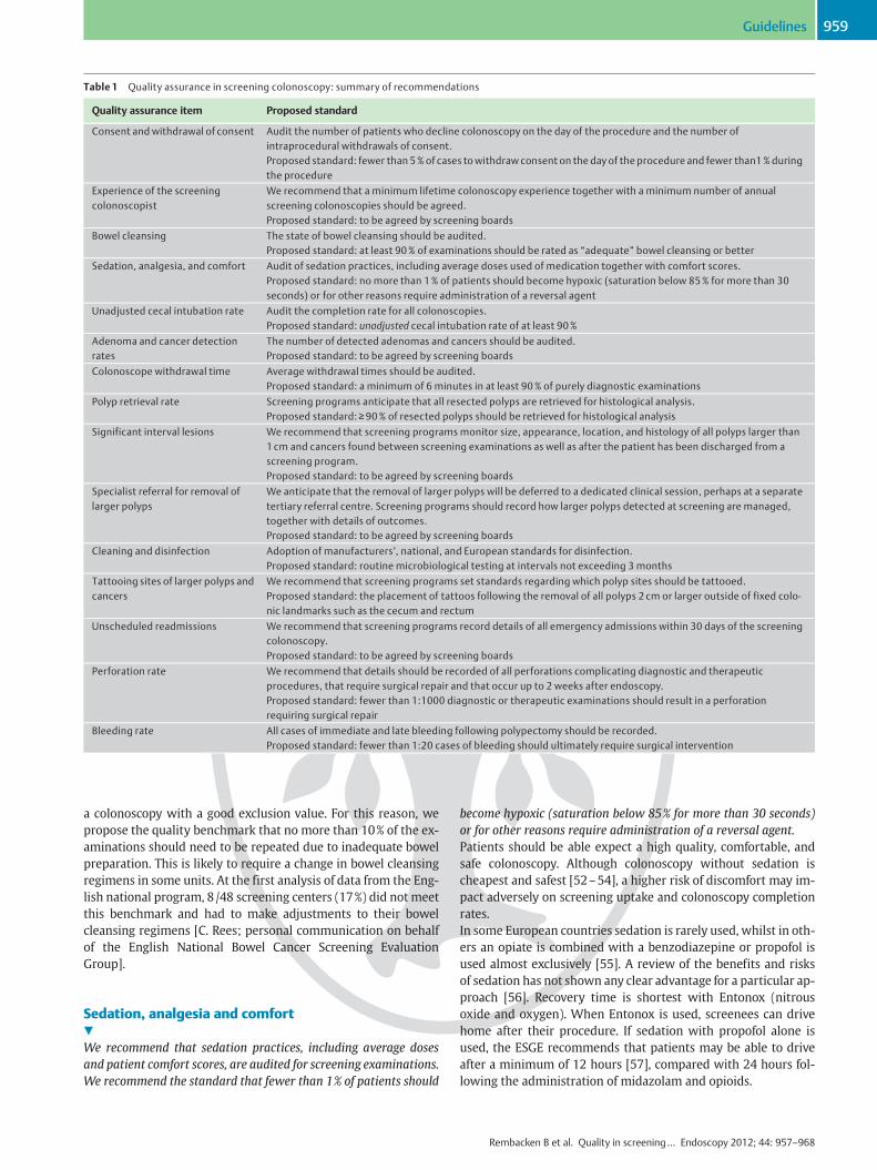
a colonoscopy with a good exclusion value. For this reason, wepropose the quality benchmark that no more than 10% of the ex-aminations should need to be repeated due to inadequate bowelpreparation. This is likely to require a change in bowel cleansingregimens in some units. At the first analysis of data from the Eng-lish national program, 8/48 screening centers (17%) did not meetthis benchmark and had to make adjustments to their bowelcleansing regimens [C. Rees; personal communication on behalfof the English National Bowel Cancer Screening EvaluationGroup].
Sedation, analgesia and comfort!
We recommend that sedation practices, including average dosesand patient comfort scores, are audited for screening examinations.We recommend the standard that fewer than 1% of patients should
become hypoxic (saturation below 85% for more than 30 seconds)or for other reasons require administration of a reversal agent.Patients should be able expect a high quality, comfortable, andsafe colonoscopy. Although colonoscopy without sedation ischeapest and safest [52–54], a higher risk of discomfort may im-pact adversely on screening uptake and colonoscopy completionrates.In some European countries sedation is rarely used, whilst in oth-ers an opiate is combined with a benzodiazepine or propofol isused almost exclusively [55]. A review of the benefits and risksof sedation has not shown any clear advantage for a particular ap-proach [56]. Recovery time is shortest with Entonox (nitrousoxide and oxygen). When Entonox is used, screenees can drivehome after their procedure. If sedation with propofol alone isused, the ESGE recommends that patients may be able to driveafter a minimum of 12 hours [57], compared with 24 hours fol-lowing the administration of midazolam and opioids.
Table 1 Quality assurance in screening colonoscopy: summary of recommendations
Quality assurance item Proposed standard
Consent and withdrawal of consent Audit the number of patients who decline colonoscopy on the day of the procedure and the number ofintraprocedural withdrawals of consent.Proposed standard: fewer than 5% of cases to withdraw consent on the day of the procedure and fewer than1% duringthe procedure
Experience of the screeningcolonoscopist
We recommend that a minimum lifetime colonoscopy experience together with a minimum number of annualscreening colonoscopies should be agreed.Proposed standard: to be agreed by screening boards
Bowel cleansing The state of bowel cleansing should be audited.Proposed standard: at least 90% of examinations should be rated as “adequate” bowel cleansing or better
Sedation, analgesia, and comfort Audit of sedation practices, including average doses used of medication together with comfort scores.Proposed standard: no more than 1% of patients should become hypoxic (saturation below 85% for more than 30seconds) or for other reasons require administration of a reversal agent
Unadjusted cecal intubation rate Audit the completion rate for all colonoscopies.Proposed standard: unadjusted cecal intubation rate of at least 90%
Adenoma and cancer detectionrates
The number of detected adenomas and cancers should be audited.Proposed standard: to be agreed by screening boards
Colonoscope withdrawal time Average withdrawal times should be audited.Proposed standard: a minimum of 6 minutes in at least 90% of purely diagnostic examinations
Polyp retrieval rate Screening programs anticipate that all resected polyps are retrieved for histological analysis.Proposed standard: ≥90% of resected polyps should be retrieved for histological analysis
Significant interval lesions We recommend that screening programs monitor size, appearance, location, and histology of all polyps larger than1 cm and cancers found between screening examinations as well as after the patient has been discharged from ascreening program.Proposed standard: to be agreed by screening boards
Specialist referral for removal oflarger polyps
We anticipate that the removal of larger polyps will be deferred to a dedicated clinical session, perhaps at a separatetertiary referral centre. Screening programs should record how larger polyps detected at screening are managed,together with details of outcomes.Proposed standard: to be agreed by screening boards
Cleaning and disinfection Adoption of manufacturers’, national, and European standards for disinfection.Proposed standard: routine microbiological testing at intervals not exceeding 3 months
Tattooing sites of larger polyps andcancers
We recommend that screening programs set standards regarding which polyp sites should be tattooed.Proposed standard: the placement of tattoos following the removal of all polyps 2 cm or larger outside of fixed colo-nic landmarks such as the cecum and rectum
Unscheduled readmissions We recommend that screening programs record details of all emergency admissions within 30 days of the screeningcolonoscopy.Proposed standard: to be agreed by screening boards
Perforation rate We recommend that details should be recorded of all perforations complicating diagnostic and therapeuticprocedures, that require surgical repair and that occur up to 2 weeks after endoscopy.Proposed standard: fewer than 1:1000 diagnostic or therapeutic examinations should result in a perforationrequiring surgical repair
Bleeding rate All cases of immediate and late bleeding following polypectomy should be recorded.Proposed standard: fewer than 1:20 cases of bleeding should ultimately require surgical intervention
Rembacken B et al. Quality in screening… Endoscopy 2012; 44: 957–968
Guidelines 959

Naturally, sedation must be delivered in line with guidance pro-duced by national screening boards. To allow comparisons of per-formance, national screening boards should agree a scoring sys-tem to monitor sedation practices, patient comfort, and level ofsedation. The use of the following types of sedation practiceshould be recorded: (i) no sedation or analgesia; (ii) conscious se-dation, including which drugs were used; (iii) propofol or generalanesthesia; or (iv) Entonox.Unfortunately, there is no validated score to record level of seda-tion and comfort, although a validation of a colonoscopy comfortscore is underway. The American Society for Gastrointestinal En-doscopy (ASGE) has adopted a “continuous model” in which se-dation ranges from simple anxiolysis to “conscious sedation,” inwhich the patient responds to verbal commands and maintainsadequate spontaneous breathing. This is followed by “deep seda-tion” when the patient is only responsive to pain and may stopbreathing spontaneously [58]. Sedation is particularly hazardousin the elderly who are more likely to have co-morbidities that af-fect cardiorespiratory reserve and are more sensitive to the effectof sedation and analgesia [59, 60].Evaluating patient comfort is more problematic. The endoscopist,the assistant, and the patient may have different opinions aboutthe level of comfort during the procedure. We recommend thatthe patient’s estimate of comfort should be recorded on a simplescale such as for example: 1, no or minimal discomfort; 2, milddiscomfort; 3, moderate discomfort; or 4, severe discomfort.A total of 14% of patients undergoing procedures in the Norwe-gian Gastronet program reported that the examination hadbeen “severely painful” and a further 20% regarded the proce-dure as “painful.” Patients undergoing colonoscopy performedby surgeons were more likely to report pain than patients exam-ined by gastroenterologists. The authors of that study concluded
that the proportion of patients reporting a painful procedure wasunacceptably high [61].In addition, organized screening programs should record cases inwhich the oxygen saturation drops below 86% and when agentssuch as naloxone or flumazenil are used to reverse the sedation[62]. In an audit of sedation practices in a range of countries, hy-poxic episodes were reported in 5% of procedures [63].It may seem illogical to term the use of a potentially lifesavingmeasure, such as administration of reversal agents, a “negativeoutcome.” Indeed, if the consequence of administration of a sin-gle dose of reversal agent is punitive to the endoscopist, this maybecome a disincentive to its use.Nevertheless, the death of a patient from a respiratory arrest isthe worst possible outcome. For this reasonwe propose the qual-ity benchmark that fewer than 1% of patients should become hy-poxic (saturation below 85% for more than 30 seconds) or forother reasons require administration of a reversal agent.
Cecal intubation rate!
We recommend that the completion rate for all screening colonos-copies is audited, and we propose a minimum standard of 90% forunadjusted cecal intubation rate.A complete examination of the colon and rectum is fundamentalto any colorectal cancer screening program. The medial wall ofthe cecum between the appendiceal orifice and ileocecal valvecan not be visualized from a distance. Cecal intubation is definedas deep intubation into the cecum with the tip of the endoscopebeing able to touch the appendiceal orifice.Failure to reach the cecum is expensive and inconvenient for pa-tients as a new attempt at colonoscopy or a radiological examina-tion is required.Rapid and reliable cecal intubation is also a proxy indicator of co-lonoscopy skill. However, other factors also have an effect. Thechances of successfully reaching the cecum are reduced in indi-viduals with advancing age and increasing body mass index(BMI) [64, 65]. Colonoscopy in a young patient in good health ismost likely to be complete [66, 67]. The use of technology suchas the variable stiffness colonoscope [68] or endoscopic imagingcan also improve the probability of successful cecal intubation[69].The English National Health Service (NHS) Bowel Cancer Screen-ing Programme (BCSP) has set aminimum, unadjusted cecal intu-bation rate of 90% [70]. The European Commission guideline alsoregards a 90% cecal intubation rate as acceptable but excludescases with obstructive cancer requiring surgery [71].The US Multi-Society Task Force on Colorectal Cancer recom-mends different benchmarks for “screening” and “symptomatic”populations (95% and 90%, respectively) [72, 73]. Similarly, Can-cer Care Ontario Colonoscopy Standards set a minimum adjustedcompletion rate of 95% (excluding cases with poor bowel pre-paration and obstructing lesions) [74].The appendiceal orifice should be photographed, preferably froma distance of 2 to 4cm, so that the photograph encompasses thececal strap fold or “crow’s foot.” A second photograph should betaken more distally to include the entire cecum and the ileocecalvalve. The terminal ileum should be photographed if intubated. Ifresources are available, video recording provides the highest levelevidence that the cecum has been intubated.In the context of an organized screening program, variables suchas the presence of an obstructing lesion are likely to be equally
Table 2 Details to be included in the colonoscopy report.
Patient details
Endoscope used Manufacturer, model, and serial number
Name and positionof endoscopist andancillary staff
Indication for theprocedure
Number of screening roundDetails and interval to the recent screeninginvestigationFollow-up polypectomy after previousscreening colonoscopy
Bowel cleansing Bowel cleansing regimen givenPatient tolerance of bowel cleansing regimenMucosal views obtained
Intubation Limit of examination including reason whyexamination was incompleteDuration of intubation to limit of examinationDuration of extubation
Disease detected andmanagement
Site of each lesionSize of the lesion (as estimated by theendoscopist)Growth morphology (Paris classification)Crypt pattern of each lesion (Kudo’sclassification)Endoscopic diagnosis of each lesionAction taken for each lesionSuccess and complications in removing eachlesionDiathermy settings used for cauteryFinal histological diagnosis
Rembacken B et al. Quality in screening… Endoscopy 2012; 44: 957–968
Guidelines960

distributed amongst all screening providers. For this reason, inthe context of an organized screening program, we recommendthat only completely unadjusted data are recorded, that is, basedon intention to examine the complete colon. Not adjusting forcases with poor bowel cleansing makes the measure more objec-tive and will allow national bowel cancer screening programs todetect endoscopy units that provide suboptimal bowel cleansingregimens.
Detection of adenomas and cancers!
We recommend that the number of adenomas and cancers is re-corded for all screening examinations. As the number of lesionsdetected depends on the details of the screening program, theaudit standard would have to be agreed by screening boards.The detection of adenomas and early cancers is fundamental inany bowel cancer program. Data from the US National PolypStudy [75] and the UK Flexible Sigmoidoscopy Screening Trial[76] have shown that the removal of colonic adenomas reducesthe risk of subsequent cancer.When a primary FOBT-based organized population screeningprogram is implemented, a secondary-test population “enriched”with adenomas and cancers is expected. Typically, screenees pre-senting for colonoscopy following a positive guiac-based FOBThave a 35% risk of detection of adenoma and a nearly 11% risk ofcancer [77]. This compares with a 15%–25% risk of detection ofbenign adenoma by ad hoc case finding [78–80].Unfortunately there is evidence that adenomas are missed at co-lonoscopy [81–83] and that some endoscopists miss morepolyps than others [84]. A marked variation in adenoma detec-tion rate (8%–16%) was found in the UK Flexible SigmoidoscopyScreening Trial [85]. A recent meta-analysis of studies of colonos-copies performed by primary care physicians in the USA found aneven greater range in adenoma detection (8.8% to>50%) [86]. Atandem study demonstrated a miss rate for advanced adenomas(>1cm) of up to 6%, and a rate as high as 27% for adenomas lessthan 5mm in size [87]. Such values have been confirmed by com-parative studies between CT colonography and colonoscopy [88].In a recent Polish study, screening colonoscopists with adenomadetection rates below 20% were more likely to have patients sub-sequently presenting with interval cancer [89].It has been demonstrated that there is a good correlation be-tween the polyp and adenoma detection rates (ADR) [90]. Never-theless, adenoma detection is a more relevant QA item thanpolyp detection which would also include a number of non-neo-plastic polyps. Although checking the histology of all polyps re-moved is a large task, it should be achievable within an organizedscreening program. In countries that are setting up bowel cancerscreening programs, the national screening boards need to agreeon minimum adenoma and cancer detection rates within theirprogram.The English Bowel Cancer Screening Programme defines “adeno-ma detection rate” as “the number of colonoscopies at which oneor more histologically confirmed adenomas is found divided bythe total number of colonoscopies performed.” Perhaps a moreuseful alternative would be the “adenoma detection index” (ADI)which signifies the total number of adenomas detected dividedby the total number of colonoscopies performed.The benchmarks set for minimum detection rates would dependon the details of the country’s colorectal cancer screening pro-gram, such as the age of those screened [91, 92] and the sensitiv-
ity and specificity of the primary stool test used [93–98]. In addi-tion, the adenoma and cancer detection rate will vary greatly be-tween men and women. Finally, when an organized screeningprogram is introduced in a country with widespread ad hoc casefinding for bowel cancer, fewer polyps and cancers may be de-tected.In the English screening program a surprisingly wide range ofadenoma detectionwas found at the first analysis [C. Rees; perso-nal communication on behalf of the English National Bowel Can-cer Screening Evaluation Group]. It is of concern that somescreening colonoscopists only detected an adenoma at 22% of ex-aminations whilst others found adenomas in 60% of cases. Asadenomas are more common in men (mean adenoma detectionrate [ADR] 52.9% in men vs. 36.5% in women), a predominanceof women in the screening population might have explainedpart, but not all, of this variation.
Withdrawal timeWe recommend that the average withdrawal time is audited dur-ing screening colonoscopies and propose a minimum of 6 minutesin at least 90% of purely diagnostic examinations.Colonoscopy withdrawal time and polyp detection are closelyrelated. Two large studies have supported a minimum withdra-wal time of 6 minutes in diagnostic colonoscopies [99, 100]. Asthe finding of polyps, followed by their removal increases theaverage duration of the colonoscopy, this figure only applies toexaminations in which no polyps are found.In the study by Barclay et al., there was a threefold difference (9.4%–32.7%) in adenoma detection rate depending on the durationof withdrawal (which ranged from 3.1 to 16.8 minutes). Colonos-copists with withdrawal times of greater than 6 minutes hadhigher detection of any neoplasia (28.3% vs. 11.8%). In addition,the detection of advanced neoplasia was also significantly differ-ent (6.4% vs. 2.6%). A recent analysis of the English screening pro-gram showed that withdrawal times of 10minutes were associat-ed with the best adenoma detection rate [101].As there is a correlation between withdrawal time and the detec-tion of adenomas we recommend that withdrawal time is audi-ted. A minimum of 6 minutes for withdrawal time is recommen-ded in cases when no therapy is undertaken. However, speed ofwithdrawal is not the only factor affecting polyp detection.In addition to withdrawal time, factors such as aspiration of li-quid, careful examination behind folds [102], position change,the use of buscopan, fitting a shallow cap on the tip of the endo-scope [103], or technology such as high resolution or “the third-eye retroscope” can also improve polyp detection [104, 105].The use of blue dye sprayed onto suspicious mucosal areas im-proves the detection of smaller lesions or polyps with a flatgrowth pattern [106, 107]. Furthermore, dye-spraying tech-niques allow prediction of histology [108] particularly whenused together with a magnifying endoscope [109]. Image proces-sing technologies such as Olympus narrow band imaging (NBI),Fuji Intelligent Chromo Endoscopy (FICE) and the Pentax i-scanhave been developed to provide quicker assessment of suspiciousareas and to allow differentiation between hyperplastic and ade-nomatous polyps [110].
Retrieval of polypsWe recommend that the number of resected and retrieved polyps isaudited for all screening colonoscopies, and propose the standardthat at least 90% of resected polyps are retrieved for histological a-nalysis.
Rembacken B et al. Quality in screening… Endoscopy 2012; 44: 957–968
Guidelines 961

Retrieval of resected polyps for histological examination is im-portant. In the UK pilot demonstration of colorectal screening,16.6% of all cancers were “polyp cancers” [81]. As expected, therisk of polyp-cancer increases with the size of the polyp (●" Table3). After piecemeal resection, or when histological analysis ofadenomas larger than 10mm cannot confirm complete excision,early follow-up is recommended (e.g. within 3–6months). Inter-estingly, when polyps are resected using Endocut current, micro-scopic evaluation of resection margins is better than if coagula-tion current is used for polypectomy [111].In organized screening programs it is expected that resectedpolyps will be retrieved for histological analysis. However, re-cently, a “resect and discard” policy for smaller polyps has beenproposed. At an expert center, optical diagnosis has been foundto be accurate in more than 90% of polyps up to 10mm in size[112]. Such a policy would result in substantial cost savings forscreening programs [113].As the effect of a “resect and discard” policy has never been test-ed outside tertiary referral centers, we recommend that nationalscreening boards monitor the retrieval rate for all resectedpolyps. Successful retrieval of at least 90% of excised polypsseems a reasonable standard.
Significant interval lesions!
We recommend that the size, appearance, location and histology ofall polyps larger than 1cm should be recorded in screening pro-grams, as well as all cancers found between screening examina-tions and those found after the patient has been discharged from ascreening program.The US National Polyp Study suggested that polypectomy canprevent up to 90% of subsequent cancers. In a study by Imperialeet al. [114] no interval cancers were found 5 years after a negativecolonoscopy in 1256 individuals. However other studies havedemonstrated a lower protective effect [115]. In a study by Farraret al. [116] 5.4% of all cancers detected were interval lesions. Apooled analysis of North American studies that had followed pa-tients with previous adenomas for a median of 4 years put therisk of subsequent cancer at 0.6% [117] (the risk of developingan “advanced neoplasia” was 11.8%). In a retrospective Dutchstudy the sensitivity of colonoscopy to detect a colorectal cancerwas estimated at 90% [118]. In a Canadian study, between 2% and6% of patients who developed colorectal cancer had undergone acolonoscopy in the previous 3 years [119].It appears that colonoscopy offers better protection against fu-ture cancer arising in the left hemi-colon (80% protection) thanthe right hemi-colon (12%–33% protection) [120–124]. One ex-planation for why colonoscopy might offer better protection
against distal cancers is that the right side of the colon tends tobe less well cleaned than the left side. Indeed, miss rates are con-sistently two- to threefold higher in the proximal than the distalcolon [125–127]. An alternative explanation is that right-sidedlesions are more aggressive [128] or that they arise from incon-spicuous flat lesions [129] that are easily missed particularly asthe right hemi-colon is more difficult to clean [130].National screening boards need to agree clear definitions for “in-terval lesions.” For example, they may be defined as adenomaslarger than 1cm or cancers, that are detected between a screen-ing episode and the scheduled next screening (surveillance) epi-sode. Data on interval lesions are an important tool for assessingthe quality of screening colonoscopies. Capturing data on adeno-mas larger than 1cm or cancers that are detected after the pa-tient has left a screening program would also be important, forexample to identify a need to extend the screening age range.We recommend that national screening bodies record the details(size, appearance, location, and histology) of all lesions detected,not just during screening examinations but also outside thescreening program. By cross-referencing data with national can-cer registries, it should be possible for national cancer screeningprograms to obtain accurate data on interval cancers.
Removal of larger polyps!
We recommend that screening programs audit how larger lesionsdetected at screening are managed, together with details of out-comes. In particular, the number of benign lesions referred for sur-gical resection should be recorded and outcomes monitored.The purpose of colorectal cancer screening is to detect early can-cers and remove precursor lesions safely and effectively, therebypotentially reducing cancer incidence. However, screening colo-noscopists may not have the expertise to remove the largestpolyps. In addition, the removal of larger polyps is associatedwith greater risks and the informed consent process must reflectthis.Unfortunately, referring patients with larger benign lesions forsurgery rather than polypectomy may be associated with higherrisks of adverse outcomes [131, 132]. There is evidence from theFrench screening program that up to 10% of entirely benignpolyps are removed surgically rather than endoscopically [133].Colonoscopists providing an enhanced therapeutic referral ser-vice may not wish to provide conventional screening. Neverthe-less, in order to provide a therapeutic referral service, we recom-mend that individuals should register as “screening colonosco-pists” and collect QA data on their activities. There is little pub-lished data on advanced therapy complication rates that can beused to establish benchmarks for such a tertiary referral service[134]. Moss et al. reviewed the outcomes following resection of479 polyps, 2cm or larger in size. A total of 1.5% of patients pres-ented with a post-polypectomy serositis, 2.1% were admittedwith pain following the procedure, 2.9% of patients suffered de-layed bleeding, and perforation complicated 1.3% of resections[135]. It seems clear that the risks are greater with larger polyps.
Tattooing the sites of suspected malignant polyps andcancers!
We recommend that screening programs introduce guidelines onthe use of ink tattoos and recommend the placement of tattoos fol-
Table 3 Risk of malignancy versus size of polyp in the English Bowel CancerScreening Programme (BCSP)
Polyp-
cancers, n
Total polyps,
n
Polyp-
cancers, %
Size range
0–9mm 103 34959 0.29%
10–19mm 370 8425 4.39%
20–29mm 240 3008 7.98%
≥30mm 174 1705 10.2%
Size not recorded 34 957 –
Total 921 49054 1.88%
Rembacken B et al. Quality in screening… Endoscopy 2012; 44: 957–968
Guidelines962

lowing the removal of all polyps 2cm or larger situated outside ofthe cecum or rectum.The sites of larger polyps, suspected malignant polyps, and can-cers should be marked with an indelible compound such as Indiaink or a pure carbon-based alternative, if they are situated out-side of an unmistakable colonic landmark such as the rectum orcecum. This assists identification at follow-up colonoscopies or atthe time of surgery (especially for laparoscopic resections).India ink is a marker which requires dilution and sterilization incontrast to pre-packed sterile pure carbon-based preparations.Concerns have been raised about the safety of tattooing, with re-ports of fever, abdominal pain, and abscess formation [136].However, prior injection with saline followed by injection of theink into the saline bleb appears safe [137].National screening bodies should agree guidelines on which le-sions detected at screening should have the site marked with atattoo. Furthermore, agreement with local colorectal surgeonsshould be sought regarding the preferred number and positionof tattoos. In most cases, it is preferable to place more than onetattoo just distal to the lesion. The placement of 2 or 3 tattoos en-sures that at least one tattoo is visible on the antemesenteric bor-der of the colon, allowing the distal resection margin to be clearof neoplasia.The risk of unexpected cancer increases with the size of thepolyp, approaching 10% for lesions 2cm in diameter or larger(●" Table3). For this reason, we recommend the placement of tat-toos following the removal of all polyps 2cm or larger situatedoutside of the rectum or cecum.
Minimum experience for screening colonoscopists!
We recommend that screening programs agree a minimum lifetimeexperience for their screening colonoscopists and set a minimumbenchmark for their annual number of screening examinations.There is a link between the experience of the endoscopist and thetime to reach the cecum, as well as with polyp detection rate andwith outcomes following polypectomy [138–140].A population-based study from Canada found that the risk ofcomplications such as perforation and bleeding was increasedthreefold with colonoscopists who performed fewer than thethreshold of 300 colonoscopies per year [141]. For this reason,the setting of a minimum annual number of screening colonosco-pies is fundamental to all other QA audits. For example, the Eng-lish NHS Bowel Cancer Screening Program set requirements of aminimum lifetime experience of 1000 examinations and a mini-mum annual number of 150 screening colonoscopies. This an-nual figure was set in order to allow meaningful analysis of QAdata from all screening colonoscopists [142].To ensure that screeners are of sufficient caliber, all nationalscreening boards should consider setting minimum standardsfor lifetime experience and annual number of procedures.
Recording early and late adverse outcomes!
We recommend that full details of all complications, including un-scheduled re-admissions following screening examinations are re-corded. We propose the quality standard that fewer than 5% ofbleeding complications should require surgical intervention andthat fewer than 1:1000 screening colonoscopies should be compli-cated by a perforation requiring emergency surgery.
Colonoscopy with polypectomy is a high risk endeavor with thepotential for life-threatening complications. Screening for colo-rectal cancer therefore has a real risk of directly harming its par-ticipants.It is difficult to draw firm conclusions from the literature on theincidence of complications. Most published series come from sin-gle centers with extensive experience in colonoscopy, withoutseparation of symptomatic and screening patients. Results maytherefore not reflect standard practice. Differences among au-thors in the definitions of complications has also hampered thesetting of firm benchmarks for screening. Recently the AmericanSociety for Gastrointestinal Endoscopy (ASGE) sponsored a work-shop that devised a useful classification system of adverse eventsto incorporate into our current Minimal Standard Terminology(MST version 3.0) lexicon [62]. We encourage national screeningboards to use the current MST terminology together with the re-cent ASGE classification of adverse events.Many adverse events are obvious direct complications of the en-doscopic procedure, e.g. bleeding, perforation, or cardiorespira-tory complications. However, at other times it can be more diffi-cult to decide whether an adverse event should be attributed tothe colonoscopy. Examples could include phlebitis at the site ofthe intravenous cannula, abdominal discomfort that resolvesspontaneously soon after the colonoscopy, development of achest infection within a week of the procedure.As it is important not to miss adverse outcomes that may havebeen caused by the endoscopic procedure, we propose that allevents should be recorded that result in: (i) an unscheduled ad-mission; (ii) a lengthening of the hospital stay; (iii) an unsched-uled further endoscopic procedure; (iv) emergency intervention,including blood transfusion; (v) emergency surgery; or (vi) deathof the patient.The capture of “late events” up to 30 days after the patient has leftthe endoscopy unit is difficult. Nevertheless it forms a bench-mark which allows comparison between screening programs.Full details of all readmissions should be sought including reasonfor admission, length of stay, medical/surgical intervention, andoutcomes.
PerforationIn study series from both Nottingham in the UK [14] and Minne-sota in the USA [16] there were approximately 7 perforations per10 000 colonoscopies. In the UK pilot program, 5 perforations per10 000 colonoscopies were reported. In the smaller NorwegianColorectal Cancer Prevention (NORCCAP) study, therewere no re-ported perforations following diagnostic examinations; however1 perforation per 336 polypectomies was reported [143].The British Society of Gastroenterology (BSG) audit of colonosco-py in the UK also demonstrated that the risk of perforation ap-proximately doubles when polypectomy is carried out [144].The risk of perforation at diagnostic examinations was 1:923comparedwith 1:460 following polypectomy. A reviewof a largerdataset (39 286 colonoscopies carried out in the US Medicareprogram) also reported a perforation rate of 1:500 examinationsbut did not report on the influence of polypectomy [145]. Theabove figures are not dramatically different from that of a Ger-man review of colonoscopies carried out in the late 1970s. Thisstudy from 40 years ago reported 1 perforation complicating ev-ery 300 polypectomies [146].A colonic perforation is usually defined as evidence of air, luminalcontents, or instrumentation outside the gastrointestinal tract.Nevertheless a small, contained perforation into the omental re-
Rembacken B et al. Quality in screening… Endoscopy 2012; 44: 957–968
Guidelines 963

flection of the colon or a microperforation which is immediatelyclosed by the application of clips may also be regarded as a per-foration. On occasion, perforations are suspected in patients whodevelop abdominal discomfort following simple mucosal biop-sies or smaller polypectomies. In these cases abdominal X-raysmay disclose the presence of a small amount of intramural gasor pericolonic edema; this can be difficult to interpret when thepatient has no clinical signs of a perforation.Most perforations complicate therapeutic procedures and somepolypectomies are more hazardous than others. The risk of per-foration appears to be greatest with the removal of larger, sessile,or right-sided polyps [147]. Provided that such therapeutic mi-croperforations are immediately recognized and managed withthe application of clips and systemic antibiotics, no harmwill en-sue.A pragmatic endpoint, which will capture all significant cases, isto only record perforations which require surgical repair. We pro-pose the quality benchmark that fewer than 1:1000 screening ex-aminations should result in a perforation requiring emergencysurgery.
BleedingBleeding at the time of polypectomy is common and is usually ofno significance when immediately managed endoscopically.However, if further intervention such as an unscheduled admis-sion is required, the bleeding should be recorded as an adverseevent. Pragmatically, post-polypectomy bleeding (PPB) may bedefined as visible blood loss or melena for up to 2 weeks follow-ing the procedure that requires transfusion, surgery, or furtherendoscopic therapy. This definition excludes the smaller amountof post-polypectomy bleeding that most patients experience fol-lowing the removal of large lesions.It is difficult to draw conclusions from published PPB rates as ahuge range (1:10 to 1:300) has been reported [148,149]. The rea-son for the wide range is that the risk of bleeding is affected bynumerous factors. Elderly patients, or those taking antithrombo-tic medication (apart from aspirin) appear to be at greatest over-all risk [150,151]. Lesion-specific factors also affect the risk ofbleeding. The risks are greater with larger and sessile lesions, par-ticularly in the right hemi-colon [147]. Finally, the diathermy set-tings can also influence bleeding rates [152]. The use of a “purecut” diathermy is associated with a higher risk of immediatebleeding [153,154] whilst “blended” and “pure coagulation” elec-trocautery are associated with a similar risk of PPB [155], with atrend to more immediate versus delayed (up to 8 days) PPB withblended versus coagulation current, respectively.The topic of PPB has recently been reviewed by the ESGE [156].The review concluded that endoscopic interventions that are ef-fective in preventing PPB include placement of a detachable loopligating device for large pedunculated polyps and submucosal in-jection of diluted adrenaline for sessile polyps. The efficacy ofother measures, including endoclip placement, injection of sal-ine, and argon plasma coagulation, has not been definitivelydemonstrated.Finally, it is perhaps not surprising that the experience of the co-lonoscopist also affects the risk. A study of outcomes following al-most 100 000 outpatient colonoscopies showed that the risk ofcomplications was 3-fold greater when the polypectomywas car-ried out by a “low volume” colonoscopist [145]. However, it islikely that it is the annual number of polypectomies that is of im-portance rather than the annual number of diagnostic examina-tions. The German quality assurance program has set a modest
annual minimum of 10 snare polypectomies to maintain accred-itation.In almost all instances of immediate and delayed bleeding, itshould be possible to manage the bleeding with supportive careand endoscopic therapy. As the rate of PPB is affected by a largenumber of factors, it is difficult to set an arbitrary benchmark.However, in all cases of late bleeding in which the patient is he-modynamically compromised or has ongoing bleeding, an at-tempt at endoscopic management should precede surgery. Wepropose that less than 5% of patients suffering a post-polypecto-my bleed, as defined above, should ultimately require surgical in-tervention
The colonoscopy report!
The report is an important record of the screening examination andwe recommend that it contains a minimum dataset documentingthe procedure.It is important that the endoscopy report is complete, with de-tails of all abnormalities. In particular, details of each lesion de-tected should be recorded together with information on methodof removal. For a complete colonoscopy report, the ESGE recom-mend a set of eight photographs to be taken from standard loca-tions [157]. A ninth photograph of the low rectal mucosawith theendoscope in a retroverted position should also be taken when-ever possible. In addition, reasons for any failed cecal intubationshould be recorded.An outline of information which should be included in thescreening colonoscopy report is detailed in●" Table2. In manycountries the patient is provided with a copy of the report imme-diately after the procedure and the endoscopist is obliged to im-mediately forward a copy to the patient’s primary care physician.Nevertheless, most would consider the endoscopy report to beincomplete before it has been updated with the final histologicalanalysis.
Cleaning and disinfection of equipment!
We recommend that standards for disinfection set by manufactur-ers and by national and European bodies are actively audited inscreening programs, and recommend routine microbiological test-ing at intervals not exceeding 3 months.Appropriate cleaning of endoscopes and accessories is a core re-quirement of endoscopy. Naturally, individuals attending forscreening must be able to be confident that all equipment hasbeen effectively cleaned.The Guideline Committee of ESGE and the European Society ofGastroenterology and Endoscopy Nurses and Associates (ES-GENA) has published detailed guidelines relating to hygiene anddisinfection in endoscopy [158,159]. In addition to these theremay be local regulations, national laws [160], and manufacturers’instructions to follow.There are also published European Standards (EN 14885) andguidelines on how the efficacy of the cleaning process should beassessed [161–163] at intervals not exceeding3months. Nationalscreening boards should ensure that relevant guidelines are fol-lowed.
Rembacken B et al. Quality in screening… Endoscopy 2012; 44: 957–968
Guidelines964
Conclusion!
Our guidance has been produced under the auspices of the ESGEwith the aim of providing clear and simple advice for countriessetting up organized screening programs, to allow assessment ofsafety and quality relevant to screening colonoscopy.Colonoscopy is fundamental to most screening programs and thesuccess of screening programs is closely related to the promptprovision of a high quality, patient-centered colonoscopy service.To minimize risks andmaximize benefit, all countries need to putrobust quality assurance frameworks in place.The adoption of our quality assurance items lays the foundationfor meaningful comparisons among individual endoscopists, dif-ferent endoscopy units, and even the services provided by differ-ent countries, to achieve better outcomes for patients.
Competing interests: None
Institutions1 Centre for Digestive Diseases, Department of Gastroenterology, The GeneralInfirmary at Leeds, Leeds, United Kingdom
2 Digestive Endoscopy Unit, Catholic University, Rome, Italy3 Stiftung Lebensblicke, Klinikum Ludwigshafen, Ludwigshafen, Germany4 Department of Gastroenterology, Kettering General Hospital, Kettering,United Kingdom
5 Department of Gastroenterology, University Hospital of North Tees,Stockton-on-Tees, United Kingdom
6 Durham University, County Durham, UK7 Department of Gastroenterology, HUG, Geneva, Switzerland8 Department of Internal Medicine, Digestive Diseases and Endoscopy,New Mowasat Hospital, Salmiya, Kuwait
9 Department of Hepatogastroenterology, Edouard Herriot Hospital, Lyon,France
References1 Benson VS, Patnick J, Davies AK et al. International Colorectal Cancer
Screening Network. Colorectal cancer screening: a comparison of 35initiatives in 17 countries. Int J Cancer 2008; 122: 1357–1367
2 Wennberg JE. Time to tackle unwarranted variations in practice. BMJ2011; 342: 687–690
3 Eisinger F, Cals L, Calazel-Benque A et al. Impact of organised programson colorectal cancer screening. BMC Cancer 2008; 8: 104
4 Bos AB, van Ballegooijen M, van Gessel-Dabekaussen AA et al. Organisedcervical cancer screening still leads to higher coverage than sponta-neous screening in The Netherlands. Eur J Cancer 1998; 34: 1598–1601
5 Ronco G, Pilutti S, Patriarca S et al. Impact of the introduction of orga-nised screening for cervical cancer in Turin, Italy: cancer incidence byscreening history 1992–98. Br J Cancer 2005; 93: 376–378
6 Ronco G, Segnan N, Giordano L et al. Interaction of spontaneous and or-ganised screening for cervical cancer in Turin, Italy. Eur J Cancer 1997;33: 1262–1267
7 Nygard JF, Skare GB, Thoresen SO. The cervical cancer screening pro-gramme in Norway, 1992–2000: changes in Pap smear coverage andincidence of cervical cancer. J Med Screen 2002; 9: 86–91
8 Lynge E, Clausen LB, Guignard R et al. What happens when organizationof cervical cancer screening is delayed or stopped? J Med Screen 2006;13: 41–46
9 Nieminen P, Kallio M, Anttila A et al. Organised vs. spontaneous Pap-smear screening for cervical cancer: A case–control study. Int J Cancer1999; 83: 55–58
10 Quinn M, Babb P, Jones J et al. Effect of screening on incidence of andmortality from cancer of cervix in England: evaluation based on routi-nely collected statistics. BMJ 1999; 318: 904–908
11 Chamot E, Charvet AI, Perneger TV. Who gets screened, and where: acomparison of organised and opportunistic mammography screeningin Geneva, Switzerland. Eur J Cancer 2007; 43: 576–584
12 Puliti D,Miccinesi G, Collina N et al. Effectiveness of service screening: acase–control study to assess breast cancer mortality reduction. Br JCancer 2008; 99: 423–427
13 International Agency for Research on Cancer. Cervix cancer screening.IARC Handbooks of cancer prevention. 10: Volume Lyon, France: IARCPress; 2005
14 Hardcastle JD, Chamberlain JO, Robinson MH et al. Randomised con-trolled trial of faecal-occult-blood screening for colorectal cancer. Lan-cet 1996; 348: 1472–1477
15 Kronborg O, Fenger C, Olsen J et al. Randomised study of screening forcolorectal cancer with faecal-occult-blood test. Lancet 1996; 348:1467–1471
16 Mandel JS, Bond JH, Church TR et al. Reducing mortality from colorectalcancer by screening for faecal occult blood. Minnesota Colon CancerControl Study.. N Engl J Med 1993; 328: 1365–1371
17 Lindholm E, Brevinge H, Haglind E. Survival benefit in a randomizedclinical trial of faecal occult blood screening for colorectal cancer. Br JSurg 2008; 95: 1029–1036
18 Kewenter J, Breving H, Engaras B et al. Results of screening, rescreening,and follow-up in a prospective randomized study for detection of colo-rectal cancer by faecal occult blood testing. Scand J Gastroenterol1994; 29: 468–473
19 Council of the European Union. Council Recommendation of 2 Decem-ber 2003 on cancer screening (2003/87/EC): OJ L327/34–38. Brussels:2003
20 Atkin WS, Edwards R, Kralj-Hans I et al. Once-only flexible sigmoido-scopy screening in prevention of colorectal cancer: a multicentre ran-domised controlled trial. Lancet 2010; 375: 1624–1633
21 Segnan N, Senore C, Andreoni B et al. Baseline findings of the Italianmulticenter randomized controlled trial of “once-only sigmoidoscopy”– SCORE. J Natl Cancer Inst 2002; 94: 1763–1772
22 Weissfeld J, Schoen R, Pinsky P et al. Flexible sigmoidoscopy in the PLCOcancer screening trial: results from the baseline screening examina-tion of a randomized trial. J Natl Cancer Inst 2005; 97: 989–997
23 European Commission. editors European guidelines for quality assur-ance in colorectal cancer screening and diagnosis.Segnan N, Patnick J,von Karsa L. 1: edition Luxembourg: Publications Office of the Euro-pean Union; 2011: DOI 10.2772/15379
24 Bourke MJ. Making every colonoscopy count: ensuring quality in en-doscopy. J Gastroenterol Hepatol 2009; 24: 43–50
25 Hoff G, Bretthauer M,Huppertz-Hauss G et al. The Norwegian Gastronetproject: Continuous quality improvement of colonoscopy in 14 Nor-wegian centres. Scand J Gastroenterol 2006; 41: 481–487
26 Seip B, Bretthauer M, Dahler S et al. Sustaining the vitality of colonosco-py quality improvement programmes over time. Experience from theNorwegian Gastronet programme. Scand J Gastroenterol 2010; 45:362–369
27 Burke CA, Church JM. Enhancing the quality of colonoscopy: the impor-tance of bowel purgatives. Gastrointest Endosc 2007; 66: 565–573
28 Froehlich F, Wietlisbach V, Gonvers JJ et al. Impact of colonic cleansingon quality and diagnostic yield of colonoscopy: the European Panel ofAppropriateness of Gastrointestinal Endoscopy European multicenterstudy. Gastrointest Endosc 2005; 61: 378–384
29 Harewood GC, Sharma VK, de Garmo P. Impact of colonoscopy prepara-tion quality on detection of suspected colonic neoplasia. GastrointestEndosc 2003; 58: 76–79
30 Thomas-Gibson S, Rogers P, Cooper S et al. Judgement of the quality ofbowel preparation at screening flexible sigmoidoscopy is associatedwith variability in adenoma detection rates. Endoscopy 2006; 38:456–460
31 Hookey LC, Vanner S. A review of current issues underlying coloncleansing before colonoscopy. Can J Gastroenterol 2007; 21: 105–111
32 Hawes RH, Lowry A, Deziel D. A consensus document on bowel pre-paration before colonoscopy. Gastrointest Endosc 2006; 63: 894–909
33 World Health Organization. WHO Pharmaceuticals Newsletter. Gene-va: WHO; 2009: No.1
34 Belsey J, Epstein O,Heresbach D. Systematic review: oral bowel prepara-tion for colonoscopy. Aliment Pharmacol Ther 2007; 25: 373–384
35 Rex DK, Vanner SJ. Colon cleansing before colonoscopy: does oral so-dium phosphate solution still make sense? Can J Gastroenterol 2009;23: 210–214
36 Frommer D. Cleansing ability and tolerance of three bowel prepara-tions for colonoscopy. Dis Colon Rectum 1997; 40: 100–104
37 Hamilton D, Mulcahy D, Walsh D et al. Sodium picosulphate comparedwith polyethylene glycol solution for large bowel lavage: a prospectiverandomised trial. Br J Clin Pract 1996; 50: 73–75
Rembacken B et al. Quality in screening… Endoscopy 2012; 44: 957–968
Guidelines 965
38 Golub RW, Kerner BA, Wise WEJr. Colonoscopic preparations – whichone? A blinded, prospective, randomized trial. Dis Colon Rectum1995; 58: 594–597
39 Rösch T, Classen M. Fractional cleansing of the large bowel with Golyte-ly for colonoscopic preparations: a controlled trial. Endoscopy 1987;19: 198–200
40 Kilgore TW, Abdinoor AA, Szary NM et al. Bowel preparation with split-dose polyethylene glycol before colonoscopy: a meta-analysis of ran-domized controlled trials. Gastrointest Endosc 2011; 73: 1240–1245
41 Aoun E, Baki HA, Azar C et al. A randomized single-blind trial of split-dose PEG-electrolyte solution without dietary restriction comparedwith whole dose PEG-electrolyte solution with dietary restriction forcolonoscopy preparation. Gastrointest Endosc 2005; 62: 213–218
42 El Sayed AMA, Kanafani ZA,Mourad FH et al. A randomized single-blindtrial of whole versus split-dose polyethylene glycol-electrolyte solu-tion for colonoscopy preparation. Gastrointest Endosc 2003; 58: 36–40
43 Park SS, Sinn DH, Kim YH et al. Efficacy and tolerability of split-dosemagnesium citrate: low-volume (2 liters) polyethylene glycol vs. sin-gle- or split-dose polyethylene glycol bowel preparation for morningcolonoscopy. Am J Gastroenterol 2010; 105: 1319–1326
44 Cohen SM, Wexner SD, Binderow SR et al. Prospective, randomized, en-doscopic-blinded trial comparing pre-colonoscopy bowel cleansingmethods. Dis Colon Rectum 1994; 37: 689–696
45 Cohen LB, Sanyal SM, von Althann C et al. Clinical trial: 2-L polyethyleneglycol-based lavage solutions for colonoscopy preparation – a random-ized, single-blind study of two formulations. Aliment Pharmacol Ther2010; 32: 637–644
46 Afridi S, Barthel J, King P et al. Prospective, randomized trial comparinga new sodium phosphate-bisacodyl regimen with conventional PEG-ES lavage for outpatient colonoscopy preparation. Gastrointest Endosc1995; 41: 485–489
47 Berkelhammer C, Ekambaram A, Silva R. Low-volume oral colonoscopybowel preparation: sodium phosphate and magnesium citrate. Gastro-intest Endosc 2002; 56: 89–94
48 Clarkston W, Tsen T, Dies D et al. Oral sodium phosphate versus sulfate-free polyethylene glycol electrolyte lavage solution in outpatient pre-paration for colonoscopy: a prospective comparison. Gastrointest En-dosc 1996; 43: 42–48
49 Sharma VK, Steinberg EN, Vasudeva R et al. Randomized, controlledstudy of pre-treatment with magnesium citrate on the quality of colo-noscopy preparation with polyethylene glycol electrolyte lavage solu-tion. Gastrointest Endosc 1997; 46: 541–543
50 Rostom A, Jolicoeur E. Validation of a new scale for the assessment ofbowel preparation quality. Gastrointest Endosc 2004; 59: 482–486
51 Lai EJ, Calderwood AH, Doros G et al. The Boston bowel preparationscale: a valid and reliable instrument for colonoscopy-orientated re-search. Gastrointest Endosc 2009; 69: 620–625
52 Eckardt VF, Kanzler G, Schmitt T et al. Complications and adverse effectsof colonoscopy with selective sedation. Gastrointest Endosc 1999; 49:560–565
53 Rex DK. Colonoscopy. Gastrointest Endosc. Clin N Am 2000; 10: 135–160
54 Rex DK, Imperiale TF, Portish V. Patients willing to try colonoscopywithout sedation: associated clinical factors and results of a random-ized controlled trial. Gastrointest Endosc 1999; 49: 554–559
55 Riphaus A, Wehrmann T, Weber B et al. S3 Guideline: Sedation for gas-trointestinal endoscopy 2008. Endoscopy 2009; 41: 787–815
56 McQuaid KR, Laine L. A systematic review and meta-analysis of ran-domized, controlled trials of moderate sedation for routine endoscopicprocedures. Gastrointest Endosc 2008; 67: 910–923
57 Dumonceau JM, Riphaus A, Aparicio JR et al. European Society of Gas-trointestinal Endoscopy, European Society of Gastroenterology andEndoscopy Nurses and Associates, and the European Society of Anaes-thesiology Guideline: Non-anesthesiologist administration of propofolfor GI endoscopy. Endoscopy 2010; 42: 960–974
58 Practice guidelines for sedation and analgesia by non-anesthesiolo-gists – An updated report by the American Society of AnesthesiologistsTask Force on Sedation and Analgesia by Non-Anesthesiologists. Anes-thesiology 2002; 96: 1004–1017
59 Greenblatt DJ, Allen MD, Shader RI. Toxicity of high-dose flurazepam inthe elderly. Clin Pharmacol Ther 1977; 21: 355–361
60 Castleden CM, George CF, Marcer D et al. Increased sensitivity to nitra-zepam in old age. Br Med J 1977; 1: 10–12
61 Seip B, Bretthauer M, Dahler S et al. Patient satisfaction with on-de-mand sedation for outpatient colonoscopy. Endoscopy 2010; 42:639–646
62 Cotton PB, Eisen GM, Aabakken L et al. A lexicon for endoscopic adverseevents: report of an ASGE workshop. Gastrointest Endosc 2010; 71:446–454
63 Froehlich F,Harris JK,Wietlisbach Vet al. Current sedation andmonitor-ing practice for colonoscopy: an international observational study(EPAGE). Endoscopy 2006; 38: 461–469
64 Eloubeidi MA, Wallace MB, Desmond R et al. Female gender and otherfactors predictive of a limited screening flexible sigmoidoscopy exam-ination for colorectal cancer. Am J Gastroenterol 2003; 98: 1634–1639
65 Harris JK, Vader JP, Wietlisbach V et al. Variations in colonoscopy prac-tice in Europe: a multicentre descriptive study (EPAGE). Scand J Gas-troenterol 2007; 42: 126–134
66 Rathgaber SW, Wick TM. Colonoscopy completion and complicationrates in a community gastroenterology practice. Gastrointest Endosc2006; 64: 556–562
67 Viiala CH, Olynyk JK. Outcomes for women in a flexible sigmoidoscopy-based colorectal cancer screening programme. Intern Med J 2008; 38:90–94
68 Othman MO, Bradley AG, Choudhary A et al. Variable stiffness colono-scope versus regular adult colonoscope: meta-analysis of randomizedcontrolled trials. Endoscopy 2009; 41: 17–24
69 Shah SG, Brooker JC, Williams CB et al. Effect of magnetic endoscopeimaging on colonoscopy performance: a randomised controlled trial.Lancet 2000; 356: 1718–1722
70 Rutter MD, Chilton A. Quality assurance guidelines for colonoscopy.NHS BCSP Publication 2011; 6: 24
71 European Commission. European guidelines for quality assurance incolorectal cancer screening and diagnosis. European Union; 2010:978-92-79-16435-4 ISBN
72 Rex DK, Bond JH,Winawer S et al. Quality in the technical performanceof colonoscopy and the continuous quality improvement process forcolonoscopy: recommendations of the U.S. Multi-Society Task Forceon Colorectal Cancer.. Am J Gastroenterol 2002; 97: 1296–1308
73 Levin B, Lieberman DA, McFarland B et al. Screening and surveillancefor the early detection of colorectal cancer and adenomatous polyps,2008: a joint guideline from the American Cancer Society, the US Mul-ti-Society Task Force on Colorectal Cancer, and the American College ofRadiology. Gastroenterology 2008; 134: 1570–1595
74 Rabeneck L, Rumble RB, Axler J et al. Cancer Care Ontario ColonoscopyStandards: standards and evidentiary base. Can J Gastroenterol 2007;21: 5D–24D
75 Winawer SJ, Zauber AG, O’Brien MJ. The National Polyp Study Work-group. et al. Randomized comparison of surveillance intervals aftercolonoscopic removal of newly diagnosed adenomatous polyps. NEngl J Med 1993; 328: 901–906
76 Atkin WS, Edwards R, Kralj-Hans I et al. Once-only flexible sigmoido-scopy screening in prevention of colorectal cancer: a multicentre ran-domised controlled trial. Lancet 2010; 375: 1624–1633
77 UK Colorectal Cancer Screening Pilot Group. Results of the first roundof a demonstration pilot of screening for colorectal cancer in the Uni-ted Kingdom. BMJ 2004; 329: 133–135
78 Lieberman DA, Weiss DG, Bond JH et al. Use of colonoscopy to screenasymptomatic adults for colorectal cancer. Veterans Affairs Coopera-tive Study Group 380. N.Engl J Med 2000; 343: 162–168
79 Regula J, Rupinski M, Kraszewska E et al. Colonoscopy in colorectal-can-cer screening for detection of advanced neoplasia. N Engl J Med 2006;355: 1863–1872
80 Schoenfeld P, Cash B, Flood A et al. Colonoscopic screening of average-risk women for colorectal neoplasia. N Engl J Med 2005; 352: 2061–2068
81 Bressler B, Paszat LF, Vinden C et al. Colonoscopic miss rates for right si-ded colon cancer: a population based analysis. Gastroenterology 2004;127: 452–456
82 Heresbach D, Barrioz T, Lapalus MG et al. Miss rate for colorectal neo-plastic polyps: a prospective multicenter study of back-to-back videocolonoscopies. Endoscopy 2008; 40: 284–290
83 Hixson L, Fennerty MB, Sampliner RE et al. Prospective study of the fre-quency and size distribution of polyps missed by colonoscopy. J NatlCancer Inst 1990; 82: 1769–1772
84 Chen SC, Rex DK. Endoscopist can be more powerful than age and malegender in predicting adenoma detection at colonoscopy. Am J Gastro-enterol 2007; 102: 856–861
Rembacken B et al. Quality in screening… Endoscopy 2012; 44: 957–968
Guidelines966

85 Atkin W, Rogers P, Cardwell C et al. Wide variation in adenoma detec-tion rates at screening flexible sigmoidoscopy. Gastroenterology2004; 126: 1247–1256
86 Wilkins T, LeClair B, Smolkin M et al. Screening colonoscopies by pri-mary care physicians: a meta-analysis. Ann Fam Med 2009; 7: 56–62
87 Rex DK, Cutler CS, Lemmel GT et al. Colonoscopic miss rates of adeno-mas determined by back-to-back colonoscopies. Gastroenterology1997; 112: 24–28
88 Pickhardt PJ, Nugent PA, Mysliwiec PA et al. Location of adenomas mis-sed by optical colonoscopy. Ann Intern Med 2004; 141: 352–359
89 Karminski MF, Regula JR, Kraszewska E et al. Quality indicators for colo-noscopy and the risk of interval cancer. N Engl J Med 2010; 362: 1795–1803
90 Denis B, Sauleau EA, Gendre I et al. Measurement of adenoma detectionand discrimination during colonoscopy in routine practice: an ex-ploratory study. Gastrointest Endosc 2011; 74: 1325–1336
91 Imperiale TF, Wagner DR, Lin CY et al. Results of screening colonoscopyamong persons 40 to 49 years of age. N Engl J Med 2002; 346: 1781–1785
92 Rundle AG, Lebwohl B, Vogel R et al. Colonoscopic screening in average-risk individuals ages 40 to 49 vs 50 to 59 years. Gastroenterology 2008;134: 1311–1315
93 Zheng S, Chen K, Liu X et al. Cluster randomization trial of sequencemass screening for colorectal cancer. Dis Colon Rectum 2003; 46: 51–58
94 Allison JE, Sakoda LC, Levin TR et al. Screening for colorectal neoplasmswith new fecal occult blood tests: update on performance characteris-tics. J Natl Cancer Inst 2007; 99: 1462–1470
95 Dancourt V, Lejeune C, Lepage C et al. Immunochemical faecal occultblood tests are superior to guaiac-based tests for the detection of colo-rectal neoplasms. Eur J Cancer 2008; 44: 2254–2258
96 Guittet L, Bouvier V,Mariotte N et al. Comparison of a guaiac based andan immunochemical faecal occult blood test in screening for colorectalcancer in a general average risk population. Gut 2007; 56: 210–214
97 Imperiale TF, Ransohoff DF, Itzkowitz SH et al. Fecal DNA versus fecal oc-cult blood for colorectal-cancer screening in an average-risk popula-tion. N Engl J Med 2004; 351: 2704–2714
98 Ahlquist DA, Sargent DJ, Levin TR et al. Stool DNA screening for colorec-tal neoplasia: prospective multicenter comparison with occult bloodtesting. Gastroenterology 2005; 128: 63
99 Barclay RL, Vicari JJ, Doughty AS et al. Colonoscopic withdrawal timesand adenoma detection during screening colonoscopy. N Engl J Med2006; 355: 2533–2541
100 Simmons DT, Harewood GC, Baron TH et al. Impact of endoscopistwithdrawal speed on polyp yield: implications for optimal colonosco-py withdrawal time. Aliment Pharmacol Ther 2006; 24: 965–971
101 Lee TJW, Blanks RG, Rees CJ. Colonoscopy withdrawal time and adeno-ma detection rate in screening colonoscopy: the optimum averagewithdrawal time is 10 min. Gut 2011; 60: A44 DOI 10.1136
102 Rex DK. Colonoscopic withdrawal technique is associated with ade-noma miss rates. Gastrointest Endosc 2000; 51: 33–36
103 Hewett DG, Rex DK. Cap-fitted colonoscopy: a randomized, tandemcolonoscopy study of adenoma miss rates. Gastrointest Endosc2010; 72: 775–781
104 East JE, Stavrindis M, Thomas-Gibson S et al. A comparative study ofstandard vs high definition colonoscopy for adenoma and hyperplas-tic polyp detection with optimized withdrawal technique. AlimentPharmacol Ther 2008; 28: 768–776
105 DeMarco DC, Odstrcil E, Lara LF et al. Impact of experience with aretrograde-viewing device on adenoma detection rates and withdra-wal times during colonoscopy: the Third Eye Retroscope study group.Gastrointest Endosc 2010; 71: 542–550
106 Brown SR, Baraza W, Hurlstone P. Chromoscopy versus conventionalendoscopy for the detection of polyps in the colon and rectum. Co-chrane Database Syst Rev 2007; 4: CD006439
107 Kudo S, Lambert R, Allen JI et al. Nonpolypoid neoplastic lesions of thecolorectal mucosa. Gastrointest Endosc 2008; 68: 3–47
108 Pohl J, Nguyen-Tat M, Pech O et al. Computed virtual chromoendosco-py for classification of small colorectal lesions: a prospective com-parative study. Am J Gastroenterol 2008; 103: 562–569
109 Emura F, Saito Y, Taniguchi M et al. Further validation of magnifyingchromocolonoscopy for differentiating colorectal neoplastic polypsin a health screening center. J Gastroenterol Hepatol 2007; 22:1722–1727
110 Raghavendra M, Hewett DG, Rex DK. Differentiating adenomas fromhyperplastic colorectal polyps: narrowband imaging can be learnedin 20 minutes. Gastrointest Endosc 2010; 72: 572–576
111 Fry LC, Lazenby AJ,Mikolaenko I et al. Diagnostic quality of polyps re-sected by snare polypectomy: does the type of electrosurgical currentused matter? Am J Gastroenterol 2006; 101: 2123–2127
112 Ignjatovic A, East JE, Suzuki N et al. Optical diagnosis of small colorec-tal polyps at routine colonoscopy (Detect InSpect Characterise Resectand Discard; Discard trial): a prospective cohort study. Lancet Oncol-ogy 2009; 10: 1171–1178
113 Hassan C, Pickhardt PJ, Rex DK. A resect and discard strategy wouldimprove cost-effectiveness of colorectal cancer screening. Clin Gas-troenterol Hepatol 2010; 8: 865–869
114 Imperiale TF, Glowinski EA, Lin-Cooper C et al. Five-year risk of colorec-tal neoplasia after negative screening colonoscopy. N.Engl J Med2008; 359: 1218–1224
115 Lakoff J, Paszat LF, Saskin R et al. Risk of developing proximal versusdistal colorectal cancer after a negative colonoscopy: a population-based study. Clin Gastroenterol Hepatol 2008; 6: 1117–1121
116 Farrar WD, Sawhney MS, Nelson DB et al. Colorectal cancers foundafter a complete colonoscopy. Clin Gastroenterol Hepatol 2006; 4:1259–1264
117 Martinez ME, Baron JA, Lieberman DA et al. A pooled analysis of ad-vanced colorectal neoplasia diagnoses after colonoscopic polypecto-my. Gastroenterology 2009; 136: 832–841
118 Loeve F, Ballegooijen M, Boer R et al. Colorectal cancer risk in adenomapatients: a nation-wide study. Int J Cancer 2004; 111: 147–151
119 Bressler B, Paszat L, Chen Z et al. Rates of new or missed colorectalcancers after colonoscopy and their risk. Gastroenterology 2007;132: 96–102
120 Singh H, Turner D, Xue L et al. Risk of developing colorectal cancer fol-lowing a negative colonoscopy examination. JAMA 2006; 295: 2366–2373
121 Cotterchio M,Manno M, Klar N et al. Colorectal screening is associatedwith reduced colorectal cancer risk: a case-control study within thepopulation-based Ontario Familial Colorectal Cancer Registry. CancerCauses Control 2005; 16: 865–875
122 Brenner H, Hoffmeister M, Arndt V et al. Protection from right and left-sided colorectal neoplasms after colonoscopy: population-basedstudy. J Natl Cancer Instit 2010; 102: 89–95
123 Singh H, Nugent Z, Demers AA et al. Rate and predictors of early/mis-sed colorectal cancers after colonoscopy in Manitoba: a population-based study. Am J Gastroenterol 2010; 105: 2588–2596
124 Baxter NN, Goldwasser MA, Paszat LF et al. Association of colonoscopyand death from colorectal cancer. Ann Intern Med 2009; 150: 1–8
125 Haseman J, Lemmel G, Rahmani E et al. Failure of colonoscopy to de-tect colorectal cancer: evaluation of 47 cases in 20 hospitals. Gastro-intest Endosc 1997; 45: 451–455
126 Farrar W, Sawhney M, Nelson D et al. Colorectal cancers found after acomplete colonoscopy. Clin Gastroenterol Hepatol 2006; 4: 1259–1264
127 Robertson DJ, Greenberg ER, Beach M et al. Colorectal cancer in pa-tients under close colonoscopic surveillance. Gastroenterology2005; 129: 34–41
128 Sawhney MS, Farrar WD, Gudiseva S et al. Microsatellite instability ininterval colon cancers. Gastroenterology 2006; 131: 1700–1705
129 Rembacken BJ, Fujii T, Cairns A et al. Flat and depressed colonic neo-plasms: a prospective study of 1000 colonoscopies in the UK. Lancet2000; 355: 1211–1214
130 Rostom A, Jolicoeur E, Dube C et al. A randomised prospective trialcomparing different regimens of oral sodium phosphate and polye-thylene glycol-based lavage solution in the preparation of patientsfor colonoscopy. Gastrointest Endosc 2006; 64: 544–552
131 McNicol L, Story DA, Leslie K et al. Postoperative complications andmortality in older patients having non-cardiac surgery at three Mel-bourne teaching hospitals. Med J Aust 2007; 186: 447–52
132 Birkmeyer JD, Siewers AE, Finlayson EVA et al. Hospital volume andsurgical mortality in the United States. NEJM 2002; 346: 1128–37
133 Manfredi S, Piette C, Durand G et al. Colonoscopy results of a Frenchregional FOBT-based colorectal cancer screening program with highcompliance. Endoscopy 2008; 40: 422–427
134 Swan MP, Bourke MJ, Alexander S et al. Large refractory colonicpolyps: is it time to change our practice? A prospective study of theclinical and economic impact of a tertiary referral colonic mucosal re-
Rembacken B et al. Quality in screening… Endoscopy 2012; 44: 957–968
Guidelines 967
section and polypectomy service.. Gastrointest Endosc 2009; 70:1128–1136
135 Moss A, Bourke MJ, Williams SJ et al. Endoscopic mucosal resectionoutcomes and prediction of submucosal cancer from advanced colo-nic mucosal neoplasia. Gastroenterol 2011; 140: 1909–1918
136 Dell’Abate P, Iosca A, Galimberti A et al. Endoscopic preoperative colo-nic tattooing; a colonic and surgical complication. Endoscopy 1999:31; 271–273
137 Sawaki A, Nakamura T, Suzuki T et al. A two-step method for markingpolypectomy sites in the colon and rectum. Gastrointest Endosc2003; 57: 735–737
138 Enns R. Quality indicators in colonoscopy. Can J Gastroenterol 2007;21: 277–279
139 Baxter NN, Sutradhar R, Forbes SS et al. Analysis of administrative datafinds endoscopist quality measures associated with post-colonosco-py colorectal cancer. Gastroenterology 2011; 140: 65–72
140 Rex DK, Rahmani EY, Haseman JH et al. Relative sensitivity of colonos-copy and barium enema for detection of colorectal cancer in clinicalpractice. Gastroenterology 1997; 112: 17–23
141 Rabeneck L, Paszat LF, Hilsden RJ et al. Bleeding and perforation afteroutpatient colonoscopy and their risk factors in usual clinical prac-tice. Gastroenterology 2008; 135: 1899–1906
142 Barton R. Validity and reliability of an accreditation assessment forcolonoscopy. Gut 2008; 57: A4
143 Gondal G, Grotmol T, Hofstad B et al. The Norwegian Colorectal CancerPrevention (NORCCAP) screening study: baseline findings and imple-mentations for clinical work-up in age groups 50–64 years. Scand JGastroenterol 2003; 38: 635–642
144 Bowles CJ, Leicester R, Romaya C et al. A prospective study of colonos-copy practice in the UK today: arewe adequately prepared for nation-al colorectal cancer screening tomorrow? Gut 2004; 53: 277–283
145 Gatto NM, Frucht H, Sundararajan V et al. Risk of perforation after co-lonoscopy and sigmoidoscopy: a population-based study. J Natl Can-cer Inst 2006; 95: 230–236
146 Fruhmorgen P,Demling L. Complications of diagnostic and therapeuticcolonoscopy in the Federal Republic of Germany. Results of an inqui-ry. Endoscopy 1979; 11: 146–150
147 Heldwein W, Dollhopf M, Rösch T et al. Munich Polypectomy Study(MUPS): prospective analysis of complications and risk factors in4000 colonic snare polypectomies. Endoscopy 2005; 37: 1116–1122
148 Rosen L, Bub DS, Reed JF et al. Hemorrhage following colonoscopic po-lypectomy. Dis Colon Rectum 1993; 36: 1126–1131
149 Nelson DB,McQuaid KR, Bond JH et al. Procedural success and compli-cations of large-scale screening colonoscopy. Gastrointest Endosc2002; 55: 307–314
150 Friedland S, Sedehi D, Soetikno R. Colonoscopic polypectomy in anti-coagulated patients. World J Gastroenterol 2009; 15: 1973–1976
151 Hui AJ, Wong RM, Ching JY et al. Risk of colonoscopic polypectomybleeding with anticoagulants and antiplatelet agents: analysis of1657 cases. Gastrointest Endosc 2004; 59: 44–48
152 Rey JF, Beilenhoff U, Neumann CD et al. European Society of Gastroin-testinal Endoscopy (ESGE) guideline: the use of electrosurgical units.Endoscopy 2010; 42: 764–771
153 Parra-Blanco A, Kaminaga N, Kojima T et al. Colonoscopic polypecto-my with cutting current: is it safe? Gastrointest Endosc 2000; 51:676–681
154 Kim HS, Kim TI, KimWH et al. Risk factors for immediate postpolyect-omy bleeding of the colon: a multicenter study. Am J Gastroenterol2006; 101: 1333–1341
155 Van Gossum A, Cozzoli A, Adler M et al. Colonoscopic snare polypecto-my: analysis of 1485 resections comparing two types of current. Gas-trointest Endosc 1992; 38: 472–475
156 Boustière C, Veitch A, Vanbiervliet G et al. Endoscopy and antiplateletagents. European Society of Gastrointestinal Endoscopy (ESGE)Guideline.. Endoscopy 2011; 43: 445–461
157 Rey JF, Lambert R. ESGE recommendations for quality control in gas-trointestinal endoscopy: guidelines for image documentation in up-per and lower GI. Endoscopy 2001; 33: 901–903
158 Beilenhoff U, Neumann CS, Rey JF et al. ESGE–ESGENA guideline:cleaning and disinfection in gastrointestinal endoscopy. Endoscopy2008; 40: 939–957
159 Beilenhoff U, Neumann CS, Rey JF et al. ESGE–ESGENA guideline forquality assurance in reprocessing: microbiological surveillance test-ing in endoscopy. Endoscopy 2007; 39: 175–181
160 Rey JF, Kruse A. Cleaning and disinfection in Europe according to theendoscopic societies’ guidelines. Endoscopy 2003; 35: 878–881
161 International Organization for Standardization. DIN EN ISO 15883Washer-disinfectors – Part 1: General requirements, terms and defi-nitions and tests. 2006: http://www.iso.org
162 International Organization for Standardization. EN ISO 15883−4:2008. Washer-disinfectors – Part 4: Requirements and tests for wash-er-disinfectors employing chemical disinfection for thermolabile en-doscopes.http://www.iso.org
163 International Organization for Standardization. ISO/TS 15883−5;(2005) Washer-disinfectors – Part 5: Test soils and methods for de-monstrating cleaning efficacy. 2005: http://www.iso.org
This ESGE position statement is intended to assist National Bod-ies developing Quality Standards for Colorectal Cancer ScreeningProgrammes. The recommendations are not rules and should notbe constructed as establishing a legal standard of care or as en-couraging, advocating, requiring or discouraging any particulartreatment.
Rembacken B et al. Quality in screening… Endoscopy 2012; 44: 957–968
Guidelines968