quantitative analysis of phospholipids containing arachidonate and docosahexaenoate chains in
TRANSCRIPT

660 Journal of Lipid Research Volume 51, 2010
Copyright © 2010 by the American Society for Biochemistry and Molecular Biology, Inc.
This article is available online at http://www.jlr.org
Brain is the most lipid-rich tissue in mammals, and lipid metabolism disorders often have prominent neurological manifestations. For example, it has been suggested that Alzheimer’s disease (AD) is caused by a disorder of lipid metabolism, and this suggestion is supported by several general observations. One such observation is that elevated levels of lipid oxidation products are consistently found in AD ( 1 ). The oxidation products most closely associated with AD include various eicosanoids and chemically reac-tive aldehydes that are derived from arachidonic acid (AA) chains ( 2–6 ). In vitro studies have demonstrated that AA oxidation products may be produced by the action of amyloid � proteins on lipid membranes ( 7 ), and that these products in turn promote amyloid fi bril formation ( 8–11 ).
Another general observation is that docosahexaenoic acid (DHA) levels are low in AD ( 12–15 ), and dietary DHA appears to be protective ( 16, 17 ). The neurological conse-quences of DHA defi ciency ( 18 ) and the benefi ts of DHA intake ( 19–23 ) have also been demonstrated in mice, which have metabolic mechanisms to conserve and retain DHA during nutritional defi ciency ( 24 ). However, not all models or protocols have found DHA to have a benefi cial effect ( 25, 26 ). In contrast to the amyloidogenicity of an AA oxidation product, the corresponding oxidation prod-uct of DHA is not amyloidogenic ( 27 ).
An important step toward a deeper understanding of these observations is the development of means to quan-tify the phospholipid species containing AA and DHA
Abstract Phospholipids containing polyunsaturated fatty acyl chains are prevalent among brain lipids, and regional differences in acyl chain distribution appear to have both functional and pathological signifi cance. A method is de-scribed in which the combined application of GC and mul-tiple reaction monitoring (MRM) MS yielded precise relative quantitation and approximate absolute quantitation of lipid species containing a particular fatty acyl chain in milligram-sized tissue samples. The method uses targeted MRM to identify specifi c molecular species of glycerophospho-choline lipids, glycerophospho-ethanolamine lipids, glycer-ophosphoinositol lipids, glycerophosphoserine lipids, glycero-phosphoglycerol lipids, and phosphatidic acids that contain esterifi ed arachidonate (AA) and docosahexaenoate (DHA) separated during normal phase LC/MS/MS analy-sis. Quantitative analysis of the AA and DHA in the LC frac-tions is carried out using negative ion chemical ionization GC/MS and stable isotope dilution strategies. The method has been applied to assess the glycerophospholipid molecu-lar species containing AA and DHA in microdissected sam-ples of murine cerebral cortex and hippocampus. Results demonstrate the potential of this approach to identify re-gional differences in phospholipid concentration and reveal differences in specifi c phospholipid species between cortex and hippocampus. These differences may be related to the differential susceptibility of different brain regions to neurodegenerative disorders. —Axelsen, P. H., and R. C. Murphy. Quantitative analysis of phospholipids containing arachidonate and docosahexaenoate chains in microdis-sected regions of mouse brain. J. Lipid Res . 2010. 51: 660–671.
Supplementary key words plasmalogen • oxidative stress • normal-phase HPLC • negative-mode MS • Alzheimer’s disease
This work was supported by grants from the NIA (AG20238), the American Health Assistance Foundation, and an Alzheimer’s Association Zenith Fellows Award to PHA, and a large scale collaborative grant, Lipid Maps, GM069338 to RCM.
Manuscript received 1 September 2009 and in revised form 18 September 2009.
Published, JLR Papers in Press, September 18, 2009DOI 10.1194/jlr.d001750
Quantitative analysis of phospholipids containing arachidonate and docosahexaenoate chains in microdissected regions of mouse brain
Paul H. Axelsen 1, * and Robert C. Murphy †, *
Departments of Pharmacology, Biochemistry and Biophysics, and Medicine,* University of Pennsylvania School of Medicine , Philadelphia, PA , 19104-6084; and Department of Pharmacology, † Mail Stop 8303, University of Colorado at Denver Health Sciences Center , Aurora, CO 80045-0511
Abbreviations: AA, arachidonic acid or arachidonate; AD, Alzheimer’s disease; CL, cardiolipin; DHA, docosahexaenoic acid or docosahexa-enoate; MRM, multiple reaction monitoring; PA, phosphatidic acid; PC, phosphatidylcholine; PE, phosphatidylethanolamine; PG, phos-phatidylglycerol; PI, phosphatidylinositol; PS, phosphatidylserine.
1 To whom correspondence should be addressed. e-mail: [email protected] (P.H.A.); [email protected] (R.C.M.)
The online version of this article (available at http://www.jlr.org) contains supplementary data in the form of one table.
methods
by guest, on April 9, 2019
ww
w.jlr.org
Dow
nloaded from
.html http://www.jlr.org/content/suppl/2009/09/18/jlr.D001750.DC1Supplemental Material can be found at:
by guest, on April 9, 2019
ww
w.jlr.org
Dow
nloaded from
.html http://www.jlr.org/content/suppl/2009/09/18/jlr.D001750.DC1Supplemental Material can be found at:
by guest, on April 9, 2019
ww
w.jlr.org
Dow
nloaded from
.html http://www.jlr.org/content/suppl/2009/09/18/jlr.D001750.DC1Supplemental Material can be found at:

Polyunsaturated lipids in mouse brain 661
Technology Corp., Eatontown, NJ). The mass of the dissected tissue samples ranged from 2.0 to 4.5 mg, and they were stored at � 80°C for up to 1 month before processing. Other than a brief period during weighing, tissue samples continuously remained frozen in liquid nitrogen ( � 196°C), dry ice ( � 78°C), or a freezer ( � 80°C) until extracted.
Lipid isolation and chromatographic separation Extraction was preceded by pulverizing the frozen samples in
the bottom of the autosampler vial under liquid nitrogen with a Tefl on pestle. Extraction was performed within the original vial using a modifi ed Bligh-Dyer procedure. For tissue pieces from cerebral cortex and hippocampus (all weighing between 2.0 and 4.5 mg), a monophasic mixture of 400 � l methanol, 200 � l di-chloromethane, and 160 � l of 5 mM ammonium acetate was added to the ground tissue, and the sample was sonicated with a tip sonicator for 60 s. Another 200 � l of dichloromethane and 160 � l of water were added to this monophasic homogenate, along with 10 � l/mg tissue of an internal standards mixture (see Table 1 for composition). This mixture was vortexed for 15 s, and the two resulting phases were clarifi ed by brief low speed centri-fugation. The lower phase ( � 350 � l) was withdrawn and trans-ferred to an autosampler vial with a PTFE lined cap (9532S, Microsolv Technology Corp., Eatontown, NJ) where it almost completely fi lled the vial. All extracts were kept at 5°C while in the autosampler awaiting analysis.
Aliquots (10 � l) of each extract were injected onto a 4.6 × 250 mm silica column (Rx-SIL, Agilent), through which solvents were pumped at 1 ml/min. Solvent A was 30 parts hexanes and 40 parts isopropanol; solvent B was 30 parts hexanes, 40 parts iso-propanol, and 7 parts 11 mM ammonium acetate in water. Sol-vent B was increased linearly from 30% to 98% over 10 min and was held at 98% for 15 min. The column was reequilibrated with 30% solvent B for at least 5 min before another sample was in-jected. Column effl uent was directed into a high precision fl ow splitter (Analytical Scientifi c Instruments, El Sobrante, CA) with precisely 25% of the effl uent directed into the standard ESI source for LC/MS/MS analysis and 75% collected in fractions for GC/MS analysis.
MS Two multiple reaction monitoring (MRM) analytical methods
were developed using an ABI 4000 QTrap mass spectrometer (Toronto, Canada). One method monitored a set of collision-induced mass transitions in the negative ion mode corresponding to the production of AA anions ( m/z 303.2) from various phos-pholipid M H[ ] and M + OAc[ ] anions, and the 17:0 heptadacano-ate anions ( m/z 269.2) derived from sn1 chains in the AA-containing standards. The other method monitored the cor-responding mass transitions for DHA anions ( m/z 327.2) and 21:0 heneicosanoate anions ( m/z 327.3). Each method divided the chromatographic separation into four periods, described be-low in detail. For all transitions, the dwell time was 100 ms, the source voltage was –4500 V, the collision voltage was –40 V, the collision gas was set to “medium”, the resolution for both the fi rst and third quadrupoles were set to “unit”, and a drying gas at 300°C was applied to the spray. Transition peaks were integrated using Analyst 1.4.2 software, although all integrations were visu-ally reviewed and many were adjusted manually.
GC/MS was performed with a 30-m (30-m × 0.2-mm inner di-ameter × 0.25-µm fi lm thickness) ZB-1 polydimethylsiloxane cap-illary gas chromatograph column (Phenomenex, Torrance, CA) attached to a ThermoFinnigan (San Jose, CA) Trace DSQ mass spectrometer. The injector temperature was maintained at 230°C, and the transfer line was kept at 290°C. The mass spectrometric experiments were performed in negative CI mode (70 eV) with a
chains in brain tissue. It is relatively easy to separate phos-pholipid classes by headgroup and quantify acyl chain con-tent ( 28, 29 ), even in specifi c brain regions ( 30 ), but this approach does not resolve differences among lipid species that may be signifi cant. Positive ion mass spectrometric analysis by headgroup provides quantitative precision and some resolution of species but cannot unambiguously identify species containing AA and DHA ( 31 ). Han et al. ( 32 ) described an internally standardized, negative mode MS approach for the analysis of phosphatidylethanolamine (PE) species in human and mouse brain, although the use of direct infusion and single-quadrupole analysis pre-cluded unambiguous identifi cation of species containing AA and DHA. A high-throughput procedure for quantify-ing plasmalogens in blood plasma by negative mode MS has also been described, although its sensitivity was not re-ported and it was designed to focus on only relatively few species ( 33 ).
This study was undertaken to develop a micro-scale method for identifying and quantifying polyunsaturated phospholipid molecular species and to document regional differences in brain tissue. Emphasis was placed on identi-fying concentration differences in adjacent brain regions that differ in function and in their susceptibility to the neurodegenerative processes associated with oxidative stress in AD.
MATERIALS AND METHODS
Key compounds and reagents The following synthetic phospholipid standards were obtained
from Avanti Polar Lipids (Alabaster, AB): 17:0a/20:4a-phosphati-dylcholine (PC), 17:0a/20:4a-PE, 17:0a/20:4a-phosphatidylglycerol (PG), 17:0a/20:4a-phosphatidylinositol (PI), 17:0a/20:4a-phosphatidylserine (PS), 17:0a/20:4a-phosphatidic acid (PA), 21:0a/22:6a-PC, 21:0a/22:6a-PE, 21:0a/22:6a-PG, 21:0a/22:6a-PI, 21:0a/22:6a-PS, and 21:0a/22:6a-PA in methanol at concentra-tions ranging from 10 to 30 � M, as well as (24:1) 3 /14:1- cardiolipin (CL), (14:1) 3 /15:1-CL, (15:0) 3 /16:1-CL, and (22:1) 3 /14:1-CL. Pen-tafl uorobenzyl bromide and N,N-diisopropylethylamine were ob-tained from Sigma-Aldrich (St. Louis, MO). d8 -AA and d5 -DHA were obtained from Cayman Chemicals (Ann Arbor, MI). Neat AA and DHA for determining standard curves were obtained from NuCheck Prep Inc. (Elysian, MN).
Brain dissection Three 9-month old 129S6/SVEV female mice (Taconic Farms,
Inc., Hudson, NY) were euthanized by cervical dislocation. They were fed PMI type 5001 rodent chow ad libitum, and each mouse weighed approximately 20 g. Each brain was removed and frozen on dry ice within 5 min postmortem. Regions of interest were dissected from 1-mm-thick unstained coronal slices of mid-diencephalon while frozen under a dissecting microscope with the aid of a joystick micromanipulator (Eppendorf Transferman NK2, Westbury, NY). The two regions of interest in this work were the hippocampus (including dentate gyrus) and an adja-cent portion of cerebral cortex of comparable size. One region was dissected from each brain hemisphere, yielding a total of six samples of hippocampus and six samples of cerebral cortex. Frozen tissue pieces were transferred to high-recovery clear boro-silicate glass autosampler vials for weighing (9512S, Microsolv
by guest, on April 9, 2019
ww
w.jlr.org
Dow
nloaded from
.html http://www.jlr.org/content/suppl/2009/09/18/jlr.D001750.DC1Supplemental Material can be found at:

662 Journal of Lipid Research Volume 51, 2010
lipid species with the same headgroup would elute at ap-proximately the same times.
Quantitative results: synthetic standards A mixture of 12 phospholipid standards was examined
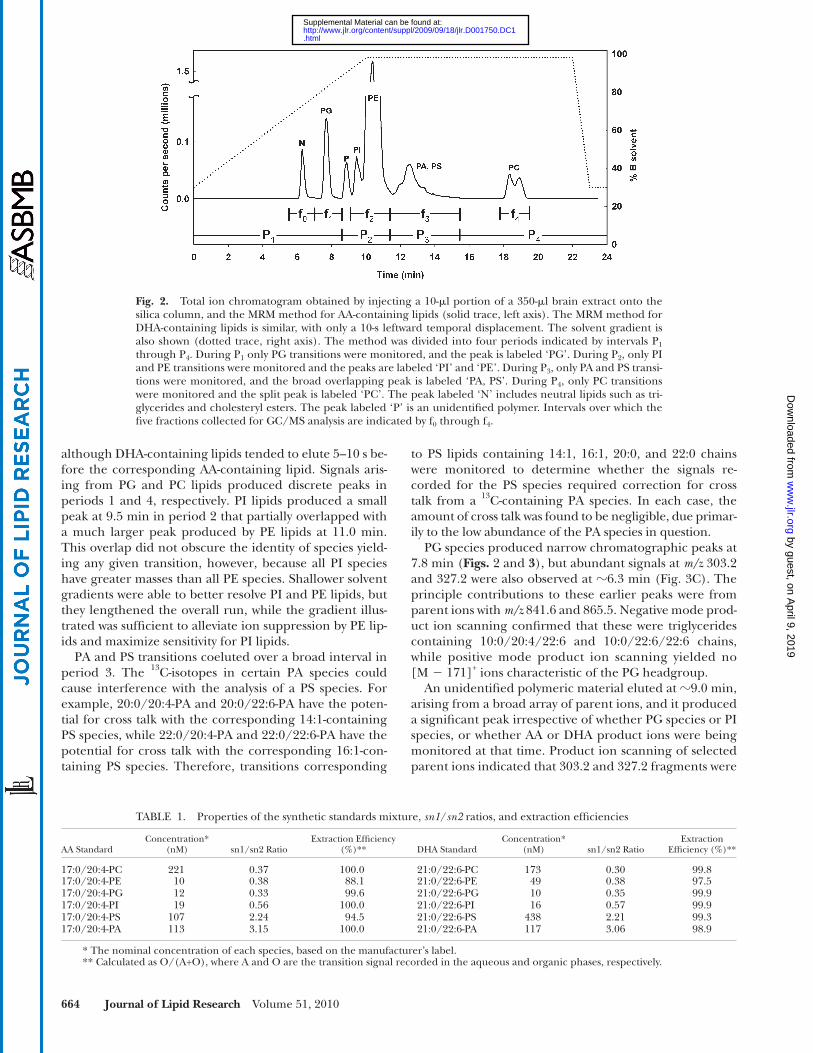
to characterize the elution characteristics of the silica column and normal phase solvent system. Based on the elution times of the major headgroup classes, the chroma-tography system described above in “Methods” was divided into four periods ( Fig. 2 ): PG standards eluted in period 1 between 7.0 and 9.5 min; PI and PE standards eluted in period 2 between 8.5 and 11.5 min; PA and PS standards eluted in period 3 between 11.5 and 13.5 min; and PC standards eluted in period 4 between 18 and 19 min. For PC standards, the yield of sn2 anions from [M + OAc � ] � ions was slightly greater than from [M � CH 3 ] � ions, so only the former were monitored.
To assess extraction effi ciency, aliquots of the standard mixture were subjected to the extraction procedure de-scribed above, and transition signals corresponding to each standard species were measured in both the upper (aqueous) and lower (organic) phase. Results suggested that extraction effi ciency was >88% in all cases, and >99% in all but 4 cases ( Table 1 ).
The fatty acyl chains at the focus of this study, AA and DHA, are highly vulnerable to oxidative damage, so spe-cial precautions were taken to protect both synthetic stan-dards and samples. These included grinding the samples under liquid nitrogen, the use of dicholoromethane in-stead of choloroform to avoid exposure of lipids to phos-gene, and a minimum of solution transfers. Transitions 16 and 32 Da greater than that of the AA and DHA standards (i.e., the monooxidized and peroxidized products) were monitored in the synthetic standard mixture and in brain. Only insignifi cant trace amounts were found.
To assess the suitability of the synthetic standards as in-ternal standards for quantitative MS, a nominally equimo-lar mixture of the 12 standards was examined several times over a period of 4 weeks. This mixture was subjected to normal phase chromatography and MRM analysis as de-scribed above, and signal responses were corrected for 13 C content. The fresh mixture yielded highly reproducible results and demonstrated that the AA-containing PE stan-dard yielded 21.1-fold more signal than the AA-containing PI standard. Corresponding values for AA-containing PA and PS standards, as well as for the DHA-containing stan-dards, are listed in Table 3 .
Between analyses, the mixture was stored in plasma-cleaned autosampler vials at � 80°C. After 4 weeks, the MRM transition signals were reexamined, and the signal responses from PG, PI, PE, PA, and PC standards were found to have decreased relative to the signals obtained for the PS standards. The losses ranged between 30% and 70%, with the greatest losses occurring in the PC stan-dards. Losses of the PS standards were not assessed. Rela-tive losses of similar magnitude were observed whether or not the vials had been plasma cleaned. The reason for these losses is not known but may involve the adherence of lipids in dilute solution to glass walls of the vial, glass-induced
source temperature of 200°C. Helium was used as the carrier gas, with a constant fl ow rate of 0.8 ml/min. Phospholipid fractions for GC/MS analysis were saponifi ed with 1 M NH 4 OH, mixed with d8-AA and d5-DHA internal standards, esterifi ed with penta-fl uorobenzyl bromide in N,N-diisopropylethylamine, and ex-tracted into isooctane as described previously ( 34 ). A standard curve for converting the internal standard signals to concentra-tion was prepared with solutions of neat AA and DHA in metha-nol that were derivatized in the same manner.
Data analysis The integrated signal for each monitored mass transition was
corrected for 13 C content and processed as described in “Results.” Quantitative sensitivity varied with the inherent diffi culty of de-tecting lipid species in some headgroup classes and the amounts of these headgroup classes present in the brain. In addition, the results were subject to several assumptions that render them somewhat approximate. One assumption was that AA and DHA chains always occupied the sn2 position, as in the synthetic stan-dards. However, lipids may have these chains in the sn1 position, they may have both an AA and a DHA chain, and they may even have two AA or two DHA chains. Precise corrections for these uncertainties are not available. When a lipid species has two AA or DHA chains, these species will be overcounted by a factor of 1 + r , where r is an sn1 / sn2 ratio listed in Table 1 . The numerical results provided for these species represent the measured values divided by this factor. No attempt was made to correct for differences in ionization effi ciency due to differ-ences in mass or due to differences between ether-linked and acyl-linked sn1 chains.
RESULTS
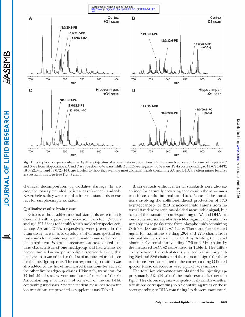
Qualitative results: brain tissue Simple mass spectra of brain tissue extracts are illus-
trated in Fig. 1 . The positive ion scans in both brain re-gions were dominated by the [M + H] + ions of 16:0/18:1-PC ( m/z 760.6), 16:0/16:0-PC ( m/z 734.6), and 18:0/18:0-PC ( m/z 790.6), as confi rmed by separate MS/MS scans. The corresponding PE lipids were minor features of the spec-trum (e.g., 16:0/18:1-PE at m/z 746.6). The most abundant PC lipids containing AA and DHA (e.g., 18:0/20:4-PC at m/z 810.6 and 18:0/22:6-PC at m/z 834.6) were also evi-dent, but lipid species containing AA and DHA in any other headgroup classes could not be detected in the posi-tive ion mode. The negative ion scans in both brain re-gions were dominated by the [M � H] � ions of 18:0/22:6-PS ( m/z 834.5) and 18:0/20:4-PI ( m/z 885.5). Some peaks, however, were most likely the superimposed ions of 18:0/22:6-PE and 18:0/18:0-PS, which were nearly isobaric at m/z 790.54 and 790.56, respectively.
Differences between brain regions were discernable in these spectra, but relatively few lipids containing AA or DHA could be positively or uniquely identifi ed, and quan-titative reproducibility was poor at this level of mass spec-trometric analysis. Chromatographic separation of most any type would help overcome these problems by deliver-ing lipid species at characteristic elution times and reduc-ing ion suppression. Normal phase separation on a silica column was chosen for further studies, because tissue ex-tracts could be injected without further processing and
by guest, on April 9, 2019
ww
w.jlr.org
Dow
nloaded from
.html http://www.jlr.org/content/suppl/2009/09/18/jlr.D001750.DC1Supplemental Material can be found at:
Polyunsaturated lipids in mouse brain 663
Brain extracts without internal standards were also ex-amined for naturally occurring species with the same mass transitions as the internal standards. None of the transi-tions involving the collision-induced production of 17:0 heptadecanoate or 21:0 heneicosanoate anions from in-ternal standard parent ions yielded measurable signal, but some of the transitions corresponding to AA and DHA an-ions from internal standards yielded signifi cant peaks. Pre-sumably, these peaks arose from phospholipids containing O-linked 18:0 and 22:0 sn1 chains. Therefore, the expected signal for transitions yielding 20:4 and 22:6 chains from internal standards were calculated by dividing the signal obtained for transitions yielding 17:0 and 21:0 chains by the measured sn1 / sn2 ratios listed in Table 1 . The differ-ences between the calculated signal for transitions yield-ing 20:4 and 22:6 chains, and the measured signal for these transitions, were attributed to the corresponding O-linked species (these corrections were typically very minor).
The total ion chromatogram obtained by injecting ap-proximately 3% (10 � l) of the brain extract is shown in Fig. 2 . The chromatogram was qualitatively similar whether transitions corresponding to AA-containing lipids or those corresponding to DHA-containing lipids were monitored,
chemical decomposition, or oxidative damage. In any case, the losses precluded their use as reference standards. Nevertheless, they were useful as internal standards to cor-rect for sample-sample variation.
Qualitative results: brain tissue Extracts without added internal standards were initially
examined with negative ion precursor scans for m/z 303.2 and m/z 327.3 ions to identify which molecular species con-taining AA and DHA, respectively, were present in the brain tissue, as well as to develop a list of mass spectral ion transitions for monitoring in the tandem mass spectrome-ter experiment. When a precursor ion peak eluted at a time characteristic of one headgroup and had a mass ex-pected for a known phospholipid species bearing that headgroup, it was added to the list of monitored transitions for that headgroup class. The corresponding transition was also added to the list of monitored transitions for each of the other fi ve headgroup classes. Ultimately, transitions for 27 individual species were monitored for each of the six AA-containing subclasses and for each of the six DHA-containing subclasses. Specifi c tandem mass spectrometric ion transitions are provided as supplementary Table I.
Fig. 1. Simple mass spectra obtained by direct injection of mouse brain extracts. Panels A and B are from cerebral cortex while panels C and D are from hippocampus. A and C are positive mode scans, while B and D are negative mode scans. Peaks corresponding to 18:0/20:4-PE, 18:0/22:6-PE, and 18:0/20:4-PC are labeled to show that even the most abundant lipids containing AA and DHA are often minor features in spectra of this type (see Figs. 5 and 6 ).
by guest, on April 9, 2019
ww
w.jlr.org
Dow
nloaded from
.html http://www.jlr.org/content/suppl/2009/09/18/jlr.D001750.DC1Supplemental Material can be found at:
664 Journal of Lipid Research Volume 51, 2010
to PS lipids containing 14:1, 16:1, 20:0, and 22:0 chains were monitored to determine whether the signals re-corded for the PS species required correction for cross talk from a 13 C-containing PA species. In each case, the amount of cross talk was found to be negligible, due primar-ily to the low abundance of the PA species in question.
PG species produced narrow chromatographic peaks at 7.8 min ( Figs. 2 and 3 ), but abundant signals at m/z 303.2 and 327.2 were also observed at � 6.3 min ( Fig. 3C ). The principle contributions to these earlier peaks were from parent ions with m/z 841.6 and 865.5. Negative mode prod-uct ion scanning confi rmed that these were triglycerides containing 10:0/20:4/22:6 and 10:0/22:6/22:6 chains, while positive mode product ion scanning yielded no [M � 171] + ions characteristic of the PG headgroup.
An unidentifi ed polymeric material eluted at � 9.0 min, arising from a broad array of parent ions, and it produced a signifi cant peak irrespective of whether PG species or PI species, or whether AA or DHA product ions were being monitored at that time. Product ion scanning of selected parent ions indicated that 303.2 and 327.2 fragments were
although DHA-containing lipids tended to elute 5–10 s be-fore the corresponding AA-containing lipid. Signals aris-ing from PG and PC lipids produced discrete peaks in periods 1 and 4, respectively. PI lipids produced a small peak at 9.5 min in period 2 that partially overlapped with a much larger peak produced by PE lipids at 11.0 min. This overlap did not obscure the identity of species yield-ing any given transition, however, because all PI species have greater masses than all PE species. Shallower solvent gradients were able to better resolve PI and PE lipids, but they lengthened the overall run, while the gradient illus-trated was suffi cient to alleviate ion suppression by PE lip-ids and maximize sensitivity for PI lipids.
PA and PS transitions coeluted over a broad interval in period 3. The 13 C-isotopes in certain PA species could cause interference with the analysis of a PS species. For example, 20:0/20:4-PA and 20:0/22:6-PA have the poten-tial for cross talk with the corresponding 14:1-containing PS species, while 22:0/20:4-PA and 22:0/22:6-PA have the potential for cross talk with the corresponding 16:1-con-taining PS species. Therefore, transitions corresponding
Fig. 2. Total ion chromatogram obtained by injecting a 10- � l portion of a 350- � l brain extract onto the silica column, and the MRM method for AA-containing lipids (solid trace, left axis). The MRM method for DHA-containing lipids is similar, with only a 10-s leftward temporal displacement. The solvent gradient is also shown (dotted trace, right axis). The method was divided into four periods indicated by intervals P 1 through P 4 . During P 1 only PG transitions were monitored, and the peak is labeled ‘PG’. During P 2 , only PI and PE transitions were monitored and the peaks are labeled ‘PI’ and ‘PE’. During P 3 , only PA and PS transi-tions were monitored, and the broad overlapping peak is labeled ‘PA, PS’. During P 4 , only PC transitions were monitored and the split peak is labeled ‘PC’. The peak labeled ‘N’ includes neutral lipids such as tri-glycerides and cholesteryl esters. The peak labeled ‘P’ is an unidentifi ed polymer. Intervals over which the fi ve fractions collected for GC/MS analysis are indicated by f 0 through f 4 .
TABLE 1. Properties of the synthetic standards mixture, sn1 / sn2 ratios, and extraction effi ciencies
AA StandardConcentration*
(nM) sn1/sn2 RatioExtraction Effi ciency
(%)** DHA StandardConcentration*
(nM) sn1/sn2 RatioExtraction
Effi ciency (%)**
17:0/20:4-PC 221 0.37 100.0 21:0/22:6-PC 173 0.30 99.817:0/20:4-PE 10 0.38 88.1 21:0/22:6-PE 49 0.38 97.517:0/20:4-PG 12 0.33 99.6 21:0/22:6-PG 10 0.35 99.917:0/20:4-PI 19 0.56 100.0 21:0/22:6-PI 16 0.57 99.917:0/20:4-PS 107 2.24 94.5 21:0/22:6-PS 438 2.21 99.317:0/20:4-PA 113 3.15 100.0 21:0/22:6-PA 117 3.06 98.9
* The nominal concentration of each species, based on the manufacturer’s label.** Calculated as O/(A+O), where A and O are the transition signal recorded in the aqueous and organic phases, respectively.
by guest, on April 9, 2019
ww
w.jlr.org
Dow
nloaded from
.html http://www.jlr.org/content/suppl/2009/09/18/jlr.D001750.DC1Supplemental Material can be found at:

Polyunsaturated lipids in mouse brain 665
, ,a n ic is the signal recorded for phospholipid species n in headgroup class a for sample i , and , ,a s ic is the signal re-corded for an internal standard in the same headgroup class and sample. Primes indicates that the signal has been corrected for 13 C content, FA is the fatty acyl content of the collected column effl uent fraction x for sample i in nmol, w is the mass of tissue sample i in g, 0.75 is the fraction of the column effl uent that was collected by the fraction col-lector, and 10/350 is the portion of the tissue extract that was injected onto the column. It should be noted that the term in parentheses in equation 1 is constant for all phos-pholipid species within a headgroup class. Therefore, the relative values of PL n for different sample sets depend only on the count ratio averages ( r a,n ) and are independent of each other.
For PI/PE and PA/PS lipids, data reduction requires information about the relative sensitivity of the MRM method for these headgroup classes, given by the ra-tios abs of signals recorded from an equimolar mixture of a lipid in headgroup class a and a lipid in headgroup class b , provided in Table 3 . Assuming that the sum of the signals recorded for all lipid species in a headgroup class, corrected for the sensitivity of the MRM method for lipids in that headgroup class, is proportional to the molar concentration of the lipids in that head-group class, it follows that a fraction containing lipids in two different headgroup classes may be divided ac-cording to:
,,
, ,
a tota x w
a tot ab b tot
cFA FA
c s c
and
,,
, ,
ab b totb x w
a tot ab b tot
s cFA FA
c s c
where
27
, ,1
a tot a nn
c c
and 27
, ,1
b tot b nn
c c
The concentration of an individual phospholipid may then be obtained by substituting either FA a or FA b for FA x,w in equation 1. This analytical approach has various strengths and potential weaknesses that will be discussed below. The one potential weakness we consider at this point is whether all of the AA and DHA chains in each fraction assayed by GC/MS were in phospholipids that corresponded to monitored MRM transitions. The two MRM lists included every fatty acyl chain with an even number of carbon atoms and every plausible number of double bonds, as well as known types of O-linked sn1 chains. Furthermore, the lists were compiled from precur-sor scans, thereby accounting for every detectable parent
produced fortuitously through multiple fragmentations of larger ions.
Quantitative results: brain tissue A total of six hippocampus and six cortical brain sam-
ples weighing between 2.0 and 4.5 mg were analyzed. The tissue pieces were reduced to 350- � l extracts, each injec-tion onto the column was 10 � l, and precisely 25% of the column effl uent was directed into the mass spectrometer. Therefore, the data was ultimately derived from 14–32 � g of tissue. Each extract was injected twice, once for each of the two MRM methods, and each MRM analysis yielded 28 measurements (27 phospholipid species plus transitions arising from the 17:0 or 21:0 chains in the internal stan-dards) for each of six phospholipid headgroup classes, or 6 × 28 × 2 = 336 individual measurements.
The column effl uent was collected in fi ve fractions as indicated in Fig. 2 . The AA and DHA content of each frac-tion were determined by GC/MS. Results grouped by brain region did not reveal signifi cant differences between regions; therefore, GC/MS results for all 12 tissue samples were averaged and are listed in Table 2 .
For PG and PC lipids, the conversion of MRM signals to quantitative results was performed by:
,, 10
350, 0.75x w
n a na sum
FAPL r
r ( 1 )
where PL n is the average concentration of an individual phospholipid species n ,
6
, , , , ,1
16a n a n i a s i
i
r c c ( 2 )
12
, ,1
1( / )
12x w x i ii
FA FA w ( 3 )
(averaged over all 12 samples)
12 28
, , ,1 1
112a sum a n i
i n
r r ( 4 )
(the sum of 28 transitions averaged over all 12 samples)
TABLE 2. Fatty acid content by GC/MS in normal phase fractions
Fraction Lipid Species AA Content* DHA Content*
0 TG 1904 ±262 828 ±2061 PG 78 ±12 9 ±32 PI,PE 5492 ±898 6040 ±10623 PA,PS 315 ±48 1064 ±2364 PC 2490 ±471 763 ±129
* nmol/g tissue ± SD of 12 samples.
TABLE 3. Sensitivity ratios for MS/MS detection
Headgroup Classes Lipids Containing AA Lipids Containing DHA
PE / PI 21.1 61.7PA / PS 3.3 6.1
by guest, on April 9, 2019
ww
w.jlr.org
Dow
nloaded from
.html http://www.jlr.org/content/suppl/2009/09/18/jlr.D001750.DC1Supplemental Material can be found at:

666 Journal of Lipid Research Volume 51, 2010
of the data would obscure the inherent precision of the MRM methods and the real differences in the amounts of individual phospholipid species in different brain regions. Therefore, none of the error in the GC/MS measure-ments was propagated through to the individual phospho-lipid results, and it should be understood that the relative quantitative precision with which brain regions may be compared is greater than the absolute quantitative preci-sion. In other words, there is more uncertainty in the ver-tical scales for each graph than is suggested by the error bars, and this uncertainty may be derived from the data in Table 2 .
Quantitative results: regional differences in brain tissue As might be expected, the predominant species in most
headgroup classes had 18:0 and 16:0 acyl-linked chains ( Figs. 5 and 6 ). Most headgroup classes also had signifi -cant amounts of 18:1 chains, while the 20:4-PE and 22:6-PE
ion yielding AA or DHA product ions. Precursor scans did detect AA and DHA in triglycerides, which eluted in frac-tion 0 and are included in Table 2 . These scans did not detect any cholesterol esters of AA or DHA that would have also eluted in fraction 0, although these compounds ionize poorly. A targeted MRM method for lysophospho-lipids containing AA or DHA was created, and none were detected throughout the 24 min elution. Another targeted MRM method for lipids with PI-phosphate headgroups was created. Only trace amounts were detected, so no fur-ther attempt to quantify PI-phosphate lipids was made.
It has been reported that CLs in mouse brain tissue have a more diverse array of fatty acyl chains than those in myo-cardial or skeletal muscle tissue and that AA and DHA chains constitute a signifi cant fraction of these chains ( 35 ). Therefore, we created a targeted MRM method for a set of synthetic CL standards and the mostly likely CL spe-cies based on the reported prevalence of fatty acyl chains in brain CLs. The method assumed that parent ions would be doubly charged. Results showed that synthetic CLs eluted at about the same time as PI and PE lipids. There were minimally detectable signals for several CL species, but they were insuffi cient for meaningful quantitation. These results are consistent with the low reported concen-trations of AA and DHA in mouse brain CLs (4–10 nmol/mg protein) ( 35 ).
Results by headgroup class are summarized in Fig. 4 . Overall, there were 10.3 � mol/g tissue of AA-containing lipids in the tissue extracts, which was somewhat greater than the 8.7 � mol/g of DHA-containing lipids. AA and DHA were most abundant in PE lipids but relatively rare in PG and PA lipids. Neutral, PI, and PC lipids contained more AA than DHA, while PE and PS lipids contained more DHA. The greater amount of DHA in PS lipids is consistent with the known preference for DHA chains among enzymes involved in PS headgroup synthesis ( 36 ).
Quantitative results for individual phospholipid species are summarized in Figs. 5 and 6 . An important feature to note in these fi gures is the high degree of quantitative pre-cision compared with the precision of the GC/MS fatty acid data listed in Table 2 . Ordinarily, the uncertainty of the fatty acid quantitation would be propagated through the equations given above and refl ected in the results for individual phospholipid species. However, this treatment
Fig. 3. Examples of extracted ion chromatograms for selected PG lipids. In panel A, the peak corre-sponding to 18:0/20:4-PG is shown eluting at 7.8 min, along with panel B as an inset showing that the syn-thetic standard 17:0/20:4-PG (labeled ‘S’) elutes at the same time. In panel C, the peak representing a minor species is also shown eluting at 7.8 min. The peak at 6.3 min is most likely an AA-containing tri-glyceride (e.g., 12:0/18:3/20:4-TG), because it coe-lutes with species shown by MS/MS analysis to represent 10:0/20:4/22:6-TG. The asterisk in panels A and C indicates that the parent ions also yield [M � 171] + fragment ions in positive mode MS/MS studies.
Fig. 4. Total AA and DHA in the six major headgroup classes and in neutral lipids. The data for neutral (NL), PG, and PC lipids were obtained directly from GC/MS analysis of fatty acyl chains in frac-tions f 0 , f 1 , and f 4 (see Fig. 1 ). The data for PI, PE, PA, and PS lipids were obtained by dividing the results from GC/MS analysis of frac-tions f 2 and f 3 as described in the text. There were no signifi cant differences between cerebral cortex and hippocampus, so the re-sults are the average of all 12 brain extracts.
by guest, on April 9, 2019
ww
w.jlr.org
Dow
nloaded from
.html http://www.jlr.org/content/suppl/2009/09/18/jlr.D001750.DC1Supplemental Material can be found at:

Polyunsaturated lipids in mouse brain 667
species in such samples may be compared with a high de-gree of precision. Several factors contribute to this preci-sion, beginning with a sample preparation protocol that emphasized ultracold tissue manipulations and the mini-mization of exposure to oxygen. These precautions are particularly important when focusing on polyunsaturated phospholipids because of their susceptibility to oxidative degradation. The addition of specifi c antioxidants may in-crease protection to these lipid species, but additives of this type were not included or examined in this study, be-cause so-called antioxidants may also have pro-oxidant activity. Another factor that contributed to the precision of these measurements was the availability of synthetic odd-length internal standards for each headgroup and for each acyl chain type examined. The odd-length chains minimized spectral overlap with natural phospho-lipid species while coeluting with analytes under normal phase chromatographic conditions. Coelution of internal standards and analytes is advantageous, because it controls for varying degrees of ion suppression by other molecules that may be present in the eluent at any given time.
It should be noted that the relative phospholipid con-centrations in the two different sample groups arise from independent measurements. This independence is achieved
subclasses also contained signifi cant amounts of 16:0, 18:1, and 18:0 plasmalogens. The 22:6-PI subclass was notable for having an abundance of long saturated second acyl chains.
Among the AA-containing subclasses, the amount of 18:1/20:4 lipids was virtually identical in the two brain re-gions, except for a small difference in PC lipids. Cortical brain contained more 16:0/20:4-PG and 18:0/20:4-PG, as well as plasmalogens 16:0p/20:4-PE and 18:0p/20:4-PE, than hippocampus. Cortical brain contained less 18:0/20:4-PS, 18:0/20:4-PE, 16:0/20:4-PC, 18:1/20:4-PC, and 18:0/20:4-PC than hippocampus.
Among DHA-containing subclasses, all signifi cant dif-ferences between brain regions refl ected greater amounts in cortical brain. There were no signifi cant differences de-tected in the 22:6-PA and 22:6-PE subclasses, but all major phospholipid species in the 22:6-PG, 22:6-PI, 22:6-PS, and 22:6-PC subclasses were signifi cantly greater in cerebral cortex compared with hippocampus.
DISCUSSION
The results demonstrate that a large array of phospho-lipids may be quantifi ed in milligram-sized samples of brain tissue and that the concentrations of specifi c lipid
Fig. 5. The concentrations of AA-containing phospholipid species in cerebral cortex (black bars) and hippocampus (gray bars) in nmol/g tissue. Each result is the mean of six different brain extracts, and the error bars represent SD. An asterisk indicates results with statistically signifi cant differences between brain regions. See text for information about the nature of quantitative uncertainty in these data.
by guest, on April 9, 2019
ww
w.jlr.org
Dow
nloaded from
.html http://www.jlr.org/content/suppl/2009/09/18/jlr.D001750.DC1Supplemental Material can be found at:

668 Journal of Lipid Research Volume 51, 2010
able reference standards for the phospholipid species be-ing examined. Without reference standards, it is necessary to assume that detection effi ciency was constant within each headgroup class and that the ion signals recorded for each transition are proportional to the concentrations of the species present. Bruegger et al. ( 37 ) showed that de-tection effi ciency in positive mode precursor scanning was a strong function of lipid mass, and a similar relationship may also apply to the negative mode approach used in this study. In addition, there are uncertainties about the ion-ization effi ciency of sn2 chains in phospholipids contain-ing O-linked versus acyl-linked sn1 chains, as well as differences in ion yields from the sn1 and sn2 positions ( Table 1 ). Positional differences in ion yields will intro-duce error into any measurements in which AA and DHA do not occupy the sn2 position as in the internal standard. There were measurable amounts of phospholipids con-taining two AA chains, two DHA chains, or one of each, so the potential for signifi cant error exists. In situations where a phospholipid has one AA and one DHA chain, it is possible to calculate the fraction in which the AA chain is in the sn1 position (see Appendix). The AA chain occu-pied the sn1 position in 70–95% of such molecules in the PE, PA, and PS headgroup classes.
by averaging r a,n over the sample groups, but averaging the remaining terms (within the parentheses of equation 1) over all samples. It might appear reasonable instead to av-erage r a,n /r a,sum over each sample group and multiply those averages by the fatty acid content of each fraction. However, that approach either forces the total phospholipid content in different samples to be precisely equivalent or causes real dif-ferences between samples to be obscured by experimental uncertainty in the fatty acid content measurements. In con-trast, the approach used in this work makes it reasonable to conclude, for example, that the two results for 18:1/20:4-PG in Fig. 5 are truly indistiguishable, while the results for 16:0/20:4-PG and 18:0/20:4-PG indicate that there is more AA-containing PG in cortex than in hippocampus.
The absolute scale on which we have placed these re-sults is only approximately accurate, for several reasons, including numerical uncertainty in the fatty acid assay re-sults, uncertainty in assigning the fatty acid assay results to lipids from different headgroup classes that coelute, and uncertainty over whether or not signifi cant sources of AA and DHA other than phospholipid exist in each fraction. These problems could have been addressed in various ways, but addressing them would have solved only a small part of the more fundamental problem, the lack of avail-
Fig. 6. The concentrations of DHA-containing phospholipid species in cerebral cortex (black bars) and hippocampus (gray bars) in nmol/g tissue. Each result is the mean of six different brain extracts, and the error bars represent SD. An asterisk indicates results with statistically signifi cant differences between brain regions. See text for information about the nature of quantitative uncertainty in these data.
by guest, on April 9, 2019
ww
w.jlr.org
Dow
nloaded from
.html http://www.jlr.org/content/suppl/2009/09/18/jlr.D001750.DC1Supplemental Material can be found at:
Polyunsaturated lipids in mouse brain 669
vitro data showing that an AA oxidation product (hy-droxynonenal) is amyloidogenic in vitro, whereas the anal-ogous DHA oxidation product (hydroxyhexenal) is not ( 27 ). The available data on AA and DHA levels in the brain do not clarify whether they are reduced by oxidative deg-radation and their oxidation products lead to neurode-generative disease, or whether reduced levels fail to protect other substances in the brain from oxidative damage. It is conceivable that the products of AA oxidation are neuro-toxic and that the products of DHA oxidation are less toxic and, hence, protective.
In conclusion, a method has been developed to quantify phospholipid molecular species containing specifi c fatty acyl chains using targeted negative ion MRM-LC/MS/MS techniques. The relative abundance of these molecular species was used to convert the total amounts of AA and DHA in each HPLC fraction obtained by GC/MS into a quantitative assessment of each molecular species. The re-sults of this study facilitate further study of polyunsatu-rated phospholipids in brain tissue and investigations into the functional and pathological signifi cance of quantita-tive differences with methods that can distinguish among phospholipid species.
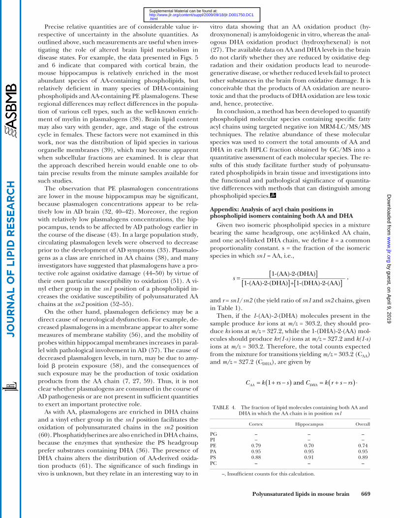
Appendix: Analysis of acyl chain positions in phospholipid isomers containing both AA and DHA
Given two isomeric phospholipid species in a mixture bearing the same headgroup, one acyl-linked AA chain, and one acyl-linked DHA chain, we defi ne k = a common proportionality constant. s = the fraction of the isomeric species in which sn1 = AA, i.e.,
1-(AA)-2-(DHA)
1-(AA)-2-(DHA) + 1-(DHA)-2-(AA) s ,
and r = sn1 / sn2 (the yield ratio of sn1 and sn2 chains, given in Table 1 ).
Then, if the 1 -(AA)- 2 -(DHA) molecules present in the sample produce ksr ions at m/z = 303.2, they should pro-duce ks ions at m/z = 327.2, while the 1-(DHA)-2-(AA) mol-ecules should produce kr(1-s) ions at m/z = 327.2 and k(1-s) ions at m/z = 303.2. Therefore, the total counts expected from the mixture for transitions yielding m/z = 303.2 (C AA ) and m/z = 327.2 (C DHA ), are given by
1 andAA DHAC k rs s C k r s rs .
Precise relative quantities are of considerable value ir-respective of uncertainty in the absolute quantities. As outlined above, such measurements are useful when inves-tigating the role of altered brain lipid metabolism in disease states. For example, the data presented in Figs. 5 and 6 indicate that compared with cortical brain, the mouse hippocampus is relatively enriched in the most abundant species of AA-containing phospholipids, but relatively defi cient in many species of DHA-containing phospholipids and AA-containing PE plasmalogens. These regional differences may refl ect differences in the popula-tion of various cell types, such as the well-known enrich-ment of myelin in plasmalogens ( 38 ). Brain lipid content may also vary with gender, age, and stage of the estrous cycle in females. These factors were not examined in this work, nor was the distribution of lipid species in various organelle membranes ( 39 ), which may become apparent when subcellular fractions are examined. It is clear that the approach described herein would enable one to ob-tain precise results from the minute samples available for such studies.
The observation that PE plasmalogen concentrations are lower in the mouse hippocampus may be signifi cant, because plasmalogen concentrations appear to be rela-tively low in AD brain ( 32, 40–42 ). Moreover, the region with relatively low plasmalogens concentrations, the hip-pocampus, tends to be affected by AD pathology earlier in the course of the disease ( 43 ). In a large population study, circulating plasmalogen levels were observed to decrease prior to the development of AD symptoms ( 33 ). Plasmalo-gens as a class are enriched in AA chains ( 38 ), and many investigators have suggested that plasmalogens have a pro-tective role against oxidative damage ( 44–50 ) by virtue of their own particular susceptibility to oxidation ( 51 ). A vi-nyl ether group in the sn1 position of a phospholipid in-creases the oxidative susceptibility of polyunsaturated AA chains at the sn2 position ( 52–55 ).
On the other hand, plasmalogen defi ciency may be a direct cause of neurological dysfunction. For example, de-creased plasmalogens in a membrane appear to alter some measures of membrane stability ( 56 ), and the mobility of probes within hippocampal membranes increases in paral-lel with pathological involvement in AD ( 57 ). The cause of decreased plasmalogen levels, in turn, may be due to amy-loid � protein exposure ( 58 ), and the consequences of such exposure may be the production of toxic oxidation products from the AA chain ( 7, 27, 59 ). Thus, it is not clear whether plasmalogens are consumed in the course of AD pathogenesis or are not present in suffi cient quantities to exert an important protective role.
As with AA, plasmalogens are enriched in DHA chains and a vinyl ether group in the sn1 position facilitates the oxidation of polyunsaturated chains in the sn2 position ( 60 ). Phosphatidylserines are also enriched in DHA chains, because the enzymes that synthesize the PS headgroup prefer substrates containing DHA ( 36 ). The presence of DHA chains alters the distribution of AA-derived oxida-tion products ( 61 ). The signifi cance of such fi ndings in vivo is unknown, but they relate in an interesting way to in
TABLE 4. The fraction of lipid molecules containing both AA and DHA in which the AA chain is in position sn1
Cortex Hippocampus Overall
PG – – –PI – – –PE 0.79 0.70 0.74PA 0.95 0.95 0.95PS 0.88 0.91 0.89PC – – –
–, Insuffi cient counts for this calculation.
by guest, on April 9, 2019
ww
w.jlr.org
Dow
nloaded from
.html http://www.jlr.org/content/suppl/2009/09/18/jlr.D001750.DC1Supplemental Material can be found at:

670 Journal of Lipid Research Volume 51, 2010
17 . Barberger-Gateau , P. , C. Raffaitin , L. Letenneur , C. Berr , C. Tzourio , J. F. Dartigues , and A. Alperovitch . 2007 . Dietary patterns and risk of dementia: the three-city cohort study. Neurology . 69 : 1921 – 1930 .
18 . Calon , F. , G. P. Lim , T. Morihara , F. S. Yang , O. Ubeda , N. Salem , S. A. Frautschy , and G. M. Cole . 2005 . Dietary n-3 polyunsaturated fatty acid depletion activates caspases and decreases NMDA recep-tors in the brain of a transgenic mouse model of Alzheimer’s dis-ease. Eur. J. Neurosci. 22 : 617 – 626 .
19 . Calon , F. , G. P. Lim , F. S. Yang , T. Morihara , B. Teter , O. Ubeda , P. Rostaing , A. Triller , N. Salem , K. H. Ashe , et al . 2004 . Docosahexaenoic acid protects from dendritic pathology in an Alzheimer’s disease mouse model. Neuron . 43 : 633 – 645 .
20 . Lim , G. P. , F. Calon , T. Morihara , F. S. Yang , B. Teter , O. Ubeda , N. Salem , S. A. Frautschy , and G. M. Cole . 2005 . A diet enriched with the omega-3 fatty acid docosahexaenoic acid reduces amy-loid burden in an aged Alzheimer mouse model. J. Neurosci. 25 : 3032 – 3040 .
21 . Calon , F. , and G. Cole . 2007 . Neuroprotective action of omega-3 polyunsaturated fatty acids against neurodegenerative diseases: evi-dence from animal studies. Prostaglandins Leukot. Essent. Fatty Acids . 77 : 287 – 293 .
22 . Calderon , F. , and H. Y. Kim . 2004 . Docosahexaenoic acid pro-motes neurite growth in hippocampal neurons. J. Neurochem. 90 : 979 – 988 .
23 . Green , K. N. , H. Martinez-Coria , H. Khashwji , E. B. Hall , K. A. Yurko-Mauro , L. Ellis , and F. M. LaFerla . 2007 . Dietary docosahexaenoic acid and docosapentaenoic acid ameliorate amyloid-beta and tau pathology via a mechanism involving presenilin 1 levels. J. Neurosci. 27 : 4385 – 4395 .
24 . DeMar , J. C. , K. Z. Ma , J. M. Bell , and S. I. Rapoport . 2004 . Half-lives of docosahexaenoic acid in rat brain phospholipids are prolonged by 15 weeks of nutritional deprivation of n-3 polyunsaturated fatty acids. J. Neurochem. 91 : 1125 – 1137 .
25 . Plourde , M. , M. Fortier , M. Vandal , J. Tremblay-Mercier , E. Freemantle , M. Begin , F. Pifferi , and S. C. Cunnane . 2007 . Unresolved issues in the link between docosahexaenoic acid and Alzheimer’s disease. Prostaglandins Leukot. Essent. Fatty Acids . 77 : 301 – 308 .
26 . Arendash , G. W. , M. T. Jensen , N. Salem , N. Hussein , J. Cracchiolo , A. Dickson , R. Leighty , and H. Potter . 2007 . A diet high in omega-3 fatty acids does not improve or protect cognitive performance in Alzheimer’s transgenic mice. Neuroscience . 149 : 286 – 302 .
27 . Liu , L. , H. Komatsu , I. V. J. Murray , and P. H. Axelsen . 2008 . Promotion of amyloid � protein misfolding and fi brillogenesis by a lipid oxidation product. J. Mol. Biol. 377 : 1236 – 1250 .
28 . Yamamoto , A. , and G. Rouser . 1973 . Free fatty-acids of normal hu-man whole brain at different ages. J. Gerontol. 28 : 140 – 142 .
29 . Ulmann , L. , V. Mimouni , S. Roux , R. Porsolt , and J. P. Poisson . 2001 . Brain and hippocampus fatty acid composition in phospho-lipid classes of aged relative cognitive defi cit rats. Prostaglandins Leukot. Essent. Fatty Acids . 64 : 189 – 195 .
30 . Prasad , M. R. , M. A. Lovell , M. Yatin , H. Dhillon , and W. R. Markesbery . 1998 . Regional membrane phospholipid alterations in Alzheimer’s disease. Neurochem. Res. 23 : 81 – 88 .
31 . Rouzer , C. A. , P. T. Ivanova , M. O. Byrne , H. A. Brown , and L. J. Marnett . 2007 . Lipid profi ling reveals glycerophospholipid remodel-ing in zymosan-stimulated macrophages. Biochemistry. 46 : 6026 – 6042 .
32 . Han , X. , D. M. Holtzman , and D. W. McKeel . 2001 . Plasmalogen defi ciency in early Alzheimer’s disease subjects and in animal mod-els: molecular characterization using electrospray ionization mass spectrometry. J. Neurochem. 77 : 1168 – 1180 .
33 . Goodenowe , D. B. , L. L. Cook , J. Liu , Y. Lu , D. A. Jayasinghe , P. W. K. Ahiahonu , D. Heath , Y. Yamazaki , J. Flax , K. F. Krenitsky , et al . 2007 . Peripheral ethanolamine plasmalogen defi ciency: a logical causative factor in Alzheimer’s disease and dementia. J. Lipid Res. 48 : 2485 – 2498 .
34 . Kayganich , K. , and R. C. Murphy . 1991 . Molecular-species analy-sis of arachidonate containing glycerophosphocholines by tandem mass-spectrometry. J. Am. Soc. Mass Spectrom. 2 : 45 – 54 .
35 . Cheng , H. , D. J. Mancuso , X. T. Jiang , S. P. Guan , J. Y. Yang , K. Yang , G. Sun , R. W. Gross , and X. L. Han . 2008 . Shotgun lipidom-ics reveals the temporally dependent, highly diversifi ed cardiolipin profi le in the mammalian brain: Temporally coordinated postnatal diversifi cation of cardiolipin molecular species with neuronal re-modeling. Biochemistry. 47 : 5869 – 5880 .
36 . Kim , H. Y. 2007 . Novel metabolism of docosahexaenoic acid in neural cells. J. Biol. Chem. 282 : 18661 – 18665 .
Given measured values of r, AAC , and DHAC for any given headgroup, s may be calculated from
1 11 1
AA
AA DHA
C rs
C C r r
With mean values of AAC and DHAC from the raw counts for each brain tissue sample, and values of r from Table 1 , we obtained the values of s listed in Table 4 . Among lipids with both an AA and a DHA chain, therefore, we conclude that the AA chain occupies the sn1 position in 70–90% of molecules in the PE, PA, and PS headgroup classes.
REFERENCES
1 . Montine , T. J. , M. D. Neely , J. F. Quinn , M. F. Beal , W. R. Markesbery , L. J. Roberts , and J. D. Morrow . 2002 . Lipid peroxidation in aging brain and Alzheimer’s disease. Free Radic. Biol. Med. 33 : 620 – 626 .
2 . Pratico , D. , V. M. Y. Lee , J. Q. Trojanowski , J. Rokach , and G. A. FitzGerald . 1998 . Increased F-2-isoprostanes in Alzheimer’s dis-ease: evidence for enhanced lipid peroxidation in vivo. FASEB J. 12 : 1777 – 1783 .
3 . Montine , T. J. , K. S. Montine , W. McMahan , W. R. Markesbery , J. F. Quinn , and J. D. Morrow . 2005 . F-2-isoprostanes in Alzheimer and other neurodegenerative diseases. Antioxid. Redox Signal. 7 : 269 – 275 .
4 . Sayre , L. M. , D. A. Zelasko , P. L. R. Harris , G. Perry , R. G. Salomon , and M. A. Smith . 1997 . 4-hydroxynonenal-derived advanced lipid peroxidation end products are increased in Alzheimer’s disease. J. Neurochem. 68 : 2092 – 2097 .
5 . Ando , Y. , T. Brannstrom , K. Uchida , N. Nyhlin , B. Nasman , O. Suhr , T. Yamashita , T. Olsson , M. El Salhy , M. Uchino , et al . 1998 . Histochemical detection of 4-hydroxynonenal protein in Alzheimer amyloid. J. Neurol. Sci. 156 : 172 – 176 .
6 . Markesbery , W. R. , and M. A. Lovell . 1998 . Four-Hydroxynonenal, a product of lipid peroxidation, is increased in the brain in Alzheimer’s disease. Neurobiol. Aging . 19 : 33 – 36 .
7 . Murray , I. V. J. , M. E. Sindoni , and P. H. Axelsen . 2005 . Promotion of oxidative lipid membrane damage by amyloid beta proteins. Biochemistry. 44 : 12606 – 12613 .
8 . Koppaka , V. , and P. H. Axelsen . 2000 . Accelerated accumulation of amyloid beta proteins on oxidatively damaged lipid membranes. Biochemistry. 39 : 10011 – 10016 .
9 . Koppaka , V. , C. Paul , I. V. J. Murray , and P. H. Axelsen . 2003 . Early synergy between A � 42 and oxidatively damaged membranes in promoting amyloid fi bril formation by A � 40. J. Biol. Chem. 278 : 36277 – 36284 .
10 . Komatsu , H. , L. Liu , I. V. Murray , and P. H. Axelsen . 2007 . A mechanistic link between oxidative stress and membrane mediated amyloidogenesis revealed by infrared spectroscopy. Biochim Biophys Acta . 1768 : 1913 – 1922 .
11 . Murray , I. V. J. , L. Liu , H. Komatsu , K. Uryu , G. Xiao , J. A. Lawson , and P. H. Axelsen . 2007 . Membrane mediated amyloidogenesis and the promotion of oxidative lipid damage by amyloid beta pro-teins. J. Biol. Chem. 282 : 9335 – 9345 .
12 . Kyle , D. J. , E. Schaefer , G. Patton , and A. Beiser . 1999 . Low serum docosahexaenoic acid is a signifi cant risk factor for Alzheimer’s de-mentia. Lipids . 34 : S245 .
13 . Morris , M. C. 2006 . Docosahexaenoic acid and Alzheimer disease. Arch. Neurol. 63 : 1527 – 1528 .
14 . Schaefer , E. J. , V. Bongard , A. S. Beiser , S. Lamon-Fava , S. J. Robins , R. Au , K. L. Tucker , D. J. Kyle , P. W. F. Wilson , and P. A. Wolf . 2006 . Plasma phosphatidylcholine docosahexaenoic acid content and risk of dementia and Alzheimer disease - The Framingham heart study. Arch. Neurol. 63 : 1545 – 1550 .
15 . Tully , A. M. , H. M. Roche , R. Doyle , C. Fallon , I. Bruce , B. Lawlor , D. Coakley , and M. J. Gibney . 2003 . Low serum cholesteryl ester-docosahexaenoic acid levels in Alzheimer’s disease: a case-control study. Br. J. Nutr. 89 : 483 – 489 .
16 . Morris , M. C. , D. A. Evans , J. L. Bienias , C. C. Tangney , D. A. Bennett , R. S. Wilson , N. Aggarwal , and J. Schneider . 2003 . Consumption of fi sh and n-3 fatty acids and risk of incident Alzheimer disease. Arch. Neurol. 60 : 940 – 946 .
by guest, on April 9, 2019
ww
w.jlr.org
Dow
nloaded from
.html http://www.jlr.org/content/suppl/2009/09/18/jlr.D001750.DC1Supplemental Material can be found at:

Polyunsaturated lipids in mouse brain 671
37 . Brugger , B. , G. Erben , R. Sandhoff , F. T. Wieland , and W. D. Lehmann . 1997 . Quantitative analysis of biological membrane lipids at the low picomole level by nano-electrospray ionization tan-dem mass spectrometry. Proc. Natl. Acad. Sci. USA . 94 : 2339 – 2344 .
38 . Farooqui , A. A. , and L. A. Horrocks . 2001 . Plasmalogens: work-horse lipids of membranes in normal and injured neurons and glia. Neuroscientist . 7 : 232 – 245 .
39 . Jones , C. R. , T. Arai , J. M. Bell , and S. I. Rapoport . 1996 . Preferential in vivo incorporation of [H-3]arachidonic acid from blood into rat brain synaptosomal fractions before and after cholinergic stimula-tion. J. Neurochem. 67 : 822 – 829 .
40 . Ginsberg , L. , S. Rafi que , J. H. Xuereb , S. I. Rapoport , and N. L. Gershfeld . 1995 . Disease and anatomic specifi city of ethanolamine plasmalogen defi ciency in alzheimers-disease brain. Brain Res. 698 : 223 – 226 .
41 . Farooqui , A. A. , S. I. Rapoport , and L. A. Horrocks . 1997 . Membrane phospholipid alterations in Alzheimer’s disease: Defi ciency of etha-nolamine plasmalogens. Neurochem. Res. 22 : 523 – 527 .
42 . Guan , Z. , Y. A. Wang , N. J. Cairns , P. L. Lantos , G. Dallner , and P. J. Sindelar . 1999 . Decrease and structural modifi cations of phos-phatidylethanolamine plasmalogen in the brain with Alzheimer disease. J. Neuropathol. Exp. Neurol. 58 : 740 – 747 .
43 . Eriksen , J. L. , and C. G. Janus . 2007 . Plaques, tangles, and memory loss in mouse models of neurodegeneration. Behav. Genet. 37 : 79 – 100 .
44 . Reiss , D. , K. Beyer , and B. Engelmann . 1997 . Delayed oxidative deg-radation of polyunsaturated diacyl phospholipids in the presence of plasmalogen phospholipids in vitro. Biochem. J. 323 : 807 – 814 .
45 . Hahnel , D. , K. Beyer , and B. Engelmann . 1999 . Inhibition of per-oxyl radical-mediated lipid oxidation by plasmalogen phospho-lipids and alpha-tocopherol. Free Radic. Biol. Med. 27 : 1087 – 1094 .
46 . Hahnel , D. , T. Huber , V. Kurze , K. Beyer , and B. Engelmann . 1999 . Contribution of copper binding to the inhibition of lipid oxidation by plasmalogen phospholipids. Biochem. J. 340 : 377 – 383 .
47 . Maeba , R. , Y. Sawada , H. Shimasaki , I. Takahashi , and N. Ueta . 2002 . Ethanolamine plasmalogens protect cholesterol-rich liposo-mal membranes from oxidation caused by free radicals. Chem. Phys. Lipids . 120 : 145 – 151 .
48 . Maeba , R. , and N. Ueta . 2003 . Ethanolamine plasmalogens prevent the oxidation of cholesterol by reducing the oxidizability of choles-terol in phospholipid bilayers. J. Lipid Res. 44 : 164 – 171 .
49 . Morandat , S. , M. Bortolato , G. Anker , A. Doutheau , M. Lagarde , J. P. Chauvet , and B. Roux . 2003 . Plasmalogens protect unsaturated
lipids against UV-induced oxidation in monolayer. Biochim. Biophys. Acta. . 1616 : 137 – 146 .
50 . Kuczynski , B. , and N. V. Reo . 2006 . Evidence that plasmalogen is protective against oxidative stress in the rat brain. Neurochem. Res. 31 : 639 – 656 .
51 . Brosche , T. , and D. Platt . 1998 . Mini-review: the biological signif-icance of plasmalogens in defense against oxidative damage. Exp. Gerontol. 33 : 363 – 369 .
52 . Khaselev , N. , and R. C. Murphy . 1999 . Susceptibility of plasmenyl glycerophosphoethanolamine lipids containing arachidonate to oxidative degradation. Free Radic. Biol. Med. 26 : 275 – 284 .
53 . Khaselev , N. , and R. C. Murphy . 2000 . Structural characterization of oxidized phospholipid products derived from arachidonate-contain-ing plasmenyl glycerophosphocholine. J. Lipid Res. 41 : 564 – 572 .
54 . Khaselev , N. , and R. C. Murphy . 2000 . Peroxidation of arachido-nate containing plasmenyl glycerophosphocholine: Facile oxida-tion of esterifi ed arachidonate at carbon-5. Free Radic. Biol. Med. 29 : 620 – 632 .
55 . Murphy , R. C. 2001 . Free-radical-induced oxidation of arachidonoyl plasmalogen phospholipids: Antioxidant mechanism and precursor pathway for bioactive eicosanoids. Chem. Res. Toxicol. 14 : 463 – 472 .
56 . Ginsberg , L. , J. H. Xuereb , and N. L. Gershfeld . 1998 . Membrane instability, plasmalogen content, and Alzheimer’s disease. J. Neurochem. 70 : 2533 – 2538 .
57 . Zubenko , G. S. 1986 . Hippocampal membrane alteration in Alzheimers disease. Brain Res. 385 : 115 – 121 .
58 . Cheng , H. , J. Xu , D. W. McKeel , and X. Han . 2003 . Specifi city and potential mechanism of sulfatide defi ciency in Alzheimer’s disease: an electrospray ionization mass spectrometric study. Cell. Mol. Biol. 49 : 809 – 818 .
59 . Murray , I. V. J. , B. I. Giasson , S. M. Quinn , V. Koppaka , P. H. Axelsen , H. Ischiropoulos , J. Q. Trojanowski , and V. M. Y. Lee . 2003 . Role of alpha-synuclein carboxy-terminus on fi bril formation in vitro. Biochemistry. 42 : 8530 – 8540 .
60 . Zemski Berry , K. A. , and R. C. Murphy . 2005 . Free radical oxidation of plasmalogen glycerophosphocholine containing esterifi ed doco-sahexaenoic acid: structure determination by mass spectrometry. Antioxid. Redox Signal. 7 : 157 – 169 .
61 . Davis , T. A. , L. Gao , H. Yin , J. D. Morrow , and N. A. Porter . 2006 . In vivo and in vitro lipid peroxidation of arachidonate esters: the effect of fi sh oil � -3 lipids on product distribution. J. Am. Chem. Soc. 128 : 14897 – 14904 .
by guest, on April 9, 2019
ww
w.jlr.org
Dow
nloaded from
.html http://www.jlr.org/content/suppl/2009/09/18/jlr.D001750.DC1Supplemental Material can be found at:

1244 Journal of Lipid Research Volume 51, 2010
ERRATA
A previously published errata in the September 2009 issue of the Journal of Lipid Research was incorrect. The authors of the article “ Structural analysis of novel bioactive acylated steryl glucosides in pre-germinated brown rice bran ” ( J. Lipid Res. 49: 2188 – 2196 ) have advised the Journal that the notation Δ8-cholesterol used in the original article is incorrect and the correct notation should be Δ8(14)-cholestenol with the following structure: 5 α -cholest-8(14)-en-3 β -ol based on “ IUPAC-IUB Joint Commission on Biochemical Nomenclature: Nomenclature of Steroids ” (Pure & Appl. Chem. 61: 1783 – 1822, 1989). This misspelling appeared online in the September 2009 issue but has since been corrected. The Journal sincerely regrets this error.
Please note that the correct structure is shown in Figure 3B as published in the original article.
DOI 10.1194/jlr.M800257ERR2
The authors of the article “ Quantitative analysis of phospholipids containing arachidonate and docosahexaenoate chains in microdissected regions of mouse brain ” ( J. Lipid Res. 51: 660 – 671 ) have advised the Journal that “ docosahexaenoate ” had been misspelled as “ docosahexenoate ” . This misspelling appeared initially online but has since been corrected.
DOI 10.1194/jlr.D001750ERR