quantitative subcellular analysis of the effects of … · quantitative subcellular analysis of the...
TRANSCRIPT

Quantitative Subcellular Analysis of the Effects of the Enigmatic Protein PCSK9
By
Nicholas Denis
Department of Biochemistry, Microbiology, and Immunology
Submitted in partial fulfillment
Of the requirements for the degree of
Masters of Science
Faculty of Graduate Studies
The University of Ottawa
Ottawa, ON
December 2010
© Nicholas Denis, Ottawa, Canada, 2011

ii
ABSTRACT
PCSK9 is the third gene implicated in autosomal dominant hypercholesterolemia, due to
its role in promoting the degradation of the low density lipoprotein receptor (LDLR). Little is
known regarding the mechanism by which it promotes the degradation of LDLR, nor the
effects PCSK9 has on other cellular proteins. I report here the first quantitative subcellular
proteomic study of proteins affected by the expression of a variant of PCSK9. I show that
the expression levels of 293 proteins were affected by the expression of the PCSK9-ACE2-
V5 construct. Of particular interest, is a protein involved in receptor recycling, EHBP1,
which shows reduced protein levels by both PCSK9-ACE2-V5 and the PCSK9-D374Y mutant.
I show that an EHBP1 binding protein, EHD4, binds with PCSK9 and LDLR. These results
establish novel effects of PCSK9 on liver cell protein levels, of which some relating to
endosomal sorting are shown to bind to PCSK9 and LDLR in complex, providing insight into
the mechanism of PCSK9 mediated LDLR degradation.

iii
Acknowledgements
My time in the lab started with a nervous phone call to a potential supervisor,
unsure of how to pronounce his name. Dr. Daniel Figeys has been a great supervisor to me
and has always been open, honest and helpful with many discussions about science, but
more so about important life matters outside of the lab. My path through graduate school
has not been that of a straight line, but one with lots of turns and changes. Throughout this,
Daniel was both critical and understanding, a great combination to keep me focused, but
also allow me to pursue my passion outside of science. For this I am very grateful, thank
you Daniel! I would also like to thank the members of my thesis advisory committee, Dr.
Mbikay and Dr. Bennett, for their support, criticisms and suggestions over the years.
Over the years I have had the great pleasure of meeting many students and post-
docs in Daniels lab, and have been lucky enough to become friends with most of them.
Weimin Hou, the first person to train me on the reactor and since then became great
friends. Dr. Julian Vasilescu, my ubiquitination colleague and friend. Dr. Jeffrey C. Smith and
Dr. Theodore Glenn Wright, two great post-docs. Dr. Hou Zhou, Dr. Riujun Tian, and of
course Dr. Houjiang Zhou, you guys have all helped me continuously in the lab and had fun
both doing it. Dr. Maroun Bou-Khalil, Mohamed Abu-Farha and Jean-Phillippe Lambert,
thanks for always taking the time to help me when I needed it. To everyone else, thanks!
Finally I would like to thank my parents John and Linda Denis, and my brother Joel
Denis, for their constant support, interest and encouragement over the years. In addition to
my supervisor, I have them to thank for always listening and talking to me about my crazy
mind-changing decisions, thank you.

iv
Table of contents
Title Page i
Abstract ii
Acknowledgements iii
Table of Contents iv
List of Abbreviations vii
List of Figures viii
List of Tables x
Chapter 1. INTRODUCTION 1
1.1 Literature Review 3
I. Proprotein Convertases 3
i. General Background 3
ii. PC Structure and Function 4
II. PCSK9, a Novel Proprotein Convertase that Regulates the Low-Density Lipoprotein Receptor 7
i. PCSK9 Discovery 7
ii. PCSK9 Structure, Activation and Post-translational modification 8
iii. PCSK9 Expression and Localization 10
iv. PCSK9 and LDLR Degradation 13
v. PCSK9 Mutations in Humans 14
vi. PCSK9 Outside of LDLR Degradation 15
vii. PCSK9 Interaction Partners 16
viii. PCSK9 as a Therapeutic Target 17
III. Low-Density Lipoprotein Receptor 17
i. LDLR Structure and Function 17
ii.LDLR Internalization 18
v
1.2 Hypothesis Statement 20
Chapter 2. MATERIALS AND METHODS 21
Reagents 21
Cell culture and SILAC metabolic labelling 21
Subcellular Fractionation and histodenz gradient preparation 21
Western blot analysis 22
HPLC-ESI-MS/MS 23
Database searching, quantitation and data analysis 24
Validation Experiments 25
PCSK9 Immunoaffinity purifications and interaction experiments 26
Chapter 3. RESULTS 28
Subcellular Fractionation and Western blot Analysis of Histodenz Fractions 28
SILAC Quantitation 29
PCSK9 promotes a decrease in cellular EH domain binding protein 1 and an
increase in actin related protein 2\3 at the protein level 27
PCSK9-ACE2-V5 and PCSK9-D374Y variants decrease both cellular LDLR and
EHBP1 protein levels 41
PCSK9-ACE2-V5 Immunoaffinity purification optimization 44
PCSK9-ACE2-V5 IP-LC-MS/MS analysis of interaction partners 47
PCSK9 and mature LDLR both interact with EHD4 49
Chapter 4. DISCUSSION 55
4.1 Conclusions 72

vi
References 77
Contributions of Collaborators 87
Appendix A. Reagents, solutions and buffers 88
CV 91

vii
List of Abbreviations
PCSK9 Proprotein convertase subtilisin kexin type 9
LDLR Low-density lipoprotein receptor
MS/MS Tandem mass spectrometry
PC Proprotein convertase
ER Endoplasmic reticulum
ACE Angiotensin converting enzyme
EV Empty vector
NARC-1 Neural apoptosis-regulated convertase 1
ADH Autosomal dominant hypercholesterolemia
LDL-C Low-density lipoprotein cholesterol
SREBP Sterol regulatory element-binding protein
EGF Epidermal growth factor
BACE1 β-site amyloid precursor protein (APP)-cleaving enzyme 1
VLDLR Very low-density lipoprotein receptor
DMEM Dulbecco's Modified Eagle Medium
SILAC Stable isotope labelling of amino acids in cell culture
FBS Fetal bovine serum
PBS Phosphate buffered saline
EDTA Ehylenediamine tetraacetic acid
PNS Post-nuclear supernatant
PBS-Tw PBS-Tween
TGN Trans Golgi network
HRP Horseradish peroxidise
HPLC High performance liquid chromatography
ESI Electrospray ionization
LTQ Linear trap quadrupole
SDS-Page Sodium dodecyl sulphate polyacrylamide gel electrophoresis
IP Immunoaffinity purification
IPP Insilicos proteomic pipeline

viii
List of Figures
Figure 1 Structure of human proprotein convertases 5
Figure 2 Structure, mutations and trafficking of PCSK9 as it relates to LDLR
processing and degradation 9
Figure 3 PCSK9 gene expression 12
Figure 4 LDLR life cycle 19
Figure 5 Schematic outlining general workflow of SILAC metabolic labelling
of HuH7 cell lines, subcellular fractionation, protein processing and
LC-MS/MS analysis of resulting peptides 30
Figure 6 Western blot analysis of subcellular fractions for specific organelle 31
markers
Figure 7 Log2 distribution of peptides quantified reveal a normal distribution
of SILAC peptides 33
Figure 8 Heat map clustering of quantitated fractionation data reveals non-
random distribution of proteins throughout subcellular fractions 40
Figure 9 Western blot validation of Arp2/3 levels increased in PCSK9-
ACE2-V5 expressing cells 42
Figure 10 Western blot validation of EHBP1 levels decreased in PCSK9-
ACE2-V5 expressing cells 43
Figure 11 PCSK9-ACE2-V5 and the natural gain of function variant PCSK9-
D374Y expressing HuH7 cells lead to decreased EHBP1 and LDLR
levels in whole cell lysates 45
Figure 12 Optimization of anti-V5 Immunoaffinity purifications for PCSK9-
ACE2-V5 pulldowns 46
Figure 13 PCSK9-ACE2-V5 Immunoaffinity Purification 48
Figure 14 PCSK9 Interaction map 50
ix
Figure 15 Co-IP Experiments reveal PCSK9 and EHD4 Interact 52
Figure 16 Co-IP Experiments Reveal EHD4 and LDLR interact 54

x
List of Tables
Table 1 Partial list of significantly affected non-nuclear proteins and 34
their biological functions
Table 2 PCSK9 Interaction partners via IP-LC-MS/MS 51
Table 3 Literature comparison between SILAC quantitation and previous
gene expression studies 59

1
Chapter 1. INTRODUCTION
The Proprotein convertase subtilisin kexin type 9 (PCSK9) has been identified in
humans to be the third genetic loci linked to autosomal dominant hypercholesterolemia.
This finding was due to the mutations found throughout this protein that affect its ability to
regulate the low-density lipoprotein receptor (LDLR) [25]. Higher levels of PCSK9 or its
gain-of-function mutants lead to increased levels of LDL, whereas lower levels of PCSK9 or
it loss-of-function mutants lead to lower levels of LDL [27,28]. PCSK9 is a member of the
family of proteins called the proprotein convertases. The primary role of proprotein
convertases is to cleave a wide variety of proteins leading to the activation or inactivation
of the proteins by cleaving a key fragment or the generation of smaller biologically active
peptides/proteins. PCSK9 is a unique convertase in many ways; despite having a functional
proteolytic active site that leads to autocatalysis, no known substrates have been identified
to date. As well, PCSK9 moves through the secretory pathway and is secreted into the
plasma. Circulating PCSK9 then binds cell surface LDLR in the liver, is internalized with the
LDLR and disrupts the normal recycling of the LDLR to instead promote the degradation of
the LDLR in acidic lysosomal vesicles. To date, the mechanism of how PCSK9 mediates the
degradation of the LDLR remains unclear, as are the other potential functions and roles that
PCSK9 has in liver cells.
In this study I have employed various proteomic technologies, including qualitative
and quantitative approaches to gain insight into novel roles PCSK9 plays in liver cells.
Through the quantitative analysis of subcellular fractionated HuH7 liver proteins, it was
possible to identify proteins whose relative abundance levels were significantly affected by

2
PCSK9 expression. Through the use of immunoaffinity purifications of PCSK9 containing
protein complexes coupled with liquid chromatography tandem mass spectrometry (LC-
MS/MS), it was possible to identify novel interaction partners of PCSK9. These proteins
have potential links to the PCSK9 mediated degradation of the LDLR, such as the endocytic
machinery proteins EHBP1 and RME-8, as well as many other interesting functions
unrelated to LDLR regulation such as vesicle trafficking, chaperone activity, post-
translational modifications, cell adhesion and cell signalling. The results found in this study,
and future studies stemming from them, provides a better understanding of PCSK9
mediated degradation of the LDLR, as well as other novel functions PCSK9 plays. Overall,
this study of PCSK9 demonstrates the advantages of taking global, large-scale and
quantitative approaches to studying a protein of limited understanding, in gaining novel
information regarding the proteins functions, both known and unknown.

3
1.1 Literature Review
I.Proprotein Convertases
i. General Background
Biologically active polypeptides and proteins are often the product of post-
translational processes and regulations. Often, such polypeptides and proteins are
activated following intracellular proteolytic cleavages of inactive precursor proteins. Such
precursor proteins regulated by proteolytic activation include hormones, cell surface
receptors, proteases, growth factors, cell signalling proteins, as well as pathogenic factors
such as viral and bacterial proteins.
The proteases responsible for such proteolytic activation of precursor proteins are
called proprotein convertases (PCs), and are secretory proteins containing serine protease
activity. The first proprotein protease to be discovered was the kexin protease (KEX2 gene)
from S. cereveisiae, which was later characterized as a Ca2+ -dependant serine
protease[1,2]. Soon after the discovery of Kex2 protease, homologues in humans were
discovered[3,4].
To date nine mammalian PCs have been characterized, and all belong to a
conserved family of subtilisin-like proteinases, due to the structural homology between
their catalytic domains and that of bacterial subtilisins. These proprotein convertases
include seven enzymes that cleave protein precursors containing the multi-basic (K/R)-(X)n-
(K/R)↓ motif, where ↓ represents the cleavage site, X represents any amino acid besides
cysteine, and n= 0,2,4 or 6. These PCs include furin, PC1/3, PC2, PC4, PACE4, PC5/6 and
4
PC7. The two remaining PCs include Subtilisin/kexin-like isozyme-1 (SKI-1) which cleaves at
(R/K)-X-(L,I,V)-Z↓ motifs, where Z represents any amino acid except cysteine, glutamic
acid, valine, or proline. The newest member of the proprotein convertase family is
proprotein convertase subtilisin/ kexin type 9 (PCSK9). To date no known substrates exist
for PCSK9, however it does undergo autocatalytic cleavage of its prodomain at the
VFAQ↓SIP motif[5-11].
ii. Proprotein Convertase Structure and Function
Each currently known PC contains a signal peptide at its N-terminus, a pro-segment
(also known as a pro-domain), a catalytic domain, followed by a C-terminal domain which is
unique to each convertase (Figure 1). Most PCs are soluble proteins, however furin, PC5-B,
PC7 and SKI-1 each contain a type-I single pass transmembrane domain near their C-
terminus. The signal peptide functions as a signal to target the newly synthesized protein to
the secretory pathway, beginning at the endoplasmic reticulum (ER). The pro-domain has
multiple functions. This domain serves both as an intramolecular chaperone, aiding in the
correct folding of the polypeptide during its translation, as well as auto-inhibition of the
catalytic domain. The pro-domain of mammalian PCs have high sequence similarity (30-
67%)[12]. Importantly, the pro-domain is always N-terminal to the catalytic domain,
ensuring the protease is synthesized as an inactive zymogen immediately upon synthesis.
By inhibiting the catalytic domain of the PC, the pro-domain allows for proper temporal and

Figure 1. Structure of human proprotein convertases. Schematic representation of the
protein structure of human proprotein convertases. Each PC contains a N-terminal signal
sequence, a self-inhibitory prodomain, a subtilisin-like catalytic domain, as well as a P
domain responsible for calcium binding and pH dependence. The PCs contain either serine
and threonine rich regions or cysteine rich regions, and sometimes contain
transmembrane domains followed by C-terminal cytoplasmic domains.

5

6
spatial activation of the catalytic activity. This in turn regulates the convertase which
ensures that proteolysis occurs only in the presence of its intended target substrates.
Following autocatalytic cleavage of the pro-domain, the pro-domain remains tightly bound
through non-covalent interactions with the catalytic domain and functions as a competitive
inhibitor. As the PC moves throughout the secretory pathway the complex remains tightly
associated until it reaches its final destination where the organelle environmental milieu
becomes optimal for a secondary cleavage to occur and/or sufficiently low pH and
increasingly high calcium concentrations favour the dissociation [13-20].
The catalytic domains of PCs are related to bacterial subtilisins as well as enzymes
found in the trypsin superfamily [22]. With the conserved active site catalytic triad of Asp,
His and Ser, PCs are able to catalyze the acyl transfer reactions [22]. Additionally, the
oxyanion hole stabilizing Asn residue (PC2 uses Asp oxyanion hole) is also conserved among
the PCs, thereby acting as a hydrogen bond donor, which acts as a hydrogen bond donor
during the transition state which undergoes an accumulation of negative charge on the
scissile carbonyl group. Following the conserved catalytic domain, PCSK1-8 contain a β-
barrel P-domain that functions in stabilizing the catalytic domain and pocket of the
convertase. Finally the C-terminal domains of PCs are unique from one another, and play a
role in regulating their trafficking and cellular localization [23]. The structure of these PC’s
can be seen in Figure1.
7
II. PCSK9, a Novel Proprotein Convertase that Regulates the Low-Density Lipoprotein
Receptor
i. PCSK9 Discovery
Proprotein convertase subtilisin kexin type 9 (PCSK9) was initially discovered and
named neural apoptosis-regulated convertase 1 (NARC-1) for the discovery of its expression
in the cerebellum and brain telencephalon cells [24]. It was found to promote cortical
neurogenesis most likely via promoting the recruitment of undifferentiated neural
progenitor cells towards a neuronal lineage, when NARC-1 was overexpressed in mice [24].
Soon after, human mutations in PCSK9 lead to the implication of PCSK9 as the third genetic
locus that can cause autosomal dominant hypercholesterolemia (ADH) [25]. It was shown
that wild type and mutant forms of PCSK9 responsible for ADH, when overexpressed in
mice and in primary hepatocytes leads to a decrease in low density lipoprotein receptor
(LDLR) levels post-transcriptionally, both in the presence and absence of LDLR adaptor
protein ARH [26]. This study provided a link between the identification of PCSK9 as a
protein involved in hypercholesterolemia in humans and its function. The link between
PCSK9 and hypercholesterolemia was found as PCSK9 leads to decreased LDLR levels post-
transcriptionally, less LDL-cholesterol (LDL-C) can be internalized from circulation into the
liver, thus leading to hypercholesterolemia. Conversely, in mice lacking PCSK9 a decrease in
plasma cholesterol was observed [27], leading to the finding that serum LDL-cholesterol
levels correlate directly with PCSK9 in serum [28].
8
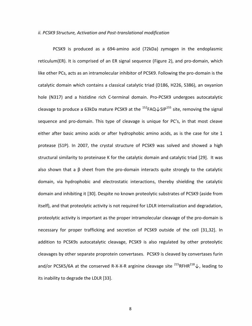
ii. PCSK9 Structure, Activation and Post-translational modification
PCSK9 is produced as a 694-amino acid (72kDa) zymogen in the endoplasmic
reticulum(ER). It is comprised of an ER signal sequence (Figure 2), and pro-domain, which
like other PCs, acts as an intramolecular inhibitor of PCSK9. Following the pro-domain is the
catalytic domain which contains a classical catalytic triad (D186, H226, S386), an oxyanion
hole (N317) and a histidine rich C-terminal domain. Pro-PCSK9 undergoes autocatalytic
cleavage to produce a 63kDa mature PCSK9 at the 152FAQ↓SIP155 site, removing the signal
sequence and pro-domain. This type of cleavage is unique for PC’s, in that most cleave
either after basic amino acids or after hydrophobic amino acids, as is the case for site 1
protease (S1P). In 2007, the crystal structure of PCSK9 was solved and showed a high
structural similarity to proteinase K for the catalytic domain and catalytic triad [29]. It was
also shown that a β sheet from the pro-domain interacts quite strongly to the catalytic
domain, via hydrophobic and electrostatic interactions, thereby shielding the catalytic
domain and inhibiting it [30]. Despite no known proteolytic substrates of PCSK9 (aside from
itself), and that proteolytic activity is not required for LDLR internalization and degradation,
proteolytic activity is important as the proper intramolecular cleavage of the pro-domain is
necessary for proper trafficking and secretion of PCSK9 outside of the cell [31,32]. In
addition to PCSK9s autocatalytic cleavage, PCSK9 is also regulated by other proteolytic
cleavages by other separate proprotein convertases. PCSK9 is cleaved by convertases furin
and/or PCSK5/6A at the conserved R-X-X-R arginine cleavage site 215RFHR218↓, leading to
its inability to degrade the LDLR [33].

Figure 2. Structure, mutations and trafficking of PCSK9 as it relates to LDLR processing
and degradation. a) Schematic showing PCSK9 structure including domains, various gain of
function (above) and loss of function (below) mutations found in humans responsible for
hyper- and hypocholesterolemia, as well as specific amino acids relating to the catalytic
triad and oxyanion hole. b) Diagram showing the general trafficking of the LDLR in the
absence (right) and presence (left) of PCSK9. In the absence of PCSK9 LDLR is internalized
into endosome vesicles and eventually recycled back to the cell surface, whereas in the
presence of PCSK9 it co-internalizes with PCSK9 into endosomes then trafficked to the
lysosome for degradation.

9

10
Besides proteolytic cleavages, PCSK9 also undergoes various other forms of post-
translational modification. These modifications include N-linked glycosylation in the
cysteine and histidine rich C-terminal domain, which occurs throughout the endoplasmic
reticulum, sulfation of the prodomain at Tyr38, and phosphorylation of both the prodomain
at Ser47 and in the cysteine and histidine rich C-terminal domain at Ser688 [24,34].
Glycosylation of PCSK9 can be expected as it is quite common for secreted proteins and
those found in the secretory pathway [34,35]. To date, sulfation of PCSK9 has no known
functional significance and appears to have no effect on PCSK9 mediated LDLR degradation.
While PCSK9 phosphorylation is not quite understood at the moment, it appears to be
dependent on cell type, and thought to play a role in shielding the prodomain from
proteolytic cleavage.
iii. PCSK9 Expression and Localization
PCSK9 was initially found to be expressed in neuroepithelioma, hepatic and colon
carcinoma cell lines, as well as in hepatocytes, kidney, pancreatic, spleen, testes, thymus
and intestinal tissues [24,46]. As a proprotein convertase, PCSK9 is synthesized in the
endoplasmic reticulum, and following proper folding, maturation and processing, moves
throughout the secretory pathway towards the cis- and trans- Golgi apparatus en route to
becoming secreted into the extracellular milieu and circulating plasma. Throughout the
circulating plasma, PCSK9 can be internalized and act intracellularly in hepatic, lung, kidney
and adipose cells [31, 32]. Interestingly, it has been shown that PCSK9 trafficking and

11
localization is affected by the presence of LDLR [36]. In the absence of LDLR, PCSK9
remains in the endoplasmic reticulum, however in the presence of LDLR, PCSK9 is found in
post-ER compartments throughout the secretory pathway (including Golgi and endosomal
compartments), as well can co-localize with LDLR at the cell surface.
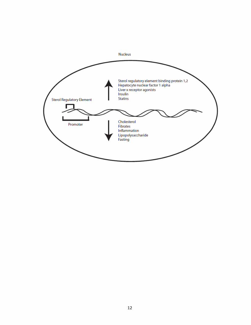
PCSK9 gene expression can be highly modulated depending on the presence of
many proteins and nutritional factors (Figure 3). PCSK9 expression is regulated by sterol
regulatory element-binding proteins 1 and 2 (SREBPs), as the promoter for PCSK9 carries a
sterol regulatory element [37], as well by the co-factor hepatocyte nuclear factor 1 α *38+.
Because of this, liver x receptor agonists (which activate the liver x receptor and thus
SREBP1c) can increase PCSK9 gene expression, as does insulin, while cholesterol, fibrates,
inflammation and lipopolysaccharide [39] and fasting cause a decrease in PCSK9 expression
[40,41].
Interestingly, despite not having been found to be a nuclear protein, studies have
been carried out to see the effects PCSK9 plays on global gene expression. To this end, it
appears that PCSK9 gene expression in turn affects the gene transcript levels of not only
genes belonging to cholesterol and steroid biosynthesis, but also affects genes related to
immune and viral response, sterol metabolism, the ubiquitin proteasome pathway, cell
cycle regulation, inflammation and stress response, suggesting that PCSK9 plays, in vivo,
other functions that have yet to be discovered [42,43].

Figure 3. PCSK9 gene expression. PCSK9 gene expression is known to be affected by
multiple stimulus, dietary factors and proteins. Gene expression is controlled by a sterol
regulatory element in the promoter of PCSK9. This leads to an increase in gene expression
in the presence of sterol regulatory element binding protein 1 and 2, HNF-1α, liver x
receptor agonists, insulin and statins, while factors that decrease gene expression include
cholesterol, fibrates, inflammation, LPS and fasting.
12

13
iv. PCSK9 and LDLR Degradation
Upon discovery that PCSK9 was implicated in hypercholesterolemia, and that PCSK9
overexpression causes a decrease in LDLR levels and an increase in circulating plasma
cholesterol, the relationship between PCSK9 and LDLR degradation has been studied
extensively. PCSK9 overexpression causes a decrease in hepatic LDLR protein levels while
mRNA levels remain unchanged, suggesting an effect at the protein level, while circulating
PCSK9 can decrease adipose, lung and kidney tissue LDLR, in addition to hepatic LDLR [44].
Oppositely, inhibition of PCSK9 expression leads to a decrease in circulating LDL levels
[45,46]. Additionally, it was found that PCSK9 mediated LDLR degradation is unaffected by
proteasome inhibition, but must occur in some post-endoplasmic reticulum compartment,
as treatment with brefeldin A inhibited the degradation of LDLR. PCSK9 was then shown to
work extracellularly. This was demonstrated by adding purified PCSK9 to hepatic cell
cultures which reduced cell surface LDLR levels, implying that LDLR mediated endocytosis
and interaction is necessary for PCSK9 internalization [48,49]. However PCSK9 mediated
degradation of the LDLR does involve an internal pathway, the importance of both
pathways remains unclear [50]. The internalization and degradation of LDLR by PCSK9
requires the transfer from a late endosomal acidic compartment to the lysosome [51,52].
Interestingly, in vitro binding assays show about an 150 fold increase in affinity of PCSK9 to
LDLR at an acidic pH of 5.3 as compared to a neutral pH of 7.4, strengthening the results
that implicate PCSK9 mediated degradation of the LDLR occur in late endosome/lysosome
compartments [53]. Following this study, it was shown that the interaction was mapped to
occur at one of LDLRs many extracellular epidermal growth factor (EGF) like-repeat

14
domains, specifically the EGF-A domain, which required calcium as a co-factor for binding.
The binding of PCSK9 to the EGF-A domain of the LDLR was also stronger (150x) at a pH of
5.2 as compared to a neutral pH of 7 [54]. This interaction occurs at the surface of the
catalytic domain of PCSK9, specifically between Asp-374 of PCSK9 and the His-306 of the
LDLR EGF-A repeat domain [55]. Interestingly, this site is known for as the strongest gain-of-
function mutation of PCSK9. In humans the D374Y mutation leads to a stronger association
with LDLR (25x) and accelerated degradation of the LDLR [56]. Most recently it was
postulated that the C-terminal cytoplasmic domain of LDLR, which interacts with endocytic
machinery, does not play a role in PCSK9 mediated internalization and degradation of the
LDLR [53b]. This was approached through studies that showed that replacement of the
cytoplasmic domain of the LDLR with that of the transferrin receptor still lead to PCSK9
mediated degradation of the chimeric LDLR in CHO cell lines [53b].
v. PCSK9 Mutations in Humans
Many variants of PCSK9 naturally occur throughout various human populations,
leading to a wide array of gain-of-function or loss-of-function PCSK9 variants, in terms of
ability to degrade LDLR(Figure 2). The study of familial hypercholesterolemia pedigrees
have led to the discovery of gain-of-function PCSK9 mutants caused from single point
mutations, such as D374Y [57], S127R, F216L [25], and N157K, among others [58]. These
mutants mechanism of action are not all fully understood, however they do have in
common the drastic effect on promoting LDLR degradation and thus increase in circulating
serum LDL and cholesterol levels.

15
PCSK9 loss-of-function mutants naturally occurring in humans are also observed,
and range from missense mutations that reduce the autocatalytic cleavage of the
prodomain, such as G106R [59]and N354I [60], to mutations that lead PCSK9 unable to exit
the endoplasmic reticulum, such as G236S, to nonsense mutations that lead to the
production of truncated forms of PCSK9 that are not processed or trafficked and secreted
properly, such as Y142X and C679X [61].
vi. PCSK9 Outside of LDLR Degradation
PCSK9 is able to bind the EGF-A domains of LDLR and other EGF-A domain
containing proteins. These proteins include cell surface receptors ApoER2 and very low
density lipoprotein receptor (VLDLR) [62], which are also subject to PCSK9 mediated
degradation, possibly through a similar mechanism, as the natural gain-of-function D374Y
mutant also increases the rate of degradation of these proteins [63].
PCSK9 was discovered to play a novel role in the endoplasmic reticulum when it was
shown to promote the degradation of non-acetylated β-site amyloid precursor protein
(APP)-cleaving enzyme 1 (BACE1) intermediates [64]. The acetylation of BACE1 in the
endoplasmic reticulum was shown to be compulsory for its trafficking from the ER into the
Golgi, however those intermediates that are not acetylated are retained in the ER and
signaled for degradation in a post-ER compartment by PCSK9, separate from the
proteasome. It was also shown that secreted PCSK9, when added to cell culture media,
becomes internalized and promotes the degradation of BACE1, just as it does for LDLR [64].
16
This was the first finding that PCSK9 has functional roles outside of LDLR degradation, as
well as in the endoplasmic reticulum.
PCSK9 has been shown to degrade the cell surface receptor CD81, a membrane
protein that plays a role in Hepatitis C viral (HCV) entry [65]. In HuH7 cells HCV infection
was reduced when PCSK9 was able to degrade CD81 protein levels, thereby providing a
potential role for PCSK9 in protecting humans from HCV infection. Despite these identified
functions of PCSK9, not much else is known regarding the mechanisms in which PCSK9
affects these proteins, as well as the other functional roles PCSK9 plays.
vii. PCSK9 Interaction Partners
The first known interaction partners of PCSK9 were the EGF-A domain containing
cell surface receptors, including LDLR, VLDLR and ApoER2. In 2008 a non-cell surface
receptor interaction partner was discovered in the 33kDa protein, annexin A2. Annexin A2
is a soluble protein found to interact with PCSK9 in its C-terminal cysteine and histidine rich
domain. This interaction occurs near the cell surface of HepG2, CHO and HuH7 cell lines,
and when alone or in complex with p11, could lead to the inhibition of PCSK9 mediated
degradation of the LDLR [66].

17
viii. PCSK9 as a Therapeutic Target
As the third identified gene responsible for autosomal dominant
hypercholesterolemia (the first two being LDLR and ApoB), its physiological role in
promoting the degradation of the LDLR and increase in circulating plasma cholesterol levels
make inhibiting PCSK9 an attractive target to treat hypercholesterolemia. To this end, using
antibodies raised against specific regions of PCSK9 responsible for interacting with LDLR is
considered a valuable means for disrupting this interaction between these two proteins
and potentially inhibiting the degradation of the LDLR [67]. Using this strategy, a
monoclonal antibody (mAb1) of PCSK9 has been generated that binds nearby the site that
mediates PCSK9-LDLR interaction. Co-incubation of mAb1 with PCSK9 inhibited this
interaction, leading to higher LDLR protein levels and LDL uptake, decreasing serum LDL-
cholesterol levels by up to 80% in cynomologus monkeys [68].
III. Low-Density Lipoprotein Receptor
i. LDLR Structure and Function
The LDLR carries an important function in vivo as it is the cell surface receptor
responsible for internalizing and removing circulating LDL and LDL-C from the blood serum.
LDLR has a very complex structure with many different subunits. The majority of the
receptor is extracellular, where the ligand-binding domain is found, it is here that
circulating LDL particles are able to interact and bind cell surface LDLR. This domain is
comprised of seven LDLR class A folds (LA1-7), followed by the EGF precursor homology

18
domain which contains EGF repeats as well as a β-propeller domain. Immediately
preceding the transmembrane domain is a domain responsible for receiving O-linked sugar
modifications [69]. The intracellular domain of the LDLR is quite short in relation to its size,
and contains the cytoplasmic tail responsible for internalization of the LDLR via protein
interactions that occur through the NPXY motif, a common receptor motif responsible for
internalization [70].
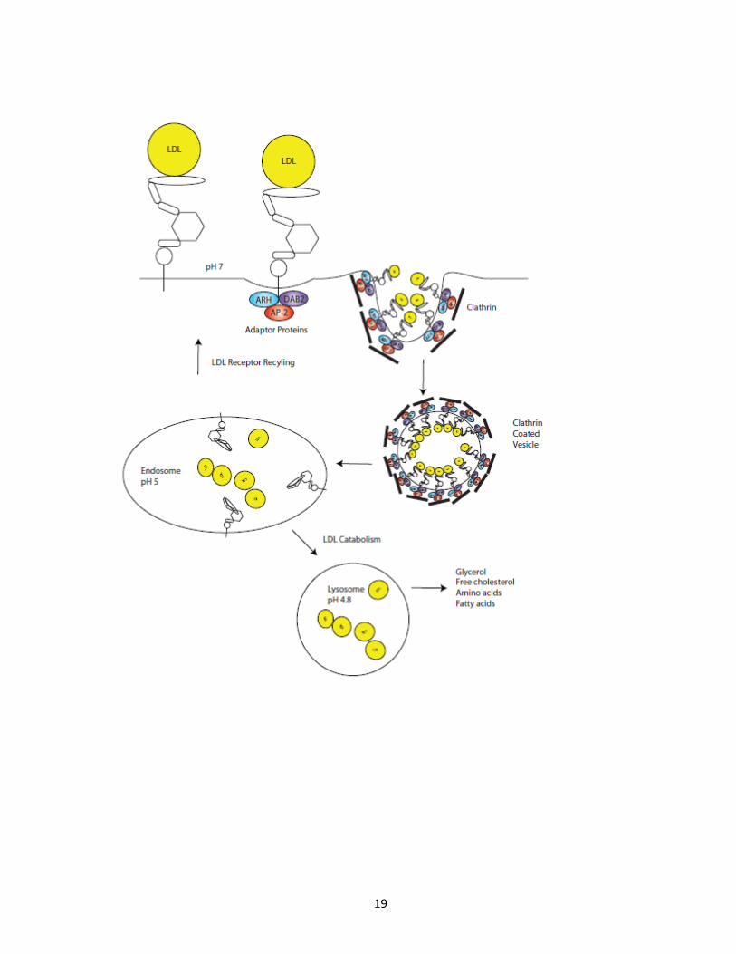
ii. LDLR Internalization
Upon binding of LDL to the LDLR, the protein complex is internalized into the cell via
endocytosis (Figure 4). This internalization process is carried out by clathrin coated pits, and
involves adaptor proteins such as autosomal recessive hypercholesterolemia (ARH). These
adaptor proteins form a complex between the cytoplasmic tail of the LDLR, to clathrin
machinery as well as other endosomal machinery [71,72]. Following internalization, the
LDL-LDLR complex is trafficked to acidic endosomal compartments. It is under these
conditions that the LDLR undergoes a conformational change in which the β-propeller
moves towards the ligand binding domain, which in turn causes the release of the LDL for
future metabolism, while the LDLR is recycled back to the cell surface to repeat this process
to allow for more LDL and LDL-C to be internalized [73,74].

Figure 4. Life cycle of LDLR. Following the binding of cell surface LDLR and circulating LDL
particles at a neutral pH, cytoplasmic adaptor proteins autosomal recessive
hypercholesterolemia (ARH), disabled 2 (Dab2) and adaptor protein complex 2 (AP-2)
participate in binding the cytoplasmic tail of LDLR and recruit endocytic machinery proteins
as well as clathrin. Invagination of the cell surface occurs leading to the internalization of
LDLR into a clathrin coated vesicle (CCV). This CCV fuses with an endosome (pH 5) where
the LDLR undergoes a conformation change, releasing LDL, where LDLR is then trafficked
back through a recycling pathway back to the cell surface while LDL is transported to the
lysosome (pH 4.8) for catabolism.
19
20
1.2 Hypothesis Statement
PCSK9 studies to date have helped to elucidate various factors that affect LDLR
degradation, expression, post-translational regulation and the effects of mutations on
human physiology. Little is known regarding the cellular functions of PCSK9 outside of its
role in LDLR degradation, nor are the mechanisms or protein complexes that participate in
PCSK9 mediated LDLR degradation. In order to increase our understanding of the effects of
PCSK9 in the cell and PCSK9 cellular functions, both known and currently uncharacterized, I
utilized quantitative subcellular fractionation, LC-MS/MS analysis and protein interaction
studies to test the hypothesis that PCSK9 has multiple effects on the liver cellular proteome,
and that these effects signify novel cellular functions and provide insight into currently
known functions of PCSK9. This approach utilizes quantitative proteomics and
immunoaffinity purification to study PCSK9 in a global view of its effects on the cellular
proteome, as well as its interaction partners. By observing the novel effects PCSK9 plays on
liver cell proteins in conjunction with purifying and identifying PCSK9 interaction partners,
information can be attained regarding PCSK9 cellular function that was otherwise
unknown. From this work future studies can be carried out to further explore these
potential novel functions of PCSK9.

21
Chapter 2. MATERIALS AND METHODS
Reagents
See Appendix A for list of reagents, buffers, and solutions used and their recipes.
Cell culture and SILAC metabolic labelling
HuH7 liver cells stably expressing PCSK9-ACE2-V5 and empty vector control cells
were generated as previously described [63] and were kindly donated by Dr. Nabil Seidah.
HuH7 cells were maintained in Dulbecco's Modified Eagle Medium (DMEM) supplemented
with 10% fetal bovine serum (FBS) and 1% anti-mycotic antibiotic reagent at 37°C in a 5%
CO2 humidified incubator. For SILAC labelling, HuH7 cells stably expressing PCSK9-ACE2-V5
were grown in SILAC ‘light’ media containing only 12C lysine amino acids, while empty
vector control cells were grown in SILAC ‘heavy’ media containing only 13C lysine amino
acids [75]. Cells were grown in SILAC media for 10 doubling times. SILAC media kit was
obtained from Invitrogen.
Subcellular Fractionation and histodenz gradient preparation
The subcellular fractionation was performed essentially as described [76]. At the
end of the labelling period, cells were washed twice with ice-cold phosphate buffered
saline (PBS) and then removed with a plastic scraper. The collected cells were centrifuged
at 2,000 × g for 5 min at 15 °C. The pellet was re-suspended in membrane solubilization
buffer and homogenized using a ball bearing homogenization apparatus. The cell

22
homogenate was then transferred to 15-ml glass Corex tubes and centrifuged at
10000 × g for 10 min at 5 °C, to remove unbroken cells, debris, nuclei and mitochondria.
The post-nuclear supernatant (PNS) was set aside on ice for further centrifugation.
Histodenz stock solution and saline buffer were used to prepare four histodenz solutions of
increasing concentrations (10, 14.66, 19.33, and 24%) and 2.5 ml of each solution were
loaded from the bottom of the tube (Beckman Polyallomer Centrifuge Tubes) in decreasing
percentage order. The tubes were then sealed with a piece of parafilm, and a linear
gradient was formed by placing the tubes horizontally for 45 min at room temperature.
Gradient maker was obtained from Sigma. The tubes were then centrifuged at 37,000 × g
for 4 h at 15 °C (Beckman L8–70 M Ultracentrifuge). The supernatant was layered on top of
the created histodenz gradient and centrifuged at 37,000 × g, for 1.5 h at 15 °C (SW41
rotor). Following ultracentrifugation, each tube was fractionated into 15 aliquots (0.8 ml
each) and used for Western blotting and liquid chromatography-mass spectrometry (LC-
MS/MS) analyses.
Western blot analysis
Protein samples from aliquots and fractions were boiled for 5 min using 5x sample
buffer, then separated on NuPage 4-12% Bis-Tris precast gels (Invitrogen) and then
transferred onto a nitrocellulose membrane (Bio-Rad). The membrane was blocked over
night at 4C and washed four times for 5 min in PBS-Tween. Antibodies, unless otherwise
stated, were added at 1:1000 (v/v) dilution in PBS-Tw + 5% skim milk for 1 hour at room

23
temperature. Membranes were washed four times at 5 min in PBS-Tw. Secondary
antibodies were added at 1:5000 dilution and the membrane was incubated for 1 h at room
temperature. Membranes were washed 4 times for 5 min in PBS-Tw. The membrane was
then incubated in SuperSignal chemiluminescent substrate (Pierce) for 1 min and exposed
to film.
HPLC-ESI-MS/MS
Following histodenz subcellular fractionation, each histodenz fraction was separated
using custom made 15cm 4-12% Tris-glycine precast denaturing gels (Jule Inc., Milford, CT)
and silver stained. Gel lanes were cut and separated into 15 separate 1cm bands. Bands
were excised from each gel lane, and digested with trypsin as previously described [77].
From these experiments about 330 samples were analyzed by HPLC-ESI-MS/MS, equating
to almost 500 hours of mass spectrometry time. The HPLC-ESI-MS/MS consisted of an
automated Agilent 1100 micro-HPLC system (Agilent Technologies, Santa Clara, CA) coupled
with an ESI LTQ mass spectrometer (Thermo Scientifics). Briefly, each peptide mixture was
reconstituted in 20 µl of 5% formic acid and loaded on a 200 μm × 50 mm fritted fused silica
pre-column packed in-house with 5 cm of reverse phase Magic C18AQ resins (5 μm; 200-Å
pore size; Michrom Bioresources; Auburn, CA). The separation of peptides was performed
on an analytical column (75 µm i.d. × 50 mm) packed with the same beads using a 90min
gradient of 5-80% acetonitrile (v/v) containing 0.1% formic acid at an eluent flow rate of
200 nl/min after in-line flow splitting. The HPLC was interfaced to an ESI LTQ linear ion trap
mass spectrometer (Thermo Electron, Waltham, MA) operated in positive ion mode. A

24
voltage of 1.8 kV was applied to generate the electrospray ionization. Data dependant
analysis were performed in which a full scan MS was first performed to detect potential
peptides which was then followed by 5 data-dependant MS/MS.
Database searching, quantitation and data analysis
Peak lists were generated from the raw file using Mascot Distiller 2.0.0.0 (Matrix
Science, London, UK) to export *mgf files. MS/MS data were searched against the human
NCBInr database Mascot 2.2.02 (Matrix Science Ltd., London, UK). The searches were
performed using the following criteria, only tryptic peptides with up to two missed cleavage
sites were allowed; the mass tolerance was set to 2 Da for MS and 0.8 Da for MS/MS
fragment ions; carbamidomethyl was set as a static modification; oxidation on methionine
and 13C6-Lys from SILAC were specified as dynamic modifications. The false discovery rate
of protein identification was limited by accepting only the results with a mascot score > 30
(P < 0.05). Following peptide identification using Mascot, peptide and protein quantitation
was carried out using Insilicos Proteomic Pipeline (IPP v1.0 rev.91, Build 200712031206,
Institute for Systems Biology, Seattle, WA). Here, dat files from Mascot search results were
converted to pepXML files for downstream quantitation. Peptide prophet was used for
peptide validation, accepting peptide results with P < 0.05. XPRESS was used for
quantitation, setting SILAC ‘heavy’ peptides to a value of 1, varying SILAC ‘light’ peptides
relative quantitation levels. Once quantitative data from all LC-MS runs were collected and
compiled, all peptides that showed a greater than 2 fold change in relative abundance were

25
subjected to manual validation. Here, the MS scans for these drastically changing peptides
were manually validated based on peak intensities for co-eluting light and heavy peptide
pairs. Labelled peptide pairs peak intensities not matching the relative quantitation
reported by IPP were removed from the dataset. The resulting quantitative peptide list was
expressed according to the log2SILAC L,H in order to produce a log2 distribution curve.
Validation Experiments
The myc-tagged EHD4 construct [78], the V5-tagged human LDLR construct [63], and
the V5-tagged PCSK9 wild-type construct [24] used for expression experiments were
previously described. The C-terminal Flag-tagged PCSK9 wild-type construct was made by
transferring the PCSK9 gene from pIRES into the pCMV-Flag-MAT-2 expression vector.
HEK293 cells were stably transfected with either human LDLR-V5 alone or in combination
with Flag-tagged PCSK9 wild-type using the calcium chloride method. Each stably
transfected HEK293 cell line was then transiently transfected with myc-tagged EHD4 using
polyethylenimine (PEI) transfection reagent, as previously described [79]. Briefly, each
stable cell line was seeded into two 150 mm tissue culture plates and was transfected when
cells reached 70% plate confluency. Plates were transfected with myc-EHD4 in the
presence of serum-free medium. In addition, native HepG2 cells were seeded into four
150 mm tissue culture plates and were ready for transfection when cells reached 70% plate
confluency. Two 150 mm plates of HepG2 cells were transfected with myc-EHD4 alone,
while another two plates were transfected with myc-EHD4 in combination with wild-type

26
PCSK9-V5, in serum-free medium. Following an initial 3 h incubation for the various
transient transfections, serum-free medium was replaced with medium containing 10% FBS
and an anti-biotic/anti-mycotic stock. Cells were grown for an additional 36 h, at which
time cells were washed twice with cold PBS, followed by scraping and resuspension in cold
PBS. Cells were pelleted at 2000 x g for 2 min, PBS was removed and cells were either lysed
directly or flash frozen using liquid nitrogen. Frozen cell pellets were stored at -80oC.
Upon confirmation of proper expression of transfected proteins via Western blot analysis,
cell lysates were subjected to immunoaffinity purifications. For anti-Flag IP experiments,
monoclonal anti-FLAG M2 beads were used (Sigma). For anti-V5 IP experiments anti-V5
agarose affinity gel was used (Sigma). For anti-Myc IP experiments anti-C-Myc agarose
affinity gel was used (Sigma). For co-IP experiments, 1mg of cell lysate and 20µL of specific
agarose affinity gel was used for each separate immunoaffinity purification. Antibody-
beads and cell lysates were mixed on a rotator at 4°C for 2 hours. Flow-through fractions
were removed and antibody-bead-antigen complexes were washed twice in modified RIPA
lysis buffer. Proteins were eluted by boiling beads at 95°C for 5 minutes in 2x sample
buffer. Eluted proteins were subjected to SDS-Page fractionation and Western blotting.
PCSK9 Immunoaffinity purifications and interaction experiments
PCSK9-ACE2-V5 immunoaffinity purifications using anti-V5 pull-downs were
optimized using the aforementioned HuH7 cell lines. Briefly, using a constant amount of
anti-V5 agarose affinity gel (10µL of 50:50 slurry), multiple IP’s were tested against

27
increasing amounts of PCSK9-ACE2-V5 and EV expressing HuH7 cell lysates. Following
mixing, antibody-beads and lysates mixtures were mixed for 2h at 4°C. Flow through was
removed, beads were washed with modified RIPA, and eluted with 2x sample buffer for
Western blot analysis using polyclonal anti-PCSK9 antibody. For large scale IP experiments
HuH7 cells expressing empty vector control or PCSK9-ACE2-V5 were grown in 15cm plates.
Following cell lysis, 15mg of protein for each sample was prepared and subjected to anti-V5
immunoaffinity purification using 150µL of anti-V5 antibody-beads for 4 hours at 4°C. Flow-
through fractions were removed and antibody-bead-antigen complexes were washed eight
times in modified RIPA lysis buffer. Proteins were eluted by boiling beads at 95°C for 5
minutes in 2x sample buffer. Eluted proteins were subjected to SDS-Page fractionation,
silver staining, LC-MS/MS analysis and database searching, as previously described, in order
to identify novel interaction partners of PCSK9-ACE2-V5. Two lists of interaction partners
were generated; those proteins identified in the empty vector control cell line anti-V5 IP,
and those proteins identified in the PCSK9-ACE2-V5 expressing cell line anti-V5 IP. Proteins
identified in the empty vector control IP were removed from the list of potential interaction
partners found in the PCSK9-ACE2-V5 IP experiment. Peptides with a mascot score of 30 or
greater were accepted. This experiment was repeated for n=2.
28
Chapter 3. RESULTS
Subcellular Fractionation and Western blot Analysis of Histodenz Fractions
PCSK9 studies in recent years have centred around its localization, expression,
mutation studies, and effects on LDLR processing. Despite the large number of studies
being presented, very little is known regarding the key players and mechanisms controlling
LDLR degradation. Additionally, some studies have revealed a possible functional role for
PCSK9 in the ER, unrelated to its processing of LDLR for degradation [62,64,65]. It is for
these reasons that we chose to investigate the effects PCSK9 plays on a human liver
subcellular proteome. For the study, human HuH7 liver cells were used. Cells were kindly
donated from Dr. Nabil Seidah. One set of cells stably expressed a variant of PCSK9,
containing an ACE2 transmembrane domain from the angiotensin converting enzyme 2
protein, followed by a c-terminal V5 tag. This PCSK9-ACE2-V5 construct has been
previously shown to promote LDLR degradation stronger than wild type PCSK9 can [63].
This is thought to occur as the ACE2 transmembrane domain localizes the protein towards
endosomal/lysosomal membranes, the sites thought to promote the degradation of the
LDLR [63]. These cells, as well as an empty vector expressing control cells were grown for
ten doubling times in SILAC light and heavy media, respectively. Following metabolic
labelling of proteins using SILAC, light and heavy labelled cells were combined in equal
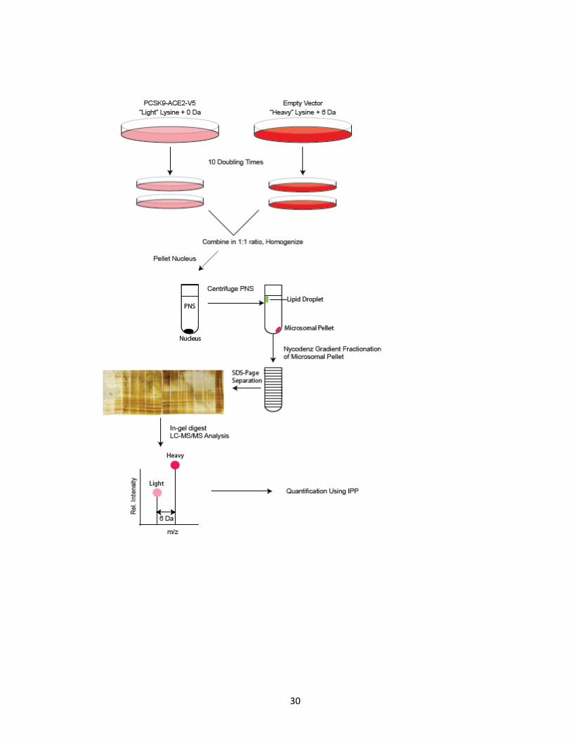
amounts, homogenized and subjected to a subcellular fractionation protocol (Figure 5). 15
29
subcellular fractions were collected and subjected to Western blot analysis using known
markers for resident proteins of specific subcellular organelles ranging from ER to
lysosomes (Figure 6). The more dense fractions (fraction 15) contained the heavier
subcellular organelles such as the ER, and as the density of the fractions decreased, so did
the size of the organelles contained within them. Found in these histodenz fractions was
that the later fractions contained mostly ER, Golgi was found in mid-level fractions, and
finally the least dense fractions contained smaller organelles such as early endosomes and
lysosomes. Nuclear contamination was also tested in the fractions, and was shown to be
present in the most dense subcellular fraction (fraction 15), but mostly absent from the
remaining fractions.
SILAC Quantitation
Following the analysis of subcellular organelle histodenz fractions were subjected to
further fractionation based on size via SDS-Page gel separation. These protein gels were
then stained with silver nitrate, and all proteins were trypsinized and subjected to LC-
MS/MS analysis for protein identification and quantitation. A total of 330 gel bands were
digested and subjected to LC-MS/MS analysis, resulting in approximately 500 hours of mass
spectrometry run time. Following peptide and protein identification, Insilico Proteomic
Pipeline software was used to perform quantitation on each of the 330 mass spectrometry
runs, from SILAC light and heavy paired peptides identified [109]. From two completed
experiments (n=2) a total of 6187 peptides were quantitated (3169 n=1, 3018 n=2). All
peptide hits found to undergo a greater than 2 fold change by IPP were manually validated

Figure 5. General workflow of SILAC metabolic labelling of HuH7 cell lines, subcellular
fractionation, protein processing and LC-MS/MS analysis of resulting peptides. HuH7 cells
stably expressing PCSK9-ACE2-V5 were grown in ‘light’ SILAC media, while empty vector
expressing cells were grown in ‘heavy’ SILAC media. Cells were labelled in their respective
media for 10 doubling times, combined in a 1:1 ratio and homogenized. PNS were
separated in 15 fractions by histodenz gradient subcellular fractionation. Fractions were
further fractionated based on size via SDS-Page and visualized by silver nitrate. Gel bands
were excised, digested with trypsin and analyzed by LC-MS/MS followed by quantitative
analysis using IPP software.
30

Figure 6. Western blot analysis of subcellular fractions for specific organelle markers. The
15 subcellular histodenz fractions were subjected to Western blot analysis with the
following organelle markers, Calnexin (ER), TGN46 (Golgi apparatus), MAN2A (Golgi
apparatus), Early endosomal antigen1 (Early endosome), Lamp1 (Lysosome), Lamin A/C
(Nuclear envelope). Cytosolic and nuclear fraction were also subjected to Lamin A/C
Western blot analysis for nuclear contamination.

31

32
based on peak intensities for co-eluting light and heavy peptide pairs from the raw mass
spectrum.
The SILAC light and heavy relative abundance ratios were converted to log2 values
to simplify data, and all quantitated data were expressed in terms of abundance (Figure 7)
in order to show the distribution of quantitated peptides. Quantitated data from both
experiments followed similar distribution patterns, centred at log2 = 0, suggesting a normal
distribution where the majority of proteins were unaffected by PCSK9-ACE2-V5 expression
in HuH7 cells. However, a small portion of the quantitated peptides did show protein levels
significantly altered as compared to the empty vector control HuH7 cells. All quantitated
peptides with relative abundances of over 2 or under 0.5 were subjected to manual
validation. A total of 293 proteins were found to be significantly affected by PCSK9
expression. Interesting proteins whose expression was determined to have been
significantly affected by the expression of PCSK9-ACE2-V5 were listed in Table 1. Proteins
that were determined to be significantly affected by the PCSK9 variant expression belonged
to many different functional classes of proteins, including cytoskeletal organization,
organelle transport and trafficking, lipid and cholesterol homeostasis, protease activity and
other post-translational modifications, chaperone and protein folding, cell adhesion and

Figure 7. Log2 distribution of peptides quantified by IPP reveals a normal distribution of
SILAC labelled peptides. Following quantitative analysis, light, heavy relative abundance
ratios for peptides were converted to a log2 scale. The distribution of these proteins were
plotted, with a distribution centred at log2 = 0. The first set of experiments (n=1) resulted
in 3169 peptides quantitated, and the second set of experiments (n=2) resulted in 3018
peptides quantitated.

33

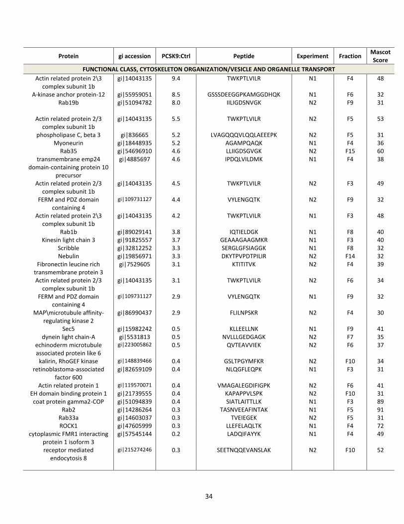
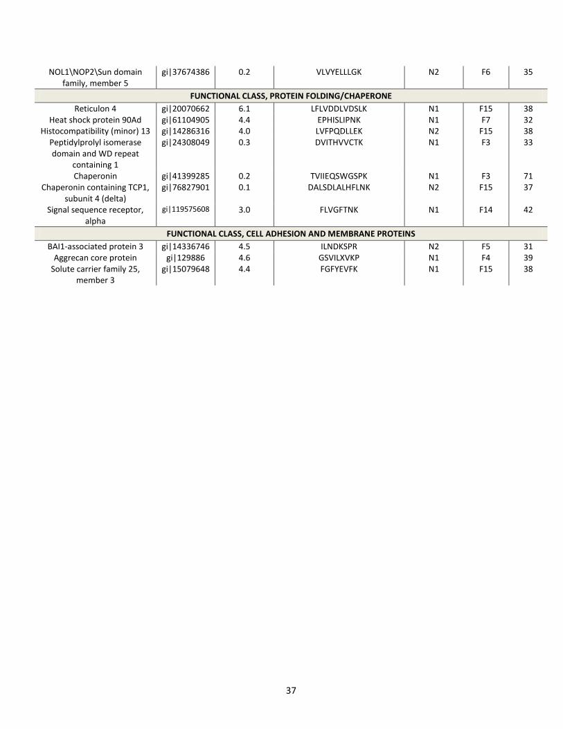
Table 1. Partial list of significantly affected non-nuclear proteins and their biological
functions, following PCSK9-ACE2-V5 expression
34
Protein gi accession PCSK9:Ctrl Peptide Experiment Fraction Mascot Score
FUNCTIONAL CLASS, CYTOSKELETON ORGANIZATION/VESICLE AND ORGANELLE TRANSPORT
Actin related protein 2\3 complex subunit 1b
gi|14043135 9.4 TWKPTLVILR N1 F4 48
A-kinase anchor protein-12 gi|55959051 8.5 GSSSDEEGGPKAMGGDHQK N1 F6 32 Rab19b gi|51094782 8.0
IILIGDSNVGK N2 F9 31
Actin related protein 2/3 complex subunit 1b
gi|14043135 5.5 TWKPTLVILR N2 F5 53
phospholipase C, beta 3 gi|836665 5.2 LVAGQQQVLQQLAEEEPK N2 F5 31 Myoneurin gi|18448935 5.2 AGAMPQAQK N1 F4 36
Rab35 gi|54696910 4.6 LLIIGDSGVGK N2 F15 60 transmembrane emp24
domain-containing protein 10 precursor
gi|4885697 4.6 IPDQLVILDMK N1 F4 38
Actin related protein 2/3 complex subunit 1b
gi|14043135 4.5 TWKPTLVILR N2 F3 49
FERM and PDZ domain containing 4
gi|109731127 4.4 VYLENGQTK N2 F9 32
Actin related protein 2\3 complex subunit 1b
gi|14043135 4.2 TWKPTLVILR N1 F3 48
Rab1b gi|89029141 3.8 IQTIELDGK N1 F8 40 Kinesin light chain 3 gi|91825557 3.7 GEAAAGAAGMKR N1 F3 40
Scribble gi|32812252 3.3 SERGLGFSIAGGK N1 F8 32 Nebulin gi|19856971 3.3 DKYTPVPDTPILIR N2 F14 32
Fibronectin leucine rich transmembrane protein 3
gi|7529605 3.1 KTITITVK N2 F4 39
Actin related protein 2/3 complex subunit 1b
gi|14043135 3.1 TWKPTLVILR N2 F6 34
FERM and PDZ domain containing 4
gi|109731127 2.9 VYLENGQTK N1 F9 32
MAP\microtubule affinity-regulating kinase 2
gi|86990437 2.9 FLILNPSKR N2 F4 30
Sec5 gi|15982242 0.5 KLLEELLNK N1 F9 41 dynein light chain-A gi|5531813 0.5 NVLLLGEDGAGK N2 F7 35
echinoderm microtubule associated protein like 6
gi|223005862
0.5 QVTEAVVIEK N2 F6 37
kalirin, RhoGEF kinase gi|148839466 0.4 GSLTPGYMFKR N2 F10 34 retinoblastoma-associated
factor 600 gi|82659109 0.4 NLQGFLEQPK N1 F3 31
Actin related protein 1 gi|119570071 0.4 VMAGALEGDIFIGPK N2 F6 41 EH domain binding protein 1 gi|21739555 0.4 KAPAPPVLSPK N2 F10 31 coat protein gamma2-COP gi|51094839 0.4 SIATLAITTLLK N1 F3 89
Rab2 gi|14286264 0.3 TASNVEEAFINTAK N1 F5 91 Rab33a gi|14603037 0.3 TVEIEGEK N2 F5 31 ROCK1 gi|47605999 0.3 LLEFELAQLTK N1 F4 72
cytoplasmic FMR1 interacting protein 1 isoform 3
gi|57545144 0.2 LADQIFAYYK N1 F4 49
receptor mediated endocytosis 8
gi|215274246 0.3 SEETNQQEVANSLAK N2 F10 52

35
FUNCTIONAL CLASS, LIPID/CHOLESTEROL HOMEOSTASIS AND METABOLIC PROTEINS
GAPDH gi|603211 5.1 IISNASCTTNCLAPLAK N1 F3 116 Cyclophilin A gi|51702775 5.1 XSELFADK N1 F15 50
gamma-butyrobetaine hydroxylase
gi|3746805 4.9 MKTGNMACTIQK N2 F4 33
Cyclophilin A gi|51702775 4.6 XSFELFADK N1 F9 54 Cyclophilin A gi|51702775 4.0 XSFELFADK N2 F5 49
Carbamyl phosphatesynthetase I
gi|62822412 3.8 ETLMDLSTK N1 F15 36
Hydroxysteroid (17-beta) dehydrogenase 12
gi|7705855 3.4 GVFVQSVLPYFVATK
N1 F14 37
ATP citrate lyase isoform 2 gi|38569423 3.4 KAKPAMPQGK N2 F14 32 Cyclophilin A gi|51702775 3.2 XSFELFADK N1 F3 54
Protein disulfide isomerase family A, member 4
gi|37674412 3.2 RSPPIPLAK
N1 F15 40
Protein disulfide isomerase family A, member 4
gi|37674412 3.1 RSPPIPLAK N1 F9 40
Malate deydrogenase 2 gi|6648067 3.0 IQEAGTEVVKAK N1 F4 37 7-dehydrocholesterol
reductase gi|4581759 3.0 FLPGYVGGIQEGAVTPAGVVNK N2 F15 141

36
Alpha enolase gi|12804749 2.9 DATNVGDEGGFAPNILENKEGLELLK
N1 F4 48
Lactate dehydrogenase B gi|12803117 2.8 MVVESAYEVIK N1 F3 35 Calreticulin gi|12803363 0.4 FYALSASFEPFSNK N1 F14 96
Methylenetetrahydrofolate dehydrogenase 1
gi|14602585 0.4 YVVVTGITPTPLGEGK N2 F7 38
Triosephosphate isomerase gi|14043688 0.3 VVLAYEPVWAIGTGK N1 F5 34 Cyclophilin A gi|51702775 0.2 KITIADCGQLE N2 F3 51
FUNCTIONAL CLASS, PROTEASE/PROTEASOME
Plasminogen gi|387026 6.1 LSSPADITDK N1 F4 38 Proteasome 26S non-ATPase
subunit 13 isoform 1 gi|12654533 3.7 YYQTIGNHASYYK N2 F4 60
ADAMTS5 gi|6049180 3.6 LMSSILTSIDASKPWSK N1 F7 33 Proteasome subunit,
alpha type, 8 gi|20379541 3.3 LYQTDPSGTYHAWK N1 F4 47
Proteasome subunit, alpha type, 2
gi|50881968 3.2 GYSFSLTTFSPSGK N1 F3 89
Proteasome alpha 6 subunit gi|82571777 3.2 LYQVEYAFK N1 F3 64 Transmembrane protease,
serine 11F gi|34527807 3.1 ALYQSLKTK N1 F4 34
Headpin gi|12643252 3.1 DLFPDGSISSSTK N2 F3 37 Proteasome 26S subunit,
non-ATPase, 3 gi|13436065 0.4 LQLDSPEDAEFIVAK N1 F3 104
Proteasome beta 7 subunit proprotein
gi|1531533 0.3 LDFLRPYTVPNK N2 F5 35
Proteasome 26S non-ATPase subunit 7
gi|15214948 0.3 VVGVLLGSWQK
N2 F6 65
PAPP-A gi|38045915 0.2 CKVLMLGGSALNHNYR N2 F15 34
FUNCTIONAL CLASS, POST-TRANSLATIONAL MODIFICATION
Protein tyrosine phosphatase gi|16876892 5.3 MEKGDDINIK N2 F4 45 Heparan sulfate 3-O-
sulfotransferase 6 gi|14336772 5.1 KAQGGSRPR N2 F9 34
Cullin 7 gi|2833262 4.8 NLLNCLIVRILK N2 F4 34
Retinoblastoma binding protein 6 isoform 1
gi|74762440 4.8 ISKLEVTEIVKPSPK N2 F9 32
Tssk4 gi|83405295 4.7 RATILDIIK N2 F15 37 E1A binding protein p400 gi|56549696 4.3 AIQPQAAQGPAAVQQKITAQQIT
TPGAQQK N1 F7 31
Tssk4 gi|83405295 4.2 RATILDIIK N1 F5 40 Cullin 7 gi|2833262 3.7 NLLNCLIVRILK N2 F5 33
Ubiquitin specific protease 4 isoform b
gi|40795667 3.3 ADTIATIEK
N2 F3 31
Protein tyrosine kinase gi|515871 3.1 TGAFEDLKENLIR N2 F4 41 TIP120 protein gi|76661742 0.5 EGPAVVGQFIQDVK N1 F5 34
N-myristoyltransferase 1 gi|10835073 0.5 GSETDSAQDQPVKMNSLPAER N2 F6 37 Putative methyltransferase
WBMT gi|23831505 0.4 AGFSGGMVVDYPNSAK N1 F7 54
Janus kinase 1 gi|62087694 0.3 MQLPELPKDISYK N2 F4 31 Ring finger protein 20 gi|83405148 0.3 LQELTDLLQEK N1 F5 52 Polypeptide GalNAc
transferase 13 gi|27530993 0.3 NQEGPGEMGK
N2 F10 39
37
NOL1\NOP2\Sun domain family, member 5
gi|37674386 0.2 VLVYELLLGK N2 F6 35
FUNCTIONAL CLASS, PROTEIN FOLDING/CHAPERONE
Reticulon 4 gi|20070662 6.1 LFLVDDLVDSLK N1 F15 38 Heat shock protein 90Ad gi|61104905 4.4 EPHISLIPNK N1 F7 32
Histocompatibility (minor) 13 gi|14286316 4.0 LVFPQDLLEK N2 F15 38 Peptidylprolyl isomerase domain and WD repeat
containing 1
gi|24308049 0.3 DVITHVVCTK N1 F3 33
Chaperonin gi|41399285 0.2 TVIIEQSWGSPK N1 F3 71 Chaperonin containing TCP1,
subunit 4 (delta) gi|76827901 0.1 DALSDLALHFLNK N2 F15 37
Signal sequence receptor, alpha
gi|119575608 3.0 FLVGFTNK N1 F14 42
FUNCTIONAL CLASS, CELL ADHESION AND MEMBRANE PROTEINS
BAI1-associated protein 3 gi|14336746 4.5 ILNDKSPR N2 F5 31 Aggrecan core protein gi|129886 4.6 GSVILXVKP N1 F4 39
Solute carrier family 25, member 3
gi|15079648 4.4 FGFYEVFK N1 F15 38

38
Translocase of inner mitochondrial membrane 44
homolog
gi|62897685 4.4 ILDIDNVDLAXGK N1 F9 32
Potassium voltage-gated channel, subfamily H, member 6 isoform 1
gi|11878259 3.6 VVERTQNVTEK N2 F11 36
Transient receptor potential cation channel, subfamily M,
member 8
gi|72537227 3.5 GLLKEIANK N1 F15 39
Semaphorin receptor gi|6010211 3.1 FRYLVPGSNGQLTFDSGFEK N1 F9 33 GPAD9366 gi|73619948 0.5 MVTFYCTTK N1 F7 34
Cadherin 8, type 2 gi|6483315 0.5 HKNEPLIIK N1 F4 33
FUNCTIONAL CLASS, SIGNALLING/CELL CYCLE
Fibroblast growth factor 11 gi|20160215 5.6 SLCQKQLLILLSK N1 F7 36 cell division cycle 2-like 6
(CDK8-like) gi|57209410 3.7 DLKPANILVMGEGPER N2 F3 34
Rotatin gi|145046269 3.4 KSAAEQLAVIMQDIK N2 F14 31 Cytokine induced protein 29
kDa
gi|109097133 3.3 FGIVTSSAGTGTTEDTEAK N2 F5 91
KH domain containing, RNA binding, signal transduction
associated 1
gi|12653853 0.4 ILGPQGNTIK N2 F6 43
Cyclin-dependent kinase 9 gi|12805029 0.3 EIKILQLLK N2 F4 33
FUNCTIONAL CLASS, OTHER
Retinol dehydrogenase 14 (all-trans\9-cis\11-cis)
gi|10190746 5.9 GGDPGLMHGK N1 F14 35
GTP-binding protein NGB gi|4191616 4.1 SSFINKVTR N2 F9 32 HLA-B associated transcript 1 gi|56207984 3.2 GLAITFVSDENDAK N1 F3 80
Carbamoyl-phosphate synthetase 2, aspartate transcarbamylase, and
dihydroorotase
gi|50403731 3.1 HPQPGAVELAAK N1 F6 32
Ribosomal protein L29 gi|60656425 3.0 AQAAAPASVPAQAPK N1 F5 88 Alpha-fetoprotein gi|31351 0.5 GYQELLEK N1 F15 31
A kinase (PRKA) anchor protein 8-like
gi|6688138 0.3 QTADFLQEYVTNK N1 F3 42
J-type co-chaperone HSC20 gi|41388876 0.2 QFLIEIMEINEK N1 F3 44

39
membrane proteins as well as other types of functionalities. This finding suggests that
PCSK9 either directly or indirectly has an effect on many types of proteins with a wide
range of functions.
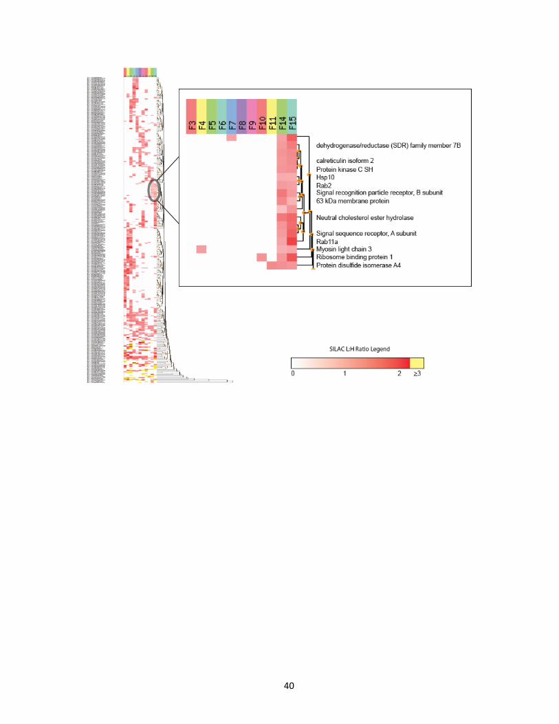
All quantitated proteins that were identified among two or more fractions were
then subjected to hierarchical clustering in order to observe any possible trends in the
SILAC data, as well as assess the data quality (Figure 8). As shown, most proteins were
identified only in similar or contiguous histodenz fractions, rather than interspersed
amongst the histodenz subcellular fractions. This suggests the proteins are not present in
all but instead are found in specific regions among the subcellular fractions. An example of
this is found in Figure 8 (see insert) where the proteins clustered together among the more
dense histodenz fractions (15 and 14) were highly enriched for ER resident proteins. This is
as expected, as it was found that these fractions were comprised of ER marker proteins and
lacked Golgi, endosome and lysosome marker proteins (Figure 6).
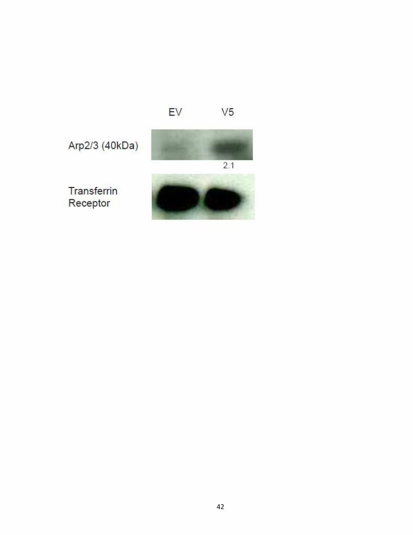
PCSK9 promotes a decrease in cellular EH domain binding protein 1 and an increase in actin
related protein 2\3 at the protein level
Beyond manual validation of quantitation results at the raw MS level, two different
proteins were chosen for further biochemical validation studies. One protein, actin related
protein 2\3 (Arp 2\3), was chosen since its protein levels were significantly higher in
response to PCSK9-ACE2-V5 expression, as well as it being seemingly unrelated to PCSK9
mediated degradation of the LDLR. SILAC quantitation results showed Arp 2\3 to have

Figure 8. Heat map clustering of quantitated fractionation data reveals non-random
distribution of proteins throughout subcellular fractions. Proteins identified in two or
more histodenz fractions were subjected to hierarchical clustering across the subcellular
histodenz fractions. Inset shows proteins clustering together in high density fractions (F14
F15) relating to the endoplasmic reticulum, (See Figure 6) were found to be highly enriched
with endopasmic reticulum resident proteins.
40

41
protein levels increased between 9.4 to 3.1 fold in subcellular fractions 3 to 6, in PCSK9
expressing cells. Cell lysates from empty vector control and those expressing PCSK9-ACE2-
V5 were subjected to Western blot analysis for Arp 2\3. Protein levels were corrected to
transferring receptor levels, a protein known to be unaffected by PCSK9 levels. Western
blot analysis revealed increased protein levels of over 2-fold of Arp 2\3 in PCSK9-ACE2-V5
expressing HuH7 cells (Figure 9). Another protein, EH domain binding protein 1 (EHBP1),
was chosen for its significantly decreased protein levels in response to PCSK9-ACE2-V5
expression, as well as for its potential link to PCSK9 mediated degradation of LDLR. SILAC
quantitation results showed EHBP1 protein levels were 2.5 fold lower in PCSK9-ACE2-V5
expressing cells. Cell lysates from empty vector control and PCSK9-ACE2-V5 expressing
HuH7 cells were subjected to Western blot analysis for EHBP1 (Figure 10). Protein levels
were corrected to transferrin receptor levels, and revealed a decreased protein levels of
roughly 2-fold of EHBP1 in PCSK9-ACE2-V5 expressing cell lysates, and a decrease in protein
levels as low as 4-fold in subcellular fractions. These results are in accordance with, and
validate the quantitation results obtained through SILAC labelling experiments.
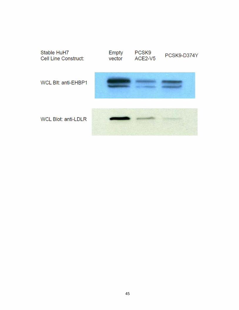
PCSK9-ACE2-V5 and PCSK9-D374Y variants decrease both cellular LDLR and EHBP1 protein
levels
PCSK9-ACE2-V5 has previously been shown to accelerate LDLR degradation greater
than wild type PCSK9. To date, however, the strongest gain of function variant, in terms of

Figure 9. Western blot validation of Arp2/3 levels increased in PCSK9-ACE2-V5 expressing
cells. HuH7 cells expressing empty vector and PCSK9-ACE2-V5 were grown in DMEM
supplemented with 10% FBS and 1% anti-mycotic. Whole cell lysates of empty vector and
PCSK9-ACE2-V5 expressing HuH7 cells were subjected to Western blot analysis for
endogenous Arp2/3. Arp2/3 levels in PCSK9-ACE2-V5 expressing cells were listed relative to
those found in empty vector cells. Sample load was corrected to transferrin receptor levels.
SILAC quantitation found Arp2/3 levels in PCSK9-ACE2-V5 expressing cells were increased
between 9.4 and 3.1 fold as compared to empty vector expressing cells, depending on the
subcellular fraction it was identified in.
42

Figure 10. Western blot validation of EHBP1 levels decreased in PCSK9-ACE2-V5
expressing cells. HuH7 cells expressing empty vector and PCSK9-ACE2-V5 were grown in
DMEM supplemented with 10% FBS and 1% anti-mycotic. Subcellular fractions between
empty vector and PCSK9-ACE2-V5 expressing HuH7 cells for fractions F2-F5 (top) and whole
cell lysates of empty vector and PCSK9-ACE2-V5 expressing HuH7 cells (bottom) were
subjected to Western blot analysis for endogenous EHBP1. EHBP1 levels in PCSK9-ACE2-V5
expressing cells were listed relative to those found in empty vector cells, and corrected for
sample loading according to transferrin receptor levels. SILAC quantitation found EHBP1
levels in PCSK9-ACE2-V5 expressing cells were decreased 2.5 fold as compared to empty
vector expressing cells.

43

44
specifically promoting the degradation of LDLR, of PCSK9 is the PCSK9-D374Y variant. With
the belief that EHBP1 may play a role in PCSK9 mediated degradation of the LDLR, EHBP1
and LDLR protein levels were analyzed from HuH7 empty vector, PCSK9-ACE2-V5, and
PCSK9-D374Y stably expressing cells (Figure 11). Compared to empty vector control cells,
both EHBP1 and LDLR levels were decreased in PCSK9-ACE2-V5 and PCSK9-D374Y variant
expressing cell lines, suggesting a possible link between EHBP1 and PCSK9 mediated LDLR
degradation.
PCSK9-ACE2-V5 Immunoaffinity purification optimization
Despite extensive biological and physiological studies of PCSK9, at the time of this
study, the only known protein interaction partners of PCSK9 were the LDLR and potentially
VLDLR, and ApoER2. Determining protein interaction partners can provide a great deal of
information regarding a proteins function, and in the case of PCSK9, potentially reveal
novel cellular functions and/or proteins it may work in concert with to promote the
degradation of the LDLR. In order to optimize the anti-PCSK9-ACE2-V5 IP conditions,
increasing amounts of PCSK9-ACE2-V5 expressing HuH7 cell lysates (from 0 to 500µg) were
incubated with a constant amount (10µL, 50% slurry) of anti-V5 agarose affinity gel, for 2h
at 4°C on a rotator. Eluted proteins were subjected to Western blot analysis for
endogenous PCSK9 (Figure 12). From the conditions tested, it was evident that from the
amounts of whole cell lysates tested, using 500µg of whole cell lysates for 10µL of beads
(50% slurry) was most optimal. This was determined since the recovery of PCSK9 was

Figure 11. PCSK9-ACE2-V5 and the natural gain of function variant PCSK9-D374Y
decreases EHBP1 and LDLR protein levels in whole cell lysates. Cell lysates of empty vector
control, PCSK9-ACE2-V5 and PCSK9-D374Y expressing HuH7 cells were subjected to
Western blot analysis for endogenous EHBP1 and mature LDLR levels revealing PCSK9-
ACE2-V5 and PCSK9-D374Y promote the degradation of both the LDLR and EHBP1. EHBP1
can appear as a single band or doublet depending on WB conditions.
45

Figure 12. Optimization of anti-V5 Immunoaffinity purifications for PCSK9-ACE2-V5
pulldowns. HuH7 cells expressing PCSK9-ACE2-V5 were lysed, and purifications using 10µL
of anti-V5 agarose affinity gel (50% slurry) were used against solutions containing 0, 25, 50,
100, 250 and 500 µg of cell lysates in modified RIPA buffer. Size marker (SM), and flow
through (FT) and eluted (E) proteins for each IP were subjected to Western blot analysis for
endogenous PCSK9. Intense lower band found in all elute lanes is IgG from anti-V5 antibody
beads.

46

47
strongest, and there was little to no increase in the amount of non-binding PCSK9, as
shown by the amount of PCSK9 found in the flow through fraction.
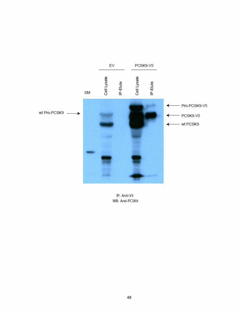
PCSK9-ACE2-V5 IP-LC-MS/MS analysis of interaction partners
A large-scale anti-V5 IP was performed in order to co-purify interaction partners of
PCSK9. 10% of starting material and eluted protein complexes were subjected to Western
blot analysis to show the efficiency and specificity of the PCSK9-ACE2-V5 purification
(Figure 13). In an empty vector expressing HuH7 cell lysates, endogenous pro-PCSK9 and
cleaved PCSK9 are present, however following an anti-V5 IP from the empty vector HuH7
cell lysate, no forms of PCSK9 can be detected in the eluted protein fraction. Conversely, in
PCSK9-ACE2-V5 expressing HuH7 cell lysates, both endogenous pro-PCSK9 and cleaved
PCSK9 are present, in addition to pro-PCSK9-ACE2-V5 and cleaved PCSK9-ACE2-V5 protein
variants. Following an anti-V5 IP, protein eluate contains solely the pro- and cleaved PCSK9-
ACE2-V5 protein variants, and endogenous PCSK9 isoforms are not purified, suggesting a
specific purification of V5 tagged PCSK9-ACE2 and its protein complexes. The remaining
90% of eluted protein complexes from both EV and PCSK9-ACE2-V5 IPs were subjected to
protein separation, tryptic digestion, protein processing and LC-MS/MS analysis in order to
identify potential interaction partners. In both IP experiments (n=2) PCSK9 was confidently
identified in PCSK9-ACE2-V5 expressing cell IPs (Mascot score 567 and 546), but not in the
EV control cell IPs, reflecting the results obtained by Western blot analysis. A complete list
of potential interaction partners was obtained and following data filtering, a list of potential

Figure 13. PCSK9-ACE2-V5 Immunoaffinity Purification. 15mg of HuH7 empty vector and
PCSK9-ACE2-V5 cell lysates were subjected to anti-V5 IP using 300 µL (50% slurry) of anti-V5
affinity agarose gel for 4h at 4°C. Flow through was collected, antibody-bead-antigen
complexes were washed 8x with ice chilled modified RIPA buffer. Protein complexes were
eluted by boiling 2x sample buffer for 10 minutes. 5% of eluted and flow through material
were subjected to Western blot analysis against endogenous PCSK9.
48

49
PCSK9 interaction partners of higher confidence was obtained (Figure 14 and Table 2).
Interestingly, a potential interaction partner of PCSK9 that was identified was EH domain-
containing protein 4 (EHD4), a member of a family of proteins that bind to EH domain
binding protein 1 (EHBP1), which was shown to have decreased protein levels in PCSK9-
ACE2-V5 expressing cells, as shown previously through SILAC quantitation and Western blot
analysis. These results provide a link between PCSK9 interaction partners and its effect on
regulating protein levels in HuH7 cells, and suggest a link to the EH domain proteins and EH
domain binding protein endosomal machinery.
PCSK9 and mature LDLR both interact with EHD4
In order to test and validate the interaction between wild type PCSK9 lacking the
ACE2 domain and EHD4, various cell systems were used. HepG2 cells expressing EHD4-Myc
tag were co-expressed with PCSK9-V5 or an empty vector. HuH7 cells expressing EHD4-Myc
tag were co-expressed with PCSK9-Flag or an empty vector. Anti-V5 and anti-Flag IP
experiments were performed in both cell systems and protein eluate was subjected to
Western blot analysis against anti-Myc (Figure 15). EHD4-myc was present specifically in
the anti-V5 protein eluate from PCSK9-V5 expressing cells, while absent in the empty vector
expressing cells. The reverse IP experiments were also performed, purifying EHD4-Myc
using anti-Myc agarose affinity gel (Figure 15). Western blot analysis against anti-V5
Figure 14. PCSK9 Interaction map. Proteins identified in anti-V5 immunopurifications to co-
purify with PCSK9-ACE2-V5.

50

Table 2. List of proteins identified to co-immunoprecipitate with PCSK9-ACE2-V5
following anti-V5 immunoaffinity purification and LC-MS/MS analysis.

51
Protein Name Mascot Score No. Peptides IP No.
Vimentin 1349 21 2 PCSK9 567/546 9/8 1/2
Heat shock protein 90 174/520 3/9 1/2 Nestin 569 8 2
Galectin 3 binding protein 99/180 2/3 1/2 TRK-fused gene/anaplastic large cell lymphoma kinase extra
long form 360 4
1
Heat shock protein 70 protein 1 337 4 2 Spectrin, beta 327 8 2
Lactate dehydrogenase A 222 4 1 Heterogeneous nuclear ribonucleoprotein K 210 3 2
Protein disulfide-isomerase precursor 194 3 2 Heat shock protein 70 protein 8 150 2 1
Heterogeneous nuclear ribonucleoprotein F 147 3 2 MTHSP75 128 2 1
Neutral alpha-glucosidase AB precursor isoforms 2 128 3 2 X-prolyl aminopeptidease 125 2 1
Desmoplakin 121 2 1 Filamin-B 120 3 2
Mitochondrial aspartate-glutamate carrier protein 115 3 2 Programmed cell death protein 8 102 2 2
Tubulin, beta 101 2 1 EH-domain containing protein 4 91 2 2
Heterogeneous nuclear ribonucleoprotein M 88 2 2 BBF2H7/FUS protein 83 2 2
Intracellular adhesion molecular 1 precursor (ICAM-1) 74 2 2 Prohibitin-2 68 2 2

Figure 15. Co-IP Experiments reveal PCSK9 and EHD4 Interact. a) HepG2 cells were
transiently transfected with EHD4-myc and either PCSK9-V5 or empty vector constructs and
subjected to anti-V5 Immunopurifications. HEK293 cells were transiently transfected with
EHD4-myc and either PCSK9-Flag or empty vector constructs and subjected to anti-Flag or
anti-V5 immunopurifications. Eluted protein complexes were subjected to Western blot
analysis against anti-myc-HRP. Reverse Co-IP experiments were performed in these cell
systems involved anti-Myc-EHD4 immunopurifications, and eluted complexes were
subjected to Western blot analysis against b) anti-Flag from HEK293 cells and c) anti-V5
from HepG2 cells.

52

53
revealed an interaction between PCSK9-V5 and EHD4 when co-expressed. In HEK293 cells,
EHD4-myc was co-expressed with either flag tagged wild type PCSK9 or an empty vector.
When anti-Flag IP experiments were performed and eluted proteins were subjected to anti-
Myc Western blot analysis, it was shown that PCSK9-Flag interacts with EHD4-Myc (Figure
15). The reverse IP experiments were also performed, purifying EHD4-myc using anti-Myc
agarose affinity gel (Figure 15). Western blot analysis against anti-Flag revealed an
interaction between PCSK9-Flag and EHD4-Myc in HepG2 cells. Taken together, these
results validate the previous IP-LC-MS/MS interaction results between PCSK9 and EHD4,
and suggest that this novel interaction is specific in nature and not due to the ACE2 domain
present in the PCSK9-ACE2-V5 variant.
With previous studies showing that the EH domain containing and binding proteins
are known to play a role in LDLR trafficking and recycling, the link between EHD4, PCSK9
and LDLR became a valid possibility, and therefore the interaction between EHD4 and
endogenous LDLR was investigated. In both HepG2 and HEK293 cells either Myc-tagged
EHD4 or an empty vector was expressed. In both cell systems, anti-Myc IP experiments
were performed, and eluted protein complexes were subjected to Western blot using an
antibody for endogenous cellular LDLR, in the mature, glycosylated form (Figure 16). The
resulting blot showed that mature endogenous LDLR co-immunoprecipitated specifically in
EHD4-Myc expressing cells. These results suggest a novel and specific interaction between
LDLR and the endosomal machinery and trafficking protein EHD4.
Figure 16. Co-IP Experiments Reveal EHD4 and LDLR interact. HepG2 cells were transiently
transfected with EHD4-myc or an empty vector construct. Cells were lysed and subjected
to anti-myc immunoaffinity purifications. The resulting eluted protein complexes were
subjected to Western blot analysis against endogenous mature LDLR.
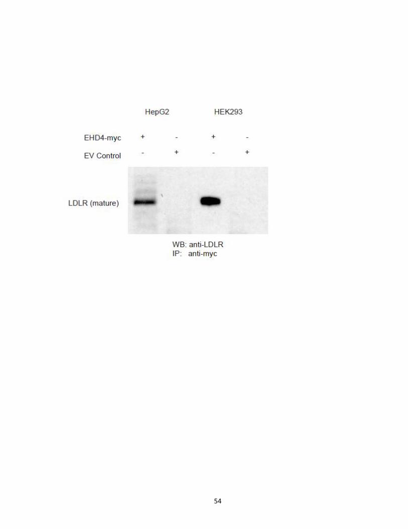
54
55
Chapter 4. DISCUSSION
In these studies I have utilized large-scale proteomic techniques including
subcellular fractionation, relative quantitation using SILAC metabolic labelling, and
immunoaffinity purification of protein complexes in order to explore cellular roles PCSK9
plays in the liver. The aim of my work was to determine whether PCSK9 affected cellular
proteins and cellular activities in liver cells. The work discussed thus far suggests that PCSK9
expression does affect the abundance of many liver cell proteins, many of which at
significant levels. Additionally, the work here also aimed to determine if PCSK9 plays any
novel cellular roles in the liver. The results obtained from the SILAC relative quantitation
and interaction were not limited to the known established functional roles of PCSK9 such as
lipid and cholesterol homeostasis and regulation, but suggests PCSK9 plays novel roles in
liver cells outside of LDLR degradation. These novel functional roles which include
cytoskeleton organization and regulation, vesicle transport and trafficking, post-
translational modification and regulation of proteins including proteasomal regulation and
chaperone regulation, cell membrane and adhesion protein regulation, chaperone
mediated autophagy and even potential roles in Alzheimer’s via amyloid precursor protein
(APP) processing and regulation. Despite this, there is a lack of understanding of
mechanisms in which PCSK9 plays a role in these cellular processes, the results obtained in
this study provide a new insight into PCSK9 cellular function and show that PCSK9 is a multi-
functional cellular protein with roles outside of LDLR regulation.
56
Subcellular fractionation and quantitative SILAC labelling of PCSK9-ACE2-V5 expressing
HuH7 cells reveals effects on cellular proteome
In order to optimally study the effects PCSK9 expression plays on the HuH7 liver cell
proteome, it was advantageous to perform subcellular fractionation on the proteome. This
method would provide another layer of protein fractionation before LC-MS/MS analysis,
identification and quantitation, as well provide cell locale relative protein quantitative
information throughout the study. Following these experiments, quantitation and quality
control, it became clear that PCSK9 expression affected much more than LDLR, lipid and
cholesterol homeostasis. PCSK9 affected the expression of 341 HuH7 cellular proteins.
Most protein expression levels were unaffected by PCSK9, including those that were known
to be unaffected by PCSK9, such as transferrin receptor. The transferrin receptor is
constantly recycled from endosomes back to the cell surface and has been previously
shown to be unaffected by PCSK9 over-expression [42], as it is seen in this study to be
unaffected, whose levels are found to range from a ratio of 1.0 to 1.2 (PCSK9-ACE2-V5
expressing cells to empty vector control cells).
Those proteins that were affected by the expression of PCSK9 were grouped into
functional classes and found to represent diverse cellular roles, ranging from cell surface
membrane and cell adhesion proteins, lipid and cholesterol metabolism, cytoskeleton
organization, endocytosis and vesicle trafficking, post-translational modification,
proteasome function, chaperone mediated autophagy, protein folding and chaperone
activity, and amyloid precursor protein processing and regulation.

57
Comparison of SILAC quantitation results with previous PCSK9 gene expression studies
Previously, two studies have been published with the aim of looking at genes whose
expression is affected by PCSK9 expression. The first study looked at the genes upregulated
when the variant PCSK9-D374Y was expressed in comparison to an empty vector control
[42]. This study revealed that most genes significantly affected by PCSK9-D374Y are related
to lipid and cholesterol metabolism, as well as stress response. The second study treated
cells with purified wild type or PCSK9-D374Y for various time points and looked at the
genes that were affected by these treatments [80]. This study was able to expand upon
novel pathways and functions that which PCSK9 affects, including cell cycle, inflammation
and protein ubiquitination, as well as confirming that lipid and cholesterol metabolism and
stress response related genes are affected.
Like the previous gene expression studies performed, this protein quantitation
study was able to expand upon the understanding of what cellular functions and pathways
are affected by PCSK9 expression. However, this study goes even further than the
previously discussed gene expression studies, by documenting changes at the protein level,
not simply at the gene expression level, as well as in a cell location specific level, not a
global level. The results from this study mirrored those obtained from the previous gene
expression studies. These studies suggest that PCSK9 expression affects proteins involved in
cellular processes such as lipid and cholesterol metabolism, stress response, cell cycle,
protein ubiquitination and proteasome function. However, in light of the data presented
here, we suggest that PCSK9 expression also affects proteins involved in other cellular

58
processes not previously known to be associated with PCSK9 function, including
cytoskeleton organization, organelle trafficking, chaperone activity and post-translational
modification, among others.
A complete comparison of proteins/genes identified overlapping between these
studies can be found in Table 3. By comparison, very few protein/genes were found to
overlap between this study and those found in the two previous mRNA expression studies.
Of those that do overlap, not all the results are harmonious, as is often the case when
comparing gene expression results and protein expression levels. For example, a protein
whose levels are almost non-existent may have high/up-regulated gene expression levels as
a feed-back mechanism in order to raise the protein levels of such a gene, as is the case for
the LDLR [80]. However there are some results that are congruent, as is the case for
gene/protein 7-dehydrocholesterol reductase, whose gene expression when treated with
purified wt PCSK9 was shown to increase in a range from 1.4 to 2.5 fold, and from 1.5 to 2.9
when treated with purified PCSK9-D374Y. Like the gene expression study, my results
showed an increased level of this protein involved in lipid and cholesterol metabolism,
ranging from 1.4 to 3.0. Baculoviral IAP repeat-containing 4, a gene/protein involved in
ubiquitination in apoptotic cells was shown to have to a 9.6 fold increase in expression
when treated with purified wt PCSK9, while I was able to show that PCSK9 expression
caused this protein level to be increased by 3.1 fold. Additionally, both the second gene
expression study utilizing exogenously treated purified wt PCSK9 and the study presented
here showed similar decreases for the Zinc finger DAZ interacting protein. Proteins

Table 3. Literature comparison between SILAC quantitation and previous gene expression
studies. Overlapping proteins/genes identified in PCSK9 overexpression studies [42,80] are
compared to the SILAC protein quantitation data. For the second study [80] wild type or
PCSK9-D374Y was added exogenously to cell media for cellular uptake. The gene
expressions listed were of those following 48hrs of wt/PCSK9-D374Y exogenous treatment.

59
Protein/Gene SILAC L:H Ratio PCSK9-D374Y Protein
overexpression studya
Wt and or PCSK9-D374Y media
treatment study Wtb DYc
kinectin 1 (kinesin receptor) 24.3 0.7 0.6
myosin IXA 17.5
14.2
13.9 6.3
2.0 1.5
palladin, cytoskeletal associated protein
8.6 5.2
6.4 7.7
9.9 10.4
11.8 14.9
15.0 12.0
10.4 9.9
cell division cycle 40 homolog (S. cerevisiae)
8.0
0.3 0.6
within bgcn homolog (Drosophila)
7.4 2.7
0.2 0.2
small nuclear ribonucleoprotein polypeptide A
7.0
9.1 2.8
protein tyrosine phosphatase, non-receptor type 2
6.2 4.9 3.8
10.4 6.1
reticulon 4 6.1 3.6 2.2
WW domain containing transcription regulator 1
6.1 4.9 7.0
7.0 5.0
heterogeneous nuclear ribonucleoprotein M
5.8 3.6
0.3 0.2
17.6 3.8
0.2 0.4
0.2 0.3
retinoblastoma binding protein 6
5.5 0.04 0.2
0.2 0.4
0.1 0.4
0.5 0.7
transmembrane and tetratricopeptide repeat containing 3
4.7 0.3 0.2
0.5 0.7
0.2 0.3
transmembrane emp24-like trafficking protein 10 (yeast)
4.6 0.5 0.7
histocompatibility (minor) 13
4.3 0.6 0.5
0.5 0.5
cell division cycle 2-like 6 (CDK8-like)
4.0 2.5 1.6
cyclin-dependent kinase 8
4.0 2.3 2.3
1.9 1.5

60
2.0 1.6
fibronectin 1
4.0 2.0 1.5
0.2 0.4
heterogeneous nuclear ribonucleoprotein C (C1/C2)
3.9 0.3 0.2
0.2 0.3
rotatin 3.9 2.2 1.7
ATP citrate lyase 3.8 1.7 1.7
1.6 1.5
ubiquitin specific peptidase 4 (proto-oncogene)
3.8 2.3 2.3
2.4 2.2
2.8 2.5
2.3 2.3
four and a half LIM domains 2 3.7 4.3 2.6
hairy and enhancer of split 1, (Drosophila)
3.5 2.0 1.7
fibronectin leucine rich transmembrane protein 3
3.5 3.1 2.8
4.8 5.8
3.1 2.4
5.8 4.7
2.4 3.1
7-dehydrocholesterol reductase
3.0
1.5 1.4
1.6 1.5
1.5 1.61
1.4 1.5
synaptotagmin binding, cytoplasmic RNA interacting protein
3.4 0.3 0.5
0.3 0.6
SFRS protein kinase 1 3.4 0.5 0.7
proteasome (prosome, macropain) subunit, alpha type, 2
3.2 0.3 0.2
0.2 0.3
HLA-B associated transcript 1 3.2 10.7 4.9
ribosomal protein L11 3.2 0.5 0.4
BAT2 domain containing 1
3.2 1.2 1.0
1.0 0.8
protein disulfide isomerase family A, member 4
3.2 3.1
0.5 0.5
0.5 0.6
high-mobility group box 1
3.1 0.4 0.5
0.3 0.5
0.5 0.7
MAX interactor 1 3.1 2.5 1.8
amyotrophic lateral sclerosis 2 (juvenile)
3.1 6.1 3.2

61
malate dehydrogenase 2, NAD (mitochondrial)
3.0 0.3 0.5
methyl-CpG binding domain protein 4
3.0 0.6 0.4
0.4 0.6
0.4 0.5
0.4 0.5
0.2 0.5
solute carrier family 23 (nucleobase transporters), member 1
3.0 0.2 0.1
0.1 0.2
bromodomain adjacent to zinc finger domain, 1A
3.0 2.0 1.9
far upstream element (FUSE) binding protein 1
2.9 0.2 0.1
0.1 0.2
0.04 0.2
signal sequence receptor, alpha
2.9 0.4 0.6
0.4 0.6
0.4 0.4
0.3 0.4
fusion (involved in t(12;16) in malignant liposarcoma)
2.9 0.5
0.3 0.2
4.7 1.5
0.1 0.1
0.3 0.4
0.2 0.3
F-box protein 11
2.8 0.3 0.4
0.4 0.3
THO complex 2
2.8 2.3 2.3
2.2 2.3
contactin associated protein-like 3
2.7 2.9 2.1
cold inducible RNA binding protein
2.7 2.1 1.3
0.4 0.5
0.5 0.8
FtsJ homolog 3 (E. coli) 2.7 0.5 0.6
proteasome (prosome, macropain) 26S subunit, non-ATPase, 7
2.7 0.4 0.4
0.4 0.4
splicing factor, arginine/serine-rich 7, 35kDa
2.6 3.6 2.2
0.2 0.4
trinucleotide repeat containing 6A
2.6 0.1 0.3
DIP2 disco-interacting protein 2 homolog C (Drosophila)
2.6 2.4 3.0
2.5 2.5
3.0 2.5

62
RNA binding motif protein 15
2.6 2.3 1.6
2.0 1.5
ataxin 1
2.6 2.0 2.4
2.4 2.0
10.0 1.5
5.2 1.2
proteasome (prosome, macropain) 26S subunit, ATPase, 2
2.5 0.2 0.1
0.1 0.2
kelch-like 14 (Drosophila) 2.5 0.1 0.3
trafficking protein particle complex 3
2.5 0.4 0.5
enhancer of mRNA decapping 4
0.5 0.4 0.3
0.4 0.6
replication factor C (activator 1) 3, 38kDa
0.5 0.4 0.5
0.3 0.5
SWI/SNF related, matrix associated, actin dependent regulator of chromatin, subfamily a, member 5
0.5 0.4 0.5
cullin-associated and neddylation-dissociated 1
0.5 7.6 3.2
TAF15 RNA polymerase II, TATA box binding protein (TBP)-associated factor, 68kDa
0.5 3.1 2.1
interleukin enhancer binding factor 3, 90kDa
0.5 0.1
0.3 0.5
heterogeneous nuclear ribonucleoprotein U (scaffold attachment factor A)
0.5 0.3 0.3
0.2 0.3
0.3 0.3
kalirin, RhoGEF kinase
0.5 2.9 3.0
3.0 2.9
0.1 0.2
KH domain containing, RNA binding, signal transduction associated 1
0.5 3.9 1.9
EH domain binding protein 1
0.5 3.7 2.4
2.4 1.0
U2 small nuclear RNA auxiliary factor 1
0.4 1.9 3.9
4.4 2.1
3.9 1.9
U2 small nuclear RNA auxiliary factor 1-like 4
0.4 1.9 1.5
Janus kinase 1 0.4 7.4 2.3

63
structural maintenance of chromosomes 4
0.4 0.1
0.4 0.6
ribonucleotide reductase M1 0.4
0.4 0.3
0.4 0.4
0.3 0.4
0.4 0.4
coatomer protein complex, subunit gamma 2
0.4 2 2.9
family with sequence similarity 118, member A
0.4 0.2 0.5
(GalNAc-T13); UDP-N-acetyl-
alpha-D-
galactosamine:polypeptide N-
acetylgalactosaminyltransferase
1 (GalNAc-T1)
0.3 0.4 0.5
0.1 0.3
0.4 0.6
A kinase (PRKA) anchor protein 8-like
0.3 2.3 1.5
intraflagellar transport 140 homolog (Chlamydomonas)
0.3 2.2 1.6
SET binding factor 2 0.2 2.1 1.7
cytoplasmic FMR1 interacting protein 1
0.2 0.5 0.6
interleukin 1, alpha 0.2 3.2 1.3
retinoblastoma binding protein 4
0.1 0.4 0.6
0.4 0.6
0.1 0.2
0.2 0.2
retinoblastoma binding protein 7
0.1 0.5 0.6
polymerase (DNA directed), epsilon
0.1 0.4 0.5
Baculoviral IAP repeat-containing 4
3.1 15.1 5.4
Zinc finger DAZ interacting protein 3
0.75 0.1 0.3 0.2 0.3
inositol 1,4,5-triphosphate receptor, type 1
1.0 1.8 1.6
2.1 2.1
2.1 2.1
cytochrome P450, family 51, subfamily A, polypeptide 1
1.4 0.9 0.8
1.2
lanosterol synthase (2,3-oxidosqualene-lanosterol cyclase)
0.9 1.5

64
ATP citrate lyase
3.8 1.2 1.1 0.2
1.2
stearoyl-CoA desaturase (delta-9-desaturase)
1.1 1.0 0.8
1.4
cold inducible RNA binding protein
2.7 1.2
1.3
DnaJ (Hsp40) homolog, subfamily A, member 1
1.6 1.1
0.7
excision repair cross-complementing rodent repair deficiency, complementation group 6-like
1.1
1.2
guanine nucleotide binding protein (G protein), beta polypeptide 1
1.4 1.3
heat shock 70kDa protein 1A 1.22 0.3 heat shock 70kDa protein 8 1 0.7 heat shock 105kDa/110kDa protein 1
1.6 0.9
0.7
polymerase (DNA directed) kappa
3.9 0.6
ubiquitin specific peptidase 1 0.6 1.2 a Taken from reference [42]. Gene expression ratio of PCSK9-D374Y over-expressing cells as
compared to wild type cells.
b Taken from reference [80]. Gene expression ratio of wt-PCSK9 treated cells to those with no
treatment at time point 48hr.
c Taken from reference [80]. Gene expression ratio of PCSK9-D374Y treated cells to those with no
treatment at time point 48hr.

65
with congruent up- and down-regulations as compared to the first gene expression study
that over-expressed PCSK9-D374Y, include upregulated proteins RECQ protein-like (DNA
helicase like), ATP citrate-lyase, cold inducible RNA binding protein, and guanine nucleotide
binding protein (G protein), and downregulated proteins signal transducer and activator of
transcription I and heat shock 105/110 kDa protein I. Furthermore, the original PCSK9-
D374Y gene expression study also showed that transferrin receptor gene levels were left
unchanged, as was observed here in this study.
Comparison between the proteomic study presented here and the previous mRNA
gene expression studies published previously suggest that there are similarities between
the studies, both in the functional groups of genes/proteins affected by PCSK9 as well as
the up- and down-regulation of specific gene/proteins recorded. Moreover, each
subsequent study performed was able to increase and expand upon the genes/proteins
affected and regulated by PCSK9, with the most expansive list presented here. It is worth
noting that the gene expression studies and proteomic quantitative analysis performed
here did not always produce similar trends and results. This could be attributed to
differences often observed between protein levels and gene expression levels as well as
small differences in experimental choices made between the three studies.
A novel unidentified role for PCSK9 in cytoskeleton organization and organelle transport
To date PCSK9 has shown no evidence of playing a role in affecting cytoskeleton
organization, and involved in organelle transport solely as a secreted protein that itself is

66
transported throughout the secretory pathway, and internalized and trafficked from
endosomes to lysosomes. The relative quantitative data obtained through SILAC
experiments here show that many proteins of this nature are affected by the expression of
PCSK9 in HuH7 cells. In PCSK9 expressing cells, actin related protein 2\3 complex subunit 1b
(Arp2\3) was shown have increased protein levels between 9.4 to 3.1 fold, in various
fractions, as compared to control cells. The increased levels of Arp2\3 was verified in
Western blot analysis (Figure 9). Arp2\3 promotes the polymerization of actin tubules for
vesicle transport and trafficking, and interestingly has been shown to regulate the fate of
epidermal growth factor receptor (EGFR) containing early endosomes. Under certain
conditions, Arp2\3 will enclose EGFR containing early endosomes with F-actin, preventing
the proper trafficking and recycling of these vesicles and thereby promotes their
degradation [81]. In addition, Arp2\3 requires Annexin A2, a known interaction partner of
PCSK9, in order to promote actin polymerization for the transport of early endosomes
towards late/mature endosomes [82]. Whether or not the increased levels of Arp2\3 plays
a role in LDLR degradation or not is yet to be tested. This finding remains quite interesting
as both of the above functions of Arp2\3 mirror what is happening in the PCSK9 mediated
degradation of the LDLR. In the presence of PCSK9, LDLR is internalized, its trafficking is
altered, unable to be recycled back to the cell surface, and is transported from an early
endosomes towards late endosomes and ultimately lysosomes.
Rho associated protein kinase 1 (ROCK1) is another major protein affecting
cytoskeletal organization in the cell that is affected by PCSK9 expression. In PCSK9-ACE2-V5
expressing cells ROCK1 is found have decreased protein levels by 3.3 fold. This decrease is
67
biologically interesting as it has been shown that in LDLR knockout mice, those that have a
ROCK1 deficiency in bone-marrow derived cells are protected against atherosclerosis [83].
In addition to the results obtained from the SILAC relative quantitation, IP-LC-MS/MS
results obtained interaction partner results for both Filamin-B, a protein involved in actin
cytoskeleton organization, and linking actin networks to membranes and cell membrane
receptors [84] and vimentin, an intermediate filament that interacts with many cell surface
proteins, and also plays a role in transport and processing of LDL derived cholesterol
[85,86]. However the meaning of these protein-protein interactions will require future
studies in order to determine both the relationship of these proteins with PCSK9, and the
biological significance of the interactions.
PCSK9 expression appears to have a diverse impact on organelle transport proteins.
Proteins responsible for regulating the transport, trafficking and recycling of internalized
cell surface receptors. In this study, Rab2a (0.3), receptor mediated endocytosis 8 (RME-8,
0.3), and EH-domain binding protein 1 (EHBP1, 0.4) are all decreased when PCSK9 is
overexpressed. Rab2 affects membrane internalization, post-endocytic trafficking in C.
Elegans, and regulates transport of membrane receptors and secretory proteins [87-89].
RME-8, as its name implies, plays a role in receptor mediated endocytosis, and also
regulates internalized cell surface receptor trafficking. RME-8 is essential for EGF receptor
recycling, and when downregulated, internalized EGFR is degraded even in the presence of
Erb2 overexpression, a protein that prevents EGFR degradation. Very little is known about
RME-8, however it appears it may have a role in PCSK9 mediated LDLR degradation, as it
has been shown previously in Hela cells that RME-8 downregulation using siRNA leads to a

68
decrease in LDLR recycling and increase in LDLR degradation [90-91]. The direct regulation
of internalized LDLR containing vesicles by RME-8 provides a potentially interesting link
between PCSK9 and its regulation of LDLR. Similarly, EHBP1 is also a protein involved in
regulating endocytic vesicles. EHBP1 interacts with EH-domain containing proteins, which
are known for functioning as endocytic machinery proteins, binding cell surface receptor
proteins, regulates their recycling back to the cell surface, and couples endocytosis to actin
cytoskeleton organization [92,93]. Decreased protein levels of EHBP1 was validated from
both subcellular fractions and whole cell lysates of PCSK9-ACE2-V5 expressing HuH7 cells by
Western blot (Figure 10). In addition, this study showed that the natural gain of function
variant of PCSK9, D374Y, also promotes decreased protein levels of EHBP1 (Figure 11).
Furthermore, upon performing IP-LC-MS/MS interaction experiments, EH domain
containing protein 4 (EHD4) was shown to interact with PCSK9. These interaction results
were further verified by various Co-IP experiments (Figure 15) which showed that EHD4
was able to interact with different epitope tagged PCSK9 variants, and in different cell
systems. Additionally, this study showed that EHD4 also interacts with the mature
glycosylated LDLR, possibly in complex with PCSK9 (Figure 16). These findings were made
even more attractive as it has been previously shown that EHD4 regulates LDL receptor
trafficking, recycling and transport, as seen by the delayed recycling of LDLR endocytic
vesicles, accumulation of LDL-C in early endosomes and increased degradation of LDLR in
EHD4 siRNA treated cells [94].
These results suggest PCSK9 plays a biological role in regulating cytoskeletal
organization and organelle transport, both through the vast effect PCSK9 plays on this

69
group of proteins expression, as well as through the identification of novel protein-protein
interaction partners, however no clear mechanisms are known. It is unclear for some of the
proteins affected by PCSK9 whether they are related to LDLR degradation or not, however
some clearly have distinct links to this PCSK9 mediated function. The other novel
cytoskeletal and organelle trafficking related functions of PCSK9 will require future studies.
A potentially novel unidentified role for PCSK9 in chaperone related functions
A recent study began to explore the possible role of PCSK9 in the ER, as it may
function as a chaperone of acetylated intermediates throughout the chaperone mediated
processing of various proteins in the ER, acting as a step of quality control for such proteins
as BACE1 [64]. Since the release of this study, the field on PCSK9 as it relates to chaperone
activity in the cell has been quiet and no further progress has been made. In this study, it
appears a set of proteins relating to chaperone activity, as well as chaperone mediated
autophagy, have had their protein levels affected by PCSK9, and been found to interact
with PCSK9 through IP-LC-MS/MS experiments. Calreticulin (PCSK9-ACE2-V5 expressing
cells levels increased by 1.6 fold) and Erp72/Protein disulfide isomerise A4 (increased levels
in PCSK9-ACE2-V5 expressing cells ranging from 3.1 to 3.2 fold) both act as chaperone
proteins together in complex in the ER in order to promote the proper folding of and
processing of proteins such as apolipoprotein B, the major constituent protein of LDL [95].
In addition to ER chaperone activity, there is some evidence that suggests that PCSK9 may
play a role in chaperone mediated autophagy (CMA), a process in which Hsc70 and Hsp90

70
act as molecular chaperones, which bind and transport proteins containing the KFERQ-like
degradation motif (K/R, F/I/L/V, D/E,K/R, Q) necessary for transport into the
autophagosome, often via a gated membrane protein such as the lysosomal antigen
membrane protein 2 (LAMP2) [96,97]. Interestingly, both PCSK9 (RFHRQ219) and a known
binding partner Annexin A2 contain KFERQ-like motifs. As well, the IP-LC-MS/MS analysis
repeatedly found Hsp90 to be a strong (via Mascot score) interaction partner of PCSK9.
From the SILAC relative quantitation experiments it was found that Rab33a protein, a
protein involved in the formation of autophagosomes, was decreased by 0.3 fold in PCSK9-
ACE2-V5 expressing cells. Although no further studies were performed in order to find the
meaning of these interactions and links of PCSK9 to chaperone/CMA activity, these results
suggest a potential functional role of PCSK9 in chaperone and CMA activity and provide a
basis for future studies on this potential novel role of PCSK9.
A potential link between PCSK9 and Amyloid precursor protein processing
Amyloid precursor protein (APP) is a single pass membrane protein that is known
best for its ability to be sequentially cleaved by β-secretase (BACE) followed by γ-secretase
to produce the neurotoxic Amyloid β (Aβ) peptide which forms aggregate plaques and leads
to Alzheimer’s progression [98]. The expression of PCSK9 suggests it may have an effect on
APP regulation as PCSK9 affects the protein levels of many proteins that in turn affect APP.
These proteins include reticulon-4 (6.1 fold in increase in protein levels in PCSK9-ACE2-V5
expressing cells) which is able to inhibit the production of Aβ peptide via interacting with

71
BACE [99], Rab1b, (3.8 fold increase in PCSK9-ACE2-V5 expressing cells) which has been
shown to play a role in processing and trafficking beta amyloid precursor protein from the
ER to the Golgi [100], cyclophilin A (increased between 3.2 and 5.1 fold in PCSK9-ACE2-V5
expressing cells) which protects PC12 cells against the cytotoxic effects of Aβ(25-35)
peptide treatment by reducing the anti-apoptotic effects of the peptide [101], and
transmembrane protein 21 (tmp21) which has been shown to form a complex which
modulates gamma secretase activity, and affects the level of Aβ production [102,103],
among other proteins.
Other evidence exists to support a possible link between PCSK9 and APP processing,
or even Alzheimer’s disease, as it is previously been reported that PCSK9 acts as a
chaperone of BACE1 in the ER, and depending on its acetylation state, can promote the
degradation of this APP processing enzyme [64]. Also of importance is the finding that the
LDLR is affected in Alzheimer’s, with a decrease in expression levels and decrease in
localization at the cell surface, and the finding that increased levels of LDLR can protect
against Alzheimer’s by promoting the clearance of Aβ [104,105]. Taken together, these
findings could implicate PCSK9 in APP processing and possibly even Alzheimer’s disease.
However, although PCSK9 was first discovered as a neuronal expressed protein (NARC-1),
the vast majority of the studies involving PCSK9 have been performed in liver tissue and
cells. Future studies would need to be carried out in tissues/cell cultures that are more
relevant to APP processing and Alzheimer’s disease in order to gain insight into this
potentially new role of PCSK9. It is quite possible that proteins that interact, or are affected

72
by PCSK9 in liver cells may not be expressed or are regulated differently than in neuronal
cells and tissues.
4.1 Conclusions
The objective of this study was to determine if the expression of PCSK9 had multiple
effects in the HuH7 liver cell line, with the intention of gaining insights into the functional
roles PCSK9 plays in the cell, both known and unknown. This study was successful in
determining that PCSK9 expression has a great and varied effect on the liver cell proteome,
causing 293 proteins relative abundance to become significantly altered. These proteins
have a wide range of cellular functions, implicating PCSK9 in all of these cellular functions,
most of which were previously uncharacterized for PCSK9. In addition, many novel PCSK9
interaction partners have been discovered in this study. These protein-protein interaction
partners also represent protein complexes that provide insight into novel cellular functions
of PCSK9, as well as those that interactions that form throughout the process of regulating
the LDLR. Although the large-scale experiments performed in this study utilized PCSK9-
ACE2-V5 variant, there is confidence in the results as congruent validation experiments
were performed with wild type PCSK9, the natural gain-of-function D374Y PCSK9 variant, as
well as PCSK9 constructs with various epitope tags.
Proteins whose relative abundance levels that significantly change due to PCSK9
expression are potential candidates for future studies relating to novel functions that PCSK9
plays in the cell. As well, the identified protein interaction partners of PCSK9 can also
73
provide a starting point for future studies to help elucidate the mechanisms of
PCSK9 functions, or how these interactions might affect PCSK9s cellular roles.
Future Directions
The approach used in this study was highly informative, and at the time of this
writing, could be improved upon in various ways. I believe future studies aimed at looking
into novel PCSK9 functions could employ a similar quantitative subcellular fractionation
approach, with the use of liver cell lines (HuH7 or HepG2) that have low-to-no expression of
PCSK9 or using cell lines with stable knock-down of PCSK9. Additionally, the study could be
repeated with expression of wild type PCSK9, rather than a gain-of-function variant of
PCSK9 (in terms of its ability to degrade the LDLR), in order to focus on novel functions of
PCSK9 outside of LDLR degradation. In these studies where protein quantitation is the main
focus, I would employ the use of a higher resolution mass spectrometer, to yield higher
quality data and to lessen the burden of having to manually verify as much raw MS results.
To determine what the meaning of a specific PCSK9 mediated protein effect,
individual proteins, or groups of proteins based on functional class, can be chosen for
future studies to be over-expressed, or have their expression silenced/knocked down.
Depending on the protein chosen and its known function, assays could be performed to see
what affects this may have on the cell.

74
The results obtained from this study are interesting and new for the field of PCSK9.
Finding a novel interaction partner of PCSK9 that is involved in endocytic processes, such as
EHBP1 and EHD4, could have great importance for both the LDLR related and unrelated
functions of PCSK9. Future studies could focus on the importance of the relationship
between PCSK9, EHD4 and EHBP1 in order to understand the functional relationship these
proteins have. To test if these proteins play a role in PCSK9 mediated degradation of the
LDLR, over-expression and knock down studies of each of these proteins can be carried out,
meanwhile monitoring the ability of PCSK9 to internalize and promote the degradation of
LDLR. If internalization of LDLR and/or LDLR levels are affected in these studies, depending
on the system used (over-expression/knock down), a model can be formulated for the roles
these proteins play in PCSK9 mediated LDLR internalization and degradation. If the results
did show that they are necessary for PCSK9 mediated LDLR internalization and degradation,
it would be worthwhile to extend the protein interaction network of this complex by
expressing epitope tagged copies of these proteins in a liver cell system and perform
Immunoaffinity purifications with the aim of identifying their interaction partners by LC-
MS/MS. Chemical cross-linking might also be utilized as these endocytic machinery proteins
might form many interaction partners, however such interactions may be transient in
nature and hard to capture without chemical cross-linking. The results of these
experiments might provide information regarding the other proteins in complex with PCSK9
and LDLR, and/or other proteins that take part in this process, and could help to elucidate
the mechanism by which PCSK9 promotes the degradation of the LDLR.

75
Furthermore, as the C-terminal EHD proteins 1,2,3 and 4 function by forming homo-
and various heterodimers with each other [78] and all are known to interact with EHBP1, a
decrease of EHBP1 may in turn affect the functionality of the other EHD proteins either
directly or indirectly through EHBP1. If this is the case, evaluation of all the EHD (1-4)
protein levels could be compared between wild type liver cells, and those over-expressing
the D374Y gain of function variant of PCSK9, in order to observe if their levels are affected.
Furthermore, the functionality of these EHD proteins should be observed in the presence
and absence of PCSK9. This could be carried out by looking at which receptors each play a
role in internalizing and endocytosis. For example, EHD1 is a known interaction partner of
insulin-like growth factor 1 receptor (IGF-1R) and plays a role in down-regulating the IGF-1
signalling pathway [106]. Alterations in EHD1 protein levels can affect the down-stream
signalling pathways of the IGF-1R [106] and thus, we could evaluate whether PCSK9 may
also have an effect on this pathway. Another great area to explore is whether PCSK9 may
affect EHD protein functions. This could be studied by characterizing the effect PCSK9
levels (overexpression vs knock out) has on glucose transporter protein 4 (Glut4) levels and
function. Studies show that EHD proteins interact with Glut4 and that siRNA mediated
depletion of EHD1 leads to a dispersion of Glut4 containing vesicles throughout the
cytoplasm reduction in Glut4 trafficking to the cell surface which is necessary for glucose
transport following insulin response [107]. Also of interest is the finding in the same study
that siRNA depletion of EHBP1 leads to a complete inhibition of Glut4 in the presence of
insulin. As this work has shown that EHBP1 protein levels are decreased in the presence of
PCSK9, this data would suggest that PCSK9 most likely affects Glut4 activity, and possibly

76
glucose homeostasis as previously suggested [108]. With this potential links between
PCSK9, EHBP1, EHD proteins and Glut4 function, various assays could be carried out tolook
at the localization and ability to internalize glucose in response to insulin in cell cultures
expressing PCSK9.These studies could also be reproduced in animal models for PCSK9 as
our collaborators have access to both transgenic mice expressing human PCSK9, and PC9 -/-
KO mice.
In addition, of interest to me would be the study of the protein RME-8 with regards
to PCSK9 mediated degradation of the LDLR. As discussed previously, it plays a direct role
in regulating and degrading internalized LDLR, and therefore future studies, such as the
ones described above for EHD4 would be beneficial to increasing our understanding of how
RME-8, PCSK9 and the LDLR relate to one another.

77
References
1. Fuller, R.S., Brake, A.J., Thorner, J. (1989) Intracellular targeting and structural
conservation of a prohormone-processing endoprotease. Science 246,482-486
2. Julius, D., Brake, A., Blair, L, Kunisawa, R., Thorner, J. (1984) Isolation of the putative
structural gene for the lysine-arginine-cleaving endopeptidase required for
processing of yeast prepro-a-factor. Cell 37,1075-1089
3. Smeekens, S.P., Steiner, D.F. (1990)Identification of a human insulinoma cDNA
encoding a novel mammalian protein structurally related to the yeast dibasic
processing protease Kex2. J Biol Chem 265,2997-3000
4. Seidah N.G., Gaspar, L., Mion, P., Marcinkiewicz, M., Mbikay, M., Chretien, M.
(1990) cDNA sequence of two distinct pituitary proteins homologous to Kex2 and
furin gene products, tissue-specific mRNAs encoding candidates for pro-hormone
processing proteinases. DNA Cell Biol 9,415-424
5. Smeekens, S.P., Avruch, A.S., LaMendola, J., Chan, S.J, Steiner, D.F. (1991)
Identification of a cDNA encoding a second putative prohormone convertase related
to PC2 in AtT20 cells and islets of Langerhans. Proc Natl Acad Sci USA 88,340-344
6. Kiefer, M.C., Tucker, J.E., Joh, R., Landsberg, K.E., Saltman, D., Barr, P.J. (1991)
Identification of a second human subtilisin-like protease gene in the fes fps region of
chromosome 15. DNA Cell Biol 10,757-769
7. Lusson, J., Vieau, D., Hamelin, J., Day, R., Chretien, M., Seidah, N.G. (1993) cDNA
structure of the mouse and rat subtilisin kexin-like PC5 – a candidate proprotein
convertase expressed in endocrine and nonendocrine cells. Proc. Natl Acad Sci USA
90,6691-6695
8. Nakagawa, T., Hosake, M., Torii, S., Watanabe, T., Murakami, K., Nakayama, K.
(1993) Identification and functional expression of a new member of the mammalian
Kex2-like processing endoprotease family, its striking structural similarity to PACE4. J
Biochem 113,132-135
9. Bruzzaniti, A., Goodge, K., Jay, P., Taviaux S.A., Lam, M.H.C., Berta, P., Martin, T.J.,
Moseley, J.M., Gillespie, M.T. (1996) C8, a new member of the convertase family.
Biochem J 314,727-731
10. Nakayama, K., Kim, W.S., Torii, S., Hosaka, M., Nakagawa, T., Ikemizu, J., Baba, T.,
Murakami, K. (1992) Identification of the fourth member of the mammalian
endoprotease family homologous to the yeast Kex2 protease. J Biol Chem 267,5897-
5900
11. Seidah, N.G., Hamelin, J., Mamarbachi, M., Dong, W., Tadros, H., Mbikay, M.,
Chretian, M., Day, R. (1996) cDNA structure, tissue distribution, and chromosomal
78
12. localisation of rat PC7, a novel mammalian proprotein convertase closest to yeast
kexin-like proteinases. Proc Natl Acad Sci USA 93,3388-3393
13. Seidah, N.G, Day, R., Marcinkiewicz, M., Chertien, M (198) Precursor convertases, an
evolutionary ancient, cell-specific, combinatorial mechanism yielding diverse
bioactive peptides and proteins. Ann. N.Y. Acad. Sci. 839,9-24
14. Vey, M., Schafer, W., Berghofer, S., Klenk, H-D., Garten, W. (1994) Maturation of the
trans-Golgi Network protease furin, Compartmentalization of propeptide removal,
substrate cleavage, and COOH-terminal truncation. J Cell Biol 127,1829-1842
15. Leduc, R., Molloy, S.S., Thorne, B.A., Thomas, G. (1992) Activation of human furin
precursor processing endoprotease occurs by an intramolecular autoproteolytic
cleavage. J Biol Chem 267,14304-14308
16. Anderson, E.D., VanSlyke, J.K., Thulin, C.D., Jean, F., Thomas, G. (1997) Activation of
the furin endoprotease is a multi-step process, requirements for acidification and
internal propeptide cleavage. EMBO J 16,1508-1518
17. Goodman, L.J., Gorman, C.M. (1994) Autoproteolytic activation of the mouse
prohormone convertase mPC1. Biochem Biophys Res Commun 201,795-804
18. Matthews, G., Shenna, K.I., Seal, A.J., Taylor, N.A., Colman, A., Docherty, K. (1994)
Autocatalytic maturation of the prohormone convertase PC2. J Biol Chem 269,588-
592
19. Shennan, K.I.J., Taylor, N.A., Jermany, J.L., Matthews, G., Docherty, K. (1995)
Differences in pH optima and calcium requirements for maturation of the
prohormone convertases PC2 and PC3 indicates different intracellular locations for
these events. J Biol Chem 270,1402-1497
20. De Bie I., Marcinkiewicz, M., Malide, D., Lazure, C., Nakayama, K., Bendayan, M.,
Seidah, N.G. (1996) The isoforms of proprotein convertase PC5 are sorted to
different subcellular compartments. J Cell Biol 135,1261-1275
21. Nour, N., Basak, A., Chretien, M., Seidah, N.G. (2003) Structure-function analysis of
the prosegment of the proprotein convertase PC5A. J Biol Chem 278,2886-2895
22. Siezen, R.J., Leunissen, J.A.M. (1997) Subtilases: the superfamily of subtilins-like
serine proteases. Protein Sci. 6,501-523.
23. Barr, P.J., Mason, O.B., Landsberg, K.E., Wong, P.A., Kiefer, M.C., Brake, A.J. (1991)
cDNA and gene structure for a human subtilisin-like protease with cleavage
specificity for paired basic-amino-acid residues. DNA Cell Biol 10,319-3285
24. Gensberg, K., Jan, S., Matthews, G.M. (1998) Subtilisin-related serine proteases in
the mammalian constitutive secretory pathway. Semin Cell Dev Biol. 9, 11-17
25. Seidah, N.G., Benjannet, S., Wickham, L., Marcinkiewicz, J., Jasmin, S.B., Stifani, S.,
Basak, A., Prat, A., Chretien, M. (2003) The secretory proprotein convertase neural

79
26. apoptosis-regulated convertase 1 (NARC-1), Liver regeneration and neuronal
differentiation. Proc Natl Acad Sci USA.100,928-833
27. Abifadel, M., Varret, M., Rabes, J.P., Allard, D., Ouguerram, K., Devillers, M., Cruaud,
C., Benjannet, S., Wickham, L., Erlich, D., et al. Mutations in PCSK9 cause autosomal
dominant hypercholesterolemia. (2003) Nat Gen 34,154-156
28. Park, S.W., Moon, Y-A., Horton, J.D. (2004) J Biol Chem 279,50630-50638
29. Rashid, S., Curtis, D.E., Garuti, R., Anderson, N.N., Bashmakov, Y., Ho, Y.K., Hammer,
R.E., Moon, Y.A., Horton, J.D. (2005) Decreased plasma cholesterol and
hypersensitivity to statins in mice lacking Pcsk9. Proc Natl Acad Sci USA. 102,5374-
5379
30. Alborn, W.E., Cao, G., Careskey, H.E., Qian, Y.W., Subramaniam, D.R., Davies, J.,
Conner, E.M., Konrad, R.J. (2007) Serum proprotein convertase subtilisin kexin type
9 is correlated directly with serum LDL cholesterol. Clinical Chemistry 53,1814-1819
31. Cunningham, D., Danley, D.E., Geoghegan, K.F., Griffor, M.C., Hawkins, J.L., Subashi,
T.A., Varghese, A.H., Ammirati, M.J, Culp, J.S., Hoth, L.R., et al. (2007) Structural and
biophysical studies of PCSK9 and its mutants linked to familial
hypercholesterolemia. Nat Structure and Mol Biol 14,413-419
32. Piper, D.E., Jackson, S., Liu, Q., Romanow, W.G., Shetterly, S., Thibault, S.T., Shan, B.,
Walker, N.P.C., et al. (2007) The crystal structure of PCSK9, A regulator of Plasma
LDL-cholesterol. Structure 15,545-552
33. McNutt, M.C., Lagace, T.A., Horton, J.D. (2007) Catalytic activity is not required for
secreted PCSK9 to reduce low density lipoprotein receptors in HepG2 cells. J Biol
Chem 282,20799-20803
34. Li, J., Tumanut, C., Gavigan, J.A., Huang, W.J., Hampton, E.R., Tumanut, R., Suen,
K.F., Trauger, J.W., Spraggon, G., Lesley, S.A., et al. (2007) Secreted PCSK9 promotes
LDL receptor degradation independently of proteolytic activity. Biochem J 406,203-
207
35. Benjannet, S., Rhainds, D., Hamelin, J., Nasoury, N., Seidah, N.G. (2006) The
proprotein convertase (PC) PCSK9 is inactivated by furin and/or PC5/6A. J Biol Chem
281,30561-30572
36. Benjannet, S., Rhainds, D., Essalmani, R., Mayne, J., Wickham, L., Jun, W., Asselin,
M.C., Hamelin, J., Varret, M., Allard, D., et al. (2004) NARC-1/PCSK9 and its natural
mutants, zymogen cleavage and effects on the low density lipoprotein (LDL)
receptor and LDL cholesterol. J Biol Chem 279,48865-48875
37. Dewpura, T., Raymon, A., Hamelin, J., Seidah, N.G., Mbikay, M., Chretien, M.,
Mayne, J. (2008) PCSK9 is phosphorylated by a golgi casein kinase-like kinase ex vivo
and circulates as a phosphoprotein in humans. FEBS Journal 275,3480-3493

80
38. Nassory, N. (2007) The cellular trafficking of the secretory proprotein convertase
PCSK9 and its dependence on the LDLR. Traffic 8,718-732
39. Jeong, H.J., Lee, H.S., Kim, K.S., Kim, Y.K., Yoon, D., Park, S.W. (2008) Sterol-
dependent regulation of proprotein convertase subtilisin/kexin type 9 expression by
sterol-regulatory element binding protein-2. J Lipid Res. 49,399-409
40. Dong, B., Wu, M., Li, H., Kraemer, F.B., Adeli, K., Seidah, N.G., Park, S.W., Liu, J.
(2010) Strong induction of PCSK9 gene expression through HNF1α and SREBP2,
mechanism for the resistance to LDL-cholesterol lowering effect of statins in
dyslipidemic hamsters. J Lip Res. 51,1486-1495
41. Feingold, K.R., Moster, A.H., Shigenaga, J.K., Patzek, S.M., Grunfeld, C. (2008)
Inflammation stimulates the expression of PCSK9. Biochem Biophys Res Commun
374,341-344
42. Costet, P., Carlou, B., Lambert, G., Lalanne, F., Lardeux, B., Jarnoux, A.L., Grefhorst,
A., Staels, B., Krempf, M. (2006) Hepatic PCSK9 expression is regulated by nutritional
status via insulin and sterol regulatory element-binding protein 1c. J Biol Chem
281,6211-6218
43. Kourimate, S., May, C.L., Langhi, C., Jarnoux, A.L., Ouguerram, K., Zair, Y., Nguyen, P.,
Krempf, M., Cariou, B., Costet, P. (2008) Dual mechanisms for the fibrate0mediated
repression of proprotein convertase subtilisin/kexin type 9. J Biol Chem 283,9666-
9673
44. Ranheim, T., Mattingsdal, M., Lindvall, J.M., Holla, O.L., Berge, K.E., Kulseth, M.A.,
Leren, T.P. (2008) Genome-wide expression analysis of cells expressing gain of
function mutant D374Y-PCSK9. J Cell Physiol 217,459-467
45. Lan, H., Pang, L., Smith, M.M., Levitan, D., Ding, W., Liu, L., Shan, L., Shah, V.V.,
Laverty, M., Arreaza, G., et al. (2010) Proprotein convertase subtilisin/kexin type 9
(PCSK9) affects gene expression pathways beyong cholesterol metabolism in liver
cells. J Cell Phsyiol 224,273-281
46. Schmidt, R.J., Beyer, T.P., Bensch, W.R., Qian, Y.W., Lin, A., Kowala, M., Alborn, W.E.,
Konrad, R.J., Cao, G. (2008) Secreted proprotein convertase subtilisin/kexin type 9
reduces both hepatic and extrahepatic low-density lipoprotein receptors in vivo.
Biocheml Biophyl Res Commun. 370,634-640
47. Graham, M.J., Lemonidis, K.M., Whippie, C.P., Subramaniam, A., Monia, B.P.,
Crooke, S.T., Crooke, R.M. (2007) Antisense inhibition of proprotein convertase
subtilisin/kexin type 9 reduces serum LDL in hyperlipidemic mice. J Lipid Res 48,763-
767
48. Zaid, A., Roubtsova, A., Essalmani, R., Marcinkiewicz, J., Chamberland, A., Hamelin,
J., Tremblay, M., Jacques, H., Jin, W., Davignon, J., Seidah, N.G., Prat, A. (2008)
Proprotein convertase subtilisin/kexin type 9 (PCSK9), Hepatocyte-specific low-

81
49. density lipoprotein receptor degradation and critical role in mouse liver
regeneration. Hepatology 48,646-654
50. Maxwell, K.N., Fisher, E.A., Breslow, J.L. (2005) Overexpression of PCSK9 acelerates
the degradation of the LDLR in a post-endoplasmic reticulum compartment. Proc
Natl Acad Sci USA. 102,2069-2074
51. Lagace, T.A., Curtis, D.E., Garuti, R., McNutt, M.C., Park, S.W., Prather, H.B.,
Anderson, N.N., Ho, Y.K., Hammer, R.E., Horton, J.D. (2006) Secreted PCSK9
decreases the number of LDL receptors in hepatocytes and in livers of parabiotic
mice. J Clin Invest. 116,2995-3005
52. Qian, Y.W., Schmidt, R.J., Zhang, Y., Chu, S., Lin, A., Wang, H., Wang, X., Beyer, T.P.,
Bensch, W.R., Li, Q., et al. (2007) Secreted PCSK9 downregulates low density
lipoprotein receptor through receptor-mediated endocytosis. J Lipid Res 48,1488-
1498
53. Poirier, S., Mayer, G., Poupon, V., McPherson, P.S., McPherson, P.S., Desjardins, R.,
Ly, K., Asselin, M.C., Day, R., Duclos, F.J., Witmer, M., Parker, R., Prat, A., Seidah,
N.G. (2009) Dissection of the endogenous cellular pathways of PCSK9-induced low
density lipoprotein receptor degradation. J Biol Chem 284,28856-28864
54. Holla, O.L., Cameron, J., Berge, K.E., Ranheim, T., L., T.P. (2007) Degradation of the
LDL receptors by PCSK9 is not mediated by a secreted protein acted upon by PCSK9
extracellularly. BMC Cell Biol. 8,1-12
55. Zhang, D.W., Lagace, T.A., Garuti, R., Zhao, Z., McDonald, M., Horton, J.D., Cohen,
J.C., Hobbs, H.H. (2007) Binding of proprotein convertase subtilisin/kexin type 9 to
epidermal growth factor-like repeat A of low density lipoprotein receptor decreases
receprot recycling an d increases degradation. J Biol Chem 282,18602-18612
56. Fisher, T.S., Surdo, P.L., Pandit, S., Mattu, M., Santoro, J.C., Wisniewski, D.,
Cummings, R.T., Calzetta, A., Cubbon, R.M., Fischer, P.A., et al. (2007) Effects of pH
and low density lipoprotein (LDL) on PCSK9-dependent LDL receptor regulation. J
Biol Chem 282,20502-20512
53b. Holla, O.L., Strom, T.B., Cameron, J., Berge, K.E., Leren, T.P. (2010) A chimeric LDL receptor
containing the cytoplasmic domain of the transferrin receptor is degraded by PCSK9. Mol
Genet Metab. 99,149-156.
57. Zhang, D.W., Lagace, T.A., Garuti, R., Zhao, Z., McDonald, M., Horton, J.D., Cohen,
J.C., Hobbs, H.H. (2007) Binding of proprotein convertase subtilisin/kexin type 9 to
epidermal growth factor-like repeat A of low density lipoprotein receptor decreases
receptor recycling an d increases degradation. J Biol Chem 282,18602-18612
58. Kwon, H.J., Lagace, T.A., McNutt, M.C., Horton, J.D. (2008) Molecular basis for LDL
receptor recognition by PCSK9. Proc Natl Acad Sci USA. 105,1820-1825

82
59. Gefhorst, A., McNutt, M.C., Lagace, T.A., Horton, J.D. (2008) Plasma PCSK9
preferentially reduces liver LDL receptors in mice. J Lipid Res 49,1303-1311
60. Timms, K.M., Wagner, S., Samuels, M.E., Forbey, K., Goldfine, H., Jammulapati, S.,
Skolnick, M.H., Hopkins, P.N., Hunt, S.C., Shattuck, D.M. (2004) A mutation in PCSK9
causing autosomal-dominant hypercholesterolemia in a Utah pedigree. Hum Genet
114,349-353
61. (Leren, T.P. (2004) Mutations in the PCSK9 gene in Norwegian subjects with
autosomal dominant hypercholesterolemia. Clin Genet 65,419-422
62. Cameron, J., Holla, O.L., Ranheim, T., Kulseth, M.A., Berge, K.E., Leren, T.P. (2006)
Effect of mutations in the PCSK9 gene on the cell surface LDL receptors. Hum Mol
Genet 15,1551-1558
63. Cameron., J., Holla, O.L., Laerdahi, J.K., Kulseth, M.A., Ranheim, T., Rognes, T.,
Berge, K.E., Leren, T.P. (2008) Characterization of novel mutations in the catalytic
domain of the PCSK9 gene. Journal of Internal Medicine. 263,420-431
64. Hallman, M., Srinivasan, S.R., Chen, W., Boerwinkle, E., Berenson, G.S. (2007)
Relation of PCSK9 mutations to serum low-density lipoprotein cholesterol in
childhood and adulthood (from the Bogalusa heart study). Am J Cardiol. 100,69-72
65. Shan, L., Pang, L., Zhang, R., Murgolo, N.J., Lan, H., Hedrick, J.A. (2008) PCSK9 binds
to multiple receptors and can be functionally inhibited by an EGF-A peptide.
Biochemical and Biophys Res Commun. 375,69-73
66. Poirier, S., Mayer, G., Benjannet, S., Bergeron, E., Marcinkiewicz, J., Nassoury, N.,
Mayer, H., Nimpf, J., Prat, A., Seidah, N.G. (2008) The proprotein convertase PCSK9
induces the degradation of low density lipoprotein receptor (LDLR) and its closest
family members VLDLR and ApoER2. J Biol Chem 283,2363-2372
67. Jonas, M.C., Costantini, C., Puglielli, L. (2008) PCSK9 is required for the disposal of
non-acetylated intermediates of the nascent membrane protein BACE1. EMBO
reports 9,916-922
68. Labonte, P., Begley, S., Guevin, C., Asselin, M.C., Nassoury, N., Mayer, G., Prat, A.,
Seidah, N.G. (2009) PCSK9 impedes hepatitis c virus infection in vitro and modulates
liver CD81 expression. Hepatology 50,17-24
69. Mayer, G., Poirier, S., Seidah, N.G. (2008) Annexin A2 is a C-terminal PCSK9-binding
protein that regulates endogenous low density lipoprotein receptor levels. J Biol
Chem 283,31791-31801
70. Duff, C.J., Scotty, M.J., Kirby, I.T., Hutchinson, S.E., Martin, S.L., Hooper, N.M. (2009)
Antibody-mediated disruption of the interaction between PCSK9 and the low-
density lipoprotein receptor. Biochem J. 419,577-584
71. Chan, J.C.Y., Piper, D.E., Cao, Q., Liu, D., King, C., Wang, W., Tang, J., Liu, Q., Higbee,
J., Xia, Z., et al. (2009) A proprotein convertase subtilisin/kexin type 9 neutralizing

83
72. antibody reduces serum cholesterol in mice and nonhuman primates. ProcC Natl
Acad Sci USA 106,9820-9825
73. Jeon, H., Meng, W., Takagi, J., Eck, M.J., Springer, T.A., Blacklow, S.C. (2001)
Implications for familial hypercholerolemia from the structure of the LDL receptor
YWTD-EGF domain pair. Nat Struct Biol 8,499-504
74. Uhlik, M.T., Temple, B., Bencharit, S., Kimple, A.J., Siderovski, D.P., Johnson, G.L.
(2005) Structural and evolutionary division of phosphotyrosine binding (PTB)
domains. J Mol Biol 345,1-20
75. He, G., Gupta, S., Yi, M., Michaely, P., Hobbs, H.H., Cohen, J.C. (2002) ARH is a
modular adaptor protein that interacts with the LDL receptor, clathrin and AP-2. J
Biol Chem 277,44044-44049
76. Mishra, S.J., Watkins, S.C., Traub, L.M. (2002) The autosomal recessive
hypercholesterolemia (ARH) protein interfaces directly with the clathrin-coat
machinery. Proc Natl Acad Sci USA. 99,16099-16104
77. Jeon, H., Meng, W., Takagi, J., Eck, M.J., Springer, T.A., Blacklow, S.C. (2001)
Implications for familial hypercholerolemia from the structure of the LDL receptor
YWTD-EGF domain pair. Nat Struct Biol 8,499-504
78. Gent, J., Braakman, I. (2004) Low-density lipoprotein receptor structure and folding.
Cell Mol Life Sci. 61,2461-2470) (Rudenko, G., Henry, L., Henderson, J., Ichtchenko,
K., Brown, M.S., Goldstein, J.L., Deisenhofer, J. (2002) Structure of the LDL receptor
extracellular domain at endosomal pH. Science. 298,2353-2358
79. Ong, S-E., Blagoev, B., Kratchmarova, I., Kristensen, D.B., Steen, H., Pandey, A.,
Mann, M. (2002) Stable isotope labelling of amino acids in cell culture, SILAC, as a
simple and accurate approach to expression proteomics. Mol Cell Proteomics.
1,376-386
80. Tran, K., Thorne-Tjomsland, G., DeLong, C.J., Cui, Z., Shan, J., Burton, L., Jamieson,
J.C., Yao, Z. (2002) Intracellular assembly of very low density lipoproteins containing
apolipoprotein B100 in rat hepatoma McA-RH7777 cells. .J Biol Chem. 277, 31187-
31200
81. Denis, N.J., Vasilescu, J., Lambert, J-P., Smith, J.C., Figeys, D. (2007)Tryptic digestion
of ubiquitin standards reveals an improved strategy for identifying ubiquitinated
proteins by mass spectrometry. Proteomics. 7, 868-874
82. George, M., Ying, G., Rainey, M.A., Solomon, A., Parikh, P.T., Gao, Q., Band, V., Band,
H. (2007) Shared as well as distinct roles of EHD proteins revealed by biochemical
and functional comparisons in mammalian cells and C. Elegans. BMC Cell Biol. 8,1-
22

84
83. Goula D., Remy, J.S., Erbacher, P., Wasowicz, M., Levi G., Abdallah, B., Demeneix,
B.A. (1998) Size, diffusibility and transfection performance of linear PEI/DNA
complexes in the mouse central nervous system. Gene Ther 5,712-717
84. Lan, H., Pang, L., Smith, M.M., Levitan, D., Ding, W., Liu, L., Shan, L., Shah, V.V.,
Laverty, M., Arreaza, G. (2010) Proprotein convertase subtilisin/kexin type 9 (PCSK9)
affects gene expression pathways beyond cholesterol metabolism in liver cells. J Cell
Physiol. 224,273-281
85. Llado, A., Timpson, P., Vila de Muga, S., Moreto, J., Pol, A., Grewal, T., Daly, R.J.,
Enrich, C., Tebar, F. (2008) Protein kinase Cdelta and calmodulin regulate epidermal
growth factor receptor recycling from early endosomes through Arp2\3 complex
and cortactin. Mol Biol Cell. 19,17-29
86. Morel, E., Parton, R.G., Gruenberg, J. (2009) Annexin A2-dependent polymerization
of actin mediates endosome biogensis. Dev Cell. 16,445-457
87. Wang, H.W., Liu, P.Y., Oyama, N., Rikitake, S., Gitlin, J., Liao, J.K., Boisvert, W.A.
(2008) Deficiency of ROCK1 in bone marrow-derived cells protects against
atherosclerosis in LDLR-/- mice. FASEB J. 22,3561-3570
88. van der Flier, A., Sonnenberg, A. (2001) Structural and functional aspects of filamins.
Biochim Biophys Acta. 1538,99-117
89. Sarria, A.J., Panini, S.R., Evans, R.M. (1992) A functional role for vimentin
intermediate filaments in the metabolism of lipoprotein-derived cholesterol in
human SW-13 cells. J Biol Chem. 267,19455-19463
90. Bhattacharya, R., Gonzalez, A.M., Debiase, P.J., Trejo, H.E., Goldman, R.D., Flitney,
F.W., Jones, J.C. (2009) Recruitment of vimentin to the cell surface by beta3 integrin
and plectin mediates adhesion strength. J Cell Sci. 122,1390-1400
91. Dong, C., Wu, G. (2007) Regulation of anterograde transport of adrenergic and
angiotensin II receptors by Rab2 and Rab6 GTPases. Cell Signal 19,2388-2399
92. Chun, D.K., McEwen, J.M., Burbea, M., Kaplan, J.M. (2008) UNC-108/Rab2 regulates
postendocytic trafficking in Caenorhabditis elegans. Mol Biol Cell. 19,2682-2695
93. Tisdale, E.J., Azizi, F., Artalejo, C.R. (2009) Rab2 utilizes glyceraldehydes-3-phosphate
dehydrogenase and protein kinase c{iota} to associate with microtubules and to
recruit dynein. J Biol Chem. 284,5876-5884
94. Girard, M., McPherson, P.S. (2008) RME-8 regulates trafficking of the epidermal
growth factor receptor. FEBS Lett. 582,961-966
95. Fujibayashi, A., Taguchi, T., Misaki, R., Ohtani, M., Dohmae, N., Takio, K., Yamada,
M., Gu, J., Yamakami, M. (2008) Human RME-8 is involved in membrane trafficking
through early endosomes. Cell Struct Funct. 33,35-50
96. Naslavsky, N., Caplan, S. (2005) C-terminal EH-domain-containing proteins,
consensus for a role in endocytic trafficking, EH? J Cell Sci. 118,4093-4101

85
97. Guilherme, A., Soriano, N.A., Bose, S., Holik, J., Bose, A., Pomerleau, D.P., Furcinitti,
P., Leszyk, J., Corvera, S., Czech, M.P. (2004) EHD2 and the novel EH domain binding
protein EHBP1 couple endocytosis to the actin cytoskeleton. J Biol Chem.
279,10593-10605
98. Sharma, M., Naslavsky, N., Caplan, S. (2008) A role for EHD4 in the regulation of
early endosomal transport. Traffic 9,995-1018
99. Linnik, K.,M., Herscovitz, H. (1998) Multiple molecular chaperones interact with
apolipoprotein B during its maturation. The network of endoplasmic reticulum-
resident chaperones (ERp72, GRP94, calreticulin, and BiP) interacts with
apolipoprotein b regardless of its lipidation state. J Biol Chem. 273,21368-21373
100. Bejarano, E., Cuervo, A.M. (2010) Chaperone-mediated autophagy. Proc Am Thorac
Soc. 7,29-39,
101. Bandyopadhyay, U., Cuervo, A.M. (2008) Entering a lysosome through a transient
gate by chaperone-mediated autophagy. Autophagy. 16,1101-1103
102. Chow, V.W., Mattson, M.P., Wong, P.C., Gleichmann, M. (2010) An overview of APP
processing enzymes and products. Neuromolecular Med. 12,1-12
103. Murayama, K.S., Kametani, F., Saito, S., Kume, H., Akiyama, H., Araki, W. (2006)
Reticulons RTN3 and RTN4-B/C interact with BACE1 and inhibit its ability to produce
amyloid beta-protein. Eur J Neurosci. 24,1237-1244
104. Dugan, J.M., deWit, C., McConlogue, L., Maltese, W.A. (1995) The Ras-related GTP-
binding protein, Rab1B, regulates early steps in exocytic transport and processing of
beta-amyloid precursor protein. J Biol Chem. 270,10982-10989
105. Ge, Y.S., Teng, W.Y., Zhang, C.D. (2009) Protective effect of Cyclophilin A against
Alzheimer’s amyloid beta-peptide (25-35)-induced oxidative stress in PC12 cells.
Chin Med J. 122,716-724
106. Chen, F., Hasegawa, H., Schmitt-Ulms, G., Kawarai, T., Bohm, C., Katayama, T., Gu,
Y., Sanjo, N., Glista, M., Rogaeva, E., et al. (2006) TMP21 is a presenilin complex
component that modulates gamma-secretase but not epsilon-secretase activity.
Nature. 440,1208-1212
107. Dolcini, V., Dunys, J., Sevalle, J., Chen, F., Guillot-Sestier, M.V., St George-Hyslop, P.,
Fraser, P.E., Checler, F. (2008) TMP21 regulates Abeta production but does not
affect caspase-3, p53, and neprilysin. Biochem Biophys Res Commun. 371,69-74
108. Kim, J., Castellano, J.M., Jiang, H., Basak, J.M., Parsadanian, M., Pham, V., Mason,
S.M., Paul, S.M., Holtzman, D.M. (2009) Overexpression of low-density lipoprotein
receptor in the brain markedly inhibits amyloid deposition and increases
extracellular A beta clearance. Neuron. 64,632-644

86
109. Abisambra, J.F., Fiorelli, T., Padmanabhan, J., Neame, P., Wefes, I., Potter, H. (2010)
LDLR expression and localization are altered in mouse and human cell culture
models of Alzheimer’s disease. PLoS. 5,e8556
110. Rotem-Yehudar, R., Galperin, E., Horowitz, M. (2001) Association of insulin-like
growth factor 1 receptor with EHD1 and SNAP29. J Biol Chem. 276, 33054-33060
111. Guilherme, A., Soriano, N.A., Furcinitti, P.S., Czech, M.P. (2004) Role of EHD1 and
EHBP1 in perinuclear sorting and insulin-regulated GLUT4 recycling in 3T3-L1
adipocytes. J Biol Chem. 279, 40062-40076.
112. Mbikay, M., Sirois, F., Mayne, J., Wang, G.S., Chen, A., Dewpura, T., Prat, A., Seidah,
N.G., Chretien, M., Scott, F.W. (2010) PCSK9-deficient mice exhibit impaired glucose
tolerance and pancreatic islet abnormalities. FEBS. 584, 701-706
113. Pedrioli, P.G. (2010) Trans-proteomic pipeline:a pipeline for proteomic analysis.
Methods Mol Biol. 604, 213-238.
87
Contributions of Collaborators
In this study, the work in its entirety was performed by myself, Nicholas J. Denis, with the
exception of the following, Histodenz gradient fractionation was performed together with
Dr. Theodore G. Wright for time constraint issues. Western blot analysis, as depicted in
Figure 11, showing the down-regulation of LDLR and EHBP1 by both PCSK9-ACE2-V5 and
PCSK9-D374Y was kindly performed by Dr. Janice Mayne. Stable and transient transfections
involving HEK293 and HepG2 cell lines with either PCSK9-V5, PCSK9-Flag and/or EHD4-Myc
were kindly performed by Dr. Theodore G. Wright.

88
Appendix A. Reagents, solutions, and buffers.
Chemicals and Reagents
Acetonitrile (ACN) with 0.1% formic acid (J.T. Baker)
Complete protease inhibitor cocktail (Sigma)
NuPage 4-12% Bis-Tris precast gels (Invitrogen)
SuperSignal chemiluminescent substrate (Pierce)
custom made 15cm 4-12% Tris-glycine precast denaturing gels (Jule Inc.)
monoclonal anti-FLAG M2 beads (Sigma)
anti-V5 agarose affinity gel (Sigma)
anti-C-Myc agarose affinity gel (Sigma)
SILAC Media labelling kit (Invitrogen)
Enzymes
Trypsin (Promega)
Antibodies
Rabbit anti-calnexin antibody (Stressgen Bioreagents)
rabbit anti-TGN46 antibody (Abcam)
mouse anti-MAN2A antibody (Abnova)
mouse monoclonal anti-V5 antibody (Abcam)
polyclonal anti-PCSK9 was kindly donated from Dr. Nabil Seidah
mouse anti-EEA1 antibody (Abcam)
rabbit anti-Lamp1 antibody (Cell Signaling Technology)
goat anti-Lamin A/C antibody (Cell Signalling Technology)
mouse anti-Transferrin Receptor (Abcam)
mouse anti-EHBP1 (American diagnostic inc.)
anti-myc-HRP (Invitrogen)
anti-flag-HRP (Sigma)
LDLR (Fitzgerald)
anti-rabbit, goat anti-mouse and donkey anti-goat horseradish peroxidise (HRP)-
conjugated secondary antibodies (Cell Signalling).

89
Solutions
10x Phosphate buffered saline (PBS)
o 80g NaCl
o 2g KCl
o 11.5g Na2PO4
o KH2PO4
o Up to 1L with distilled H2O
Membrane solubilisation buffer
o 10mM Tris-HCl pH 7.4
o 250mM sucrose
o 5 µM ethylenediaminetetraacetic acid (EDTA)
o complete protease inhibitor mixture
Histodenz stock solution
o 27.6% Histodenz
o 10 mM Tris-HCl pH 7.4
o 3 mM KCl
o 1 mm EDTA
Saline buffer
o 0.75% NaCl
o 10mM Tris-HCl pH 7.4
o 3mM KCl
o 1mM EDTA
5x Sample loading buffer
o 10% SDS
o 10mM beta-mercaptoethanol
o 50% Glycerol
o 250mM Tris-HCl pH 6.8
o 0.1% Bromophenol blue
0.1% PBS-Tween
o 1mL of Tween-20 in 1L of 1x PBS
20x MOPS buffer
o 209.2 MOPS
o 121.1 Tris Base
o 20g SDS
o 6g EDTA
o Up to 1L with distilled water
20x Transer buffer

90
o 81.6 Bicine
o 104.8 Bis-Tris (free base)
o 6g EDTA
o Up to 1L with distilled water
Membrane blocking solution
o 5g skin milk powder in 100mL of 0.1% PBS-Tween
Modified RIPA Buffer
o 50mM Tris-HCl, pH 7.4
o 1% NP-40
o 0.25% Na-deoxycholate
o 150mM NaCl
Silver Staining
o Fixing Solution
50% Methanol
5% Acetic acid
o Solution A
50% methanol
o Solution B
0.2g/L sodiumthiosulphate
o Solution C
0.2g/100mL Silver nitrate
o Developing Solution
5% sodium carbonate
o Stopping Solution
5% acetic acid

91
Nicholas Denis
Qualifications
Strong analytical mind and abilities
Driven by critical, analytical and systematic reasoning
Experience with proteomics based technologies, including immunoaffinity purification, Western blotting, mass spectrometry, chromatography, HPLC and micro-fluidic devices
Experience with standard molecular biology techniques, from PCR to cloning and cell culture
Experience with Windows, Internet, Word Processing and Excel
Academic Training
2006-Present M.Sc. Candidate, Biochemistry, University of Ottawa
In Progress
2002-2006 B.Sc., Honours in Biochemistry, University of Ottawa Diploma GPA 9.2/10
Honours Thesis, Differential analysis of protein ubiquitination using a
novel gel-free approach
Supervisor, Dr. Daniel Figeys
Employment History
Winter 2007 University of Ottawa Laboratory Course Teaching Assistant
Experience in communicating and demonstrating various lab
experiments, supervision over students and marking of their work
Summer 2006 Laboratory Technician (Ottawa Institute of Systems Biolog

92
Experience in applying proteomics based-studies towards mapping
ubiquitinated proteins by mass spectrometry
Summer 2005 FSWEP Summer student program (Agriculture Canada)
Experience with plant molecular biology, tissue culture and molecular
cloning
2001-2005 Casual Custodian (Ottawa Carleton Catholic School Board) Responsible for managing multiple Ottawa high schools and the
safety of those using the facilities during off-school hours
Scholarships, Awards and Achievements
2008-2011 Natural Sciences and Engineering Research Council of Canada
(NSERC) Alexander Graham Bell Canada Graduate Scholarship (CGS-
D)
Value, $105 000
2007-2008 Natural Sciences and Engineering Research Council of Canada
(NSERC) Alexander Graham Bell Canada Graduate Scholarship (CGS-
M)
Value, $17 500
Declined Ontario Graduate Studies (OGS) Scholarship
Value, $15 000/year
2006 Ontario Genomics Institute Travel Award
Value, $1 667
2006-Present University Of Ottawa Graduate Studies Excellence Award
Value, $4 500/year
2003-Present Golden Key International Honour Society Member
2002-2006 University of Ottawa Undergraduate Entrance Award Value, $2 500/year
93
Attended Conferences and Presentations
2008 Gordon Research Conference, Proprotein Processing, Trafficking
and Secretion
Poster Presentation, The effects of PCSK9 on the HuH7 cellular
proteome using quantitative proteomics.
International conference concerned with the biological sorting and
trafficking of bioactive proteins and peptides. Held in New London,
NH, USA.
2006 Progress in Systems Biology 2006
Second annual systems biology symposium hosted by the Ottawa
Institute of Systems Biology
Poster Presentation. Probing the ubiquitome, A large-scale
characterization of ubiquitination sites using a novel gel-free
approach.
2006 Human proteome organization 5th annual world congress
International conference concerned with the latest developments in
human proteome research. Held in Long Beach, California Poster
Presentation. Probing the ubiquitome, A large-scale
characterization of ubiquitination sites using a novel gel-free
approach.
2005 Progress in Systems Biology
First annual systems biology symposium hosted by the Ottawa
Institute of Systems Biology

94
Publications
Gyamera-Acheampong, C., Sirois, F., Denis, N.J., Mishra, P., Figeys, D., Basak, A., Mbikay,
M. (2010) The precursor to the germ cell-specific PCSK94 proteinase is inefficiently
activated in transfected somatic cells: evidence of interaction with the BiP chaperone. Mol.
Cell Biochem. Manuscript number: mcbi-3625R1
Zhou, H., Elisma, F., Denis, N.J., Wright, T.G., Tian, R., Zhou, H., Hou, W., Zou, H., Figeys, D.
(2010) Analysis of the subcellular phosphoproteome using a novel phosphoproteomic
reactor. J. Proteome Res. 9,1279-1288.
Zhou, H., Hou, W., Denis, N.J., Zhou, H., Vasilescu, J., Zou H., Figeys, D. (2009)
Glycoproteomic reactor for human plasma. J. Proteome Res. 8,556-566.
Vasilescu, J., Smith, J.C., Zweitzig, D.R., Denis, N.J., Haines, D.S., Figeys, D. (2007) Systematic
determination of ion score cutoffs based on calculated false positive rates, application for
identifying ubiquitinated proteins by tandem mass spectrometry. J. Mass Spectrom.
43,296-304.
Denis, N.J., Vasilescu, J., Lambert, J.P., Smith, J.C., Figeys, D. (2007) Tryptic digestion of
ubiquitin standards reveals an improved strategy for identifying ubiquitinated proteins by
mass spectrometry. Proteomics. 7,868-74.
Vasilescu, J., Zweitzig, D.R.*, Denis, N.J.,* Smith, J.C., Ethier, M., Haines, D.H., Figeys, D.
(2007) The proteomic reactor facilitates the analysis of affinity-purified proteins by mass
spectrometry, application for identifying ubiquitinated proteins in human cells. J Proteome
Res. 6,298-305.
*These authors contributed equally.