quasispecies analysis in hepatitis c virus infection by fluorescent single strand conformation...
TRANSCRIPT

ELSEVIER Journal of Virological Methods 64 (1997) 95- 102
Quasispecies by fluorescent s
Thomas Peters, Hans-Joac
Journal of Virological Methods
analysis in hepatitis C virus infection ngle strand conformation polymorphism
lim Schlayer, Bernhard Hiller, Bernd Riisler, Hubert Blum, Jens Rasenack”
Unioersity Hospital, Department of’ Medicine II, Hugstetter StraJe 55, D-79106 Freihurg, Germany
Accepted 15 October 1996
Abstract
Hepatitis C virus (HCV) results frequently in chronic hepatitis and its sequelae liver cirrhosis and hepatocellular carcinoma. Interferon-a is at present the most effective treatment, resulting in a sustained response in about 20-25”/;, of patients. HCV genotype, titer and quasispecies determine the success of treatment. In this study, fluorescent single strand conformation polymorphism (f-SSCP) was evaluated for the analysis of HCV quasispecies. Two sera from a chronically HCV-infected patient, obtained 6 years apart, were examined. The hypervariable region I (HVRI) of the HCV genome was amplified by reverse transcription and PCR. The PCR products were cloned and sequenced or fluorescein-labeled and subjected to f-SSCP. Both methods demonstrated a single HCV species in the early serum and multiple quasispecies in the late serum. Single clones of the heterogenous virus population were used to optimize conditions for f-SSCP. The most important factors were the gel temperature and virus titer. At the optimal running temperature one base exchange in 218 bases was detectable. Repeat extractions and amplifications gave identical results. Dilution of the serum containing multiple quasispecies resulted in a ‘loss’ of species. Provided the running temperature is optimal and virus titer is sufficient, f-SSCP is shown to be fast and reliable for HCV quasispecies analysis, Copyright 0 1997 Elsevier Science B.V.
Kqword.s: HCV; Quasispecies: Single strand conformation polymorphysm
1. Introduction Ahhreciations: FSSCP, fluorescent single stranded confor-
mation polymorphysm: HCV, hepatitis C virus; RT, reverse
transcription; HVRI, hypervariable region 1. * Corresponding author. Present address: Medizinische Uni-
versitatsklinik Freiburg, Hugstetter Str. 55, D-79106 Freiburg,
Germany. Tel.: +49 761 2703401; fax: +49 761 2703287.
The hepatitis C virus (HCV) is a single stranded
RNA-virus which frequently causes chronic liver
disease (Rubin et al., 1994). Since the cloning and
expression of its genome (Choo et al., 1989; Kuo
0166.0934/97:$17,00 Copyright Q 1997 Elsevier Science B.V. All rights reserved
PI1 SO166-0934(96)02144-l
et al., 1989) many different HCV-sequences have been reported from all over the world. These genomes are classified into six HCV genotypes (Simmonds et al., 1994) which show varying de- grees of homology in different regions of the genome (Okamoto et al., 1991). The highest ho- mology is found in the 5’-non-coding region, whereas the nucleotide sequence of the N-termi- nus of the putative envelope protein, the so-called hypervariable region I (HVRI), is extremely vari- ant (Kremsdorf et al., 1991; Hijikata et al., 1991; Tanaka et al., 1992). In the blood of a given HCV infected patient a population of closely related mutants, termed quasispecies, is observed. The number of these quasispecies or complexity, may be a predictor of the response of HCV infection to treatment with interferon-a (Kato et al., 1993; Okada et al., 1992; Moribe et al., 1995). Two methods have been used for HCV quasispecies analysis, cloning followed by sequencing of indi- vidual clones and single stranded conformation polymorphism (SSCP) analysis of radioactively labelled PCR-products. SSCP analysis is a new method to detect point mutations in nucleic acids (Makino et al., 1992; Spinardi et al., 1991). Elec- trophoreses of single-stranded nucleic acids in non-denaturing gels at low temperature allows the detection of single base substitutions due to differ- ent migration of the strands because of changes in the secondary structure. This method has been applied successfully for the detection of point mutations in the human immunodeficiency virus (Lin et al., 1995). Using fluorescent primers with photo-detection and a temperature controlled electrophoresis system we tested f-SSCP for HCV quasispecies in a patient with chronic post-trans- fusion hepatitis C.
2. Materials and methods
2.1. Patient
The patient’s HCV infection was discovered by a prospective study on the frequency of post- transfusion hepatitis C undertaken between 1985 and 1988 (Schlayer et al., 1992; Peters et al., 1994). Four weeks after transfusion he developed
clinical hepatitis. Anti-HCV antibodies and HCV- RNA became positive 3 weeks after the transfu- sions. Viremia and clinical hepatitis persisted for more than 5 years.
2.2. Oligonucleotides
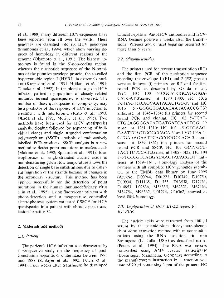
The primers used for reverse transcription (RT) and the first PCR of the nucleotide sequence encoding the envelope 1 (El) and 2 (E2) protein were as follows: (i) primers for RT and the first round PCR as described by Okada et al., 1992, HC 100 5’-CGCATGGCATGGGA- CATGAT-3’:sense, nt 1280-1300; HC 1Ola S’GGAGTGAAGCAATACACTGG-3’, and HC 1Olb 5’ - GGGGTGAAACAATACACCGG3’: antisense, nt 18455 1864; (ii) primers for second round PCR and cloning, HC 102 5’-TCAT- CTGCAGGGGACATGATGATCAACTGG - 3’: sense, nt 1281-1310; HC 103a 5’-GTGAAG- GAATTCACTGGGCCACA-3’ and HC 103b 5’- GTGAAAGAATTCACCGGGCCACA-3’ : anti- sense, nt 183991861; (iii) primers for second round PCR and SSCP, HC 105 GCTTGCC- TACTTCTCCATG:sense, nt 1405- 1423; HC 104 5’-f-TCCCGTCAGGACAACTACACGGT anti- sense, nt 1589- 1611. Homology analysis of the primers with all complete HCV genomes submit- ted to the EMBL data library by June 1995 (Act-No. D00944, D01221, D10749, D10750, D10934, D11168, D11355, D13558, D17763, D14853, L02836, M58335, M62321, M67463, M84754, M96362, U01214, U16362) showed at least 80% homology.
2.3. Atnpll’ficution of HCV El-E2 region by’
RT-PCR
The nucleic acids were extracted from 100 /ll serum by the guanidinium thiocyanate-phenol- chloroform extraction method with minor modifi- cations using the RNA isolation kit from Stratagene (La Jolla, USA) as described earlier (Peters et al., 1994). The RNA was reverse transcribed using AMV reverse transcriptase (Boehringer, Mannheim, Germany) according to the manufacturers instruction in a reaction vol- ume of 20 /II containing 1 pm of the primers HC

T. Peters et ul. i Journal of’ Virological Methods 64 (1997) 95-102 97
1Ola and HC 101b. The cDNA was amplified by hot start PCR with 40 cycles using the primers mentioned above (94°C for 1 min, 55°C for 2 min, 72°C for 3 min) and 1 U of Taq polymerase (Boehringer) according to the manu- facturer’s recommendations. For cloning and se- quencing, 1 ~1 of PCR 1 was subjected to another 15 cycles using primers HC 102, 103a/ 103b. Fragments for SSCP analysis were am- plified from PCR I by another 15 cycles using primer HC 104 and HC 105. Sensitivity was controlled by serial dilution of a standard serum from a patient with chronic HCV infection. PCR was positive in all assays at a dilution of 10 ~ 4 of the standard serum.
natively, for the detection of double strands, a 40% sucrose solution substituted for the stop so- lution and samples were loaded without heat de- naturation. Five microliters were loaded on a 30 cm non-denaturing acrylamide gel of varying concentration (5%, lo”/0 and 12”/0) and cross- linking (19:l and 29:l) containing 5% glycerol. Gel temperatures were either 10, 15, 20 or 30°C. The power was set to 30 W and the current to 38 mA. The preload run time was 1 h.
2.6. HCV-titer
Proof of specificity was obtained by running negative controls together with the patient’s samples and by sequencing the amplified DNA.
The concentration of HCV was determined using the quantitative Amplicor test kit (Hoff- mann-LaRoche, Grenzach-Wyhlen, Germany)
according to the manufacturer’s instructions.
2.4. Cloning mu’ sequencing 3. Results
The fragments obtained by the nested PCR reaction were purified on Sephadex G50 columns (Boehringer) cut with PstI and EcoRI (Boehringer) and ligated in pUC 18. The plas- mids were transformed into competent E. coli JM 109. Recombinant clones were identified as white colonies on AIX plates (LB medium plus Ampicillin, IPTG and X-gal) and by the Eck- hardt technique (Eckhardt, 1978). Recombinant plasmids were purified with Qiagen-tip 100 columns (Diagen, FRG). The eluted DNA sub- jected to a cycle sequencing reaction using the ‘fmol-DNA Sequencing System’ (Promega, Madison, Wis) and fluorescent sense primer u, 5’-Fluorescein-d CGACGTTGTAAAACGACG- GCCAGT-3’ or antisense primer r: 5’-Fluores- cein-d CAGGAAACAGCTATGAC-3’. Sequence analysis was performed with the automated laser fluorescent DNA sequencer (ALF, Pharmacia, LKB, Sweden).
3.1. Patient duta
HCV-RNA was amplified from two specimens of a patient with chronic post-transfusion hep- atitis C which were obtained 3 weeks and 6 years after infection, respectively. The clinical course of the patient is shown in Fig. 1. The preoperative serum sample was negative for anti-HCV antibodies as well as for HCV RNA.
Anti-HCV - + + + + + + +
HCV-RNA - + + 60, + + + +
2.5. SSCP
For SSCP analysis 2 ~1 of PCR product am- plified with primers HC 104 and HC 105 were added to 8 ~1 of stop solution (formamide, blue dextran) and heated to 94°C for 15 min. Alter-
h, 3” ,r ‘I 9w 17.a 24w 1, 67
Fig. 1. Clinical course of the patient with post transfusion
hepatitis. Anti-HCV: - . negative; + , positive; HCV-RNA:
~, negative; +, positive; J, indicates the time point of quasispecies analysis.
Clinical Course
I

98 T. Peters et al. : Journal of’ Virological Methods 64 (1997) 95- 102
Aminoacidsequence Hypervariable Region I ClOlle Yea~G GTHVTGGSAAHTTSGFVGJ.FSOGAR SSCP week3
GTI1VTGGSAAHTTSGFVGL,FSQGPl?
240 270 300
[min]
Fig. 2. (a) Quasispecies analysis of early and late serum sample by sequencing the HRVI of multiple clones. data are shown as amino
acid sequences in the one letter code. (b) Quasispecies analysis of early and late serum sample by f-SSCP. Intensity of fluorescence
of DNA is shown as a function of analysis time (min).
Anti-HCV antibodies became detectable in the first postoperative serum sample 3 weeks after surgery, and remained positive again for 6 years. Elevated ALT levels were found between week 4 and 24 and were normal after 2 and 6 years, although ALT elevations were observed by the patients’ general practitioner during this period of time.
3.2. HCV RNA detection
HCV RNA was detectable from post-opera- tive week three and remained so for at least 6 years. The HCV titer was 248 000 virus parti- cles/ml in the serum sample 6 years after the infection. The fragments obtained for sequence analysis contained nucleotide sequences from the N-terminus of the El-protein and the first half of the E2-protein which code for the amino acid sequence of the hypervariable region I (HVRI).
3.3. HCV RNA analysis
The genotype of the patient’s virus population was type la according to Simmonds et al. and was determined by homology analysis of the conserved sequence of the El-protein. The amino acid se- quence of 30 clones of each specimen of this region from the third week and 6 years after infection are shown in Fig. 2. The early sample showed very few point mutations scattered over the whole popula- tion. In contrast, the clones of the late sample showed more mutations leading to at least four groups of related clones. Despite the large number of sequenced clones from each point of time, both virus populations had no clones in common.
3.4. SSCP
To test the specificity of SSCP four representa- tive clones of the heterogenous population of the late serum sample were chosen. The analyzed

T. Peters et al. /Journal of Virological Methods 64 (1997) 95-102
fragments were 208 bases long from which 42
were primer-derived. The hypervariable region
consisted of 75 bases, the remaining 91 bases were
part of the higher conserved El-region. Their
sequences are listed in Fig. 3(a) and the number of
nucleotide variations in Fig. 3(b). Two clones
(clone A and D) differed by only one base.
The variables of SSCP tested were acrylamide-
concentration (5, 8, 10 and 12%) degree of cross-
linking (19:1, 29:l) and running temperature (10,
15, 20 and 30°C). The acrylamide-concentration
as well as the cross-linking is determined by the
fragment length to be analyzed. For the 208 bases
fragments tested here, acrylamide concentrations
higher than 10% resulted in a rapid decrease in
migration, at lower concentrations the one-base
substitution in two of the fragments could not be
resolved any more. The same was true for the
degree of crosslinking. Higher cross linking re-
sulted in better resolution, but the time for analy-
sis increased sharply (data not shown). Standard
conditions used for this fragment therefore were a
10% acrylamide-gel at an acrylamide-bisacry-
lamide ratio of 29:1, which allowed the detection
of a single point mutation in a 6 h run time (Fig.
4).
D _______-----------~--------_-_--__--_-__________---_-
D ________--------_---~~~~~~------------_-------------_
A CC*GCTGGTTAACACCAACGGCAGTTGGaccgtgtagtt~~==~~~=~~~~ B -------__~------_-____________________-------------
c ______---~------_-_----__-____________-----__--_--- D -__---_--------------------------------------------
Fig. 3. (a) Sequence variation of standard clones A, B, C, D.
The hypervariable region is underlined. Primer-derived se- quences are written in small letters. (b) Number of amino acid
substitutions between the four related clones from the 6th year after infection.
t
t
Fig. 4. Influence of temperature on the resolution of different
clones in the SSCP.
The running temperature proved most crucial as shown in Fig. 4. At 10°C the fluorescence- marked DNA-strand of a single HCV clone did not, as expected, migrate in a single band, but in
up to three different bands. With higher running temperature the monoclonal fragments formed one single peak. Testing at 10” intervals the best resolution and reliability were obtained at 20°C.
At higher temperature the clones B and C ran at almost the same speed. The clones A and B differing by a single point mutation could be resolved at an interval of 1 min 40 s at 20°C and
1 min 2 s at 30°C. The results obtained by SSCP with respect to
this patient’s HCV quasispecies corresponded well to the sequence data from multiple clones (Fig. 2). The fragments from the early serum sample which contained only a few quasispecies differing only
by few point mutations migrated as a single peak in the SSCP-gel (Fig. 2). From the nucleic acids amplified from the late serum sample at least four

main virus clone groups could be classified. They were discriminated well by SSCP analysis,
Reproducibility of SSCP was tested by analyz- ing eight independent HCV preparations (extrac- tion and RT-PCR, Fig. 5(a)) of the same serum sample 6 years after infection. Fluorescence of the peaks was quantitated by integration of the peak area (Fig. 5(b)). Denaturation was very constant within one gel, 65.6% of fluorescence was found in the peaks of denaturated strands. The distribution of fluorescence in the denatured strands was quite similar between the different preparations.
In sera with high virus titers quasispecies analy- sis by SSCP was reproducible. Fig. 6 demon- strates the dependence of the resolution of quasispecies in SSCP on the viral titre. The serum was diluted prior to extraction. At a concentra- tion close to the detection limit of 250 copies/ml only one single clone was detected, which corre- sponded to the most predominant clone in the undiluted serum.
4. Discussion
HCV infection causes chronic hepatitis in up to 95% of patients. Factors influencing clinical out- come have been studied. Host factors such as age,
a) b) SSCP distribution of
fluorescence
l”hl
dS I II I:1 I”
Fig. 5. Reproducibility of SSCP: groups A-H are independent
preparations of HCV fragments of the same serum sample. (a) SSCP analysis; (b) relative distribution of fluorescence among
peak non-denatured double strands (ds) and I-!V.
PCR hcv titer
IcoDies/mll
248000
24800
2480
240
co
MG
Fig. 6. Interdependence of virus titer and quasispecies analysis. The standard serum contained 248000 virus particles/ml and
was diluted with control serum (co) lo-. IOO- and IOOO-fold.
Fragments obtained are shown on a agarose gel stained with
ethidium bromide (PCR) and after strand separation on the
acrylamide gel detected by their fluorescence (SSCP); MC.
molecular weight marker.
gender, MHC-class and immunocompetence as
well as viral factors such as virus titer and geno-
type have been shown to be important (Mita et
al.. 1994; Bjoro et al., 1994; Pozazato et al.. 1994;
Peano et al., 1994; Soni et al., 1995; Kurosaki et
al., 1994). In addition, the heterogeneity and com- plexity of the patient’s virus population may be a
predictor of his response to interferon therapy
(Kato et al.. 1993: Okada et al., 1992; Moribe et
al., 1995). Cloning of RT-PCR-amplified frag-
ments followed by sequencing of these clones
(Kato et al., 1993; Okada et al., 1992) or analysis
of radioactively labelled PCR fragments by single-
strand conformation polymorphism (SSCP) (Moribe et al., 1995) have been used to analyze
the viral population. However, sequencing of a
large number of HCV clones to obtain representa-
tive results is time consuming and laborious.
SSCP analysis of fragments obtained by RT-PCR
could be a useful alternative as the heterogeneity
of one’s patient viral population is easily detected.
This method can be performed using either ra-
dioactively labelled PCR products followed by
autoradiography or fluorescence labelled primers
and on-line fluorescence detection system, that gives quantitative and qualitative results. We
studied factors which influence resolution and re-
liability of f-SSCP of the hypervariable region of
HCV.

T. Peters et al. ! Journal of’ Virological Me1hod.s 64 (1997) 95-102 101
4. I. Influence of the HCV sequence on
quasispecies analysis
One important factor is the region to be examined and the primer design. The sequence of HCV genome is variable, the variations of the sequence are different in distinct segments of the genome, however. The 5’-non-coding region is the most conserved region, whereas the HVRI and II, coding for parts of the E2-protein, are extremely variable (Kremsdorf et al., 1991; Hijikata et al., 1991; Tanaka et al., 1992). These regions are of special interest because the extensive sequence variation leads to escape mutants (Kurosaki et al., 1994; Kato et al., 1994). The sequence variability also causes difficulties in PCR analysis as ideal primers should hybridize to all types and subtypes. To circumvent this problem we used primer mix- tures for first round PCR, cloning and sequenc- ing that covered sequences of type l-3 genomes. The fragments obtained were 600 bases in length.
4.2. Variables of SSCP analysis and reliability
SSCP analysis may be influenced by a variety of factors, sequence with respective alterations of the tertiary structure, fragment length, HCV concentration of the blood, composition of the gel and temperature. For SSCP analysis only single primer pairs can be used for the HVRI region of the HCV which should cover all types. The optimal fragment length for this method is 100-300 bases. The consequence is, however, that only 85% of our patients with chronic HCV infection can be tested. Different gel ma- trices may be used like agarose or acrylamide gels. Higher matrix concentrations and cross- linking lead to retardation of migration and are therefore applied for smaller fragments. Another important factor is the running temperature. In the literature running temperatures for SSCP range from 4 to 25°C (Makino et al., 1992; Orita et al., 1989) but little information is given on their effect. We tested systematically different temperatures with a temperature controlled gel plate and found additional peaks at low temper-
atures, probably due to additional conforma- tions of the nucleic acids, that are stabilized at low temperatures. In our experiments, 20°C
running temperature gave the best resolution
without producing unexpected peaks. These con- ditions may vary for nucleic acids with different
secondary structures.
Finally, reproducibility for independent serum extractions and different virus titers were tested. These aspects have not been considered until now. Very good reproducibility was found with
the number of clones detected as well as the
distribution within the whole population. Quan- titative analysis in SSCP of radioactively labeled
fragments usually requires densitometry as a further step. Our method, with application of
fluorescent primers is reliable and fast. Virus titers in serum had a significant influence on
the SSCP pattern. If low titered sera of a com- plex virus population were analyzed by PCR
and SSCP, fewer peaks than expected were found. In this experiment the most frequent clone in the diluted serum formed a single band. In conclusion, representative analysis of a het-
erogenous virus population can only be achieved, if the virus titer is significantly above the detection limit of the method used. The effi-
cacy of the extraction and the amplification de- termine whether high complexity is identified.
On the other hand in patients with low virus titers results of the SSCP analysis may underes-
timate the number of quasispecies. Further stud- ies have to be done to determine whether low virus titer and low complexity are independent predictors of response to interferon or lead to
false results of SSCP analysis in low titered samples.
If this is true, f-SSCP may become a useful clinical tool for choosing candidates for inter- feron therapy.
Acknowledgements
This work was supported by the Deutsche Forschungsgemeinschaft, DFG, Bonn-Bad Godesberg through grant Ra 258/4-l.

References
Bjoro, K., Froland, S., Yun. Z.. Samdal, i-1. and Haland, T.
(1994) Hepatitis C infection in patients with primary hy-
pogammaglobulinemia after treatment with contaminated
immune globulin. N. Engl. J. Med. 331, 1607~1614.
Choo, Q.L., Kuo, G.. Weiner, A.J., Overby, L.R., Bradley,
D.W. and Houghton, M. (1989) Isolation of a cDNA clone
derived from a blood-borne Non-A Non-B viral hepatitis
genome. Science 244, 359 -362.
Eckhardt, R. (1978) A rapid method for the identification of
plasmid deoxyribonucleic acid in bacteria. Plasmid I, 584-
588.
Hijikata, M., Kato, N., Ootsuyama. Y.: Nakagawa. M..
Ohkoshi. S. and Shimotohno, K. (1991) Hypervariable
regions in the putative glycoprotein of the hepatitis C virus.
Biochem. Biophys. Res. Commun. 175, 220-228.
Kato, N., Sekiya, H., Ootsuyama, Y., Nakazawa, T., Hijikata.
M., Ohkoshi, S. and Shimotohno K. (1993) Humoral
immune response to hypervariable region I of the putative
envelope glycoprotein (gp70) of hepatitis C virus. J. Viral.
67, 3923-3930.
Kato, N.. Ootsuyama, Y., Sekiya, H., Ohkoshi, S., Nakazawa,
T.. Hijikata, M. and Shimotohno, K. (1994) Genetic drift
in hypervariable region I of the viral genome in persistent
hepatitis C virus infection. J. Virol. 68, 4776-4784.
Kremsdorf, D., Porchon, C., Kim, J.P.. Reyes, G.R. and
B&hot, C. (1991) Partial nucleotide sequence analysis of a
French hepatitis C virus: implications for HCV genetic
variability in the EZ/NSl protein. J. Gen. Viral. 72. 2557
2561.
Kuo, G., Choo, Q.L., Alter, H.J., Gitnik, G.L., Redeker, A.G.,
Purcell, R.H., Miyamura, T.. Dienstag, J.L., Alter, M.J.,
Stevens, C.E., Tegtmeier, T.E., Bonino, F., Colombo, M.,
Lee, W.S., Kuo, C., Berger, K., Shuster, J.R., Overby,
L.R., Bradley, D.W. and Houghton, M. (1989) An assay
for circulating antibodies to a major etiologic virus of
human non-A. non-B hepatitis. Science 244, 362-364.
Kurosaki, M., Enomoto, N., Marumo, F. and Sato, C. (1994)
Evolution and selection of hepatitis C virus variants in
patients with chronic hepatitis C. Virology 205, I61 169.
Lin, H.J., Siwak, E.B., Lauder. I.J. and Hollinger, F.B. (1995)
Single-strand conformation polymorphysm study of human
immunodeficiency virus type I RNA and DNA in plasma,
peripheral blood mononuclear cells. and their virologic
cultures. J. Infect. Dis. 171, 1619-22.
Makino, R., Yazyu, M., Kishimoto, Y., Sekiya, T. and
Hayashi, K. (1992) F-SSCP: Fluorescence-based poly-
merase chain reaction-single-strand conformation polymor-
physm (PCR-SSCP) analysis. PCR Methods Appl. 2.
10-13.
Mita. E.. Hayashi, N., Hagiward. H., Ueda, K., Kanazawa. Y., Kasahard. A., Fusamoto, H. and Kamada. T. (1994) Pre- dicting interferon therapy efficacy from hepatitis C virus
genotype and RNA titer. Dig. Dis. Sci. 39 (5). 977-982. Moribe, T.. Hayashi, N., Kanazawa, Y., Mita, E., Fusamoto,
H., Negi, M., Kaneshige. T., Igimi, H.. Kamada, T. and
Uchida. K. ( 1995) Hepatitis C viral complexity detected by single-strand conformation polymorphism and response to
interferon therapy. Gastroenterology 108, 7X9-795.
Okada, S.. Akahane, Y., Suzuki, H., Okamoto, H. and Mishiro, S. (1992) The degree of variability in the amino
terminal region of the E2!NSI protein of hepatitis C virus
correlates with responsiveness to interferon therapy in
viremic patients. Hepatology 16, 619 624.
Okamoto, H., Okada, S., Sugiyama, Y., Kurai, K., Iizuka, H., Machida, A.. Miyakawa, Y. and Mayumi M. (1991) Nucle-
otide sequence of the genomic RNA of hepatitis C virus isolated from a human carrier: comparison with reported
isolates for conserved and divergent regions. J. Gen. Virol.
72, 2697 -2704.
Orita. M., Iwahana, H., Kanazawa, H., Hayashi, K. and
Sekiya, T. (1989) Detection of polymorphisms of human DNA by gel electrophoresis as single-strand conformation
polymorphism. Proc. Nat]. Acad. Sci. USA 86. 2766- 2770.
Peano, G.. Menardi, G., Ponzetto, A. and Fenoglio, L. (1994) HLA-DR5 antigen. A genetic factor influencing the out-
come of hepatitis C virus infection? Arch. Int. Med. 154,
2733-2736. Peters. T., Mohr. L., Scheiffele, F., Schlayer, H. and Preisler,
S. (19Y4) Antibodies and viremia in acute post-transfusion
hepatitis C: a prospective study. J. Med. Virol. 42. 420
427.
Pozazato. G.. Kaneto, S.. Moretti. M., Croce, L.. Franzin. F., Unuora. M., Bercich. L., Tiribelli, C.. Crovatto, M., San-
tini. G. and Kobayashi, K. (1994) Different genotypes of
hepatitis C virus are associated with different severity of
chronic liver disease. J. Med. Virol. 43. 291-296.
Rubin, R.A., Falestiny, M. and Malet. P.F. (1994) Chronic
Hepatitis C. Advances in diagnostic testing and therapy. ,Arch. Int. Med. 154, 387-392.
Schlayer, H.J., Peters, T., Preisler, S., Berthold, H.. Gerok, W.
and Rasenack, J. (I 992) Cause and frequency of posttrans- fusion hepatitis after open-heart surgery. Clin. Invest. 70,
579%584. Simmonds, P., Alberti, A., Alter, H.J., Bonino, F., Bradley, D..
Brechot, C., Brouwer, J.T., Chan, S.W., Chayama, K.,
Chen. D.S.. Choo, Q.L., Colombo, M., Cuypers. H.T.M..
Date, T.. Duwheiko, G.M.. Estebdn, J.I., Fay, O., Hadziyannis, S.J., Han, J., Hatzakis. A., Holmes, E.C. and
Hotta, H. (I 994) A proposed system for the nomenclature
of hepatitis C viral genotypes. Hepatology 19. 1321- 1324.
Soni, P., Dusheiko. G., Harrison, T. and Dhillon, A. (1995) Genetic diversity of hepatitis C virus: Implications for
pathogenesis, treatment and prevention. Lancet 345, 562-
566. Spinardi, L., Mazars, R. and Theillet. C. (1991) Protocols for
an improved detection of point mutations by SSCP. Nu-
cleic Acids Res. 19, 4009.
Tanaka, T., Kato, N., Nakagawa, M.. Ootsuyama, Y., Cho. M., Nakazawa, T., Hijikata. M., Ishimura, J. and Shimo- tohno, K. (1992) Molecular cloning of hepatitis C virus
genome from a single Japanese carrier; sequence variation within the same individual and among infected individuals.
Virus Res. 23. 39- 53.