quaternary structures and complex enzymes · pdf fileno cooperative activity . p50. ... assume...
TRANSCRIPT
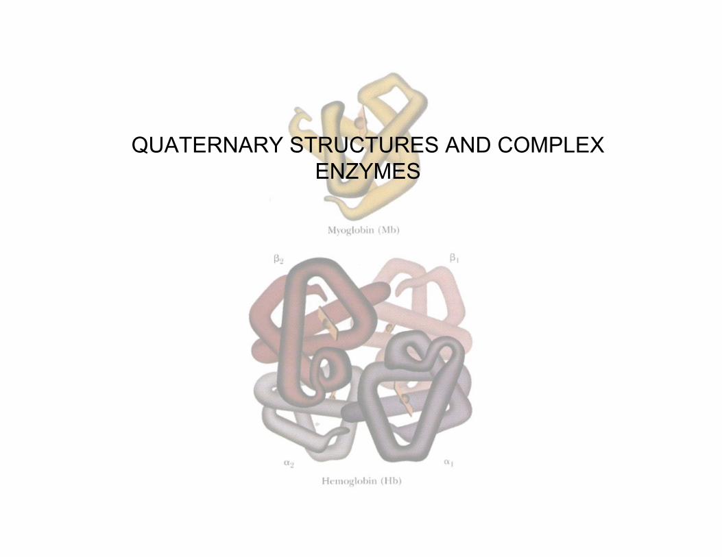
QUATERNARY STRUCTURES AND COMPLEXENZYMES
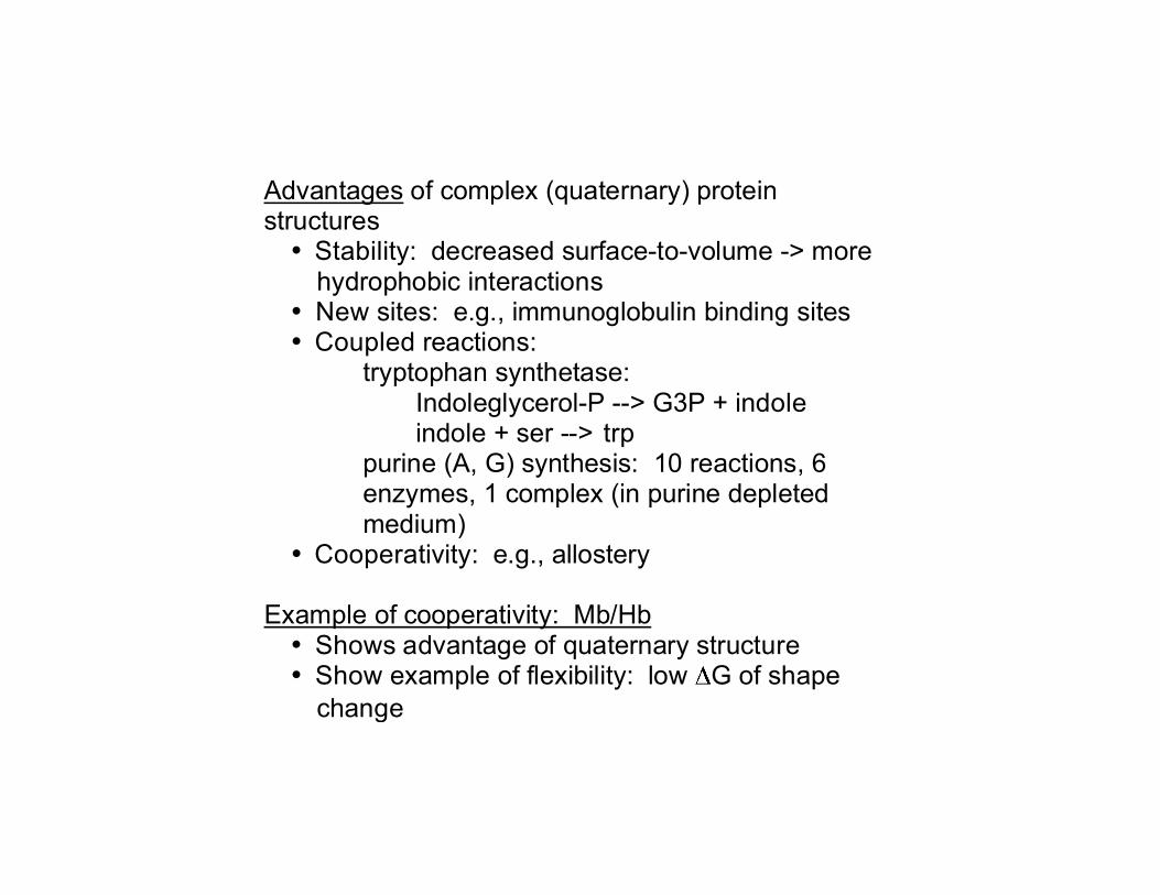
Advantages of complex (quaternary) protein structures • Stability: decreased surface-to-volume -> more
hydrophobic interactions • New sites: e.g., immunoglobulin binding sites • Coupled reactions:
tryptophan synthetase: Indoleglycerol-P --> G3P + indole indole + ser --> trp
purine (A, G) synthesis: 10 reactions, 6 enzymes, 1 complex (in purine depleted medium)
• Cooperativity: e.g., allostery Example of cooperativity: Mb/Hb • Shows advantage of quaternary structure • Show example of flexibility: low G of shape
change
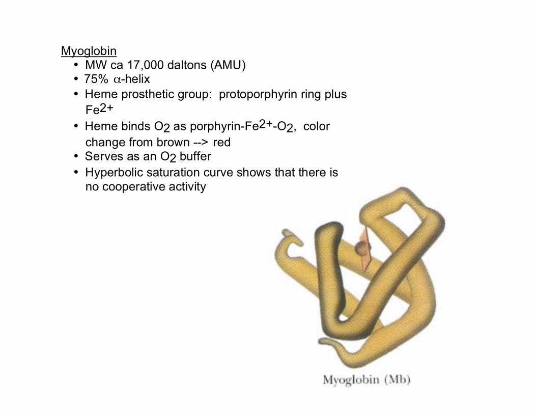
Myoglobin • MW ca 17,000 daltons (AMU) • 75% -helix • Heme prosthetic group: protoporphyrin ring plus
Fe2+ • Heme binds O2 as porphyrin-Fe2+-O2, color
change from brown --> red • Serves as an O2 buffer • Hyperbolic saturation curve shows that there is
no cooperative activity
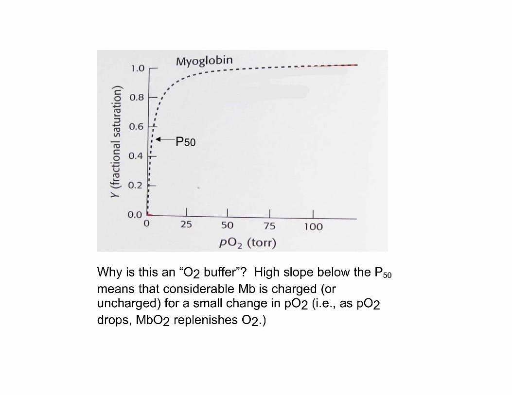
P50

Hemoglobin • Tetramer of myoglobin-like subunits, each with... • Heme prosthetic groups: protoporphyrin ring plus
Fe2+ • MW ca 4 x 17,000 daltons • 75% -helix • Complexed with O2, porphyrin-Fe2+-O2, brown
--> red
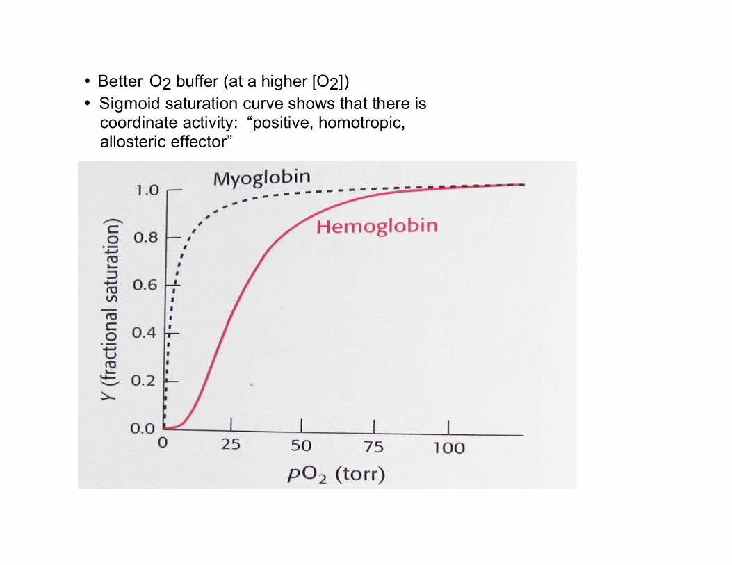
• Better O2 buffer (at a higher [O2]) • Sigmoid saturation curve shows that there is
coordinate activity: “positive, homotropic, allosteric effector”

• Better O2 buffer (at a higher [O2]) • Sigmoid saturation curve shows that there is
coordinate activity: “positive, homotropic, allosteric effector”

• Bohr effect: H+, CO2 promote dissociation of O2 from Hb-O2: “negative, heterotropic, allosteric effector”
! 2,3-bisPGA also promotes dissociation of O2
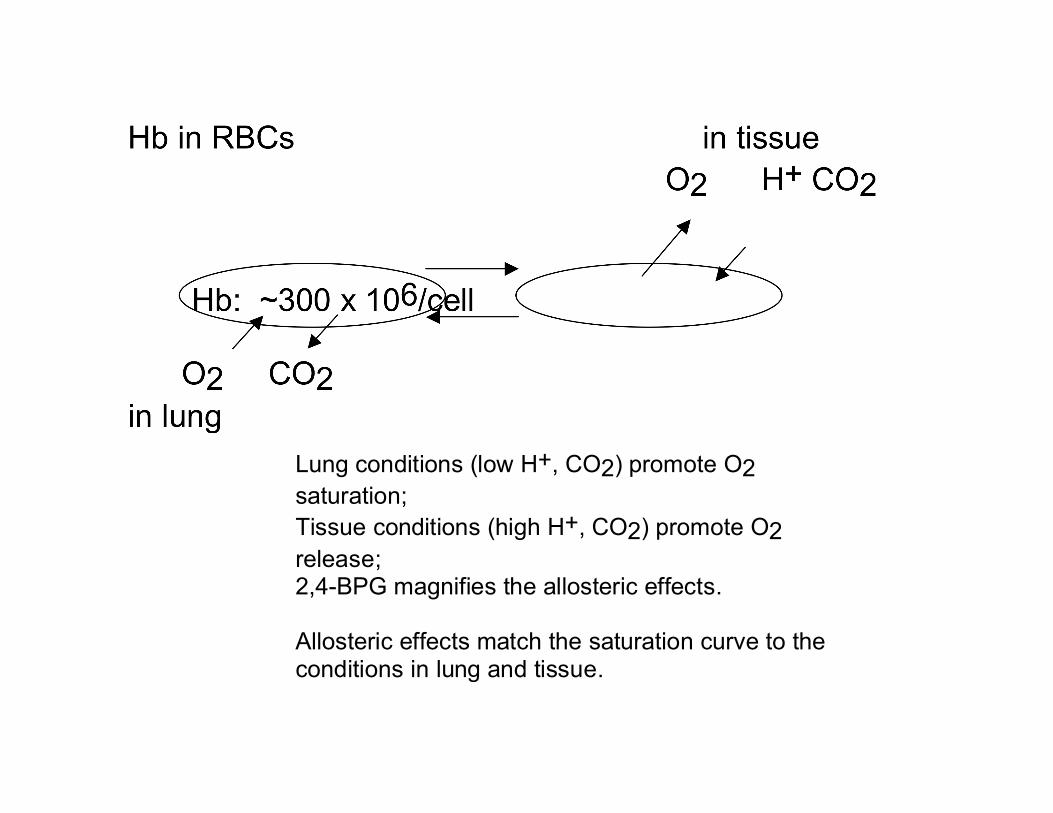
Lung conditions (low H+, CO2) promote O2 saturation; Tissue conditions (high H+, CO2) promote O2 release; 2,4-BPG magnifies the allosteric effects. Allosteric effects match the saturation curve to the conditions in lung and tissue.
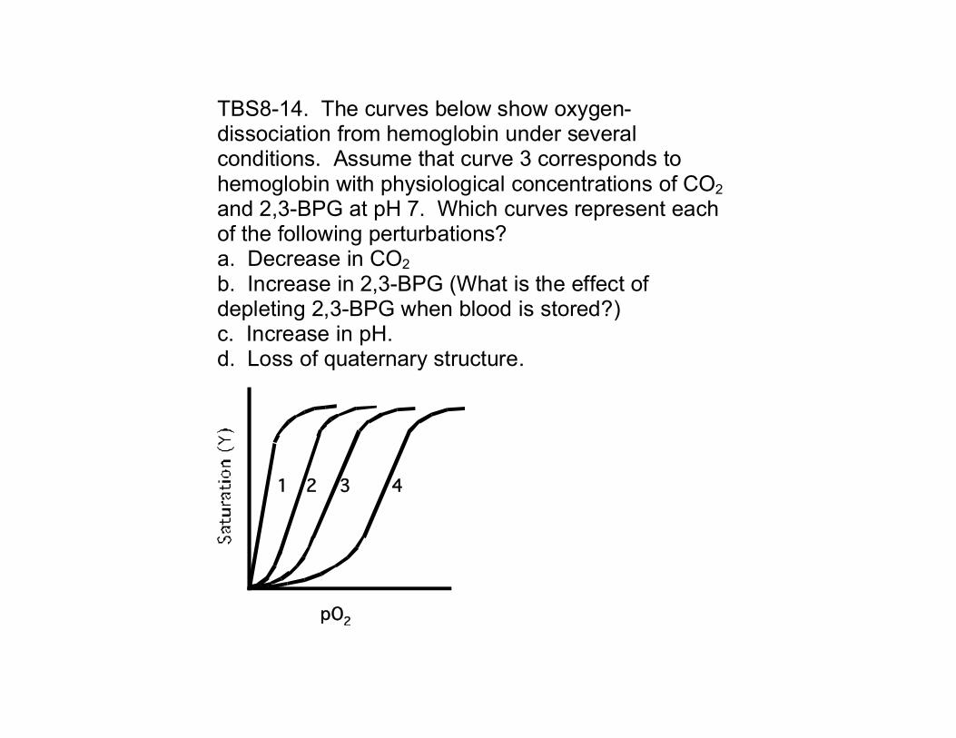
TBS8-14. The curves below show oxygen-dissociation from hemoglobin under several conditions. Assume that curve 3 corresponds to hemoglobin with physiological concentrations of CO2 and 2,3-BPG at pH 7. Which curves represent each of the following perturbations? a. Decrease in CO2 b. Increase in 2,3-BPG (What is the effect of depleting 2,3-BPG when blood is stored?) c. Increase in pH. d. Loss of quaternary structure.
1 2 3 4
pO2
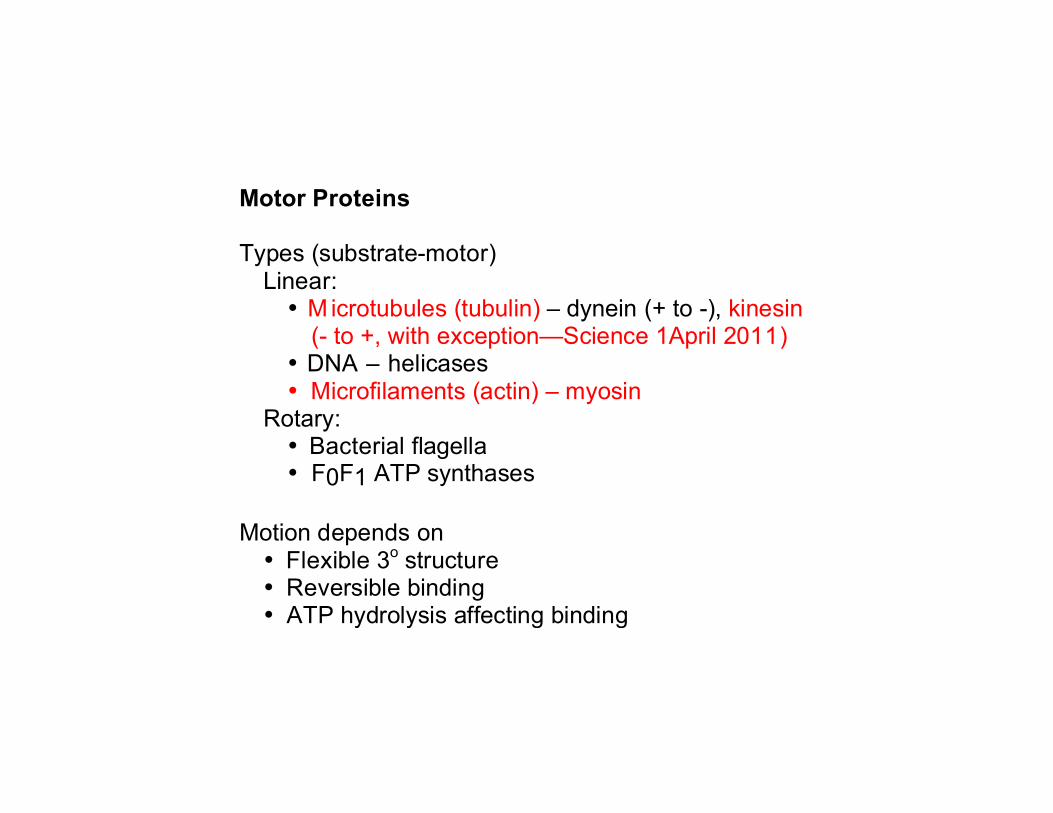
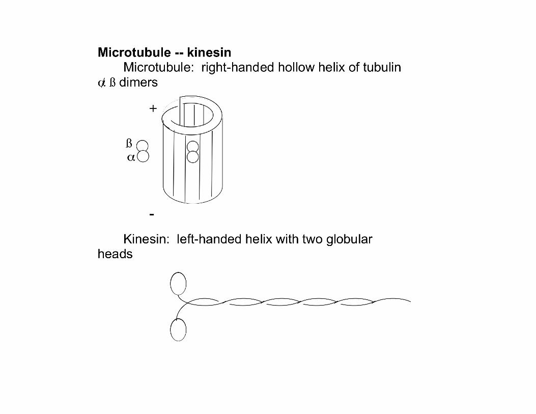
Motor Proteins Types (substrate-motor)
Linear: • M icrotubules (tubulin) – dynein (+ to -), kinesin
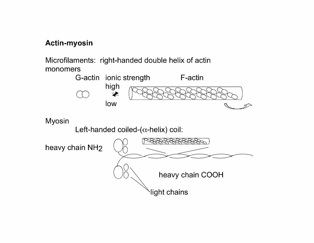
(- to +, with exception—Science 1April 2011) • DNA – helicases • Microfilaments (actin) – myosin
Rotary: • Bacterial flagella • F0F1 ATP synthases
Motion depends on • Flexible 3o structure • Reversible binding • ATP hydrolysis affecting binding
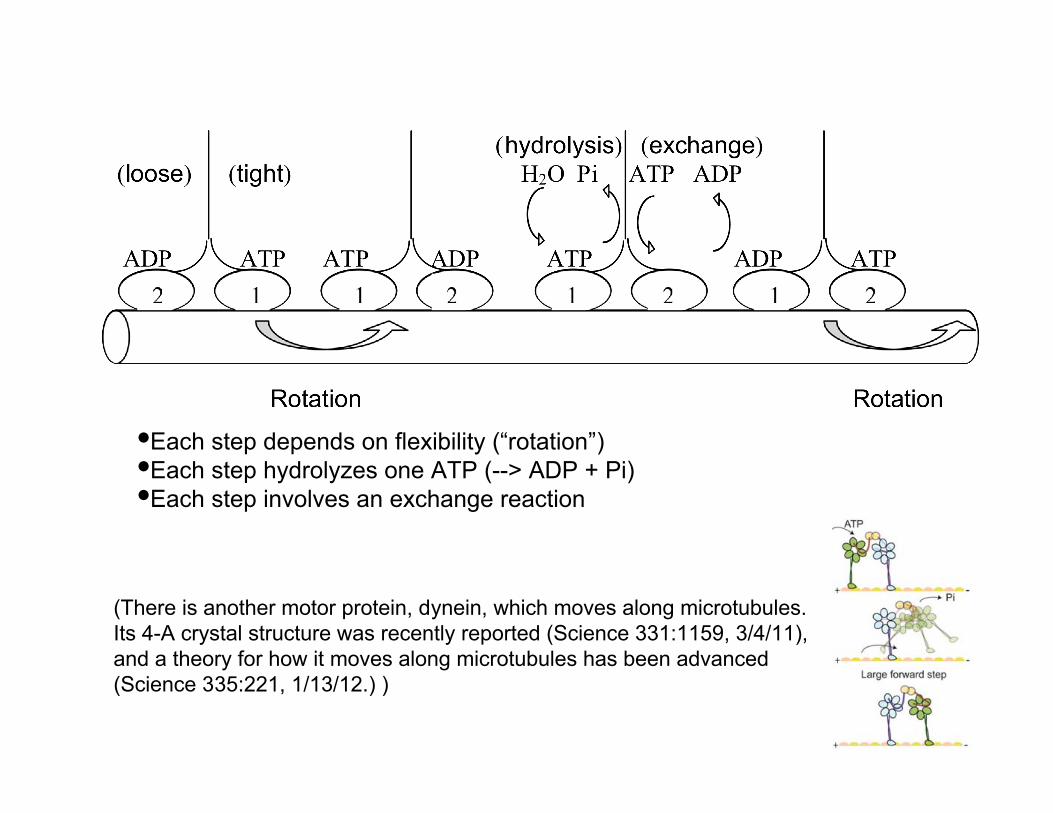
•Each step depends on flexibility (“rotation”)•Each step hydrolyzes one ATP (--> ADP + Pi)•Each step involves an exchange reaction
(There is another motor protein, dynein, which moves along microtubules. Its 4-A crystal structure was recently reported (Science 331:1159, 3/4/11), and a theory for how it moves along microtubules has been advanced (Science 335:221, 1/13/12.) )
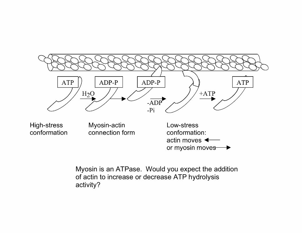
Myosin is an ATPase. Would you expect the addition of actin to increase or decrease ATP hydrolysis activity?
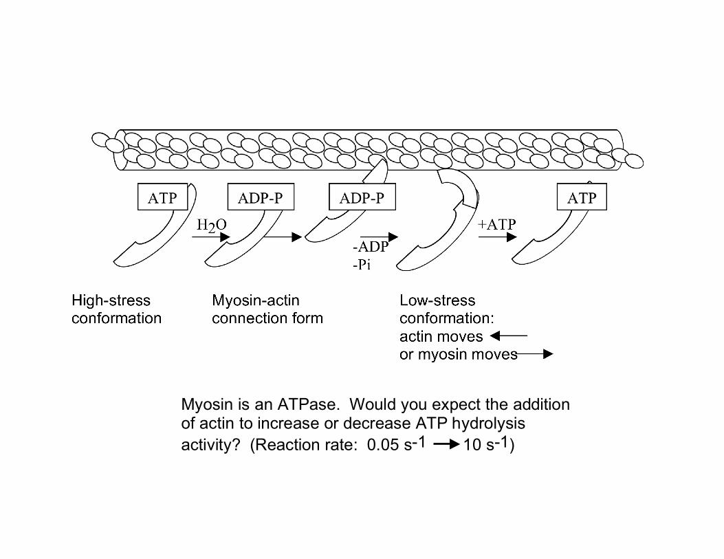
Myosin is an ATPase. Would you expect the addition of actin to increase or decrease ATP hydrolysis activity? (Reaction rate: 0.05 s-1 10 s-1)
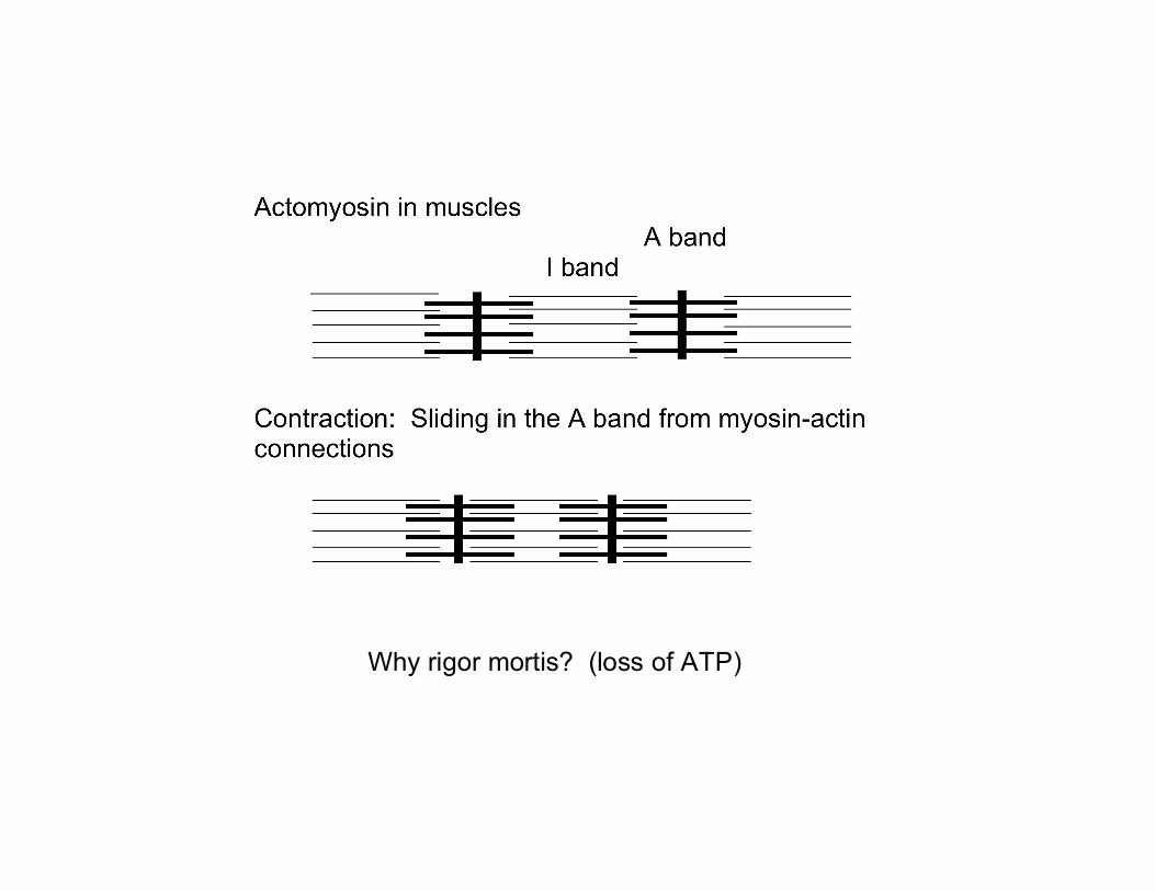
Why rigor mortis? (loss of ATP)

Conclusion:
Flexibility in protein structures allows more complex functions
•Reversible O2 and CO2 binding •Reversible protein-protein (kinesin-MT) binding
Shows the importance of low ΔG in protein shape changes