quicklase a-z lasers · operculectomy incision of abscess and sterilisation sterilisation of gum...
TRANSCRIPT

1 06122017v1.5
Quicklase A-Z Lasers
This document is intended for advice on lasers including laser knowledge, benefits,
applications, FAQ’s and LPA - laser protection advice for health and safety.
Introduction to LASERS
Types
- Diode
- Nd:YAG
- CO2
- Argon
- Erbium:Yag
- Ho:YAG
- Nd:YAP
Clinical Applications
Diode (used mostly in Dentistry)
- Soft tissue management (crown lengthening, Veneers re-contouring to
curettage
- Hygienists gum management
- Replaces Electrosurgery better cutting
- Smallest type of Lasers (2.5 pounds, 1.1Kg)
- Portable, fast easy and effective
- Very competitively priced
Erbium:YAG with water
- Soft tissue surgery (you will get bleeding)
- Hard tissue (Caries removal, Cavity preparation)
Argon
- Soft tissue surgery
- Curing of Composite materials
- Tooth whitening
- Illumination for caries detection
- Illumination for endodontic orifice location
CO2
- Soft tissue surgery
- Curing of Composite materials
- Tooth whitening
Nd:YAG Like Diode (used for years in dentistry)
2 06122017v1.5
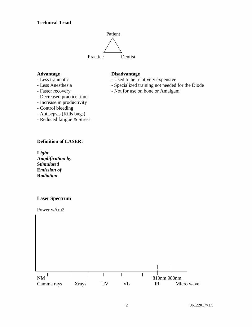
Technical Triad
Patient
Practice Dentist
Advantage Disadvantage
- Less traumatic - Used to be relatively expensive
- Less Anesthesia - Specialized training not needed for the Diode
- Faster recovery - Not for use on bone or Amalgam
- Decreased practice time
- Increase in productivity
- Control bleeding
- Antisepsis (Kills bugs)
- Reduced fatigue & Stress
Definition of LASER:
Light
Amplification by
Stimulated
Emission of
Radiation
Laser Spectrum
Power w/cm2
| |
|
NM 810nm 980nm
Gamma rays Xrays UV VL IR Micro wave

3 06122017v1.5
Laser components
- Active medium
- Pumping mechanism
- Optical resonator
pumping mechanism
optical resonator (optical resonator)
active medium

4 06122017v1.5
Benefits of the lasers versus Electrosurge, scalpel and retraction cord
- Bloodless
- Coagulation of bleeding tissue
- Clear visual field allows for better visual working area
- Creates better contouring for accurate impression
- Precise crown lengthening and veneers margin re-contouring
- Less damage to soft tissue, no collateral damage
- Simple removal and shaping of soft tissue surface
- Easy to use
- Faster healing time
- Can touch hard tissue (teeth), implant and metal posts
- Can be used on pacemaker patients
Integrating the Diode Laser into dental practices
- Easy training
- Easy technique
- Easy care & fiber maintenance & sterilisation
- Follow up support by our staff
- Staff & Patient education & training reinforcement
- BILD clinical laser training – Core of knowledge
Soft tissue Applications
Soft tissue management
- Troughing (accurate impression)
- Gingival incisions & excisions
- Gingivoplasty (crown lengthening & veneers re-contouring)
- Gingivectomy (cutting gum)
- Vestibuloplasty (reshaping)
- Papillectomy (growth)
- Ulcers (good revenue)
- Implant Recovery and relieving tissue for abutment placement
- Sulcular debridement (Curettage - tissue flap)
Hygienists gum management - Laser Sulcular Debridement and curettage -Perio, reduces the risk of CardioVascular diseases)
Sterlisation - Endo & Perio sterlisation
Therapy and whitening -TMJ therapy and teeth Whitening prisms

5 06122017v1.5
Uses for the QuickLase Lasers – Dr Ashley Watson
The QuickLase is only for use on soft tissue. It is an 810-980nm wavelengths laser
operating in continuous and pulsation modes which make it the best laser for coagulation
and cutting tissue that contains Haemoglobin, Oxyhaemoglobin, and Melanin. Being
solid state it is also the most reliable as well as fast, easy and effective.
It is a proven fact that laser surgery heals faster with less scarring than a scalpel or
electrosurgery, and requires no suturing.
The following procedures are therefore the most likely ones that a general dental
practitioner will use more frequently.
1 Crown preparation impressions – (Tissue retraction for Impression)
If an impression is not clear it will need to be taken again or the crown will not fit
leading to extra clinical time adjusting it (money) or embarrassment to the dentist
(loss of goodwill).
2 Stopping Gums Bleeding – (Haemostasis)
Now that more fillings rely on glue (bonding) any contamination such as blood
from the gums can frustrate a dentist trying to do a good job.
3 Cutting or Reshaping the gums – (Gingivectomy/Gingivoplasty)
Good looking restorations rely on having the correct shaped gums as well as the
correct colour and shape of teeth and lasers are the best and least painful way of
doing this.
4 Frenectomy
Very often this can be done with little or no anaesthesia and rarely requires
suturing, even lingual frenectomy can be carried out with small amounts of LA
and no sutures.
5 Cauterising exposed nerve tissue – (High Pulpotomy)
If the nerve is exposed during a dental procedure bleeding can be stopped, the
area sterilised and the patient has a greater chance of healing without further
problems meaning the tooth is less likely to need root canal treatment.
6 Sterilisation of tooth sockets following extractions –
By using the laser in a recent extraction site haemostasis can be achieved quicker
and the patients report quicker and less painful healing times. This is due to an
increase in temperature locally above 60’C which causes plasmolysis of the
bacteria and denaturation of bacterial proteins.
6 06122017v1.5
7 Sterilisation of gum pockets – (Sulcular Debridement)
If the laser is used in a recently cleaned gum pocket the area can be sterilised for
up to 60 days resulting in less after pain following extensive periodontal (gum)
treatments. This is also true for wisdom tooth problems which are extremely
common in young patients and for which there is no other minimally invasive
treatment as comfortable.
8 Removal of unwanted tissue –
If a denture does not fit well because of flabby tissue or “extra” gum then this can
be removed more predictably with less postoperative pain than conventional
techniques.
9 Stopping the pain of ulcers –
This can be an extremely painful condition for some people and use of the laser
can ensure the ulcer is non-painful within minutes without anaesthetic.
There are many other applications for the laser and it is futile to list its ability completely,
as the list of oral surgical procedures is vast.
In general the laser replaces two instruments in the vast majority of cases, the scalpel
(scalpel knife – lots of blood and unpredictable scarring and healing) and the
electrosurgery unit (electrical burn – very useful but not as refined as the laser).
It is generally accepted that lasers are a far superior way of carrying out most procedures
and they are used in the best plastic surgeon’s theatres for good reason, however until
now the cost has been restrictive.
The diode laser is generally accepted as the most reliable laser as it consists of electrical
circuits only and has a very flexible fibre optic cable making it very easy to use.
The size use to betrays its ability and will allow even the most minimalistic dentist to
hide it somewhere. But now with the QuickwhiteTM
Lase this is not an issue.
However the best idea is to show these instruments off as investment in a laser suggests
high-tech dentistry to the vast majority of patients and very often just advertising the
possession of a laser will increase a dental surgery revenue without even using it!
List of most useful applications of soft tissue laser in order of most common
Veneers re-contouring
Haemostasis after cavity preparation
Gingivectomy to expose cavities
Tissue troughing for perfect impressions every time
Removal of hyperplastic gum tissue that has grown into cavity
Crown lengthening prior to preparation
Gingival recontouring prior to aesthetic work
Sterilisation of tooth sockets post extraction
Pulpotomy for painless recovery
Removal of fibromatous or hyperplastic tissue around dentures

7 06122017v1.5
Curing ulcers
Operculectomy
Incision of abscess and sterilisation
Sterilisation of gum pockets
Frenectomy
Excisional biopsies
Vestibuloplasty
Removal of drug induced gingival hypertrophy

8 06122017v1.5
Diode Laser Dentistry (Patients Frequently Asked Questions)
What will I notice?
No vibration, no noise, more relaxing treatment all round. Your dentist will now be
able to do much more work on gum problems and so help you to keep your teeth much
longer.
Will I feel anything?
Even without an injection - you may only feel that something is going on - nothing
particularly unpleasant.
What are the sounds?
There are no sounds when your dentist is using the laser – just a beeping sound to
indicate the use of the laser.
Will the drill be used as well?
The drill will only be used to work on the teeth to remove any fillings and to polish
your teeth.
Will laser treatment be available on the NHS?
No, but you will quickly realise that this treatment is very good value - even relaxing!
How new is this?
It has been available throughout the world for some years.
How common are lasers?
Very common: Laser printers, CD players, Supermarket checkouts, Wheel alignment
on cars.
Is it safe?
Yes, very safe. But your dentist will give you a pair of protective glasses to wear to
protect your eyes.
What have other patients said about this sort of treatment?
Most patients are quite pleased with laser treatment using words like: relaxing and
enjoying! Because it there is nothing touching you – really there is no pressure and
NO vibrations even when drilling deep into a tooth.
Will it whiten my teeth?
The tooth whitening treatment will not only make the teeth more white, but will give them
an overall luster. The improvement in whiteness will depend on a number of factors -
your dentist will explain more.

9 06122017v1.5
The Need for a laser protection Advisor - LPA
Similar to x-ray users, all laser users (including Employers and Employees) have a
responsibility for maintaining a safe place of work, and for ensuring that their work
activities do not present unacceptable levels of risk to themselves or to others.
In any place of work in which lasers are in use, it is the employer's responsibility to
ensure that the risks to health arising from the use and reasonably foreseeable misuse of
laser equipment are properly assessed. The employer must take all necessary steps to
ensure that these risks are either eliminated or, where this is not reasonably practicable,
reduced to an acceptably low level.
Wherever potentially hazardous lasers are in use, the employer needs to establish a
general policy for the safe management of these hazards, although specific safety tasks
may be delegated to others. This policy, which should be an integral part of the
organisation's overall safety policy, should require that all reasonably foreseeable hazards
arising from laser use are identified and that a risk assessment is carried out. Significant
findings of this assessment should be documented and appropriate protective measures
implemented wherever necessary to reduce these risks. The effectiveness of such
protective measures should be reviewed regularly.
The laser safety standard also requires that, where lasers in Class 3B or Class 4 are in use,
a Laser Safety Officer, a member of staff (often called a Laser Protection Supervisor in
healthcare environments) should be appointed to take day-to-day responsibility, on behalf
of the employer, for maintaining safe laser use. Quite often, however, especially when
undertaking the initial risk assessment, in determining the safety controls and procedures
that are necessary, and in providing safety training to staff. It is the role of the Laser
Protection Adviser (As Quicklase are the manufacturers, they could be the LPA) to
provide this expertise (see Risk Assessment Check List below).
Source: Association of Laser Safety Professional

10
06122017v1.5
LASER PROTECTION ADVICE - LPA
LASER RISK ASSESSMENT OPERATIONAL CHECKLIST
Before the procedure � Make sure that only persons who need to be there and who are sufficiently
trained are present. � Do not lase till you are inside the mouth cavity. � Make sure the caution/warning sign is on your surgery door. � The person using the laser has removed all unnecessary reflective items
from the area in which the beam will be located to avoid unwanted reflections/stray radiation. This includes shiny medical instruments, jewellery, watches, plastic ID card, etc.
� Ensure that there are sufficient pairs of protective eyewear available for dentist, technicians, and patients and that each pair is the correct type with regards to the laser wavelength.
� Airborne contaminant control systems (local exhaust suction, etc.) must be operational. During the procedure � Everybody present who has a reasonable likelihood of being exposed to
laser radiation must wear protective eyewear. Keep protective eyewear on during the entire process. This includes the patient being treated especially if the treatment area is near his/her face.
� The beam must be carefully controlled in the oral cavity at all times. � Never leave a laser operating and unattended in an unsafe condition. � Laser firing foot pedals and finger switches must be separate from any
similar switches to avoid confusion. � Keep in mind that the most dangerous lasers are the invisible beam
lasers, particularly near infrared lasers. Use a high degree of caution when operating these types of lasers.
After the procedure � Keep protective eyewear on until the laser system is off or placed into a
safe mode (standby). � Remove any temporary warning signs and lift restricted access to
operatory. � Always store the protective eyewear near the lasers for which it is worn
such that it will not get lost, scratched, or broken. Keep the eyewear in protective cases, if possible, in a clean and dry location.
This assessment checklist is offered as an example and is in addition to the health and safety guidelines within your surgery.
Quicklase Ltd accept no legal responsibility for its completeness or its use, this is only a guide.

11
06122017v1.5
Laser damage threshold and beam divergence – Dr Colin Pett
Dental lasers are not used like “conventional”, medical, surgical lasers. Generally the dental lasers do not
have enough power/energy to ablate tissue in the classical surgical context where a beam of energy is
applied directly to the tissue to vaporize it. Dental lasers are generally operated in a manner similar to hot-
wire cautery (See section 2.2.2).
When tested in its “clean” state the power emitted from the fibre will be typically 70-80% of that injected
into the fibre pigtail emission by the laser. The analysis below shows that the due to the divergence of the
emission (from a perfectly clean, cleaved fibre end) the power density falls off dramatically with distance
from the fibre end. In actual normal operation the level of laser emission will be substantially reduced, thus
further reducing any residual risks of collateral (accidental) damage to surrounding tissue.
The highest level laser emission would be from a clean, perfectly cleaved fibre-end. In practice this is very
unlikely hence is considered to be the absolute “worse-case”.
The multimode optical fibres used on the equipment will only propagate light that enters the fibre within a
range of angles, known as the acceptance cone of the fibre. The half-angle of this cone is called the
acceptance angle, θmax. For step-index multimode fibre, the acceptance angle is determined only by the
indices of refraction:
where n1 is the refractive index of the fibre core, and n2 is the refractive index of the cladding and n is the
refractive index of air (taken to be 1). This has the same form as the numerical aperture in other optical
systems, so it has become common to define the NA of any type of fibre to be,
Thus θmax = sin-1
(NA). Due to reciprocity, this is also determines the maximum exit angle for the
particular fibre.

12
06122017v1.5
From the above it is apparent that.
r(D) = D tan (θmax) + ro = D tan (sin-1
(NA)) + ro
And that the Power Density = P/AD = P/( r2) =
P/ . (D tan (sin
-1(NA)) + ro)
-2
The plots below show the variation of normalized power density (Log10) with distance in mm from the exit
of the fibre.
The fibres used in the product have the following range of parameters,

13
06122017v1.5
Parameter Ro (low) Ro (high)
NA = 0.22 0.2 mm 0.4 mm
The damage threshold power density is 38.8
W/mm2. This level will ablate
approximately 0.5mm depth of tissue,
0.4mm in diameter, in approximately 0.5
seconds. Hence in the worst practical case
for the equipment the damage threshold
would only be exceeded within < 1mm
from the end of the fibre.
Power (Worst case) ~10 W
“Transmission” factor 0.8
NA 0.22
Ro 200 um
Dthreshold << 1 mm

14
06122017v1.5

15
06122017v1.5
For the wavelength in question C4~ 3.6 and C7= 1, giving a “Class 1” ~ 1.4 mW. Assuming this falls on a
fully dilated pupil (8mm diameter), this gives a “safe” power density of .03mW.mm-2 The high level of
divergence means that the emission would drop below the “Class 1” level in less than 0.5 m from the end of
the fibre. This does show that the use of laser goggles is essential for close in work.
Conclusion
The Quicklase laser units (single and Dual wavelengths) are considered to comply with the requirements of
EN 60601-2-22:1996 Medical electrical equipment. Particular requirements for safety. Specification for
diagnostic and therapeutic laser equipment. and EN60601-1-2 :2007 Medical Electrical Equipment –
Electromagnetic Compatibility and the Essential Requirements of given in Annex 1, and hence is
considered to be compliant with the Medical devices Directive 93/42/EEC as amended.

16
06122017v1.5
Laser References relevant to Diode Laser Therapy
Amin Z.
Diode Lasers. Dent Today
1997;16(1):114-115
Arrastia AMA,
Machida T, Wilder
Smith P,
Matsumoto K.
Comparative study of the thermal
effects of four semiconductor lasers on
the enamel and pulp chamber of a
human tooth.
Lasers Surg Med
1994;15(4):382-389
Kim SK, Yoon SH,
Kim JS, Lee JH
Effect of Ga-As Diode laser irradiation
on tooth pulp responses.
J Dent Res
1992;72(Special
Issue);657, Abstract
1133
Kurumada F A study on the application of Ga-As
semiconductor laser to endodontics.
The effects of laser irradiation on th
eactivation of inflammatory cells and
the vital pulpotomy.
J Clin Pediatr Dent
1995;19(3):232,
Abstract
Moritz A,
Gutknecht N,
Doertbudak O, et al.
Bacterial reduction in periodontal
pockets through irradiation with a diode
laser: A pilot study
J Clin Laser Med
Surg
1997;15(1):33-37
Moritz A,
Gutknecht N,
Goharkhay K,
Schoop U,
Wernisch J, Sperr
W.
In vitro irradiation of infected root
canals with a diode laser: Results of
microbiologic, infreared spectrometric,
and stain penetration examinations.
Quintessence Int
1997;28(3):205-209
Myers TD, White
JM.
Soft tissue histologic effects of a
gallium aluminium arsenide laser.
4th
Annual
Conference of the
Academy of Laser
Dentistry. 1996:20-
21, Abstract
Recent
Presentations at the
8th
International
Conference of The
Academy of Laser
Dentistry 2001
Janet Hatcher Rice
DDS
Bristol, Tennessee
Implant placement and uncovering
utilising diode laser technology: A
clinical case study.
Claus P. Neckel
DDS
Bad Neustadt,
Germany
Clinical and Histological Study on
Continuous Wave and Pulsed Biopsies
with a Diode Laser
Gloria E.Monzon,
RDH
Milpitas, California
Periodontal Therapy for Advanced
Periodontal Disease