r n a f s o u pi j ne medhkour et al., j spine 2015, 4:3 journal ... et al., j spine 2015, 4:3 d:...
TRANSCRIPT

Medhkour et al., J Spine 2015, 4:3DOI: 10.4172/2165-7939.1000232
Research Article Open Access
Volume 4 • Issue 3 • 1000232J Spine, an open access journalISSN: 2165-7939
En Bloc Resection of Cervical Sarcoma Involving C1: Report of Two Cases and Surgical ConsiderationsDeshdeepak Sahni1, Akaanksh Shetty1, Jeffrey T Vrabec2, Donald T. Donovan2 and Rex AW Marco1
1Department of Orthopedic Surgery, Houston Methodist Hospital, 6550 Fannin Street, Smith Tower, Suite 2600, Houston, USA2Department of Otolaryngology, Houston Methodist Hospital, 6550 Fannin Street, Smith Tower, Suite 1701, Houston, USA
AbstractStudy background: A two patient case series describing the surgical management of upper cervical sarcoma.
Due to the density of critical neurovascular structures in the upper cervical spine, these rare sarcomas require primary surgical treatment that preempts local recurrence. Recurrence secondary to tumor spillage is problematic due to scar tissue formation and radiation effect creating surgically inaccessible tissue planes. En bloc resection of sarcomas during an index procedure provides the best chance at cure and prevention of local recurrence. Meticulous planning, familiarity with anatomy and surgical technique is critical for the success of these operations.
Methods: Two patients: a 30-year-old and 36-year-old female, were referred to our institution with malignant spine tumors involving C1. The first was found to have a left sided synovial sarcoma anterolateral to C1 and C2. The second presented with metastatic alveolar soft tissue sarcoma at C1. Both patients underwent multi-stage en bloc surgical removal of their tumors.
Results: Successful en bloc tumor excision and instrumented stabilization of the cervical spine without neurovascular complication was performed. Tumor margins were negative and x-rays demonstrated adequate spinal alignment. At six month followup, MRI evaluation demonstrated no local recurrence in either patient.
Conclusions: En bloc resection is a highly effective, but technically demanding method of treating upper cervical sarcomas. In conjunction with adjuvant radiotherapy, en bloc surgery has the lowest risk of local recurrence and highest quality of life outcomes. Due to the proximity of critical neurovascular structures in the upper cervical spine, meticulous planning, staging and technique is required. A multidisciplinary surgical team should be assembled that includes a head and neck, skull-base, neuro-endovascular and spine surgeon. With appropriate planning, understanding of anatomy and surgical technique, en bloc resections of upper cervical sarcomas can be successfully performed.
Keywords: Upper cervical spine; C1; Synovial sarcoma; Soft tissuesarcoma; en bloc resection; Vertebral artery ligation
Abbreviations: ICA: Internal Carotid Artery; MCA: MiddleCerebral Artery
IntroductionSarcomas of the spine are uncommon; sarcomas of the upper
cervical spine are exceedingly rare. Synovial sarcomas comprise 5-10% of all sarcomas and less than 1% of all malignancies. Few spinal synovial sarcomas have been reported [1-13]. Similarly, soft-tissue sarcomas localizing to the spine are also very rare [14-16].
In the absence of contraindications, en bloc surgical resection is the gold standard for treatment of spinal sarcomas [1]. A 2002 study on patients with primary spinal sarcomas by Talac et al demonstrated local recurrence rates of 11%, 33%, and 70% for patients undergoing en bloc resections, piecemeal resections, and all resections, respectively [17]. Despite diminished risk of local recurrence, en bloc resection of upper cervical lesions is complicated by the density and proximity of important anatomical structures such as the vertebral arteries, carotid arteries, jugular veins, esophagus, trachea and lower cranial nerves. Extensive neck dissection, arterial ligation and nerve root sacrifice are often required in order to mobilize tumors away from vital structures [18,19]. Consequently, sarcoma surgery in the atlantoaxial region requires a multidisciplinary, staged approach with extensive use of operating room technology. Navigation-guided biopsy, spinal and neuroangiography, intraoperative cranial nerve EMG monitoring, high resolution imaging and experienced ICU care are required for planning, execution and recovery. The coordinated cooperation of specialists in head and neck, skull-base, neuroendovascular and spine surgery is essential. Here we describe two successful en bloc resections of atlantoaxial sarcomas and the knowledge acquired in planning, staging and surgical anatomy.
*Corresponding author: Rex A.W. Marco, Vice-Chair, Department of OrthopedicSurgery, Houston Methodist Hospital, 6550 Fannin Street, Smith Tower, Suite2600, Houston, TX 77030, USA, Tel: 713-363-7510; Fax: 713-790-6202; E-mail:[email protected]
Received May 11, 2015; Accepted June 27, 2015; Published June 30, 2015
Citation: Sahni D, Shetty A, Vrabec JT, Donovan DT, Marco RAW (2015) En Bloc Resection of Cervical Sarcoma Involving C1: Report of Two Cases and SurgicalConsiderations. J Spine 4: 232.doi:10.4172/21657939.1000232
Copyright: © 2015 Sahni D, et al. This is an open-access article distributed under the terms of the Creative Commons Attribution License, which permits unrestricted use, distribution, and reproduction in any medium, provided the original author and source are credited.
CasesCase 1
A 30-year-old female presented to our clinic with a left-sided neck mass and increasing pain of 2 months duration. Past medical history was significant only for migraine headaches. Neurologic assessment was without abnormal findings. Imaging studies, including a CT scan and an MRI of the neck, revealed a 2 x 4 x 4 cm mass encasing the left vertebral artery extending from the lateral aspect of the C1 lateral mass distally to the C3-C4 facet (Figure 1A-B). PET scan, which was done to rule out metastatic disease, found clinically significant uptake only in the cervical lesion (Figure 2). Needle biopsy was performed and the results were most consistent with Ewing’s sarcoma.
The patient was initially treated with neoadjuvant chemotherapy. Post-chemotherapy MRI revealed no evidence of tumor regression (Figure 3A-B). The patient was offered further options for local control including surgery and radiation therapy. She decided to proceed with en bloc surgery in order to minimize local recurrence and decrease the likelihood of post-radiation tumorigenesis. Due to the encasement of
Journal of Spine
ISSN: 2165-7939
Journal of Spine

Citation: Sahni D, Shetty A, Vrabec JT, Donovan DT, Marco RAW (2015) En Bloc Resection of Cervical Sarcoma Involving C1: Report of Two Cases and Surgical Considerations. J Spine 4: 232.doi:10.4172/21657939.1000232
Page 2 of 7
Volume 4 • Issue 3 • 1000232J Spine, an open access journalISSN: 2165-7939
Figure 1: a) Axial CT scan slices demonstrating bony erosion on the left lateral aspects of C2 and C3 with a visible soft tissue mass. b) Axial T2 MRI slices at the C2 and C3 levels demonstrate tumor encasement around the left vertebral artery and internal carotid artery.
Figure 2: PET revealed no evidence of metastatic disease. The left cervical region demonstrated increased uptake of 18 F-FDG.
Figure 3: a) Pre-neoadjuvant chemotherapy sagittal T2 MRI sequence image showing tumor volume. b) Post chemotherapy treatment sagittal T2 MRI sequence image showing no apparent evidence of tumor regression.

Citation: Sahni D, Shetty A, Vrabec JT, Donovan DT, Marco RAW (2015) En Bloc Resection of Cervical Sarcoma Involving C1: Report of Two Cases and Surgical Considerations. J Spine 4: 232.doi:10.4172/21657939.1000232
Page 3 of 7
Volume 4 • Issue 3 • 1000232J Spine, an open access journalISSN: 2165-7939
Stage 2: In anticipation of the next stage of surgery requiring ICA sacrifice, a balloon test occlusion of the left ICA was performed. While some cross-over flow from the anterior communicating artery into the left middle cerebral artery distribution was noted, it was insufficient to prevent onset of contralateral motor deficits (Figure 6). Consequently, in order to bolster blood flow into the left anterior circulation, a low-flow superficial temporal artery to MCA bypass was performed. Post-bypass catheter angiogram and CT-angiogram demonstrated incomplete filling of the left MCA from the donor vessel (Figure 7). The patient was closely monitored in the ICU for signs of neurological deficit. It was determined that, despite the risk of leaving tumor behind, the left ICA had to be spared and the decision was made to proceed with the final stage of surgery.
Stage 3: With the assistance of head and neck surgery, the patient was positioned in right lateral decubitus position for a left-sided far lateral approach to the tumor. Transdermal electrodes for continuous EMG monitoring of the facial, glossopharyngeal, vagus, hypoglossal
the left vertebral artery and the need to ligate and sacrifice it, the patient was first evaluated by a neuro-endovascular surgeon. She underwent a balloon test occlusion of the left vertebral artery (which was tolerated) followed by coil-assisted embolization (Figure 4). The patient was scheduled to undergo an initial posterior approach with left vertebral artery sacrifice. This was to be followed by a test occlusion of the left ICA at a later date in anticipation of a final anterolateral approach with ICA sacrifice and en bloc removal of the tumor mass.
Operative procedure: Stage 1: The patient was positioned prone in a Mayfield head-clamp. Posterior midline incision was made and subperiosteal dissection of the paraspinal muscles was performed. Synthes posterior cervical instrumentation was placed on the right: a C1 lateral mass screw, a C2 pedicle screw and a C3 lateral mass screw. Decompressive left-sided hemilaminectomies at C1, C2 and C3 were performed and the C2 and C3 nerve roots were transected. The left vertebral artery was then ligated. A partial vertebrectomy was performed with sagittal cuts through the anterior columns of C1 and C2 on the left (Figure 5). The patient tolerated the surgery without complication and was admitted to the ICU.
Figure 4: Coil-assisted embolization was performed to occlude the patient’s left vertebral artery prior to sacrifice.
Figure 5: Axial CT slices demonstrate spinal instrumentation at C1-2-3 and osteotomies through the anterior columns of C1 and C2 for creation of partial vertebrectomies in anticipation of anterior moblization of the tumor mass
Figure 6: Insufficient right to left flow into the left MCA candelabra was noted on left ICA test occlusion.
Figure 7: Incomplete filling from the superficial temporal artery donor vessel into the left MCA noted on both catheter angiogram (left) and CT angiogram (right). Red circles highlight sites of anastomosis.
Citation: Sahni D, Shetty A, Vrabec JT, Donovan DT, Marco RAW (2015) En Bloc Resection of Cervical Sarcoma Involving C1: Report of Two Cases and Surgical Considerations. J Spine 4: 232.doi:10.4172/21657939.1000232
Page 4 of 7
Volume 4 • Issue 3 • 1000232J Spine, an open access journalISSN: 2165-7939
and spinal accessory nerves were placed and EMG responses verified. A horizontal incision made laterally at the level of the antitragus running across the occiput and temporal bone was joined to the previous midline incision. An occipito-cervical musculocutaneous flap was raised in a subperiosteal fashion. The previous cuts through C1 and C2 were identified and further dissection was performed to mobilize the tumor in the anteromedial, caudal, lateral and cephalad directions. The jugular vein was identified and it, along with the ICA, were mobilized laterally as the dissection proceeded along the anterior and cranial aspect of the tumor. The soft tissues around the tumor were divided and the tumor was excised en bloc. Frozen section analysis of the tumor suggested synovial sarcoma rather than Ewing’s sarcoma. Frozen section analysis
of the margins was negative with some suspicion of tumor cells along the posterior margin. The wound was closed in layers and a hemovac drain was placed.
Postoperative course: The patient was taken to the ICU where she was monitored. Mild right-sided sensory and motor deficits were noted that resolved by the time of discharge. She was discharged home on postoperative day five in a Miami-J collar with instructions to follow-up in clinic.
Case 2
A 36 year old female with a history of metastatic alveolar soft tissue sarcoma presented to clinic with increasing neck pain secondary to a known C1 mass that had been previously treated with external beam radiation. She had also undergone surgical treatment for tumor deposits in her pelvis, humerus and lungs. Her neurologic exam upon representation revealed no findings. MRI with and without contrast of the cervical spine was performed demonstrating interval growth of the C1 mass with encasement of the left V3 segment of the vertebral artery (Figure 8A-B). The patient no longer desired non-operative treatment and opted for surgical management. As a prologue to ligation, she underwent balloon test occlusion of her left vertebral artery followed by endovascular occlusion, which was well tolerated (Figure 9A-B).
Operative procedure: Stage 1: As in case one, the patient was positioned prone and underwent subperiosteal exposure. Synthes posterior spinal instrumentation was placed with an occipital plate, right-sided C1 lateral mass screw and C2 pedicle screw (Figure 10). Left hemilaminectomies were performed from C1 to C3. The C1 and C2 nerve roots were transected and the left vertebral artery was ligated. A partial vertebrectomy of C2 was performed and a silastic sheet was placed lateral to the spinal cord and medial to the C1 lateral mass to prevent tumor spillage. Due to erosion of the C1 lateral mass, the vertebrectomy was planned for the next stage via an anterolateral approach. The patient tolerated surgery well and was admitted to the ICU for observation.
Stage 2: On postoperative day eight, the patient underwent the second stage of surgery. In conjunction with a skull-base and head and neck surgeon, a far lateral approach to the cervical mass was performed. The lower cranial nerves were again monitored with EMG. The patient was positioned right lateral decubitus and a musculocutaneous flap was raised (Figure 11A-C). The internal carotid artery, digastric and sternocleidomastoid muscles, facial, vagus, spinal accessory and hypoglossal nerves were mobilized and the skull base dissected free of
Figure 8: a) Axial T1 post-contrast images at the C1 and C2 reveal an avidly contrasting mass infiltrating into the left occipital condyle and C1lateral mass. b) Coronal T2-weighted slices demonstrate encasement of the V3 segment of the left vertebral artery.
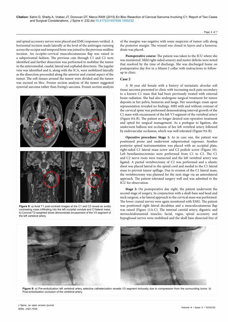
Figure 9: a) Pre-embolization left vertebral artery selective catheterization reveals V3 segment tortuosity due to compression from the surrounding tumor. b) Post-embolization occlusion of the vertebral artery.

Citation: Sahni D, Shetty A, Vrabec JT, Donovan DT, Marco RAW (2015) En Bloc Resection of Cervical Sarcoma Involving C1: Report of Two Cases and Surgical Considerations. J Spine 4: 232.doi:10.4172/21657939.1000232
Page 5 of 7
Volume 4 • Issue 3 • 1000232J Spine, an open access journalISSN: 2165-7939
muscle and tissue attachment. The tip of the mastoid and supracondylar bone was drilled away to improve visualization. At this point, our spinal surgery team entered into the operation. The C2 vertebrectomy was completed with an osteotome directed posterior to anterior. The C1 vertebrectomy was performed in a similar fashion across the anterior ring. The occipital condyle was finally removed with a burr and osteotome thus mobilizing the tumor medially, anteriorly, superiorly, laterally, and inferiorly. The tumor was removed entirely in an en bloc fashion (Figure 12 and 13). The wound was closed in layers and a deep drain was placed.
Postoperative course: The patient was monitored in the ICU. No postoperative complications were noted. On postoperative day 4, a halo was placed for stabilization due to removal of her condyle in the absence of `bilateral fixation. She was discharge home on postoperative day six with clinic follow up.
DiscussionHistology, anatomy, location, systemic considerations and disease
burden are all important considerations in the management of primary spinal tumors. Management can include radiation therapy, chemotherapy and surgery. Most spinal tumors, however, are treated through combinations of these modalities rather than the exclusive use
of any one method [18-20]. Treatment algorithms are lacking for most spinal tumor histologies due to their rarity, a dearth of well-controlled studies and reliance on traditional treatment methods.
Radiation therapy is often used to treat primary spinal tumors in lieu of surgery, however, its administration is fraught with the risk of radiation-induced myelitis, secondary cancers, tissue fibrosis, hypothyroidism, infertility and a host of other complications – both acute and chronic [17,18,21-23]. The relative radio-resistance of many spinal tumors, especially sarcomas, requires high dose radiation protocols often approaching 50-60 grey fractions – the threshold at which radiation myelitis develops [21,22].
Neoadjuvant chemotherapy is often used in soft tissue sarcomas located near neurovascular structures to decrease the size of the tumor [24]. However, surgery remains the definitive final treatment pathway in patients who receive chemotherapy.
While surgery is the mainstay for treatment of primary malignant spinal tumors, not all surgical methods are equal in benefit or efficacy. The existing literature has documented en bloc surgical resection as providing the best chance for cure and prevention of local recurrence of spinal tumors [25-32]. The prevention of local recurrence is a critical consideration in the treatment of upper cervical sarcomas due to the density of critical anatomy in this region as well as the variable response of spinal sarcomas to chemotherapy and radiation. Boriani et al, in a study of 22 patients with spinal chondrosarcoma, demonstrated a local
Figure 10: Status post stage one occipital to C2 instrumentation.
Figure 11: a) Positioning for a far lateral approach to the skull base. b) Proposed incision. c) Raising of the musculocutaneous flap.
Figure 12: En bloc surgical specimen.
Figure 13: six month followup MRI post contrast imaging. No definitive recurrence is seen in either patient. (top row - patient 1, bottom row – patient 2).

Citation: Sahni D, Shetty A, Vrabec JT, Donovan DT, Marco RAW (2015) En Bloc Resection of Cervical Sarcoma Involving C1: Report of Two Cases and Surgical Considerations. J Spine 4: 232.doi:10.4172/21657939.1000232
Page 6 of 7
Volume 4 • Issue 3 • 1000232J Spine, an open access journalISSN: 2165-7939
recurrence rate of 21.4% in patients treated with en bloc resection versus 100% in patients who underwent piecemeal curettage [25]. In support of Boriani’s data, Cloyd et al performed a meta-analysis on cervical spinal tumor patients treated with en bloc resection that identified a combined recurrence rate of 22%, with a mean follow-up time of 47 months. In these patients, disease-free survival rates were, respectively, 88% and 76% at one and five years [26].
As pathology manifests on increasingly rostral levels of the spine, anatomic considerations make treatment a matter of art as much as science. Despite the significant number of studies on the en bloc treatment of primary spinal tumors, few studies have been published on en bloc resections of spinal tumors in the upper cervical spine. An extensive literature search reveals several high cervical en bloc resection manuscripts but only one study involving C1 [25-33]. We attribute the dearth of these studies to the technical difficulty of en bloc removal of tumor masses in the densely neurovascular region of C1-2. These surgeries require high levels of therapeutic artistry in the form of disciplined forethought, interdisciplinary coordination and skill.
The two cases we have outlined describe the insights acquired in charting relatively unexplored territory in the en bloc treatment of atlantoaxial spinal tumors. These entities cannot be managed by a single surgical subspecialist. Successful treatment requires the presence of a skull-base surgeon for far lateral surgical exposure, the preservation of lower cranial nerves and the possible removal of basilar structures such as occipital condyles. A head and neck surgeon is recommended to mobilize the carotid sheath and establish tissue planes around the tumor in the posterior, lateral and anterior directions without tumor spillage. Neuroendovascular specialists might be required to assess the feasibility of vascular ligation through balloon occlusion testing of tumor encased vertebral or internal carotid arteries. A vascular neurosurgeon may be needed to perform bypasses that augment blood-flow in patients who poorly tolerate ICA sacrifice. Finally, a spine surgeon is required to sever diseased segments of the spine for en bloc removal and perform instrumentation and fusion of destabilized spinal levels.
During manipulation, dissection and exploration of the skull-base, we recommend transdermal triggered EMG monitoring of the lower cranial nerves. This can help reduce disabling deficits in swallowing, voice and tongue function that may ensue from iatrogenic injury to the cranial nerves.
We also suggest that the results of needle biopsy cannot be blindly relied upon as was the case with our first patient. Presuming tumor histology to be radio- or chemosensitive based on needle biopsy and then obviating surgical intervention for neoadjuvant management might result in perilous tumor progression in the upper cervical spine. C1 neoplasms must be considered surgical lesions unless there is compelling evidence to support a diagnosis of multiple myeloma or lymphoma without frank spinal instability. It goes without saying that a tertiary referral center with advanced imaging facilities and ICU capability is a prerequisite for the performance of these surgeries. We also recommend cadaveric practice to become familiar with the three dimensional features of the surgical anatomy that will be encountered.
Finally, toleration of vertebral or internal carotid artery sacrifice is not a given. Be prepared to add an additional surgical stage in the form of a bypass procedure requiring a cerebrovascular surgeon as we did with our first patient. Or be prepared to accept an incomplete en bloc resection due to the preservation of an artery encased in tumor. Patients should be adequately informed of all the risks and benefits of vessel sacrifice or how its preservation can induce local recurrence. The wishes of an informed patient should, at all times, guide operative management.
En bloc resection of sarcomas or spinal tumors involving the C1 and C2 level are challenging surgeries that push the envelope of surgical planning, management and execution. With the requisite armamentarium in the form of hospital resources, technology, coordination, knowledge and skilled specialists, these surgeries can be performed with substantial benefit to patients.
References
1. Mattei T, Teles A, Mendel E (2014) Modern surgical techniques for management of soft tissue sarcomas involving the spine: Outcomes and complications. J Surg Oncol 111: 580-586.
2. Rao G, Suki D, Chakrabarti I, Feiz-Erfan I, Mody M, et al. (2008) Surgical management of primary and metastatic sarcoma of the mobile spine. J Neurosurg Spine 9: 120-128.
3. Merimsky O, Kollender Y, Bokstein F, Issakov J, Flusser G, et al. (2004) Radiotherapy for Spinal Cord Compression in Patients with Soft-tissue Sarcoma. International Journal of Radiation Oncology 58: 1468-473.
4. Cao Y, Jiang C, Chen Z, Jiang X (2014) A rare synovial sarcoma of the spine in the thoracic vertebral body. Eur Spine J 23: 228-235.
5. Kim J, Lee SH, Choi YL, Bae GE, Kim ES, et al. (2014) Synovial sarcoma of the spine: a case involving paraspinal muscle with extensive calcification and the surgical consideration in treatment. Eur Spine J 23: 27-31.
6. Kim KW, Park SY, Won KY, Jin W, Kim SM, et al. (2013) Skeletal Radiol 42: 303-308.
7. Yonezawa I, Saito T, Nakahara D, Won J, Wada T, et al. (2012) Synovial sarcoma of the cauda equina. J Neurosurg Spine 16: 187-190.
8. Naphade PS, Desai MS, Shah RM, Raut AA (2011) Synovial sarcoma of cervical intervertebral foramen: a rare cause of brachial weakness. Neurol India 59: 783-785.
9. Zairi F, Assaker R, Bouras T, Chastanet P, Reyns N (2011) Cervical synovial sarcoma necessitating multiple neurosurgical procedures. Br J Neurosurg 25: 769-771.
10. Foreman SM, Stahl MJ (2011) Biphasic synovial sarcoma in the cervical spine: Case report. Chiropr Man Therap 19: 12.
11. Arnold PM, Roh S, Ha TM, Anderson KK (2010) Metastatic synovial sarcoma with cervical spinal cord compression treated with posterior ventral resection: case report. J Spinal Cord Med 33: 80-84.
12. Koehler SM, Beasley MB, Chin CS, Wittig JC, Hecht AC (2009) Synovial sarcoma of the thoracic spine. Spine J 9: 1-6.
13. Ravnik J, Potrc S, Kavalar R, Ravnik M, Zakotnik B (1976) Dumbbell synovial sarcoma of the thoracolumbar spine: a case report. Spine 34: 363-366.
14. Suh SI, Seol HY, Hong SJ, Kim JH, Kim JH, et al. (2005) Spinal epidural synovial sarcoma: a case of homogeneous enhancing large paravertebral mass on MR imaging. AJNR Am J Neuroradiol 26: 2402-2405.
15. Theermann R, Krödel A (1989) The spine as an unusual location for a malignant synovioma. Z Orthop Ihre Grenzgeb 127: 575-578.
16. Signorini GC, Pinna G, Freschini A, Bontempini L, Dalle Ore G (1986) Synovial sarcoma of the thoracic spine. A case report. Spine 11: 629-631.
17. Talac R, Yaszemski MJ, Currier BL, Fuchs B, Dekutoski MB, et al. (2002) Relationship between surgical margins and local recurrence in sarcomas of the spine. Clin Orthop Relat Res 397: 127-132.
18. Kaloostian Paul, and Ziya Gokaslan (2014) Surgical Management of Primary Tumors of the Cervical Spine: Surgical Considerations and Avoidance of Complications. Neurological Research 36: 557-65.
19. 19. Sundaresan N, Rosen G, and Boriani S (2009) Primary malignant tumors of the spine. Orthop Clin North Am 40: 21-36.
20. Chapman, Michael W (2001) Tumors and Infections of the Cervical Spine, Chapman’s Orthopaedic Surgery. (3rdedn), Lippincott Williams & Wilkins, Philadelphia, USA.
21. Anusheel M, Prabhakar R, Mohanti BK (2006) Radiation injury to the spinal cord in head and neck cancers: Does field arrangement have a role. J Can Res Ther 2: 65-67.
22. Tidy C, Jackson C (2013) Radiotherapy.

Citation: Sahni D, Shetty A, Vrabec JT, Donovan DT, Marco RAW (2015) En Bloc Resection of Cervical Sarcoma Involving C1: Report of Two Cases and Surgical Considerations. J Spine 4: 232.doi:10.4172/21657939.1000232
Page 7 of 7
Volume 4 • Issue 3 • 1000232J Spine, an open access journalISSN: 2165-7939
23. Cho W, Chang UK (2013) Survival and Recurrence Rate after Treatment for Primary Spinal Sarcomas. Journal of Korean Neurosurgical Society 53: 228-234.
24. Buecker, Peter J (2006) Sarcoma Treatment: An Overview. Liddy Shriver Sarcoma Initiative.
25. Boriani S, De Iure F, Bandiera S, Campanacci L, Biagini R, et al. (2000) Chondrosarcoma of the mobile spine: report on 22 cases. Spine 25: 804-812.
26. Cloyd, JM, Chou D, Deviren V, Ames CP (2009) En Bloc Resection of Primary Tumors of the Cervical Spine: Report of Two Cases and Systematic Review ofthe Literature. The Spine Journal 9: 928-35.
27. Eck JC, Dekutoski MB (2009) En Bloc Resection of Primary Spinal Tumors. Seminars in Spine Surgery 21: 93-98.
28. Guppy KH, Chakrabarti I, Isaacs RS, Jun JH (2013) En Bloc Resection of a Multilevel High-cervical Chordoma Involving C-2: New Operative Modalities. Journal of Neurosurgery: Spine 19: 232-42.
29. Bailey CS, Fisher CG, Boyd MC, Dvorak M (2006) En Bloc Marginal Excision of a Multilevel Cervical Chordoma. Journal of Neurosurgery: Spine 4: 409-14.
30. Hsieh PC, Gallia GL, Sciubba DM, Bydon A, Marco RAW, et al. (2011) En bloc excisions of chordomas in the cervical spine: review of five consecutive cases with more than 4-year follow-up. Spine 36: E1581-E1587.
31. Rhines LD, Fourney DR, Siadati A, Suk I, Gokaslan ZL (2005) En bloc resection of multilevel cervical chordoma with C-2 involvement. Case report and description of operative technique. J Neurosurg Spine 2: 199-205.
32. Gulati D, Aggarwal A, Kumar S, Chaturvedi S (2011) Primary Ewing’s Sarcoma of the Second Cervical Vertebra: A Rare Entity. Journal of PediatricOrthopaedics B 20: 408-12.
33. Molina CA, Ames CP, Chou D, Rhines LD, et al (2014) Outcomes following attempted en bloc resection of cervical chordomas in the C-1 and C-2 region versus the subaxial region: a multiinstitutional experience. J Neurosurg Spine21: 348-56.