rad tech a week 2 career’s in radiology radiographic equipment spring 2009
TRANSCRIPT

RAD TECH A WEEK 2
CAREER’S IN CAREER’S IN RADIOLOGYRADIOLOGY
RADIOGRAPHIC EQUIPMENT
Spring 2009

HISTORY REVIEW
• WHO
• WHAT
• WHEN
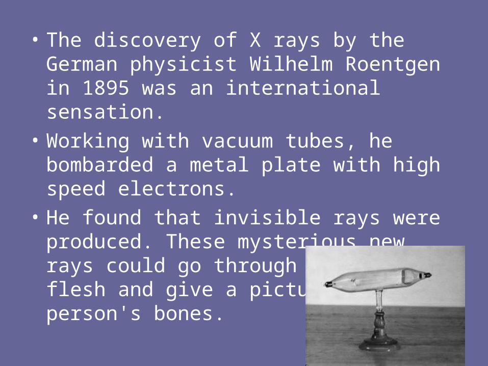
• The discovery of X rays by the German physicist Wilhelm Roentgen in 1895 was an international sensation.
• Working with vacuum tubes, he bombarded a metal plate with high speed electrons.
• He found that invisible rays were produced. These mysterious new rays could go through skin and flesh and give a picture of a person's bones.

Collaborative EventsCollaborative Events
Crookes tubeCrookes tube Air evacuated glass tubeAir evacuated glass tube Cathode sideCathode side Anode sideAnode side Electrical supplyElectrical supply
Screen or board painted with barium Screen or board painted with barium platinocyanideplatinocyanide
Low light work areaLow light work area

X-rays – the Basic Radiological ToolX-rays – the Basic Radiological Tool
Roentgen’s experimental apparatus (Crookes tube) that led to the discovery of the new radiation on 8 Nov. 1895 – he demonstrated that the radiation was not due to charged particles, but due to an as yet unknown source, hence “x” radiation or “x-rays”
Known as “the radiograph of Bertha Roentgen’s hand” taken 22 Dec. 1895

Hand images Today !


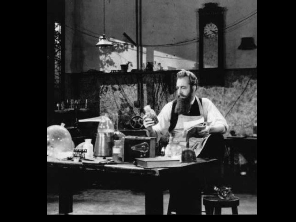
“Willie Roentgen”
• Honored in 1901 with the first Nobel prize in physics for his efforts.
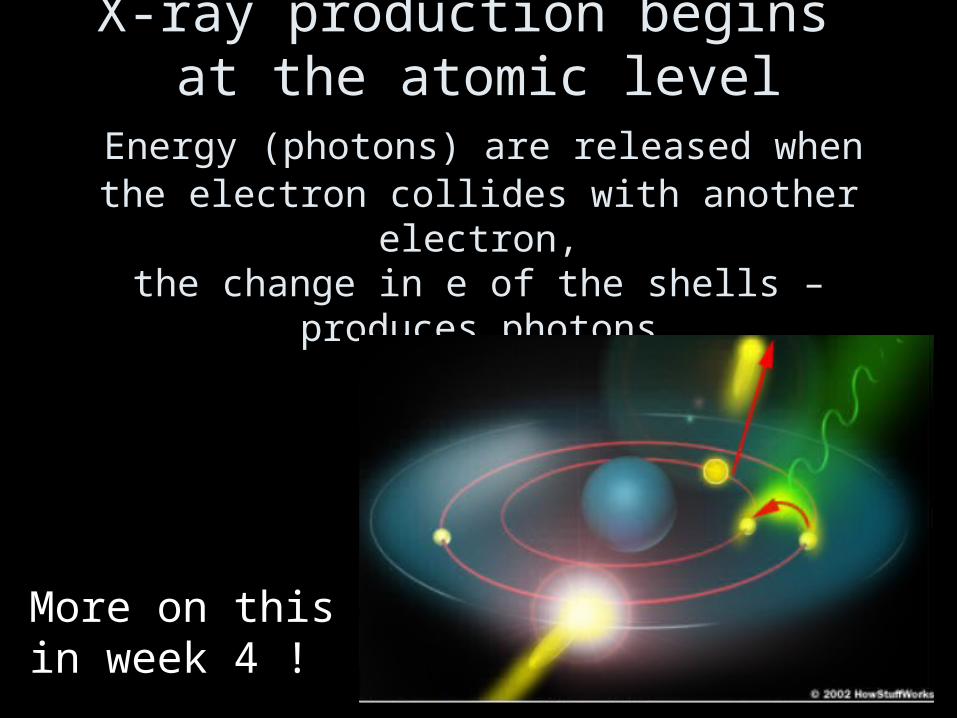
X-ray production begins at the atomic level
Energy (photons) are released when the electron collides with another electron,
the change in e of the shells –produces photons
More on thisin week 4 !

CAREERS IN RADIOLOGIC TECHNOLOGY
• RADIOLOGIC TECHNOLOGIST– RADIOGRAPHER– X-RAY
TECHNOLOGIST
• TECHNICIAN
• RADIOLOGIST

Objectives
• Define the Radiology Team
• Illustrate Careers in and around Radiology
• Identify work environments for Imaging Technologists
What does a What does a Radiologic Radiologic Technologist do?Technologist do?
Radiologic Radiologic TechnologistTechnologist Help othersHelp others Work with peopleWork with people Make a differenceMake a difference Think criticallyThink critically Demonstrate creativity Demonstrate creativity Achieve resultsAchieve results

FIG. 4–2FIG. 4–2 Radiologic science professionals must use advanced critical thinking skills when handling all patient care Radiologic science professionals must use advanced critical thinking skills when handling all patient care situations.situations.
Elsevier items and derived items © 2007, 2003 by Saunders, an imprint of Elsevier Inc. Elsevier items and derived items © 2007, 2003 by Saunders, an imprint of Elsevier Inc.
Radiologic Technologist
•Must be able to COMMUNICATE EFFECTIVELY with
•PATIENTS, STAFF & PEERS
•Must use GOOD JUDGEMENT & CRITICAL THINKING SKILLS
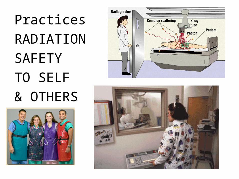
Practices
RADIATION
SAFETY
TO SELF
& OTHERS


Early years in Radiologic Technology
• Nurses or nurses aides taught how to “take an x-ray”
• NO special education• Only “ON THE JOB” training• Experience the best teacher
• The first Technologist is credited to be EDWARD C. JERMAN.

ARRTarrt.org
• 275,000 Registered RT (R)
• 2007: 15,285 exams – 89% Radiography– 3.6 % Nuclear Med– 7.1 Radiation Therapy
NATIONAL LICENSURE
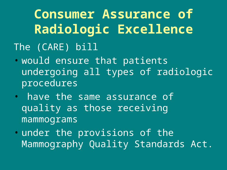
Consumer Assurance of Radiologic Excellence
The (CARE) bill
• would ensure that patients undergoing all types of radiologic procedures
• have the same assurance of quality as those receiving mammograms
• under the provisions of the Mammography Quality Standards Act.

CARE BILL
• Consistency, Accuracy, Responsibility, and Excellence...ARRT Among Supporters Seeking To Ensure Patient Safety Through Adoption of CARE Bill
• ASRT – American Society of Radiologic Technologist ASRT.org

As of Sept 2001- 16 states have no licensure lawsAs of Sept 2001- 16 states have no licensure laws

Diagnostic Radiologic Technologist
Aka: Radiographer
Not a “Technician”
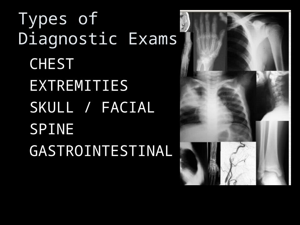
Types of Diagnostic Exams
• CHEST
• EXTREMITIES
• SKULL / FACIAL
• SPINE
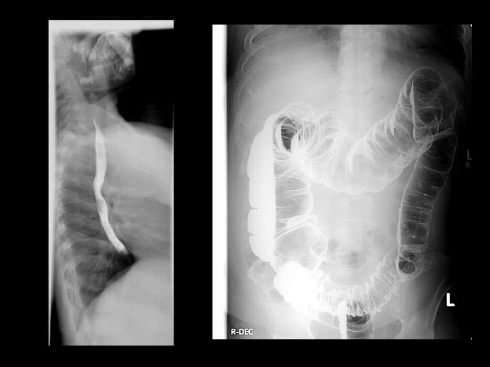
• GASTROINTESTINAL
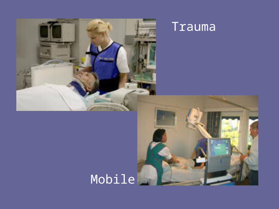
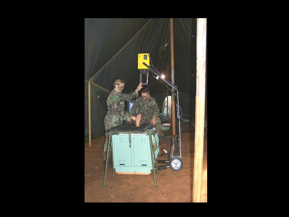
Trauma
Mobile

“TRAUMA”


ALL PATIENTS

ELDERLY

INFANTS & CHILDREN

UNIQUE, UNUSAL, INTERESTING, ETC
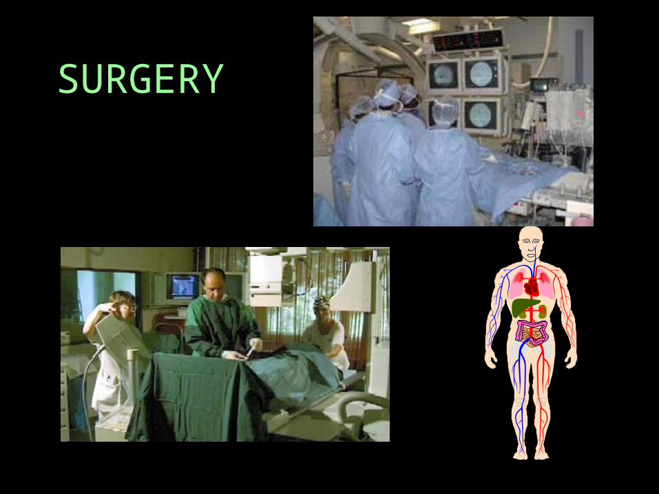
SURGERY


TECHNOLOGIST
• FLUOROSCOPY
• CONTRAST MEDIA (X-RAY DYE)
• KELLY HOLT, ARRT R.T. (R)
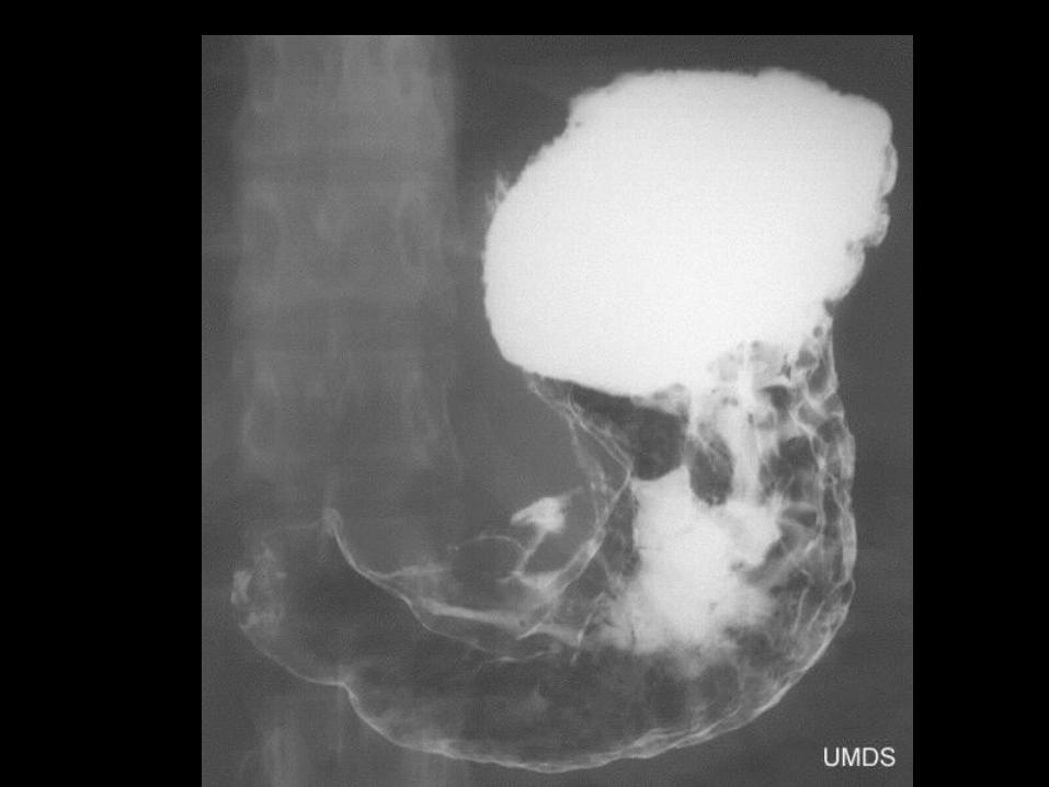
FLUOROSCOPY- XRAYS IN MOTION
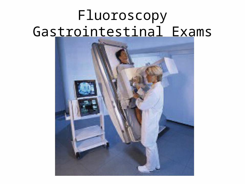
Fluoroscopy Gastrointestinal Exams
ARRT ExamsPrimary Examinations
• Five “Primary Exams”
offered by the ARRT

5 Primary Exams
• Radiography (R)• Nuclear Medicine Technology (NM)• Radiation Therapy (T)• Sonography (US) (RDMS)• Magnetic Resonance Imaging (MR)
• candidates must have successfully completed a formal educational program in the respective discipline that is accredited by a mechanism
acceptable to ARRT.

Beyond Diagnostic Beyond Diagnostic RadiographyRadiography
Advanced ModalitiesAdvanced Modalities

POST – PRIMARY EXAMSPOST – PRIMARY EXAMS(TAKEN AFTER COMPLETIONS OF ARRT – RT License)(TAKEN AFTER COMPLETIONS OF ARRT – RT License)
(M) Mammography(M) Mammography (CT) Computed Tomography(CT) Computed Tomography (MRI) Magnetic Resonance Imaging(MRI) Magnetic Resonance Imaging (QM) Quality Management(QM) Quality Management (US) Sonography(US) Sonography
Vascular Sonography Vascular Sonography Breast Sonography Breast Sonography (BD) Bone Densitometry(BD) Bone Densitometry (CV) Cardiac-Interventional Radiography(CV) Cardiac-Interventional Radiography (CIV) Vascular-Interventional Radiography(CIV) Vascular-Interventional Radiography
Advanced PracticeAdvanced Practice (R.R.A) Registered Radiologist Assistant(R.R.A) Registered Radiologist Assistant Imaging Informatics Certification ProgramImaging Informatics Certification Program
Merging IT and Medical technology Merging IT and Medical technology
PET PET MANAGEMENT (BS or BA)MANAGEMENT (BS or BA) EDUCATION (BS,MEd, PhD EDD)EDUCATION (BS,MEd, PhD EDD)

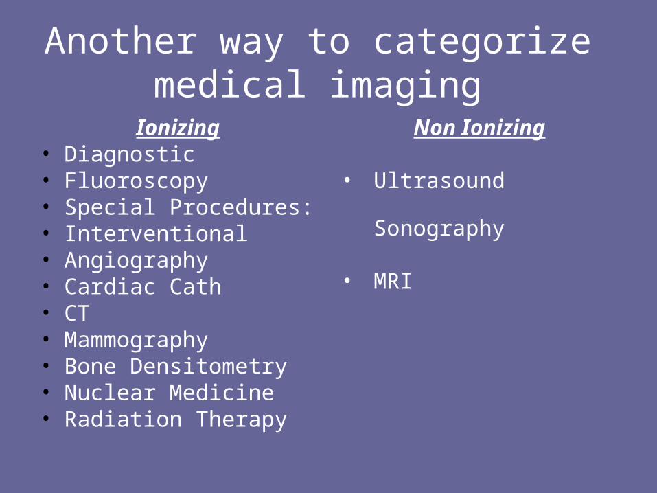
Another way to categorize Another way to categorize medical imaging medical imaging
IonizingIonizing DiagnosticDiagnostic FluoroscopyFluoroscopy Special Procedures:Special Procedures: InterventionalInterventional AngiographyAngiography Cardiac CathCardiac Cath CTCT MammographyMammography Bone DensitometryBone Densitometry Nuclear MedicineNuclear Medicine Radiation TherapyRadiation Therapy
Non IonizingNon Ionizing
UltrasoundUltrasound SonographySonography
MRIMRI
HEALTH CARE TEAMHEALTH CARE TEAM MEDICAL CENTERMEDICAL CENTER
RADIOLOGYRADIOLOGY
RADIOLOGISTRADIOLOGIST
TECHNOLOGIST VS TECHNOLOGIST VS TECHNICIANTECHNICIAN
RADIATIONRADIATION

RADIATIONRADIATION
ENERGYENERGY IONIZATION- IONIZATION-
adding or removing adding or removing an electronan electron
IONIZING IONIZING RADIATIONRADIATION
NON-IONIZING NON-IONIZING RADIATIONRADIATION

What is Ionization?

When an electron is added or removed from the atom- it is
ionized
Another way to categorize medical imaging
Ionizing• Diagnostic• Fluoroscopy• Special Procedures:• Interventional• Angiography• Cardiac Cath• CT• Mammography• Bone Densitometry• Nuclear Medicine• Radiation Therapy
Non Ionizing
• Ultrasound Sonography
• MRI

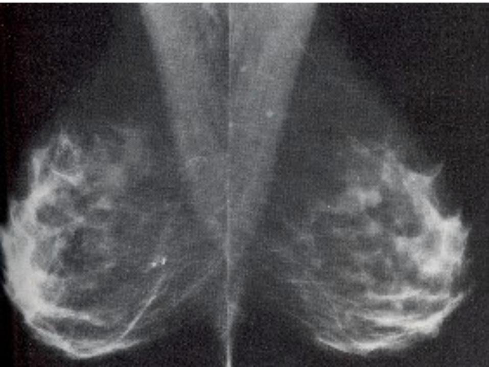
MAMMOGRAPHY
Colleen McFaul, RT (R) (M)
RADIOGRAPHIC IMAGING OF THE
BREAST Dx for breast cancer
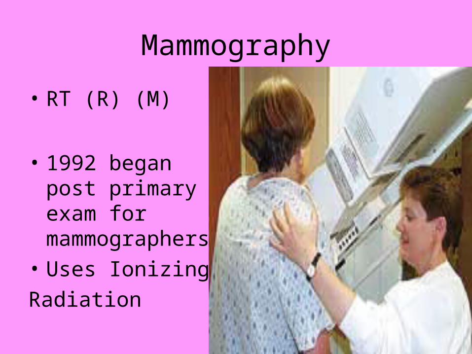
Mammography
• RT (R) (M)
• 1992 began post primary exam for mammographers
• Uses Ionizing
Radiation

Bone Densitometry
“low dose
Ionizing Radiation”

BONE DENSITOMETRY
• JOE RUIZ, RT (R) (BD)
• POST PRIMARY EXAM BEGAN IN JANUARY 2001
• May also have certification w/o RT
• MEASURING MINERAL CONTENT AND DENSITY OF BONES


Bone densitometry

CT SCAN & MRI
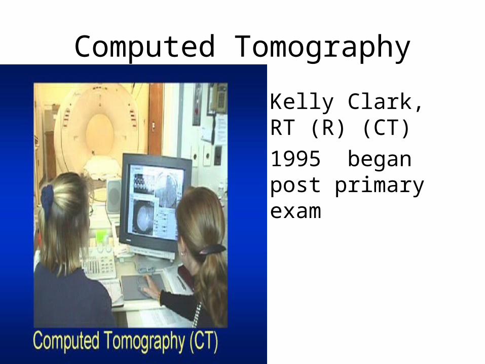
Computed Tomography
• Kelly Clark, RT (R) (CT)
• 1995 began post primary exam
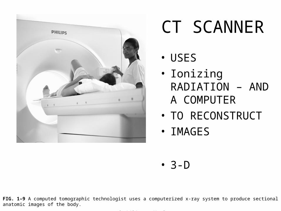
FIG. 1–9 A computed tomographic technologist uses a computerized x-ray system to produce sectional anatomic images of the body.
(Courtesy of Philips Medical Systems.)
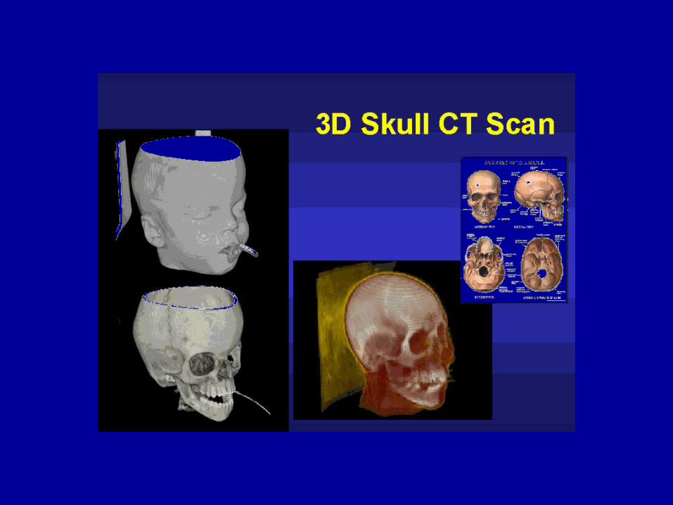
CT SCANNER
• USES• Ionizing RADIATION
– AND A COMPUTER
• TO RECONSTRUCT
• IMAGES
• 3-D

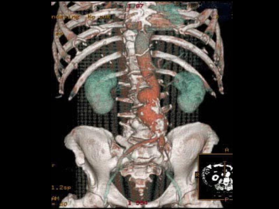
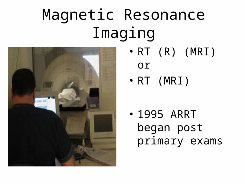
Magnetic Resonance Imaging
• RT (R) (MRI) or
• RT (MRI)
• 1995 ARRT began post primary exams
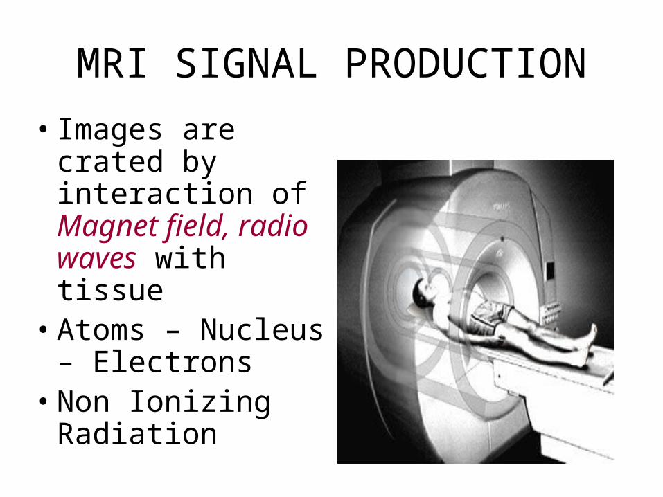
MRI SIGNAL PRODUCTION
• Images are crated by interaction of Magnet field, radio waves with tissue
• Atoms – Nucleus – Electrons
• Non Ionizing Radiation

MRI EXAMS
• MRI is growing as an alternative to traditional x-ray mammography.


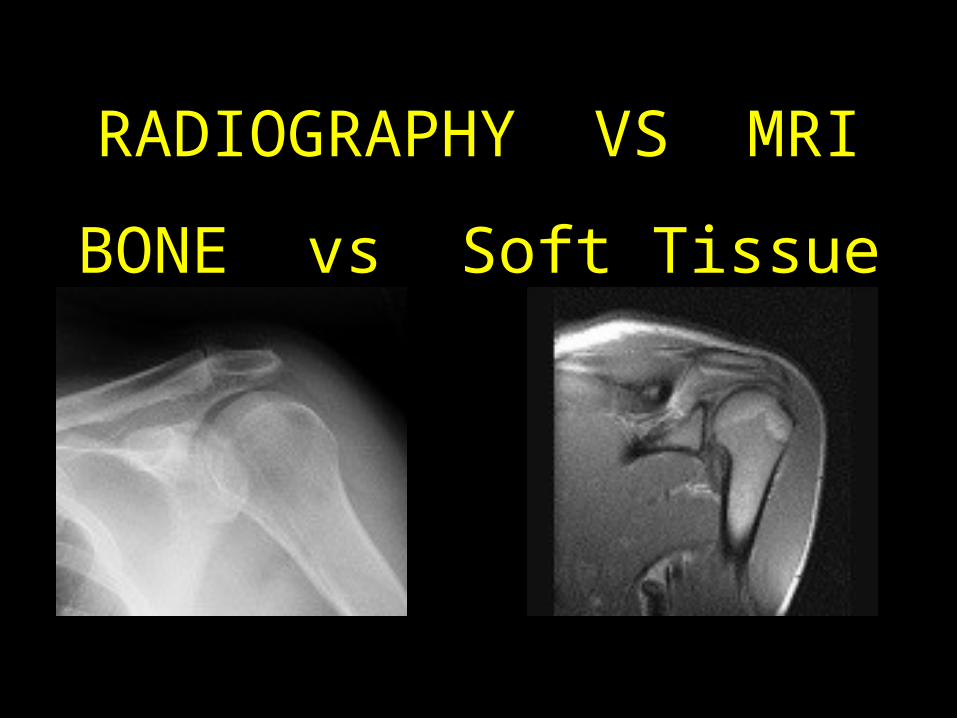
RADIOGRAPHY VS MRI
BONE vs Soft Tissue
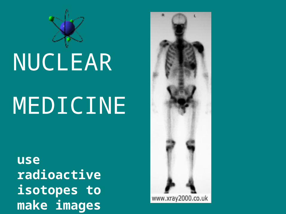
NUCLEAR
MEDICINE
use radioactive isotopes to make images
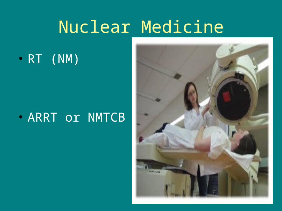
Nuclear Medicine
• RT (NM)
• ARRT or NMTCB

scans use radioactive
isotopes to make images

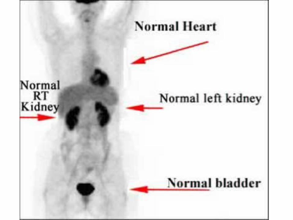
NM – Images collected afterinjections of a RADIOISOTOPE
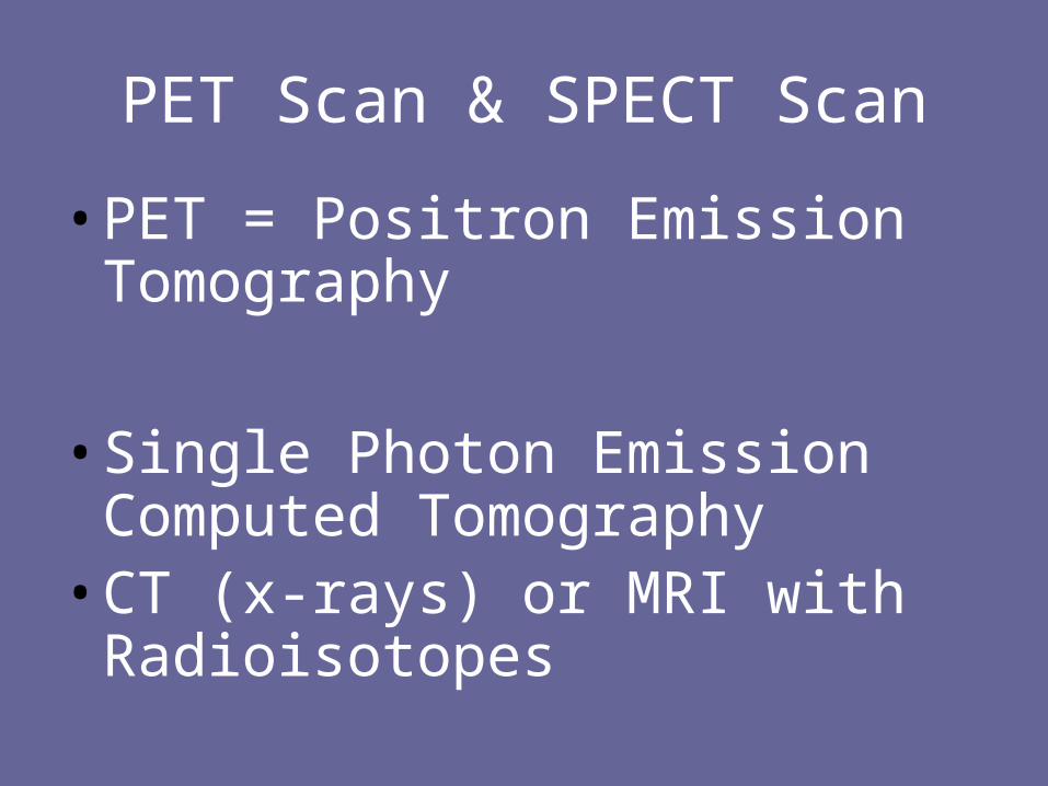
PET Scan & SPECT Scan
• PET = Positron Emission Tomography
• Single Photon Emission Computed Tomography
• CT (x-rays) or MRI with Radioisotopes


ANGIOGRAPHY/ CARDIOVASCULAR INTERVENTIONAL
TECHNOLOGYis a specialized radiographic examination where the images of the blood vessels of the body are demonstrated by injection of contrast media.

Cardiovascular Interventional Radiography
• Jane Smith, RT (R) (CVI)
• Angiography
• RT (CV) (CIT) (CVIT)
• 1991 began post primary exam for CVIT Radiographers

INTERVENTIONAL RADIOGRAPHY
• Uses
• Fluoroscopy
• Ionizing
Radiation
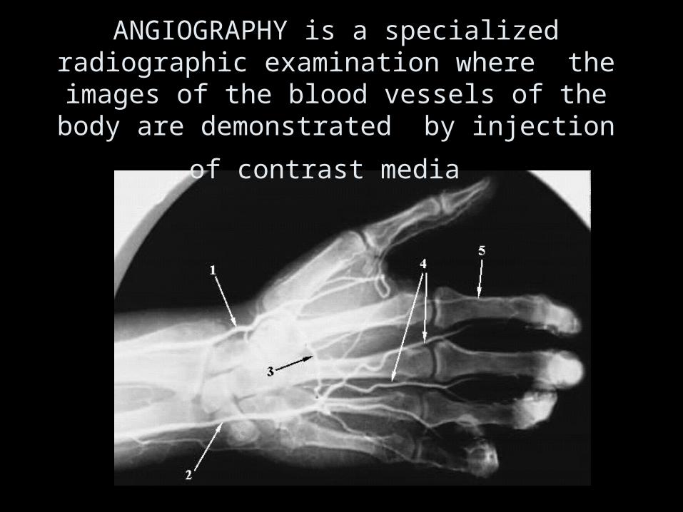
ANGIOGRAPHY is a specialized radiographic examination where the images of the blood
vessels of the body are demonstrated by injection
of contrast media


RADIATION THERAPY
R.T.(T)• 1 - 4 YRS ADDITIONAL EDUCATION
• CITY OF HOPE
• LLU
• CAL STATE LONG BEACH
• The branch of Radiology that involves the treatment of disease by means of high energy x-rays or radioactive substances


Radiation Therapy
• RT (T) = Registered technologist in radiation therapy technology
• Medical dosemetrists are involved in treatment planning and dose calculations
• 1-4 year program


ULTRASOUND
uses a technique similar to Navy SONAR to produce diagnostic images.
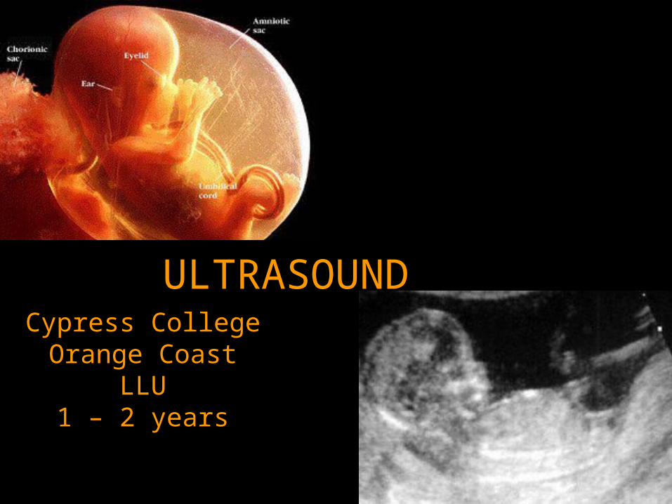
ULTRASOUNDCypress CollegeOrange Coast
LLU1 – 2 years

FIG. 1–10 A diagnostic medical sonographer uses high-frequency sound waves to create images.
(Courtesy of Philips Medical Systems.)
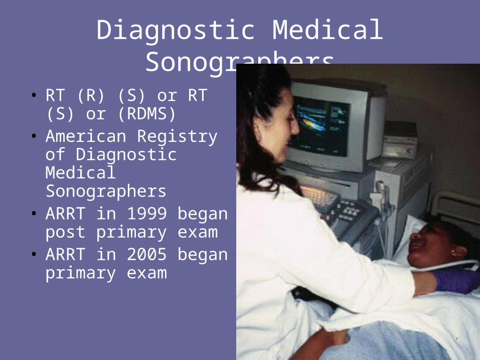
Diagnostic Medical Sonographers
• RT (R) (S) or RT (S) or (RDMS)
• American Registry of Diagnostic Medical Sonographers
• ARRT in 1999 began post primary exam
• ARRT in 2005 began primary exam

Uses SOUND WAVES (NOT X-RAYS)
“real time” images

U/S & the “real thing”

Breast Sonography
• Post Primary Exam
• Valuable for Technologists that specialize in Mammography
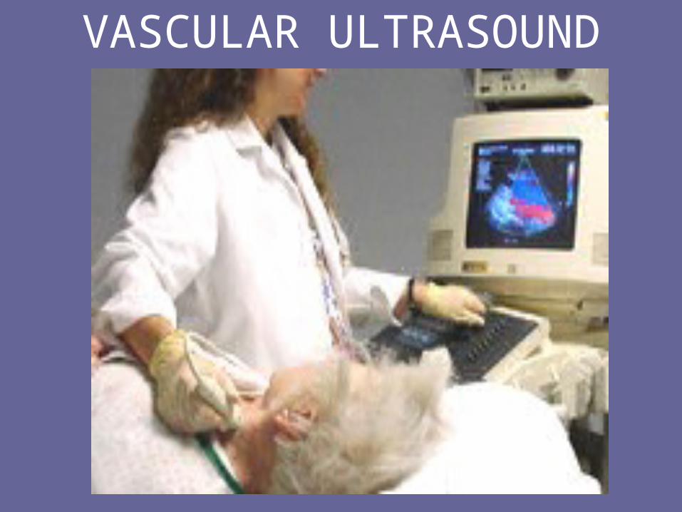
VASCULAR ULTRASOUND

Additional Opportunities
• Education
• Administration
• Management (QM)
• Commercial
• Radiologist Assistant = RRA
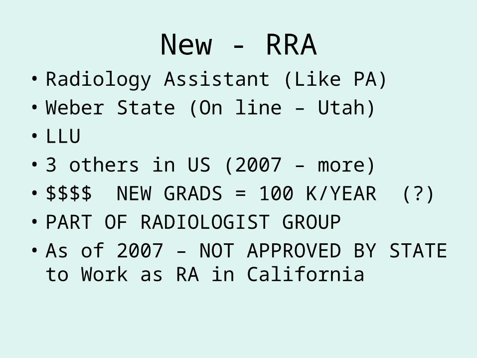
New - RRA• Radiology Assistant (Like PA)
• Weber State (On line – Utah)
• LLU
• 3 others in US (2007 – more)
• $$$$ NEW GRADS = 100 K/YEAR (?)
• PART OF RADIOLOGIST GROUP
• As of 2007 – NOT APPROVED BY STATE to Work as RA in California

RADIOLOGY ASSISTANT RA
• LLU • 2 YRS RT• BA DEGREE
• WEBER STATE• 6YR RT
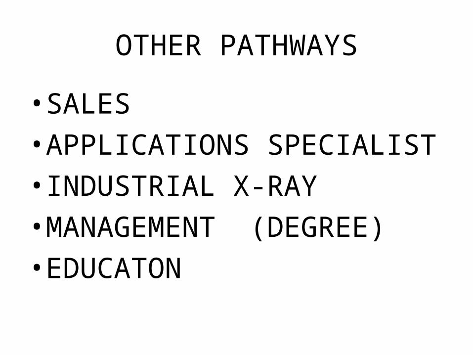
OTHER PATHWAYS
• SALES
• APPLICATIONS SPECIALIST
• INDUSTRIAL X-RAY
• MANAGEMENT (DEGREE)
• EDUCATON

Work Opportunities
• Hospital
• Imaging Center
• Doctor’s Office
• Mobile Trailer
• Research

Variety of Work Settings• physicians offices,• clinical outpatient facilities, • free standing imaging centers, • mobile imaging centers • portable services to rehabs • Mammo’s to under privileged
areas• Urgent care

Other working opportunities…
• Registry (local)
• Registry (out of state)
• X rays taken around the world !!

TRAVELING TECHNOLOGIST = SEE THE WORLD AND GET $$$

NEED – JOB SECURTIY
• PEOPLE ARE GETTING OLDER – LIVING LONGER
• TECHNOLOGIST ALSO GETTING OLDER
• NEED NEW, YOUNG PEOPLE TO TAKE OVER THE JOBS & TAKE CARE OF THE SICK AND ELDERLY
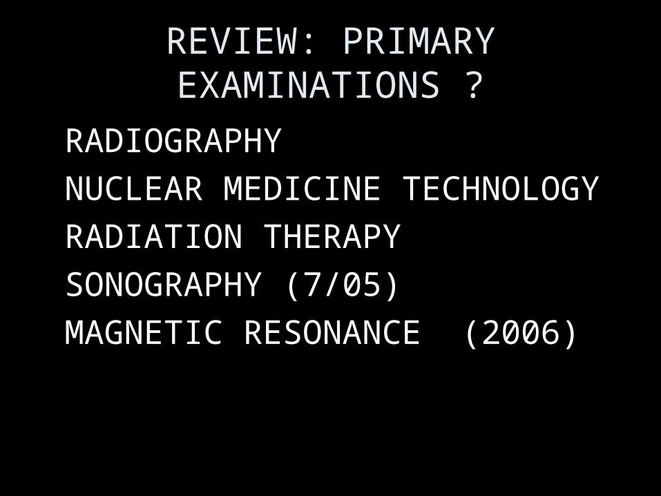
REVIEW: PRIMARY EXAMINATIONS ?
• RADIOGRAPHY
• NUCLEAR MEDICINE TECHNOLOGY
• RADIATION THERAPY
• SONOGRAPHY (7/05)
• MAGNETIC RESONANCE (2006)

Review
• Radiology Team
• Patient Care Careers
• Other Careers Opportunities
• Work Settings

HISTORY REVIEW
• WHO
• WHAT
• SERIES OF EVENTS
• WHEN

QUESTIONS?