radiation biology and radiation protection - j.o. liljenzin
TRANSCRIPT

474
CHAPTER 18
Radiation Biology and Radiation Protection
Contents
18.1. The biological target 47618.2. Radiation effects on the molecular level 478
18.2.1 Radiation physics 47818.2.2 Radiation chemistry 47818.2.3 Radiation weighing factors 480
18.3. Radiation effects on different types of cells 48118.4. Some concepts of radiation biology 48218.5. Further regularities at large doses 48518.6. Epidemiological observations of effects at large radiation doses 486
18.6.1. Radiation sickness after accidental exposure 48718.6.2. Radiation therapy and deterministic studies 48818.6.3. Stochastic cancer induction 48918.6.4. Mental retardation 491
18.7. Radiation sterilization 49218.8. Genetic effects 49218.9. Radiomimetic substances 49418.10. Radiation background 49418.11. Somatic effects of low radiation doses 495
18.11.1. Epidemiological results 49518.11.2. Problems in studies of low-level low-LET radiation 496
18.12. The dose-effect curve 49818.13. Regulatory recommendations and protection standards 498
18.13.1. Risk of cancer induction by radiation 49818.13.2. Recommended dose limits 50018.13.3. The collective dose 50018.13.4. Committed doses 50118.13.5. Internal radiation and ALI values 50218.13.6. Radiotoxicity and risks 50518.13.7. Classifications, working rules, etc 507
18.14. Protective measures for radiochemical laboratory work 50818.14.1. Tracer work with moderate $-(-levels 50818.14.2. "-laboratories 50918.14.3. High-level $-(-emitters 510
18.15. Control of radiation protection measures 51218.16. Exercises 51218.17. Literature 513
The biological effects of ionizing radiation were discovered soon after the production ofintense radiation sources in the form of X-ray machines and radioactive elements about a

Radiation Biology and Radiation Protection 475
Physical parametersKind of radiation: ", $, or (Internal or external sourceSize of total doseDose rateExposure time: instantaneous, temporary, recurrent, chronic
Microscopic effectsmolecular: DNA changes chromosome: abberationscellular (single or a few cells, in vitro or in vivo):
kind of cells (nervous system, bone marrow, liver, etc)cell cycle (stage, rate of cell division, inactivation, etc)temperatureoxygen contentcell poisons present (increasing radiation sensitivity)protective agents present (anti-oxidants)
tissue and organ: cancer growth or deathMacroscopic effects (inactivation, lethal dose (LD), cancer, etc.)
Somatic: rapid (within a month), delayed (up to 20 years)Genetic: observed in offspring (one or several generations later)
TABLE 18.1. Parameters in biological effects of radiation
century ago. Radiation was found to stop cell division and could therefore be usedtherapeutically to stop cancer growth. On the other hand, radiation applied locally was foundto cause wounds, which were difficult to heal, and to induce cancer. Many serious accidentsoccurred as a result of the use of radiation before an adequate understanding of its biologicaleffects led to formulation of rules for protection of workers. By 1922 approximately 100radiologists (not patients) had died as a result of biological radiation damage. The biological effect of very large whole-body doses is radiation sickness and early death,while large organ doses lead to local cell destruction and, possibly, organ death. The effectsat lower doses are cell changes (decreased surviving fraction, decreased rate of division,chromosomal abberations, etc), which usually can be observed by microscope soon afterirradiation. The induction of cancer may take years to observe and genetic changes may not bediscovered until after several generations. The creation of our world occurred in intense radiation fields and, consequently, we haveinherited an Earth drenched in radiation from cosmic sources and the minerals in the ground(Ch. 5, 10 and 17). Though the intensity of these radiation sources is much smaller thanproduced by human techniques, no human can avoid these natural sources. Therefore, theeffects of the natural radiation background has become an important health issue, particularlyradon levels in houses. Closely related to this problem is the effects of man-made sources ofsimilarly low levels, such as the storage of nuclear waste. Much research is presently devotedto the effects of low-level radiation. Through extensive research in this field, our knowledge has grown enormously. Initially, theeffects of radiation on local organs were studied; then the effects on various specific tissues andcell types were of concern; today, the focus is on the effects at the molecular level. This deve-lopment is understandable as cancer induction and genetic changes are

Radiochemistry and Nuclear Chemistry476
believed to have their roots in alterations in the DNA molecules in the cells. In Table 18.1 welist a number of factors which affect the actual extent of the biological damage caused byradiation; most of these factors are discussed in this chapter. Many groups are actively engaged in research on the biological effects of ionizing radiation:radiologists use radiation for diagnosis and treatment of tumors; health physicists have theresponsibility of controlling the use of irradiation equipment and protecting people fromunnecessary exposure to radiation; in collaboration with oncologists (tumor researchers) andgeneticists, radiobiologists conduct research to explain the effects of radiation on the cellularand molecular level, radiation chemists are interested in the interaction between radiation andthe DNA molecule.
18.1. The biological target
The discussion of the effects of radiation on biological systems requires some familiarity withthe target composition and common terminology. The human body contains some 10 cells. Figure 18.1.A shows schematically the cell and14
its nucleus, which contains thin thread-like DNA-molecules (deoxyribonucleic acid). The DNAcarries the "genetic code" and are the most important molecules of an organism. It consists oftwo strands of sugar-phosphate chains, attached together by base pairs (forming so-callednucleotides), in a double helix form (Fig. 18.1.H!J). The human DNA contains 2.9×10⁹
nucleotides; the DNA of simpler organism have fewer nucleotides (down to about 5000). Thenucleotides are combined in triplets (called codons), each one with the ability to produce(through some intermediate steps) a certain protein. The codons are ordered in long groups; theordering is referred to as the genetic code. Because these long groups carry the informationnecessary to produce proteins for the different tissues, they are called genes ("makers"). Inhumans, the DNA is distributed over 23 pairs (altogether 46) chromosomes. In thechromosome, the DNA is wound around histone protein cores (Fig. 18.1.G) and highly twined("condensed") to facilitate cell division; the DNA+histone unit (containing some 200base-pairs) is referred to as the chromatin; these repeating units are known as nucleosomes.Figure 18.1.D shows some of the 23 chromosome pairs, and Figure 18.1.E a single one. Thetotal amount of genetic information in the cell is called the genom. Stretched out the totalgenetic material is about 1.5 m long, but its diameter is only 2 nm; the molecular weight isabout 10 per chromosome. While the cells comprise most of the body, the volume of the11
chromosomes only occupy ~1% of the cell volume and only about 1/10 of the chromosomevolume carries a genetic message; thus the genetically significant target is rather small. The nucleotides are held together by hydrogen bonds between the nitrogen bases, which havetheir nitrogen rings perpendicular to the plane of Figure 18.1.I (i.e. turned 90E compared to thepaper surface). The nitrogen rings contain some B-electrons, which interact between the planesand stabilize the chain structure. The unsaturated bonds in the nitrogen bases are sensitive tooxidation. Further, the phosphate oxygens are ionic. Thus, the DNA presents all types ofchemical bonding and is sensitive to many types of reactants. The cell cycle plays an important role in radiation damage (Figures 18.1.B and C) (i) duringthe S-stage proteins are synthesized, (ii) in the "gap 2", G , stage, the cell is being prepared2for division (the DNA-chains are split up and copied), (iii) in the cell division

Radiation Biology and Radiation Protection 477
FIG. 18.1. The cell and DNA biochemical system.
Radiochemistry and Nuclear Chemistry478
(mitosis) the chromatin condenses into chromosomes, which are split and move in oppositedirections, while the original cell is divided at mid-level (Figs. 18.1.C.3 and C.4), after which(iv) the cell "rests" or matures in the G -state.1
18.2. Radiation effects on the molecular level
When a high energy particle (whether ", $ or () strikes a human cell it produces a narrowtrack, less than 1µm thick (cf. Figs. 6.5 and 7.1). The interaction with water, which is the mainconstituent of the cell, leads to radiolysis products, as described in Chapter 7. To a lesserextent, the radiation may interact directly with the molecules of the cell ("direct hit"). Only theeffects (direct or indirect) on the cell nucleus DNA are thought to cause severe biologicaldamage.
18.2.1. Radiation physics
High energy (-rays lose most of their energy in a few compton scattering events. A "typical"natural radiation background gamma quantum of 1 MeV loses about 75% of its energy in 20cm of water (a "typical" human thickness). This loss occurs to 50 ! 80% in a single comptonscattering event, producing an electron with up to 0.8 MeV kinetic energy, depending on thescattering angle. In the next compton interaction, the $ 0.2 MeV ( loses $ 50% of its energy;the distance between these initial two interactions exceeds cellular dimensions. Thus, the 1 MeV(-absorption on the cellular level can be considered as the production of a single high energyelectron. However, at very low energies (compton ('s and X-rays), low energy electrons areproduced at densities of several electron pairs per µm (cf. Fig. 6.5.D and Table 6.2; 1 mm airis considered equivalent to about 1 µm of water or tissue). High energy electrons lose most of their energy in low energy collisions (cf. Figures 6.5 and6.7). A typical LET-value is 200 eV/µm (Table 6.2), corresponding to 5 ! 10 ion pairs/µm;a 10 keV electron produces about 10 ion pairs/µm according to Figure 6.7. Alpha particles cause dense ionization (Figs. 6.5 and 6.7) with typical LET-values of 200keV/µm (Table 6.2) and production of several thousand ion pairs per µm (i.e. several ionpairs/nm). High energy neutrons are absorbed in water mainly by collisions with H-atoms, formingenergetic protons which ionize similar to alpha particles (Table 6.2 and Fig. 6.7). In thecollision process the H-atoms are knocked from their positions in the molecule; also other atomsmay be knocked out. The neutron ends its life by being absorbed in an (n,() process or leavesthe system.
18.2.2. Radiation chemistry
The absorbtion of low-LET radiation and high-LET radiation is illustrated in Figure 18.1.Eand 18.1.H, respectively. Table 18.2 gives the number of ion pairs formed in the DNA,nucleosome and chromatin by "'s and ('s ( Co). Table 18.3 shows the damage caused to the60
DNA and cell nucleus. The wide range of values in parentheses indicate the
Radiation Biology and Radiation Protection 479
Average number of ionizations in________________________________________________________________________
Radiation DNA segment Nucleosome Chromatin segment(2 nm long) (i ~ 10 nm) (25 nm long)
_________________________________________________________________________________________________
(-rays 1 (0 to <8) 2 (0 to <20) 2 (0 to <45)"-particles 2 (0 to <15) 10 (0 to <90) 50 (0 to <200)_________________________________________________________________________________________________
UNSCEAR = United Nations Scientific Committee on the Effects of Atomic Radiation.†
TABLE 18.2. Ionization clusters produced directly in a DNA-related target by a single radiationtrack. (From Goodhead, UNSCEAR)†
Average number of induced breaks_________________________________________
Radiation Average number DNA single DNA doubleof ionizations strand strand
___________________________________________________________________________________________
(-rays 70 (1-1500) 1 (0-20) 0.04 (0-few) "-particles 23 000 (1-100 000) 200 (0-400) 35 (0-100)
TABLE 18.3. Damage products in a single-cell nucleus traversed by a single radiation track(From Goodhead, UNSCEAR)
uncertainty as well as the dependence on the particle energy (in general, higher ionization atlower energy). The radiation is largely absorbed in water, as the cell contains some 70% H O, and produces2ions, free radicals and excited atoms. A momentaneous lethal dose of 20 Sv produces aconcentration of 14 FM of reactive products (@OH, e , H O , etc). Trace metals in the body!
aq 2 2(e.g. Cu, Cr, Se) are also poisonous at this concentration level, but essential at lower levels(e.g. a daily intake of 40 Fg Se is recommended). The ions produced will probably have littleeffect as the DNA contains numerous ionizable positions at the phosphate group. Free radicalslike HO· and oxidizing products like H O are highly reactive and can add to unsaturated bonds,2 2which upsets the sensitive hydrogen-B-bonding and may break the bonding between the twohelices. Excited hydrolysis products may transfer the excitation energy to the DNA, leading toa localized break in the sugar-phosphate chain. The damage to the DNA may also lead to asubstitution reaction in the nucleotide or a loss of a segment. Actually, hundreds of differentDNA-damage products have been identified. If the damage is limited to one of the strands ofthe helix, it is referred to as a single strand break. The cell is protected by different DNA repair mechanisms, which try to restore the damage.We don't know the details, except when the repair goes wrong (e.g. a replacement of a lostnucleotide by a "wrong" base-pair, etc.). It is believed that most single strand breaks arecorrectly repaired. If not, this may lead to somatic effects for the organism (e.g. cancer) or toan inheritable DNA-defect. The repair system is believed to be more effective in a livingorganism, where the cells are in continuous exchange with surrounding cells and body fluids,than in the tissue samples often studied in the laboratory. This should be kept in mind below,where effects on whole organisms as well as on cell cultures in "test tubes" (in vitro studies)are described.

Radiochemistry and Nuclear Chemistry480
The cell contains natural radical scavengers. As long as they are in excess of the radiolysisproducts, the DNA may be protected. When the products exceed the amount of scavengers,radiation damage and cancer induction may occur. In principle, there could thus be a thresholddose for radiation damage, at which the free radicals formed exceed the capacity of scavenging.The scavenging capacity may differ from individual to individual, depending on his/her physicalcondition. A low-LET gamma ray "hit" may cause 1 ionization (or <8) in a 2 nm DNA-segment. Thiscorresponds to a deposition of some 30 eV (or <250 eV) in about 6 base-pairs +sugar-phosphates. The radiation energy may distribute over a large number of bonds, so thatno bond gets enough energy to dissociate. Thus, the radiation interaction may leave theDNA-segment mainly unchanged. Low-LET radiation sometimes form clusters of ions along the particle track, i.e. produceshigh-LET "spots". Such spots mean increased risk of damage because a larger amount ofenergy is then deposited in the small volume occupied by the DNA-helix. High-LET spotstherefore increase the possibility of damage to both strands of the helix, causing a double strandbreak. It has been found that for each 100 single strand breaks produced by low-LET radiation about3 double strand breaks occur. Because the DNA-chain is rather rigid, a double strand breakdoes not lead to the two halves snapping away from each other. Instead, they are resting,waiting for the repair mechanism to start. Nevertheless, the repair is more difficult and thechances of "repair errors" (mutations) are much larger than for the single strand break. Thisis demonstrated by two facts: (i) the repair time for a single break is on average 10 minutes,while that for the double break some hours; (ii) chromosome abberations (Fig. 18.1.D) onlyoccur after double strand breaks. The chromosome abberations may be due to that the cellundergoes mitosis before the double strand break has been repaired. Cells with severechromosome abberations are not viable. Minor repair errors may be carried on to next genera-tion of cells as a change in the genetic code or may stop the cell division process. In addition to double strand breaks being more difficult to repair than single strand breaks,double strand breaks caused by high-LET radiation are more difficult to repair than thosecaused by low-LET radiation. The high energy deposited at the hit area causes "havoc" on theDNA (cf. Table 18.3). A few percent of low-LET radiation causes double-strand breaks incontrast to 10-20% for high-LET radiation. After a low-LET dose of 3 Gy, every cell in the human body contains 3000 single strand andsome 100 double strand breaks; if given in a short time the cell (and organism) has great diffi-culties in repairing this damage (resulting in death in the case of no medical treatment).However, spread out over a week only chromosomal abberations will be noted. In thesubsequent sections, we shall discuss macroscopic effects of various kinds and doses ofradiation.
18.2.3. Radiation weighing factors
In order to take into account the biological effects of different kinds of radiation, radiationweighing factors, w , have been introduced and are given in Table 18.4. Earlier, two similarRconcepts were used with about the same meaning: the "quality factor", Q, and "rela-

Radiation Biology and Radiation Protection 481
LET in water Weighting factor Type and energy of radiation (eV/nm) wR_____________________________________________________________________________________________
0.2 - 35 1 photons (X-rays and ('s)0.2 ! 1.1 1 all electrons > 5 keV 20 5 slow neutrons < 10 keV 50 20 intermediate n's 0.1 - 2 MeV
10 fast n's 2 - 20 MeV5 protons > 2 MeV
130 20 "-particles ~5 MeV, high energy ions
TABLE 18.4. Radiation weighting factors, w (ICRP 1990)R
Doses for inactivation (Gy): enzymes > 20 000virus (dry) 300-5000bacteria 20-1000human cells $ 1
__________________________________________________________________________________
Flowers (Senecio) survive at 10 Gy/d : during theTrees do not survive at > 1 Gy/d ; growing seasonTrees normally survive at # 0.02 Gy/d < (normally spring)__________________________________________________________________________________
LD (Gy) for amoeba 100050/30fruit fly (Drosophila) $600shellfish 200goldfish 20tortoise 15song sparrow 8rabbit 8monkey 6man ~4dog 3.5
TABLE 18.5. Effects of (-radiation doses on micro organisms, plants and animals
tive biological effectiveness", RBE. The ICRP 1990 dose concept, the biologically effectivedose, is termed equivalent dose and abbreviated H , and defined as:T
H (Sv) = E w D (18.1)T R T,R
D is the absorbed dose averaged over the tissue organ T, due to radiation R. H is measuredT,R Tin sieverts (Sv); 1 Sv = 1 J kg . The earlier, but similar concept was the rem (radiation-1
equivalent man), where 1 Sv equaled 100 rem. Until 1990, H was called dose equivalent. DT T,Ris measured in the units of gray (Gy). The summation is taken over all sources irradiating thetarget.

Radiochemistry and Nuclear Chemistry482
0.20 gonads0.12 lung, bone marrow, stomach, colon0.05 thyroid, liver, oesophagus, bladder; "remainder"0.01 skin, bone surface
TABLE 18.6. Tissue weighing factors, w (ICRP 1990)T
18.3. Radiation effects on different types of cells
We can distinguish between two types of cells: those which are directly involved in thefunctioning of the organ (e.g. the cells of bone marrow, liver, or the nervous system) and thosewhich are associated with reproduction. Radiation damage gives rise in the former to somaticeffects (i.e. limited to the organism irradiated) such as cancer induction or cell death, and in thelatter to genetic effects (i.e.limited to future generations). Cells which are undergoing frequent division, and organs and tissues in which the cells arereplaced slowly, exhibit high radiation sensitivity. Of the some 200 different kinds of cells inour body, some never divide (e.g. in the ovary, some sense organs and part of the centralnervous system, except in the embryo state), while others divide frequently (bone marrow,intestinal epithelium, male gonads). The cell cycle time varies from hours to days. Usually,tumor cells divide much faster (3-5 times) than surrounding healthy tissue. In general, the more differentiated the cells of an organ (i.e. the higher the organ is on thebiological evolutionary scale), the greater the sensitivity to radiation. This also holds fordifferent organisms, as reflected in Figure 18.2 and Table 18.5. Figure 18.2 shows theinactivation dose (i.e. leading to no further cell division) for organisms with different cell sizes.Table 18.5 gives doses which are inactivating or lethal to organisms within 30 days (LD )50/30after short-time irradiation. A 10-fold dose is required to kill rather than to inactivate micro-organism cells. Because of the varying radiation sensitivities of the different cell types, a tissue weighingfactor, w , is introduced. It represents the relative contribution of that tissue to the totalTdetriment from uniform irradiation of the body; Table 18.6 gives tissue weighing factors. Thisleads to another dose concept (cf. eqn. 18.1), the effective equivalent dose, H , which is theEsum of the weighted equivalent doses in all tissues, as given by
H (Sv) = E w H (18.2)E T T
The weighing factors are important in medical radiation therapy and e.g. for evaluating theeffects of internal radionuclides, §18.13.5. For low-LET whole-body irradiation w and EwR T= 1, i.e. the indici in (18.1) and (18.2) may be dropped.
18.4. Some concepts of radiation biology
In radiology, the biological effects of radiation are usually discussed along two lines: thematrix effect and target theory. The matrix effect considers the particle-water interaction inwhich ions, radicals, and excited atoms are produced. This is the dominating effect at large

Radiation Biology and Radiation Protection 483
FIG. 18.2. Inactivation doses for cells and particles of different sizes.
radiation doses and dose rates; the lethal dose of 10 Gy generates about 10 000 electron tracksin each human cell. Free radicals and oxidizing products interact directly with cell DNA,causing the DNA-strands to break as described in §18.2.2. One can state that at such high dosesthe cell is simply poisoned by decomposition products, and the whole organ may be destroyed.It should be noted that in cancer therapy even much higher doses are given. Inactivation of cell populations (Figure 18.2) and organisms (Table 18.5) requires comparati-vely large doses and the matrix effect should dominate. However, a 10 Gy dose to cells 1/1000the size of human cells produces only 10 electron tracks per cell. The cell repair mechanismsare able to overcome the poisonous products. Consequently, doses required for inactivation ofenzymes and viruses must be 10 ! 10 times larger than the ones that are lethal to man.5 6
Target theory depicts the DNA molecule as the site of reaction. Even if matrix effects cannever be excluded, target theory is the essential model at low levels of irradiation because asingle change in a DNA molecule may convert it into an oncogene (tumor producing gene) withfatal consequence to the organism. Natural background radiation is mainly low-LET. At acommon level of 1 mGy/y, each human cell receives on average one track intersection per year.Nevertheless, it is claimed (e.g. UNSCEAR 1993) that this low-level low-LET radiation maybe responsible for many of the malignant cancers in the population, though no laboratory expe-riments have confirmed this, as larger doses are required to produce observable laboratoryresults.

Radiochemistry and Nuclear Chemistry484
FIG. 18.3. Survival curves for Chinese hamster cells at different stages of the cell cycle. (Sinclairand Morton 1966, from Nias.)
Figure 18.3 shows typical dose-response curves: the effects of X-ray irradiation of hamstercells (in vitro) at different stages of the cell cycle. The surviving fraction of cells decreases withincreasing dose. Cells in the late G stage and in mitosis are more sensitive than cells at G and2 1the S stage. This is a general observation, which can be explained by assuming that thelikelihood for double strand breaks of the DNA is greater and the cell repair mechanism lessefficient when the DNA is in the condensed chromosome state as compared to when it is in thechromatin state (S-state). In Figure 18.3, the response function for mitosis seems to be linearwhile the "late S" function is curved. Extrapolation of the straight part of the S-curve extendsit to the zero dose line at ~100; this number is referred to as the extrapolation number, N. Itssignificance is not quite clear. The typical values for human cells and X- and (-radiation are2 ! 10. The value of the extrapolated line at 100% cell survival (or N = 1) is referred to as the"quasi-threshold dose", D . The "shoulder" in the dose-response curve is typical for X- andq(-irradiation of human cells while neutrons and alpha particles hardly produce any shoulder.Also, a shoulder does not form when very simple organisms, such as viruses, are irradiated byX- and (-rays. A simple straight dose-response relation (the mitosis line) is taken as supportof a single-cell hit killing, which is purely random. We have already concluded that n and "radiations are likely to severely damage DNA, particulary during the G and mitosis stages.2

Radiation Biology and Radiation Protection 485
FIG. 18.4. Dose response curves of dicentric abberations yields for several qualities of radiation(Lloyd and Purrot 1981, from Nias.)
The existence of a shoulder is taken as support of a multi-target model: the DNA must bedamaged at several points by X- or (-rays in order to cause the cell to die because of theefficient repair mechanism for single-strand breaks. The slope of the straight part of the line allows calculation of the mean lethal dose, D , whichois the dose required to inactivate the fraction 1 ! e (i.e. 63%) of the cells. Designating the!1
surviving fraction as f givesS
f = e (18.3.a)S!D/Do
for the straight line (mitosis). The curve can be described by
f = 1 ! (1 ! e ) (18.3.b)SD/Do N
18.5. Further regularities at large doses
Figure 18.4 shows the number of dicentric chromosome abberations observed in a cellpopulation as a function of neutron or gamma/X-ray energy. Dicentric abberations (Fig.18.1.D) seem to be the most consistent index of radiation damage and represents about 60%of all observable unstable abberations following acute irradiation. Their background

Radiochemistry and Nuclear Chemistry486
frequency is very low (about 1 in 1000 lymphocyte cells). From Figure 18.4, it is seen that thelower the radiation particle energy is, the larger the damage (of course, until the energy hasdecreased to such low levels that its ionization effects begin to drop abruptly, see Figure 6.7).This is attributed to the increasing ionization density with decreasing particle energy, asdescribed in Ch. 6; see also Tables 18.2 and 4. Figure 18.4 also shows that the damage increases with increasing dose rate. This is a generaland significant phenomenon which is probably related to the cell (DNA) repair mechanism. Athigh dose rates, the repair mechanism may become saturated. If a large number of cells aredamaged almost simultaneously, the tissue may cease to function. This is seen in the nerve cellsat large doses and dose rates. In animal experiments, it has been found that a higher dose rate also produces earliercancerogenesis (i.e. shorter latency) and more severe forms of the tumor (higher malignancy). Figure 18.5.A illustrates the effect of dose fractionation on cell survival at high doses anddose rates. After each succeeding dose fraction, the curve exhibits the same shoulder and slope,indicating that within a given period part or all of the damage is repaired. Figure 18.5.B illustrates a matrix effect during irradiation in air and in an oxygen deficient(hypoxic) system. OER, the oxygen enhancement ratio, is the relative dose increase needed toproduce the same biological effect in the hypoxic as the oxic case. For X-rays and (, cell survi-val increases when oxygen is reduced; the addition of oxidants has the opposite effect. Thisindicates that it is the oxidative radiolysis products of water which are most damaging to thecell. However, no oxygen effect is seen for " radiation, thereby supporting the model of doublestrand breaks by direct hits of the "'s. This result suggests that the addition of a reductant would reduce the radiation effect whichis also observed. For example, 20 Gy is required to kill 80% of a cell population in thepresence of cysteine (or cysteamine), while in the reference system without cysteine the sameeffect is caused by only 5 Gy. Cysteine is oxidized to cystine. Several other radiation protection agents are known. These compounds are typically aminothiols, similar to the natural amino acid cysteamine. They probably function as scavengers forthe products of water radiolysis. Their effectiveness is evaluated by determination of the dosereduction factor (DRF), which is the ratio of LD for protected and unprotected animals.50/30Because of their chemical toxicity, many can only be administered in small doses. No evidence has been found for cells or of higher organisms supporting the development oflong-lasting radiation resistance. Certain bacteria have been shown to develop a seemingresistance to radiation after receiving small radiation doses over a long period of time.However, this is possibly due to the formation of mutated organisms with a different sensitivityto radiation than the original ones.
18.6. Epidemiological observations of effects at large radiation doses
We may distinguish between accidental (e.g. Japan and Chernobyl) and deliberate (as givenin radiotherapy) exposure to large radiation doses. The former are said to be stochastic becausethe harm caused is statistically distributed over the exposed population; the frequency of tumorinduction is assumed to increase linearly with the dose. Deliberate

Radiation Biology and Radiation Protection 487
FIG. 18.5. Fraction of surviving mammalian cells as a function of dose. A) Effect of dosefractionation. B) Irradiation by X-rays and by "-particles under aerated and hypoxic conditions.
large irradiations are deterministic because the damage is caused intentionally to a certain organor population; such irradiations are considered to have a threshold value, below which noeffects are observed. We begin by discussing the deterministic cases.
18.6.1. Radiation sickness after accidental exposure
Very large instantaneous doses (>10 Gy) occur in explosions of nuclear weapons, in accidentsinvolving nuclear reactors, or from carelessness in working with accelerators, X-ray equipmentor radioactive installations (e.g. Co sources used for technical and therapeutic purposes),60
criticality accidents, and in handling unshielded strong radiation sources or unshieldedradioactive waste. Such doses are very unlikely to be received in work involving amounts of# 1 GBq of radioactivity. Instantaneous whole body doses (i.e. those received within a few hours) of o 10 Sv lead todeath within 24 h through destruction of the neurological system. At 7 ! 8 Sv, the main damageis to the gastrointestinal tract, causing severe bleeding which may lead to death within severaldays to a month. Doses < 0.5 Sv are rarely lethal. For doses between these two levels, inten-sive hospitalization is required for survival. At the higher end of this range, death usuallyoccurs from 4 to 8 weeks after the accident through infection as because of the destruction ofthe leukocyte ("white blood cells") forming organs. Those surviving this period usually recovercompletely. For doses < 0.5 Sv, the only proven effect is a decrease in the white blood count(leukopenia). The threshold value for early somatic damage for short irradiation times appearsto be about 0.25 Sv. The most common type of overexposure in radioactive work involves high instantaneous dosesto the hands. Fortunately, the hands, of which the skin is the most sensitive tissue,

Radiochemistry and Nuclear Chemistry488
<2 Gy No proven effect~4 Gy Erythema, skin scaling, follicle deaths6 ! 7 Gy Skin reddening after a few hours, which then decreases, later strongly increases after 12
! 14 d, then finally disappears within a month; pigmentation>8.5 Gy As above, but irreversible degeneration of the skin is visible to the naked eye (the skin
becomes hard and cracked);degeneration of the binding tissue with increasing dose50 ! 80 Gy Development of non-healing skin cancer; amputation necessary
TABLE 18.7. Observed effects of instantaneous radiation doses to the hands
can stand fairly large doses (Table 18.7). If they do receive extremely high doses (o 10 Sv $or (), amputation is usually required. Although in some cases, skin transplants have providedtemporary relief.
18.6.2. Radiation therapy and deterministic studies
Radiation therapy usually consists of the delivery of large instantaneous or fractionated dosesto tissues for which surgical operation is impossible or undesirable. The effects of large organdoses is well described in the literature and is one of the main forms of tumor treatment.Radioactive Co (up to 200 TBq) or Cs (up to 20 TBq) (-sources are used for treatment of60 137
deeply located organs; otherwise, X-rays are used more commonly. The organ doses formalignant tumors are usually <100 Sv for mature breast, prostate and blood; typically # 50Sv for other internal organs and skin (often given in a series of smaller doses) and 10 ! 20 Svfor the gonads, breast and bone marrow. In comparison, diagnostic radiation doses are # 0.1Sv. The use of internal radiation sources for therapy is described in §9.5.4. A special form of radiation therapy uses heavy high energy ions from accelerators. Thedecreasing velocity of charged particles in matter results in a very high specific ionization nearthe end of the path (the Bragg peak, Fig. 6.7.b). The energy of the particles is selectedaccording to the depth and type of tissue to be penetrated so that the particles have the properrange to provide a very high local dose in the proper volume of the sick tissue. This technique(the "proton knife") has been particularly important in treating diseases of the pituitary gland,which is located deep inside the brain. In some cases, local irradiation of some tissues is produced by the use of radioactive nuclidesimplanted in the tissue by means of needles or small capsules. For example, needles of
Sr! Y, pellets of Au, etc., have been implanted in the pituitary gland (for acromegalia,90 90 198
Cushing's disease, and cancer), in the breast (for breast cancer), in the prostate, and in thenerves (to reduce pain). The local dose may be 100's of Sv. Also, radioactively labeled tumorseeking compounds are used. It should be noted that a patient's response to a certain radiation dose is highly individual;thus, all patients cannot be treated alike for the same tumor. Statistically up to 20% of thepatients may be "lost" if this individuality is neglected! It is explained by the different "normallevels" of the patient's immune defense, which may have a hereditary cause. Small radiationdoses seem to activate the immune response system by stimulating antigen

Radiation Biology and Radiation Protection 489
FIG. 18.6. Radiation effects on population in Hiroshima and Nagasaki as a function of dose. A.Chromosome abberations. B. Leukemia frequency.
production. This is used in radiation therapy by exposing the patient to a comparably small"pre-treatment" dose (~0.5 Gy) before the large organ doses are delivered in order to hastenthe recovery of the patient after the treatment. The immune system then rapidly takes care ofthe radiation products.
18.6.3. Stochastic cancer induction
The first radiation induced cancer in a human was reported in 1902 when skin cancer wasobserved on the hand of a radiologist working with X-rays. During the following decades, quitea number of such cases were reported and occurred in either persons employed in medicalradiology or in patients treated with radiation for benign lesions (e.g. eczema, birth marks ortuberculous lymph nodes). Other types of radiation induced cancers were reported as thyroidcancer and sarcoma in bone or soft tissues. The cumulative doses were usually quite high(several 10ths of Gy). Today this type of radiation induced cancer is rather uncommon due toincreased knowledge in radio-physics and clinical radiobiology. The A-bomb survivors in Japan are the most important source of information on humanwhole-body irradiation. The population was large and varied as regards age and sex, the timeof exposure was short and well defined, and it has been possible to make reliable estimates ofthe dose to each individual. The most recent evaluation (BEIR V, the so-called DS86dosimetry) comprises 76 000 persons, who had been exposed to # 7 ! 8 Gy of instantaneousgamma and neutron irradiation. Their health and their children's health has now been followedin detail for 50 years. Figure 18.6 shows in (A) the chromosomal aberration frequency, and in(B) the number of leukemias observed as a function of dose. The excess leukemia frequencyis 3.16 by the original 1965 estimation (T65) and 3.10 by the new 1986 dosimetry (DS86). Theexcess malignancies are rather low for other types of cancer. The raw data in Figure 18.6.A show "best" lines for the Hiroshima and the Nagasaki victims;the central line is a common fit. The difference between Hiroshima and Nagasaki is believedto be due to a much higher contribution of neutrons in Hiroshima due to the construction of thebomb; Nagasaki was probably only gamma radiation effects. This is in agreement with thehigher damage produced by high-LET radiation in Figures 18.4 and 18.5.B.

Radiochemistry and Nuclear Chemistry490
The total leukemia frequency is five times lower for the Japanese bomb victims who receivedonly a slightly larger radiation dose than the average of the whole Japanese population. Thismay be caused by an early removal of "leukemia sensitive" individuals (i.e. pre-destinedvictims of leukemia because of inherited leukemia oncogenes) from the group or "protectionby radiation" related to the stimulation of the DNA repair mechanism as discussed above, whichreduces the susceptibility to the normal incidence of leukemia. It is important to bear in mind that the number of radiation induced cancers in this cohort (agroup of persons with a common statistical characteristic) is relatively small and thus thestatistical uncertainty is large: in 1950-1985 a total of 5,936 cases of cancer were reportedcompared to the statistically expected 5,596 in an unirradiated reference group of similarsex-age composition, i.e. an excess of only 6% (350 cases). Following large dose exposure, leukemia appears first after an approximately short latentperiod of about 2 ! 3 years. The incidence of leukemia reaches peak frequency around 6 ! 8years and then declines and almost disappears about 25 years after exposure. Cancers othersthan leukemia, however, tend to exhibit a different behavior. They appear after a latent periodof 10 years following the exposure and then show progressive increase with time. The breastseems to be one of the most susceptible organs for radiation induced cancer, but the A-bombcohorts indicate a risk only in the younger age group (<40 y at exposure). Except for thebreast, the thyroid is the most susceptible organ in humans for radiation induced solid tumors.This is the only cancer induction (an 80-fold increase among children) observed so far amongthe Chernobyl victims. The mortality risk, however, is very low because most of these tumorsare benign. Lung cancer can be induced by (-rays, but interest is presently focused on the riskfrom radon daughters in air. Bone sarcoma may develop after local exposure to X-rays or (-rays. The latency period is 5 ! 10 years for large doses (20 ! 70 Gy). For the Japanese survivors, increased detection of leukemia (13 times higher than naturallyexpected), multiple myeloma (6 times), cancer in the colon, urinary tract and breast (abouttwice) have been reported. In comparing an observed frequency of cancer with an expected one,it is important to correct for differences in sex and age groups. Information on radiation effects from ingestion or inhalation of large amounts of radioactivesubstances is mainly limited to four cases: (i) uranium mine workers who inhaled and ingestedRn and Rn-daughters; (ii) the painters of luminous dials in Europe (before 1930, they ingestedradium while sharpening their brushes by licking them); (iii) people living where the air has ahigh Rn concentration; (iv) patients treated by high amounts of I for thyroid disorders (a131
non-stochastic investigation). The first two groups have been followed for more than fivedecades with the lifetime risks for fatal cancers estimated as 0.05% per Sv for bone sarcomaand 0.2% per Sv for cancer of the bone marrow. Radon is discussed in §§18.10, 18.11.1 and18.13.5. Saenger et al. (1968) studied 36 000 patients with hyperthyroidism (a cancer causing thethyroid to grow), of which 22 000 were treated with I (only $, no (; local doses of several131
hundred Sv) while the rest underwent surgery or chemical therapy. Although the I patients131
received bone marrow doses of about 100 mSv, no difference was observed in relation to thenon-irradiated group with respect to incidence of leukemia. In 1988 Holm et al. reported a similar comprehensive study of 35 000 patients who had beenadministered 2×10 Bq I each, which caused a thyroid dose of about 0.5 Sv per person. The6 131
mean time of examination was 44 years for patients of all ages. In this group 50 thyroid cancerswere discovered (compared to the expected 40 cases for untreated),

Radiation Biology and Radiation Protection 491
FIG. 18.7. Tumor frequency as a function of absorbed dose; see text.
indicating a relative risk of 1.27±0.33 (95% confidence interval). The excess of 10 thyroidcancers is so small that it falls within statistical uncertainty. Thus, Holm et al. conclude that"these data provide little proof that I is carcinogenic in humans" at these doses and dose131
rates. The examples illustrate the difficulty in proving carcinogenesis even at relatively highdoses. Information on the effects of large "whole-body internal" radiation doses comes mainly fromlaboratory experiments on animals. Figure 18.7 shows the excess tumor frequency for miceirradiated with doses up to 120 Gy in a comparison of the effects of different radiation sources:A, Sr-induced osteosarcomas in female CBA mice (Nilsson 1975); B, bone tumors in humans90
from incorporated Ra (Rowland 1971); C, kidney tumors in rats by X-radiation (Maldague226
1969); D, skin tumors in rats by electrons (Burns 1968). Figure 18.7 seems to indicate a thres-hold dose at # 5 Gy below which no effect is observed.
18.6.4. Mental retardation
In Hiroshima and Nagasaki, both severe mental retardation and lower intelligence levels(obtained from intelligence tests) occurred following prenatal exposure during the 8-15 weeksafter pregnancy. In the following 16 ! 25 weeks after ovulation, lesser vulnerability wasobserved. The exposure to 1 Gy during the early period increased the frequency of mentalretardation 40% (normal frequency about 0.8%), and lowered IQ by 25 ! 30 points. No casesof severe mental retardation has been observed at exposures <0.5 Gy. No radiation effects onthe brain have been observed at low dose rates.

Radiochemistry and Nuclear Chemistry492
18.7. Radiation sterilization
Figure 18.2 and Table 18.5 illustrate that at appropriate dosages radiation is an effectivemeans of destroying higher organisms with less harm to simpler ones. This has a practicalconsequence because radiation can be used for the conservation of food in a quite differentmanner than the conventional methods of heat, canning, and freezing. Conservation by radiationattempts to achieve the complete destruction of all bacteria with minimum change in the tasteof the food (due to the formation of small amounts of decomposition products). Radiationpasteurization (i.e. partial sterilization with lower doses) and irradiation at low temperaturescause correspondingly smaller taste changes. The safety of food irradiation has been accepted by IAEA, FAO (Food and AgriculturalOrganization) and WHO. It has been used for several decades without any negative healtheffects. More than 35 countries have approved some 50 different irradiated food products forunrestricted human consumption. Examples of the foods are (country, maximum dose): potatoes(Canada, 100 Gy), onions (Israel, 100 Gy), shrimp (Netherlands, 1000 Gy), fried meat inplastic bags (Soviet Union, 8000 Gy), and wheat and wheat products (USA, 500 Gy). Medical supplies, which must be sterile, can be manufactured and packed by conventionaltechniques, after which the packages may be exposed to high energy penetrating radiation (e.g.
Co, Cs or electrons from small accelerators). The radiation kills all bacteria with little60 137
damage to low molecular weight compounds. In this common technique, the irradiation sourceis quite large (0.1!1 MCi; around 10 Bq). The packages slowly pass through the source, such11
that the doses are on the order of 10 ! 30 kGy. The same facilities can be used also for otherpurposes such as food sterilization. Radiation doses of ~2 Gy produce sterility in the human male for about 12 months whilehigher doses lead to permanent sterility; libido and potency are unchanged. Permanent sterilityoccurs in 60% of young women given 8 Gy of fractionated irradiation to the ovary, butoccasional pregnancies can occur after doses of up to 5 ! 8 Gy; the children are apparentlynormal at birth (BEIR V). Radiation has been used to sterilize (80 Sv) the males of certain insect species which whenreleased (the ratio between the sterilized and untreated males was approximately 4:1), mate withfemales and prevent further reproduction of the species. This technique has been used in theUS, Mexico, Egypt, Libya, etc to eradicate screw worm flies which can cause huge damageto the cattle industry. The technique is now also used against other insect species which arethreats to agriculture.
18.8. Genetic effects
In 1927, Muller showed how irradiation of fruit flies (Drosophila M.) could produce newspecies that were defective with regard to the parents (e.g. lacked wings). This defect wascarried in the genes to later generations. This dramatic demonstration of the mutagenic effectof radiation has been extended to other primitive species. The vast majority of such changes arerecessive, i.e. in order to become dominant both parents must carry the same DNA-damage.From all experience as well as the theoretical considerations described

Radiation Biology and Radiation Protection 493
Source Value____________________________________________________________________________________________________________________
Cosmic rays 0.3 6 2.0 (LaPaz) mSv/yTerrestrial, external 0.3 6 4.3 (Kerala) mSv/yIntake 0.052 (ingestion), 0.01 (inhalation) mSv/yRadionuclides in body 0.23 6 0.6 mSv/y†
Rn in water supplies and air: see §5.6Rn in body 1.3 6 10 mSv/yBuildings : wood/gypsum 0.18 mSv/y (K 150, Ra 20, Th 5 Bq/kg)‡
" : typical masonry 0.7 " (K 500, Ra 50, Th 50 Bq/kg) " : alum shale concrete 5.9 " (K 770, Ra 1300, Th 67 Bq/kg)Nuclear power plant: to critical group (Sweden) <0.1 mSv/yX-ray investigation: barium meal (intestine) 5-8 mGy
intravenous urography 4-5 "abdomen and lung ~1 "mean gonadal dose (abdomen), <0.2 "genetically significant dose 0.1-0.5 "dental 0.02 "
Total average 2 - 4 mSv/y____________________________________________________________________________________________________________________
Except Rn. Staying indoors at 8760 h/y; masonry building in temperate climate.† ‡
TABLE 18.8. Effective dose (normal 6 elevated value) in mSv/y (UNSCEAR 1993) or averageannual dose values (Nias 1990)
above, the likelihood for two identical DNA impairments to come together in the fertilized cellis extremely small. No radiation induced hereditary effects have been observed in mammals.Irradiation of the sexual organs of 2000 carefully selected mice (~2 Sv each) for 19 genera-tions (in total 38 000 mice, all inbred) showed no genetic changes (Lüning 1970). A thoroughinvestigation of the 75 000 children born to parents who were exposed to the A-bomb irradiationin 1945 have not shown any increase in the frequency of hereditary diseases (or cancer). The35 000 children born to parents of which at least one had been exposed to #3 Sv (averageexposure 0.2 Sv) showed no genetic differences (analyzing still births, birth weight, congenitalabnormalities, infant mortality, childhood mortality, leukemia or sex ratio) from children bornto non-irradiated parents within statistical uncertainty. Large doses to plants have caused mutations which have either improved the quality of thespecies or produced effects which are disadvantageous to the species, but desirable to society.Although irradiation of plant seeds results in a ratio of about 1000 to 1 of the harmful to theadvantageous mutations, by cultivating those few plants showing improvement in properties,new plant variations have been obtained. This has resulted in species of grains and legumeswhich have stronger stocks, higher yields, and improved resistance to mold and to fungi. Innorthern countries such as Sweden, most of the grain that is grown today is radiation-producedspecies possessing much greater cold resistance.

Radiochemistry and Nuclear Chemistry494
Natural sources 87%radon 47%food and drink 12%('s from ground and buildings 14%('s from cosmic rays 10%
Artificial sources 13%medical 12%fall-out 0.4%work 0.2%occupational <0.1%†
others 0.4%__________________________________________________________________________
Sellafield reprocessing plant, average 5 mSv/y†
TABLE 18.9. Relative contribution of sources of radiation to the population (UK NationalRadiation Protection Board, 1989); annual average dose is 2.2 mSv
18.9. Radiomimetic substances
Many chemical substances when administrated to the body show the same effects asirradiation. Because cancer caused by radiation has been investigated more than cancer inducedby chemicals, such substances are called radiomimetic (mimetic = imitative). In order toqualify as a radiomimetic agent, the substance must do the following: stop cell division, stoptumor formation, produce chromosome aberrations, cause mutations, kill lymphocytes, and becarcinogenic. Chemical substances which meet a few, but not all of these requirements are notconsidered radiomimetic. The effects depend on the concentration of the substance; e.g. celldivision is interrupted by many radiomimetic substances at concentrations # 10 mole l .!5 !1
Typical radiomimetic substances are organic peroxides (e.g. ethylene oxide), ethylene di-imine, mustard gas and derivatives, aliphatic dichloro-amines, etc. These compounds orchemical groups occur in many familiar materials such as tobacco smoke. The effect of acertain amount of a radiomimetic substance can be calculated to correspond to a radiation dose.Thus, smoking a pipe of tobacco corresponds to an average radiation dose of about 0.04 mSv.
18.10. Radiation background
Through life, everyone is constantly exposed to ionizing radiation from a variety of sources.Table 18.8 gives radiation doses which are representative for a large number of countries withthe exception of extreme conditions like cosmic radiation at high altitude (e.g La Paz at altitude3900 m) and locations on monazite sand (e.g. Kerala, India, and Esperito Santo, Brazil). Table18.9 gives the relative contribution of sources of radiation to the UK population. The values arerepresentative for most countries in the northern hemisphere. Within the same country, the totalbackground dose commonly varies within a factor of 3 ! 5 between lowest and highest areas.Brick and concrete houses contain varying amounts of uranium and/or radium, depending onthe source of the building material. Because of radioactive soil, all foods contain some naturalradioactivity. An approximate average value

Radiation Biology and Radiation Protection 495
FIG. 18.8. Malignant mortality rates per 100 000 by state and natural average radiationbackground. (From Frigerio, Eckerman and Stowe, 1973.)
in northern Europe is <0.3 Bq/kg from U-decay products in food or beverage, whilevegetables in the monazite areas in India (e.g. roots) have been found to have >10 Bq/kg. Presently great concern is directed towards people living in dwellings with high radonconcentration ($400 Bq/m ) because the radon and daughters are assumed to make the largest3
contribution to the present radiation background. Typical indoor values are 50 ! 400 Bq/m air;3
we discuss this further in §18.13.5.
18.11. Somatic effects of low radiation doses
The effect of low radiation doses such as the natural radiation background or from the nuclearpower fuel cycle (and possible accidents) is controversial because the evidence is inconclusive.
18.11.1. Epidemiological results
Of the large number of epidemiological investigations on the effects of low-LET low-levelradiations, we will only mention a few representative ones. Figure 18.8 shows the malignant mortality rate of the US white population accumulated over18 years (1950 ! 1967) in 46 states as a function of the natural radiation background. Eachpoint represents an average of about 100 000 deaths and is thus significant. The averagebackground value for the whole US population is 1.3 mSv/y and the average annual mortalityrate in cancer (horizontal line) corresponds to 0.15%. The only significant trend seems to bea decrease in cancer deaths with increasing background up to about 2.5 mSv/y. Although thebackground radiation in the 7 highest states is 2.1 mSv/y as compared to 1.3 mSv/y for the USaverage, the frequency of all malignancies is lower (126 per 100 000) in the former than in thelatter (150 per 100 000) case. The frequency of leukemia, breast cancer, thyroid cancer, andall child malignancies (age 0 ! 9 y) is slightly lower or the same. Although attempts have beenmade to correlate the cancer decrease rate with other

Radiochemistry and Nuclear Chemistry496
factors, such as living habits (including sexual frequency and divorce!), no convincing explana-tion has been given. In the Han province of China, 150 000 peasants with the same genetic background and lifestylewere examined. Half of the group lived in a region where they received almost a threefoldhigher radiation exposure because of radioactive soil. The investigation of radiation effects suchas chromosomal abberations, frequencies of hereditary diseases and deformities, frequency ofmalignancies, growth and development of children, and status of spontaneous abortions failedto disclose any difference between the two groups. There are many similar studies from the US, UK, Canada, France, Sweden, Finland, etc,covering millions of people which fail to show a correlation of cancer incidence with smallexcesses in low-LET radiation. For example, the county in Sweden (Västergötland) with thehighest average background radiation has the lowest total cancer frequency. However, opposite results have also been reported. Kendall et al. (1992; in all 13 authors)have studied the cancer mortality and occupational exposure of 95 000 radiation workers at 24different sites in England; the mean life-time dose was 34 mSv. They concluded that thefrequency of cancers correlated slightly with dose. For multiple myeloma the correlation was"strong" and "for leukemia the trend was significant". 52 leukemia cases were observed whilethe expected range was 45±13, excluding chronic lymphatic cancer which is assumed not tobe related to radiation. Nevertheless, the observed leukemia frequency was lower than the UKaverage. The number of all cancers observed was 1363, as compared to an expected 1569±20in the general population. These "low results" are explained by "the healthy worker syndrome";i.e. they belonged to a selected group of people with healthy living conditions and good healthcare and, consequently, the group was assumed not to be representative of the population ingeneral. A somewhat similar study of Hanford, USA, (see Ch. 19 & 20) workers during 1945 ! 1981(Gilbert et al. 1989) failed to discover any increase in leukemia, but instead found an increasein multiple myeloma, which also was explained by "the healthy worker syndrome". Multiplemyeloma is a skin disease which is common among people who spend much time outdoors andexpose themselves excessively to sunshine. Many studies have tried to relate lung cancer frequency with the Rn-concentrations in ordinaryair (#200 Bq/m ). Except for the study of uranium miners (§§18.11 and 18.13.5), all these3
investigations, though carried out with utmost care, have failed to statistically prove such arelation. However, such a relation is still assumed to exist.
18.11.2. Problems in studies of low-level low-LET radiation
In searching for effects of low-level low-LET radiation, the research has focused on theformation of cancer, particulary leukemia, which is a proven effect of large radiation doses,but is a rare form of cancer in the normal population. The normal frequency of leukemia in theUS population is 0.012%/year (i.e. 12 cases per 100 000 deaths). The few cases in a largepopulation leads to statistical problems and large uncertainty in the results, as well as difficultiesin obtaining a homogenous cohort. Corrections have to be made for sex, aging, changes in theenvironment (which contain a large number of genotoxic agents), etc. For example, in orderto be certain of an increased incidence of breast cancer, which is >10 times more common thanleukemia, following an acute exposure of 10 mSv to both

Radiation Biology and Radiation Protection 497
FIG. 18.9. Left, distribution of raindrops on a window. Right, matrix, to be put on top of theleft Figure. Counting drops in the squares yields the figures noted; error (1F) is ± 6.
breasts at age 35, would require a cohort of 100 million women (Land, Walinder). To illustrate the difficulty with low numbers of incidents, we use Figure 18.9 which showsa purely accidental distribution of 165 raindrops on a window. Subdividing the window into 20squares, A1 to D5 (right figure), one finds a range of 4 (A2) to 13 (D3) drops per square. Thedifference between the lowest and highest value is over a factor 3! Small excess numbers ofcancers, presumably assigned to radiation, carry little weight. Walinder has pointed out thatfrom an epistemological (science theoretical) standpoint, it is impossible to prove that lowradiation doses (in the 1 mSv range) are harmful or harmless. It has been convincingly proven that several damages to the DNA must occur almostsimultaneously before a cancerogenic process starts. In §18.2, it was described that low-LETradiation mainly causes single-strand breaks, which are likely to be repaired by the biologicalsystem. It was also mentioned that at low dose rates, the radical scavenging system (moregenerally, the DNA repair system) is more effective than at high dose rates. Further, it waspointed out that at high doses, each cell in a tissue may be hit several times, while at low dosesa "hit" cell usually is surrounded by undamaged cells. Low-level low-LET dose effect studiesare further complicated by the fact that damage to the stem cells in the M phase is thedominating risk. About 0.01 ! 0.1% of all cells are stem cells, which act as breeding sources(especially in the bone marrow) for other cells (which then mature in the thymus or elsewherefor specialized purposes). Thus, research may have to be concentrated on a very small fractionof cells. Many researchers claim that there are several arguments for the assumption that responsesfrom high-level and high-LET radiation may not be directly extrapolated to low-dose low-LETradiation. Many are of the opinion that the natural radiation background is harmless, and someeven claim that it is beneficial (radiation hormesis). Thus, looking for effects at low doses canbe a vain study. Walinder has pointed out that it is impossible to arrive to a reliable dose-effectrelation at doses <50 mSv by epidemiological studies. A complicating factor in judging the risk of low-LET low-dose radiation is the differentsensitivity of the exposed individuals. Though rats etc. can be inbred to produce a single

Radiochemistry and Nuclear Chemistry498
genetic strand, that is not possible with humans. At the beginning of the 20:th century, dozensof young people, mainly female, were employed in the laboratory of Marie Curie makingcalibrated radium sources, during this work they exposed themselves to radiation doses of manySv a year. It is reported ! though supressed at that time ! that the health of many of theseyoung people rapidly deteriorated so much that they had to leave, some having accured anemiaand other radiation related deceases, while others worked for decades without any obviousdetrimental health effects. The explanation for this difference is assumed to be due todifferences in the individuals immune defence system. A similar conclusion can be drawn from Figure 18.6.A, where the number of cell abberations! measured a long time after exposure ! varies with at least a factor of 5 (i.e. 500% !) for thesame dose.
18.12. The dose-effect curve
Figure 18.10 shows a number of hypothetical dose-effect relations: The "unmeasurable range"is indicated within the circle. The dashed-dotted line outside the circle indicates the uncertaintyin the "measurable range". Line a is based on the ICRP recommendations and the message isclear: the risk is zero only at zero radiation dose. Curve b indicates a threshold around 50 mSv,below which their is no increase in cancer (or other radiation induced diseases); manyradiologists support this hypothesis. Curve d assumes that there is a constant risk at the lowestdoses. Curve c illustrates the "quadratic-linear" model, which presently seems to be favoredby several radiation protection agencies (incl. ICRP), who assume that the slope near zero isone half of the slope at higher doses and dose rates. As this slope is unknown, it could as wellbe less. Many studies suggest the existence of a dose threshold level around 100 mSv, below whichno health effects from radiation are observed. Other studies claim that health effects areobserved even in the range 10 - 50 mSv. This has lead to "schools" of radiation health experts,those who clain that the NTLE (No Threshold Linear Effect) hypothesis is scientificallyvalidated, and those who claim that it is not. Figures 18.6, 18.7, and 18.8 show that it is impossible to assess the risk at the 1 ! 5 mSvlevel, which is the range of natural radiation background. As epidemiological investigationsseem to fall short, it has been suggested (Sondhaus, Yalow, Sagan, Cohen etc) that the onlyway to resolve this question is through in vivo radiation studies at the molecular level.
18.13. Regulatory recommendations and protection standards
18.13.1. Risk of cancer induction by radiation
The International Commission on Radiation Protection (ICRP) was formed in 1928 and hasbeen the main international organization solely devoted to recommendations to prevent humansfrom being harmed by ionizing radiation. Their recommendations form the basis for nationalregulatory decisions.

Radiation Biology and Radiation Protection 499
FIG. 18.10. Dose!effect relations in the known (with spread) and in the unknown (withincircle) range, and some hypotheses.
Exposed Fatal Non-fatal Severe hereditarypopulation cancer cancer effects Total______________________________________________________________________________________________________________
Adult workers 4.0 0.8 0.8 5.6Whole population 5.0 1.0 1.3 7.3
TABLE 18.10. Nominal probability coefficients for stochastic effects in detriment percent per Sv(10 Sv ) (ICRP #60, 1990)!2 !1
In 1990, the ICRP concluded (Publ. # 60) that the risk of cancer induction, L , is 5% per Svcor
L = 0.05 H (18.4)c E
for low-levels of low-LET radiation (i.e. NTLE is assumed). Thus, for a dose of 20 Sv, theprobability of acquiring cancer is set at 100%. Here L is the probability for an individual tocacquire (and die) of cancer if exposed to the effective equivalent dose H (§18.4). This rule isEbased on the probabilities in Table 18.10. In §18.6.1, it was stated that the lethal dose for instantaneous low-LET radiation is 10 Sv. Thevalue 5%/Sv is half as large, which is a concession to the fact that biological harm depends ondose-rates as described in §18.5. In ICRP terminology, the dose reduction factor is chosen tobe 2, although UNSCEAR 1993 (p.688) notes that it is more likely 2 ! 10.

Radiochemistry and Nuclear Chemistry500
UNSCEAR collaborates closely with the United Nations Environment Program (UNEP), theWorld Health Organization (WHO), the International Atomic Energy Agency (IAEA), theInternational Commission on Radiation Units and Measurements (ICRU) and the ICRP. In the light of previous discussion, one may state that the ICRP rules are "extra-safe".However, it is unlikely that the risk is underestimated, which is an important safety aspect. TheICRP does not recognize a threshold value, below which there is no harm at all. The ICRPnotes (see e.g. BEIR V or UNSCEAR 1993) that the straight line a in Figure 18.10 mayoverestimate the true risks at low radiation doses and low dose rates, but should neverthelessbe adhered to for safety reasons. However, it should not be used for prediction of cancerinduction in large populations.
18.13.2. Recommended dose limits
The ICRP and the IAEA regularly issue recommendations for proper handling of radiationsources. The purposes of the recommended system of dose limitations are to ensure that noexposure is unjustified in relation to its benefits, that all necessary exposure is kept as low asis reasonably achievable (the ALARA principle), and that the doses received do not exceed thespecified limits. The ICRP stresses the values given in Table 18.11; they apply to the sum ofthe relevant doses from external exposure in the specified period and the 50-year committeddose (for children 70 years; see also next §) in the same period. These values must be respec-ted. They are intended to limit somatic effects in individuals, hereditary effects in the immediateoffspring of irradiated individuals, and hereditary and somatic effects in the population as awhole. The effective equivalent dose limit refers to the sum of the equivalent doses to all tissues fromexternal sources and from radioactivity taken into the body. The limits do not include contri-butions from any medical procedure or from normal natural radiation. The dose limits should not be regarded as a dividing line between safety and danger. Whenlimits have been exceeded by a small amount, it is generally more significant that there has beena failure of control than that one or more individuals have exposed themselves to dangerousradiation levels.
18.13.3. The collective dose
The linear relationship between dose and effect, as illustrated by eqn. (18.4) and the line a inFigure 18.10, are based on the assumption that cancer induction is a stochastic single hit processindependent of dose rate or dose fractionation. Thus the detriment to the population is the samewhether one person receives 20 Sv, or 20 000 receive 1 mSv each. Using the dose-effectrelation of (18.4) there will be a 100% chance of cancer in both cases. We can express this bysaying that the collective effective dose is 20 man sieverts (manSv). The collective effectivedose, S , is the sum of the effective doses to all n individuals:coll
nS (manSv) = E H n (18.5)coll E,i i

Radiation Biology and Radiation Protection 501
Applications Occupational Public___________________________________________________________________________________
Effective dose 20 mSv per year 1 mSv in a year† ††
Annual equivalent dosein the lens of the eye 150 mSv 15 mSvin the skin 500 mSv 50 mSvin the hands and feet 500 mSv !___________________________________________________________________________________
Averaged over defined periods of 5 years, but the effective dose should not exceed 50 mSv in one year.†
The value may be exceeded as long as the 5-year average dose does not exceed 5 mSv.††
TABLE 18.11. Recommended dose limits (ICRP #60, 1990)
For simplicity, we consider only whole body doses although the collective dose concept isequally useful for organ or tissue risk evaluations (mine workers, thyroid patients, etc). In theequation, i refers to a situation where each of the n persons has received different personaldoses, H .E,i For example, in the Chernobyl accident (1986), it is estimated that the 24 000 people that wereevacuated from the Pripyat area received a collective dose of 11 000 manSv. According to(18.4), this would mean an expected increase in cancer incidents of 550 cases spread over thelifetime of these people. The collective (effective) dose concept is commonly applied to natural radiation background.At an average level of 3 mSv/y; 0.015% (18.4) of the population should die of cancer each yearfrom natural radiation. For a population of 50 M people, the collective dose becomes 150 000manSv/y and corresponds annually to 7 500 additional cancers (out of an expected 100 000cancer deaths/y). Consequently, the background radiation may be responsible for about 5 !10% of all cancers; a more prudent statement is "#10% of all cancers". This claim is notpossible to confirm by epidemiological investigations. Suggestions have been made to also apply the collective dose concept to chemical poisons. Weillustrate the consequence of this with an example: copper is a natural and needed constituentof our body (0.00010%) that takes part in enzyme reactions. However, an amount of ~6 g ofcopper as a salt is lethal (minimum lethal dose). Using the collective dose concept, one findsthat one of every 6×10 persons should die of copper poisoning. This is a condition for which6
there is no scientific support.
18.13.4. Committed doses
In order to relate the emissions of radioactivity from nuclear power installations or theaccumulation of radioactivity in the body from fall-out to a resulting dose to the population, theICRP has introduced the committed dose concept (equivalent or effective), S . S is thecomm commtotal dose contribution to the population over all future years of a specific release or exposure.It is defined as the infinite time integral of (i) the per caput dose rate ( ÿH = dH/dt), or (ii) theman-Sievert dose rate (ÿS = dS/dt) due to a specific event for a population (such as a criticalgroup or the world population):
. H (t) (Sv) = * H(t) dt (18.6a)comm

Radiochemistry and Nuclear Chemistry502
.S (t) (manSv) = * S(t) dt (18.6b)comm
The concept is best described by Figure 18.11, where each rectangle represents the dosedelivered in one year from an annual release. A is the dose from the first year's release; thenext year contains a smaller dose B because of radioactive decay (or other removal processes);the third year yields an even smaller dose C. We assume that the fourth year is the last year thatthe dose contribution (D) is significant. In this same fourth year, we have a "first year release",A', equal in amount to A, plus what is left from the previous years, B' and C'. Thus, theannual dose at release equilibrium is equal to the dose commitment for one year. Eqns. (18.6) are integrated between the time of release and infinite time. If infinity isconsidered unreasonable, one uses "truncated time": e.g. for a person it may be 70 years, whilefor the human population a common figure of 500 years is often used. It is sometimes claimedin such calculations that improvements in medical science are incorporated (e.g. improvedleukemia treatments reduce mortality probabilities). The collective dose concept allows for extrapolation of the consequences from large scaleintroduction of nuclear power, which, in turn, establishes the need to ensure that the total annualdose stays within agreed safe limits. If it is assumed that fission power will be used for onlyabout 100 y, the dose commitment integral may be limited to 100 y (sometimes called "incomp-lete collective dose").
18.13.5. Internal radiation and ALI values
When a radioactive substance is taken into the body by ingestion or inhalation in sufficientamounts to be a hazard, the procedure is to attempt to remove it as fast as possible so that itdoes not have time to become incorporated into tissues that have relatively long biologicallifetimes, such as the bones. Administration of chemical complexing agents, which form stablecomplexes with the radioactive substances, provides a mechanism for the removal ofradioactivity from the body. A substance which represents a hazard within the body due to its radioactivity is referred toas radiotoxic. The radiotoxicity depends on the properties of the radiation and on a number ofphysical, chemical, and biological conditions such as mode of intake (via air, in water or food,through wounds, etc.), the size of the ingested or inhaled particles, their chemical properties(e.g. solubility), metabolic affinity, and ecological conditions. Most of these conditions areconsidered in the ALI concept. ALI (Annual Limits of Intake) and DAC (Derived AirConcentrations, from the ALI value) and other relevant data are presented in Table 18.12. Anannual intake of 1 ALI corresponds to an annual committed dose equivalent of 50 mSv. In Table 18.12, t is the effective half-life of the radionuclide in the body, defined byeff
t = t (biol) + t (18.7)eff 2 2!1 !1 !1
where t (biol) is the biological half-life. For example, taking the whole body into account2
t (biol) is 230 d for C, 19 d for Na, 38 d for K, 130 d for I, 190 d for Sr, and 20 000 d2

Radiation Biology and Radiation Protection 503
FIG. 18.11. Dose commitment for continuous radioactive decay.
for Ra. For strontium incorporated in bone, t (biol) is 4000 d. Radium is assumed to enter into2
the bone structure. In Table 18.12, a critical organ is selected by weighing of two factors. Itis the organ for which the nuclide has the greatest metabolic affinity and for which the damageby radiation is greatest. Some of the commonly used nuclides are listed in Table 18.13according to their relative radiotoxicities. Eqn. (18.7) is quite general and may also include terms for other removal processes, such aswashing of deposits of Sr or Cs from the soil, etc.90 137
Beta-ray emitters dissipate their energy over a somewhat larger volume than that ofalpha-emitters, but the energy absorbed per unit length is still sufficient to be very damagingto the tissue. Elements such as sodium and potassium represent slight hazards as their bodychemistry does not tend to localize them in any particular organ and their exchange rate is high,leading to rapid elimination. Strontium and iodine, on the other hand, are localized andretained, and therefore are more hazardous. Heavy elements such as radium and plutonium are often concentrated in the most sensitiveareas of the bone where their "-emissions provide essentially lifetime irradiation since the rateof exchange is quite small. The energy is dissipated in the small volume where the element isconcentrated and considerably increases the local biological damage. It is now believed that the largest contributor to population radiation dose comes from lowradon and radon daughter concentrations in air (see §18.10 and Tables 18.8 and 18.9). Thesesources were discussed in §5.6. In the UK, the average radon dose is 1.2 mSv/y, with avariation between 0.3 and 100 mSv/y. Though these are large differences, the epidemiologicalinvestigations have failed to show any statistically significant correlation between the radondoses and lung cancer. However, at larger doses there is clear evidence of correlation. Of theuranium miners in Erzgebirge from 1875 through 1912, 25 ! 50% (the statistics is somewhatuncertain) died by lung cancers due to inhaled radon. Note that the "Rn-dose" is not deliveredby isotopes of radon (when inhaled they are rapidly expelled), but by the Rn-daughters (see Fig.1.2) which are associated with particles in the air (aerosols) containing the Rn. It has also beenfound that smokers among uranium miners in the United States have an incidence of lung cancer10 times higher than nonsmoker

Radiochemistry and Nuclear Chemistry504
Nuclide t t Conversion Ingestion Inhalation2 eff(a) (days) factors (Sv/Bq) ALI (Bq) DAC (Bq/m )3
(g, h) (b) (f)_________________________________________________________________________________________________________________
H body tissue 12.35 y 12 2×10 h 10 8×10³ !11 9 5
C fat 5730 y 12 2×10 g 4×10 2×10 c14 !9 7 4
Na GI(SI) 15.0 h 0.17 5×10 2×10 c 24 7 4
P bone 14.3 d 14 2×10 h 8×10 1×1032 -9 6 4
S testis 87.4 d 76 7×10 3×10 c35 7 4
K GI(S) 12.4 h 0.04 5×10 2×10 c42 7 4
Cr GI(LLI) 27.7 d 0.75 3×10 h 5×10 7×1051 !11 8 5
Fe spleen 2.7 y 390 1×10 4×10 c55 8 4
Fe GI(LLI) 44.5 d 0.75 4×10 h 1×10 5×1059 !9 7 3
Co GI(LLI) 5.27 y 0.75 8×10 g 7×10 W 3×10 W60 !9 6 3
Cu GI(LLI) 12.7 h 0.75 2×10 8×10 c64 8 4
Zn total 243.9 d 190 5×10 Y 2×10 c65 6 3
Sr total 50.5 d 2×10 h 6×10 1×1089 !9 6 4
Sr bone 29.1 y 6000 4×10 g 6×10 3×1090 !8 5 2
Zr bone surface 64.0 d 0.75 5×10 h 2×10 3×1095 !9 7 2
Tc GI(LLI) 2.1×10 y 0.75 3×10 g 3×10 1×10 c99 5 !10 7 4
Ru GI(LLI) 370 d 0.75 2×10 h 2×10 1×10106 !8 6 3
I thyroid 1.57×10 y 140 6×10 g 2×10129 7 !8 5
5×10 h 1×10!8 2
I thyroid 8.04 d 7.6 9×10 h 8×10 7×10131 !9 5 2
Cs total 30.0 y 70 1×10 g 1×10137 !8 6
9×10 h 2×10!9 3
Ba GI(LLI) 12.7 d 0.75 6×10 3×10 c140 6 3
Ce GI(LLI) 284 d 0.75 5×10 h 2×10 4×10144 !8 6 2
Au GI(LLI) 2.7 d 0.75 1×10 4×10 c198 7 3
Po spleen 138 d 42 5×10 g 9×10 4×10 c210 -7 4 1
Rn k lung 3.8 (3.8) (k) ~70 (k)222
Ra k bone 1600 y 16000 3×10 g 9×10 W226 !7 4
2×10 h 1×10 W!6 1
Th bone 1.4×10 y 73000 4×10 h 5×10 W 2×10 W232 10 !4 4 !1
U bone,lung 1.58×10 y 300 3×10 g 7×10 3×10 c233 5 !7 5 2
U lung,kidney 4.5×10 y 15 3×10 g 8×10 2×10238 9 !7 5 1
3×10 W 1×10 W6 1
Pu bone 87.7 y 23000 1×10 g 4×10 W 1×10 W238 !4 4 !1
Pu bone 24065 y 72000 1×10 g 4×10 W239 !6 4
1×10 h 1×10 W!4 !1
Am kidney 432 y 23000 9×10 g 3×10 W 1×10 W241 !7 4 !1 _________________________________________________________________________________________________________________
(a) GI gastrointestinal, LLI lower large intestine, S stomach, SI small intestine. (b) From ICRP#61. (f) FromICRP #54, or calculated "c" by the relation DAC=ALI/2400 Bq/m . (g) For ingestion; ICRP #30 or #56. (h)3
For inhalation; ICRP #54. (k) Including daughter products;conversion factor for a 1 year exposure is 0.08mSv/Bq m ; see text.3
TABLE 18.12. ICRP values (1993) for effective half-lives, dose equivalent conversion factors foringestion, ALI and DAC values. DAC is Class D (except when W or Y), particle size 1µm
miners. Such synergistic effects seem common to cancer. In this particular case, there areseveral synergistic factors such as the "mine dust" (sharp mineral fragments in the air), but

Radiation Biology and Radiation Protection 505
I. Very high: Sr, Ra, Pa, Pu90
II. High: Ca, Fe, Y, Ce, Pm, Bi, Po45 55 91 144 147 210
III. Medium: H, C, Na, P, S, Cl, Mn, Fe, Co, Sr,3 14 22 32 35 36 54 59 60 89
Nb, Ru, Ru, Te, Te, Cs, Ba, La,95 103 106 127 129 137 140 140
Ce, Pr, Nd, Au, Au, Hg, Hg141 143 147 198 199 203 205
IV. Low: Na, K, Cu, Mn, As, As, Kr, Hg24 42 64 52 76 77 85 197
TABLE 18.13. Classification of radionuclides according to their radiotoxicity
it is not considered in the investigation. The lifetime risk of lung cancer from radon daughters have been estimated to be 0.2 ! 3 lungcancers per year in a population of one million people continually exposed to 1 Bq/m . The3
probability of obtaining lung cancer from inhaling Rn-daughters is given by ICRP as 1 !4×10 WLM ; 1 Working Level Month is reached after 170 hours of exposure to 3700 Bq!4 !1
(0.1 µCi) Rn-daughters/m , corresponding to 72 Bq/m y; in SI-units 1 WLM = 3.5×10 J3 3 !3
h m . Authorities apply an expected death rate value for lung cancer of 1.2% for a person!3
living in a space of 100 Bq/m in 80% of the time for 60 years. In several countries, radon3
"action levels" have been set: e.g. at #70 Bq/m no action is taken, but at $400 Bq/m action3 3
must be taken to reduce the level. An international "action limit" is proposed for dwellingsexceeding 200 ! 600 Bq/m . 3
It may be noted that the carcinogenic effect of low levels of Rn (i.e. <400 Bq/m ) is not3
statistically proven, and therefore contested by some scientists.
18.13.6. Radiotoxicity and risk
A common question in practical work with a particular amount of radioactivity is how"hazardous" it is. Considering both the intrinsic properties, In, and the extensive (external)conditions, Ex, we may designate the hazard (or risk) as a product
Ha = In × Ex (18.8)
It is often assumed that a risk Ha is high if the probability for its occurrence is $ 1%.Authorities usually state that the risk to an individual's health shall be < 0.1% to permit anundertaking, such as work with radioactive material. Thus, the probability to induce cancershall not increase by more than 1/1000. The intrinsic properties are the amount of radioactivity and factors which give a measure ofthe risk for the worker (e.g. the radiotoxic properties of the particular nuclide as given by theALI or DAC values). It is not possible to draw any definite conclusions about the hazard froma certain amount of a radiotoxic substance. The hazard risk may only be evaluated from itsradiotoxicity value. For that purpose, it is also necessary to consider its chemical form andpathways to man, which are considered to be extensive properties. ALI values do take intoaccount if the chemical form is "soluble" or "insoluble", but this is, of course, a rather crudesubdivision. The DAC values consider the particle size and time of exposure to that particularair condition (e.g. in a factory). However, it does not consider the particular ways by whichthe substance is released to the environment (cf. §21.11.1).
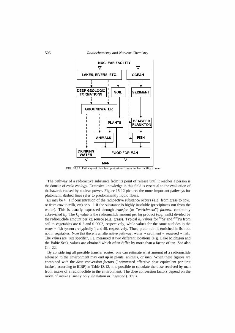
Radiochemistry and Nuclear Chemistry506
FIG. 18.12. Pathways of dissolved plutonium from a nuclear facility to man.
The pathway of a radioactive substance from its point of release until it reaches a person isthe domain of radio ecology. Extensive knowledge in this field is essential to the evaluation ofthe hazards caused by nuclear power. Figure 18.12 pictures the more important pathways forplutonium; dashed lines refer to predominantly liquid flows. Ex may be > 1 if concentration of the radioactive substance occurs (e.g. from grass to cow,or from cow to milk, etc) or < 1 if the substance is highly insoluble (precipitates out from thewater). This is usually expressed through transfer (or "enrichment") factors, commonlyabbreviated k . The k value is the radionuclide amount per kg product (e.g. milk) divided byd dthe radionuclide amount per kg source (e.g. grass). Typical k values for Sr and Pu fromd
90 239
soil to vegetables are 0.2 and 0.0002, respectively, while values for the same nuclides in thewater 6 fish system are typically 1 and 40, respectively. Thus, plutonium is enriched in fish butnot in vegetables. Note that there is an alternative pathway: water 6 sediment 6 seaweed 6 fish.The values are "site specific", i.e. measured at two different locations (e.g. Lake Michigan andthe Baltic Sea), values are obtained which often differ by more than a factor of ten. See alsoCh. 22. By considering all possible transfer routes, one can estimate what amount of a radionuclidereleased to the environment may end up in plants, animals, or man. When these figures arecombined with the dose conversion factors ("committed effective dose equivalent per unitintake", according to ICRP) in Table 18.12, it is possible to calculate the dose received by manfrom intake of a radionuclide in the environment. The dose conversion factors depend on themode of intake (usually only inhalation or ingestion). Thus

Radiation Biology and Radiation Protection 507
FIG. 18.13. Label for shipment of radioactive material of class II. "II" in red with a yellowbackground.
the dose factor of Pu is 10 larger for inhalation then for ingestion. Both kinds of data are239 2
given in Table 18.12, depending on available data (see ICRP publications). Such calculations are applied in the risk analyses of contemplated nuclear waste repositoriesand in the risk evaluation of radium or plutonium content in drinking water. They are also usedto establish limitations for intake of food from a contaminated area; e.g. in Sweden reindeermeat with >1500 Bq/kg is considered unfit as food (reindeer meat is the main protein intakefor the Laps living in Northern Sweden).
18.13.7. Classifications, working rules, etc
Radioactive substances of various activities, concentrations (or specific activities), decaymodes, etc., constitute quite different hazards, and must be handled accordingly. Variouscountries classify radioactive material differently and issue different working rules. We givea few classifications and rules, adhering primarily to the recommendations of the IAEA. Figure 18.13 shows the international transportation label with the symbol for ionizing radiation(with a white or yellow background, wbkg or ybkg). The radiation source and its activity shouldbe given. The category number is shown in red, according to:
I-wbkg, radiation level # 5 µSv/h : at surface ofII-ybkg, " # 0.5 mSv/h ; package; for III-III-ybkg " # 2 mSv/h < #1 mSv/h at 1 m

Radiochemistry and Nuclear Chemistry508
For high levels, the source must be transported as a special cargo. The transportation carriagemust fulfill a number of requirements with regard to resistance against fire, mechanical damage(drop tests), leakage (immersion tests), etc. Special regulations apply for spent reactor fuels andhigh-level waste (usually > 4 000 TBq; see Chapter 20). IAEA has issued rules fortransportation of radioactive materials, which are of special importance to large nuclearfacilities. For radio-tracer work in common non-nuclear research laboratories, some general rules canbe recommended (see also next section). Spills may result in increase in the radiationbackground. They may not constitute a hazard to the workers, but may ruin the scientificexperiments if not cleaned up immediately. In all work with radionuclides, radioactive wasteis produced. It is common practice to collect all such waste in special containers, and to disposeof it according to national rules. For short lived radionuclides of low hazard and low levels ofradioactivity (e.g. as in C-work), it is common practice to dispose of such waste by normal14
flushing to the sewer with several liters of tap water if such procedures are permitted by thenational radiation protection organizations.
18.14. Protective measures for radiochemical laboratory work
Three basic principles are recommended for keeping radiation exposure to a minimum:shielding, control, and distance. If a radiochemical laboratory is designed properly and the workperformed in such a manner that the general background contamination is sufficiently low todo valid low level tracer experiments, then the health aspects of radiation control are satisfied.We indicate the main principles for work with radioactive substances, but in each notion,special rules may apply.
18.14.1. Tracer work with moderate $-(-levels
Ordinary chemical laboratories may be used for radiochemical work at low levels of shortlived $-( radionuclides (e.g. half-lives <14 d and activity levels <10 kBq). However, it isrecommended that a special room be used for radioactive work. In the design of such alaboratory it is important that airborne contamination be prevented from spreading to countingrooms and to offices. Therefore, a pressure difference between the laboratories and the otherareas is desirable. The air velocity in the fume hoods should never be below 0.25 m s , and!1
0.5 m s is recommended. With such a flow velocity, radioactive dust and fumes are retained!1
in the hood and removed through the vents. The fume hoods should have filters for collectingradioactive particulates. To minimize the possibility of ingestion as well as the chances of ruining experiments throughaccidental contamination, limiting the radioactive work to a minimum area is essential. Thereshould be no radioactivity except in the immediate working area, and upon completion of theparticular experiment, all activity should be removed and the area cleaned (decontaminated) ifnecessary. For low levels, this means working in a good hood with easily cleaned nonporoussurfaces. One operation which commonly results in contamination involves evaporation of a solution todryness either on a hot plate or under a heat lamp. Although the percentage of the

Radiation Biology and Radiation Protection 509
sample carried away by spray may be very small, it may result in appreciable amounts ofactivity being spread around the area of the evaporation. Consequently, all evaporation shouldbe performed in a hood and the vicinity should be protected from the active spray by a coveringof absorbent paper. For nuclides which emit only $-particles, the glass walls of the container may providesufficient shielding. Sheets of glass or plastic (such as lucite) are commonly used to shieldexposed solid samples. For work with higher levels of $-(-emitters of longer half-lives, special radiochemicallaboratories should be used. The working surfaces and floors should be even, nonporous, andwith a minimum of seams. Surfaces of plastic material, stainless steel, and artificial stone areacceptable bench materials. It is recommended that fume hood and benches be covered with anabsorbing material such as absorbing paper, and that experiments be conducted when possiblein trays of stainless steel or plastic. Such arrangements ensure that the radioactive material doesnot contaminate a larger area if a spill occurs. Radionuclide workers should wear surgicalgloves and laboratory coats in the laboratory. If there is any danger of splashing, plastic face-hoods are recommended. It is extremely important that oral contamination be avoided.Beta-radiation from radioactive sources has ranges which rarely exceed 1 g cm .!2
Consequently, in a laboratory in which the level of $-emission is less than 1000 MBq,protection from the radiation can be achieved with a 1 cm plexiglass shield.
18.14.2. "-laboratories
For "-emitters the main hazard is internal, not external, to the body. For moderate activitylevels a glove box under slightly reduced pressure provides a simple and convenient closedchemical laboratory. Samples of the actinide elements usually have high specific radioactivities.Therefore, special care must be exercised in working with them. Because these "- emittersusually are associated with weak (-radiation or X- rays, chemical work with these elementsmust be conducted in more advanced gloveboxes (Fig. 18.14), which are kept at a pressureslightly below the surrounding atmosphere by circulation of pure air or inert gas through thebox. The boxes should have alarm systems for monitoring hazards such as interrupted watercirculation, electrical short circuits, oxygen in the inert gas, heat, etc. In Figure 18.14, thecontrol panel for these protective arrangements is shown above the box. In large alpha-box laboratories, one of the main hazards is radioactive dust. All room surfacesshould, therefore, be made with as few seams and sharp corners as possible; particularly thefloor must be of high quality. Electric power, water, waste, ventilation, etc., should beconnected to piping in the ceiling. The air into the laboratory must pass through filters as mustthe air exiting the laboratory. The exit air should be monitored for "-activity. Entering andleaving the laboratory should be through airlocks, and the hands and feet must be monitored foractivity on exit. All these protective measures make "-laboratories quite expensive, but smallerlaboratories working with lower levels of "-activity can be constructed in simpler fashion forcorrespondingly less cost.

Radiochemistry and Nuclear Chemistry510
FIG. 18.14. Glove-box for plutonium experiments. A radiochemical fume hood withfilter is seen in the background. Note that all connections are in the ceiling.
18.14.3. High level $-(-emitters
Since the intensity decreases as the inverse square of the distance, maintaining maximumdistance (by use, when necessary, of remote control apparatus such as tongs) when workingwith moderate or high levels of activity reduces the exposure appreciably. High levels of (, >1000 MBq, require shielding with layers of concrete, water, steel, orlead, and the operations must be carried out by remote control. The eyes can be protected bythe use of lead glass windows of high density, periscopes, mirrors, etc., see
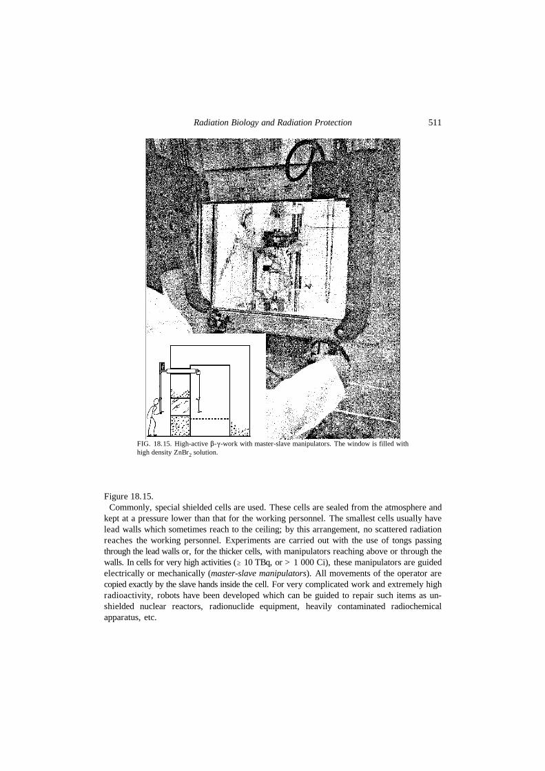
Radiation Biology and Radiation Protection 511
FIG. 18.15. High-active $-(-work with master-slave manipulators. The window is filled withhigh density ZnBr solution.2
Figure 18.15. Commonly, special shielded cells are used. These cells are sealed from the atmosphere andkept at a pressure lower than that for the working personnel. The smallest cells usually havelead walls which sometimes reach to the ceiling; by this arrangement, no scattered radiationreaches the working personnel. Experiments are carried out with the use of tongs passingthrough the lead walls or, for the thicker cells, with manipulators reaching above or through thewalls. In cells for very high activities ($ 10 TBq, or >1 000 Ci), these manipulators are guidedelectrically or mechanically (master-slave manipulators). All movements of the operator arecopied exactly by the slave hands inside the cell. For very complicated work and extremely highradioactivity, robots have been developed which can be guided to repair such items as un-shielded nuclear reactors, radionuclide equipment, heavily contaminated radiochemicalapparatus, etc.

Radiochemistry and Nuclear Chemistry512
18.15. Control of radiation protection measures
In larger organizations, the control of radiation hazards is the responsibility of specialistsknown as health physicists or, sometimes, health chemists. Their main duty is to ensure thatwork is carried out without hazard to the health of the people involved. The protection follows three stages: prevention, supervision, and after-control. Preventivemeasures include use of fume hoods, "-boxes, radiation shielding, tongs, etc., as discussedabove. The supervision stage involves the use of radiation instruments to monitor the radiationlevel (see Ch. 8). Small TLD, film or pocket pen dosimeters are used for individual monitoring(§7.9). For spills and contamination of hands, shoes, etc., special contamination instruments(counters) are used which are more sensitive than the monitoring dose instruments. Contamination in the laboratory must be avoided. This is controlled by smear tests; i.e., afilter paper is wiped over the surface and the paper is checked with a suitable instrument. In aso-called "clean area", the fixed contamination should not exceed 2 Bq for ", and 4 Bq for $-(on a surface of 100 cm . For an "active area" the rules are a maximum of 20 Bq for " per 1002
cm , and 0.01 mGy h from $-( at a distance of 2 cm from the surface. Radioactive aerosols2 -1
are monitored by air samplers in which a certain amount of air is drawn through a fine filterpaper after which the paper activity is measured. The after-control usually consists of checking personal dosimeters and a medical examination.Depending on the kind of work, the dosimeters are checked from twice a week to once a month.A medical examination is given once or several times a year, depending on the work conditions.In danger of inhalation or ingestion of "- or soft $-emitters, urine samples are analyzed. Suchanalyses are very sensitive and much less than a kBq in the body is easily detected. For workerswho handle hazardous amounts of "-emitters (e.g. plutonium in more than milligram amounts),urine samples should be taken regularly. If necessary whole-body counts are also taken. Whole-body counting is carried out with thesubject being surrounded by numerous scintillation or solid state detectors in a heavily shieldedroom. The natural body content of K is easily detected and is a control of the efficiency of40
this technique.
18.16. Exercises
18.1. "Reference man" consists of 18%C, 66% H O, 0.2% of K per body weight. He may also have accumulated 102pCi Ra in the body; assume 0.3 decay for each of the following 5 daughters. Calculate for a body weight of 70 kg the226
number of radioactive decays per unit time from H, C, K and Ra. Assume 30 TU in water.3 14 40 226
18.2. Using the information above, how many grams of the body's molecules (assume average mole weight of 10 ) will5
be damaged in a year if the G(damage) value is 3.1×10 mol/J? Assume E ($) = E /3.!7abs max
18.3. Under the same assumption as above, what amount of damage will be caused by cosmic radiation? Assume thatthe cosmic particles produce 3×10 ion pairs s m of the body.9 !1 !3
18.4. With the information in exercise 18.1, calculate annual doses received from (a) K, and (b) Ra and daughters.40 226
Assume w (") = 20.r 18.5. Ten mg U has been collected in the kidneys. Considering the biological half-life of uranium and assuming only238
one "-emission in U decay, calculate the dose (in Sv) received by the organ if the uranium is evenly distributed. The238
weight of a kidney is 150 g. 18.6. A (-dose rate of 1 Sv is assumed to inactivate (kill) human cells. The body contains 6x10 cells in a cell weight13
of 42 kg for a 70 kg man. (a) What average energy (in eV) has to be deposited in a cell to kill it? (b)
Radiation Biology and Radiation Protection 513
Calculate the number or kidney cells destroyed for the dose received in exercise 18.5. For simplicity assume the cellsto be cubic with a side length of about 11 µm. 18.7. A tumor has the weight of 80 g and we wish to destroy 20% of the cells by irradiating with 180 MeV protons withsuch penetration that half of the energy is deposited in the tumor. The particle beam is 5 µA. For what time must theirradiation be? A cell of weight 10 g is assumed to be killed on the absorption of 200 keV and no cell is assumed to be!9
killed twice.
18.17. Literature
E. H. SCHULTZ, Vorkomnisse und Strahlenunfälle in kerntechnischen Anlagen, Karl Thiemig, 1965.K. Z. MORGAN and J. E. TURNER (Eds.), Principles of Radiation Protection, J. Wiley, 1967.D. J. REES, Health Physics, Butterworths, 1967.Z. M. BACQ and P. ALEXANDER, Fundamentals of Radiobiology, Pergamon Press, 1967.E. L. SAENGER, G. E. THOMAS and E. A. TOMPKINS, Incidence of leukemia following treatment of hyperthyroidism, J.Amer. Med. Assoc. 205 (1968) 855.H. KIEFER and R. MAUSHART, Radiation Protection Measurement, Pergamon Press, 1972.IAEA, Population Dose Evaluation and Standards for Man and his Environment, Vienna, 1974.K. K. MANOCHA and R. K. MOHINDRA, Estimate of natural internal radiation dose to man, Health Physics 30 (1976) 485.G. WALINDER (Ed.), Tumorigenic and Genetic Effects of Radiation, National Swedish Environmental Protection Board1976.B. L. COHEN, The cancer risk from low-level radiation, Health Physics 39 (1980) 659.E. S. JOSEPHSON and M. S. PETERSON, Preservation of food by ionizing radiation, CRC Press, 1983.C. FERRADINI and J. PUCHEAULT, Biology of ionizing radiation, Masson, 1983.J. E. COGGLE, Biological Effects of Radiation, Taylor & Francis Ltd, 1983.M. TUBIANA, J. DUTREIX and A. WAMBERSIE, Radiobiologie, Hermann, 1986.J. ROTBLAT, A tale of two cities, New Scientist, January 7 (1988) 46.L. STRYER, Biochemistry, W H Freeman & Co, 1988.L.-E. HOLM, K. E. WIKLUND, G. E. LUNDELL, N. C. BERGMAN, G. BJELKEGREN, E. S. CEDERQUIST, U.-B. C. ERICSSON,L.-G. LARSSON, M. E. LIDBERG, R. S. LINDBERG, H. V. WICKLUND and J. D. BOICE JR, Thyroid cancer after diagnosticdoses of iodine-131: A retrospective cohort study. J. Natl. Cancer Inst. 80 (1988) 1132.R. YALOW, Biological effects of low-level radiation, Ch. 12 in M. E. BURNS (Ed), Low Level Radioactive WasteRegulation: Science, Politics, and Fear, Lewis Publishers, 1988.E. S. GILBERT, G. R. PETERSAN and J. A. BUCHANAN, Mortality of workers at the Hanford site: 1945-1981, HealthPhysics 56 (1989) 11.NATIONAL RESEARCH COUNCIL, Health effects of exposure to low levels of ionizing radiation. BEIR V, National AcademyPress, 1990.A. H. W. NIAS, An Introduction to Radiobiology, J Wiley & Sons, 1990.C. A. SONDHAUS, V. P. BOND and L. E. FEINENDEGEN, Cell-oriented alternatives to dose, quality factor, and doseequivalent for low-level radiation, Health Physics 59 (1960) 35.Annals of the ICRP, Pergamon Press, Oxford: Publ. #54 (1988), Individual Monitoring for Intakes of Radionuclides byWorkers: Design and Interpretation. #58 (1989), RBE for Deterministic Effects. #60 (1991), 1990 Recommendations ofthe International Commission on Radiological Protection. #61 (1991), Annual limits of Intake of Radionuclides byWorkers Based on the 1990 Recommendations.E. M. FIELDEN and P. O'NEILL (Eds.), The early effects of radiation on DNA, NATO Symposium, Springer Verlag,1991.R. MUKHERJEE and J. MIRCHEVA, Radiological effects of low-level radiation and cancer risks, IAEA Bulletin 2 (1991)32.United Nations Scientific Committee on the Effects of Atomic Radiation (UNSCEAR): Genetic and somatic effects ofionizing radiation, United Nations, 1993.