radiation information for hospital personnel - aapm · pdf fileradiation safety committee ......
TRANSCRIPT
AAPM REPORT NO. 53
RADIATION INFORMATION FOR HOSPITAL PERSONNEL
Published for theAmerican Association of Physics in Medicine
by the American Institute of Physics
AAPM REPORT NO. 53
RADIATION INFORMATIONFOR HOSPITAL PERSONNEL
RADIATION SAFETY COMMITTEECharles A. Kelsey
(Chairman)
MEMBERS OF THE TASK GROUPMorris I. Bank (Chairman)
Ralph LietoJeff ColvinSam Lott
Casmir EubigJanet Schlueter
Mary FoxDouglas ShearerFrances HarshawMichael TkacikHsin M. Kuan
Terry Yoshizumi
EDITORSLibby F. Brateman
Indra J. Das
April 1995
Published for theAmerican Association of Physicists in Medicine
by the American Institute of Physics

DISCLAIMER: This publication is based on sources and informationbelieved to be reliable, but the AAPM and the editors disclaim anywarranty or liability based on or relating to the contents of thispublication.
The AAPM does not endorse any products, manufacturers, orsuppliers. Nothing in this publication should be interpreted asimplying such endorsement.
Further copies of this report ($10 prepaid) may be obtained from:
American Association of Physicists in MedicineOne Physics Ellipse
College Park, MD 20740-3843
International Standard Book Number: 1-56396-480-5International Standard Serial Number: 0271-7344
©1995 by the American Association of Physicists in Medicine
All rights reserved. No part of this publication may be reproduced,stored in a retrieval system, or transmitted in any form or by anymeans (electronic, mechanical, photocopying, recording, or otherwise)without the prior written permission of the publisher.
Published by the American Institute of Physics, Inc.500 Sunnyside Blvd.. Woodbury, NY 11797
Printed in the United States of America
RADIATION INFORMATION FOR HOSPITAL PERSONNELTable of Contents1. INTRODUCTION . . . . . . . . . . . . . . . . . . . . . . . . . . . . . . . . . . . . . . . . . . . . . . . . . . . . . . . . . . . . . . . . . . . . . . . . . . . . . . . . . 12. RADIATION . . . . . . . . . . . . . . . . . . . . . . . . . . . . . . . . . . . . . . . . . . . . . . . . . . . . . . . . . . . . . . . . . . . . . . . . . . . . . . . . . . . . . . 1
2.1 Radiation Terms . . . . . . . . . . . . . . . . . . . . . . . . . . . . . . . . . . . . . . . . . . . . . . . . . . . . . . . . . . . . . . . . . . . . . . . . . . . . . . 2
3. BACKGROUND RADIATION . . . . . . . . . . . . . . . . . . . . . . . . . . . . . . . . . . . . . . . . . . . . . . . . . . . . . . . . . . . . 33.1 Natural Radiation Sources . . . . . . . . . . . . . . . . . . . . . . . . . . . . . . . . . . . . . . . . . . . . . . . . . . . . . . . . . . . . . .4
3.1.1 External Sources . . . . . . . . . . . . . . . . . . . . . . . . . . . . . . . . . . . . . . . . . . . . . . . . . . . . . . . . . . . . . . . . . . . .43.1.2 Internal Sources . . . . . . . . . . . . . . . . . . . . . . . . . . . . . . . . . . . . . . . . . . . . . . . . . . . . . . . . . . . . . . . . . . . . .4
4. MAN-MADE RADIATION SOURCES . . . . . . . . . . . . . . . . . . . . . . . . . . . . . . . . . . . . . . . . . . . . . . 54.1 Diagnostic Uses . . . . . . . . . . . . . . . . . . . . . . . . . . . . . . . . . . . . . . . . . . . . . . . . . . . . . . . . . . . . . . . . . . . . . . . . . . . . . . 54.2 Therapeutic Uses . . . . . . . . . . . . . . . . . . . . . . . . . . . . . . . . . . . . . . . . . . . . . . . . . . . . . . . . . . . . . . . . . . . . . . . . . . . . 6
5. MEDICAL SOURCES OF RADIATION . . . . . . . . . . . . . . . . . . . . . . . . . . . . . . . . . . . . . . . . . . . . 65.1 Diagnostic Sources . . . . . . . . . . . . . . . . . . . . . . . . . . . . . . . . . . . . . . . . . . . . . . . . . . . . . . . . . . . . . . . . . . . . . . . . . 6
5.1.1 Fixed X-Ray Machines . . . . . . . . . . . . . . . . . . . . . . . . . . . . . . . . . . . . . . . . . . . . . . . . . . . . . . . . . 75.1.2 Portable or Mobile X-Ray Machines . . . . . . . . . . . . . . . . . . . . . . . . . . . . . . . . . . . 75.1.3 Computed Tomography Scanners (CT) ................................ 95.1.4 Radioactive Materials . . . . . . . . . . . . . . . . . . . . . . . . . . . . . . . . . . . . . . . . . . . . . . . . . . . . . . . . . . . 105.1.5 Laboratory Departments . . . . . . . . . . . . . . . . . . . . . . . . . . . . . . . . . . . . . . . . . . . . . . . . . . . . . . . 11
5.2 Therapeutic Sources . . . . . . . . . . . . . . . . . . . . . . . . . . . . . . . . . . . . . . . . . . . . . . . . . . . . . . . . . . . . . . . . . . . . . . . 1 15.2.1 Radiation Therapy Machines . . . . . . . . . . . . . . . . . . . . . . . . . . . . . . . . . . . . . . . . . . . . . . . . 115.2.2 Radioactive Sources for Therapeutic Purposes . . . . . . . . . . . . . . . . . . . . 12
6. RADIATION PROTECTION METHODS . . . . . . . . . . . . . . . . . . . . . . . . . . . . . . . . . . . . . . . . . . 136.1 Protection Against Radiation . . . . . . . . . . . . . . . . . . . . . . . . . . . . . . . . . . . . . . . . . . . . . . . . . . . . . . . . . 1 5
6.1.1 Time . . . . . . . . . . . . . . . . . . . . . . . . . . . . . . . . . . . ................................................. 1 56.1.2 Distance ................................................................................. 166.1.3 Shielding . . . . . . . . . . . . . . . . . . . . . . . . . . . . . . . . . . . . . . . . . . . . . . . . . . . . . . . . . . . . . . . . . . . . . . . . . . . . . . . .16
6.2 Protection Against Radioactive Material Contamination . . . . . . . . . . . . 167. RESTRICTED AREAS . . . . . . . . . . . . . . .......................................................... 17
7.1 Recognizing Radiation Areas . . . . . . . . . . . . . . . . . . . . . . . . . . . . . . . . . . . . . . . . . . . . . . . . . . . . . . . . .178. SPECIFIC INSTRUCTIONS FOR ALLIED MEDICAL WORKERS .... 19
8.1 Housekeeping Personnel . . . . . . . . . . ....................................................... 198.2 Security Personnel . . . . . . . . . . . . . . . . . . . . . . . . . . . . . . . . . . . . . . . . . . . . . . . . . . . . . . . . . . . . . . . . . . . . . . . . . . 198.3 Maintenance Personnel . . . . . . . . . . . . . . . . . . . . . . . . . . . . . . . . . . . . . . . . . . . . . . . . . . . . . . . . . . . . . . . . . . . 1 98.4 Clerical Personnel . . . . . . . . . . . . . . . . . . . . . . . . . . . . . . . . . . . . . . . . . . . . . . . . . . . . . . . . . . . . . . . . . . . . . . . . . . . 19
9. RADIATION SURVEYS AND PERSONNEL MONITORING . . . . . . . . . 2010. RADIOACTIVE MATERIAL PACKAGE RECEIPT . . . . . . . . . ................ 2011. RADIATION EMERGENCIES . . . . . . . . . . . . . . . . . . . . . . . . . . . . . . . . . . . . . . . . . . . . . . . . . . . . . . . . . . . . 2112. RADIATION SAFETY OFFICER . . . . . . . . . . . . . . . . . . . . . . . . . . . . . . . . . . . . . . . . . . . . . . . . . . . . . . . 2113. RADIATION AND PREGNANCY ................................................................ 2214. RADIATION RISKS . . . . . . . . . . . . . . . . . . . . . . . . . . . . . . . . . . . . . . . . . . . . . . . . . . . . . . . . . . . . . . . . . . . . . . . . . . . . . 2315. ACKNOWLEDGMENTS .................................................................... 24
i
RADIATION INFORMATION FOR HOSPITAL PERSONNELList of Figures
Figure 1 - Electromagnetic Spectrum ........................................................ 2Figure 2 - Background Radiation.. ............................................................. 4Figure 3 - External and Internal Radiation Sources .................................. 5Figure 4 - Diagnostic X-Ray Machine.. ..................................................... 8Figure 5 - Fluoroscopy X-Ray Machine .................................................... 8Figure 6 - Mobile X-Ray Machine.. ........................................................... 9Figure 7 - CT scanner ................................................................................. 10Figure 8 - Gamma Camera in Nuclear Medicine ...................................... 11Figure 9 - Linear Accelerator in Radiation OncologyFigure 10 - Brachytherapy Sources Used in Radiation Oncology..Figure 11 - Time/Distance/ShieldingFigure 12 - Radiation Warning Sign(s).Figure 13 - Radioactive Source StorageFigure 14 - Hospital PersonnelFigure 15 - Counseling
.............................. 12.......... 1 4
......................................................... 15..................................................... 18.................................................... 18
Inspecting a Package ............................... 21Pregnant Worker .................................................. 22
List of Tables
Table 1 - Radiation Units . . . . . . . . . . . . . . . . . . . . . . . . . . . . . . . . . . . . . . . . . . . . . . . . . . . . . . . . . . . . . . . . . . . . . . . . . . . 3Table 2 - Background Radiation Sources . . . . . . . . . . . . . . . . . . . . . . . . . . . . . . . . . . . . . . . . . . . . . . . . . . 7
Table 3 - Estimate of Risks ....................................................................... 23
iii
RADIATION INFORMATION FORHOSPITAL PERSONNEL
1. INTRODUCTIONX-ray machines and radiation emitting sources are used in hospitals for
the diagnosis and treatment of diseases. Some of the hospital employeeswho work in radiology, nuclear medicine, radiation oncology, and somelaboratories are specifically trained in the operation of radiation machinesand the handling of radioactive materials and sources. These personnel arecalled “occupational workers.” Other hospital workers may work aroundradiation sources, and may be indirectly exposed to radiation during perfor-mance of their normal duties. These employees are “allied medical work-ers” and may belong to nursing, housekeeping, maintenance, security, ship-ping/receiving, and clerical departments. In addition, patient transport, op-erating room, and recovery room personnel may come in contact with brachy-therapy (radioactive implant) and nuclear medicine patients.
This booklet is designed to inform allied medical workers about the na-ture of radiation, its use in the hospital, and methods of radiation protection.The major areas covered in this booklet are:
• sources of radiation in the medical environment,
• radiation protection methods,
• instructions for workers,
• radiation risks and biological effects,
• and radiation exposure and pregnancy.While the potential exposure to allied medical workers from radiation is
very low, and the hazard (risk) is usually minimal, all radiation exposureshould be kept to a minimum. Further information can be obtained fromyour Radiation Safety Officer (RSO). Let us begin by defining “radiation.”
2. RADIATIONWhat is radiation?
Radiation is a general term used to describe a bundle of energy in theform of electromagnetic waves. Radio waves, microwaves, ultraviolet (UV),x rays, gamma rays, and visible light are all forms of electromagnetic or EMwaves. All EM radiation travels at the speed of light, 300,000 km/s (186,000miles/s). Among all the EM radiations, only light is visible to the humaneye. All other EM radiations cannot be seen and special instruments are
1
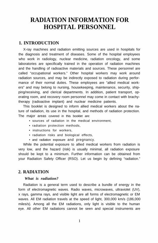
required to detect the presence of the invisible types of EM radiation. Fig-ure 1 shows the electromagnetic spectrum, a comparison of energies andproperties associated with different types of EM radiation.
The term radiation also is used to describe very fast moving particles,such as electrons and neutrons. These particles are found in the atom, whichis the smallest part of any material. When the energy of the radiation is highenough, it can remove electrons from the atoms or molecules of a substanceand is called ionizing radiation. Not all electromagnetic radiation causesionization. Ionizing radiation can pass through materials, and is also calledpenetrating radiation. X rays and gamma rays are high-energy ionizing EMradiations and may simply be called “radiation.”
In this document the term “radiation” refers to electromagnetic radiationthat causes ionization. Penetrating radiations are useful in the diagnosis andtreatment of diseases and are part of the backbone of modern medicine.However, because radiation can ionize and excite molecules, it can causedamage to living tissues. Therefore we must take precautions when usingand working around it.
2.1 Radiation TermsTo detect the presence and measure the amount of radiation, sensitive
and specialized instruments are used. Radiation is measured in radiationunits: roentgen, rad, and rem. The “roentgen” is a measure of exposure-the amount of ionization in air produced by radiation at a location. The“rad” is the radiation absorbed dose and refers to the amount of energyabsorbed by any material from the radiation. The “rem” determines the ra-diobiological equivalent and refers to the biological effect of the absorbedradiation on living things. From a practical, radiation safety concern, theseradiation terms are frequently used interchangeably despite their differentscientific definitions.
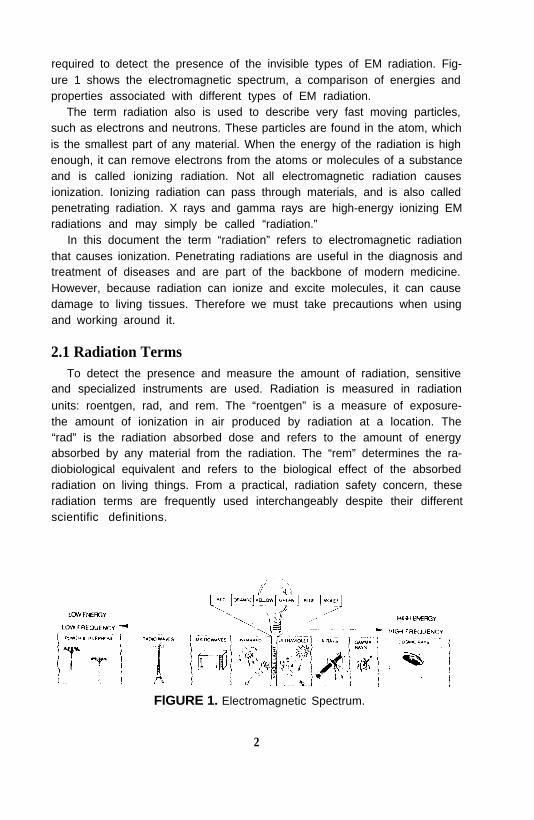
FlGURE 1. Electromagnetic Spectrum.
2
The roentgen, rad, and rem represent large quantities of radiation. Be-cause only low levels of radiation are routinely present in the medical envi-ronment to which allied medical workers are exposed, smaller units areused. These are milliroentgen (mR), millirad (mrad) and millirem (mrem),and are one one-thousandth (1/1000) of the roentgen, rad and rem, respec-tively. Most personnel exposures and measurements are expressed in thesesmaller units. You may also encounter other, newer Système International(SI) radiation units for exposure, absorbed dose, and dose equivalent, suchas the coulomb per kilogram (C/kg), gray (Gy), and sievert (Sv), respec-tively.
For radioactive materials, the amount of radioactivity is measured in unitsof the curie (Ci). A smaller unit, the millicurie (mCi) is often used, which isone one-thousandth of a curie. This term describes the rate at which theradioactive material emits radiation. The SI unit of radioactivity is also be-ing used and is the becquerel (Bq). Table 1 shows the old and the SI radia-tion units for various radiation quantities.
The amount of radioactivity present in a material decreases over time asa result of radioactive “decay.” The period of time that it takes for a materialto lose one half of its radioactivity is called its half-life. The half-life fordifferent radioactive materials varies from fractions of a second to thou-sands of years. Radioactive materials are potential sources of contamina-tion (radioactivity in places where it is not supposed to be). Contaminationcan cause radiation exposure.
3. BACKGROUND RADIATIONThe radiations discussed in this booklet are x rays and gamma rays, which
TABLE 1. Radiation Units
Quantity Unit New Unit
Radiation Exposure roentgen, R coulomb/kgAbsorbed Dose rad gray, GyDose Equivalent rem sievert, SvRadioactivity curie, Ci becquerel, Bq
1 R = 1000 mR1 rad = 1000 mrad1 rem = 1000 mrem1 Gy = 100 rad1 Sv = 100 rem1 Ci = 37,000,000,000 Bq
3
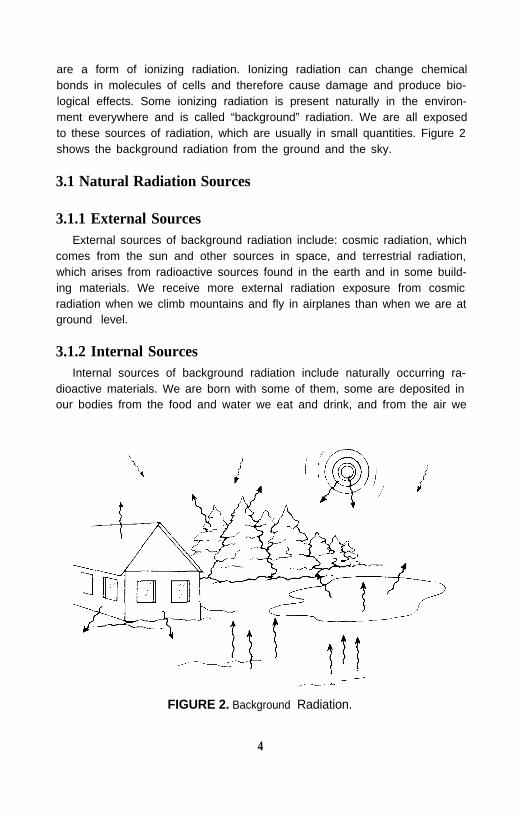
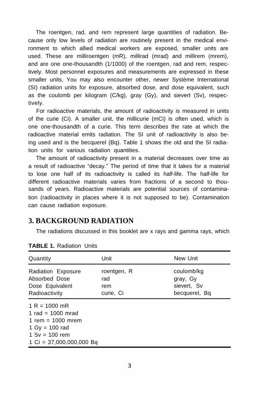
are a form of ionizing radiation. Ionizing radiation can change chemicalbonds in molecules of cells and therefore cause damage and produce bio-logical effects. Some ionizing radiation is present naturally in the environ-ment everywhere and is called “background” radiation. We are all exposedto these sources of radiation, which are usually in small quantities. Figure 2shows the background radiation from the ground and the sky.
3.1 Natural Radiation Sources
3.1.1 External SourcesExternal sources of background radiation include: cosmic radiation, which
comes from the sun and other sources in space, and terrestrial radiation,which arises from radioactive sources found in the earth and in some build-ing materials. We receive more external radiation exposure from cosmicradiation when we climb mountains and fly in airplanes than when we are atground level.
3.1.2 Internal SourcesInternal sources of background radiation include naturally occurring ra-
dioactive materials. We are born with some of them, some are deposited inour bodies from the food and water we eat and drink, and from the air we
FIGURE 2. Background Radiation.
4
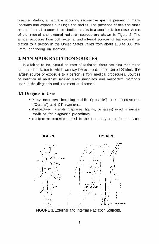
breathe. Radon, a naturally occurring radioactive gas, is present in manylocations and exposes our lungs and bodies. The presence of this and othernatural, internal sources in our bodies results in a small radiation dose. Someof the internal and external radiation sources are shown in Figure 3. Theannual exposure from both external and internal sources of background ra-diation to a person in the United States varies from about 100 to 300 mil-lirem, depending on location.
4. MAN-MADE RADIATION SOURCESIn addition to the natural sources of radiation, there are also man-made
sources of radiation to which we may be exposed. In the United States, thelargest source of exposure to a person is from medical procedures. Sourcesof radiation in medicine include x-ray machines and radioactive materialsused in the diagnosis and treatment of diseases.
4.1 Diagnostic Uses• X-ray machines, including mobile (“portable”) units, fluoroscopes
(“C-arms”) and CT scanners.• Radioactive materials (capsules, liquids, or gases) used in nuclear
medicine for diagnostic procedures.• Radioactive materials used in the laboratory to perform “in-vitro”
FIGURE 3. External and Internal Radiation Sources.
5
or test-tube studies on blood, urine, or cells for the diagnosis ofdiseases.
4.2 Therapeutic Uses• Linear accelerators or teletherapy machines used in radiation therapy
for the treatment of cancer and other diseases.• Radioactive sources in small, sealed containers used for patient
implants for treatment of cancer.• Radioactive drugs used to treat patients.
Patients who receive large, therapeutic doses of radioactive drugs orbrachytherapy implanted sources may pose risks to others and, as a conse-quence, they are typically confined to their hospital rooms. Workers whoencounter any of the radiation sources described above should ask the fol-lowing questions:
• What are the radiation sources in the area?• Are they x-ray machines or radioactive materials?• Where are the sources located?• What safety precautions should I take to minimize my exposure?• What should I do if I am accidentally exposed to these radiations?• Whom do I ask for more information regarding radiation in my
hospital?
• Is my exposure to radiation at an acceptable level?The answers to these questions and others are discussed in the following
sections.Smaller amounts of radiation exposure also arise from consumer and elec-
tronic devices such as airport baggage inspection machines, televisions,computer terminals, and smoke detectors. Table 2 shows some backgroundsources and the associated radiation levels.
5. MEDICAL SOURCES OF RADIATION5.1 Diagnostic Sources
As listed earlier, diagnostic sources include x-ray machines and radioac-tive materials. X-ray machines are used in radiography and fluoroscopy,and they may be permanently installed (“fixed”) or mobile. Radiation pro-tection methods are employed to reduce radiation exposure to the patientand others. In radiography, the exposure time is very short, usually less thanone second, and x rays are emitted from the machine only when the controlswitch to the unit is turned ON by the operator, Personnel are typically notin the x-ray room during the time the x rays are being emitted.
In fluoroscopy the exposure time may be lengthy, and personnel usually
6

TABLE 2. Background Radiation Sources
SourcesAverage Annual Dose(mrem/year)
NaturalCosmicTerrestrialInternalRadon (Estimate)Total Natural
Man-MadeMedicalConsumer ProductsFalloutNuclear Power PlantTotal Man-Made
303040200300
545<1<1<60
Adapted from NCRP Report No. 94. Exposure of the Population in the United States andCanada from Natural Background Radiation. National Council on Radiation Protectionand Measurements, Bethesda, MD, 1987.
work in the room while the machine is emitting radiation; therefore, theywear protective aprons to minimize their risks. The control panel of themachine has lights and audible signals that indicate when the machine isemitting radiation. When the control switch is in the OFF position, x raysare no longer produced from the machine, and no radiation risks exist.
5.1.1 Fixed X-Ray MachinesThese are primarily located in the X-Ray or Radiology Department, but
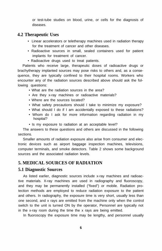
they may be located in other areas of the hospital, such as operating roomsand emergency rooms. These x-ray machines are used in the diagnosis ofdisease. These are located in a specially shielded room and are typicallyoperated by personnel trained in the proper use of the equipment. Figures 4and 5 show typical diagnostic x-ray units for radiography and fluoroscopy,respectively.
5.1.2 Portable or Mobile X-Ray MachinesMobile x-ray machines are similar in function to the fixed machines;
however, they are mobile and are transported to the patients who cannot bemoved. Mobile machines are typically used to examine patients in the Op-erating or Recovery Room during or after surgery, trauma victims in the
7
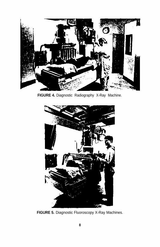
FIGURE 4. Diagnostic Radiography X-Ray Machine.
FIGURE 5. Diagnostic Fluoroscopy X-Ray Machines.
8
Emergency Room, patients located in intensive care units, neonatal units,and other bedridden patients. Figure 6 shows a mobile x-ray unit being usedin a patient’s room. Personnel and other patients may receive a small amountof exposure to x rays during the time that the x-ray machine is ON. Person-nel who assist in holding patients should wear protective aprons, as shouldthe operator.
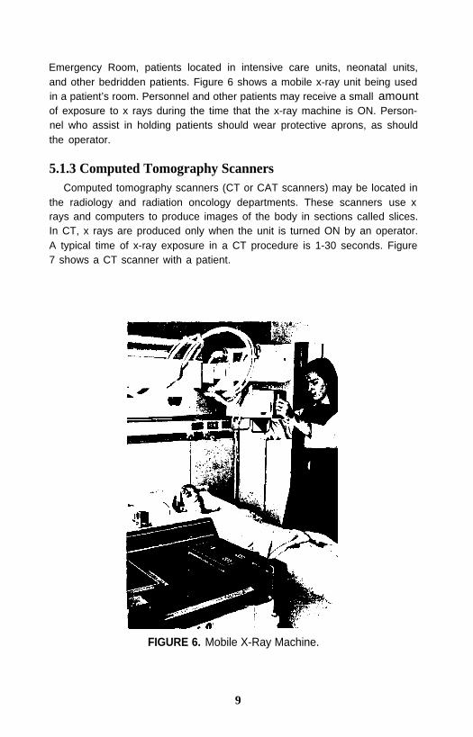
5.1.3 Computed Tomography ScannersComputed tomography scanners (CT or CAT scanners) may be located in
the radiology and radiation oncology departments. These scanners use xrays and computers to produce images of the body in sections called slices.In CT, x rays are produced only when the unit is turned ON by an operator.A typical time of x-ray exposure in a CT procedure is 1-30 seconds. Figure7 shows a CT scanner with a patient.
FIGURE 6. Mobile X-Ray Machine.
9
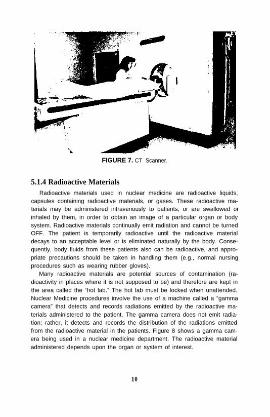
FIGURE 7. CT Scanner.
5.1.4 Radioactive MaterialsRadioactive materials used in nuclear medicine are radioactive liquids,
capsules containing radioactive materials, or gases. These radioactive ma-terials may be administered intravenously to patients, or are swallowed orinhaled by them, in order to obtain an image of a particular organ or bodysystem. Radioactive materials continually emit radiation and cannot be turnedOFF. The patient is temporarily radioactive until the radioactive materialdecays to an acceptable level or is eliminated naturally by the body. Conse-quently, body fluids from these patients also can be radioactive, and appro-priate precautions should be taken in handling them (e.g., normal nursingprocedures such as wearing rubber gloves).
Many radioactive materials are potential sources of contamination (ra-dioactivity in places where it is not supposed to be) and therefore are kept inthe area called the “hot lab.” The hot lab must be locked when unattended.Nuclear Medicine procedures involve the use of a machine called a “gammacamera” that detects and records radiations emitted by the radioactive ma-terials administered to the patient. The gamma camera does not emit radia-tion; rather, it detects and records the distribution of the radiations emittedfrom the radioactive material in the patients. Figure 8 shows a gamma cam-era being used in a nuclear medicine department. The radioactive materialadministered depends upon the organ or system of interest.
10

FIGURE 8. Gamma Camera.
5.1.5 Laboratory DepartmentsSome laboratories use small amounts of radioactive material for “in vitro,”
or test tube, diagnostic tracer studies. The amounts of radioactive materialused are typically only a fraction of those used in nuclear medicine studiesand generally do not pose any radiation risk as long as proper proceduresare followed.
5.2 Therapeutic SourcesYou will recall that the therapeutic sources include radiation therapy
machines, radioactive materials in therapeutic amounts, and scaled radioac-tive sources that are implanted in patients.
5.2.1 Radiation Therapy MachinesRadiation therapy machines are located in heavily shielded rooms in the
radiation therapy or radiation oncology departments. These machines de-liver high doses of radiation for the treatment of cancer and other diseases.The radiation dose is prescribed by a radiation oncologist and administeredby a radiation therapist. The radiation therapy machine may be a high-en-ergy x-ray machine or may be a sealed radioactive source unit, which housesa high-activity sealed Cesium-137, Iridium-192, or Cobalt-60 radioactivesource.
11
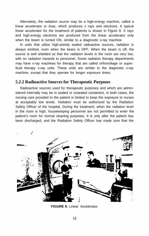
Alternately, the radiation source may be a high-energy machine, called alinear accelerator or linac, which produces x rays and electrons. A typicallinear accelerator for the treatment of patients is shown in Figure 9. X raysand high-energy electrons are produced from the linear accelerator onlywhen the beam is turned ON, similar to a diagnostic x-ray machine.
In units that utilize high-activity sealed radioactive sources, radiation isalways emitted, even when the beam is OFF. When the beam is off, thesource is well shielded so that the radiation levels in the room are very low,with no radiation hazards to personnel. Some radiation therapy departmentsmay have x-ray machines for therapy that are called orthovoltage or super-ficial therapy x-ray units. These units are similar to the diagnostic x-raymachine, except that they operate for longer exposure times.
5.2.2 Radioactive Sources for Therapeutic PurposesRadioactive sources used for therapeutic purposes and which are admin-
istered internally may be in sealed or unsealed containers. In both cases, thenursing care provided to the patient is limited to keep the exposure to nursesat acceptably low levels. Visitation must be authorized by the RadiationSafety Officer of the hospital. During the treatment, when the radiation levelin the room is high, housekeeping personnel are not permitted to enter thepatient’s room for normal cleaning purposes. It is only after the patient hasbeen discharged, and the Radiation Safety Officer has made sure that the
FIGURE 9. Linear Accelerator.
12
room is free from any source of radiation, that the room may be released tohousekeeping personnel for cleaning purposes. Generally, patients treatedby radioactive sources are hospitalized for a period of one to five days.Patients treated with sealed sources are not radioactive after the sources areremoved.
Nuclear Medicine Sources: Radioactive materials in therapeutic amountsare administered to patients in the form of a liquid or capsule. These mate-rials are highly radioactive, which means that the patients become sourcesof emitted radiation and radioactive contamination for a period of time.They remain radioactive until the administered radioactive material decaysto an acceptable level, or is eliminated naturally by the body. The patient istypically confined to one room until the radioactive decay results in accept-ably low radiation levels. The patient may cause significant contaminationof items which they touch, and all such items must be tested before dis-posal.
Radiation Therapy Sources: Another method of therapeutic use of ra-diation is to implant radioactive sources in the form of sealed seeds or rodsin patients. These patients are sources of radiation until the radioactive sourcesare removed or they have decayed to acceptably low radiation levels. Thesepatients are sources of radiation but not contamination. The patient withtemporary implants remains in isolation and is confined to a room until allthe radioactive sources have been removed.
Very rarely, sealed sources come off of a patient and wind up in bed linenor on the floor. If you should ever see or suspect that such a source is dis-lodged do not touch or pick it up. Keep everyone away from it. Call theRadiation Safety Officer to investigate immediately. Some types of brachy-therapy sources used in hospitals are shown in Figure 10. High dose-ratesources are contained in a specially shielded container. They are positionednear the patient’s tumor for brief periods of time and then retracted backinto the shielded container. The patient is usually treated as an outpatientand is not radioactive when she leaves the treatment room.
Summary: X-ray machines can be switched ON or OFF. They only emitradiation when they are turned ON. Radioactive material is always “ON”constantly emitting radiation. Radioactive materials can be shielded so thatno radiation escapes from the container.
6. RADIATION PROTECTION METHODSWhat safety precautions should one take to minimize radiationexposure?
Radiation protection is employed to protect against radiation produced
13
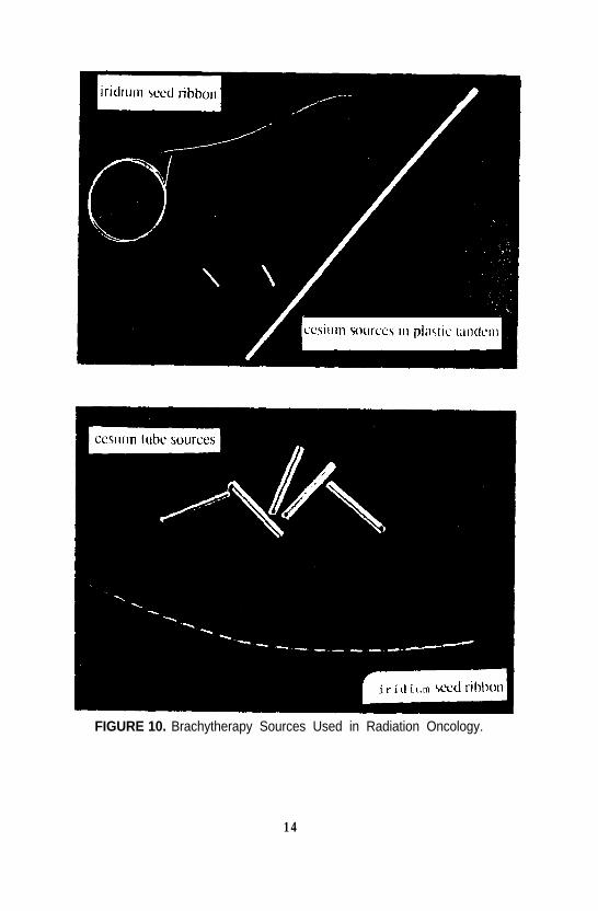
FIGURE 10. Brachytherapy Sources Used in Radiation Oncology.
1 4
from x-ray and gamma-ray machines, radiation emitted from radioactivematerials, and to protect against contamination from radioactive materialand sources.
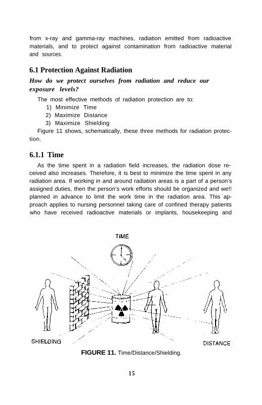
6.1 Protection Against RadiationHow do we protect ourselves from radiation and reduce ourexposure levels?
The most effective methods of radiation protection are to:1) Minimize Time2) Maximize Distance3) Maximize Shielding
Figure 11 shows, schematically, these three methods for radiation protec-tion.
6.1.1 TimeAs the time spent in a radiation field increases, the radiation dose re-
ceived also increases. Therefore, it is best to minimize the time spent in anyradiation area. If working in and around radiation areas is a part of a person’sassigned duties, then the person’s work efforts should be organized and we!!planned in advance to limit the work time in the radiation area. This ap-proach applies to nursing personnel taking care of confined therapy patientswho have received radioactive materials or implants, housekeeping and
FIGURE 11. Time/Distance/Shielding.
1 5
maintenance personnel, and security personnel responsible for off-hours ra-dioactive material package receipt and delivery. For radiation therapy treat-ments no one except the patient is allowed in the room so the exposure timeto everyone else is zero.
6.1.2 DistanceReduction of exposure due to an increase in distance is governed by the
inverse-square law. As the distance from a radiation source increases, theradiation exposure decreases rapidly. Doubling the distance between a per-son and the radiation source reduces the radiation exposure to as little asone-fourth (1/4) of the original exposure. It is good practice to keep as muchdistance between yourself and the radiation source as is reasonably pos-sible, even simply taking one step backward. Operating room or emergencyroom nurses are not always able to leave a patient unattended during a ra-diographic or fluoroscopic exam, but they can move away from the sourceas much as possible and wear lead aprons. An increase in distance from asource always reduces radiation exposure.
6.1.3 ShieldingMaterial that absorbs the radiation is a shield. The thicker the shielding,
the more the radiation exposure decreases. Some materials are better thanothers. Lead and concrete are the most commonly used materials for shield-ing x rays and gamma rays. They are very effective in stopping or blockingthe radiation beam. The walls of x-ray rooms are lead-lined to reduce theradiation exposure to those areas on the other side of the wall. Lead aprons,thyroid shields, and lead gloves are commonly used to shield body partsfrom diagnostic radiography and when portable x-ray machines are used.Lead bricks, lead vials, lead syringe shields and various other tools are usedto reduce radiation exposure in nuclear medicine and radiation oncologydepartments.
6.2 Protection Against Radioactive Material ContaminationContamination is the undesirable presence of radioactivity, such as a liq-
uid spill on the floor, or on clothing. It is a potential hazard whenever un-sealed radioactive materials are present. Avoid contaminating an area. Con-tamination can spread radioactivity to outside areas, including cars andhomes, and can result in the accidental ingestion or inhalation of radioac-tive materials. Prevent contamination by using the same precautions fol-lowed when handling infectious agents, and biological and chemical sub-
16
stances. Wear rubber gloves and protective clothing. Because the radioac-tive contamination emits radiation, one must practice the protection meth-ods described in the previous section. Remember that radioactivity or con-tamination cannot be seen. Radiation detecting instruments (survey meters)are used to survey when contamination is suspected.
Additional safe laboratory practices should be observed when personnelwork with unsealed radioactive materials. Do not eat, drink, smoke, or ap-ply cosmetics in any radiation areas. These precautions reduce the possibil-ity of the accidental ingestion or inhalation of radioactive materials.
7. RESTRICTED AREAS7.1 Recognizing Radiation Areas
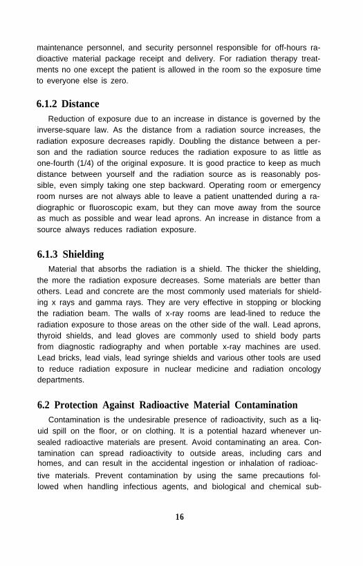
The protection methods described above are effective when radiationareas are known, but how can allied medical workers know if they are in aradiation area? Areas with radiation sources or radioactive materials aredefined to be “Restricted Areas,” which are required by federal or state lawto be posted with one or more of the following warning signs:
“Caution Radiation Area”"Caution High Radiation Area"“Caution Radioactive Materials.”
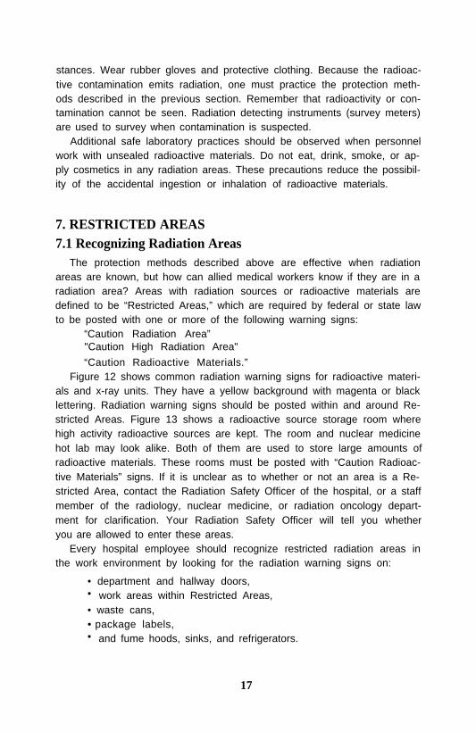
Figure 12 shows common radiation warning signs for radioactive materi-als and x-ray units. They have a yellow background with magenta or blacklettering. Radiation warning signs should be posted within and around Re-stricted Areas. Figure 13 shows a radioactive source storage room wherehigh activity radioactive sources are kept. The room and nuclear medicinehot lab may look alike. Both of them are used to store large amounts ofradioactive materials. These rooms must be posted with “Caution Radioac-tive Materials” signs. If it is unclear as to whether or not an area is a Re-stricted Area, contact the Radiation Safety Officer of the hospital, or a staffmember of the radiology, nuclear medicine, or radiation oncology depart-ment for clarification. Your Radiation Safety Officer will tell you whetheryou are allowed to enter these areas.
Every hospital employee should recognize restricted radiation areas inthe work environment by looking for the radiation warning signs on:
• department and hallway doors,• work areas within Restricted Areas,• waste cans,• package labels,• and fume hoods, sinks, and refrigerators.
17
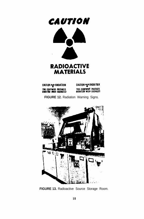
FIGURE 12. Radiation Warning Signs.
FIGURE 13. Radioactive Source Storage Room.
18
8. SPECIFIC INSTRUCTIONS FOR ALLIED MEDICALWORKERS
8.1 Housekeeping PersonnelAll housekeeping employees should be aware of the locations of all Re-
stricted Areas in order to practice good radiation protection measures. Thesemeasures are:
1) Recognize Restricted Areas in your work environment.2) Get user permission and instructions from the Radiation Safety
Officer before cleaning any spill in a Restricted Area.3) Do not clean counter tops, hoods, refrigerators, or sinks in Restricted
Areas unless specially requested and instructed by the areasupervisor or Radiation Safety Officer.
4) Do not remove bedclothes, dishes, trash, or other items from roomsposted with radiation signs, unless specifically instructed by amember of the Radiation Safety Staff.
8.2 Security PersonnelAll security personnel should be aware of the locations of all Restricted
Areas, and be able to recognize packages containing radioactive material inorder to practice good radiation protection measures.
8.3 Maintenance PersonnelAll maintenance personnel should be aware of the locations of all Re-
stricted Areas so that they may practice good radiation protection measures.These measures are:
1) Recognize Restricted Areas in your work environment.2) Obtain permission before working in an area that is in or adjacent
to a Restricted Area.3) Be aware of hoods, sinks, refrigerators, and storage areas used for
radioactive materials or sources.
8.4 Clerical PersonnelAll clerical personnel in the departments that use radiation should be
aware of the locations of Restricted Areas so that they may practice goodradiation protection measures. Good practice includes:
1) Recognizing Restricted Areas in your work environment.2) Refraining from eating, drinking, applying cosmetics, and smoking
in areas where radioactive materials are used.
19

3) Prohibiting food or soft drink storage in refrigerators used for thestorage of radioactive materials.
9. RADIATION SURVEYS AND PERSONNEL MONITORINGHow do I know that radiation levels are within acceptable limits?
Radiation surveys are measurements of radiation levels with specializedinstruments. A radiation survey of x-ray equipment and adequacy of roomshielding is conducted by the Medical Physics or Radiation Safety staff todocument the radiation levels present during operation. At the time of in-stallation, an in-depth safety evaluation of the radiation machine is per-formed before it is used for patient examinations or treatments. In additionto these surveys, periodic quality assurance checks are required.
Areas where radioactive materials are used, prepared, or stored are moni-tored for radiation levels on a periodic basis, which may be daily, weekly,monthly, or quarterly, depending on the type of use and the legal require-ments. These surveys are conducted with the use of hand-held radiationsurvey instruments. The measured radiation levels are then compared toallowable limits. Surveys are conducted in an effort to keep radiation expo-sures As Low As Reasonably Achievable (ALARA) for everyone.
In addition to the physical surveys described above, some workers mustbe monitored for radiation exposure with personnel dosimetry devices, suchas film badges, ring badges, or pocket dosimeters. These devices are in-tended to record only the radiation exposure that an individual receives as aresult of employment at a particular facility. The personnel monitoring de-vice should never be exchanged between individuals. After the device isworn for a specified period of time, it is returned to the place of evaluation.The radiation dose is evaluated and recorded as a permanent record of expo-sure for that individual, which becomes a legal document. Personnel expo-sure records for each individual are maintained for a long period of time,and the individual’s occupational dose is for that individual’s lifetime.
10. RADIOACTIVE MATERIAL PACKAGE RECEIPTRadiation surveys should be performed on all packages containing ra-
dioactive materials delivered to or shipped from the hospital. Packages con-taining radioactive material are typically delivered to a designated location,such as the nuclear medicine department, during normal working hours whenthe staff is available to conduct the required monitoring. Sometimes deliv-eries may be made during off-hours. Each facility is required to have spe-cific procedures for radioactive material package receipt, which include pro-
20
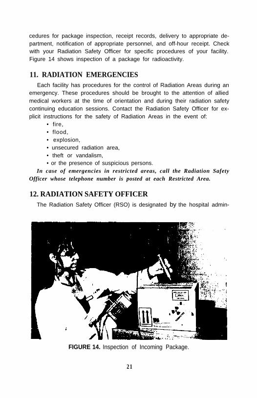
cedures for package inspection, receipt records, delivery to appropriate de-partment, notification of appropriate personnel, and off-hour receipt. Checkwith your Radiation Safety Officer for specific procedures of your facility.Figure 14 shows inspection of a package for radioactivity.
11. RADIATION EMERGENCIESEach facility has procedures for the control of Radiation Areas during an
emergency. These procedures should be brought to the attention of alliedmedical workers at the time of orientation and during their radiation safetycontinuing education sessions. Contact the Radiation Safety Officer for ex-plicit instructions for the safety of Radiation Areas in the event of:
• fire,• flood,• explosion,• unsecured radiation area,• theft or vandalism,• or the presence of suspicious persons.
In case of emergencies in restricted areas, call the Radiation SafetyOfficer whose telephone number is posted at each Restricted Area.
12. RADIATION SAFETY OFFICERThe Radiation Safety Officer (RSO) is designated by the hospital admin-
FIGURE 14. Inspection of Incoming Package.
21
istration and authorized by the State and/or Nuclear Regulatory Commis-sion (NRC) to oversee the radiation safety program in the hospital for radio-active materials. An RSO must meet specific training and experience crite-ria. The name of the RSO and a 24-hour telephone contact must be postedwherever radioactive materials are used or stored.
In addition to the emergencies described above, an RSO can be con-tacted for the following:
• personnel exposure data, if you are monitored for radiation or feelthat you should be;
• regulations, license, and inspection reports;• if you are pregnant and you work in Restricted Areas;• if you have questions or suspect problems about radiation;• or if you want to know about the NRC and other federal or state
regulatory agencies regarding radiation protection.
13. RADIATION AND PREGNANCYIt has been known since the early 1900s that cells that reproduce more
frequently are more sensitive to radiation damage. Because embryos, fe-tuses, and children are growing, and therefore their cells are reproducingmore often, they are typically more sensitive to radiation than adults. Whenthe abdomen of a pregnant female is exposed to radiation, a fraction of thatexposure is also received by the embryo or fetus. The most radiosensitive
FIGURE 15. Counseling Pregnant Worker.
22
period for the embryo is from 8 to 15 weeks gestation age.Allied medical workers who may be exposed to radiation should contact
the RSO if they become pregnant or are planning to become pregnant.Instructions given to radiation workers should include information re-
garding prenatal exposure risks to the developing embryo and fetus. Thisinformation is used by the employee in the event of pregnancy to assess therisks associated with her particular employment duties and any possiblealternative work environments if deemed necessary. The radiation doseequivalent to the embryo/fetus is considered separately from the maternaldose, and the radiation limits during the nine-month gestation period are500 mrem for the embryo/fetus.
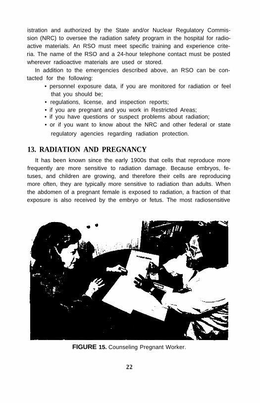
It is important to note that the mother assumes all risk until she specifi-cally declares her pregnancy, in a written and signed statement, to her su-pervisor or the RSO. At that time, the hospital is responsible for assuringthat the duties of a female worker will not result in a dose equivalent that ismore than 500 mrem to the fetus. Figure 15 shows a pregnant worker with aradiation counselor. Personnel should be encouraged to contact the RSO todiscuss any matter regarding radiation.
14. RADIATION RISKSIn today’s society, many of our daily activities involve risks. Generally, a
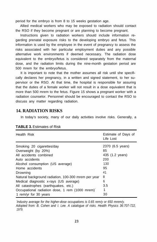
TABLE 3. Estimates of Risk
Health Risk Estimate of Days ofLife Lost
Smoking 20 cigarettes/day 2370 (6.5 years)Overweight (by 20%) 85All accidents combined 435 (1.2 years)Auto accidents 200Alcohol consumption (US average)Home accidentsDrowningNatural background radiation, 100-300 mrem per yearMedical diagnostic x-rays (US average)All catastrophes (earthquakes, etc.)Occupational radiation dose, 1 rem (1000 mrem)*
1 rem/yr for 30 years
1309541863.5130
*Industry average for the higher-dose occupations is 0.65 rem/y or 650 mrem/y.Adopted from: B. Cohen and I. Lee. A catalogue of risks. Health Physics 36:707-722,1979.
23

risk can be defined as “the possibility or chance of illness, injury, or evendeath that may result from some activity.” For a patient, the risk associatedwith the radiation exposure received from a particular exam is typicallyoutweighed by the benefit of the diagnosis or treatment received.
A useful measure for comparing the risk associated with radiation toother kinds of health risks is the average number of days of life expectancylost per unit of exposure for each type of risk. These estimates indicate thatmany of our day-to-day activities represent a higher health risk than our riskfrom the radiation levels encountered in the medical work environment.Some risk factors associated with various activities are presented in Table 3.
15. ACKNOWLEDGMENTSThe Radiation Protection Committee greatly appreciates the help of Kirah
V. Sickle and Steven Thackston of the Medical College of Georgia, Au-gusta, GA for illustration and photography; and Kathy Buchheit of the FoxChase Cancer Center, Philadelphia, PA for the preparation of the manu-script.
24