radiation protection officer study material
DESCRIPTION
Study material for Industrial Radiation Safety/Protection OfficersTRANSCRIPT

TECHNOLOGY EXPERTS GROUP
BASIC PRINCIPLES OF
RADIATION PROTECTION
FOR RPO
Prepared by
Prof. Dr. M. FAROUK AHMAD
RIYADH
APR. 2006
2
FOREWORD
The use of man-made ionizing radiation and radioactive sources
are now a day widespread, and continue to increase around the world.
Nuclear techniques are in growing use in industry, agriculture, medicine,
well logging, and research benefiting the society as a whole. Irradiation
is used around the world to preserve foodstuffs. Sterilization techniques
have been used to eradicate diseases, and ionizing radiation are widely
used in diagnosis and therapy of different diseases. Industrial
radiography is widely used to examine welds and detect cracks and
microscopic bubbles in metallic pipes, tanks and other devices, and help
prevent the failure of engineered structures.
It has been recognized that exposure to a an acute dose of ionizing
radiation causes clinical damage to the tissues of the human body. In
addition, long term studies of populations exposed to ionizing radiation
have demonstrated that this exposure has a potential for the delayed
induction of malignancies. Due to these risks all activities involving
radiation exposure shall be subjected to certain national and international
safety standards, in order to protect radiation workers, general public and
environment from exposure to ionizing radiation.
One of the requirement of the national and international safety
standards is that any installation, that is acquiring any of the radiation
sources shall appoint a radiation protection officer, RPO, (or officers), to
oversee the application of the requirements of the radiation protection
and safety of radiation sources. According to the Saudi national and
international regulations, this individual shall be technically competent
in radiation protection scientific and organizational matters, relevant for
a given type of practice. In Accordance with Saudi national regulations
shall be licensed by the national regulatory authority through passing a
qualification exam, which is held periodically by this authority.
For successfully passing this qualification exam, one should study
different scientific and organizational topics, which are existing in
different English books, and are specialized very deep in the subjects of
interest. It may be very difficult for individuals non specialized in
radiation physics to follow this subjects.

3
For this reason this booklet is prepared, and will be issued, by the
technology experts group, to cover the fundamentals and all scientific
and organizational topics that are necessary for any radiation protection
officer to be qualified as a RPO. Together with the included topics in
this booklet the practical lessons are essential part of the qualification of
the RPO. This practice in the different relevant fields may be gained
easily through these practical lessons.
We hope that the booklet will be helpful in acquiring the necessary
knowledge in the field.
Technology experts Group
and the author

4
PATRT 1
SCIENTIFIC AND TECHNOLOGICAL ASPECTS
OF RADIATION PROTECTION

5
CONTENTS
Part 1: Scientific and technical aspects of radiation protection.
Chapter 1: Radioactivity and radioactive decay.
1-1 Some properties of atomic nuclei.
1-2 Some properties of alpha decay and alpha particles.
1-3 Some properties of beta decay and beta particles.
1-4 Some properties of gamma disintegration.
1-5 The x-rays.
1-6 The neutrons and their sources.
1-7 Calculation of the source activity
1-8 The units of activity.
1-9 The physical half-life time.
1-10 The biological and effective half-life times.
1-11 The radioactive decay law..
1-12 The relation between the decay constant and the half-life time.
1-13 Some important multipliers.
Chapter 2: Interaction of radiation with matter.
2-1 Introduction.
2-2 Interaction of heavy charged particles with matter.
2-3 Interaction of beta particles with matter.
2-4 Interaction of x and gamma radiation with matter.
2-5 Interaction of neutrons with matter.
Chapter 3: Radiation detectors, survey meters and monitors.
3-1 General.
3-2 The gas detectors.
3-3 The scintillation detectors.
3-4 The semi-conductor detectors.
3-5 The survey meters.
3-6 The contamination monitors.
3-7 Devices for personal dosimetry.
Chapter 4: Some radiation measurement techniques and statistical
fluctuations.
4-1 Introduction.
4-2 The solid angle.
4-3 The detector intrinsic efficiency.

6
4-4 Relation between the counting rate and source activity.
4-5 Other factors affecting the measurements.
4-6 Dead time correction.
4-7 The statistical fluctuation of radiation measurements.
Chapter 5: Dosimetry quantities and their units.
5-1 The exposure.
5-2 The absorbed dose.
5-3 The equivalence between the Roentgen, the rad and Gray.
5-4 The Kerma
5-5 The radiation weighting factor.
5-6 The equivalent dose.
5-7 The tissue weighting factor.
5-8 The effective dose.
5-9 The committed equivalent or effective dose.
Chapter 6: Biological effects of radiation.
6-1 Direct and indirect action of ionizing radiation on cell.
6-2 Radiation effects.
6-3 Deterministic and stochastic effects.
6-4 Acute deterministic effects.
6-5 The stochastic effects.
6-6 Hereditary effects.
Chapter 7: Dose calculation.
7-1 Dose calculation from point sources.
7-2 Dose calculation for beta emitters.
7-3 Dose calculation from external gamma sources.
7-4 Dose calculation from neutron sources.
7-5 The inverse square low for external exposure
7-6 Dose calculation from internal exposure.
7-7 The annual limit on intake.
7-8 The derived air concentration.
Chapter 8: Radiation shielding.
8-1 Shielding of sources of alpha particles.
8-2 Shielding of sources of beta particles.
8-3 Shielding of x and gamma ray sources.
8-4 Shielding of the neutron sources.

7
Part 2: Organizational aspects of radiation protection.
General framework and requirements for radiation protection.
1- Introduction.
2- Administrative requirements.
3- Management requirement for radiation protection.
4 - The principle requirements.
5- Verification of safety.
6- Condition of service.
Responsibilities of parties.
1- Responsible parties for radiation protection.
2- Responsibilities of the licensee.
3- Cooperation between licensees and employers.
National (SA) dose limits.
1- The terms limit and level.
2- Radiation exposures.
3- The occupational dose limits.
4- The dose limits for general public.
5- The dose limits for medical exposures.
6- The dose limits for emergency exposures.
The radiation Protection Program (RPP).
1- Introduction.
2- The structure of the RPP.
The safe transport of radioactive material.
1- Introduction.
2- Definitions.
3- General provisions.
4- Determination of the transport index.
5- Categories of packages.
6- Marking and labeling.
7. Storage in transit.

8
CHAPTER 1
RADIOACTIVITY AND RADIOACTIVE DECAY
1-1 Some properties of the atomic nuclei:
- Any atom is composed of the atomic nucleus, around which
electrons are orbiting in elliptical shells.
- The radius of the atom is in the order of 10-10
m, while the radius
of the nucleus is in the order of 10-15
m, so that the volume of the
nucleus is smaller than that of atom by about thousand trillions times
(trillion = 1012
). Due to these dimensions, the atom is similar to the solar
system, with its inter- planetary distances.
- Any atomic nucleus consists of nucleons, which are protons or
neutrons. The proton mass is, approximately, higher than that of the
electron by about 1836 times, while the neutron mass is higher by about
1838 times. So, the neutron and the proton may be considered as
particles with the same mass. From these data the atomic mass is
concentrated in the atomic nucleus, and the nuclear density is,
approximately, constant and equals 1017
kg/m3 (about 100 millions
ton/cm3).
- The charge of the proton equals to the electron charge in
magnitude (1.6x10⁻¹⁹
Coulomb), but it is positive in sign, while the
neutron is neutral (e.g. its total charge equals zero). So, in a neutral atom
the number of the protons in the nucleus equals the number of the orbital
electrons.
- The number of the protons in a nucleus is called its atomic
number Z, while the total number of protons and neutrons, in it, is
called the mass number A. So the number of neutrons N in a nucleus is
N = A – Z. Symbolically, any atom is represented by the first letter
written in capital, or by the first one in capital and other one written in
small. The atomic number is written in the lower left corner, while the
mass number is written in the upper left one. Example of that is C12
6 (or
carbon-12), Cl35
17 (or chlorine-35), Cr51
23 (chrome-51) and Cd114
48 (or
cadmium-114).
- The nucleus of any element is composed of the same number of
protons Z, but it may have different numbers of neutrons N. these

9
different forms of the same element are called isotopes of the element.
For example, hydrogen exists in three forms (the nucleus of each
contains one proton), H1
1 without any neutron, H2
1 (or deuterium) with
one neutron and, H3
1 (or tritium) with two neutrons. The isotopes of the
element are characterized by the same chemical properties while they
have different physical properties. Some Elements have more than 40
isotopes.
- Some nuclides are stable, while some others are unstable and
they may, spontaneously, decay to daughter nuclides through the
emission of alpha or beta particle, or may disintegrate through the
emission of gamma radiation. These nuclides are called radio-nuclides
and there atoms are called radio-active isotopes. So, there are three types
of the radioactive decay, which are:
a) alpha decay (α decay)
b) beta decay (β decay), and
c) gamma disintegration (γ disintegration)
1-2 Some properties of α-decay and α-particles:
- In α decay of a nucleus, an alpha particle (α), which is the
nucleus of a helium-4 atom ( He4
2 ), is emitted. This particle is composed
of 2 protons and 2 neutrons. So, in an α decay of a parent radionuclide
the mass number of the daughter nuclide is reduced by 4 while the
atomic number is reduced by 2. An example of alpha decay is the decay
of uranium-238 to thorium-234 with the emission of an alpha particle α,
which is symbolically represented as:
U238
92 Th234
90 + He4
2
Another example is the decay of polonium ( Po210
84 ) to the stable
lead-206 ( Pb206
82 ) which is symbolically represented as:
Po210
84 Pb206
82 + α
- Alpha particles emitted from a certain radionuclide are
characterized by, so called, discrete spectrum. This means that all alpha
particles emitted from that radionuclide will have the same energy value
or separated but fixed values. So, by measuring the energy value or
values of α particles the radionuclide can be easily identified. In other
1
0
words, it is known that U238
92(for example) emits α particles with two
energy values which are 4.196 and 4.149 MeV. So, if these two energy
values for any alpha emitter are detected, then it mean that this emitter is
U238
92.
1-3 Some properties of β-decay and β –particles:
- There are three types of beta decay, which are:
1-3-1 Electron or β-negative decay:
- in this type of β decay one of the neutrons n of the parent nucleus
decays, spontaneously, to a proton p, negatron β- (which is a β-negative
particle i.e. electron) and a third particle, named anti-neutrino υ-. This is
represented symbolically as;
n p + β- + υ
-
- One example of β- (or electron decay) is the decay of
Co60
27 (Cobalt-60) to Ni60
28 (Nickel-60) with the emission of β- particle and
anti-neutrino υ-(see fig. 1-1), which is expressed symbolically as:
Co60
27 Ni60
28 + β- + υ
-
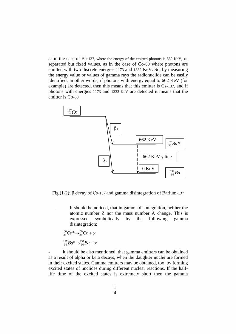
- Other example is the decay of cesium-137 to barium-137 with the
emission of the same two particles (see fig. 1-2). This is expressed as:
Cs137
55 Ba137
56 + β- + υ
-
- It should be mentioned that the decay energy which is a fixed
amount for each parent radionuclide to decays to a daughter one is
distributed randomly between the two emitted particles, β- and υ
-. In
some decays of the parent radionuclide the majority of the fixed decay
energy is acquired by beta particle, and the remaining small amount of
energy is acquired by the anti-neutrino. In other decays of the same
parent radionuclide the beta particles acquire a medium or a small
amount of the decay energy, and hence the anti-neutrino will get a
medium or a large amount of the decay energy. That is the reason of
emission of beta particles from the same radionuclide with energies
varying from zero up to the maximum decay energy. This is expressed,
in other words, in that the beta spectrum of any beta emitter is a

1
1
continuous one for different types of beta decay, and by studying beta
spectra it is impossible to identify the beta-emitting radionuclide.
- In beta-negative decay the mass number A of both parent and
daughter radio-nuclides remains constant and does not change, while the
atomic number Z of the daughter nuclide is increased by one with
respect to that of the parent one, since a neutron is converted into a
proton in the nucleus.
1-3-2 Positron or beta positive decay:
- In this type of β decay one of the protons of the parent nucleus
decays spontaneously to a neutron, β+ (which is a β-positive particle i.e.
positron) and a third particle, named neutrino υ. This is represented
symbolically as;
p n + β+ + υ
- One example of β+ (or positron decay) is the decay of Na-22
(Sodium-22) to Ne-22 (Neon-22) with the emission of β+ particle and
neutrino υ (see fig. 1-1), which is expressed symbolically as:
Na22
11 Ne22
10 + β+ + υ
- In beta-positive decay the mass number A of both the parent and
daughter radio-nuclides remains constant and does not change, while the
atomic number Z of the daughter nuclide is decreased by one with
respect to that of the parent one, since one proton of the parent nucleus is
converted into a neutron.
1-3-3 The electron capture:
- In this type of β decay one of the protons of the parent nucleus
captures an orbital electron from the shells, which are very close to the
nucleus, forming a neutron and a neutrino υ is emitted during this
process. This is represented symbolically as;
p + e- n + υ
- One example of the electron capture is the capture of an orbital
electron by Na-22 (Sodium-22) nucleus to form a Ne-22 (Neon-22)
nucleus with the emission of a neutrino υ. This is expressed symbolically
as:
1
2
e- +
Na2211 Ne22
10 + υ
- In the electron capture no beta particle is emitted, but the only
emitted particle is the neutrino. Moreover the mass number A of both the
parent and daughter nuclides remains constant and does not change, as in
all other types of beta decay, while the atomic number Z of the daughter
nuclide is decreased by one with respect to that of the parent one, since a
proton is converted into a neutron, by the analogy to the beta positive
decay.
1-4 Some properties of gamma disintegration:
- If an atomic nucleus is formed in, so called, excited energy state
(i.e. in a state with excess energy) it may disintegrate to a state with a
lower excitation energy or to the so called, the ground state (i.e. to the
state with zero excitation energy). This disintegration is accompanied
with the emission of a gamma (γ) photon, that carries an amount of
energy equal to the difference between the excitation energies of the
initial and final states. So, the energy Eγ of the emitted γ photon is given
as:
Eγ = Ei - Ef
where Ei and Ef are the excitation energies of the initial and final states
of the gamma emitting nucleus, respectively.
- Each γ photon is an electromagnetic wave (with zero rest mass)
with an ultra-high frequency f of a given value, which is, in its turn, a
characteristic value for this disintegration.
- An example of gamma disintegration is the disintegration of
*60
28 Ni nucleus, which is formed in an excited state, as a result of beta
decay of the Co60
27 , with an excitation energy equal to 2505 KeV, and then
it disintegrates, promptly, to a lower excited state with an excitation
energy equal to 1332 KeV, which, in its turn, disintegrates promptly to
the ground state with zero excitation energy. This means that the *60
28 Ni
emits two γ photons, one with energy Eγ1 = 2505 – 1332 =1173 KeV,
and the second with energy Eγ2 = 1332 – 0 = 1332 KeV. These two
gamma ray photons are characteristic lines (i.e energies) for the gamma
disintegration of *60
28 Ni , and hence for the decay of the Co60
27 to *60
28 Ni .

1
3
So, the detection of two gamma ray lines with energies 1173 and 1332
KeV is an indication that the original radio-nuclide is Co60
27 .
Fig (1-1): β decay of Co-60 and gamma disintegration of Nickel-60
- Other example of gamma disintegration is the disintegration of
*137
56 Ba nucleus, which is formed in an excited state, as a result of beta
decay of the Cs137
55 , with an excitation energy equal to 662 KeV, and then
it disintegrates, promptly to the ground state with zero excitation energy.
This means that the *137
56 Ba nucleus emits one γ photon with energy Eγ
= 662 – 0 = 662 KeV. This gamma ray photon is a characteristic line for
the gamma disintegration of *137
56 Ba , and hence for the decay of the
Cs137
55 to *137
56 Ba . So, the detection of one gamma ray line with energy
662 KeV is an indication that the original radio-nuclide is Cs137
55 .
- Gamma ray photons emitted from a certain radionuclide are
characterized by, so called, discrete spectrum. This means that all
photons emitted from that radionuclide will have the same energy value,
2505 KeV
1332 KeV
1173 KeV γ photon
1332 Kev γ photon
Ni60
28
Co60
27
1
4
as in the case of Ba-137, where the energy of the emitted photons is 662 KeV, or
separated but fixed values, as in the case of Co-60 where photons are
emitted with two discrete energies 1173 and 1332 KeV. So, by measuring
the energy value or values of gamma rays the radionuclide can be easily
identified. In other words, if photons with energy equal to 662 KeV (for
example) are detected, then this means that this emitter is Cs-137, and if
photons with energies 1173 and 1332 KeV are detected it means that the
emitter is Co-60
Fig (1-2): β decay of Cs-137 and gamma disintegration of Barium-137
- It should be noticed, that in gamma disintegration, neither the
atomic number Z nor the mass number A change. This is
expressed symbolically by the following gamma
disintegration:
CoCo 60
28
60
28 *
BaBa 137
56
137
56 *
- It should be also mentioned, that gamma emitters can be obtained
as a result of alpha or beta decays, when the daughter nuclei are formed
in their excited states. Gamma emitters may be obtained, too, by forming
excited states of nuclides during different nuclear reactions. If the half-
life time of the excited states is extremely short then the gamma
Cs137
55
*137
56 Ba
Ba137
56
662 KeV
KeV
lineγ662 KeV oβ
β1
0 KeV
1
5
disintegration will be prompt. In case, if the half-life time of the excited
states is long, then this state is called metastable, and the gamma
disintegration occurs during relatively long time. An example of the
metastable radio-nuclides, which is widely used in medicine as a gamma
emitter, is technicium-99 (Tc-99).
1-5 The x-rays:
- The x-rays are electromagnetic radiation, emitted either: a) as a
result of the interaction of the charged particles (mainly light particles
such as the electrons) with the negative orbital electrons or the positive
atomic nuclei or, b) as a result of the transfer of an orbital electron from
an orbit with higher energy to another one with lower energy. So, based
on the origin of x-ray there are two types which are bremstrahlung
and characteristic x-rays. The frequencies of these rays lay in the
region from about 1x10¹⁷
up to about 1x10²²
Hz and even higher. So, the
x and gamma radiation are widely overlapping with respect to their
energies.
- An example of the bremstrahlung x-rays, is the x-rays which
are emitted from x-ray tubes as a result of acceleration of the electrons
by a voltage difference, and then braking these electrons by high Z
elements (e.g. in the electric field of the orbital electrons and nuclei).
These bremstrahlung rays are characterized by a continuous energy
spectrum, (e.g energies of the photons may vary from zero up to the
maximum energy of the accelerated electrons). With some
approximation, the average energy of the x-ray photons may be
considered equal 0ne third of the energy of the accelerated electrons.
- An example of the characteristic x-rays, is these x-rays which
are emitted as a result of the transfer of an electron from an orbit with
higher energy to another one with lower energy, when there is an
electron vacancy in the lower shell. Since electronic orbits have definite
discrete energy values for each element, there will be a characteristic x-
ray discrete spectrum for each element. This means that x-ray will be
emitted from all atoms of same element with the same definite energy
values, which are characteristic values for this element.
1-6 The neutrons and their sources:
- As it has been mentioned, the neutron is a neutral particle (e.g.
with total charge equal zero and with rest mass, very slightly, higher
1
6
than that of the proton. There are no naturally occurring radionuclides
that can emit neutrons. There is only one artificial (man-made)
radionuclide which can partially decay through the emission of a neutron
or with the emission of alpha particles. This is the californium-252 (Cf-
252) which is an alpha and neutron emitter with a half-life time of 2.64
years
- The most commonly used neutron sources in industrial and other
applications are: the americium-beryllium (Am242-Be9) source, the
californium- 252 and the neutron generators. The nuclear reactors are
used as a very powerful neutron sources with a neutron density ranging
from 1013
up to 1018
per cm3. These reactors are used for energy
production, as well as for thermal neutron irradiation for production of
different artificial radioisotopes.
- Neutrons emitted from all neutron sources, generators and even
reactors are fast neutrons, and their energies varies about zero up to
about 14 MeV.
1-6-1 The americium-beryllium neutron sources:
- The (Am242-Be9) neutron source is made by mixing a certain
amount of a very fine powder of americium-242 with a certain
weight of a very fine powder of beryllium-9. The Am-242 is a
source of alpha particle, which interacts with a beryllium
nucleus and produces a neutron, in accordance with the
following nuclear reaction:
He4
2 + Be9
4 C12
6 + n1
0
- This reaction is expressed in other form of writing as (, n)
reaction on beryllium, where denotes the projectile alpha
particle and n denotes the resultant neutron emitted in the
reaction, while beryllium denotes the target atom. Activity of
one Curie (1Ci) of Am-242 with about one gram of Be-9
produces a neutron source, with a neutron yield of about,
2.2x10⁶ neutrons / second. Earlier, neutron sources were
made of radium-226 or Po-210, (as alpha emitters) with
beryllium-9. However, but the production of such sources has
been stopped due to the explosion hazards of Ra-226 or
relatively short half life time of Po-210. In all alpha beryllium
1
7
neutron sources, fast neutrons are emitted with energies
varying between zero and about 10 MeV
1-6-2 The californium-252:
The californium-252, which is an isotopic neutron sources, is
produced in nuclear reactors. 1 microgram (1 μg) of Cf-252 produces
about 2.3x10⁶ fast neutrons per second. Neutron sources with different
yields ((up to more than 10 milligrams, e.g. 2.3x10¹⁰
neutrons/second)
are available in the market. Energies of the emitted neutrons from this
source vary from about zero up to more than 8 MeV.
1-6-3 The Photo-neutron source:
- In this type of neutron sources a gamma source which can emit
photons with energy higher than 1.67 MeV is used to interact with
beryllium-9 and split it to two alpha particles and a neutron according to
the following photonuclear reaction:
γ + Be9
4 2 He4
2 + n1
0
- The most commonly used gamma emitter in the photo-neutron
sources is sodium-24 (Na-24), which emits gamma photons with energy
of 2.76 MeV. The fast neutrons emitted from this source are
characterized by a mono-energetic value (e.g. all emitted neutrons
will have the same energy) instead of the continuous energy
spectrum which is obtained from all alpha-beryllium sources.
1-6-2 The neutron generators:
- These devices are small accelerators in which deuterons (denoted
as d, H2
1 or D2
1 , which is an isotope of the hydrogen) are accelerated
using a potential difference of about 150 Kilo- Volt (KV), to gain energy
of about 150 KeV, and then they collide a tritium (denoted as H3
1 or T3
1 )
target (tritium is another isotope of the hydrogen) to yield an alpha
particle and fast neutrons in accordance with the following nuclear
reaction:
D2
1 + T3
1 He4
2 + n1
0
which is known as (deuteron, neutron) reaction on tritium, and
which can be written as (d, n) reaction on tritium.
1
8
- The neutrons are emitted from this reaction with a fixed energy
value of 14.1 MeV. Neutron generators of this type are produced with
different neutron yields, varying from about 106 up to 10
12
neutrons/second.
1-6-3 The nuclear reactors:
The nuclear reactor is a facility in which neutrons are obtained as
a result of the fission of a fissile material, such as U-235 or Pu-239, in
sustained chain reactions. The emitted neutrons from the nuclear fission
are fast. However, they are moderated (slowed down) to thermal
neutrons by a moderators which ,usually, is light or heavy water or
graphite. Most of the reactors used for different applications are operated
with thermal neutrons. The neutron density in the reactor core varies
from about 1013
up to 1018
neutrons/cm3, depending on the reactor
power.
1-7 Calculation of the source activity A:
- The activity A (in decay per second) of a certain radioactive
source or sample is defined as the number of decays (or disintegrations)
that occur in this source or sample in a unit of time. In the SI system
units the time is expressed in seconds (s). If the source contains at a
certain moment N radioactive atoms, and if the probability for a single
atom of this type, to decay per second is λ (1/s) then the activity of this
source is equal λ N decays/second: e.g:
A = λ N (1-1)
1-8 The specific activity:
- The specific activity is the activity of a unit of mass, volume,
area or length. It represents the amount of activity existing in any of
these massive, volumetric, surface or line samples or species.
1-9 The decay (or disintegration) constant λ:
The probability for a single atom of a certain radionuclide to
decay per second is called the decay constant λ of this nuclide and its
unit in SI system is (1/s) i,e s-1
.
1
9
1-10 The units of Activity, The Becquerel and the Curie:
- In the SI system of units the activity A is measured in Becquerel
(Bq), which is one decay (disintegration) per second. So, in a sample
with 15 Bq activity, 15 decays occur per second from the parent nuclide
to the daughter one.
- In the old system of units source activity was expressed in
Curie (Ci). One Ci was defined as the activity of one gram of pure
radium-226. Later, it has been determined that one Ci is equal to 3.7 x
1010
decays/second. So, the relation between the Ci and the Bq is:
1 Ci = 3.7 x 10¹⁰
Bq
- The SI units of the specific activity are:
* Bq/Kg for massive species, such as food, soil and other
samples
* Bq/m3 for volumetric samples, such as air, water and
other samples
* Bq/m2 for surface samples such as surface contamination.
* Bq/m for line samples such as long pipes or rods.
- In other systems of units the specific activity may be expressed
in Curies/gm, Bq/liter, Ci/m3, Ci/cm
2, Ci/cm, or many other units. One
should be able to transfer from these units to those of the SI system and
vice verse.
1-11 The physical half-life time T1/2:
- The physical half-life time Tp1/2 of a radio-nuclide, or simply the
half-life time T1/2 is defined as the time period during which one half of
the total number of that nuclide decays (disintegrate) and the other half
remains without decay (disintegration). So, if (for example) the T1/2 of a
certain radio-nuclide is 5.27 years, and if at a certain moment we have a
sample of that nuclide containing 4000 radioactive atoms, then during
5.27 years 2000 atoms decay and the other 2000 remain without decay.
During the second 5.27 years one half of the remaining atoms decays
(e.g 1000 atoms decay and the other 1000 remain without decay).
During the third 5.27 years 500 atoms decay and the other 500 remain
without decay etc.
2
0
1-12 The biological and effective half-life times:
- When a human being is ingesting or inhaling, any radio-active
isotope (or radio-nuclide) by injection or through a wound, then the
amount of the radio-nuclide in the body will be reduced as a function of
time due to two different effects, which are:
a) The physical decay of the radionuclide, with the physical
half-life time T1/2, which is not affected by any physical,
chemical or biological factors.
b) The different biological excretion processes, such as urine
and other excreta, with biological have life-time Tb1/2
- The biological half-life time Tb1/2 is defined as the time period
during which one half of the total number of that ingested, inhaled or
injected radio-nuclide will be excreted out from the human body,
through all excretion processes, and the other half remains inside the
body. It should be mentioned that although the Tb1/2 is considered
constant, it may vary in limited way, from man to other, depending on
the human dietary food habits.
- The effective half-life time Te1/2 is defined as the time period
during which one half of the total number of that ingested, inhaled or
injected radio-nuclide will be decayed or excreted out from the human
body, through the physical decay process and all excretion processes,
and the other half will remain inside the body without decay. The
effective half-life time Te1/2 is related with both the physical half-life
time Tp1/2 and the biological half-life time Tb1/2 by the following simple
relation:
(1/ Te1/2) = (1/Tp1/2) + (1/Tb1/2) (1-2)
1-13 The radioactive decay law:
- This law relates the number of remaining atom without decay N
with respect to its initial number N0 as a function of the time t. This
relation is expressed as:
N = N0 e – λ t
(1-3)
- The same law is used to express the exponential decrease of a
sample activity A with respect to its reference activity A0 at a certain
2
1
reference moment t = 0, as a function of time t. It is expressed in the
following form:
A = A0 e – λ t
(1-4)
1-14 The relation between decay constant λ and the half- life time
T1/2:
- Using the radioactive decay law and the definition of the half-life
time T1/2 it is easy to show that the decay constant λ is related with the
half-life time T1/2 by the following simple relation:
λ = ln2 / T1/2 or
λ = 0.693 / T1/2 (1-5)
- The biological decay constant λb is related with the biological
half-life time Tb1/2 with a relation of the similar form e.g:
λb = 0.693 / Tb1/2
and the effective decay constant λe is related with the effective
half-life time Tb1/2 with a relation of the form:
λe = 0.693 / Te1/2
- The effective decay constant λe is related with the effective the
physical decay constant and the biological decay constant as:
λe = λp + λb (1-6)

2
2
1-15 Some important multipliers
Subscripts Notation The multiplier
1 deci 1 d 1 x 10⁻¹
1centi 1 c 1 x 10⁻²
1 milli 1 m 1 x 10⁻³
1 micro 1 μ 1 x 10⁻⁶
1 nano 1 n 1 x 10⁻⁹
1 pico 1 p 1 x 10-12
1 femto 1 f 1 x 10-15
Superscripts
1 Deco 1 D 1 x 101
1 Hekto 1 H 1 x 10²
1 Kilo 1 K 1 x 10³
1 Mega 1 M 1 x 10⁶
1 Gega 1 G 1 x 10⁹
1 Tera 1 T 1 x 10¹²
1 Exa 1 E 1 x 10¹⁵

2
3

2
4
CHAPTER 2
INTERACTION OF RADIATION WITH MATTER
2-1 Introduction
From the view point of interaction between particles or radiations
and matter, particles and radiations are divided into four different
groups. These are:
a- Heavy charged particles, such as alpha particles, deuterons, and
protons.
b- Light charged particles, such as beta particles (which are
electrons and positrons).
c- Electromagnetic radiations, such as x-rays and gamma radiations.
d- neutral particles such as neutrons.
2-2 Interaction of heavy charged particles, with matter:
- When a parallel beam of heavy charged particles, such as α
(alpha) particles or protons is incident on a matter, these particles
interact, mainly, with the orbital electrons of the atoms, which form this
matter, through the Coulomb forces that arise between the charge of the
incident particle and the orbital electrons. The interaction between the
incident particles and the atomic nuclei of the matter is too limited, from
the point of view of radiation protection. This Coulomb interaction
(due to Coulomb force between the incident charged particle and the
orbital electrons) results in transferring a portion of the energy from the
incident particle to the orbital electrons. If the transferred energy is
relatively low (within some eV), then the affected electron can be
removed from its orbit to another one in the same atom with higher
orbital energy, in a process called "excitation". If the transferred
energy is relatively large, then the affected electron will be kicked
out from its mother atom, in a process called "ionization", where
the electron (with its negative charge) becomes free and the atom
becomes ionized with positive charge, e.g. positive ion. In other words
the energy transfer will lead to formation of the so called electron-ion
pair. In case, if the transferred energy is larger enough (within some
hundreds of eV) then the kicked electron, in its turn, may ionize a
2
5
neutral atom forming a new electron-ion pair or pairs. In this case
electrons are called delta () electrons. The main properties of the
interaction between heavy charged particles and matter can be
summarized in the following:
- The main processes by which alpha particles with relatively low
energies (5-10 MeV) transfer their energy to the matter is the ionization
and excitation.
- The track of any heavy charged particle in the matter is a straight
line (due to the large mass of the incident particle with respect to the
electron mass).
- The energy is transferred from the incident heavy charged
particle to the electrons in relatively very small portions. This means
that the energy of the incident heavy charged particle is reduced
gradually as it penetrates through the matter. At the end of the track, the
alpha particle will capture two electrons from the neighbor atoms
forming an inert atom of helium-4.
- The average energy w, which is required to form one
electron-ion pair in air or human tissue is about 34 eV, so that, the
average number of electron-ion pairs formed in the whole range of 5 MeV
alpha particles is about 150000 pairs.
- The delta electrons represent about 70 % of the total number
of free electrons, while the primary electrons represent about 30 %
only.
- Different particles with the same incident energy will have
slightly different rang inside the matter. This effect is called :stragling".
- the range of 5 MeV alpha particles is about 35- 40 mm in air at
standard temperature and pressure, and about 40 micrometers in water or
human tissues.
- The specific ionization s of alpha particles with about 5 MeV
energy in air, which is defined as the number of electron - ion pairs,
formed in 1 mm of their track, varies from about 2000 pairs/mm at the
beginning of the track to more than 6000 pairs/mm at the end of the track.
Fig. (2-1) shows the variation of s as a function of penetration distance
in the matter.
- The stopping power (dE/dx) of alpha particles in a matter, which
is defined as the amount of energy transferred per unit length of the track
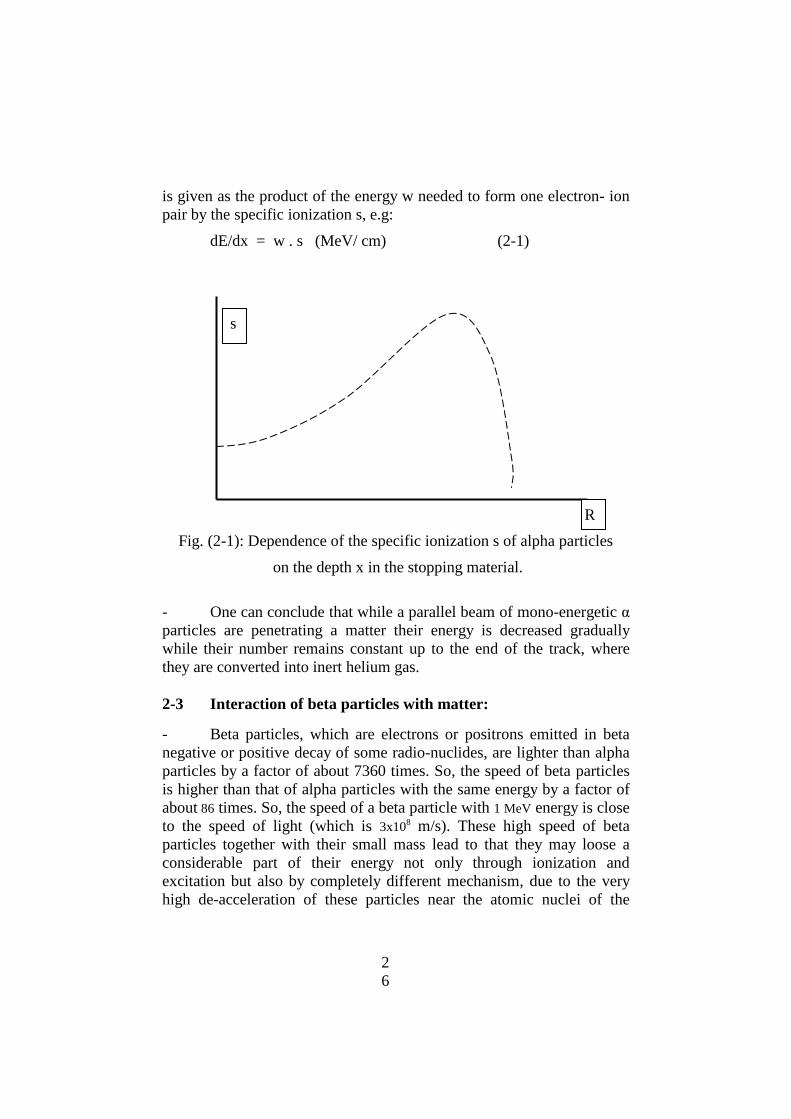
2
6
is given as the product of the energy w needed to form one electron- ion
pair by the specific ionization s, e.g:
dE/dx = w . s (MeV/ cm) (2-1)
Fig. (2-1): Dependence of the specific ionization s of alpha particles
on the depth x in the stopping material.
- One can conclude that while a parallel beam of mono-energetic α
particles are penetrating a matter their energy is decreased gradually
while their number remains constant up to the end of the track, where
they are converted into inert helium gas.
2-3 Interaction of beta particles with matter:
- Beta particles, which are electrons or positrons emitted in beta
negative or positive decay of some radio-nuclides, are lighter than alpha
particles by a factor of about 7360 times. So, the speed of beta particles
is higher than that of alpha particles with the same energy by a factor of
about 86 times. So, the speed of a beta particle with 1 MeV energy is close
to the speed of light (which is 3x10⁸ m/s). These high speed of beta
particles together with their small mass lead to that they may loose a
considerable part of their energy not only through ionization and
excitation but also by completely different mechanism, due to the very
high de-acceleration of these particles near the atomic nuclei of the
s
R
2
7
matter. This mechanism is the emission of electromagnetic radiation (x-
ray) known as bremstrahlung radiation.
- As the velocities of beta particles are very high comparing with
alpha particles with the same energies, the interaction time between the
incident beta particle and the orbital electrons and the nuclei of the
atoms is very small, in comparison with the interaction time of an alpha
particle. Moreover, the beta particle and orbital electrons are of the same
mass. So, all these factors strongly affect the character of interaction
between beta particles and matter. The main discrepancies between beta
and alpha interaction with matter can be summarized in the following:
- Beta particles transfer their energy to the matter via two
mechanisms which are: ionization and excitation, and emission of
bremstrahlung radiation. At comparatively low energy of particles
(few hundreds KeV) the main process for energy loss is the ionization
and excitation. As the energy of these particles increases the contribution
of emission of bremstrahlung radiation increasesd ant at very high
energies, this contribution becomes the predominant process of energy
loss. Moreover, the role of emission of bremstrahlung radiation is
strongly dependent on the atomic number Z of the matter, where it
increases with the increase of Z. For this reason high Z material should
not be used for shielding sources. The best material that can be used to
shield sources are the light solid material, such as plastic or aluminum
to reduce the emission of bremstrahlung radiation (x-ray).
- The energy percentage f of beta particles, which is lost via the
emission of bremstrahlung radiation as a function of both beta particles
maximum energy Emax and the atomic number Z is determined as:
f = 0.035 Emax Z %
- The track of any beta particle in the matter takes the form of a
broken line (due to the similar mass of the two interacting particles).
- The energy transferred from the incident beta particle to the
orbital electron in a single collision varies from a very low portion of the
particle energy up be very high portion of this energy, so that the
complete energy of the incident particle may be transferred in a single
collision. This means that the delta electrons are predominant in
interaction with matter.

2
8
Fig. (2-2): The broken track of particles in the material
- The specific ionization s in beta interaction is much less than that
for alpha interaction (by a factor of about one hundred due to the smaller
interaction time). So the range of beta particles is much larger than that
of alpha particles. The range of 1 MeV particles is about 4- 5 m in air, 6-
8 mm in water, plastic or human tissue, and about 2- 3 mm in aluminum.
- Both particles (e.g. the electron and the positron) behaves in
the matter in accordance with the previously mentioned two
mechanisms, although they have different sign of the charge. However,
there is an essential difference between the two particles at the end of the
track. When the energy of the positron becomes very low, it annihilates
with one of the electrons of the matter, where they completely vanishes
as a mass, and these two masses are converted into electromagnetic
energy in the form of two photons, each with energy of 511 KeV. This last
process is known as the annihilation process and the two photons with
511 KeV are called annihilation photons.
- It is important to conclude that while a parallel beam of β
particles are penetrating a matter, not only their energies are decreased
as a function of depth in the matter, but also their number will be
decreased, due to two facts which are: (a) the continuous energy
spectrum of β particles, so that low energy particles will loose their
energy through, relatively, a very thin layer of the matter while high
energy particles can penetrate to much higher depth, (b) a large number
of β particles will be deflected from their initial direction due the their
broken track.

2
9
- Due to the above mentioned factors, the number of β particles
which penetrate a certain thickness of matter x is decreased
exponentially, in accordance with the following (2-2) relation:
N = N0 e – μ x
(2-2)
where N is the number of particles penetrating the thickness x,
N0 is the number of particles reaching the same point in the absence of
the absorber, and μ is known as the attenuation factor. This factor is
strongly dependent on both atomic number Z of the absorber and energy
E of the particles.
2-4 Interaction of x-ray and gamma radiation with matter:
- When a beam of x-ray or mono-energetic gamma radiation fall
on a matter, its photons may interact with this matter via one of the
following mechanisms, depending on the photon energy as well as on
the atomic number of the matter:
a- The photo-electric effect,
b- Compton scattering, and
c- The pair production.
- Other types of interaction between incident photons and the
matter, such as the interaction with the atomic nuclei, is considered
negligible from the point of view of radiation protection.
2-4-1 The photo-electric effect:
- In this process, the incident photon interacts with one of the
strongly bound orbital electrons of the atom (e.g. with any of electrons
belonging mainly to K or L shells, which are the closest shells to the
nucleus). In this type of interaction the photon delivers its total energy
Eγ to the orbital electron and completely vanishes, and correspondingly,
the electron will be knocked out from the atom, carrying an amount of
energy Ee equal to:
Ee = Eγ – B (2-3)
where, B is the binding energy of the electron in the corresponding shell,
defined as the amount of energy that should be delivered to the electron
just enough to liberate it from this shell (it varies from less than 1 to
about 100 KeV depending on the atomic number Z of the matter). If Eγ <
B, then the process will not occur. Correspondingly, the photo-electric

3
0
effect will yield one electron which carries approximately the photon
energy.
- The cross- section σph (sigma) of the photo-electric effect, which
is defined as the probability of occurrence of this effect, when a single
photon is incident on a unit area (1 cm2) containing a single atom,
strongly depends on the photon energy Eγ as well as on the atomic
number of the matter Z. This probability σph decreases very fast with
increasing the photon energy Eγ, while it increases very rapidly with
increasing Z, as Z4 up to Z5. The unit of σph is barn(1 barn = 10
-24 cm
2).
- Dependence of the photo-electric cross section σph on photon
energy Eγ is shown 0n figure (2-3) where the photon energy is expressed
in a logarithmic scale.
K-edge
σph
ln Eγ
Fig: (2-3): Dependence of the photo-electric cross section on photon
energy
2-4-2 Compton scattering:
- In this process, the incident photon interacts with one of the
very loosely bound orbital electrons of the atom, or with a free
electron (e.g. with any of electrons belonging to the outermost shells,
which are far away from the nucleus). In this type of interaction the
photon delivers a part of its energy Eγ to the electron and the photon
well be deviated (scattered) from its original direction, carrying the
remaining amount of energy. Correspondingly, the Compton scattering

3
1
of a photon will yield a photon with lower energy and a free Compton
electron, that carries the remaining amount of energy.
σc
ln Eγ
Fig: (2-4): Dependence of the Compton cross section on photon energy
- the cross-section σc of Compton scattering decreases
approximately slowly with increasing of the photon energy, while it
depends linearly on Z of the matter.
2-4-3 The pair production:
- In this process, the incident photon interacts with the strong
electric field of the atomic nucleus, when approaching it very closely
(e.g. interaction between the incident photon and the atomic nucleus),
and if the photon energy is higher than 1022 KeV. In this type of
interaction the photon vanishes completely, and one electron-positron
pair with rest mass equivalent to 1022 KeV is produced. If the energy of
the incident photon Eγ is higher than 1022 KeV, then the excess energy is
delivered to the produced electron and positron, in approximately equal
portions. Correspondingly, the pair production will yield two particles
which are the electron and the positron.
- The electron and the positrons behave inside the stopping matter
in the same way as beta particles, e.g. they loose there energy on
ionization and excitation of the atoms of this matter as will as on
emission of bremstrahlung radiation, depending on the atomic number of
the atoms of the absorbing matter. When its energy becomes very low
each positron annihilates with one of the orbital electrons, (e.g. this
positron and electron vanish as a mass converting into two photons, each

3
2
with energy of 511 KeV). These two photons may interact with matter via
photo-electric process or Compton scattering, or they both may escape
out from the matter without interaction, in a process known as a double
escape, or one photon may interact while the other may escape in a
process known as a single escape.
- The cross-section σp of the pair production process increases with
the photon energy increase. This increase is relatively slow after the
threshold value of 1022 KeV and becomes fast with increasing the energy.
This probability σp depends on the atomic number of the matter as Z2.
σp
1022 KeV ln E γ
Fig: (2-5): Dependence of the pair production cross section on photon
energy
- Due to the formation of energetic electrons and positrons,
resulting from the three processes of interaction between gamma
radiation or x-rays and the matter this radiation, is known as indirectly
ionizing radiation.
2-4-4 The total gamma cross section σ:
- The total gamma cross-section σ is defined as the total
probability for a single incident photon to interact with one atom
existing in a target of 1 cm2 when it collide this area via any of the three
processes, e.g:
σ = σph + σc + σp

3
3
- The unit of the total cross section σ is the barn (1 barn = 10-24
cm2).
2-4-5 The linear attenuation coefficient μ:
- By definition, the linear attenuation coefficient μ for a certain
matter and at a certain photon energy, is defined as the probability of the
interaction of a single photon that have this energy with all atoms
existing in a cube of 1 cm3 (1 cm
2 area and 1 cm depth) of this matter, on
which it falls by all the three processes. So, if the number of atoms in 1
cm3 is n, and the total interaction cross-section is σ, then it is clear that:
μ = n σ
σ
1022 KeV ln Eγ
Fig: (2-6): Dependence of the total cross section on photon energy
- The unit of the linear attenuation coefficient μ is cm-1
(e.g. per
cm). It is also clear from the behavior of σ as a function of the energy
that μ depends strongly on the atomic number Z of the attenuating
material, specially for both low and high energy photons. Moreover, μ is
strongly dependent on the photon energy Eγ.
2-4-6 The mass attenuation coefficient μm:
- In different references another physical quantity, known as the
mass attenuation coefficient μm is used instead of the linear attenuation
3
4
coefficient μ. This new quantity μm is defined by dividing the linear
attenuation coefficient μ by the density ρ of the attenuator, e.g:
m = μ / ρ
- It is seen that the unit of the mass attenuation coefficient μm is
(cm2/ gm). The reason for using μm instead of μ is that its value may be
considered, approximately, constant for different attenuating materials,
for the same photon energy.
2-4-7 The exponential attenuation of x and gamma radiation:
When a narrow beam of mono-energetic x-ray or gamma
radiation falls on a matter of thickness x cm, a part of the incident
number of photons No from this beam will interact with the matter via
any of the three known processes, resulting in the reduction of this
incident number as a function of the thickness x of the matter. Number
of the photons N, that will penetrate the thickness x without any
interaction with the matter will proceed in the same direction and do not
loose any part of their energies. This is expressed, mathematically, by
the following exponential law:
N = No e - μ x
- The exponential attenuation (e.g. exponential reduction of the
number of photons) is valid when specific conditions are applied. These
conditions are:
a) A very narrow beam consisting of parallel mono-
energetic photons.
b) A very small thickness x of the attenuator, so that,
multiple Compton scattering is negligible.
- In all other cases this exponential law is not valid due to
Compton scattering of photons from the broad beam as well as the
multiple Compton scattering of some photons due to the thick layer of
the attenuator. This will be discussed, in details, in a later chapter on
build-up.
- If the linear attenuation coefficient μ is used (in cm-1
) then the
thickness x of the attenuator should be expressed in (cm), to get non-
dimensional value of the product μ x. However, when the mass
attenuation coefficient μm is used (in cm2/gm), then the thickness of the
attenuator should be expressed in the so called mass-thickness xm, which

3
5
is obtained as the product of the linear thickness x of the attenuator and
its density ρ, e.g:
xm = x ρ
The unit of the mass-thickness xm is (gram/cm2).
- The exponential attenuation of x-rays and gamma radiation
makes the concept of the range for this type of electromagnetic radiation
is not valid. A definite portion of the incident beam will penetrate
through the attenuating matter, even when its thickness is too large. For
example, if a Co-60 source is shielded (surrounded) by more than 2 m
thick concrete wall some emitted photons from this cobalt will penetrate
through this shield, without suffering any kind of interaction.
2-4-8 The half value layer (HVL):
- The half value layer (HVL), or half value thickness, of a matter
at a certain gamma energy, is defined as the thickness of that matter,
which is necessary to attenuate the original number of the incident
photons No, with this energy, to its half value ( e.g. to N = 1/2 No). The
HVL is related with the linear attenuation coefficient μ with the
following simple relation:
HVL = 0.693 / μ
- Since μ is dependent on the radiation energy E and the material
of the attenuator Z, the HVL is also dependent on these factors.
- The unit of the HVL is cm when the μ is expressed in cm-1
, and
its unit is (gm/ cm2), when μ is expressed in cm
2/ gm.
2-4-9 The tenth value layer (TVL):
- The Tenth value layer (TVL), or Tenth value thickness, of a
matter at a certain gamma energy, is defined as the thickness of that
matter, which is necessary to attenuate the original number of the
incident photons No, with this energy, to one tenth of this value ( e.g. to
N = 1/10 No). The TVL has the same units as the HVL, and it is related
with last value with the following relation:
TVL = 3.32 HVL
3
6
2-4-9 The energy absorption coefficient μa:
- The energy absorption coefficient represents the portion of
energy absorbed from x-ray or gamma radiation in a definite volume of
the matter. This coefficient is used to account for the so called "kerma"
or absorbed dose from x or gamma radiation into the interacting matter,
(e.g. in dose calculations). It should be mentioned that authors of some
references are using, by fault, this coefficient to express the attenuation
coefficient μ. These Two coefficient (μa and μ, both linear and mass)
have different values, specially at medium and high photon energies, and
should not replace each other, except at very low photon energies (less
than few hundreds of KeV) where they are very close to each other.
- The reason of the discrepancy between μa and μ is the Compton
scattering and the pair production. In Compton scattering the photon is
deviated from its original direction, transferring only undefined part of
its energy to the matter, and the scattered photon may escape out from
this matter, so that although it has been omitted out from the beam, it
does not transfer its complete energy to the matter. In the pair production
the energy may not be transferred completely to the matter, since one or
even the two photons, resulting from the annihilation of the positron
with one electron may escape out of the matter.
- Due to the above mentioned reasons μ is almost higher than μa ,
specially with increasing the photon energy
2-5 Interaction of the neutrons with the matter:
- Since the neutrons are neutral particles (e.g. uncharged particles),
they do not interact neither with any of the orbital electrons nor electro-
statically with the atomic nuclei. They may interact only with nuclei via
nuclear forces, when they very closely approach any of them. This is the
reason of the high penetrating power of neutrons in the matter.
- the most important and efficient mean for energy transfer from
neutrons to the matter is the elastic scattering of the neutron on light
nuclei, such as hydrogen (in wax, water, polyethylene, or plastic),
deuterium (in heavy water) beryllium, carbon, and oxygen. With
decreasing the mass number of the interacting nucleus, the average
energy, transferred from the neutron to this nucleus, in a single collision,
increases. For this reason the hydrogen nuclei are considered the best
moderator for neutrons, and the materials which contain high
3
7
concentration of hydrogen, such as wax, water, Polyethylene, and plastic
are extensively used for effective slowing down of the fast neutrons. In a
single collision with a hydrogen nucleus, the neutron loses, in average,
63 % of its energy. This portion of energy is transferred to a proton,
which is the hydrogen nucleus.
- Since the recoil protons are heavy charged particles, they ionize
the matter. So, the neutrons are considered as indirectly ionizing
particles.
2-5-1 The neutron moderation:
- The neutron moderation means the slowing down of fast
neutrons (e.g. decreasing their energies from the MeV range to about
0.025 eV. Neutrons with such low energies are called thermal neutrons,
since their motion is controlled by the prevailing temperature.
- For slowing down of the fast neutrons (with energy of about
several MeV) to thermal neutrons, these neutrons should be subjected, in
average, to about 18-19 collisions with hydrogen nuclei. This number of
collisions requires a thickness of a hydrogen rich material, such as wax
or water of about 15- 25 cm.
- The thickness of the wax or water may be increased over the
mentioned values for radiation protection purposes, since these materials
absorb thermal neutrons with a certain probability forming deuterium
atoms which are stable.
- The role of inelastic scattering of neutrons for neutron
moderation is negligible.
2-5-2 The neutron capture:
- when a neutron approach very closely to a nucleus it may be
captured in it, forming a new isotope of the same element, with the
emission of a prompt gamma photon. An example of the neutron capture
reaction is:
no
1 + Cd114
47 Cd115
47 + γ
- The probability of the neutron capture is strongly dependent on
the neutron energy. The reaction cross-section (which represents the
probability of the neutron capture) increases strongly with the decrease
of the energy, reaching very high values for thermal and slow neutrons
3
8
(the slow neutrons are those with energies just higher than that of
thermal neutrons). Moreover, at certain energy values for the slow and
thermal neutrons, and for some nuclides the probability of the neutron
capture reaches very high values, known as a resonance neutron capture
or absorption. The energy values at which the resonance neutron capture
occurs depend on the absorbing nuclide. For example for Cd114
47 , it has
been found that the resonance capture occurs at thermal and low
energies, and the capture probability at resonance reaches extremely
high values. For this reason Cd114
47 is considered one of the best absorber
for thermal and slow neutrons.
- One of the most effective method to shield a neutron source and
to reduce effective doses around it is to put three layers of different
materials in the following consequence from the source: a) About 20 cm
of wax, plastic or any other solid (or liquid) material, rich with hydrogen
content to moderate fast neutron and convert them into thermal or slow
neutrons, then b) A thin sheet of Cd114
47 (with about 1 mm thickness) to
absorb thermal and slow neutrons, and finally c) a certain thickness of
lead to attenuate the prompt gamma radiation emitted in the neutron
capture in Cd114
47 .
- There are other materials that can be used practically to reduce
the neutron doses arising from different neutron sources, by moderation
and absorption of these neutrons, such as water (normal or light water),
boron and others
- In the absence of all of the mentioned materials one can use other
commonly existing materials in the field, such as the sand and other
types of soil. Although their shielding properties is too limited in
comparison with other materials, a large thickness of these sand or soil
may reduce neutron doses to lesser values due to the presence of some
light elements such as oxygen and carbon.

3
9
CHAPTER 3
RADIATION DETECTORS, SURVEY METERS
AND CONTAMINATION MONITORS
3-1 General:
- The main two processes which are used for detection of different
types of ionizing radiation are based on the use of:
a) Ionization of the detector material and formation of
electron-ion pairs, or electron hole pairs, and collection of this
charges or their current.
b) Excitation of the detector material and then measurement
of the emitted light during the de-excitation process, and
collection of this light or their current.
- There are other processes, which are used for detection and
counting of ionizing radiation. For example, one of these processes is the
use of activation of a certain nuclides by irradiation of certain material
by neutrons and then by measurement of the induced activity due to the
neutron capture.
- The type of the detector that should be used for detection and
counting and identifying of ionizing radiation depends strongly on:
a) The type of the radiation (e.g. heavy or light charged particles,
neutrons, x, or gamma radiation.
b) The energy of the measured particles or photons.
c) The intensity of the radiation field (e.g. the particle or photon
flounce).
d) The purpose of detection and measurement.
3-2 The gas detectors:
- In all gas detectors, detection of directly and indirectly ionizing
radiation is done through the ionization of some mixture of a gas
contained in a vessel with certain shape and volume.
- For directly ionizing radiation, such as heavy charged particles or
beta particles, the ionization of the gas atoms or molecules occurs inside
4
0
the detector vessel. The average number of the resulting primary
electron-ion pairs in the detector is defined by dividing the particle
energy (in eV) by 34 eV, which is the average energy needed to form
one electron- ion pair. For detection of heavy charged particles (such as
alpha), the detector wall should be equipped with a very thin window of
low Z material (less than 40 gm/cm2 of a light material) to permit the
entrance of these particles inside the detector, without loosing a
considerable part of its energy in this window. For the detection of beta
particles the window can be done from a thicker material, since the
range of these particles is much higher than that of alpha particles.
- For the indirectly ionizing radiation, namely x and gamma
radiation, ionization of the detector’s gas is done by the primary charged
electrons and positrons, emitted as a result of the interaction of the
incident photons with a very thin layer of a heavy material, such as lead,
fixed inside the wall of the detector. For detection of x and gamma
photons, There is no need to make a window in the detector wall due to
the very large range of photons.
- For neutrons, which are indirectly ionizing radiation too, the
ionization is done by charged particles such as protons emitted as a
result of the elastic scattering of the incident fast neutrons with hydrogen
nuclei existing in a very thin layer of polyethylene fixed inside the
detector wall, or by alpha particles, which are emitted as a result of the
neutron capture of thermal neutrons in certain gas materials with high
reaction cross-section, which is filling the detector, such as BF3 gas
(Boron tri-Fluoride) or others. Due to the high penetrability of neutrons,
there is no need to make any window in neutron detectors.
- There are three types of gas detectors which are:
a) the ionization chamber,
b) the proportional counter, and
c) the Geiger- Muller (GM) counter.
- For all types of gas detectors, the intrinsic detection efficiency
is 100 % only for all heavy charged particles. For beta particles the
efficiency is slightly less than 100 %, due to their continuous energy
spectrum, so that a part of the low energy particles will be absorbed
inside the window thickness. The efficiency of all gas detectors for
measuring photons or neutrons is extremely low, and strongly dependent
on their energy. For example the intrinsic efficiency of these detectors
4
1
for photons may vary from few percents (2-4 %) to very low values (less
by many orders of magnitude) with increasing the energy of photons.
Remark: the intrinsic efficiency of a detector, for a certain type of
indirectly ionizing radiation at a certain energy, is defined as the ratio of
the number of particles or photons with the mentioned energy detected
by the detector from a given source, in a certain time period to the total
number of these particles or photons, with the same energy, incident
from the source on the detector surface, during the same time period. To
get the efficiency in percent this ratio should be multiplied by 100. For
example, if the intrinsic detector efficiency for photons with 662 KeV
energy is 2.5 % then this detector will detect only 2.5 % of photons
incident on its sensitive surface with this energy.
3-2-1 The ionization chamber:
- It is a detection device (see fig. (3-1), which consists of::
a- Two electrodes (anode a and cathode c) connected to a
moderate potential difference V (about 50- 100 volts depending
on the chamber volume and pressure) to secure collection of the
majority of the electrons and ions, which are generated by the
ionizing radiation inside the chamber on the anode and the
cathode respectively.
b- A guard grid g between the anode and the cathode to
secure independency the collected current, or consequently
voltage of the output pulse signal, resulting due to the passage of
this current through a high Ohmic resistance R, on the track
position of the incident particle.
- The ionization chambers can be used in a current regime (e.g. to
measure the very small average electric current, resulting by ionization
by a large number of incident particles or photons, and the chamber is
then known as a current type ionization chamber. They, also, can be
used to measure consequence pulses resulting from individual ionization
events (particles or photons), and hence to determine the number and
energies of these particles or photons, and in this case the chamber is
known as a pulse type ionization chamber.
- Since the collected current in the ionization chamber is too low
(in the range of pico-Ampers), the ionization chamber should be

4
2
connected with a direct current amplifier (or pulse height amplifier) with
a very high amplification gain (thousands or more).
a C
g c V R
Fig (3-1): A diagram of an ionization chamber
- Ionization chambers are characterized by certain characteristics.
Some of these characteristics are:
a) The multiplication gain of any chamber equals 1, which
means that there is no multiplication of the electric current
resulting by ionizing radiation.
b) Relatively, high energy resolution r, which means that it
can be used to differentiate between particles or photons with
relatively close energies. The energy resolution of the ionization
chambers r varies between about 2.5 and 7 %, depending on its
volume and on the gas pressure.
Remark: the energy resolution r is defined as the ratio of the
energy fluctuation E caused by the detection process, to the
energy E of the particle multiplied by 100 (to get it as a percent)
e.g:
r = (E/E)x100 %.

4
3
c) Relatively, a constant energy response curve in a wide
range of energies, comparing with all other detectors, when the
chamber is used as a detector in dose or dose-rate survey meters.
A constant energy response means that the ratio of the
measured dose (or electric current) from ionizing radiation with a
given energy E to that at a reference one Er remains constant in a
wide range of energies when the radiation field is homogeneous.
This is a very important property of ionization chambers.
d) In some cases the wall of the chamber is made from a
material having a similar composition as air to correct for energy
absorption in different materials, for more accurate determination
of doses or dose rates. In these cases the chamber is known as
air-wall ionization chamber.
e) For measurement of relatively high energy beta particle
or photons, it is necessary to increase the gas pressure inside the
chamber to secure full stopping of the ionizing beta particles
within it. In This case the chamber is known as a pressurized
ionization chamber. Such cambers are important for dose
measurements in a radiation field with a wide energy range.
- The shape of the output pulse from a pulse type ionization
chamber, which represents the detection of a single particle or
photon with a given energy value is demonstrated in fig.(3-2).
The polarity of th pulse on this figure is inverted, since it is
originally negative. The vertical axis shows the output voltage
amplitude of the pulse which is proportional to the energy of the
particle or photon, while the horizontal axis shows the time
duration of the pulse and dependence of its amplitude on time.
The voltage amplitude of the output pulses lies in the range of
less than one microvolt up to about one hundred microvolts,
depending on the particle energy. The pulse durations lies
between less than a 100 microseconds up to more than 1000
microseconds depending on the geometrical dimensions of the
chamber as well as on its internal capacitance and resistance. The
values of the used electronic devises such as the input impedance
and capacitance of the of this circuit strongly affect the duration
of the output pulses

4
4
The pulse amplitude
The time (microsecond)
Fig (3-2): The pulse shape at the output of an pulse type
ionization chamber
3-2-2 The proportional counter:
- The proportional counter, (see fig 3-3) is a gas detector of a
cylindrical form, where a metallic cylinder is acting as the detector
cathode, while a very thin coaxial metallic wire with a regular diameter
is used as the anode.
- The applied voltage difference between the anode and the
cathode for the proportional counter is much higher than that used in an
ionization chamber with the same dimensions. This increase in the
applied voltage difference leads to the acceleration of ions and electrons,
so that they become capable to ionize new atoms, while they are moving
to the cathode and anode respectively. This yields in a high increase of
the electric current caused by ionizing radiations. So, the proportional
counter is acting as a detector and a current multiplier.
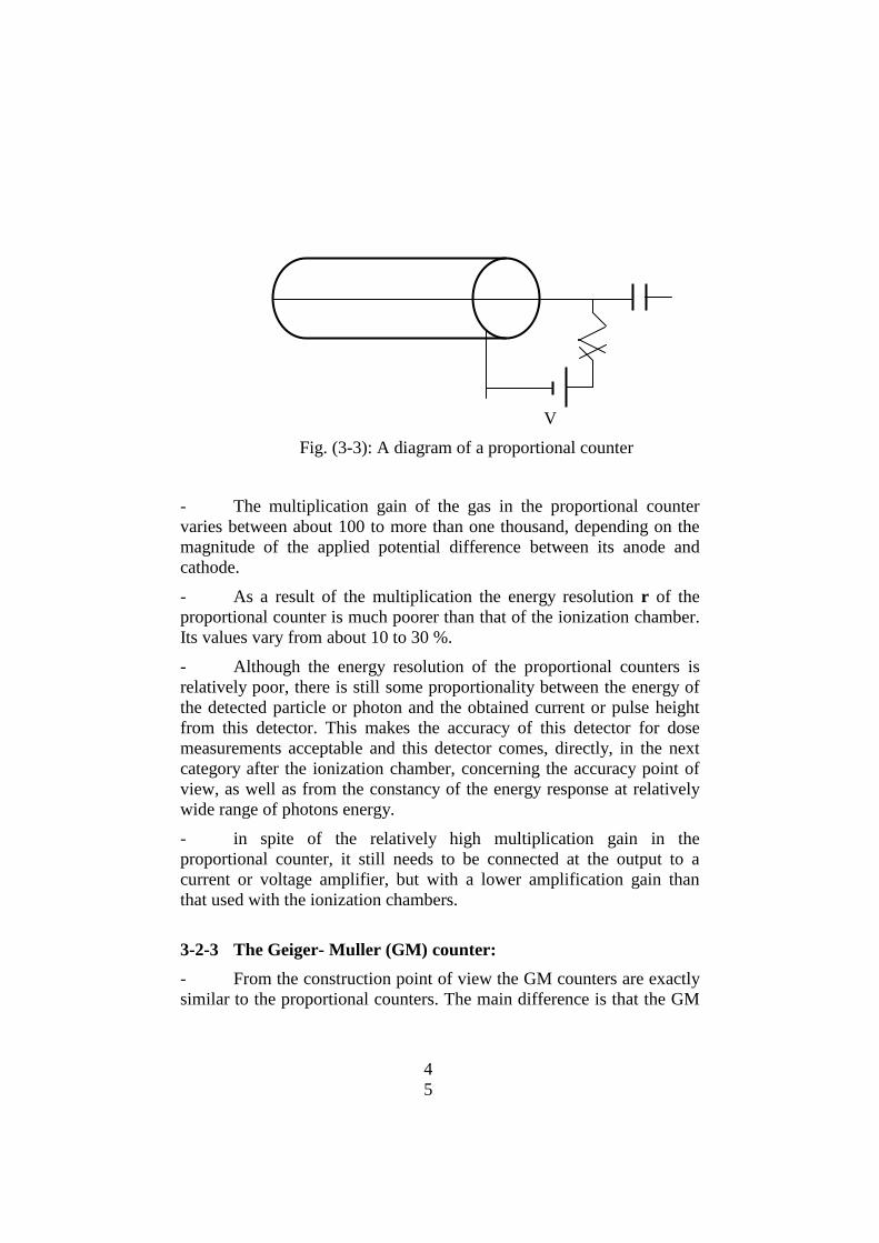
4
5
V
Fig. (3-3): A diagram of a proportional counter
- The multiplication gain of the gas in the proportional counter
varies between about 100 to more than one thousand, depending on the
magnitude of the applied potential difference between its anode and
cathode.
- As a result of the multiplication the energy resolution r of the
proportional counter is much poorer than that of the ionization chamber.
Its values vary from about 10 to 30 %.
- Although the energy resolution of the proportional counters is
relatively poor, there is still some proportionality between the energy of
the detected particle or photon and the obtained current or pulse height
from this detector. This makes the accuracy of this detector for dose
measurements acceptable and this detector comes, directly, in the next
category after the ionization chamber, concerning the accuracy point of
view, as well as from the constancy of the energy response at relatively
wide range of photons energy.
- in spite of the relatively high multiplication gain in the
proportional counter, it still needs to be connected at the output to a
current or voltage amplifier, but with a lower amplification gain than
that used with the ionization chambers.
3-2-3 The Geiger- Muller (GM) counter:
- From the construction point of view the GM counters are exactly
similar to the proportional counters. The main difference is that the GM
4
6
counter is operated at relatively higher potential difference between the
anode and the cathode.
- With increasing the applied voltage the current multiplication in
the gas of the tube becomes very high and almost reaches infinity. When
an ionizing particle or photon inters the GM tube, and when it interacts
with the detector material causing even one electron– ion pair a series of
consequent ionization occurs making avalanche multiplication. This will
cause occurring of electric discharge of the detector gas.
- The gas discharge will continue unless, it will be stopped by
internal or external reason in a process called quenching. The external
quenching is secured by inserting a large Ohmic resistance R in series
with the high voltage source, while the internal quenching is secured by
the addition of a certain ratio of a mono-atomic gas. The second
technique of quenching is preferred, since the first one leads to a serious
increase in the detector dead time, due to the increase of the magnitude
of the resistance.
- As a result of infinite amplification of the GM tubes, particles or
photons with different energies will give the same electronic signals with
the same pulse amplitude, so that, it can be measured without further
amplification.
- Due to the complete discharge through the detector tube, the
proportionality between the energy of the particle and the pulse height of
corresponding signal is completely lost. In other words the GM counter,
completely, does not differentiate between different energies, and it can
be only used to count the number of pulses (detected particles or
photons) independent of their energies.
- The dead time of a pulse type detector is defined as the time
period through which the electrons and ions are collected and treated as
a pulse. During the dead time the detector will not detect any other
ionization event, so If the time separation between two sequent ionizing
events (e.g. two consequent registered particles or photons) is less than
the detector dead time, then they will be detected as a single particle or
photon, and hence there will be some loss of the detected number of
particles or photons.
- The energy response curve of the GM counter is, comparatively,
worse than that of the proportional counter. For this reason, special
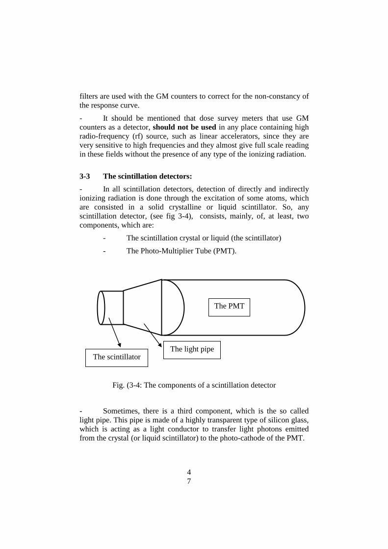
4
7
filters are used with the GM counters to correct for the non-constancy of
the response curve.
- It should be mentioned that dose survey meters that use GM
counters as a detector, should not be used in any place containing high
radio-frequency (rf) source, such as linear accelerators, since they are
very sensitive to high frequencies and they almost give full scale reading
in these fields without the presence of any type of the ionizing radiation.
3-3 The scintillation detectors:
- In all scintillation detectors, detection of directly and indirectly
ionizing radiation is done through the excitation of some atoms, which
are consisted in a solid crystalline or liquid scintillator. So, any
scintillation detector, (see fig 3-4), consists, mainly, of, at least, two
components, which are:
- The scintillation crystal or liquid (the scintillator)
- The Photo-Multiplier Tube (PMT).
Fig. (3-4: The components of a scintillation detector
- Sometimes, there is a third component, which is the so called
light pipe. This pipe is made of a highly transparent type of silicon glass,
which is acting as a light conductor to transfer light photons emitted
from the crystal (or liquid scintillator) to the photo-cathode of the PMT.
The PMT
The light pipe The scintillator

4
8
- All the components are matched together, without any air voids
or bubbles by putting a small drop of silicon oil between any of these
components and pressing so that no air bubbles are existing in between.
The detector components are enclosed inside a hermetically sealed
metallic enclosure, so that no light can penetrate through it.
- The function of the scintillator is to emit photons of visible light,
The number of these photons is linearly dependent on the energy of the
incident particle. As these emitted photons fall on the photo-cathode of
the PMT, a limited number of electrons will be emitted from this photo-
cathode. The number of these photo-electrons is linearly dependent on
the number of the incident photons on the photo-cathode, and
consequently, on the energy of the incident particle on the scintillator.
- The role of the photo-multiplier tube (PMT) is to multiply the
number of emitted electrons from the photo-cathode, by a very large
factor (at least some thousands times and much more). For this purpose
the PMT contains a large number of dynodes (about 9- 13 dynodes),
each of which is covered with a material with high coefficient of the
secondary emission. The emitted photo-electrons are accelerated toward
the first dynode by a positive voltage difference V, so that they gain an
amount of kinetic energy equal V electron volts, and become capable to
induce secondary electron emission from the dynode, so that their
number will be multiplied by a factor equal to the coefficient of
secondary emission . This coefficient is strongly dependent on the
voltage difference V and may reach, relatively, high values (up to 3 and
more) with the increase of V. Electrons emitted from the first dynode
are, again, accelerated toward the second dynode by another positive
voltage difference V, giving rise to another step of a secondary emission
from this second dynode, and yielding second multiplication . Then the
consequent acceleration processes toward the next dynodes with a
multiplication factors of on each one of these dynodes will yield a total
multiplication factor of n (if the value of is the same for all dynodes),
where n is the number of dynodes in the PMT. After multiplication a
huge number of electrons are emitted from the last dynode and they are
collected on the anode of the PMT, giving a negative pulse on the output
of this anode due to the presence of a high ohmic resistance.
- The anode pulse represents the registration of a single particle in
the detector, and the amplitude of this pulse is proportional to the energy
of the particle. So, the number of the registered pulses is proportional to

4
9
the number of the incident particles or photons, while the amplitude of
each pulse represents the energy of the registered particle or photon.
Output pulses on the anode of the PMT have a similar form of the pulses
from an ionization chamber shown on fig. (3-2), but the time duration of
the pulse may be more less than that of the ionization chamber for some
types of scintillation crystals.
- It should be mentioned that the electron multiplication gain M of
the PMT, (which is approximately equal to the coefficient raised to the
power n (i.e. M n))
is strongly dependent on the biasing voltage V
which is supplied to the PMT Anode or cathode. This voltage is divided
by a potential divider using a set of resistances to bias the cathode, all
dynodes and the anode with the nominal voltages. It is recommended to
supply the PMT with the nominal voltage, since the increase of V will
increase the factor M, but at the same time it will shorten, strongly, the
service life-time of the PMT.
- different types of radiations are detected using different
scintillators. Table (3-1) represents the most widely used scintillators for
different types of radiations. All these scintillators emit violet light with
wave length shown in table (3-1).
- Alpha particles and protons can be easily detected using a thin
layer (about 1mm thickness) zinc sulphide crystal doped with silver ZnS
(Ag), while electrons and positrons can be detected using organic
crystals or liquids.
- The Sodium Iodide crystal with Thallium NaI(Tl) is the best
scintillation crystal that can be used to detect gamma radiation with a
higher efficiency, due to its high density. Moreover, the addition of a
small ratio of Thallium to the Sodium iodide makes the crystal capable
for emission of light photons at room temperature. To meat the required
detection efficiency of gamma radiation, the NaI(Tl) crystal is grown
with a different thicknesses. These crystals are available in the market,
mainly, in a cylindrical form with dimensions ranging from 1/2 inch
diameter x 1/2 inch height, up to more than 10 "
x 10 ". Generally
speaking, the scintillation gamma detectors are much sensitive to detect
gamma radiation, in comparison with gas detectors, and the detector
with 3" x 3
" NaI(Tl) crystal is considered as a reference one, so that, the
relative efficiency of any other gas and solid detectors, is given referring
to this reference one.
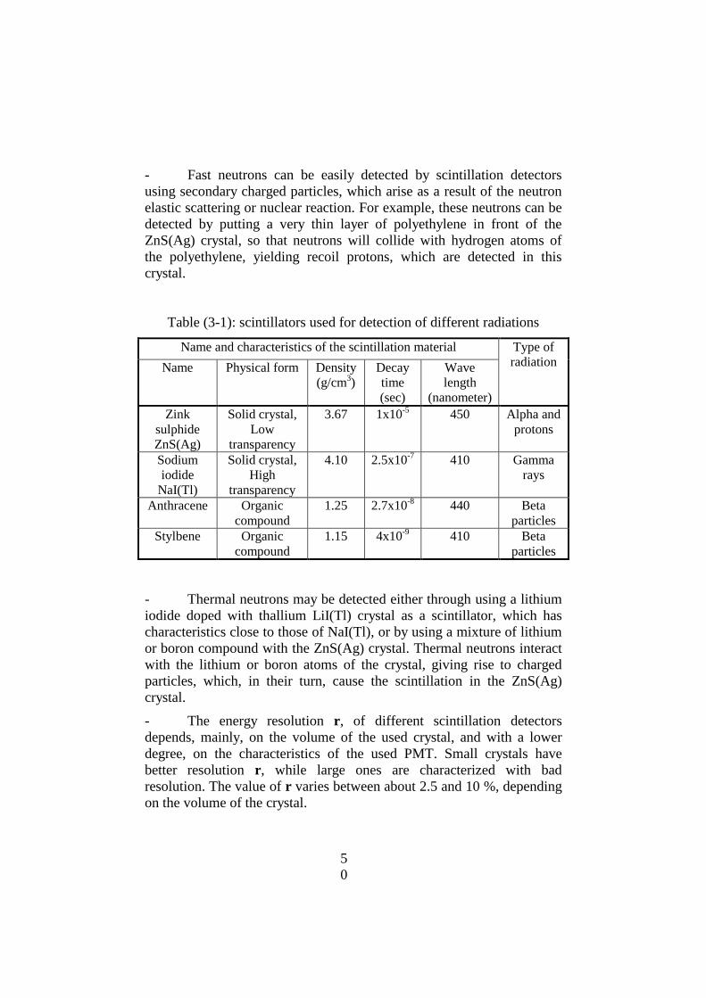
5
0
- Fast neutrons can be easily detected by scintillation detectors
using secondary charged particles, which arise as a result of the neutron
elastic scattering or nuclear reaction. For example, these neutrons can be
detected by putting a very thin layer of polyethylene in front of the
ZnS(Ag) crystal, so that neutrons will collide with hydrogen atoms of
the polyethylene, yielding recoil protons, which are detected in this
crystal.
Table (3-1): scintillators used for detection of different radiations
Name and characteristics of the scintillation material Type of
radiation Name Physical form Density
(g/cm3)
Decay
time
(sec)
Wave
length
(nanometer)
Zink
sulphide
ZnS(Ag)
Solid crystal,
Low
transparency
3.67 1x10⁻⁵
450 Alpha and
protons
Sodium
iodide
NaI(Tl)
Solid crystal,
High
transparency
4.10 2.5x10⁻⁷
410 Gamma
rays
Anthracene Organic
compound
1.25 2.7x10⁻⁸
440 Beta
particles
Stylbene Organic
compound
1.15 4x10⁻⁹
410 Beta
particles
- Thermal neutrons may be detected either through using a lithium
iodide doped with thallium LiI(Tl) crystal as a scintillator, which has
characteristics close to those of NaI(Tl), or by using a mixture of lithium
or boron compound with the ZnS(Ag) crystal. Thermal neutrons interact
with the lithium or boron atoms of the crystal, giving rise to charged
particles, which, in their turn, cause the scintillation in the ZnS(Ag)
crystal.
- The energy resolution r, of different scintillation detectors
depends, mainly, on the volume of the used crystal, and with a lower
degree, on the characteristics of the used PMT. Small crystals have
better resolution r, while large ones are characterized with bad
resolution. The value of r varies between about 2.5 and 10 %, depending
on the volume of the crystal.

5
1
- However the efficiency of the scintillation detectors for x and
gamma radiations is much higher than that of all gas detectors, its
response curve to dose variation with radiation energy is very poor,
comparing with all other detectors. For this reason scintillation detectors
are not widely used in different survey meters, for dose or dose-rates
measurements or.for radiation dosimetry, but they are very widely
used to search for a lost gamma source as will as for radiation
counting and spectroscopic measurements as well as in surveying
ground resources of nuclear ores.
3-4 The Semi-conductor detectors:
- The semi-conductor materials used in manufacturing electronic
devices and radiation detectors are the silicon and germanium. Both of
these elements have tetravalent atoms, and their crystalline structure is
formed, so that, each atom has a covalent bond with four neighbor
atoms. When radiation interacts with one of these atoms an electron of
the four outermost electrons is ejected, and it becomes free, and then its
atom is left without an electron, This free of electron place is known as a
hole. So, while interacting with a silicon or germanium crystal radiation
will generate electron-hole pairs. The energy required to form one
electron-hole pair in silicon is about 1.1 eV , in average, while the
energy required for germanium is about 0.7 eV. For this reason, the
number of electron-hole pairs formed in silicon by a particle or photon
with certain energy is higher than the number of electron-ion pairs
formed in an air ionization chamber by a factor of about 30 times for
silicon and of about 48 times for germanium. As a result of that, the
energy resolution of semiconductor detectors is much better than that of
the ionization chamber. For example, the energy resolution r for a
germanium gamma detector with a cylindrical crystal of about 60 mm
diameter and 60 mm height is about 1.75 KeV for the 1332 KeV gamma
ray line of Co-60, (which is about 0.13 %).
- At present, hyper-pure germanium crystals of different shapes
and volumes are produced for use as a powerful tool for high resolution
gamma ray spectrometry in the fields as well as in the fixed laboratories,
to meat the required efficiencies. Their relative efficiency cover a very
wide range starting from about 10 % up to more than 100 % with respect
to the 3" x 3" NaI(Tl) detector. The only disadvantage of these detectors
is that they required a very deep cooling, prior to their operation, and

5
2
this is achieved, mainly, through cooling by liquid nitrogen (- 189 oC) or
by electric cooling.
- Silicon surface-barrier detectors are produced, since the sixties of
the 20th
century up to now, with different shapes and thicknesses, to
detect heavy charged particles of different energies, and they are widely
used in high resolution alpha spectrometry, as well as in spectroscopic
measurements of heavy charged particles (such as protons, deuterons
and others). Their energy resolution is as good as about 0.4- 0.5 %, and
they do not require any cooling.
- Other pure silicon crystals are produced to be used for high
energy resolution spectroscopic measurements of x-ray and low energy
gamma radiation up to about 100 KeV. These detectors, again. require
the deep cooling as germanium ones prior to their operation.
3-5 The survey meters:
- Surveying the radiation areas and measurements of radiation
doses and dose rates is one of the required activities that must be
conducted, regularly, in all areas, where radiation sources are used, and
around these areas, to evaluate the radiation levels and, consequently to
assess the radiation doses to the occupational workers as well as to the
general public.
- There is no single survey meter, which can be used to survey all
types of radiations, and the choice of the survey meter is strongly
dependent on the type of radiations or particles, their energy, as well as
on their intensity.
- Any survey meter consists, mainly, of:
a- A radiation probe or detector, which is assigned for a
certain type of radiation, and for a certain range of energy, as
well as for a certain range of radiation intensities ore doserates,
b- An electronic circuit for current or voltage amplification.
c- A measuring device to measure the amplified electric
current or to count the pulse rate or the number of pulses during a
defined time interval.
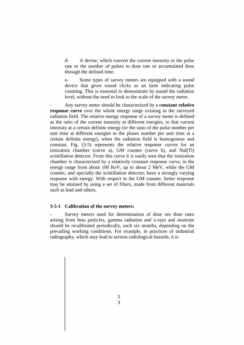
5
3
d- A devise, which convert the current intensity or the pulse
rate or the number of pulses to dose rate or accumulated dose
through the defined time.
e- Some types of survey meters are equipped with a sound
device that gives sound clicks as an larm indicating pulse
counting. This is essential to demonstrate by sound the radiation
level, without the need to look to the scale of the survey meter.
- Any survey meter should be characterized by a constant relative
response curve over the whole energy range existing in the surveyed
radiation field. The relative energy response of a survey meter is defined
as the ratio of the current intensity at different energies, to that current
intensity at a certain definite energy (or the ratio of the pulse number per
unit time at different energies to the pluses number per unit time at a
certain definite energy), when the radiation field is homogenous and
constant. Fig. (3-5) represents the relative response curves for an
ionization chamber (curve a), GM counter (curve b), and NaI(Tl)
scintillation detector. From this curve it is easily seen that the ionization
chamber is characterized by a relatively constant response curve, in the
energy range from about 100 KeV, up to about 2 MeV, while the GM
counter, and specially the scintillation detector, have a strongly varying
response with energy. With respect to the GM counter, better response
may be attained by using a set of filters, made from different materials
such as lead and others.
3-5-1 Calibration of the survey meters:
- Survey meters used for determination of dose ore dose rates
arising from beta particles, gamma radiation and x-rays and neutrons
should be recalibrated periodically, each six months, depending on the
prevailing working conditions. For example, in practices of industrial
radiography, which may lead to serious radiological hazards, it is
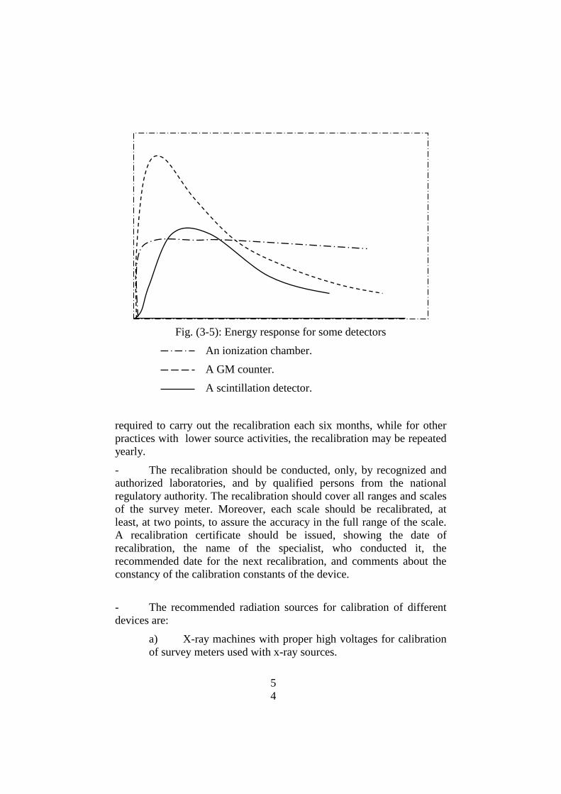
5
4
Fig. (3-5): Energy response for some detectors
An ionization chamber.
A GM counter.
A scintillation detector.
required to carry out the recalibration each six months, while for other
practices with lower source activities, the recalibration may be repeated
yearly.
- The recalibration should be conducted, only, by recognized and
authorized laboratories, and by qualified persons from the national
regulatory authority. The recalibration should cover all ranges and scales
of the survey meter. Moreover, each scale should be recalibrated, at
least, at two points, to assure the accuracy in the full range of the scale.
A recalibration certificate should be issued, showing the date of
recalibration, the name of the specialist, who conducted it, the
recommended date for the next recalibration, and comments about the
constancy of the calibration constants of the device.
- The recommended radiation sources for calibration of different
devices are:
a) X-ray machines with proper high voltages for calibration
of survey meters used with x-ray sources.
5
5
b) Cesium-137 and Cobalt-60 sources for calibration of
gamma survey meters.
c) Sr-90 sources for calibration of beta survey meters.
d) Am-Be source or Cf-252 sources for calibration of
neutron survey meters
3-6 The contamination monitors:
- A contamination monitor is a device used to detect
contamination of surfaces, hands and feet, clothes and surface
contamination on equipments with any radio-nuclides. There are other
contamination monitors that are used to detect contamination of air with
radio-nuclides, such as iodine monitors, which are, widely, used in
laboratories of the nuclear medicine in the hospitals. Any contamination
monitor should be able to detect very small contamination (up to 185 Bq
or 0.005 micro-Curie). For detection of a lower contamination, another
procedure, known as wipe test, should be conducted.
- To detect surface contamination with any radio-nuclide (except
Tc-99m), only alpha or beta particles should be detected, since gamma
radiation and neutrons have a very high penetration power, and hence,
they will be detected independent of their location inside the sealed
container or on the external surface of this container. For this reason, any
contamination monitor consists, mainly, of:
a- An alpha or beta particle detector, which is prepared with
a very thin window to permit these particles to pass to the
detector to be detected inside it.
b- An electronic amplifier circuit for voltage amplification,
to get measurable pulses.
c- A measuring device to count the pulse rate or the number
of pulses during a defined time interval.
d- The contamination monitors are, always, equipped with a
sound device that gives clicks indicating pulse counting. This is
essential to demonstrate by sound the contamination level,
without the need to look to the scale of the monitor.
- To detect surface contaminations with alpha emitters or with beta
emitters, with relatively high beta particles energy, using a wipe test, an

5
6
appropriate gross alpha beta counter should be used for high
contamination levels.
- For detecting surface contaminations, of low contamination
levels, with alpha emitters or with low energy beta emitters, such as
tritium (H-3), using a wipe test, a liquid scintillation detector should be
used for increasing the solid angle and the detection probability.
- The wipe test, for detection of any contamination on the outside
surface of any radio-active source should be conducted, periodically,
each six months. If the counting facilities needed to detect the surface
contamination of the sources are not available at the licensee, then he
should contract a qualified and recognized party to conduct these tests
on behave of him.
3-7 Devices for personal dosimetry:
- In all controlled areas monitoring of the personal doses of the
workers must be done using, internationally, recognized personal
devices, such as either the Thermo-Luminescent Dosimeter (TLD) or the
Film badge. In the Kingdom of Saudi Arabia., the TLD are the
recognized device.
- Some of the widely used TLDs are the lithium fluoride (LiF) or
calcium fluoride (CaF) non-metal crystals. When the ionizing radiations
fall on any of these crystals and interact with orbital electrons of their
atoms, some of the electrons are transferred from the, so called,
equivalence band to a higher band. One of the main characteristics of
these crystals is that the transferred electrons remain in the new band at
the prevailing temperature. When the crystal is heated up to a
temperature of 200 Celsius, the transferred electrons return back to their
original band, with the emission of a visible light. The amount of the
emitted light linearly depends on the amount of radiation energy
absorbed in the crystal. So, measurement of the amount of the emitted
light using a device, such as a photo-multiplier tube, is a good indication
of the amount of energy delivered from the ionizing radiation to the air
or the human body.
- The CaF crystal is characterized by a high sensitivity to
radiation, however its energy response is limited, while the LiF is
characterized by a good energy response, but its sensitivity is limited.

5
7
- Another, internationally recognized, personal dosimeter is the, so
called "film badge", consisting a plastic film covered with a silver
bromide (AgBr) emulsion. When ionizing radiations interact with the
emulsion some electrons are ejected out, breaking the covalent bond
between silver and brome. When the film is processed the silver atoms
are collected in dots forming some darkness in the plastic film. This
darkness is a measure of the amount of radiation energy, to which the
film was exposed. To differentiate between different radiations and
energies, different filters are used between the film and its badge.
- The advantages of the film badge, in comparison with the TLD,
is that it is much cheaper, and it is considered as a document, since the
darkness remains for long time, together with its simplicity. Its
disadvantages, with respect to the TLD, is its limited accuracy and the
need to isolate it from the direct light.
_ In the supervised areas personal doses may be evaluated by
measuring the maximum dose rates in the place where the workers are
working, and by registering the total time during which the worker is
existing inside the area. In this case the dose rate should be measured as
the maximum value between the head and the knee.
5
8
CHAPTER FOUR
SOME RADIOATION MEASURMENT TEQNIQUES
AND STATISTICAL FLUCTUATIONS
4-1 Introduction:
- In this chapter a very limited number of radiation measurement
techniques, including both relative and absolute measurements, together
with some factors affecting the accuracy of these measurements, will be
touched.
- Our discussion will be limited to the case when the radiation
source is located outside the radiation detector. In this case, there are
different source-detector configurations, described as good or bad
geometrical configurations, depending on the source and detector sizes,
as well as on the distance R between the source and the detector.
- The good geometrical configuration is defined as that one, at
which the size of the source can be considered as a point, and the source
detector distance R is than the largest dimension of the detector, so
that the different rays emerged from the source toward the detector are
approximately parallel in the detector. For realistic situations, with some
approximation, the good geometrical configuration is considered that
one, in which the source-detector distance R is, at least, ten times larger
than the larger dimension of the source or the detector. For radioactive
sources, with relatively low activity, it is impossible to realize a good
geometrical configuration, since the number of particles or photons
emitted from the source toward the detector will be too limited, so that
the measurement can consume a very long time, or it cannot be carried
out due to the existence of an intensive background radiation, which may
much exceed the intended radiation emitted from the source. In these
conditions, the measurements can be conducted, with a good accuracy,
in the so called bad geometrical configuration. So, one can conclude that
the source detector configuration is determined by many factors, such as
size and shape of the source and the detector, the source activity, the
type of radiation, and purpose of measurements. Fig. 4-1 represents
some of the experimentally used source-detector configurations both
good and bad.
- The advantages of a good geometrical configuration is that the
detector solid angle , (fig. 4-1 a) through which the detector sees the
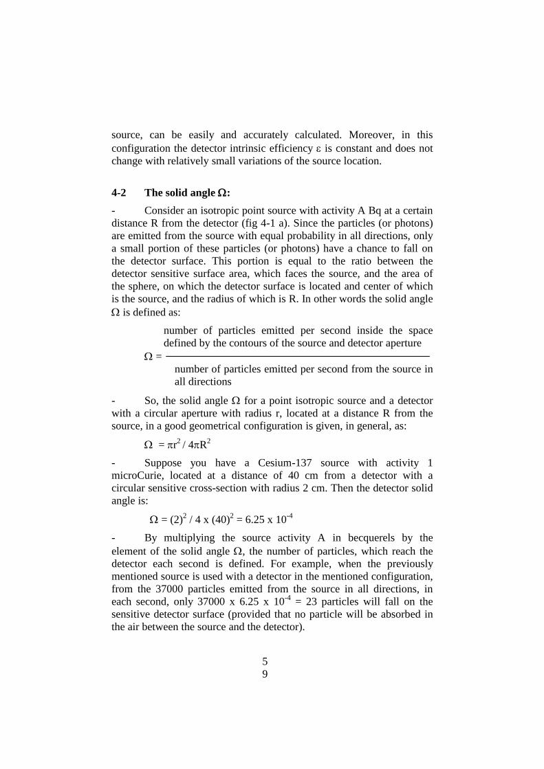
5
9
source, can be easily and accurately calculated. Moreover, in this
configuration the detector intrinsic efficiency is constant and does not
change with relatively small variations of the source location.
4-2 The solid angle :
- Consider an isotropic point source with activity A Bq at a certain
distance R from the detector (fig 4-1 a). Since the particles (or photons)
are emitted from the source with equal probability in all directions, only
a small portion of these particles (or photons) have a chance to fall on
the detector surface. This portion is equal to the ratio between the
detector sensitive surface area, which faces the source, and the area of
the sphere, on which the detector surface is located and center of which
is the source, and the radius of which is R. In other words the solid angle
is defined as:
number of particles emitted per second inside the space
defined by the contours of the source and detector aperture
=
number of particles emitted per second from the source in
all directions
- So, the solid angle for a point isotropic source and a detector
with a circular aperture with radius r, located at a distance R from the
source, in a good geometrical configuration is given, in general, as:
= r2
/ 4R2
- Suppose you have a Cesium-137 source with activity 1
microCurie, located at a distance of 40 cm from a detector with a
circular sensitive cross-section with radius 2 cm. Then the detector solid
angle is:
= (2)2 / 4 x (40)
2 = 6.25 x 10
-4
- By multiplying the source activity A in becquerels by the
element of the solid angle , the number of particles, which reach the
detector each second is defined. For example, when the previously
mentioned source is used with a detector in the mentioned configuration,
from the 37000 particles emitted from the source in all directions, in
each second, only 37000 x 6.25 x 10-4
= 23 particles will fall on the
sensitive detector surface (provided that no particle will be absorbed in
the air between the source and the detector).
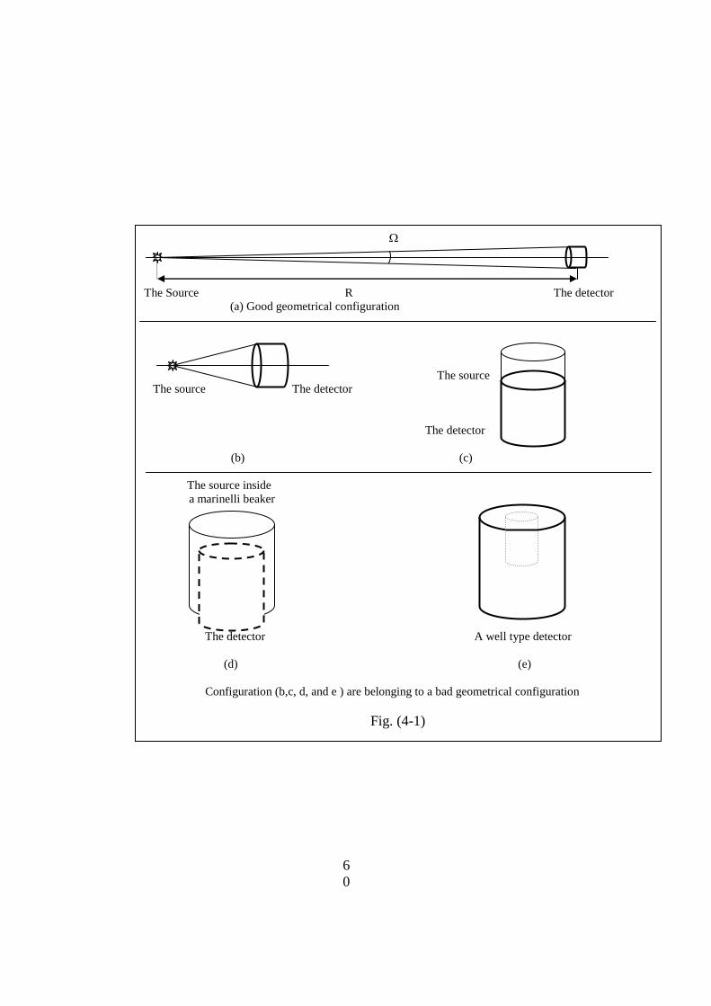
6
0
The Source R The detector
(a) Good geometrical configuration
The source
The source The detector
The detector
(b) (c)
The source inside
a marinelli beaker
The detector A well type detector
(d) (e)
Configuration (b,c, d, and e ) are belonging to a bad geometrical configuration
Fig. (4-1)
6
1
- If the activity of the alpha or beta source is A Bq then it emits A
alpha or beta particles per second in all directions. If the resultant
daughter radionuclide is a gamma emitter, and if the probability of
emission of a gamma ray line with a definite energy E is f, then the
number of photons emitted with this energy in all direction, in one
second, is defined as (f A) photons. For example, it is known, that f =
0.85, for the 662 KeV gamma ray line emitted from barium 137, formed
by the beta decay of Cs-137. Then the number of 662 KeV photons
emitted from 1 Ci Cs-137 source is = f A = 31450 photons per second.
From this last number only 31450 x 6.25 x 10-4
= 19.66 photons will
impinge on the sensitive area of the detector in the configuration
mentioned previously.
- Finally, one can defined the total number N of photons with
known probability f, that impinge on the detector surface from a source
with activity A, when the detector solid angle is as:
N = f A
4-3 The detector intrinsic efficiency :
- In case of the indirectly ionizing radiation, such as gamma
photons and neutrons, the detector can only detect a certain portion of
photons or particles, incident on its sensitive surface. The detector
intrinsic efficiency is defined as:
number of photons detected in the detector per second
=
number of photons impinging on the detector per second
- For photon detectors, there are different intrinsic efficiencies,
such as the photo-peak intrinsic efficiency ph, Compton intrinsic
efficiency c, and full peak intrinsic efficiency f.
- All intrinsic efficiencies are, strongly, dependent on the energy
of detected photons or particles, the type and dimension of the detector,
the density of the material used as a detector, and in some conditions on
the nature of the source, its density and dimension. So, the efficiency
determined for a certain detector and at a given radiation energy must
not be used for another detector or at other radiation energy or for a
source of other density or shape.

6
2
4-4 Relation between the counting rate C and source activity A:
- The counting rate C of a counting system is the number of counts
(photons or particles) detected by this system in one second 9In the SI of
units). So, using the definition of the intrinsic efficiency , and the
number of photons or particles N = f A, impinging on the detector
sensitive surface, it is then clear that
= C / f A
- So, the source activity A is related with the counting rate C,
efficiency of the detector, the solid angle and f value of the certain
gamma line with a simple relation, which is:
C = f A
- In a bad geometrical configuration this relation is not used, due
to the large uncertainties in calculation of even by using very
complicated computer programs, as well as in determination of the
detector intrinsic efficiency . For this reason, another quantity known as
the overall efficiency is introduced, which combines both intrinsic
efficiency and element of the solid angle together, i.e: = . The
relation between the counting rate C, the overall efficiency and the
source activity then is:
C = f A
- When this last relation is used the overall efficiency is
determined experimentally, using a standard source with an accurately
determined activity A. the obtained efficiency is used to determined
activities A of unknown sources, provided that all sources are measured
in the same geometrical configuration (i.e sample volume, shape,
location and density) and using the same energy lines.
4-5 Other factors affecting the measurements:
- In performing relative and absolute measurements there are many
other factors, which may, strongly, affect the accuracy of the obtained
results. Some of these factors are the source itself, the medium between
the source and the detector, and the detector itself.
6
3
4-5-1 Role of the source effects:
- One of the important effects of the source is the self absorption
inside the source. The size, and in particular the way the source is made
may have strong effect on the measurement results. Whether the source
is a solid material or a thin deposited evaporated layer on a metal disc
this may make a difference. For gamma and neutron measurements, the
effect of the source thickness is relatively limited, while it is very strong
in measurement of charged particles, and specially alpha particles. In all
cases, self absorption factor fa in the source should be taken into
consideration, since it reduces the experimentally determined activity.
For this reason, sources of the charged particles (especially alpha)
should be very thin (not more than few micrograms/cm2
- The second important effect of the source is the backscattering
effect on source backing. The source is, always, deposited on a metallic
backing or support. This backing may lead to the scattering of particles,
(especially beta particles). The particles which are directed from the
source toward the backing may suffer backscattering, so that they will be
reflected back to the detector, increasing the count rate over the real
value. The backscattering factor fb, strongly depends, on the atomic
number Z of the backing material, backing thickness X and kinetic
energy E of the particles. Increasing Z, X or E will strongly increase the
backscattering factor fb. For this reason the source backing should be
made from a material with low Z and should have the minimum
thickness. The backscattering may increase the real count rate by a value
up to 70 – 80 %.
4-5-2 The role of the medium:
- The effect of the medium between the source and the detector is
important, too, in some circumstances. Normally the medium between
the source and the detector is air, which have a very low density. For this
reason this medium will have ignored effect on the measurement results
for photons and neutrons. If the measured particles are charged, then all
the particles suffer some energy loss, and some of them (especially those
with low energy) may be completely absorbed, while some others may
be scattered in or out of the detector. To eliminate these effects, source
of the charged particles and detector should be placed in an evacuated
chamber.
6
4
- To demonstrate the role of the medium, it is important to remind
that alpha particles with energy 5 MeV loose completely, their energy
during about 4 cm of air, while beta particles with an end point of about
1 MeV loose their energy in the air within a layer of 4- 5 meters.
4-5-3 The role of the detector:
- In most cases the source is located outside the detector. The
radiation must penetrate the wall of the detector in order to be counted.
Interaction between the impinging radiation on the detector window and
the material from which this window is made, may scatter and (or)
absorb some of the impinging charged particles and even low energy
photons. This will lead to lowering the measured count rate with respect
to the real one.
4-6 Dead- time correction:
- the dead time , or resolving time of a detector, is defined as the
minimum time that can elapse between the arrival of two successive
particles at the detector, so that two distinct pulses are produced. The
most important is the dead time of the system as a whole which may be
composed of a detector, preamplifier, amplifier, ADC (Analogue-to Digital
Converter) and the multi-channel analyzer (MCA). However, since the
dead time of the detector is much longer than that of the electronics, the
later may be ignored (except the dead time of the MCA). So, the total or
detector dead time , as appropriate, should be taken into consideration
when counting ionizing radiation..
- As a result of the dead time, some pulses are not produced in the
detector, or not registered in the MCA, so that they are lost. The effect of
lost counts will be particularly important in the case of high counting
rates. Obviously, the measured counting rate should be corrected for the
loss of some counts due to the dead time. When the counting rate is too
high, then the system will stop functioning (counting) and it seems to be
dead all the time.
- If the dead time of the system (or the detector) is seconds, and
the experimentally measured counting rate is C (count per second), then
the fraction of time during which the system was dead is C second.
When the product C is 1, then the system will stop counting. The
6
5
relation between the true counting rate Ctr, the measured count rate C,
and the system dead time is:
Ctr = C / (1 – C )
- For clarifying the role of the dead time suppose that the dead
time of a system is 400 microseconds (s), and that the measured
counting rate is 30000 counts per minute (cpm). In this case the system
will be dead for 400 x 10-6
x ( 30000 / 60) = 0.2 seconds during the one
second, which means that the percentage of the dead time is 0.2 x 100 =
20 %. The true counting rate is then:
Ctr = 30000 / (1-0.2) = 30000 / 0.8 = 37500 counts per minute.
- For more clarification, suppose that the same system will be used
to register a measured counting rate of 150000 (cpm). In this case the
system, during one second, will be dead for 400 x 10-6
x ( 150000 / 60) =
1 second, which means that the percentage of the dead time is 1 x 100 =
100 %, i. e. the system will be dead all the time and it will stop counting.
4-7 The statistical fluctuation of radiation measurements:
- the radioactive decay is a truly random process, since no one can
predict when a certain single atom will be subjected to decay. So, all
radiation measurements are subjected to two types of errors which are
statistical and systematic errors. The next paragraphs will deal briefly
with some concepts of the statistical errors, which should be described in
statistical terms. The random processes obey the, so called, Gaussian
distribution.
- According to this Gaussian distribution, the standard error in a
mean value n of a set of readings ni, consisting of N readings, is defined
in terms of the, so called, standard deviation of the distribution. This
deviation is defined as:
= [(1/ N) ( ni – n )2 ]
1/2
- In practical situations, scientists are making a single
measurement rather than many measurements to determine the true
mean. In this case if the number of the detected events is m then the
standard deviation of this value may be given as:
= (m)1/2

6
6
So, any measured value is reported as m = m (m)1/2
.
- the percentage statistical error E is defined as:
E % = ( / m) x 100 = 100 / (m)1/2
%
So, it is seen that the percentage statistical error E % decreases
when the amount of the measured counts m is increased. This fact is
represented in table (4-2), showing the number of the registered counts
in each measurement, together with its standard deviation and
percentage error in three cases known as; lower, medium and higher
confidence level, corresponding to 1 , 2 , and 3 respectively.
Table (4-2)
Number of
counts in the
reading
The standard
deviation
The percentage Error %
1 2 3
1 1 100 200 300
4 2 50 100 150
16 4 25 50 75
25 5 20 40 60
100 10 10 20 30
400 20 5 10 15
1000 31.6 3.16 6.32 9.48
10000 100 1 2 3
100000 316 0.316 0.632 0.948
1000000 1000 0.1 0.2 0.3
- To get acquainted with the so called lower, medium and higher
confidence level, suppose that a certain experiment with a long lived
radioactive isotope, such as uranium (with a half life time of 4.468 x 10⁹
years), have been repeated 1000 times, with a mean count of 400. the
long half life is intended to be sure that no change has been occurred
during the 1000 measurements. In this case according to the laws of the
statistical distribution, the counts measured in these thousand runs will
be as shown in table (4-3)
4-7-1 The standard error in the counting rate:
- In practice, the number of counts recorded in the presence of a
given source, is usually recorded either in a scaler or as the total (gross)
counts G in a peak of interest in a multi-channel analyzer, during a

6
7
certain time period of measurement tg. However, the reported result is
the counting rate, i.e., counts recorded per unit time (namely per
second), which is Cg = (G / tg). In some cases, especially, when dealing
with a gamma source, the amount of the background gamma radiation
may be comparable with that radiation emitted from this source, and it
should be taken into consideration, to get the net count rate Cnet from the
source.
For this reason, the background counts B should be measured, in
the absence of the source, during an appropriate time period tb, and the
background count rate Cb = (B / tb) is determined. To get the net count
rate Cnet, resulting from the source alone, the background count rate Cb
should be subtracted from the gross count rate Cg, i.e.,
Cnet = Cg – Cb = ( G / tg ) – ( B / tb )
Table: (4-3)
The range of the experimental readings among
the thousand readings
The number of readings
380 – 420 which meat (m ) 680
360 – 380 which lie between [(m-2) and (m-) 136
420 – 440 which lie between [(m+) and
(m+2)
136
340 – 360 which lie between [(m-3) and (m-
2)
23
440 – 460 which lie between [(m+2) and
(m+3)
23
Less than 340 1
More than 460 1
The lower confidence level includes all reading higher than (m-1) i.e higher
than 380 or lower than (m+1), i.e. lower than 420. These are 680 +136 + 23
+1 = 840 readings among the thousand, with 84 % confidence
The medium confidence level includes all reading higher than (m-2) i.e higher
than 360 or lower than (m+2) i.e. lower than 440. These are 680 +136 +136 +
23 +1 = 976 readings among the thousand, with 97.6 % confidence.
The higher confidence level includes all reading higher than (m-3) i.e higher
than 340 or lower than (m+3), i.e. lower than 460. These are 680 +136 + 136
+ 23 + 23 +1 = 999 readings among the thousand, with 99.9 % confidence

6
8
- The standard deviation net in the net count rate Cnet is defined as:
net = [G /( tg)2 + B /( tb)
2 ]
1/2
- To reduce the error which may arise due to the background
radiation in measurements of low activity gamma sources, both the
source and the detector are placed inside a special shield.
6
9
CHAPTER FIVE
DOSIMETRY QUANTITIES AND THEIR UNITS
- The quantities used to measure the dosimetrical quantities of
ionizing radiation are based on the gross number of this radiation in a
defined situation or, on the gross amount of energy, deposited in a
defined mass of material.
5-1 The Exposure:
- The exposure is defined as the exposure of the dry air at standard
temperature and pressure (STP), to x-rays or low energy gamma ray (up
to 3 MeV).
- The old unit of the exposure is the ROENTGEN – R
- The SI (Systeme Internationale) unit of the exposure is Coulomb
per Kg dry air.
- The Roentgen is defined as the amount of exposure of the dry air
at the standard temperature and pressure, which yields a charge of 2.58 x
10-4
Cooulomb/Kg dry air of each sign (Electrons or ions).
5-2 The absorbed dose D:
- The fundamental dosimetric quantity in radiation protection is
the absorbed dose. The absorbed dose D is defined as the ratio of the
amount of energy E deposited from the ionizing radiation to a mass
element of m of a matter e.g:
D = E / m
In other words, the absorbed dose is defined as the amount of
energy deposited from any type of ionizing radiation, into a unit mass of
any matter.
- the units of the absorbed dose are the "rad" in the old (CGS
which is Centimeter Gram Second) system of units, and the "Gray Gy"
in the standard international system of units.
- The rad is the old unit of the absorbed dose (e.g. in CGS system
of units). One rad is defined as 100 erg of energy deposited into one

7
0
gram of matter. The word rad is the abbreviation of a sentence which is
"radiation absorbed dose”..
1 rad = 100 erg / 1 gram
- The Gray "Gy" is the SI unit of the absorbed dose (e.g. in MKS
system of units, Meter, Kilogram, Second). One Gray is defined as 1
Joule of energy imparted into one Kilogram of matter.
- It should be mentioned that the absorbed dose is defined in terms
that allow it to be specified at a point, but it is used to mean the average
dose over a tissue or organ.
- Using the relation between Joule and erg which is 1 Joule = 107
erg, it is clear that
1 Gy = 100 rad
5-3 The equivalence between the Roentgen R, rad and Gy:
1 R is equivalent to 87 erg/gm = 0.87 rad = 0.0087 Gy in air
1 R is equivalent to 96 erg/gm = 0.96 rad = 0.0096 Gy in the human
tissue
- So, one can consider, with acceptable approximation that:
1 R 1 rad 0.01 Gy
5-4 The Kerma K:
- The Kerma K is defined as:
K = Etr /m
Where, Etr is the sum of the initial kinetic energies of all
charged ionizing particles, liberated by the uncharged ionizing particles
in a material of mass m.
- The units of the Kerma are the same units of the absorbed dose.
- It should be mentioned that the Kerma is approximately equal
the absorbed dose at very low photon energies, but it becomes less than
the absorbed dose, at medium and relatively high photon energies.
5-5 The Radiation Weighting Factor WR:
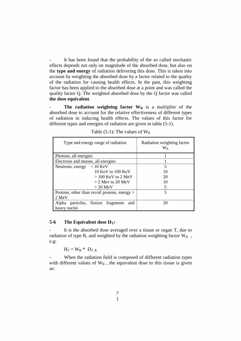
7
1
- It has been found that the probability of the so called stochastic
effects depends not only on magnitude of the absorbed dose, but also on
the type and energy of radiation delivering this dose. This is taken into
account by weighting the absorbed dose by a factor related to the quality
of the radiation for causing health effects. In the past, this weighting
factor has been applied to the absorbed dose at a point and was called the
quality factor Q. The weighted absorbed dose by the Q factor was called
the dose equivalent.
- The radiation weighting factor WR is a multiplier of the
absorbed dose to account for the relative effectiveness of different types
of radiation in inducing health effects. The values of this factor for
different types and energies of radiation are given in table (5-1).
Table (5-1): The values of WR
Type and energy range of radiation Radiation weighting factor
WR
Photons, all energies 1
Electrons and muons, all energies 1
Neutrons, energy < 10 KeV
10 KeV to 100 KeV
> 100 KeV to 2 MeV
> 2 Mev to 20 MeV
> 20 MeV
5
10
20
10
5
Protons, other than recoil protons, energy >
2 MeV
5
Alpha particles, fission fragments and
heavy nuclei
20
5-6 The Equivalent dose HT:
- It is the absorbed dose averaged over a tissue or organ T, due to
radiation of type R, and weighted by the radiation weighting factor WR ,
e.g:
HT = WR * DT, R
- When the radiation field is composed of different radiation types
with different values of WR , the equivalent dose to this tissue is given
as:
7
2
HT = R
WR * DT, R
- One should differentiate between the equivalent dose HT in a
tissue or organ and the dose equivalent H, which was used by the ICRP
before 1990. The dose equivalent H represented the dose in a point
rather than in a tissue or organ, since the quality factor Q, used for
weighting, represented this factor at the point.
- The units of the equivalent dose are the "rem" in the old CGS
system, or the Seivert "Sv" in the SI system.
- The "rem" (roentgen equivalent man) is the unit of the
equivalent dose in the old CGS system, where the absorbed dose is
measured in "rad".
- The Seivert "Sv" is the unit of the equivalent dose in the SI
system, where the absorbed dose is measured in Gray "Gy".
5-7 The tissue weighting factor WT:
- The relationship between the probability of stochastic effects and
equivalent dose is found, also, to depend on the tissue or organ
irradiated. The factor which represents the relative contribution of a
certain tissue or organ to the total detriment, from a uniform irradiation
of the whole body is called the tissue weighting factor WT.
- The tissue weighting factor WT is a multiplier of the equivalent
dose HT to an organ or tissue, to accounts for the different sensitivities of
different tissues and organs to the induction of stochastic effects. The
recommended by the ICRP values of the tissue weighting factors are
given in table (5-2)
5-8 The effective dose E:
- The effective dose E is defined as the sum of the weighted
equivalent doses in all the tissues and organs of the human body. In
other words it is defined as the sum of the tissue equivalent doses each
multiplied by the appropriate tissue weighting factor. It is given by the
Expression:
E = T
WT * HT

7
3
- The units of the effective dose are the same units which are used
for effective dose in both CGS, and SI systems, i.e: the "rem" and the
Seivert "Sv' .
- Both effective and equivalent doses are quantities intended for
use in radiation protection including the assessment of the risks in
general terms. They provide a basis for estimating probability of
stochastic effects, for absorbed doses well below the thresholds for
deterministic effects.
Table (5-2): Tissue weighting factors averaged over the sexes and ages
Tissue or organ Tissue weighting factor WT
Gonades 0.20
Red bone marrow 0.12
Colon 0.12
Lung 0.12
Stomach 0.12
Bladder 0.05
Breast 0.05
Liver 0.05
Oesophagus 0.05
Thyroid 0.05
Skin 0.01
Bone surface 0.01
The remainder 0.05
The whole body 1.00
5-9 The committed equivalent or effective dose:
- Following an intake of a radio-nuclide into the human body,
there is a period during which this nuclide gives rise to equivalent doses
in the tissues of the body at varying rates. The time integral of the
equivalent dose rate is called the committed equivalent dose H().
Where is the integration time in years following the intake. If is not
specified, it is considered to be 50 years for adults and 70 years for
children.
- The committed effective dose E() is similarly defined as the
committed equivalent dose.

7
4
- Both committed equivalent dose and committed effective dose
have the same units as equivalent or effective doses.
5-10 The collective equivalent or collective effective dose:
- All the dosimetric quantities mentioned before relate to the
exposure of an individual. However, other quantities related to the
exposure of a group of workers or population are necessary. These
quantities are the collective equivalent dose ST, in a certain tissue of a
group of people and the collective effective dose S of this group.
- The collective equivalent dose ST is the equivalent dose
incurred in a defined tissue or organ by a group of workers or by a
critical group of people. The collective equivalent dose is defined as
the product of the number N of exposed individuals and their
average equivalent dose HT, when the amount of this dose is equal
for each member of the group.
- The collective effective dose S is the effective dose incurred by
a group of workers or by a critical group. The collective effective dose
is defined as the product of the number N of exposed individuals and
their average effective dose E.
- If several groups are involved, in the exposure, then the total
collective effective dose is the sum of the collective doses for all groups.
- The old unit of the collective equivalent dose or collective
effective dose is the "man.rem", while the SI unit is man.Seivert"
man.Sv".
- The collective quantities represent the total consequences of
exposure of a population or group of workers. For example, when the
risk factor R for lethal cancer is given, it is easy to assess the additional
cancer deaths, induced by radiation, among a group of exposed people
by multiplying their collective dose and the risk factor R.

7
5
CHAPTER SIX
BIOLOGICAL EFFECTS OF RADIATION
6-1 Direct and indirect actions of ionizing radiation on cells:
- The gross biological effects, resulting from exposure to ionizing
radiation are due to long and complex series of events, which are
initiated by ionization or excitation of relatively few molecules in the
organism. For example, the lethal dose for 50 % of the exposed men (or
women) within 30 days (LD-50/30) is known to be about 4 Gy of
gamma rays. This high and lethal dose affects only 1 atom from each 10
millions atoms.
- It is known that most of the human body is water, and most of
the direct action of radiation is therefore on water. The result of energy
absorption from radiation by water molecules is the production of
highly reactive free radicals, which are chemically toxic, and may exert
their toxicity on other molecules (a free radical is a fragment of a
compound or an element, that contains an unpaired electron). When a
water molecule is irradiated, it may become ionized, i.e;
H2O H2O+ + e
- (a physical stage which occurs within 10
-15
seconds from the moment of irradiation).
The positive ion dissociates immediately according to:
H2O+ H
+ + OH
and the electron will be captured by a neutral water molecule forming a
negatively charged water molecule according to the reaction:
e- + H2O H2O
-
This last negative ion dissociates immediately as:
H2O- H + OH
-
The last three reactions are known as the physio-chemical stage,
and they occur within 10-6
second of the moment of irradiation. The
positive and negative ions H+ and OH
- are of no consequence, since all
body fluids contain significant concentrations of them. The free radicals
H and OH may combine with like radicals, or with other molecules,
specially in case of irradiation of the human body with high LET
(Linear Energy Transfer) particles, such as alpha particles or fast

7
6
neutrons. This combination yield a hydrogen peroxide molecule, which
is a stable compound and capable to diffuse far from the point of its
generation.
OH + OH O2H2
Moreover H2O2 is a very powerful oxidizing agent and can affect
cells and molecules that did not suffer radiation damage directly. If the
irradiated water contains dissolved oxygen molecules, then the free
hydrogen radical may combine with the oxygen, to form the hydro-
peroxyl radical:
H + O2 HO2
This radical has a greater stability and can diffuse away and
combine with a free H radical to form hydrogen peroxide, thereby
further enhancing the toxicity of the ionizing radiation.
- Our knowledge is still too limited concerning the gross of
biological effects, that may occur long after irradiation.
6-2 Radiation effects:
- As a result of the processes discussed in the previous paragraph,
the living cell may be damaged. The most important damage is that
which may occur in the DNA. Damage in the DNA may prevent the
survival of the cell, affect its reproduction, or modify the cell itself.
- If enough cells in the organ or tissue are killed or prevented from
functioning normally, there will be a loss of the organ function, which is
known now as a deterministic effect. The loss of function will become
more serious as the number of affected cells is increased. Many organs
and tissues are not affected by small reductions in the number of the
available cells and the body will attempt to repair this damage.
However, if the decrease is large enough, then the body cannot repair
the damage, and the end result will be the death.
- The response of the body to develop a clone of modified somatic
cells is complex. The development of such a clone may be inhibited,
unless it is promoted by an additional agent, before or after irradiation,
and the clone may be eliminated or isolated by the body’s defenses.
However, if it is not, it may result after a prolonged or variable delay,
called the "latency period", in the development of a malignant

7
7
conditions in which the proliferation of modified cells is uncontrolled.
Such condition are grouped together and called cancer. A modified
germ cell in the gonads will transfer genetic information to the
descendants of an exposed individual, which may cause severe harm to
some of these descendants, known as hereditary effects. The somatic
and hereditary effects are known as "stochastic effects".
- There is some experimental evidence that radiation appears to
enhance immunological responses and to modify the balance of
hormones in the body, thus strengthening the natural defense
mechanisms of the body. Most of the data on such effects termed
"hormesis" have been inconclusive because of statistical difficulties at
low doses.
6-3 Deterministic and stochastic effects:
- The deterministic effects are the radiation effects for which a
threshold level of dose exists, and above which the severity of the effect
is greater for higher doses. Deterministic effects occur from acute doses
and some of these effects are radiation disease, cataract, erythema and
others. Deterministic effects are belonging to prompt somatic effect,
which means that they appear promptly after the threshold on the
exposed person.
- The stochastic effects are radiation effects, generally occurring
without a threshold level of dose (i.e they may occur from low doses as
well as from high doses), and their probability is proportional to the
incurred dose, and their severity is independent of the dose. Examples
of the stochastic effects are different cancers, leukemia‟s, and hereditary
effects. The stochastic effects are considered as delayed effects.
6-4 Acute deterministic effects:
- Acute exposure is defined as any single exposure to high dose of
radiation, during a short period of time, and which produce biological
effects within a short time after exposure so, they are called prompt
effects. All deterministic effects arise due to acute exposures. These
exposures may lead, also, to stochastic effects. The acute radiation
syndrome is subdivided into three classes. In the order of increasing
severity, these are:
7
8
a) The hemopoietic syndrome.
b) The gastrointestinal syndrome
c) The central nervous system syndrome
Certain diseases or effects are common to all these classes, which
are grouped under one name as "radiation disease or sickness", which
includes nausea and vomiting, malaise and fatigue, increased
temperature, and blood changes.
- The blood changes is reflected in the changes of the blood count.
These changes, usually, do not appear before gamma ray doses of 250-
500 mGy, but they, certainly, appear after 500 mGy. The white blood
cells known as leucocytes, which are counted in healthy adults as
7000/mm3 of blood are responsible for combating the infecting
organisms. There are two main types of the leucocytes, which are
granulocytes and lymphocytes, with relative proportion of 70- 75 %
and 30- 25 % respectively. The granulocytes are produced in the red
bone marrow and circulate for about 3 days before death, while
lymphocytes are produced in the lymph nodes and spleen, and remain
alive for 24 hours. After an acute exposure in the sub-lethal range there
is a sharp increase in the number of granulocytes, followed within a day
by a very sharp decrease to reach the minimum for several weeks or
months after exposure. The lymphocytes drop sharply after the
exposure, and remain depressed for several months.
- The hemopoietic syndrome appears after a gamma dose of about
2 Gy. This disease is characterized by depression or ablation of the red
bone marrow. The onset of the disease is, rather, sudden, and is heralded
by nausea and vomiting within several hours after overexposure. At 4-6
Gy complete ablation of the bone marrow occurs. An exposure of about
7-8 Gy or greater leads to irriversable ablation of the bone marrow. The
LD-50/30 is in the range of 3-5 Gy.
- The gastrointestinal disorders may appear from relatively small
doses (about 1-2 Gy) due to the death of a part of cells of the intestinal
epithelium, but the syndrome is severe after about 10 Gy. This
syndrome is a consequence of the desquamation of the intestinal
epithelium, and its signs are severe nausea, vomiting and diarrhea,
which begins very soon or immediately after exposure and the death
within 1-2 weeks is the most likely outcome.
7
9
- Central nervous system syndrome occurs, after relatively high
dose of acute exposure which is not less than 20 Gy. Its sign is the
occurring of unconsciousness, within minutes after exposure, and the
death occurs during several hours to few days.
- The skin may be subjected, due to its location to more radiation
exposure, especially in the case of low energy x-ray and beta particles.
An exposure of the skin to about 300 R in the diagnostic x-ray
(approximately 3 Gy) results in erythema, while higher doses may cause
pigmentation, blistering and ulceration.
- The gonads are particularly radiosensitive. A dose of about 150-
200 mGy to the tests in a single exposure results in temporary sterility
among men, but in case of prolonged exposure the dose rate threshold is
0.4 Gy/year The corresponding values for permanent sterility are about
3-6 Gy for acute exposure and 2 Gy/year for prolonged one. For women,
the threshold for permanent sterility is an acute absorbed dose to the
ovaries, in the rang of about 2.5- 6 Gy.
- The threshold for opacities of the eye lens (cataract), which occur
after some delay, seems to be in the range of about 5-10 Gy for an acute
exposure to low LET radiation. For high LET radiation the absorbed
dose threshold is 2 -3 times less.
6-5 The stochastic effects:
- As it has been mentioned, all cancers and hereditary effects
belong to the stochastic effects. For these effects there is no threshold
for their induction. As a somatic effect in humans, the period between
exposure to radiation and recognition of a cancer lasts a number of
years, known as latency period. The median latency period seems to be
about 7- 8 years for leukemia, while it seems to be two three times
longer for many solid tumors, such as breast or lung cancers. However
there are some types of cancers that may appear after about two years
latency.
- Up to now, Epidemiologic data on carcinogenicity of low doses
of radiation are contradictory and inconclusive. For this reason it is
prudent to estimate the risk probability at low doses by extrapolation
from the probabilities at high doses. At high doses, there are many
evidences that the cancer dose response is linear or linear-quadratic, for
human beings and for some other biological species. So, at present, the

8
0
accepted evaluation model for excess cancer deaths due to radiation is
based on a linear zero threshold for the extrapolation to low doses. A
typical dose to cancer relationship for low and high-LET radiations is
illustrated on fig. (6-1).
Fig. (6-1): A typical dose effect relationship for low and high- LET
radiations
- The excess mortality from all cancers, attributed to a collective
effective dose of 1 man.Sv, in an acute uniform whole body exposure by
low LET irradiation, (or in other words the risk factor R, which
represents the probability of death from induced cancer by radiation per
1 man.Seivert) is illustrated in table (6-1). These values were obtained
and corrected by different national and international scientific
committees, as indicated in this table.
- The relative probabilities of fatal cancers in different organs and
the total Risk factor for Japanese population, sexes averaged, ages 0- 90
years, 0- 19 years, 20- 64 years, Evaluated by Japan and the National
Institute of Health (NIH) of USA are tabulated in table (6-2). It is
evident that the relative probabilities vary with age group by a factor of
about 2 for leukemia and colon cancer.
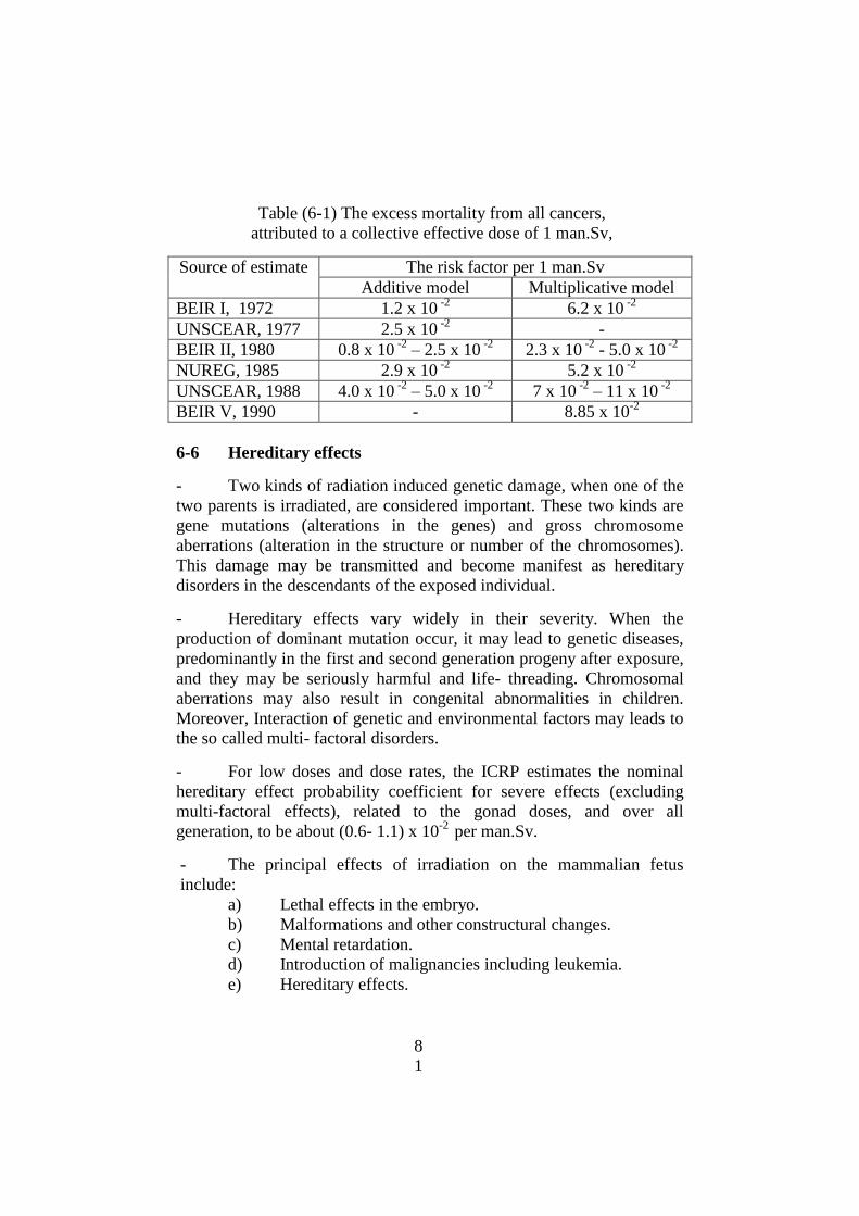
8
1
Table (6-1) The excess mortality from all cancers,
attributed to a collective effective dose of 1 man.Sv,
Source of estimate The risk factor per 1 man.Sv
Additive model Multiplicative model
BEIR I, 1972 1.2 x 10 -2
6.2 x 10 -2
UNSCEAR, 1977 2.5 x 10 -2
-
BEIR II, 1980 0.8 x 10 -2
– 2.5 x 10 -2
2.3 x 10 -2
- 5.0 x 10 -2
NUREG, 1985 2.9 x 10 -2
5.2 x 10 -2
UNSCEAR, 1988 4.0 x 10 -2
– 5.0 x 10 -2
7 x 10 -2
– 11 x 10 -2
BEIR V, 1990 - 8.85 x 10⁻²
6-6 Hereditary effects
- Two kinds of radiation induced genetic damage, when one of the
two parents is irradiated, are considered important. These two kinds are
gene mutations (alterations in the genes) and gross chromosome
aberrations (alteration in the structure or number of the chromosomes).
This damage may be transmitted and become manifest as hereditary
disorders in the descendants of the exposed individual.
- Hereditary effects vary widely in their severity. When the
production of dominant mutation occur, it may lead to genetic diseases,
predominantly in the first and second generation progeny after exposure,
and they may be seriously harmful and life- threading. Chromosomal
aberrations may also result in congenital abnormalities in children.
Moreover, Interaction of genetic and environmental factors may leads to
the so called multi- factoral disorders.
- For low doses and dose rates, the ICRP estimates the nominal
hereditary effect probability coefficient for severe effects (excluding
multi-factoral effects), related to the gonad doses, and over all
generation, to be about (0.6- 1.1) x 10-2
per man.Sv.
- The principal effects of irradiation on the mammalian fetus
include:
a) Lethal effects in the embryo.
b) Malformations and other constructural changes.
c) Mental retardation.
d) Introduction of malignancies including leukemia.
e) Hereditary effects.
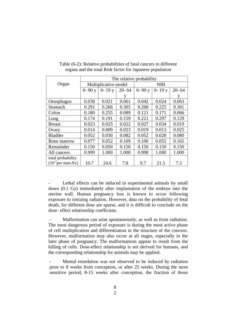
8
2
Table (6-2): Relative probabilities of fatal cancers in different
organs and the total Risk factor for Japanese population
Organ
The relative probability
Multiplicative model NIH
0- 90 y 0- 19 y 20- 64
y
0- 90 y 0- 19 y 20- 64
y
Oesophagos 0.038 0.021 0.061 0.042 0.024 0.063
Stomach 0.291 0.266 0.305 0.268 0.225 0.301
Colon 0.180 0.255 0.089 0.121 0.171 0.066
Lung 0.174 0.191 0.159 0.221 0.297 0.129
Breast 0.023 0.025 0.022 0.027 0.034 0.019
Ovary 0.014 0.009 0.023 0.019 0.013 0.025
Bladder 0.052 0.030 0.082 0.052 0.028 0.080
Bone marrow 0.077 0.052 0.109 0.100 0.055 0.165
Remainder 0.150 0.050 0.150 0.150 0.150 0.150
All cancers 0.999 1.000 1.000 0.998 1.000 1.000
total probability
(10-2
per man.Sv)
10.7
24.6
7.8
9.7
21.5
7.3
- Lethal effects can be induced in experimental animals by small
doses (0.1 Gy) immediately after implantation of the embryo into the
uterine wall. Human pregnancy loss is known to occur following
exposure to ionizing radiation. However, data on the probability of fetal
death, for different dose are sparse, and it is difficult to conclude on the
dose- effect relationship coefficient.
- Malformation can arise spontaneously, as well as from radiation.
The most dangerous period of exposure is during the most active phase
of cell multiplication and differentiation in the structure of the concern.
However, malformation may also occur at all stages, especially in the
later phase of pregnancy. The malformations appear to result from the
killing of cells. Dose-effect relationship is not derived for humans, and
the corresponding relationship for animals may be applied.
- Mental retardation was not observed to be induced by radiation
prior to 8 weeks from conception, or after 25 weeks. During the most
sensitive period, 8-15 weeks after conception, the fraction of those

8
3
exposed which became severely mentally retarded increased by
approximately 0.4 per Sv. For exposure during weeks 16-25, it
increased by about 0.1 per Sv. Moreover mental impairment of lower
severity is also apparent in children exposed in uteri. Evidence of such
impairment is still being collected.
- Irradiated fetuses seem to be susceptible to childhood leukemias
and the childhood cancers, which are expressed during, approximately,
the first decade of life. The risk of fetal childhood cancer due to prenatal
exposure has been estimated to be 2.8 x 10 -2
Per Sv. Constancy of risk
throughout the pregnancy was assumed.

8
4
CHAPTER SEVEN
DOSE CALCULATION
7-1 Dose calculation from point sources:
- Effective dose rates from point exposed sources, existing outside
the human body, which emit beta particle, gamma radiation and
neutrons can be easily calculated with high accuracy, using very simple
mathematical relations. For alpha emitters the external irradiation of the
human body is, completely of no importance, since the energy of alpha
particles is completely absorbed in about 4 cm of air. Even, if a person
is very close to an alpha emitting source, the emitted particles will be
fully absorbed in a very thin layer of the dead skin. However, the
radionulides which emit alpha particles are considered the most
hazardous particles if these radionuclides are ingested or inhaled or
taken by any other mean of intake,(internal exposure), due to the high
linear energy transfer from these particles and, correspondingly, due to
the high specific ionization and high radiation weighting factor of heavy
charged particles.
7-2 Dose calculation for the beta emitters:
- For any beta emitting source, which is relatively small in
dimension, so that it may be considered as a point source, and which is
very thin enough, to neglect the self absorption inside the source, the
dose rate Ė (in microSeivert/hour Sv/h) resulting at a point p, existing
at a distance d (in meters) from the source center can be calculated with
a good accuracy using eq.(7-1):
Ė (Sv/h) = (5 A Eav) / d2 (7-1)
where Eav is the average energy of the beta particles emitted from
the beta source (in MeV), and A is the source activity in Mega
Becquerell, MBq). If the average energy Eav of the beta emitting
radionuclide is not known, then it can be easily approximated with
accepted degree of accuracy from the maximum energy Emax (i.e. end
point energy) of this radionuclide as; Eav = Emax /3 .
- When the relation (7-1) is used to calculate the dose rate Ė of
beta emitters, the distance d between the source and the point of interest
should be limited enough (up to about 2-3 m), to ignore the absorption
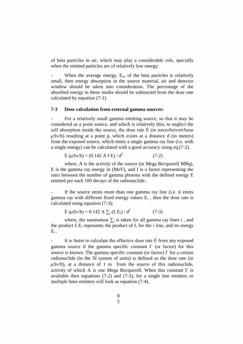
8
5
of beta particles in air, which may play a considerable role, specially
when the emitted particles are of relatively low energy.
- When the average energy, Eav of the beta particles is relatively
small, then energy absorption in the source material, air and detector
window should be taken into consideration. The percentage of the
absorbed energy in these media should be subtracted from the dose rate
calculated by equation (7-1).
7-3 Dose calculation from external gamma sources:
- For a relatively small gamma emitting source, so that it may be
considered as a point source, and which is relatively thin, to neglect the
self absorption inside the source, the dose rate Ė (in microSeivert/hour
Sv/h) resulting at a point p, which exists at a distance d (in meters)
from the exposed source, which emits a single gamma ray line (i.e. with
a single energy) can be calculated with a good accuracy using eq.(7-2).
Ė (Sv/h) = (0.142 A f E) / d2 (7-2)
where, A is the activity of the source (in Mega Becquerell MBq),
E is the gamma ray energy in (MeV), and f is a factor representing the
ratio between the number of gamma photons with the defined energy E
emitted per each 100 decays of the radionuclide..
- If the source emits more than one gamma ray line (i.e. it emits
gamma ray with different fixed energy values Ei , then the dose rate is
calculated using equation (7-3);
Ė (Sv/h) = 0.142 A ∑i (fi Ei) / d2 (7-3)
where, the summation ∑i is taken for all gamma ray lines i , and
the product fi Ei represents the product of fi for the i line, and its energy
Ei .
- It is faster to calculate the effective dose rate Ė from any exposed
gamma source if the gamma specific constant (or factor) for this
source is known. The gamma specific constant (or factor) for a certain
radionuclide (in the SI system of units) is defined as the dose rate (in
Sv/h), at a distance of 1 m from the source of this radionuclide,
activity of which A is one Mega Becquerell. When this constant is
available then equations (7-2) and (7-3), for a single line emitters or
multiple lines emitters will look as equation (7-4).
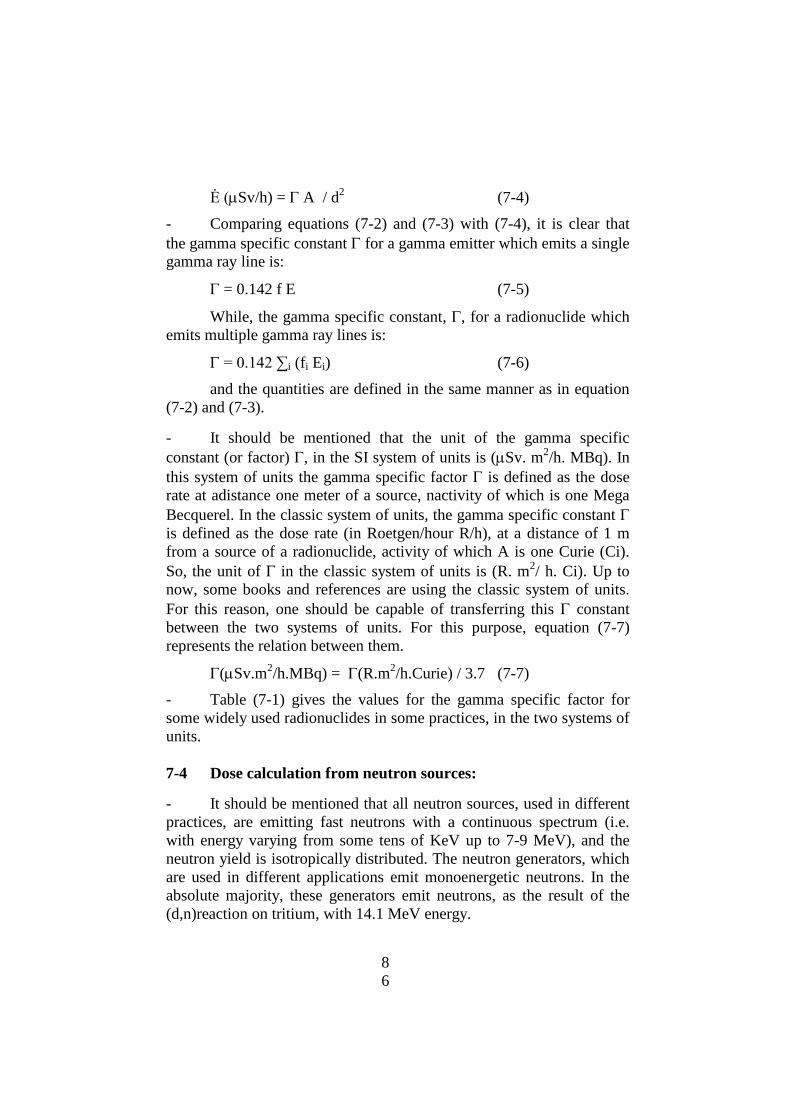
8
6
Ė (Sv/h) = A / d2 (7-4)
- Comparing equations (7-2) and (7-3) with (7-4), it is clear that
the gamma specific constant for a gamma emitter which emits a single
gamma ray line is:
= 0.142 f E (7-5)
While, the gamma specific constant, , for a radionuclide which
emits multiple gamma ray lines is:
= 0.142 ∑i (fi Ei) (7-6)
and the quantities are defined in the same manner as in equation
(7-2) and (7-3).
- It should be mentioned that the unit of the gamma specific
constant (or factor) , in the SI system of units is (Sv. m2/h. MBq). In
this system of units the gamma specific factor is defined as the dose
rate at adistance one meter of a source, nactivity of which is one Mega
Becquerel. In the classic system of units, the gamma specific constant
is defined as the dose rate (in Roetgen/hour R/h), at a distance of 1 m
from a source of a radionuclide, activity of which A is one Curie (Ci).
So, the unit of in the classic system of units is (R. m2/ h. Ci). Up to
now, some books and references are using the classic system of units.
For this reason, one should be capable of transferring this constant
between the two systems of units. For this purpose, equation (7-7)
represents the relation between them.
(Sv.m2/h.MBq) = (R.m
2/h.Curie) / 3.7 (7-7)
- Table (7-1) gives the values for the gamma specific factor for
some widely used radionuclides in some practices, in the two systems of
units.
7-4 Dose calculation from neutron sources:
- It should be mentioned that all neutron sources, used in different
practices, are emitting fast neutrons with a continuous spectrum (i.e.
with energy varying from some tens of KeV up to 7-9 MeV), and the
neutron yield is isotropically distributed. The neutron generators, which
are used in different applications emit monoenergetic neutrons. In the
absolute majority, these generators emit neutrons, as the result of the
(d,n)reaction on tritium, with 14.1 MeV energy.
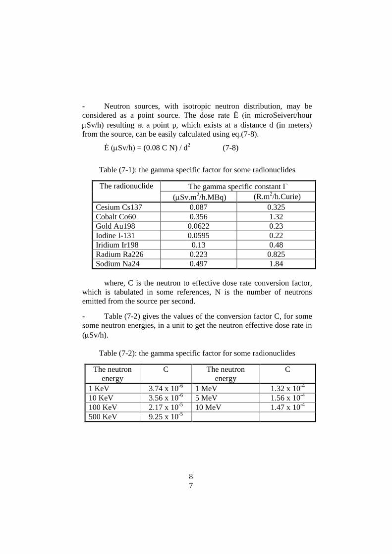
8
7
- Neutron sources, with isotropic neutron distribution, may be
considered as a point source. The dose rate Ė (in microSeivert/hour
Sv/h) resulting at a point p, which exists at a distance d (in meters)
from the source, can be easily calculated using eq.(7-8).
Ė (Sv/h) = (0.08 C N) / d2 (7-8)
Table (7-1): the gamma specific factor for some radionuclides
The radionuclide The gamma specific constant
(Sv.m2/h.MBq) (R.m
2/h.Curie)
Cesium Cs137 0.087 0.325
Cobalt Co60 0.356 1.32
Gold Au198 0.0622 0.23
Iodine I-131 0.0595 0.22
Iridium Ir198 0.13 0.48
Radium Ra226 0.223 0.825
Sodium Na24 0.497 1.84
where, C is the neutron to effective dose rate conversion factor,
which is tabulated in some references, N is the number of neutrons
emitted from the source per second.
- Table (7-2) gives the values of the conversion factor C, for some
some neutron energies, in a unit to get the neutron effective dose rate in
(Sv/h).
Table (7-2): the gamma specific factor for some radionuclides
The neutron
energy
C The neutron
energy
C
1 KeV 3.74 x 10⁻⁶
1 MeV 1.32 x 10⁻⁴
10 KeV 3.56 x 10⁻⁶
5 MeV 1.56 x 10⁻⁴
100 KeV 2.17 x 10⁻⁵
10 MeV 1.47 x 10⁻⁴
500 KeV 9.25 x 10⁻⁵
8
8
7-5 The Inverse square law for external exposure:
- This law is applicable to all gamma and neutron point sources.
For beta particle sources, the law may be applied, only for relatively
small distances d, due to the absorption of a fraction of beta particles
energy in air. This law states that, the effective dose rate Ė from a point
source inversely depends on the square of the distance d between the
source and the intended point. This means that when the distance from
the source is doubled the dose rate will decrease four times, and when
the distance is increased three times, the dose rate will decrease 9 times
(32 = 9). In mathematical representation, when there are two points from
a source, located at distances d1 and d2 , and the dose rates at these
points are Ė1, and Ė2 respectively, then the inverse square law states
that, the dose rates and their distances are related by the following
equation:
Ė1 d12
= E2 d2 2 (7-9)
7-6 Dose calculation from internal exposure:
- When radionuclides are taken inside the human body via
ingestion, inhalation, or through the skin, the exposure, then, is called
internal exposure and the effective or equivalent dose arising due this are
described as committed doses. Any element or compound, when it is
ingested or inhaled inside the human body behaves in a different
manner, and its metabolic behavior depends on many factors including
dietary habits of the human beings. The simplest and most accurate
approach to evaluate the effective committed dose E, (in Seivert) from
the internal contamination with a certain radionuclide, via ingestion or
inhalation can be easily done using equation (7-10), in case of intake of a
different radionuclides I the total comotted dose is determined as::
E (Sv) = i Ci Ni (7-10)
where Ci is the dose conversion factor of the intake of one
Becquerell of a radionuclide i (in Seivert/Bq), and Ni is the activity of
the itaked amount of the radionuclide i (in Bq). The summation should
be taken for all these radionuclides. (by ingestion or by inhalation,
depending on the pathway),

8
9
- It should be mentioned that the dose conversion factor Ci
strongly depends on the pathway of the intake (ingestion, inhalation or
through skin), as well as on the chemical and physical form of the
intaked radionuclide and on the solubility of the chemical compound in
which it is existing. Moreover the factor is strongly age dependent. For
this reason The ICRP, IAEA and UNSCEAR have Published these
factors separatelely for ingestion and inhalation and for different age
groups (less than 1 year, from 1- 2 years, from 2- 7, from 7- 17, and
adults). The obtained committed doses using these conversion factors
refer to the dose incurred up to 70 years age.
7-7 The Annual Limit on Intake ALI:
- It is the intake by ingestion, inhalation or via the skin, of a given
radionuclide in a single year, by the reference man (70 kg mass), which
would result in a committed effective dose equal to the relevant annual
dose limit for the occupational workers. The ALI is expressed in the unit
of activity.
- For occupational workers, where the dose limit, now, is 20 mSv,
the ALI for a certain radionuclide i (in Bq) can be determined, using the
dose conversion coefficient Ci for this radionuclide, (in Sv/Bq) which
represents the committed effective dose per intake of 1 Bq, via the
defined mode of intake by the following (7-11) relation.
ALIi (Bq) = 20x10-3
/ Ci (7-11)
- It should be mentioned that intake of 1 ALI by any mean of
intake in one year is equivalent to an effective committed dose of 20
mSv/year
7-8 The Derived Air Concentration (DAC)
- It is defined as the maximum concentration (in Bq/m3) of a single
radionuclide I in the air at the working place of an occupational
reference man, breathing of which during the whole working hours
through the year (2000 hours/year), would result in a committed
effective dose equal to the relevant occupational dose limit (i.e 20 mSv).
- The DAC of a certain radionuclide I is derived using the volume
of the breathed air during the work hours. According to the used model,
9
0
the reference man breathes, in average, 1.25 m3 of air during one
working hour, if the work is of a moderate type. This means that the
inhaled volume of air during the 2000 working hours/year is 2500 m3.
So, the (DAC) is defined in terms of ALI, as:
(DAC)I (Bq/m3) = (ALI)I / 2500 (7-12)
- It should be mentioned that inhalation of 2000 DAC hours by
any worker in one year is equivalent to an effective committed dose of
20 mSv/year

9
1
CHAPTER EIGHT
RADDIATION SHIELDING
8-1 Shielding of sources of alpha particles:
- It has been mentioned, in chapter 2, that alpha particles, emitted
from all their natural sources, are absorbed within a relatively thin layer
of air (about 4 cm for 5 MeV particles). So all external sources of alpha
particles do not require any shield, provided that there are no other types
of radiation (beta, gamma or neutrons) emitted from them.
8-2 Shielding of sources of beta particles:
- To make a proper shield for a beta source one should use, only,
light rigid material, with low atomic mass number Z, since high Z
materials interact with these particles, yielding a considerable portion of
highly penetrating x rays. The portion f (in percent) of beta particle
energy transferred to emit bremstrahlung radiation (i.e x-ray) is defined
from the maximum energy of the beta spectrum Emax (in MeV), and the
atomic number Z of the interacting material as:
f = 0.035 Z Emax % (8-1)
So, to make a proper shield for a beta source one should use,
only, light rigid material, with low atomic mass number Z, since high Z
materials interact with these particles, yielding a considerable portion of
highly penetrating x- rays
- Although beta particles are characterized by a continuous energy
spectrum, their mass range Rm in any matter can be easily calculated
using the maximum energy Emax of the beta spectrum. For this purpose,
the mass range Rm in (gram/cm2) is defined as the linear range R (in cm)
in the defined material, multiplied by its density , i.e:
R m = R . (8-2)
- The mass range Rm of beta particles in a given material is defined
as a function of the maximum energy Emax of the beta spectrum from the
given radionuclide, using the following (8-3) relation, provided that Emax
is expressed in MeV:
Rm (gm/cm2) = 0.412 Emax (1.265 - 0.0954 ln Emax) (8-3)

9
2
- Equation (8-3) is applied when the beta particles maximum
energy is in the range between 0 up to 2.5 MeV. At higher beta particles
energies other equation is used for determination of Rm, which is:
Rm (gm/cm2) = 0.53 Emax - o.106 (8-4)
- It should be mentioned that the thickness Rm of the shield for
beta particles, which is sufficient to, completely, absorb these particles
does not depend on the activity of the source, so that the shield which is
sufficient for any small activity is also sufficient to shield any other
much larger activity of the same beta emitter. This principle is not
correct for x-ray or gamma radiation, where the source activity is the
most important factor in determination of the thickness of the shield.
- Due to the energy loss of some beta particles energy via
emission of the bremstrahlung radiation (x- ray), the shields of all beta
sources, which must be made from low Z material should be surrounded
by another shield, which is made of a high Z material to absorb the x-
rays, emitted during the interaction of the beta particles with the beta
shield. Calculation of the later shield will be covered in the next
paragraph.
8-3 Shielding of x and gamma ray sources
- In chapter 2, the linear attenuation coefficient μ for a certain
matter and at a certain photon energy as well as the mass attenuation
coefficient μa have been defined and used in an exponential form to
express the attenuation of the number of x-ray or gamma photons as a
function of the thickness x of the material. Number of the photons N,
that will penetrate the thickness x without any interaction with the matter
was expressed, mathematically, by the exponential law:
N = No e - μ x
- This exponential attenuation (e.g. exponential reduction of the
number of photons as a function of x) is valid for calculation of the
thickness of the shield for electromagnetic radiation, only, when the
beam of parallel mono-energetic photons is very narrow, and the
thickness x of the attenuator is very thin.
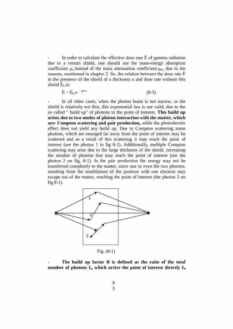
9
3
- In order to calculate the effective dose rate Ė of gamma radiation
due to a certain shield, one should use the mass-energy absorption
coefficient μa instead of the mass attenuation coefficient μm, due to the
reasons, mentioned in chapter 2. So, the relation between the dose rate Ė
in the presence of the shield of a thickness x and dose rate without this
shield Ė0 is:
Ė = Ė0 e – μa x
(8-5)
- In all other cases, when the photon beam is not narrow, or the
shield is relatively not thin, this exponential law is not valid, due to the
so called " build up" of photons in the point of interest. This build up
arises due to two modes of photon interaction with the matter, which
are: Compton scattering and pair production, while the photoelectric
effect does not yield any build up. Due to Compton scattering some
photons, which are emerged far away from the point of interest may be
scattered and as a result of this scattering it may reach the point of
interest (see the photon 1 in fig 8-1). Additionally, multiple Compton
scattering may arise due to the large thickness of the shield, increasing
the number of photons that may reach the point of interest (see the
photon 2 on fig. 8-1). In the pair production the energy may not be
transferred completely to the matter, since one or even the two photons,
resulting from the annihilation of the positron with one electron may
escape out of the matter, reaching the point of interest (the photon 3 on
fig 8-1).
Fig. (8-1)
- The build up factor B is defined as the ratio of the total
number of photons It, which arrive the point of interest directly Id
1
2
e+
3

9
4
from the source and due to scattering or pair production Is to the
number of photons, which arrive the same point directly Id, i.e:
B = It / Id =
= ( Is + Id ) / Id (8-6)
- The build up factor B strongly depends on photon energy E, as
well as on the atomic number Z of the shield and on the thickness of this
shield. Its magnitude may vary from 1 in an ideal geometry (i.e. when
the photon beam is very narrow and the shield thickness is very thin) to
some orders of magnitude for the practical conditions. This makes the
application of the relation (8-5) for calculation of the shield thickness
practically invalid for real conditions, since it will yield much less
thickness. For this reason, the build up factor should be taken into
consideration, in shield calculation. The correct equation that should be
used, to take into consideration the build up factor is:
Ė = Ė0 B e – μa x
(8-7)
- It should be mentioned that the thickness x , which is sufficient
to decrease the dose rate at the point of interest to a certain value, is
dependent on the activity of the source. Increase of the activity of the
gamma-ray source requires corresponding increase of the shield
thickness to reach the required dose rate outside the shield.
8-3 Shielding of the neutron sources:
- It has been mentioned in chapter 2 that the material with low
atomic number Z, especially hydrogen, are considered as the best
moderators for fast neutrons, since these neutrons (with energy higher
than 1 MeV) requires not more than 18 collision, in average, with the
hydrogen nuclei (protons) to moderate them to thermal neutrons with
energies of about 0.025 eV. In light materials, rich with hydrogen, such
as paraffin wax, plastics, water, and 0thers, the thickness which is
required to moderate or slow down the fast neutrons varies within 20-25
cm. So, the use of a layer with this thickness, of any of these materials,
(or any other equivalent light materials), will be sufficient to moderate
fast neutrons to thermal ones.
- One of the main principles used to shield the neutron sources is
to moderate fast neutrons, which are emitted from all neutron sources
9
5
and generators, then to absorb moderated neutrons by other material
with a high probability for capture (absorption) of thermal and slow
neutrons, and finally, to attenuate the gamma radiation emitted during
the neutron capture.
- There are some elements, such as cadmium, boron and others,
which are characterized by a very high cross section for thermal and
slow neutron radiative capture (i.e. thermal neutron absorption with the
emission of gamma photon). So, after moderation of fast neutrons, a
layer of 1 mm thick of cadmium (Cd) is sufficient to absorb the majority
of the moderated neutrons.
- Due to the radiative capture of the thermal neutrons gamma ray
photons will be emitted, so that, an additional layer of a material with
high atomic number Z, such as lead (Pb) is required to attenuate these
radiations. For this reason, a third layer, with a reasonable thickness of
lead, is used to absorb gamma photons emitted from cadmium layer.
- So the ideal shield fort neutron sources consists of three
consequent layers which are: 20-25 cm wax, plastic or water, surrounded
by about 1 mm cadmium sheet, which in its turn, is surrounded by a
reasonable thickness of lead or any other high Z material.
- Neutron shields may be made by a single layer of a low Z
material, such as paraffin wax, plastic, water or others. This is related
with the limited ability of hydrogen and some other light (or low Z)
material to absorb thermal neutrons after their moderation. The main
requirement for such shields is to increase the thickness of the layer to a
sufficient value, so that the reduction of the dose rate, arising from the
neutron source outside the shield is achieved. Examples of such shields
are the neutron semi-spherical howitzers made of wax around the
neutron sources used for educational and other purposes. The thickness
of the paraffin wax or the water around the source may vary from about
50 to more than150 cm, depending on the neutron yield of the source.
- In case of accidents with neutron sources, one may use any
available light materials to shield the exposed neutron source, including
water bags, sands and rocks, and even pieces of wood.

9
6
THE ORGANIZATIONAL ASPECTS
OF RADIATION PROTECTION
9
7
GENERAL FRAMEWORK AND REQUIREMENTS
FOR RADSIATION PROTECTION
1. INTRODUCTION:
- Occupational exposure to radiation can occur as a result of
various human activities, including the use of radioactive sources and x-
ray machines in medicine, scientific research, agriculture and industry,
work associated with the different stages of the nuclear fuel cycle, and
occupations that involve the handling of materials containing enhanced
concentrations of naturally occurring radionuclides
- This lecture addresses the organizational aspects of radiation
protection, in situations of both normal and potential exposure. The
intention is to provide an integrated approach to the control of normal
and potential exposures due to external and internal irradiation from both
artificial and natural sources of radiation.
2. ADMINISTARATIVE REQUIREMENTS:
2-1 The practice and the intervention:
- The practices are defined as the human activities that add
radiation exposure to that which people normally receive from existing
radiation sources, or that increase the likelihood (i,e probability) of their
incurring exposure. For a practice, provisions for radiation protection
and safety can be made before its commencement, and the associated
radiation exposures and their likelihood can be restricted from the outset.
- The interventions are human activities that seek to reduce the
existing radiation exposure, or the likelihood of incurring exposure, and
which are not part of a controlled practice. In the case of intervention,
the circumstances giving rise to exposure or the likelihood of exposure
already exist, and their reduction can only be achieved by means of
protective or remedial actions.
2-2 The requirements of radiation protection:
a- The basic obligation:
- No practice shall be adopted, introduced, conducted,
discontinued or ceased and no source within a practice shall, as
9
8
applicable, be mined, milled, processed, designed, manufactured,
constructed, assembled, acquired, imported, exported, distributed, sold,
loaned, hired, received, sited, located, commissioned, possessed, used,
operated, maintained, repaired, transferred, decommissioned,
disassembled, transported, stored or disposed of, except in accordance
with the national requirements, unless the exposure from such practice
or source is excluded from the requirements, including the requirements
of notification and authorization.
b- The notification
- Any legal person, who intends to carry out any of the actions
specified under the basic obligation for a practice or source shall submit
a notification to the Regulatory Authority of such an intention.
c- Authorization: registration or licensing
- The legal person responsible for any sealed source, unsealed
source or radiation generator (including x-ray machines, accelerators and
neutron generators) shall, unless the source is exempted, apply to the
Regulatory Authority for an authorization which shall take the form of
either a registration or a license.
- The legal person responsible for any irradiation installation, mine
or mill processing of the radioactive ores, installation processing
radioactive substances, nuclear installation or radioactive waste
management facility, or for any use of a source which the Regulatory
Authority has not designated as suitable for registration, shall apply to
the Regulatory Authority for an authorization which shall take the form
of a license.
- Any legal person applying for an authorization shall:
(a) submit to the Regulatory Authority and, if applicable, the
relevant information necessary to support the application;
(b) refrain from carrying out any of the actions described in
the basic obligation until the registration or license has been
granted;
(c) make an assessment of the nature, magnitude and
likelihood of the exposures attributed to the source and take all
9
9
necessary steps for the protection and safety of both workers and
the public; and
(d) if the potential for an exposure is greater than any level
specified by the regulatory authority, have a safety assessment
made and submitted to the regulatory authority as part of the
application.
- The legal person responsible for a source to be used for medical
exposure shall include in the application for authorization:
(a) the qualifications in radiation protection of the medical
practitioners who are to be so designated by name in the
registration or license; or
(b) a statement that only medical practitioners with the
qualifications in radiation protection specified in the relevant
regulations or to be specified in the license will be permitted to
prescribe medical exposure by means of the authorized source.
- Licensee shall bear the responsibility for setting up and
implementing the technical and organizational measures that are needed
for ensuring protection and safety for the sources for which he is
authorized. He may appoint other people to carry out actions and tasks
related to these responsibilities, but He shall retain the responsibility for
the actions and tasks himself.
- licensee shall specifically identify the individuals responsible for
ensuring compliance with the national requirements.
- licensee shall notify the Regulatory Authority of his intentions to
introduce modifications to any practice or source for which he is
authorized, whenever the modifications could have implications for
protection or safety, and shall not carry out any such modification unless
specifically authorized by the Regulatory Authority.
d- Inspection:
- The Licensee shall permit duly authorized representatives of the
Regulatory Authority, and of the relevant Sponsoring Organizations
when applicable, to inspect their protection and safety records and to
carry out appropriate inspections of their authorized activities. Some
inspection should be announced and the others must not be announced.

1
0
0
e- None-compliance
- In the event of a breach of any applicable requirement of the
regulations, the licensee shall investigate the breach and its causes,
circumstances and consequences, take appropriate action to remedy the
circumstances that led to the breach and to prevent a recurrence of
similar breaches, and communicate to the regulatory authority on the
causes of the breach and on the corrective or preventive actions taken or
to be taken.
- The communication of a breach of the regulations shall be
prompt and it shall be immediate whenever an emergency exposure
situation has developed or is developing. Failure to take corrective or
preventive actions within a reasonable time in accordance with national
regulations shall be grounds for modifying, suspending or withdrawing
any authorization that had been granted by the Regulatory Authority.
2-3 The basic framework of radiation protection:
- The principles of radiation protection and safety for are as
follows:
(a) Justification of practices:
“Any practice, or a source within the practice should not be
authorized unless this practice produces sufficient benefits to the
exposed individuals or to society to offset the radiation harm that it
might cause; that is: unless the practice is justified, taking into account
the social and economic factors.”
- The process of determining whether a practice is justified
involves consideration of all the radiation doses received by workers and
members of the public, for present and next generations
(b) Optimization of protection and safety:
- In relation to exposures from any particular source within a
practice, except for therapeutic medical exposures, protection and safety
shall be optimized in order that the magnitude of individual doses, the
number of people exposed and the likelihood of incurring exposures all
be kept as low as reasonably achievable, (ALARA principle), economic
1
0
1
and social factors being taken into account, with the restriction that the
doses to individuals delivered by the source be subject to dose
constraints.
- Senior management should translate their commitment to
optimization of radiation protection into effective action by establishing
appropriate radiation protection program, and rules commensurate with
the level and the nature of the radiological risk presented by the practice.
- In order to reduce or avert exposures in intervention situations,
protective actions or remedial actions shall be undertaken whenever they
are justified.
(c) Dose limitation: “The normal exposure of individuals shall be restricted so that
neither the total effective dose nor the total equivalent dose to relevant
organs or tissues, caused by the possible combination of exposures from
authorized practices, exceeds any relevant dose limit, specified by the
national Regulatory Authority.
- The dose limits are applied, only, for occupational exposures and
general public exposures, while they are not applied for medical
exposures and exposures during emergencies.
- The limit on effective dose represents the level above which the
risk of stochastic effects due to radiation is considered to be
unacceptable, while it is much less than the thresholds for deterministic
effects. For localized exposure of the lens of the eye, extremities and the
skin, this limit on effective dose is not sufficient to ensure the avoidance
of deterministic effects, and therefore limits on equivalent dose are
specified for such situations.
(d) Guidance levels for medical exposure:
- Guidance levels for medical exposure shall be established for use
by medical practitioners. The guidance levels are intended to be a
reasonable indication of doses for average sized patients. They are
needed to provide guidance on what is achievable with current good
practice rather than on what should be considered optimum
performance;

1
0
2
3- MANAGEMENT REQUIREMENTS FOR PROTECTION:
3-1 Safety culture:
- One of the definition of the safety culture is that it is consisting
of the assembly of characteristics and attitudes in organizations and
individuals which establishes that, as an overriding priority, protection
and safety issues receive the attention warranted by their significance
- A safety culture shall be fostered and maintained to encourage a
questioning and learning attitude to protection and safety and to
discourage complacency, which shall ensure that policies and procedures
be established that identify protection and safety as being of the highest
priority and problems affecting protection and safety be promptly
identified and corrected
3-2 Quality assurance:
- Quality assurance programs shall be established, that provide
adequate assurance that the specified requirements relating to protection
and safety are satisfied and quality control mechanisms for assessing the
effectiveness of protection and safety measures are fulfilled.
3-3 Human factors:
- Provision shall be made for reducing as far as practicable the
contribution of human error to accidents and other events that could give
rise to exposures, by ensuring that all personnel on whom protection and
safety depend be appropriately trained and qualified so that they
understand their responsibilities and perform their duties according to
defined procedures, and appropriate equipment, safety systems, and
procedural requirements be provided and other necessary provisions be
made to reduce, as far as practicable, the possibility that human error.
3-4 Qualified experts:
- Licensee should identify qualified experts and shall made
available the expertise for providing advice on the observance of the
regulations.
1
0
3
4- THE PRINCIPAL REQUIREMENTS:
4-1 Security of sources:
- Sources shall be kept secure so as to prevent theft or damage and
to prevent any unauthorized legal person from carrying out any of the
actions specified in the basic obligation for practices, by ensuring that
control of the sources is ensured against lost or theft and any source
shall not be transferred unless the receiver possesses a valid
authorization. A periodic inventory of all sources, especially movable
shall be conducted at appropriate intervals to confirm that they are in
their assigned locations and are secure.
4-2 Defense in depth:
- A multilayer (defense in depth) system of provisions for
protection and safety, commensurate with the magnitude and likelihood
of the potential exposures involved, shall be applied to sources such that
a failure at one layer is compensated for or corrected by subsequent
layers, for the purposes of preventing accidents that may cause exposure
and mitigating the consequences of any such accident that does occur;
and restoring sources to safe conditions after any such accident.
4-3 Good engineering practice:
- As applicable, the sitting, location, design, construction,
assembly, commissioning, operation, maintenance and decommissioning
of sources within practices shall be based on sound engineering which
shall take account of approved codes and standards and other
documented instruments. This include taking into account of relevant
developments in technical criteria, as well as the results of any relevant
research on protection or safety and lessons from previous experiences.
5- VERIFICATION OF SAFETY:
5-1 Safety assessments:
- Safety assessments related to protection and safety measures for
sources within practices shall be made at different stages, including
sitting, design, manufacture, construction, assembly, commissioning,
operation, maintenance and decommissioning, as appropriate, in order to
identify the ways in which normal and potential exposures could be
1
0
4
incurred, account being taken of the effect of events external to the
sources, as well as events directly involving the sources and their
associated equipment and to assess the quality and extent of the
protection and safety provisions.
5-2 Monitoring and verification of compliance:
- Monitoring and measurements shall be conducted of the
parameters necessary for verification of compliance with the
requirements and regulations. For this purpose, suitable equipment shall
be provided and verification procedures introduced. The equipment shall
be properly maintained and tested and shall be calibrated at appropriate
intervals with reference to standards traceable to national or international
standards.
5-3 Records:
- Different records shall be maintained for the practices and
sources and of the results of monitoring and verification of compliance,
including records of the tests and calibrations carried out in accordance
with the Standards.
6- CONDITION OF SERVICE:
6-1 Pregnant workers:
- A female worker should, on becoming aware that she is pregnant,
notify the employer in order that her working conditions may be
modified if necessary. The notification of pregnancy shall not be
considered a reason to exclude a female worker from work; however, the
employer of a female worker who has notified pregnancy shall adapt the
working conditions in respect of occupational exposure so as to ensure
that the embryo or foetus is afforded the same broad level of protection
as required for members of the public.
6-2 Conditions for young persons
- No person under the age of 16 years shall be subjected to
occupational exposure, and no person under the age of 18 years shall be
allowed to work in a controlled area unless supervised, and then only for
the purpose of training.

1
0
5
6-3 Classification of areas
- The licensee shall designate controlled and supervised areas, in which
specific protective measures or safety provisions are required for
controlling normal exposures or preventing the spread of contamination
during normal working conditions. The licensee shall delineate
controlled and supervised areas by physical means.
- According to Saudi national regulation, the controlled area is
defined as the area in which the annual effective dose may reach 3/10
the occupational annual dose limit (i.e may reach 6 mSv/year).
- According to Saudi national regulation, the supervised area is
defined as the area in which the annual effective dose may reach 1/10
the occupational annual dose limit (i.e may reach 2 mSv/year).
6-4 The local rules and supervision:
- The licensee shall establish in writing local rules and procedures
as are necessary to ensure adequate levels of protection and safety for
workers and general public, and shall include in these rules the values of
any relevant investigation level or authorized level, and the procedure to
be followed in the event that any such value is exceeded.
- The licensee shall make local rules and the protective measures
and safety provisions known to those workers, to whom they apply and
to other persons who may be affected by them.
6-5 Personal protective equipment:
- The licensee shall ensure that workers are provided with suitable
and adequate personal protective equipment which meets any relevant
specifications, including protective clothing, protective respiratory
equipment, protective aprons and gloves, and organ shields.
- Workers must receive adequate instruction in the proper use of
protective equipment, especially respiratory equipment, including testing
for good fit. All personal protective equipment shall be maintained in
proper condition and tested at regular intervals.
- Tasks requiring the use of some specific personal protective
equipment shall be assigned, only to workers whom the basis of medical
advice, are capable of safely sustaining the extra effort necessary;

1
0
6

1
0
7
RESPONSIBILITIES OF PARTIES
1- RESPONSIBLE PARTIES FOR RADIATION PROTECTION:
- The Regulatory Authority (and the intervening organizations in
the case of intervention) shall be responsible for the enforcement of the
regulations.
- The principal party having the main responsibilities for the
compliance with the regulations is he licensee (the employer).
- Other parties, who have subsidiary responsibilities for the
application of the regulations include:
(a) the radiation protection officers;
(b) the qualified experts;
(c) the medical practitioners;
(b) the workers;
(e) the suppliers;
(f) the Ethical Review Committees; and
(g) any other party to whom a principal party has delegated specific
responsibilities.
- All the parties shall have the general and specific responsibilities
set out in the national regulations.
2- RESPONSIBILITIES OF THE LICENSEE:
2-1 The general responsibilities:
- The general responsibilities of the principal party (the licensee),
within the requirements specified by the Regulatory Authority, are:
(a) to establish protection and safety objectives in conformity with
the requirements.
(b) to develop, implement and document a protection and safety
program, commensurate with the nature and extent of the risks
associated with the practices and interventions under his
responsibility, and sufficient to ensure compliance with the
requirements.

1
0
8
(c) to determine the measures and resources needed to achieve the
protection and safety objectives and to ensure that the resources
are provided, and the measures are properly implemented.
(d) to keep the measures and resources continually under review, and
to verify that the protection and safety objectives are being
achieved.
(e) to identify any failures and shortcomings in the protection and
safety measures and resources, and to take steps to correct them
and prevent heir recurrence.
2-2 Specific responsibilities of the licensee:
- The licensee, who is engaged in activities involving normal or
potential exposure, shall appoint radiation protection officer (officers)
RPO, for carrying out the technical actions and tasks related to his
responsibilities, but the licensee shall retain the responsibility for these
actions.
- The licensee (employer of workers) shall be responsible for the
protection of workers from occupational exposure and for compliance
with the relevant requirements of the regulations.
- To fulfill his responsibilities, the licensees shall ensure, for all
workers engaged in activities that involve or could involve
occupational exposure, that:
(a) The occupational exposures are limited to the national limits.
(b) The occupational protection and safety are optimized in
accordance with the requirements.
(c) Decisions regarding measures for occupational protection and
safety are recorded and made available to the relevant parties.
(d) Policies, procedures and organizational arrangements for
protection and safety are established for implementing the
relevant requirements.
(e) Suitable and adequate facilities, equipment and services for
protection and safety are provided, the nature and extent of

1
0
9
which are commensurate with the expected magnitude and
likelihood of the occupational exposure.
(f) Necessary health surveillance and health services are provided.
(g) Appropriate protective devices and monitoring equipment are
provided and arrangements are made for its proper use.
(h) Suitable and adequate human resources and appropriate training
in protection and safety are provided, as well as periodic
retraining and updating, as required, in order to ensure the
necessary level of competence
(i) Adequate records are maintained as required by the regulations
(j) Arrangements are made to facilitate consultation and co-
operation with workers with respect to protection and safety.
(k) Necessary conditions to promote a safety culture are provided.
2-3 Specific responsibilities of the RPO:
- The RPO is an individual, technically competent in radiation
protection matters, relevant to a given type of practice, who is qualified
through the judgment of the Regulatory Authority, and who is
designated by the licensee to oversee the implementation of the
requirements of the regulations.
- The responsibilities of the RPO
(a) Preparation of all technical aspects and procedures of radiation
protection program (including emergency plan and quality
assurance program) under the supervision of the qualified expert.
(b) Following-up of the implementation of the rules and procedures
for protection and safety, specified by the licensee, and
overseeing of the proper use of the surveying and monitoring
devices, the protective equipment, and all other equipments.
(c) Conduct all technical aspects, related with radiation protection
and safety, including different radiological surveys, monitoring,
tests and calibrations, or supervise their conduction at the
authorized parties.

1
1
0
(d) Co-operate with the licensee and the regulatory authority with
respect to protection and safety and the operation of radiological
health surveillance and dose assessment programs.
(e) Register all technical data and personal dose information in the
records timely, and notify the workers who approach dose limit,
and discuss all protection affairs with workers and involved
parties.
(f) Conduct demonstrations and technical training on job concerning
radiation protection and safety of the authorized practices.
(g) Stop, promptly, any violation of the local rules or regulation, and
report to both the licensee and regulatory authority.
2-4 Specific responsibilities of the workers:
- Workers can by their own actions contribute to the protection and
safety. Workers shall:
(a) Follow the rules and procedures for protection and safety,
specified by the licensee;
(b) Use properly the monitoring devices, the protective equipment,
and clothing provided.
(c) Co-operate with the licensee with respect to protection and safety
and the operation of radiological health surveillance and dose
assessment programs.
(d) Provide to the licensee information on their past and current
work as is relevant to ensure effective and comprehensive
protection and safety for themselves and others;
(e) Abstain from any willful action that could put themselves or
others in situations that contravene the requirements of
regulations.
- Workers are also responsible for providing feedback to the
management, particularly when adverse circumstances arise
related to the radiation protection program.

1
1
1
- Female workers have responsibilities regarding the protection of
the embryo or foetus. The worker herself “should, on becoming aware
that she is pregnant, notify the licensee in order that her working
conditions may be modified if necessary.
3. COOPRATION BETWEEN LICENSEES AND EMPLOYERS:
- The management of the occupational protection and safety of
transient, temporary or itinerant workers, and others who are employed
under contracts to organizations other than the operator, presents a major
concern. In order that these workers are adequately protected and do not
exceed any appropriate dose limit, there should be an adequate degree of
co-operation between the employer, the workers and the management of
the plants, for whom contracts are being undertaken.
- If workers are engaged in work that could involve a source that is
not under the control of their employer, the licensee responsible for the
source and the employer, shall co-operate by the exchange of
information to facilitate proper protective measures and safety
provisions of workers.

1
1
2
1
1
3
NATIONAL (SA) DOSE LIMITS
FOR RADIATION EXPOSURES
1. THE TERMS "LIMIT" AND "LEVEL":
- The term "limit" is defined, for radiation protection purposes, as
the value of a quantity used in a certain specified activities or
circumstances, such as effective dose or equivalent dose, which must not
be exceeded.
- The term "level" is defined, for radiation protection purposes, as
the value of a specified quantity above which appropriate actions should
be considered:
- Among levels used in radiation protection, some most important
levels will be defined which are:
(a) The action level: is the level of dose rate or activity
concentration above which remedial actions or protective actions
should be carried out in chronic exposure or emergency exposure
situations, such as sheltering, immigration or others. Action
levels often serve to protect members of the public, but they also
have relevance in the context of occupational exposure in chronic
exposure situations, particularly that involving exposure to radon
in workplaces.
(b) The clearance level: is a value, established by the regulatory
authority, and expressed in terms of activity concentration or
total activity, below which sources of radiation may be released
from regulatory control.
(c) The guidance level for medical exposures: is the value of dose,
dose rate, or activity, selected by professional bodies in
consultation with the regulatory authority, to indicate a level
above which there should be a review by medical practitioners,
to determine wither or not the value is excessive, taking into
account the particular circumstances and applying sound clinical
judgment
(d) The intervention level: is the level of avertable dose at which a
specific protective action or remedial action is taken in an
1
1
4
emergency exposure situation or chronic exposure situation. The
use of this term is normally confined to interventions related to
the protection of members of the public.
(e) The investigation level: The value of a quantity such as
effective dose, intake, or contamination per unit area or volume
at which an investigation should be conducted.
(f) The recording level: is a level of dose, exposure or intake
specified by the Regulatory Authority at which values of dose,
exposure or intake received by workers are to be entered in their
individual exposure records.
(g) The Reference level: is defined as a general term that can refer
to an action level, an intervention level, an investigation level or
a recording level. Such levels are helpful in the management of
operations as „trigger levels‟ above which some specified action
or decision should be taken.
2- RADIATION EXPOSURES:
- Radiation exposure is, generally, defined as the act or condition
of being subject to irradiation by ionizing radiation. The term exposure
is also used, in radiodosimetry, to express the amount of ionization,
produced in dry air by x-ray and low energy gamma radiation.
- In the general definition, exposure can be either external
exposure, when the irradiating source or sources are located outside the
body, or internal exposure when the source or sources are inside the
body (by inhalation, ingestion, injection or any other pathway of intake).
Moreover, exposure can be classified as:
(a) either normal or potential exposure.
(b) either occupational, medical or general public exposure.
(c) in intervention situation, either emergency or chronic exposure.
2-1 The normal exposure:
- The normal exposure is defined as an exposure which is expected
to be received under normal operating conditions of an installation or a
source, including possible minor mishaps that can be kept under control.

1
1
5
2-2 The potential exposure:
- It is defined as the exposure that is not expected to be delivered
with certainty, but may result from an accident at a source or owing to
an event or sequence of events of a probabilistic nature, including
equipment failures and operating errors.
2-3 The occupational exposure:
- It is defined as all exposures of workers incurred in their work
due to this work, with the exception of exposures excluded from the
national regulations, and exposures from practices or sources exempted
by these regulations. The exposure of medical doctors, and other
technical and nursing staff belongs to occupational exposure.
2-4 The medical exposure:
- It is defined as exposure incurred by:
(a) patients as a part of their own medical or dental diagnosis or
treatment.
(b) exposures incurred by persons, other than those occupationally
exposed, knowingly while voluntarily helping in the support and
comfort of patients.
(c) exposures incurred by volunteers in a program of biomedical
research involving their exposure.
2-5 General public exposure:
- It is defined as exposure incurred by the members of the general
public from radiation sources, excluding any occupational or medical
exposure and the normal local natural background radiation, but
including exposure from authorized sources and practices and
intervention situations.
2-6 Chronic exposure:
- It is defined as exposure persisting in time and incurred with,
relatively, small dose rates.
1
1
6
2-6 Acute exposure:
- It is defined as exposure incurred in a short time interval
(minutes, hours or days) with very high dose rate.
2-6 Emergency exposure:
- It is defined as exposure incurred in an emergency situation.
3- THE OCCUPATIONAL DOSE LIMITS:
3-1 Occupational limits for adult workers:
- The limits on effective dose for occupational exposure apply to
the sum of effective doses from external sources and committed
effective doses from intakes in the same period.
- The occupational exposure of any worker must not be exceeded
the following values:
(a) an effective dose of 20 mSv per year averaged over five
consecutive years.
(b) an effective dose of 50 mSv in any single year, provided that the
effective dose does not exceed 100 mSv over five consecutive
years.
(c) an equivalent dose to the lens of the eye of 150 mSv in a year
(d) an equivalent dose to the extremities (hands and feet) or the skin
of 500 mSv in a year.
3-2 Occupational limits for apprentices of 16- 18 years age:
- Separate limits are specified for apprentices of age 16–18 years,
who are training for employment involving exposure to radiation, and
for students of age 16–18 years, who need to use sources in the course of
their studies.
- The occupational exposure for this age category of trainees must
not exceed:
(a) an effective dose of 6 mSv in a year.
(b) an equivalent dose to the lens of the eye of 50 mSv in a year.

1
1
7
(c) an equivalent dose to the extremities or the skin of 150 mSv in a
year.
3-3 Occupational limits for pregnant workers:
- The Occupational exposure for pregnant woman must not exceed
an effective committed dose of 1mSv in the tatal period of pregnancy,
when the irradiation is internal (i.e intake), and the effective dose must
not exceed 2 mSv, when the irradiation is external, during the 9 months
of pregnancy, in order to keep the effective dose to the embryo and
foetus not more than 1 mSv.
4- THE DOSE LIMITS FOR GENERAL PUBLIC:
- The estimated average doses to a relevant critical group that are
attributable to practices must not exceed the following limits:
(a) an effective dose of 1 mSv per year averaged over five
consecutive years.
(b) in a special circumstances, an effective dose of up to 2 mSv/year
provided that the effective dose does not exceed 5 mSv over five
consecutive years.
(c) an equivalent dose to the lens of the eye of 15 mSv in a year
(d) an equivalent dose to the extremities (hands and feet) or the skin
of 50 mSv in a year.
5- THE DOSE LIMITS FOR MEDICAL EXPOSURES:
- For medical exposure there are no limits. Instead of that the
principle of guidance levels is used.
- In medical exposure there is a dose limit for those persons, who
are offering comfort or supporting patients, undergoing medical
diagnosis or treatment, or visitors of such patients. The dose for these
comforters or visitors of patient shall be constrained to 5 mSv, during
the period of diagnosis or treatment of the patient.
- The dose to children visiting patients who have ingested
radioactive material shal be constrained to 1 mSv.
1
1
8
6- THE DOSE LIMITS FOR EMERGENCY EXPOSURES:
- For emergency situations the objective should be to keep doses to
intervening personnel below an effective dose of 100 mSv or equivalent
doses of 1 Sv to the skin and 300 mSv to the lens of the eye in some
situations.
- However, where life saving actions are concerned significantly
higher levels of dose could be justified, although every effort should be
made to keep doses below ten times the maximum single year dose limit
(i.e. below 500 mSv) in order to avoid deterministic effects on health.
1
1
9
THE RADIATION PROTECTION PROGRAM (RPP)
1. INTRODUCTION
- It has been mentioned that the licensee shall establish a radiation
protection program (RPP), which is one of the requirements for all
phases of a practice, and to the lifetime of a facility, from design through
process control to decommissioning. The general objective of RPP is to
reflect the application of the management responsibility for radiation
protection and safety through the adoption of management structures,
policies, procedures and organizational arrangements that are
commensurate with the nature and extent of the risks.
- Prior to establishment of the RPP for a practice, a radiological
evaluation shall be conducted to describe, as precisely as necessary, the
situation involving occupational, medical and public exposures. This
evaluation should include, all aspects of operations an identification of
the sources of routine and potential exposures and a realistic estimate of
the relevant doses and probabilities
- the legal person (licensee) applying for an authorization should
make an assessment of the nature, magnitude and likelihood of the
exposures and, if necessary, a safety assessment. Such a safety
assessment should contribute to the design of the RPP.
2- STRUCTURE OF THE RPP
- The RPP covers the main elements contributing to protection and
safety of the practices and is therefore a key factor for the development
of protection and safety.
- The RPP is composed of 6 main elements (or components),
which are:
(a) A committed administration to safety and protection.
(b) Selection of personnel and their training.
(c) An effective surveillance for occupational exposure.
(d) An effective surveillance for public exposure.

1
2
0
(e) A proper quality assurance program.
(f) An emergency response plan.
2-1. A COMMITTED ADMINISTRATION AND ASSIGNMENT
OF RESPONSIBILITIES
- The highest managerial level should submit, in written, the
policy statements which ensure that radiation protection in the practices,
related with radiation exposure, deserves the highest consideration at all
levels. The licensee shall appoint other people to carry out actions and
tasks related to responsibilities in radiation protection aspects, but he
shall retain the responsibility for the actions and tasks himself. The
licensee shall specifically identify the individuals responsible for
ensuring compliance with the national regulations. The responsibilities
of each level, from the top management to the workers, regarding each
aspect of the RPP should be clearly delineated and documented in
written policy statements to ensure that all are aware of them. Radiation
protection officer (or officers) must be appointed, to oversee the
application of the regulatory requirements.
- The organizational structures at the licensee should reflect the
assignment of responsibilities, and the commitment of the organization
to protection and safety. The management structure should facilitate co-
operation between the various individuals involved. The RPP should be
designed in such a way that the relevant information is provided to the .
2-2. SELECTION OF PERSONNEL AND THEIR TRAINING:
- Criteria for selection of personnel should be defined, including
medical and moral aspects, and technical educational levels
- It may be appropriate, depending on the size of the organization,
to create a specific committee with representatives of those departments
concerned with radiation exposures. The main role of this committee
would be to advise senior management on the RPP individuals in charge
of the various aspects of the work.
- Qualified experts in radiation protection should be identified and
made available for providing advice on the observance of the Standards.
- Senior management should be trained in the risks associated with
ionizing radiation, the basic principles of radiological protection, their

1
2
1
main responsibilities regarding radiation risk management and the
principal elements of the RPP.
- Training for those workers directly involved in work with
radiation sources should include relevant information, presented in the
form of documents, lectures and applied training, which emphasizes
procedures specific to the worker‟s job assignment.
- Training programs should be documented and approved at an
appropriate level within the organization. Such programs should be
reviewed periodically to ensure that they remain up to date.
2-3. AN EFFECTIVE SURVEILLANCE FOR OCCUPATIONAL
EXPOSURE: - An effective surveillance for occupational radiation protection
shall be established. This surveillance shall include:
- Systems and procedures for securing radioactive source (or
sources) and an accountability system, which includes records of these
sources.
- A health surveillance program should be prepared and health
criteria should be established for radiation workers..
- Classification of working areas, whenever there is occupational
exposure to radiation. These areas should be clearly defined as part of
the RPP, and their classification should result from the prior radiological
evaluation. The two types of areas, which are controlled and supervised,
shall be delineated. Restriction of access to the controlled and supervised
by permits, and physical barriers, locks or interlocks shall be provided.
- Establishment of occupational radiation protection and safety
measures, including rules and procedures that are appropriate,
- Local rules, describing the organizational structures and the
procedures to be followed in classified areas, should be developed by
management and written down. The rules should be prominently
displayed or readily available in the workplace. and they should include
procedures and values of any relevant investigation level or authorized
level, and the procedure to be followed in the event that any such value
is exceeded. The local rules and procedures and the protective measures
1
2
2
- A radiological monitoring program for workplaces shall be
established and implemented. The nature and extent of the monitoring
program shall provide the primary justification for radiological
protection Monitoring program can be divided and subdivided into a
number of different types. The first division relates to the objectives of
the monitoring. At this level, three types of monitoring are conducted for
radiation protection purposes. These are routine monitoring, which is
associated with continuing operations and is intended to demonstrate
that the working conditions, including the levels of individual dose,
remain satisfactory, and to meet regulatory requirements, task related
monitoring which applies to a specific operatio, and special monitoring
which is investigative in nature and typically covers a situation in the
workplace for which insufficient information is available to demonstrate
adequate control.
- Individual monitoring for internal or external dose assassment
shall be undertaken for any worker who is regularly employed in a
controlled or supervised areas or who enters a controlled area only
occasionally. Individual monitoring in a supervised area shall not be
required but the occupational exposure of the worker shall be assessed
This assessment shall be on the basis of the results of monitoring of the
workplace or individual monitoring. The nature, frequency and precision
of individual monitoring shall be determined with consideration of the
magnitude and possible fluctuations of exposure levels and the
likelihood and magnitude of potential exposures.
- To secure the necessary accuracy and precision, individual
dosimetry should be performed, whenever possible, by an approved
dosimetry service. The regulatory authority should give consideration to
the establishment of a national accreditation procedure as a basis for the
approval of dosimetry services.
- Record keeping is an essential part of the individual monitoring
process. In making records of dose assessments it is important to
establish the
- Many of records, for example the full details of a particular
radiation survey, are transitory in nature are only relevant for the lifetime
of an established review period, and there may be no need to retain such
records for extended periods. Other records may be related to decisions
1
2
3
about the definition of the workplace, and these records may be relevant
for the lifetime of the workplace.
- Exposure records for each worker shall be preserved during the
worker‟s working life and afterwards at least until the worker attains or
would have attained the age of 75 years, and for not less than 30 years
after the termination of the work involving occupational exposure.
individual or if required by regulation.
2-4. AN EFFECTIVE SURVEILLANCE FOR PUBLIC
EXPOSURE: - An effective surveillance for radiation protection of general
public and the environment shall be established. This surveillance shall
include:
- Establishment of the efficient systems that will ensure securing
of the radioactive sources against accessing them for any unauthorized
person, and against loss and theft.
- Establishment of an effective surveillance for conduction of all
necessary radiological surveys and monitoring in all places, accessed by
the general public around the controlled and supervised areas, either
locally or by contracting with an authorized party.
- A radiological monitoring program for areas, which may be
affected from the licensed sources or authorized releases of
radionuclides, shall be established and implemented. The nature and
extent of the monitoring program shall provide the primary justification
for radiological protection for general public. This monitoring should
include conduction of environmental radiological monitoring in these
areas, by studying samples taken from these areas and their radiological
analyses to assess any environmental hazards.
- Establishment of safety measures for radioactive releases to the
environment, including rules and procedures that are appropriate,
- Optimization of the generation of radioactive waste, as low as
reasonably achievable, and establishment of measures, criteria and
procedure for safe interim storage of the generated radioactive waste
from the authorized practices and for safe disposal of that waste, in
accordance with the national regulations for waste disposal.
1
2
4
- Record keeping, of all information related with all radioactive
releases, waste generation, and waste disposal of used sources,
radionuclides, or wastes.
- Establishment of measures, rules and procedures, that should be
implemented for safe transport of the radioactive material, and record
keeping for all transport process, so that to minimize the general public
exposures from the transport activities.
2-5 THE QUALITY ASSURANCE PROGRAM:
- A quality assurance (QA) program shall be established as part of
the RPP. The licensee shall be responsible for establishing the quality
assurance program required by the principal requirements of the national
regulation, and the nature and extent of the quality assurance program
shall be commensurate with the magnitude and the likelihood of the
potential exposures from the sources for which they are responsible
- The quality assurance program shall provide for planned and
systematic actions, aimed at providing adequate confidence that the
specified design and operational requirements related to protection and
safety are satisfied, including provisions for feedback of operational
experience. Additionally, it shall provide for validation of designs, and
supply and use of materials, of manufacturing, inspection and testing
methods, and of operating procedures.
- Maintaining the effectiveness of any RPP relies on the ability of
those in charge of implementing its various components to adopt a QA
program and to pay as much attention as possible to lessons learned from
experience. The evaluations through appropriate reviews and audits, of
the way in which the RPP is implemented are key elements of an
effective quality assurance program.
- Management should be committed to QA and should provide the
financial and human resources necessary to achieve quality standards
and to maintain them continuously.
- The RPP should be assessed on a regular basis. Audits and
reviews of activities within the RPP should be scheduled on the basis of
the status and importance of the activity. Management should establish a
process for such assessments to identify and correct administrative and
management problems that may prevent the achievement of program
1
2
5
objectives. Audits and reviews should be conducted by persons who are
technically competent to evaluate the processes and procedures being
assessed, but do not have any direct responsibility for those activities.
- Audits and reviews should be performed in accordance with
written procedures and checklists.
2-6 THE EMERGENCY PLANNING:
- The licensee, responsible for sources for which prompt
intervention may be required, shall ensure that an emergency plan exists
that defines on-site responsibilities and takes account of off-site
responsibilities appropriate for the source and provides for
implementation of each form of protective action.
- The emergency or contingency plans should specify how the
responsibilities for the management of interventions will be discharged
on the site, off the site and across national boundaries.
- The emergency and contingency plans should include objectives
of the plans, scenarios for all possible accidents and incidents with the
authorized practices and sources and actions that should be taken in each
scenario.
- The emergency plans prepared in advance should include
definition of the roles and responsibilities of all workers concerned in
the emergency response. Details of protective actions to be taken,
protective clothing and monitoring instruments to be used, and
dosimetry arrangements should also be specified.
- The dose limits for workers should be assumed to apply unless
there are overriding reasons not to apply them. However, exceeding the
dose limit of exposure in an emergency situation may be permitted,
exclusively, for volunteers who know how to act correctly in the
prevailing situation. There are three situations where it would be
justified for the dose limits to be exceeded, which are
(a) for the purpose of saving life or preventing serious injuries.
(b) if actions intended to avert a large collective dose or to
prevent the development of catastrophic conditions.

1
2
6
- An emergency team should be formed, and this team should be
trained on actions hat should be undertaken in different scenarios.
Additional training should be provided on use of protective clothing,
respiratory protective equipments, the means of shielding, and iodine
prophylaxis. Where workers may be exposed to radiation fields with
relatively high dose rates, pre-established guidance should be given on
dose, dose rates and air concentrations for the appropriate time period.
- Doses incurred by workers during the emergency phase of the
intervention should be recorded separately, if possible, from the doses
incurred during routine work, but should be noted on the workers‟ dose
records.
- In accordance with the conditions of authorization, management
should draw up formal plans to deal with situations in which workers
might be overexposed. These plans should address the management of
overexposed workers and the health consequences that might be
encountered. They should specify the necessary actions to be taken, and
management should allocate resources for carrying out those actions.

1
2
7
SAFE TRANSPORT OF RADIOACTIVE MATERIAL
1. INTRODUCTION
- The transport regulations establish standards of safety which
provide an acceptable level of protection against radiation and thermal
hazards to persons, property and the environment that are associated
with the transport of radioactive material. This protection is achieved by
requiring:
(a) proper containment of the radioactive contents.
(b) control of the external radiation levels
(d) prevention of damage caused by heat.
- These requirements are satisfied firstly, by applying a graded
approach to contents limits for packages and conveyances, to the
performance standards applied to package designs depending upon the
hazard of the radioactive contents. Secondly, they are satisfied by
imposing requirements on the design and operation of packages and on
the maintenance of packaging, including a consideration of the nature of
the radioactive contents. Finally, they are satisfied by requiring
administrative controls including, approval by regulatory authorities.
2. DEFINITIONS
- For safe transport of radioactive material, special definitions are
applied. Some of these definitions are:
2-1 A1 and A2
- A1 means the activity value of a special form radioactive material
which is used to determine the activity limits in a type A package.
- A2 means the activity value of a radioactive material, other than
special form radioactive material, which is used to determine the activity
limits in a type A package.
2-2 Approval

1
2
8
- Unilateral approval means an approval of a design which is
required to be given by the regulatory authority of the country of origin
of the design only
- Multilateral approval means approval by the regulatory authority
of the country of origin of the design or shipment and also, where the
consignment is to be transported through or into any other country,
approval by the regulatory authority of that country. The term “through
or into” specifically excludes “over”, i.e. the approval shall not apply to
a country over which radioactive material is carried in an aircraft,
provided that there is no scheduled stop in that country.
2-3 Carrier
- Carrier means any person, organization or undertaking the
carriage of radioactive material by any means of transport. The term
includes both carriers for hire or reward (known as common or contract
carriers in some countries) and carriers on own account (known as
private carriers in some countries).
2-4 Consignee
- Consignee means any person, organization or government which
receives a consignment.
2-5 Consignment
- Consignment means any package or packages, or load of
radioactive material, presented by a consignor for transport.
2-6 Consignor
- Consignor means any person, organization or government which
prepares a consignment for transport.
2-7 Contamination
- Contamination means the presence of a radioactive substance on
a surface in quantities in excess of 0.4 Bq/cm2 for beta and gamma
emitters and low toxicity alpha emitters, or 0.04 Bq/cm2 for all other
alpha emitters.

1
2
9
- Non-fixed contamination means contamination that can be
removed from a surface during routine conditions of transport.
- Fixed contamination means contamination other than non-fixed
contamination.
2-8 Conveyance
- Conveyance means:
(a) for transport by road or rail: any vehicle,
(b) for transport by water: any vessel, or any hold,
compartment, or defined deck area of a vessel, and
(c) for transport by air: any aircraft.
2-9 Exclusive use
- Exclusive use means the sole use, by a single consignor, of a
conveyance or of a large freight container, in respect of which all initial,
intermediate and final loading and unloading is carried out in accordance
with the directions of the consignor or consignee.
2-10 Low dispersible radioactive material
- Low dispersible radioactive material means either a solid
radioactive material or a solid radioactive material in a sealed capsule,
that has limited dispersibility and is not in powder form.
2-11 Low specific activity material
- Low specific activity (LSA) material means radioactive material
which by its nature has a limited specific activity, or radioactive material
for which limits of estimated average specific activity apply. External
shielding materials surrounding the LSA material shall not be considered
in determining the estimated average specific activity. LSA material
shall be in one of three groups:
(a) LSA-I (b) LSA-II (c) LSA-III
2-12 Overpack

1
3
0
- Overpack means an enclosure such as a box or bag, used by a
single consignor to facilitate as a handling unit a consignment of one or
more packages for convenience of handling, stowage and carriage.
2-13 Package
- Package means the packaging with its radioactive contents as
presented for transport. The types of packages which are subject to the
activity limits and material restrictions are:
(a) Excepted package;
(b) Type A package;
(c) Type B(U) package;
(d) Type B(M) package;
(e) Type C package.
2-14 Packaging
- Packaging means the assembly of components necessary to
enclose the radioactive contents completely. It may, in particular, consist
of one or more receptacles, absorbent materials, spacing structures,
radiation shielding and service equipment for filling, emptying, venting
and pressure relief; devices for cooling, or absorbing mechanical shocks.
2-15 Radiation level
- Radiation level means the corresponding dose rate expressed in
millisieverts per hour.
2-16 Shipment
- Shipment means the specific movement of a consignment from
origin to destination.
2-17 Special arrangement
- Special arrangement means those provisions, approved by the
regulatory authority, under which consignments which do not satisfy all

1
3
1
the applicable requirements of the transport regulations may be
transported.
2-18 Special form radioactive material
- Special form radioactive material means either an indispersible
solid radioactive material or a sealed capsule containing radioactive
material.
2-19 Surface contaminated object
- Surface contaminated object (SCO) means a solid object which is
not itself radioactive but which has radioactive material distributed on its
surfaces. SCO shall be in one of two groups:
(a) SCO-I (b) SCO-II:
2-20 Transport index
- Transport index (TI) assigned to a package, overpack or freight
container means a number which is used to provide control over
radiation exposure.
3. GENERAL PROVISIONS
3-1 Radiation protection
- Doses to persons shall be below the relevant dose limits.
Protection and safety shall be optimized in order that the magnitude of
individual doses, the number of persons exposed, and the likelihood of
incurring exposure shall be kept as low as reasonably achievable,
economic and social factors being taken into account.
- A Radiation Protection Program shall be established for the
transport of radioactive material. The nature and extent of the measures
to be employed in the program shall be related to the magnitude and
likelihood of radiation exposures.
- A radioactive material shall be segregated from the transport
workers and from members of the public. For the purpose of calculation
of segregation distance, the following values should be used:
(a) For workers a dose of 5 mSv/year

1
3
2
(b) For members of the public 1 mSv/year.
- Radioactive material shall be segregated from undeveloped
photographic films. The basis for determining the segregation distances,
is that the dose to these films shall not exceed 0.1 mSv for the whole
transport period.
- A package shall not contain any items other than those that are
necessary for the use of the radioactive material. The interaction
between these items and the package, under the conditions of transport
applicable to the design, shall not reduce the safety of the package.
- Tanks and intermediate bulk containers used for the transport of
radioactive material shall not be used for the storage or transport of other
goods unless decontaminated to below the level of 0.4 Bq/cm2 for beta
and gamma emitters and low toxicity alpha emitters and 0.04 Bq/cm2 for
all other alpha emitters.
- The transport of other goods with consignments being
transported under exclusive use shall be permitted provided the
arrangements are controlled only by the consignor and it is not
prohibited by other regulations.
- Consignments shall be segregated from other dangerous goods
during transport, in compliance with the transport regulations for these
goods.
3-2 Control of contamination and leaking packages
- The non-fixed contamination on the external surfaces of any
package shall be kept as low as practicable and, under routine conditions
of transport, shall not exceed the following limits:
(a) 4 Bq/cm2 for beta and gamma emitters and low toxicity alpha
emitters, and
(b) 0.4 Bq/cm2 for all other alpha emitters.
These limits are applicable when averaged over any area of 300
cm2 of any part of the surface.
- If it is evident that a package is damaged or leaking, or if it is
suspected that the package may have leaked or been damaged, access to

1
3
3
the package shall be restricted and a qualified person shall, as soon as
possible, assess the extent of contamination and the resultant radiation
level of the package. The scope of the assessment shall include the
package, the conveyance, the adjacent loading and unloading areas, and,
if necessary, all other material which has been carried
4. DETERMINATION OF THE TRANSPORT INDEX
4-1 Determination of the TI:
- The transport index (TI) for a package, overpack or freight
container, or for unpackaged LSA-I or SCO-I, shall be the number
derived in accordance with the following procedure:
(a) Determine the maximum radiation level (experimentally) in
units of millirem per hour (mrem/h) at a distance of 1 m from the
external surfaces of the package, overpack, freight container. The
resulting number is the transport index.
For uranium and thorium ores and their concentrates, the
maximum radiation level at any point 1 m from the external
surface of the load may be taken as:
(i) 40 mrem/h for ores and physical concentrates of ores
(ii) 30 mrem/h for chemical concentrates of thorium;
(iii) 2 mrem/h for chemical concentrates of uranium, other than
uranium hexafluoride.
(b) For tanks, freight containers and unpackaged LSA-I and
SCO-I, the value determined in step (a) above shall be multiplied
by the appropriate factor from Table 1.
Table 1: Multiplication factors for large dimension load
Size (i.e area) of load Multiplication factor
1 m2 1
1 m2 < area 5 m
2 2
5 m2 < area 20 m
2 3
20 m2 < area 10

1
3
4
(c) The value obtained in steps (a) and (b) above shall be rounded
up to the first decimal place (e.g. 1.13 becomes 1.2), except that
a value of 0.05 or less may be considered as zero.
- The transport index for each overpack, freight container or
conveyance shall be determined as either the sum of the TIs of all the
packages contained, or by direct measurement of radiation level, except
in the case of non-rigid overpacks, for which the transport index shall be
determined only as the sum of the TIs of all the packages.
4-2 Limits on the TI and radiation level:
- Except for consignments under exclusive use, the transport index
of any package or overpack shall not exceed 10.
- Except for consignments transported under exclusive use or
special arrangement the maximum radiation level at any point on the
external surface of a package or overpack shall not exceed 2 mSv/h.
- The maximum radiation level at any point on the external surface
of a package or overpack, under exclusive, use shall not exceed 10
mSv/h.
5. CATEGORIES OF PACKAGES:
- Packages and overpacks shall be assigned to either category
WHITE-I, YELLOW-II or YELLOW-III in accordance with the
conditions specified in Table 2 and with the following requirements:
(a) For a package or overpack, both the transport index and
the surface radiation level conditions shall be taken into account
in determining the appropriate category for it. Where the
transport index satisfies the condition for one category but the
surface radiation level satisfies the condition for a different
category, the package or overpack shall be assigned to the higher
category. For this purpose, category WHITE-I shall be regarded
as the lowest category.
(b) If the surface radiation level is greater than 2 mSv/h, the
package or overpack shall be transported under exclusive use.
(c) A package transported under a special arrangement shall be
assigned to category YELLOW-III.

1
3
5
Table 2: Categories of packages and overpacks
Conditions
Category Transport Index Maximum radiation level at
any point on the surface
0 Not more than 0.005 mSv/h WHITE -I
0 < TI 1 More than 0.005 mSv/h, but
not more than 0.5 mSv/h
YELLOW-II
1 < TI 10 More than 0.5 mSv/h, but not
more than 2 mSv/h
YELLOW-III
10 < TI More than 2 mSv/h YELLOW-III under exclusive use
6. MARKING AND LABELLING:
6-1 Marking:
- Each package shall be legibly and durably marked on the outside
of the packaging with an identification of either the consignor or
consignee, or both.
6-2 Labelling:
- Each package, overpack and freight container shall bear the
labels which conform to the models in Fig. 1, Fig. 2 or Fig. 4, except as
allowed under the alternative provisions for large freight containers and
tanks, according to the appropriate category. Any labels which do not
relate to the contents shall be removed or covered.
- The labels conforming to the models in Fig. 1, Fig. 2 and Fig. 3
shall be affixed to two opposite sides of the outside of a package or
overpack or on the outside of all four sides of a freight container or tank.
7. STORAGE IN TRANSIT:
7-1 Segregation during transport and storage in transit:
- Packages, overpacks and freight containers containing
radioactive material and unpackaged radioactive material shall be
segregated during transport and during storage in transit:
1
3
6
(a) from workers in regularly occupied working areas by
distances calculated using a dose criterion of 5 mSv in a year and
conservative model parameters.
(b) from members of the critical group of the public, in areas
where the public has regular access, by distances calculated using
a dose criterion of 1 mSv in a year and conservative model
parameters;
(c) from undeveloped photographic film by distances calculated
using aradiation exposure criterion for undeveloped photographic
film due to the transport of radioactive material of 0.1 mSv per
consignment of such film; and
(d) from other dangerous goods.
- Category II-YELLOW or III-YELLOW packages or overpacks
shall not be carried in compartments occupied by passengers, except
those exclusively reserved for couriers specially authorized to
accompany such packages or overpacks.
7-2 Stowage during transport and storage in transit:
- Consignments shall be securely stowed. Provided that its average
surface heat flux does not exceed 15 W/m2 and that the immediately
surrounding cargo is not in sacks or bags, a package or overpack may be
carried or stored among packaged general cargo without any special
stowage provisions except as may be specifically required by the
regulatory authority in an applicable approval certificate.
- Loading of freight containers and accumulation of packages,
overpacks and freight containers shall be controlled as follows:
(a) Except under the condition of exclusive use, and for
consignments of LSA-I material, the total number of packages,
overpacks and freight containers aboard a single conveyance
shall be so limited that the total sum of the transport indexes
aboard the conveyance does not exceed the values 50. The same
rule is applied for storage in transit. For this purpose packages
are grouped in separate groups each with TI not exceeding 50.
(b) The radiation level under routine conditions of transport shall
not exceed 2 mSv/h, at any point on the external surface, and 0.1
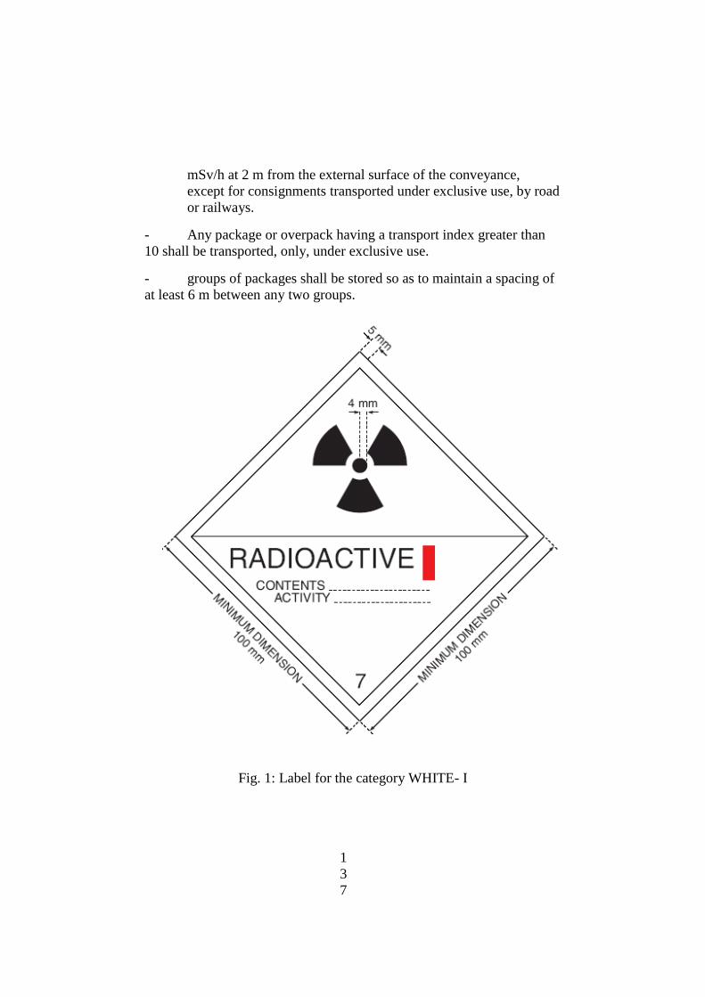
1
3
7
mSv/h at 2 m from the external surface of the conveyance,
except for consignments transported under exclusive use, by road
or railways.
- Any package or overpack having a transport index greater than
10 shall be transported, only, under exclusive use.
- groups of packages shall be stored so as to maintain a spacing of
at least 6 m between any two groups.
Fig. 1: Label for the category WHITE- I

1
3
8
Fig. 2: Label for the category YELLOW-II

1
3
9
Fig. 3: Label for the category YELLOW-III
a