radiation safety training manual - policy repository · radiation safety training manual university...
TRANSCRIPT

RADIATION SAFETY TRAINING MANUAL University of Notre Dame Revised April 2008
Page 1
RADIATION SAFETY
TRAINING MANUAL
University of Notre Dame

RADIATION SAFETY TRAINING MANUAL University of Notre Dame Revised April 2008
Page 2
RADIATION SAFETY TRAINING MANUAL
University of Notre Dame Notre Dame, Indiana
Prepared by: The Risk Management & Safety Department Course Participant_____________________________________ Responsible Investigator________________________________ Department___________________________________________

RADIATION SAFETY TRAINING MANUAL University of Notre Dame Revised April 2008
Page 3
Table of Contents
Title Page I Introduction 4 II Atomic Structure and Particles 7 III Units of Radiation Measurements, Biological Effects Of Radiation and Exposure Limits 15 IV Principles of Radiation Film Badges and Detection Instruments 23 V Basic Principles of Radiation Protection 32 Emergency Information 50 Tables Table 1. Cancer Risk Estimates from Exposure to Low Level Radiation 20 Table 2. Estimated Loss of Life Expectancy from Health Risks 20 Table 3. Permissible Occupational Exposure Limits 21 Table 4. Specific Gamma Ray Constants 27 Table 5. Maximum Beta Energies 31 Figures
Figure 1. Organizational Structure 2 Figure 2. Chart of the Nuclides 6 Figure 3. Effects of Acute Radiation Exposure 16 Figure 4. Effect of Varying the Voltage applied across the electrodes 22 of an ionization chamber Figure 5. The Inverse Square Law 30 App. A Exponential Functions 41 - 45

RADIATION SAFETY TRAINING MANUAL University of Notre Dame Revised April 2008
Page 4
I. INTRODUCTION
All employees, graduate students and undergraduate students working in or frequenting a radioisotope laboratory should understand the hazards of radiation and follow the procedures necessary for working safely around them. This training manual and associated training session is designed to provide principles, procedures and practices which new radioisotope users should know. Hopefully, this training program will correct any misconceptions you may have about radiation and safety and prepare you for routine use of radionuclides and for certain emergencies that may arise in the laboratory. The organization structure of the University of Notre Dame's Radiation Safety Program is outlined on the following page. The duties and responsibilities of each group indicated are listed in the University of Notre Dame's Radiation Safety Manual. Specific procedures for radionuclide procurement and use are also included in the University's Radiation Safety Manual. The Risk Management and Safety Staff is concerned with your understanding of radiation, proper evaluation or radiation hazards, and keeping radiation exposures As Low As Reasonable Achievable (ALARA). Do not hesitate to call the Risk Management and Safety Department at 631-5037 if you have any questions concerning radiation protection. The University of Notre Dame is authorized to use radioactive material by the United States Nuclear Regulatory Commission under broad scope license number, 13-01983-15.

RADIATION SAFETY TRAINING MANUAL University of Notre Dame Revised April 2008
Page 5
Figure 1. Organizational Structure
Radiation Safety Program Chart of Organization
President, of the University
Radiation Control Committee
Biological Sciences - South
Radiation Safety Officer
Biological Sciences - North
Chemistry Area Safety Officer
Physics Area Safety Officer
Responsible Investigators
Radiation Users
Engineering Area Safety Officer
Radiation Lab
IUSM

RADIATION SAFETY TRAINING MANUAL University of Notre Dame Revised April 2008
Page 6
What is Radiation?
Radiation consists of energy or particles, emitted from a source, that travel in wave-like motions. You are familiar with many kinds of radiation. Heat, light, and sound are kinds of radiation. The kind of radiation that we work with at most laboratories is called ionizing radiation. We cannot feel, see, hear, taste, nor smell this radiation. It is capable of producing small electrically charged particles called ions as it passes through matter. Such ions, produced in the cells of our body in sufficient quantities, can cause malfunctioning or damage in varying degrees. Man is being continuously bombarded by ionizing radiation. This radiation comes from outer space and from natural radioactive elements in the earth. The amount of such radiation varies but usually it is not sufficient to produce harmful effects. This radiation in man's natural environment is called BACKGROUND or NATURAL radiation. Only recently (since 1895) man has learned to produce large amounts of ionizing radiation. This has been done by concentrating natural radioactive materials, by using certain electrical operation devices (e.g. X-ray machines), and synthesizing material through the use of nuclear reactors and accelerators (e.g. radioisotopes). There are 106 different kinds of unique atoms, called elements. All atoms of an element behave the same chemically, even though they may vary in weight. Atoms which vary in weight but behave the same chemically are said to be isotopes of the same element. Isotopes which emit radiation because they are too heavy compared to their neighbors are called radioactive isotopes. Many of these radioactive isotopes are used in research laboratories at Notre Dame. The types of ionizing radiation with which we are mainly concerned are alphas, betas, gammas, X-rays and neutrons and each of these will be defined in the following chapters.
RADIATION SAFETY TRAINING MANUAL University of Notre Dame Revised April 2008
Page 7
II. ATOMIC STRUCTURE AND RADIATION
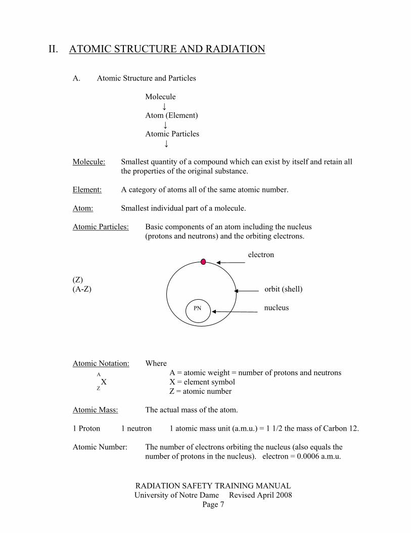
A. Atomic Structure and Particles Molecule ↓ Atom (Element) ↓ Atomic Particles ↓ Molecule: Smallest quantity of a compound which can exist by itself and retain all the properties of the original substance. Element: A category of atoms all of the same atomic number. Atom: Smallest individual part of a molecule. Atomic Particles: Basic components of an atom including the nucleus (protons and neutrons) and the orbiting electrons.
electron
(Z) (A-Z) orbit (shell) PN nucleus Atomic Notation: Where A A = atomic weight = number of protons and neutrons X X = element symbol Z Z = atomic number Atomic Mass: The actual mass of the atom. 1 Proton 1 neutron 1 atomic mass unit (a.m.u.) = 1 1/2 the mass of Carbon 12. Atomic Number: The number of electrons orbiting the nucleus (also equals the number of protons in the nucleus). electron = 0.0006 a.m.u.

RADIATION SAFETY TRAINING MANUAL University of Notre Dame Revised April 2008
Page 8
Example:
3H (tritium) 14C (carbon) 32P (phosphorous) 1 6 15 Nuclide: A species of atom having specified numbers of neutrons and protons in its nucleus. To be regarded as a distinct nuclide, the atom must be capable of existing for a measurable time. Isotopes: Nuclides all having the same atomic number. (equal number of protons, different number of neutrons.) 1H hydrogen-stable isotope (non-radioactive) 1
2H deuterium - stable isotope (non-radioactive) 1
3H tritium - radioactive isotope 1
Atomic number determines chemical properties of an atom. example: 1 proton = hydrogen 1H
6 proton = carbon 6C
8 proton = oxygen 8O
Isobars: Nuclides all having the same atomic weight. example: 3H 3H
1 2 Isotones: Nuclides all have the same number of neutrons.
15P 17N
6P 8N
1P 2N
K shell
L shell
M shell

RADIATION SAFETY TRAINING MANUAL University of Notre Dame Revised April 2008
Page 9
Figure 2. Chart of Nuclides
Periodic Table of the Elements
Ia 0
1
H Hydrogen
1.0079
IIa
IIIa IVa Va VIa VIIa
2
He Helium 4.0026
3
Li Lithium 6.941
4
Be Berylliu
m 9.0122
5
B Boron10.811
6
CCarbon 12.011
7
N Nitrogen 14.0067
8
O Oxygen15.9994
9
F Fluorine18.9984
10
Ne Neon 20.179
11
Na Sodium 22.9898
12
Mg Magnes
ium 24.305 IIIb
IVb Vb VIb VIIb _VIII_ Ib IIb
13
Al Aluminum
26.9815
14
Si Silicon 28.086
15
P Phosphorus
30.9738
16
S Sulfur32.066
17
ClChlorine35.453
18
Ar Argon 39.948
19
K Potassium
39.098
20
Ca Calciu
m 40.078
21
Sc Scandiu
m 44.956
22
Ti itanium 47.90
23
V Vanadium
50.941
24
Cr hromium 51.996
25
Mn Manganese
54.938
26
FeIron5.847
27
Co Cobalt58.933
28
Ni Nickel58.71
29
CuCopper63.546
30
Zn Zinc 65.38
31
GaGallium
69.72
32
Ge German
ium 72.59
33
As Arsenic 74.922
34
Se Seleniu
m 78.96
35
BrBromine79.904
36
Kr Krypton
83.80
37
Rb Rubidium
85.468
38
Sr Stronti
um 87.62
39
Y Yttrium 88.906
40
Zr rconium 91.22
41
Nb Niobium 92.906
42
Mo olybdenu
m 95.94
43
Tc Technetium
44
Ruuthenium01.07
45
Rh Rhodium102.906
46
Pdalladium106.4
47
AgSilver07.868
48
Cdadmium112.40
49
In Indium114.82
50
Sn Tin
118.69
51
Sb Antimony
121.75
52
Te Telluriu
m 127.60
53
I Iodine
126.905
54
Xe Xenon 131.30
55
Cs Cesium 132.905
56
Ba Barium 137.34
57
La Lanthanu
m 138.906
72
Hf afnium 178.49
73
Ta Tantalum 180.948
74
W Tungsten
183.85
75
Re Rhenium
186.2
76
Ossmium190.2
77
Ir Iridium192.22
78
Pt latinum195.09
79
AuGold96.967
80
HgMercury200.59
81
Tl Thallium204.37
82
Pb Lead 207.2
83
Bi Bismuth 208.98
84
Po Poloniu
m
85
AtAstatine
86
Rn Radon
87
Fr Francium
88
Ra Radium 226.03
89
Ac Actinium
227.03
104
Rf utherfordium
105
Db Dubnium
106
Sg eaborgium
107
Bh Bohrium
108
Hsassium
109
Mt eitnerium
110
Dsarmstadt
ium
111
Rooentgeni
um
58-71 lanthanides
58
Ce Cerium 140.12
59
Pr raseodymium
140.908
60
Nd Neodymium
144.24
61
Pm omethium
62
Sm Samarium
150.4
63
Eu Europium
151.96
64
Gdadoliniu
m 157.25
65
Tb Terbium 158.92
66
Dy ysprosium
162.50
67
Ho Holmium
164.93
68
Er Erbium 167.26
69
TmThulium168.93
70
Yb Ytterbium
173.04
71
LuLutetiu
m 174.97
90-103 actinides
90
Th Thorium 232.04
91
Pa Protactinium
231.04
92
U Uranium 238.03
93
Np eptunium
94
Pu Plutonium
95
Am Americium
96
CmCurium
97
Bk Berkelium
98
Cf Californium
99
Es Einsteiniu
m
100
Fm Fermium
101
Md Mendeleviu
m
102
No Nobelium
103
LrLawren
cium
RADIATION SAFETY TRAINING MANUAL University of Notre Dame Revised April 2008
Page 10
B. Radioactivity and Types of Radiation Radioactivity: An atom having an unstable nucleus thereby emitting a part of the nucleus (called corpuscular radiation) in order to go to a more stable state. This action is often accompanied by release of electromagnetic radiation called gamma rays. Unit of measure of radioactivity is the Curie (Ci) Curie = 3.7 x 10¹⁰ dps = 2.22 x 10¹² dpm (mCi) millicurie = 3.7 x 10⁷ dps = 2.22 x 10⁹ dpm (uCi) microcurie = 3.7 x 10⁴ dps = 2.22 x 10⁶ dpm dps = disintegrations per second International unit of measurement: Becquerel Becquerel (Bq) = (sec-1) reciprocal second 1 Curie = 3.7 x 10¹⁰ sec-1
Example: 600 dpm = 600 events x 1 min = 10 events = 10Bq min 60 sec sec Energy: 1 electron volt (dV) = 1.6 x 10⁻¹⁹ joules 1 keV = 103 eV 1 meV = 106 eV Major Types of Radiation Symbol alpha (stripped helium nucleus - He2+) α beta (electron) B- beta (positron) - positively charged electron B+ gamma δ x-ray x neutron n Alpha radiation: Particles - two protons and two neutrons (a helium nucleus) are ejected as a single particle from the nucleus. These particles usually carry high amounts of energy and are too heavily ionizing to travel father than a few inches in air. An alpha particle is easily stopped by such materials as a sheet of paper or the cells on the outside of our skin. However, if alpha particles get inside the body, they can produce greater damage then any other kind of radiation. Laboratories at Notre Dame seldom use alpha-emitting materials and thus such a hazard as this would not ordinarily be
RADIATION SAFETY TRAINING MANUAL University of Notre Dame Revised April 2008
Page 11
encountered. The recent alarm concerning 210Po in tobacco smoke is due to the fact that 210Po is an alpha emitter. The same is true for radon. Beta radiation An electron or positron emitted from the nucleus of an atom. Beta particles travel at very high speeds and may have a range in air of 12 to 15 feet, depending upon their energy. This energy and speed may cause them to penetrate several layers of the skin. You can usually shield out or greatly reduce exposure with a thin piece of aluminum, glass or plastic. Positrons will annihilate with electrons forming (usually) two 511 keV photons. Gamma radiation: Electromagnetic (waves) - energy is released from the nucleus in the form of radiant energy, not as particles. Gamma rays, like visible light and x-rays, are small bits of energy called photons traveling in straight lines in the form of waves. They are more energetic than visible light. X-rays and gamma rays have enough energy to go completely through the body where visible light is stopped by the body. Gamma rays are usually attenuated with thick sheets of lead or concrete. Neutron radiation: Neutrons are particles, about 1/4 the mass of alpha particles, and are difficult to stop. Neutrons are electrically neutral, as opposed to alpha and beta particles, which are electrically charges. Therefore, neutrons are more difficult to stop and have a very good chance of passing through materials without interacting or striking anything. Neutrons are usually attenuated with such materials as paraffin, cadmium, or water. Bremsstrahlung radiation: X-radiation produced as high energy beta particle slows down quickly as it passes through matter. Bremsstrahlung radiation is German for "Braking Radiation" example: To prevent bremsstrahlung production by 32P, plexiglass (low atomic number) shielding is used.

RADIATION SAFETY TRAINING MANUAL University of Notre Dame Revised April 2008
Page 12
Decay Schemes: Alpha decay
Beta decay
4.194 α 0.00007%
0.599
0.447
4.340 α 0.00051%
0.186
226Ra (1602 yr)
4.595 α 5.44%
0
4.782 α 94.6%
4.87 Nuclear Disintegration Energy Q-value Energy above 222Rn Ground state (Mev)
Discrete energies
#2
Eα
a. Negatron emission 1.71
32P15 (14.3d)
B- 1.71 100%
0
32S16
#B- 0.55 MeV
EB- 1.71 MeV

RADIATION SAFETY TRAINING MANUAL University of Notre Dame Revised April 2008
Page 13
Gamma decay - radiative transition
b. Positron emission
2.22
13N (10.0 Min) 7
B+ 100%
13C 6
0
#B+
E B+ (2.22 -2moc2)
c. Electron capture 0.232 26 (2.6 yr)
E. C. 100%
0 55Mn 25
(X-rays of Mn are observed)
2.88 10.5m
2.82
60Co 27 (5.26 yr)
B- 0.12%
B- 99+% t1/2 not
measurable
t 1/2 = 7x 10s
60Ni28
2.51
a. Isomeric transition
I. T. proceeds by mainly internal conversion
#e-
Ee-
1.33
0
k
l
m

RADIATION SAFETY TRAINING MANUAL University of Notre Dame Revised April 2008
Page 14
C. Interaction with Matter Charged Particles: The primary mode is via ionization of the atoms in the material. Ionization is the process of producing ions. Ions are electrons, atoms or groups of atoms carrying an electric charge - positive or negative.
Neutrons: The primary mode (in hydrogenous material) is by collisions with protons. The recoiling protons can then produce ionization (say in tissue).
Ionizing PathIonizing Particle
Negative Ion (free electron)
Positive Ion
Ion pair
Impact
b. Internal conversion
1.176
0.662
0
137Cs 55
(30.0 yr)
B- 6.5%
B- 93.5%
137M Ba 56 t1/2 = 2.55 min
137Ba56
0.662 MeV state in 137Ba can decay via several alternate Modes, including gamma rays, ejection of K,L, M, N, etc, electrons from the atomic shells.
#e-
Ee-
B- k
l
m

RADIATION SAFETY TRAINING MANUAL University of Notre Dame Revised April 2008
Page 15
Photons: Must interact with the atoms in the stopper by either A. Photoelectric effect, an ionization of the atoms. B. Compton effect, an inelastic scattering of the photon by a free electron. C. Pair production, conversion of a photon into an electron positron pair. Threshold is 1.022 Mev. Radioactive Decay: If the activity of a source is measured over a period of time, it can be observed that the disintegration rate, and thus the number of radioactive atoms present, decreases with time. When half of the radioactive atoms in the source have decayed, the disintegration rate is also cut in half. The time in which the source activity decreases to half its original value is called half life (t1/2) for that particular radionuclide. The half-lives of commonly used radionuclides cover a wide range, e.g. the half-life for 32P is 14.3 days, for 3H is 12.3 years, and for 14C is 5570 years. The equation for radioactive decay is: A = Aoe
-n (t)
Where: Ao = activity before decay A = activity remaining after decay e = base of natural log t = decay time n = decay constant =0.693 t1/2 t1/2 = half life of the radionuclide See Appendix A = Exponential Functions
III. UNITS OF RADIATION MEASUREMENTS, BIOLOGICAL EFFECTS OF RADIATION AND EXPOSURE LIMITS A. Exposure Units 1. Roentgen (R) - Unit of External Radiation Exposure The Roentgen is a unit of external radiation exposure for x-ray and gamma radiation only. It measures that amount of ionization produced by photon radiation in a given sample of air at STP (1cc). The Roentgen measures TOTAL EXPOSURE in a given field. One Roentgen equals 2.58 x 10⁻⁴ coulomb per kilogram of air.
RADIATION SAFETY TRAINING MANUAL University of Notre Dame Revised April 2008
Page 16
2. Rad (r) - Unit of Radiation Absorbed Dose The rad is a measure of LOCAL ENERGY deposited per unit mass of absorber by ionizing radiation. The rad is used to express dose from alpha, beta or gamma radiation. 1 rad = 100 ergs absorbed energy = 0.01 Joules 1 gm of absorber Kg The rad depends upon: A. Type of radiation (alpha, beta, gamma). B. Type of absorber (density) C. Intensity of radiation field (mR). New S.I. Unit - Gray which is equal to 100 rads or 1 Joule/kG 3. Rem (rem) - Roentgen Equivalent Man The rem is related to the rad by the relative biological effectiveness (RBE) of the radiation…..also know as Quality Factor (QF). Number of rem = QF x rads example: 0.05 rad of alpha radiation produces the same biological effect as 1.0 rad of x or gamma radiation. RBE or QF for alphas = 1.0 rad gamma = 20 0.05 rad alpha Quality Factors (QF) for various types of radiation are: Alpha Particles 20 Protons 10 Fast Neutrons 10 Thermal Neutrons 5 Gamma Rays 1 Beta Particles 1 International S. I. Unit - Sievert which is equal to 1 Joule/Kg = 100Rems

RADIATION SAFETY TRAINING MANUAL University of Notre Dame Revised April 2008
Page 17
B. Biological Effects of Radiation 1. Effect of Ionizing Radiation on Biological Tissue. example: Water molecules (80 percent body composition). H2O ↓ ionizing radiation electron e- + H20+ positive ion H2O ← → H2O Hydrated electron eaq- (H2O)* OH + H3O+ free radical ↓ H2 + 0* molecular hydrogen and free radical or •OH + H• free radical •OH + H• → H2O water, dominant product •OH + OH• → H2O2 hydrogen peroxide, very corrosive •H + H• → H2 hydrogen gas, explosive •OH + OH• → strand breaks, base damage, etc. 2. Sources of Radiation Exposure to Man Earth Man-Made Cosmic Ray 40 mrem/yr Medical 100 mrem/yr Earth 260 mrem/yr Occupational 1 mrem/yr Internal Man 25 mrem/yr Fallout <1 mrem/yr Total Natural ≈ 325 mrem/yr Total Man-Made ≈ 102 mrem/yr

RADIATION SAFETY TRAINING MANUAL University of Notre Dame Revised April 2008
Page 18
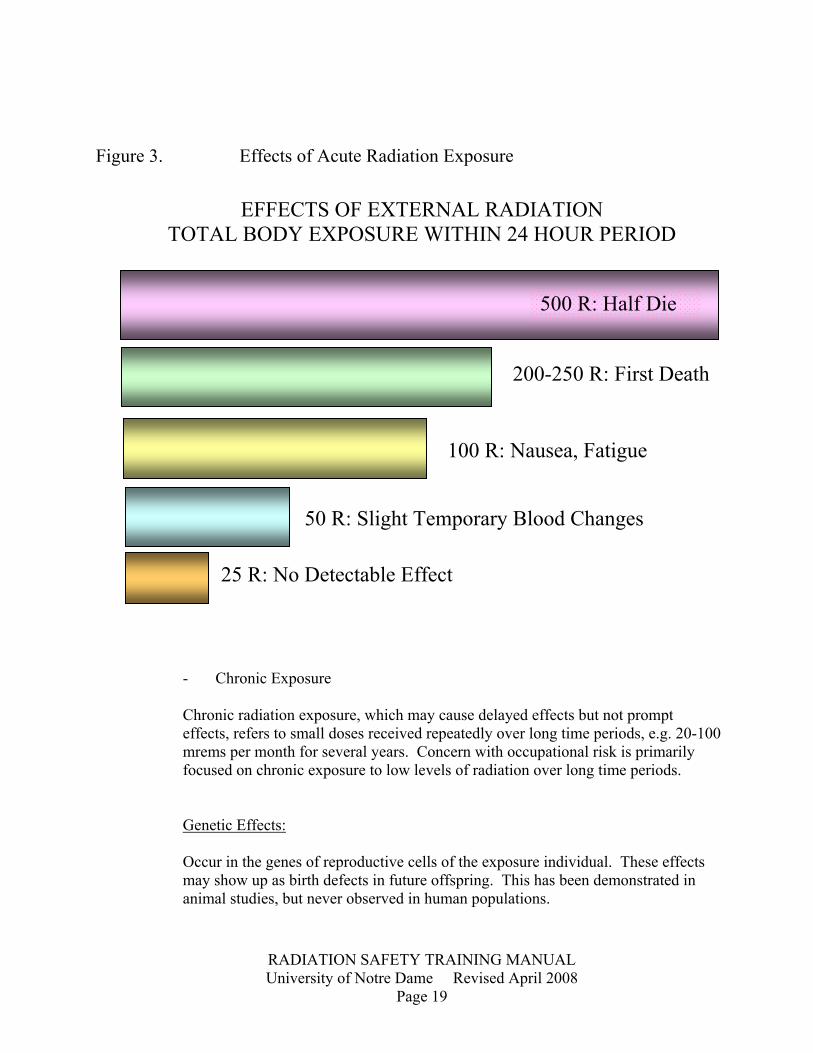
Local Dose from Medical Diagnostic Procedures Radiology Nuclear Medicine Chest x-ray 50 mrad Liver Scan 1 rad Mammogram 1 rad Lung Scan 1 rad G. I Series 1-5 rad Bone Scan 0.5 rad 3. Risk vs. Benefit Benefits: Medical diagnosis and therapy Research Nuclear Power Household convenience Employment (radiation workers) Risks: Risk can be defined in general as the probability (chance) of injury, illness, or death resulting from some radiation exposure. The effects of radiation are categorized as either somatic or genetic. Somatic effects: Directly affect the health of the person exposed. - Acute Exposure Acute radiation exposures, which causes prompt effects and may cause delayed effects, refers to a large does of radiation received in a short period of time, e.g., exposure following a nuclear bomb explosion. (See figure 3.)
RADIATION SAFETY TRAINING MANUAL University of Notre Dame Revised April 2008
Page 19
Figure 3. Effects of Acute Radiation Exposure
EFFECTS OF EXTERNAL RADIATION
TOTAL BODY EXPOSURE WITHIN 24 HOUR PERIOD
- Chronic Exposure Chronic radiation exposure, which may cause delayed effects but not prompt effects, refers to small doses received repeatedly over long time periods, e.g. 20-100 mrems per month for several years. Concern with occupational risk is primarily focused on chronic exposure to low levels of radiation over long time periods. Genetic Effects: Occur in the genes of reproductive cells of the exposure individual. These effects may show up as birth defects in future offspring. This has been demonstrated in animal studies, but never observed in human populations.
25 R: No Detectable Effect
50 R: Slight Temporary Blood Changes
100 R: Nausea, Fatigue
200-250 R: First Death
500 R: Half Die

RADIATION SAFETY TRAINING MANUAL University of Notre Dame Revised April 2008
Page 20
Table 1. Cancer Risk Estimates From Exposure to Low-Level Radiation Number of additional cancers estimated to occur in 1 Source million people after exposure of each to 1 rem of radiation BEIR 1979 268-399 ICRP 1977 300* UNSCEAR 1977 300* _______________________________________________________________ *Only one-third of excess cancer causes would be fatal. That is, 100 excess delayed cancer deaths per 1 million people after 1 rem exposure. BEIR - Biological Effects of Ionizing Radiation, Report of the Science Work Group of the Interagency Task Force on Radiation, DHEW, June 1979. ICRP - International Commission on Radiation Protection, ICRP, Publication 26, Pergamon Press January 1977. UNSCEAR - Sources and Effects of Ionizing Radiation, United Nations Scientific Committee on the Effects of Atomic Radiation, 1977. Table 2. Estimated Loss of Life Expectancy from Health Risks Estimates of Days of Life Health Risk Expectancy Lost, Average Smoking 20 cigarettes/day 2370 (6.5 years) Overweight (by 20%) 985 (2.7 years) All accidents combined 435 (1.2 years) Auto Accidents 200 Alcohol consumption (U.S. average) 130 Home accidents 95 Drowning 41 Safest jobs (such as teaching) 30 Natural background radiation, calculated 8 Medical x-rays (U.S. average), calculated 6 All catastrophes (earthquake, etc). 3.5 1 rem occupational radiation dose, calculated (industry average is 0.34 rme/yr) 1 1 rem/yr for 30 years, calculated 30 5 rems/yr for 30 years, calculated 150 These estimates indicate that the health risks from occupational radiation exposure are not greater than the risks associated with many other events or activities we encounter in normal day to day activities

RADIATION SAFETY TRAINING MANUAL University of Notre Dame Revised April 2008
Page 21
Table 3. Permissible Occupational Exposure Limits -External Radiation ANNUAL * Whole Body (W.B.) 5 rem Lens of the eye 15 rem Skin of W.B. 50 rem Extremities 50 rem (hands, etc.) * Minors are limited to 0.5 rem/yr W.B. and the unborn child to 0.5rem/gestation period. Dose to the whole body includes a dose to head and trunk; active blood forming organs; gonads. This is the sum of both external and internal radiation doses. Monitoring must be provided for any individual who might receive a dose from external radiation which exceeds the following: Over 18 years of age Under 18 years of age 500 mrem/yr W.B. 50 mrem/yr W.B. 1500 mrem/yr eye 150 rmem/yr eye dose 5000 mrem/yr extremities 500 mrem/yr extremities 5000 mrem/yr skin of W.B. 500 mrem/yr skin of W.B. As a general rule at the University of Notre Dame, film badges must be worn by personnel working in the following instances: a. When working with 1 MeV or greater beta emitters. b. When working with gamma emitters of any energy. c. When working with neutron sources or neutron generating devices. d. When working with x-ray producing devices. Internal Radiation Bioassays for determining the dose from internal radiation exposure are required for all personnel likely to receive 10% of the maximum permissible dose. Reference NRC Regulatory Guide 8.34 for further information.

RADIATION SAFETY TRAINING MANUAL University of Notre Dame Revised April 2008
Page 22
ALARA Principle (As Low As Reasonably Achievable) The ALARA Principle is the Philosophy in which radiation exposures are kept as low as reasonably achievable. Any unnecessary radiation exposure to employees should be eliminated by careful planning of laboratory procedures and by employing common radiation safety techniques. 1. Problems in Establishing Permissible Doses. a. Non-uniformity of ionizing radiation 1. probability of interacting with tissue 2. energy transfer example: If an H2O molecule is hit by a large particle, there is much disruption but the probability of reassocation is great. If an H2O molecule is exposed to a gamma ray, there is a low probability of interaction. But if the molecule is hit, the products are scattered farther and the probability of reassociation is reduced. b. Non-uniformity of tissue response 1. Depends on tissue age, type, activity, etc. c. Species differences 1. Most data is from species other than man. Data on man is very sparse. d. Susceptibility varies with age 1. Young are much more susceptible to radiation effects e. Extrapolation to Low Doses 1. Data for low doses not available 2. Becomes a measurement of probabilities 3. Extrapolation assumes either a. linear relationship b. threshold level

RADIATION SAFETY TRAINING MANUAL University of Notre Dame Revised April 2008
Page 23
4. dose rate dependency
IV. Principles of Radiation Film Badges and Detection Instruments All radiation detection devices rely on the production of ions or deposition of energy in matter. alphas - ionize and excite atoms → ion pairs betas - ionize and excite atoms → ion pairs (also deposit energy by bremsstrahlung process → x-rays) x-rays & gamma rays - give energy to electrons via: 1. photoelectric effect 2. compton effect 3. pair production Electrons then lose energy → ion pairs neutrons - collisions with protons in nuclei result in recoiling protons that deposit energy and produce ion pairs. A. Radiation Film Badges Photographic film is one of the first radiation detectors used after the discovery of x-rays. Photographic emulsions in the film are divided into 2 major categories according to the intended use. They are x-ray film, usually in dental packet size which can be calibrated for x- or gamma- ray exposures in terms of blackening and nuclear emulsions on film or glass plates, which are used in studying ionizing particle tracks.
Dose or Dose Rate
# of deaths or lesions
known

RADIATION SAFETY TRAINING MANUAL University of Notre Dame Revised April 2008
Page 24
The radiation sensing element in these emulsions is a silver halide, most often sliver bromide. Small crystals of silver bromide, ranging in size from less than 0.5 microns in nuclear emulsion to a few microns in the most sensitive x-ray emulsions, are suspended in a gelatin which is generally deposited on a cellulose acetate film or on a glass plate. Electrons traversing the emulsion become trapped in the crystal lattice, causing the reduction of silver ions to atomic silver. Theses silver atoms, in turn, result in deeper traps, which capture electrons, reducing more silver ions, eventually forming microscopic aggregates of silver atoms which constitute the latent image. Upon development, the latent images are reduced to metallic silver which appears to the eye as the blackening of x-ray or gamma-ray film or microscopic tracks in the nuclear emulsions. B. Ionization Detectors - Gas Filled Principle- As radiation traverses as gas, ions are produced and in an electric field will migrate toward the appropriate electrode. If the voltage is large enough, ion pairs will not recombine before collection. 1. Electroscope Discharge Type Ions cause discharge of electroscope by neutralizing some of the charges.
e-
+
+
- -
+
+ +
Separation of "leaves" indicates amount of radiation passing through.

RADIATION SAFETY TRAINING MANUAL University of Notre Dame Revised April 2008
Page 25
1014
1012
1010
108
106
104
1
102
Number of ions collected at anode
Applied voltage
Saturation region
Ionization chamber
Proportional region
Proportional counter
Limited proportionality region
Geiger muller region
Geiger muller counter
Continuous discharge region
Specific ionization (air) a ≈ 104 - 105 ion pairs/cm b ≈ 50 - 100 ion pairs/cm δ ≈ 1 - 10 ion pairs/cm but actually about the same as beta because of electrons formed.
a
b
δ
Recombination region
2. Ionization Chamber (Air Filled) This chamber operates in the saturation region. (see figure 4) all of the ions formed by the radiation are collected. This collection results in what is called a saturation current in the external circuit. 3. Proportional Counter This detector operates in the proportional region. The radiation produces ionization as it passes through the gas. This is called primary ionization. Primary ions are formed. The electrons from these primary ions are then accelerated by the applied voltage. They acquire enough kinetic energy to produce further ionization of the gas. This is called secondary ionization. Secondary ions are formed. The production of secondary ion pairs is called gas multiplications or gas amplification. Figure 4. Effect of varying the voltage applied across the electrodes of an ionization chamber.
Ionization chamber
B R
Insulator
C
Electrometer Measuring Circuit S

RADIATION SAFETY TRAINING MANUAL University of Notre Dame Revised April 2008
Page 26
Number of ion pairs collected = mNØ Where m = multiplication factor which is dependent on V N = initial number of primary ions produced Ø = efficiency of ion collection The primary ionization produced by an alpha particle is large compared to the primary ionization produced by either a beta particle or a gamma ray. Therefore, the total number of ion pairs collected from an alpha particle is large compared to the number collected from a beta particles and gamma rays.
Limited Proportionality Region The gas multiplication factor begins to become dependent on the primary ionization. 4. "Geiger-Muller" Counters These counters operate in the Geiger Muller region where the total number of ion pairs collected at a given voltage is independent of primary ionization. Therefore, the total number of ion pairs collected is the same for all ionizing radiations and one cannot distinguish between different kinds. At any given voltage, the total numbers depend only on the voltage and the construction of the ionization chamber. G-M tubes are very sensitive and little amplification of the pulse is necessary to drive a counting circuit. G-M tubes are also relatively inexpensive.
G-M Tube
Cathode
High Voltage Supply
Collector C
R
Counter Scaler
1500
1400
900 1000 1200 1400
1300
1200
1100
1000
Volts
Halogen Organic
8 %
12 %
Typical plateau slopes of organic-halogen quenched G-M counters

RADIATION SAFETY TRAINING MANUAL University of Notre Dame Revised April 2008
Page 27
Scintillation P.M. Tube
HV
Pre-amp Amp Pulse Height Analyzer
Recorder Scaler
Electrons
A sodium iodide crystal and photomultiplier
Light
Reflector
Nat (tl) Crystal
gamma-ray
Photosensitive Layer
If the count rate is greater than 1000 counts per minute in a G-M tube, a correction factor is necessary to reflect the lost of counts due to the "dead time or resolving time" in each system. Resolving time is defined as the minimum time interval between two distinct events which will permit both to be counted. Resolving time for most G-M tubes is usually between 100 to 300 usec. To determine the true counting rate of a system, the following equation is used: Rtrue = R obs Where; Rtrue = True counting rate
1- R obs R obs = Observed counting rate
= Resolving time Organic molecules or halogen molecules are added to the gas in the G-M tubes are sensitive to quench or reduce the resolving time in a system. Though G-M tubes are sensitive to gamma radiation their efficiency for gamma detection is low (approximately 1%). C. Scintillation Detectors System: Scintillation detectors consist of: 1. A scintillation; usually an inorganic solid (Nal), an organic plastic or organic liquid. 2. A photomultiplier tube 3. High Voltage power supply (700-2000 volts) 4. Preamplifier 5. Amplifier 6. Pulse height analyzer 7. Recorder-Scaler, etc.
RADIATION SAFETY TRAINING MANUAL University of Notre Dame Revised April 2008
Page 28
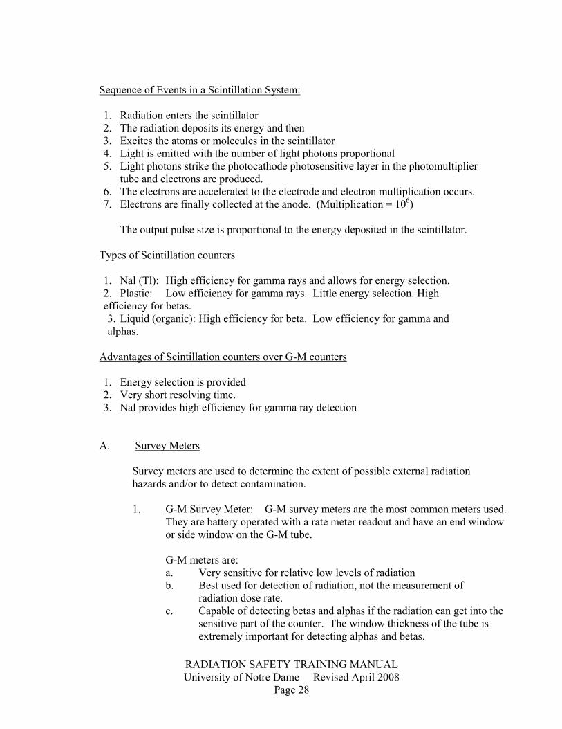
Sequence of Events in a Scintillation System: 1. Radiation enters the scintillator 2. The radiation deposits its energy and then 3. Excites the atoms or molecules in the scintillator 4. Light is emitted with the number of light photons proportional 5. Light photons strike the photocathode photosensitive layer in the photomultiplier tube and electrons are produced. 6. The electrons are accelerated to the electrode and electron multiplication occurs. 7. Electrons are finally collected at the anode. (Multiplication = 106) The output pulse size is proportional to the energy deposited in the scintillator. Types of Scintillation counters 1. Nal (Tl): High efficiency for gamma rays and allows for energy selection. 2. Plastic: Low efficiency for gamma rays. Little energy selection. High efficiency for betas. 3. Liquid (organic): High efficiency for beta. Low efficiency for gamma and alphas. Advantages of Scintillation counters over G-M counters 1. Energy selection is provided 2. Very short resolving time. 3. Nal provides high efficiency for gamma ray detection A. Survey Meters Survey meters are used to determine the extent of possible external radiation hazards and/or to detect contamination. 1. G-M Survey Meter: G-M survey meters are the most common meters used. They are battery operated with a rate meter readout and have an end window or side window on the G-M tube. G-M meters are: a. Very sensitive for relative low levels of radiation b. Best used for detection of radiation, not the measurement of radiation dose rate. c. Capable of detecting betas and alphas if the radiation can get into the sensitive part of the counter. The window thickness of the tube is extremely important for detecting alphas and betas.

RADIATION SAFETY TRAINING MANUAL University of Notre Dame Revised April 2008
Page 29
G-M meters can be calibrated for dose rate (mR/hr) for a particular gamma ray energy by using a source of know activity (mCi) and the following equation:
I = 1000 A where I = Intensity (mr/hr) d2 A = Activity (mCi)
= Specific gamma ray constant R-cm2/mCi-hr) d = distance in cm example: What is the exposure rate at 60 cm of a 100 mCi 137Cs source?
( for Cs is 3.3) 1000 mR/hr (3.3R-cm2/mCi-hr) (100 mCi) = 91.6 mR/hr (60 cm)2 Exposure rate will vary according to the inverse square law. All meters should be calibrated on several scales; x 0.1; x 1.0, x 100 mR/hr. This equation is used to convert between radioactivity (mCi) and exposure intensity (mR/hr). Any user can determine the exposure rate from a source they are using by using this equation and the gamma constant for the particular source. Gamma constants for many common radionuclides are listed in Table 4. Operation of a G-M meter 1. Check to make sure the meter has been calibrated in past 12 months. 2. Check batteries - most meters have a batter check setting. 3. Check against a source of known dose rate. 4. Start reading at the highest scale, e.g. x 100, and work done to lowest scale when entering a radiation area. 5. Some G-M tubes have a beta shield (end cap or side shield). Readings with and without the shield allow one to measure the relative number of high-energy betas and gamma vs. number of low energy betas. Caution - When interpreting mR/hr readings for betas, the meter reading will be incorrect or lower than the real exposure due to window absorption.

RADIATION SAFETY TRAINING MANUAL University of Notre Dame Revised April 2008
Page 30
2. Ion Chamber Survey Meters "Cutie Pie" or "Juno" are two popular styles of ion chambers. Most ion chambers are air-filled and directly measure exposure rate (R/hr or mR/hr) over a wide range of photon energies. These instruments are reliable and hold their calibration well. The ion chamber's major use is in measuring x- or x-ray dose rates at moderate to high radiation levels Table 4. Specific Gamma Ray Constants (R-cm2/mCi-hr) Nuclide
rt
Nuclide
rt
Nuclide
rt
Actinium-227 Antimony-122 Antimony-124 Antimony-125 Arsenic-72 Arsenic-74 Aresenic-76 Barium-131 Barium-133 Barium-140 Beryllium-7 Bromine-82 Cadmium-115m Calcium-47 Carbon-11 Cerium-141 Cerium-144 Cesium-134 Cesium-137 Chlorine-38 Chromium-51 Cobalt-56 Cobalt-57 Cobalt-58 Cobalt-60 Copper-64 Europium-152 Europium-154 Europium-155 Gallium-67 Gallium-72
2.2 2.4 9.8 2.7 10.1 4.4 2.4 3.0 2.4 12.4 0.3 14.6 0.2 5.7 5.9 0.35 0.4 8.7 3.3 8.8 0.16 17.6 0.9 5.5 13.2 1.2 5.8 6.2 0.3 1.1 11.6
Gold-198 Gold-199 Hafnium-175 Hafnium-181 Indium-114m Iodine-124 Iodine-125 Iodine-126 Iodine-130 Iodine-131 Iodine-132 Iridium-192 Iridium-194 Iron-59 Krypton-85 Lanthanum-140 Lutecium-177 Magnesium-28 Manganese-52 Manganese-54 Manganese-56 Mercury-197 Mercury-203 Molybdenum-99 Neodymiun-147 Nickel-65 Niobium-95 Osmium-191 Palladium-109 Platinum-197 Potassium-42
2.3 0.9 2.1 3.1 0.2 7.2 0.7 2.5 12.2 2.2 11.8 4.8 1.5 6.4 0.04 11.3 0.09 15.7 18.6 4.7 8.3 0.4 1.3 1.8 0.8 3.1 4.2 0.6 0.03 0.5 1.4
Potassium-43 Radium-226 Radium-228 Rhenium-186 Rubidium-86 Ruthenium-106 Scandium-46 Scandium-47 Selenium-75 Silver-110m Silver-111 Sodium-22 Sodium-24 Strontium-85 Tantalum-182 Tellurium-121 Tellurium-132 Thulium-170 Tin-113 Tungsten-185 Tungsten-187 Uranium-234 Vanadium-48 Xenon-133 Ytterbium-175 Yttrium-88 Yttrium-91 Zinc-65 Zirconium-95 Technetium-99m
5.6 8.25 5.1 0.2 0.5 1.7 10.9 0.56 2.0 14.3 0.2 12.0 18.4 3.0 6.8 3.3 2.2 0.025 1.7 0.5 3.0 0.1 15.6 0.1 0.4 14.1 0.01 2.7 4.1 0.78
Jaeger, R.G., et al., Engineering compendium on Radiation Shielding, Vol. 1, (New York: Springer-Verlag, 1968), pp. 21-30. t ſ = R-cm2/hr-mCi or ſ/10 = R/hr at 1 mCi A Manual of Radioactivity Procedures (National Bureau of Standards Handbook No. 80 [Washington, D.C.: Supt. Of Docs., U. S. Government Printing Office, Nov. 1961]), Appendix A, pp. 137-140.
RADIATION SAFETY TRAINING MANUAL University of Notre Dame Revised April 2008
Page 31
B. Personnel Monitoring Devices 1. Film badges - As mentioned in Section A, radiation badges are typically composed of 2 pieces of dental film covered by light-tight paper in a compact plastic container. Various filters in the badge holder allows areas to be restricted to x and gamma, hard gamma, and B+ gamma, which allows for determination of the energy level and type of external radiation exposure. Typically, the sensitivity of film badges are as follows: Gamma ray - 10-1800 mrem Beta - 50-1000 mrem The disadvantages of film badges are that they take 3 to 6 weeks for exposure records to be returned to the user and that they are not effective in measuring 3H and 14C. 2. Pocket Dosimeters (Ion Chambers - Electroscope type) - Fountain pen size dosimeter that measures total exposure over time from charging to time reading. This is considered an active device in that radiation exposure measurement can be read immediately as opposed to the passive film badge which cannot be read immediately as mentioned previously. C. Liquid Scintillation Detectors Liquid Scintillation detectors are used mainly to measure low energy beta emitters such as 3H and 14C. Radioactive samples are placed in scintillation cocktail mix which contains a fluor in an organic media. When radiation interacts with the fluor, the fluor emits light photons. These light photons must reach the photomultiplier tube in order to be detected. A major problem associated with liquid scintillation counting is called "quenching". This refers to the absorption of light pulses by the sample. The absorbed light pulses do not reach the photomultiplier tubes to be counted are, therefore, "lost". As more counts are lost, the instruments counting efficiency decreases. A correction factor must, therefore, be used to determine the actual total count. The most accurate method of determining the correction factor for quenching is called "internal standardization". An accurately measured activity, called a standard, is added to the sample vial which contains the unknown activity. The counting efficiency is then given by the ratio of the observed count rate to the theoretical activity of the standard. The observed count rate is obtained by subtracting the cpm of the unknown sample (CPMu) before the standard (uCi) is added, from the cpm of the unknown sample plus standard (CPMu + std).

RADIATION SAFETY TRAINING MANUAL University of Notre Dame Revised April 2008
Page 32
Then, Efficiency (cpm-uCi) = (CPMu + std) - CPMu Activity of std. (uCi) Once the efficiency has been determined, the activity of the original unknown sample is calculated: Unknown sample (uCi) = CPMu - CPM BKG Efficiency (CPM/uCi)
V. Basic Principles of Radiation Protection A. Time, Distance, Shielding 1. Time Exposure dose is a direct proportion of the amount of radiation you are exposed to and the amount of time you are being exposed. Exposed Dose (mR) = Exposure rate (mR/hr) x Time (hr) General Rule: Work as fast as possible without jeopardizing safety by being careless. 2. Decay Time As the radioactivity (millicuries) of a source decreases with time, according to the half life (t1/2), so dose the exposure rate (mR/hr) decrease. After one half-life, only half the activity remains and, therefore, only half the radiation intensity is present. 3. Distance Inverse Square Law (gamma radiation) Radiation intensity (I) from a point varies inversely as the square of distance from the source. The equation for inverse square is law is: I1 = (d2)
2 or I2 = I1 (d1)2
I2 (d1)
2 (d2)2
Where I1 = Radiation intensity (mR/hr) at distance d1 from the source I2 = Radiation intensity (mR/hr) at distance d2 from the source

RADIATION SAFETY TRAINING MANUAL University of Notre Dame Revised April 2008
Page 33
1 meter
d 0 d 1 2 meters
d 0 d 2
I 1
I 2
10mR/hr I 2
10mR/hr
I 2
10mR/hr I 2
10mR/hr
Point Source I 40mR/hr
Figure 5. The Inverse Square Law
As radiations are emitted from a point source, at 1 meter the radiation intensity is 40 mR/hr. At 2 meters (twice the distance from the point) source that same radiation field of 40mR/hr has expanded to an area four times the size of the original area. Consequently, the original area (I1) now receives only one-fourth of the entire radiation, i.e., 10mR/hr. 4. Shielding - Gamma Radiation Shielding is one of the most important principles for gamma radiation protection. It may be defined as absorption of energy (radiation) in an absorbing material (shielding). The absorption of energy by matter depends upon: a. Energy of radiation b. Density of absorbing medium c. Thickness of the absorber transversed. General rule at the University is that any containers of radioactive material that exceed 5.0mR/hr at the surface of the container must be shielded with material to sufficiently reduce the dose rate below 5.0 mR/hr.

RADIATION SAFETY TRAINING MANUAL University of Notre Dame Revised April 2008
Page 34
- Beta Radiation Beta particles are very small and light weight (9.1086 x 10⁻²⁸ grams) with maximum range dependent upon the maximum beta energy. TABLE 5. MAXIMUM BETA ENERGY Radionuclide Max. energy (MeV) 3H 0.018 14C 0.158 32P 1.709 35S 0.167 45Ca 0.254 59Fe 1.560 60Co 1.478 63Ni 0.066 90Sr 0.554 The absorbing thickness or range of beta particles in any material depends upon the density and thickness of the shielding material and the maximum energy of the beta particle. In general applications at Notre Dame, shielding for 3H and 14C solutions will not be necessary or required. For higher energy beta emitters such as 32P, lucite or plexiglass shielding should be used. High energy beta emitters will produce bremsstrahlung radiation when passing through high density shielding material. - Alpha radiation Alpha particles are very large and heavy (6.642 x 10⁻²⁴ gms), and usually easily shielded by a piece of paper. B. Reducing Internal Radiation Exposure It is extremely important to prevent radionuclides from being taken into the body. This is because there is no quick and easy way of speeding up the natural processes of removing the radioactivity from the body. It is also, in many cases, more difficult to measure how much radioactivity a person has ingested then to measure his external exposure. There are many situations, through, when radionuclides are taken into the body, either by accident or by controlled exposures of radiation workers or medical patients.

RADIATION SAFETY TRAINING MANUAL University of Notre Dame Revised April 2008
Page 35
The routes by which radionuclides can enter the body and their control measures are listed below: 1. Inhalation - An example of this is the inhalation of tritium around certain accelerators or when using tritiated water in synthesizing compounds. Control - Work with radioactive material in a fume hood when possible. If not, work in a well-ventilated area and wear an appropriate respirator. 2. Swallowing - This is probably the most common way in which radioactivity gets into the body. Usually these results are from contaminated hands or mouth pipetting. Control - Never mouth pipette radioactive materials. When working with radioactive materials, always use protective gloves. After working with radioactive materials, wash hands, and then monitor them before eating or smoking. 3. Absorption through the skin - A labeled chemical which can penetrate the skin will, of course, carry the radioactive materials. Control - Wear protective clothing such as lab coat and gloves. Immediately wash away any radioactive material spilled on the skin. 4. Absorption through wounds - This is a rare but easy way for radioactivity to enter the body. Control - Wear protective clothing and in some cases wear extra protective material. 5. Injection - This is again a rare route of entry but several individuals have accidentally injected themselves with radioactive material when attempting to inject research animals or nuclear medicine patients. Control - Be extra careful when performing injections.
RADIATION SAFETY TRAINING MANUAL University of Notre Dame Revised April 2008
Page 36
C. Personnel Protection Rules The following rules should be observed when handling or storing radioactive materials; 1. Become familiar with the requirements of the University of Notre Dame's Radiation Safety Manual. 2. Work Carefully. 3. Observe Time, Distance and Shielding principles while working with radioactive materials or while in a radiation area. 4. Film badges (when required) must be worn at all times when an individual is in a radioactive materials area. 5. Protective gloves should be worn when working with radioactive materials. 6. Do not touch clean areas such as bench tops, water faucets, door knobs and light switches with contaminated gloves. Remove the gloves or use a tissue between the glove and item to be touched. 7. A laboratory coat should be worn while you are in the isotope laboratory and removed when you leave for lunch, etc. 8. Monitor yourself for contamination at frequent intervals when working with high energy beta or gamma emitting materials. Monitoring should include hands, body, shoes and protective clothing. 9. A radiation contamination survey must be conducted at least once in every calendar month in which unsealed radioactive material is used. More frequent surveys may be required; you will be notified by the Radiation Safety Officer if this is required for the isotopes and quantity that your lab is using. A survey is required every time that more than one millicurie of P-32, I-125, or I-131 is used. 10. Prior to leaving the laboratory, wash your hands thoroughly. 11. No mouth pipetting of radioactive material is permitted. 12. Eating, drinking, smoking or application of cosmetics is prohibited in the radioisotope laboratory.

RADIATION SAFETY TRAINING MANUAL University of Notre Dame Revised April 2008
Page 37
13. Procedures involving loose (particulate) or volatile radioactive material should be conducted in an approved glove box or fume hood. All work and storage of iodine must be conducted in a properly working fume hood. 14. Clean up after yourself. Don not leave the laboratory contaminated or in a mess. Check all work areas for contamination. 15. Be familiar with and abide by information concerning the increased risks of prenatal radiation exposures. 16. Report immediately to your Responsible Investigator the details of spills or accidents involving radioactivity. 17. Conduct decontamination procedures as outlined in this manual (Section V.F.). 18. Know what steps to take in the event of an emergency. See emergency procedures in Section V.F. of this manual. D. Laboratory Facilities 1. The laboratory must be properly posted. 2. All containers of radioactive material should be identified by radionuclide, compound, radioactivity and date of assay. 3. Absorbent paper should cover bench tops where radioactive materials are handled or stored. Whenever possible, place a tray between the lined bench top and container of radioactive material. 4. Radioisotope areas should be maintained in clean and orderly conditions. 5. Storage of food or beverage is prohibited in radioisotope laboratories. 6. Radioactive material must be secured (locked) against unauthorized removal when left unattended. 7. Conduct surveys at required frequencies, and record data, including counting instruments used, on appropriate reporting form. Refer to the Radiation Safety Manual for frequency of surveys and contamination levels for respective radionuclides.

RADIATION SAFETY TRAINING MANUAL University of Notre Dame Revised April 2008
Page 38
E. Emergency Procedures An emergency is any incident resulting from the use of radioactive substances that presents or threatens to present an internal or external radiation hazard to personnel. In an emergency, the primary concern must always be the protection of human life and health. The secondary concern is the confinement of the contamination to the local area of the accident if possible and the protection of personnel from radiation hazards. 1. Procedures In the event of an emergency or suspected emergency, e.g. major spill, overexposure, etc., the Radiation Safety Officer and the Responsible Investigator shall be notified immediately without such action as to cause excessive spread of contamination. See page 47 for telephone numbers and additional emergency information. The user and Responsible Investigator shall be responsible for the decontamination procedures necessary and shall carry out these procedures under the direction of the Radiation Safety Officer or persons designated by him. a. Minor spills - Spills involving no significant radiation hazard to personnel. 1. Notify all other persons in the area at once. 2. Permit only the minimum number of persons necessary to deal with the spill into the area. 3. Confine the spill immediately. a) Liquid spills - Don protective gloves - Drop absorbent paper on spill - Wipe up and dispose of contaminated paper in solid radioactive trash. - Smear area and count smear to determine if decontaminated. b) Dry spills - Don protective gloves - Gently, damped area thoroughly and cover it with absorbent paper taking care not to spread contamination.

RADIATION SAFETY TRAINING MANUAL University of Notre Dame Revised April 2008
Page 39
- Wipe up and dispose of paper properly - Smear area and count smear to determine if area is decontaminated. 4. Notify the Radiation Safety Officer and Responsible Investigator as soon as possible, giving all details of the spill. b. Major spills - Spills involving radiation hazard to personnel: 1. Notify all persons not involved in the spill to vacate the room at once. 2. Make no immediate attempt to clean up the spill. a) If the spill is liquid and the hands are protected, right the overturned container. b) If the spill is on the skin, flush thoroughly with water. Do not scrub or use strong detergents. c) If spill is on clothing, discard outer or protective clothing at once. 3. Switch off all fans and air conditioners. 4. Vacate the room and prohibit unauthorized entrance to contaminated are. 5. Notify the Radiation Safety Officer and Responsible Investigator at once and give all details of the accident. 6. The spread of radioactive contamination can be diminished by restricting the movements of potentially contaminated persons to a local zone just outside of the spill area until the extent of shoe and clothing contamination is ascertained. 7. Anyone who might have been contaminated should be monitored for radioactivity and if contaminated, you should discard that clothing and be decontaminated. If no means are available for monitoring, it should be assumed that the person is contaminated.

RADIATION SAFETY TRAINING MANUAL University of Notre Dame Revised April 2008
Page 40
8. Immediately take the necessary steps to decontaminate personnel involved. Under no circumstances should an untrained person attempt to examine or clean up the radioactive material. 9. Decontaminate the area under the supervision of the Radiation Safety Officer or his designate. 10. Monitor all persons involved in the spill and cleaning to determine the effectiveness of decontamination. 11. Permit no person to resume work in the area until a survey is made and approval of the Risk Management and Safety Office is secured. c. Accidents involving radioactive dusts, mites, fumes, organic vapors and gases 1. Notify all other persons to vacate the room immediately. 2. Hold breath and close escape valves. Switch off air- circulating devices if possible and if time permits. 3. Vacate the room. 4. Notify the Radiation Safety Officer and Responsible Investigator at once giving all details of the accident. 5. Ascertain that all doors giving access to the room are closed and sealed by the use of wide masking tape or adhesive tape and heavy paper. Post conspicuous warning signs or guards to prevent accidental opening of doors. 6. Report at once all known or suspected inhalations of radioactive materials. 7. Decontaminate the area under the supervision of the Radiation Safety Officer or designate. 8. Monitor all persons suspected of contamination. d. Injuries to personnel involving radiation hazards 1. Wash minor wounds immediately under running water while spreading the edges of the wound.

RADIATION SAFETY TRAINING MANUAL University of Notre Dame Revised April 2008
Page 41
2. Report all radiation accidents (wounds, over exposures, ingestion, inhalation, etc.) to the Radiation Safety Officer A.S.A.P. @ 631-5037. 3. Permit no person involved in a radiation injury to return to work without the approval of the Radiation Safety Officer and the attending physician. 4. Have appropriate bioassay performed as specified. e. Fires involving radioactive material 1. Notify all persons in the room and building at once. 2. Notify the Fire Department and Radiation Safety Officer of the emergency. 3. Attempt to put out minor fires if radiation hazard is not immediately present. 4. Following the emergency, monitor the area and determine the protective devices necessary for safe decontamination. 5. Decontaminate under the supervision of the Radiation Safety Officer or his designate. 6. Monitor all persons involved in combating the emergency. 7. Permit no person to resume work without approval of the Radiation Safety Officer. F. Decontamination Procedures 1. Personal Contamination - External The recommended procedures for removing contamination on the body are as follows; a. Hands, Arms, and Face - Wash for not less than two minutes, nor more than three minutes with mild soap in warm water. Clean the entire affected area thoroughly. Give special attention to areas between the fingers and around the fingernails. The outer edges of the hands are readily contaminated and often neglected in the washing.

RADIATION SAFETY TRAINING MANUAL University of Notre Dame Revised April 2008
Page 42
Do not use highly alkaline soaps or abrasives. Rinse thoroughly and monitor for contamination. If monitor indicates contamination still present, repeat washing until decontaminate is achieved. However, washings should not be repeated more than four times as the skin may become sensitive following repeated application of detergents to the same area. In any case, one must avoid the use of organic solvents that may increase the probability of the radioactive material penetrating through the pores of the skin. After each decontamination operation, the treated area should be dried with a fresh non-contaminated towel or swab and then monitored. All materials used in decontamination should be disposed of in the approved manner. 2. Personnel Contamination - Wounds When the skin is lacerated by glassware, hypodermic needles, or other instruments containing radioactive material, the wounded area must be washed immediately under a stream of cold water. If the radioactive material is extremely toxic, a tourniquet should be applied and emergency procedures followed as outlined in Section V.E. 3. Personnel Contamination - Internal Internal contamination is essentially a medical problem, similar in some ways to the absorption or chemical toxins. Special corrective procedures should therefore be carried out only medical advice and supervision. The aims of the corrective procedures are: a) Try to eliminate as much of the internally introduced contaminant still remaining in the mouth, gastrointestinal or respiratory tract, as quickly as possible, and try to prevent or reduce its uptake into the bloodstream and tissues. b) Try to prevent the fixation of the contaminant in the body or try to increase its excretion from the body. For the first of these aims, it is sometimes necessary that the contaminated person or another non-medical person takes immediate action, for instance, to promote the mechanical elimination of the contaminant by vomiting or expectoration.
RADIATION SAFETY TRAINING MANUAL University of Notre Dame Revised April 2008
Page 43
For the second of these aims, more complicated chemical or physicochemical methods are required. Hence, treatment is a medical matter and should be undertaken as soon as possible and the emergency procedures as listed in Section V.E. followed. 4. Non-Human Contamination Procedure for the decontamination of equipment and facilities are listed in the University's Radiation Safety Manual. These steps should be reviewed before decontamination of equipment and facilities are listed in the University's Radiation Safety Manual. These steps should be reviewed before decontamination of equipment is attempted.

RADIATION SAFETY TRAINING MANUAL University of Notre Dame Revised April 2008
Page 44
APPENDIX A Exponential Functions x e-x
value x e-x
value x e-x
value x e-x
value x e-x
value 0.00 1.00000 0.50 .60653 1.00 .36788 1.50 .22313 2.00 .13534
0.01 .99005 0.51 .60050 1.01 .36422 1.51 .22091 2.01 .13399
0.02 .98020 0.52 .59452 1.02 .36060 1.52 .21871 2.02 .13266
0.03 .97045 0.53 .58860 1.03 .35701 1.53 .21654 2.03 .13134
0.04 .96079 0.54 .58275 1.04 .35345 1.54 .21438 2.04 .13003
0.05 .95123 0.55 .57695 1.05 .34994 1.55 .21225 2.05 .12873
0.06 .94176 0.56 .57121 1.06 .34646 1.56 .21024 2.06 .12745
0.07 .93239 0.57 .56553 1.07 .34301 1.57 .20805 2.07 .12619
0.08 .92312 0.58 .55990 1.08 .33960 1.58 .20598 2.08 .12493
0.09 .91393 0.59 .55433 1.09 .33622 1.59 .20393 2.09 .12369
0.10 .90484 0.60 .54881 1.10 .33287 1.60 .20190 2.10 .12246
0.11 .89583 0.61 .54335 1.11 .32956 1.61 .19989 2.11 .12124
0.12 .88692 0.62 .53794 1.12 .32628 1.62 .19790 2.12 .12003
0.13 .87809 0.63 .53259 1.13 .32303 1.63 .19593 2.13 .11884
0.14 .86936 0.64 .52729 1.14 .31982 1.64 .19398 2.14 .11765
0.15 .86071 0.65 .52205 1.15 .31664 1.65 .19205 2.15 .11648
0.16 .85214 0.66 .51685 1.16 .31349 1.66 .19014 2.16 .11533
0.17 .84366 0.67 .51171 1.17 .31037 1.67 .18825 2.17 .11418
0.18 .83527 0.68 .50662 1.18 .30728 1.68 .18637 2.18 .11304
0.19 .82696 0.69 .50158 1.19 .30422 1.69 .18452 2.19 .11192
0.20 .81873 0.70 .49659 1.20 .30119 1.70 .18268 2.20 .11080
0.21 .81058 0.71 .49164 1.21 .29820 1.71 .18087 2.21 .10970
0.22 .80252 0.72 .48675 1.22 .29523 1.72 .17907 2.22 .10861
0.23 .79453 0.73 .48191 1.23 .29229 1.73 .17728 2.23 .10753
0.24 .78663 0.74 .47711 1.24 .28938 1.74 .17552 2.24 .10646
0.25 .77880 0.75 .47237 1.25 .28650 1.75 .17377 2.25 .10540 0.26 .77105 0.76 .46767 1.26 .28365 1.76 .17204 2.26 .10435
0.27 .76338 0.77 .46301 1.27 .28083 1.77 .17033 2.27 .10331
0.28 .75578 0.78 .45841 1.28 .27804 1.78 .16864 2.28 .10228
0.29 .74826 0.79 .45384 1.29 .27527 1.79 .16696 2.29 .10127

RADIATION SAFETY TRAINING MANUAL University of Notre Dame Revised April 2008
Page 45
APPENDIX A Exponential Functions
x e-x
value x e-x
value x e-x
value x e-x
value x e-x
value 0.30 .74802 0.80 .44933 1.30 .27253 1.80 .16530 2.30 .10026
0.31 .73345 0.81 .44486 1.31 .26982 1.81 .16365 2.31 .09926
0.32 .72615 0.82 .44043 1.32 .26714 1.82 .16203 2.32 0.9827
0.33 .71892 0.83 .43605 1.33 .26448 1.83 .16041 2.33 .09730
0.34 .71177 0.84 .43171 1.34 .26185 1.84 .15882 2.34 .09633
0.35 .70469 0.85 .42741 1.35 .25924 1.85 .15724 2.35 .09537
0.36 .69768 0.86 .42316 1.36 .25666 1.86 .15567 2.36 .09442
0.37 .69073 0.87 .41895 1.37 .25411 1.87 .15412 2.37 .09348
0.38 .68386 0.88 .41478 1.38 .25158 1.88 .15259 2.38 .09255
0.39 .67706 0.89 .41066 1.39 .24908 1.89 .15107 2.39 .09163
0.40 .67032 0.90 .40657 1.40 .24660 1.90 .14957 2.40 .09072
0.41 .66365 0.91 .40252 1.41 .24414 1.91 .14808 2.41 .08982
0.42 .65705 0.92 .39852 1.42 .24171 1.92 .14661 2.42 .08892
0.43 .65051 0.93 .39455 1.43 .23931 1.93 .14515 2.43 .08804
0.44 .64404 0.94 .39063 1.44 .23693 1.94 .23693 2.44 .08716
0.45 .63763 0.95 .38674 1.45 .23457 1.95 .14227 2.45 .08629
0.46 .63128 0.96 .38289 1.46 .23224 1.96 .14086 2.46 .08543
0.47 .62500 0.97 .37908 1.47 .22993 1.97 .13946 2.47 .08458
0.48 .61878 0.98 .37531 1.48 .22764 1.98 .13807 2.48 .08374
0.49 .61263 0.99 .37158 1.49 .22537 1.99 .13670 2.49 .08208
0.50 .60653 1.00 .36788 1.50 .22313 2.00 .13534 2.50 .08208

RADIATION SAFETY TRAINING MANUAL University of Notre Dame Revised April 2008
Page 46
APPENDIX A Exponential Functions x e-x
value x e-x
value x e-x
value x e-x
value x e-x
value 2.50 .08208 3.00 .04979 3.50 .03020 4.00 .01832 4.50 .01111
2.51 0.8127 3.01 .04929 3.51 .02990 4.01 .01813 4.51 .01100
2.52 0.8046 3.02 .04880 3.52 .02960 4.02 .01795 4.52 .01089
2.53 .07966 3.03 .04832 3.53 .02931 4.03 .01777 4.53 .01078
2.54 .07887 3.04 .04784 3.54 .02901 4.04 .01760 4.54 .01067
2.55 .07808 3.05 .04736 3.55 .02873 4.05 .01742 4.55 .01057
2.56 .07730 3.06 .04689 3.56 .02844 4.06 .01725 4.56 .01046
2.57 .07654 3.07 .04642 3.57 .02816 4.07 .01708 4.57 .01036
2.58 .07577 3.08 .04596 3.58 .02788 4.08 .01691 4.58 .01026
2.59 .07502 3.09 .04550 3.59 .02760 4.09 .01674 4.59 .01015
2.60 .07427 3.10 .04505 3.60 .02732 4.10 .01657 4.60 .01005
2.61 .07353 3.11 .04460 3.61 .02705 4.11 .01641 4.61 .00995
2.62 .07280 3.12 .04416 3.62 .02678 4.12 .01625 4.62 .00985
2.63 .07208 3.13 .04372 3.63 .02652 4.13 .01608 4.63 .00976
2.64 .07136 3.14 .04328 3.64 .02625 4.14 .01592 4.64 .00966
2.65 .07065 3.15 .04285 3.65 .02599 4.15 .01576 4.65 .00956
2.66 .06995 3.16 .04243 3.66 .02573 4.16 .01561 4.66 .00947
2.67 .06925 3.17 .04200 3.67 .02548 4.16 .01545 4.67 .00937
2.68 .06856 3.18 .04159 3.68 .02522 4.18 .01530 4.68 .00928
2.69 .06788 3.19 .04117 3.69 .02497 4.19 .01515 4.69 .00919
2.70 .06721 3.20 .04076 3.70 .02472 4.20 .01500 4.70 .00910
2.71 .06654 3.21 .04036 3.71 .02448 4.21 .01485 4.71 .00901
2.72 .06587 3.22 .03996 3.72 .02423 4.22 .01470 4.72 .00892
2.73 .06522 3.23 .03956 3.73 .02399 4.23 .01455 4.73 .00883
2.74 .06457 3.24 .03916 3.74 .02375 4.24 .01441 4.74 .00874
2.75 .06393 3.25 .03877 3.75 .02352 4.25 .01426 4.75 .00865
2.76 .06329 3.26 .03839 3.76 .02328 4.26 .01412 4.76 .00857

RADIATION SAFETY TRAINING MANUAL University of Notre Dame Revised April 2008
Page 47
APPENDIX A Exponential Functions
x e-x
value x e-x
value x e-x
value x e-x
value x e-x
value 2.77 .06266 3.27 .03801 3.77 .02305 4.27 .01398 4.77 .00848
2.78 .06204 3.28 .03763 3.78 .02282 4.28 .01384 4.78 .00840
2.79 .06142 3.29 .03725 3.79 .02260 4.29 .01371 4.79 .00831
2.80 .06081 3.30 .03688 3.80 .02237 4.30 .01357 4.80 .00823
2.81 .06020 3.31 .03652 3.81 .02215 4.31 .01343 4.81 .00815
2.82 .05961 3.32 .03615 3.82 .02193 4.32 .01330 4.82 .00807
2.83 .05901 3.33 .03579 3.83 .02171 4.33 .01317 4.83 .00799
2.84 .05843 3.34 .03544 3.84 .02149 4.34 .01304 4.84 .00791
2.85 .05784 3.35 .03508 3.85 .02128 4.35 .01291 4.85 .00783
2.86 .05727 3.36 .03474 3.86 .02107 4.36 .01278 4.86 .00775
2.87 .05670 3.37 .03439 3.87 .02086 4.37 .01265 4.87 .00767
2.88 .05613 3.38 .03405 3.88 .02065 4.38 .01253 4.88 .00760
2.89 .05558 3.39 .03371 3.89 .02045 4.39 .01240 4.89 .00752
2.90 .05502 3.40 .03337 3.90 .02024 4.40 .01228 4.90 .00745
2.91 .05448 3.41 .03304 3.91 .02004 4.41 .01216 4.91 .00737
2.92 .05393 3.42 .03271 3.92 .01984 4.42 .01203 4.92 .00730
2.93 .05340 3.43 .03239 3.93 .01964 4.43 .01964 4.93 .00723
2.94 .05287 3.44 .03207 3.94 .01945 4.44 .01180 4.94 .00716
2.95 .05234 3.45 .03175 3.95 .01926 4.45 .01168 4.95 .00708
2.96 .05182 3.46 .03143 3.96 .01906 4.46 .01156 4.96 .00701
2.97 .05130 3.47 .03112 3.97 .01887 4.47 .01145 4.97 .00694
2.98 .05079 3.48 .03081 3.98 .01869 4.47 .01133 4.98 .00687

RADIATION SAFETY TRAINING MANUAL University of Notre Dame Revised April 2008
Page 48
APPENDIX A Exponential Functions
e-x
value x e-x
value x e-x
value x e-x
value 5.00 .00674 7.50 .000553 6.30 .00184 8.80 .000151
5.05 .00641 7.55 .000526 6.35 .00175 8.85 .000143
5.10 .00610 7.60 .000501 6.40 .00166 8.90 .000136
5.15 .00580 7.65 .000476 6.45 .00158 8.95 .000130
5.20 .00552 7.70 .000453 6.50 .00150 9.00 .000123
5.25 .00525 7.75 .000431 6.55 .00143 9.05 .000117
5.30 .00499 7.80 .000410 6.60 .00136 9.10 .000112
5.35 .00475 7.85 .000390 6.65 .00129 9.15 .000106
5.40 .00452 7.90 .000371 6.70 .00123 9.20 .000101
5.45 .00430 7.95 .000353 6.75 .00117 9.25 .000096
5.50 .00409 8.00 .000336 6.80 .00111 9.30 .000091
5.55 .00389 8.05 .000319 6.85 .00106 9.35 .000087
5.60 .00370 8.10 .000304 6.90 .00101 9.40 .000083
5.65 .00352 8.15 .000289 6.95 .00096 9.45 .000079
5.70 .00335 8.20 .000275 7.00 .00091 9.50 .000075
5.75 .00319 8.25 .000261 7.05 .00087 9.55 .000071
5.80 .00303 8.30 .000249 7.10 .00083 9.60 .000068
5.85 .00288 8.35 .000236 7.15 .00078 9.65 .000064
5.90 .00274 8.40 .000225 7.20 .00075 9.70 .000061
5.95 .00261 8.45 .000214 7.25 .00071 9.75 .000058
6.00 .00248 8.50 .000204 7.30 .00068 9.80 .000056
6.05 .00236 8.55 .000194 7.35 .00064 9.85 .000053
6.10 .00224 8.60 .000184 7.40 .00061 9.90 .000050
6.15 .00123 8.65 .000175 7.45 .00058 9.95 .000048
6.20 .00203 8.70 .000167 7.50 .00055 10.00 .000045
6.25 .00193 8.75 .000159
RADIATION SAFETY TRAINING MANUAL University of Notre Dame Revised April 2008
Page 49
References
Morgan, K. Z., and Turner, J.E., editors, Principles of Radiation Protection, John Wiley & Sons, Inc., New York, 1967. Radiological Health Handbook, U.S. Department of HEW, U.S. Government Printing Office, Washington, D.C., 1970. Quain, B. and Komorek, M., Radiation Safety Training Manual, State University of New York at Buffalo, April, 1981. Brannigna, F. L., Living with Radiation, The Problems of the Nuclear Age for the Layman, U. S. Department of Commerce, Springfield, Virginia, 1976. Instruction Concerning Risk From Occupational Radiation Exposure, U. S. Nuclear Regulatory Commission, May 1980.

RADIATION SAFETY TRAINING MANUAL University of Notre Dame Revised April 2008
Page 50
EMERGENCY INFORMATION
1. Campus Security Emergency Number EMERGENCY # 9 1 1 (fire, explosion, personal injury) Security Dispatch 1-5555(after hours spill, non injury accidents) (This number is open 24 hours and should be called to arrange for an ambulance.) 2. Risk Management and Safety Department 8:00 am to 5:00 pm Radiation Safety Officer / Director - Robert Zerr 1-5037 After Hours 674-4154 Radiation Safety Specialist - Andrew Welding 1-5037 After Hours 272-0450 Assistant Director - Michael McCauslin 1-5037 After Hours (269) 683-2494 Chemical Safety Specialist - Lisa Bognar-Phillips 1-5037 After Hours 287-2407 Industrial Hygienist - Scott Knight 1-5037 After Hours (269) 435-2200 General Safety Specialist – Kirk Flickinger 1-5037 After Hours (574)344-0798 3. Radiation Control Committee Chairman - Dr. Peter Burns 1-7852 After Hours 258-9034 4. Notre Dame Fire Department 1-6200 5. Memorial Hospital 284-6800 Memorial Hospital of South Bend is the designated Treatment center for radiation ingestion or injury due to the "Poison Control Center" facilities located there and the existence of radioisotope treatment program at the hospital. 6. University Heath Services Days 1-7567 Nights 1-7154