radiation therapy oncology group rtog …rpc.mdanderson.org/rpc/credentialing/files/0534.pdf · do...
TRANSCRIPT
RTOG 0534
RADIATION THERAPY ONCOLOGY GROUP
RTOG 0534
A PHASE III TRIAL OF SHORT TERM ANDROGEN DEPRIVATION WITH PELVIC LYMPH NODE OR PROSTATE BED ONLY RADIOTHERAPY (SPPORT) IN PROSTATE CANCER
PATIENTS WITH A RISING PSA AFTER RADICAL PROSTATECTOMY
Study Chairs Principal Investigator/Radiation Oncology Alan Pollack, MD, PhD Department of Radiation Oncology University of Miami Miller School of Medicine 1475 NW 12th Avenue, Suite 1500 (D-31) Miami, FL 33136 305-243-4916/FAX 305-243-6493 [email protected] Medical Physics Co-Chair Daniel Low, PhD Washington University School of Medicine 660 S. Euclid Avenue, Campus Box 8224 St. Louis, MO 63110 314-362-2636/ FAX 314-362-8521 [email protected] Quality of Life/Outcomes Co-Chair Deborah Watkins-Bruner, RN, PhD University of Pennsylvania Claire M. Fagan Hall, Room 330 418 Curie Boulevard Philadelphia, PA 19104-6096 215-746-2356/Fax 215-573-7507 [email protected] CALGB Co-Chair Joycelyn L. Speight, MD, PhD UCSF Comprehensive Cancer Center 1600 Divisadero Street H1031, Box 1708 San Francisco, CA 94143 415-353-7175/FAX 415-353-9883 [email protected]
Urology Co-Chair Leonard G. Gomella, MD Thomas Jefferson University 1025 Walnut Street, Suite 1102 Philadelphia, PA 19107 215-955-1702/FAX 215-923-1884 [email protected] Pathology Co-Chair Mahul B. Amin, MD Cedars-Sinai Medical Center 8700 Beverly Blvd., Suite 8728 Los Angeles, CA 90048 310-423-6631/FAX 310-423-0170 [email protected] Neuropsychology Co-Chair Christina A. Meyers, PhD, ABPP M.D. Anderson Cancer Center P.O. Box 301402 Neuro-Oncology, Unit 431 Houston, TX 77230-1402 713 792-8296/FAX 713 794-4999 [email protected] ECOG Co-Chair Richard Whittington, MD Department of Radiation Oncology University of Pennsylvania 3400 Spruce Street; 2 Donner Philadelphia, PA 19104 215-662-6515/FAX 215-349-5445 [email protected]
Activation Date: February 13, 2008 Version Date: December 23, 2010
(Includes Amendments 1-3) (Broadcast Date: January 11, 2011) Update Date: October 22, 2009
RTOG Headquarters/Department of Statistics
215-574-3189 1-800-227-5463, ext. 4189
This protocol was designed and developed by the Radiation Therapy Oncology Group (RTOG) of the American College of Radiology (ACR). It is intended to be used only in conjunction with institution-specific IRB approval for study entry. No other use or reproduction is authorized by RTOG nor does RTOG assume any responsibility for unauthorized use of this protocol.
RTOG 0534
This study is supported by the NCI Cancer Trials Support Unit (CTSU).
Institutions not aligned with the RTOG will participate through the CTSU mechanism as outlined
below and detailed in the CTSU logistical appendix (Appendix V). • The study protocol and all related forms and documents must be downloaded from the protocol-
specific Web page of the CTSU Member Web site located at http://members.ctsu.org • Send completed site registration documents to the CTSU Regulatory Office. Refer to the CTSU
logistical appendix for specific instructions and documents to be submitted. • Patient enrollments will be conducted by the CTSU. Refer to the CTSU logistical appendix for specific
instructions and forms to be submitted. • Data management will be performed by the RTOG. Case report forms (with the exception of patient
enrollment forms), clinical reports, and transmittals must be sent to RTOG Headquarters unless otherwise directed by the protocol. Do not send study data or case report forms to CTSU Data Operations.
• Data query and delinquency reports will be sent directly to the enrolling site by the RTOG. Please
send query responses and delinquent data to the RTOG and do not copy the CTSU Data Operations. Each site should have a designated CTSU Administrator and Data Administrator and must keep their CTEP AMS account contact information current. This will ensure timely communication between the clinical site and RTOG Headquarters.

RTOG 0534
INDEX
Schema
Eligibility Checklist
1.0 Introduction
2.0 Objectives 3.0 Patient Selection
4.0 Pretreatment Evaluations/Management
5.0 Registration Procedures
6.0 Radiation Therapy
7.0 Drug Therapy
8.0 Surgery
9.0 Other Therapy
10.0 Tissue/Specimen Submission
11.0 Patient Assessments
12.0 Data Collection
13.0 Statistical Considerations
References Appendix I - Sample Consent Form Appendix II - Study Parameters Appendix III - Performance Status Scoring Appendix IV - Staging System
Appendix V - CTSU Logistics Appendix VI - Blood and Urine Collection Kits and Instructions Appendix VII - Neurocognitive Battery: Certification Process and Test Instructions

RTOG 0534
RADIATION THERAPY ONCOLOGY GROUP
RTOG 0534
A Phase III Trial of Short Term Androgen Deprivation with Pelvic Lymph Node or Prostate Bed Only Radiotherapy (SPPORT) in Prostate Cancer Patients with a Rising PSA After Radical Prostatectomy
SCHEMA (1/8/09) (3/24/10)
SV Involvement 1. No S 2. Yes R Arm 1: PBRT Alone T A PBRT 64.8-70.2 Gy R Prostatectomy Gleason Score N A 1. Gleason ≤ 7 D T 2. Gleason 8-9 O Arm 2: PBRT + NC-STAD I M PBRT 64.8-70.2 Gy + NC-STAD for 4-6 months, F Pre-Radiotherapy PSA I beginning 2 months before RT Y 1. PSA ≥ 0.1 and ≤ 1.0 ng/mL Z 2. PSA > 1.0 and < 2.0ng/mL E Arm 3: PLNRT + PBRT + NC-STAD Pathology Stage PLNRT to 45 Gy and PBRT to 64.8-70.2 Gy, 1. pT2 and margin negative NC-STAD for 4-6 months, 2. All others beginning 2 months before RT
SV = seminal vesicle; RT = radiotherapy; PBRT = prostate bed RT; PLNRT = pelvic lymph node RT; NC-STAD = neoadjuvant and concurrent short term androgen deprivation
NOTE: It is mandatory the treating physician determine the radiation therapy technique (3D-CRT vs. IMRT) to be used prior to the site registering the patient. See pre-registration requirements in Section 5.1. See details of radiation therapy and hormone therapy in Sections 6.0 and 7.0.
Patient Population: (See Section 3.0 for Eligibility) (3/31/09) (3/24/10) Lymph node negative adenocarcinoma of the prostate treated with radical prostatectomy Post-radical prostatectomy PSA of ≥ 0.1 - < 2.0 ng/mL; pathologic T3N0/Nx disease or pathologic T2N0/Nx disease, with or without a positive prostatectomy surgical margin; Gleason ≤ 9 Required Sample Size: 1764

RTOG 0534
RTOG Institution #
RTOG 0534 ELIGIBILITY CHECKLIST (2/13/08) (1/8/09) (3/24/10)
Case # (page 1 of 4) (Y) 1. Is there adenocarcinoma of the prostate treated primarily with radical prostatectomy,
pathologically proven to be lymph node negative by pelvic lymphadenectomy (pN0) or lymph node status pathologically unknown (undissected pelvic lymph nodes [pNx]?
(Y) 2. Is the post-radical prostatectomy entry PSA ≥ 0.1 and < 2.0 ng/mL at least 6 weeks after
prostatectomy and within 30 days of registration? (Y) 3. Does the patient meet one of the following pathologic classifications: T3N0/Nx disease; or T2N0/Nx disease……….Margin Negative______ Margin Positive______? (Y) 4. Is the prostatectomy Gleason score 9 or less? (Y) 5. Is the Zubrod Performance Status 0-1? (Y) 6. Is the age ≥ 18? (Y) 7. Was there a digital rectal exam within 8 weeks prior to registration? (Y) 8. Was a history/physical examination done within 8 weeks prior to registration? (N) 9. Are there distant metastases, based upon the following minimum diagnostic work up?
• A CT scan (with contrast if renal function is acceptable) or MRI of the abdomen and pelvis within 120 days prior to registration;
• Bone scan within 120 days prior to registration and plain films and/or MRI if the bone scan is suspicious
(Y) 10. Is there adequate bone marrow function, within 90 days prior to registration, defined as
follows? • Platelets ≥ 100,000 cells/mm3 based upon CBC; • Hemoglobin ≥ 10.0 g/dl based upon CBC
(Y) 11. Is the AST or ALT < 2 x the upper limit of normal within 90 days prior to registration? (Y) 12. Was serum total testosterone obtained within 90 days prior to registration and ≥40% of
the lower limit of normal of the assay used? Assay Lower Limit _____, Value_____? (Y) 13. Did the patient sign a study-specific informed consent prior to study entry? (N/Y) 14. Was there a palpable prostatic fossa abnormality/mass suggestive of recurrence? (Y) If yes, was the abnormality/mass shown by biopsy under ultrasound
guidance not to contain cancer? (N) 15. Does the patient have N1 disease? (N/Y) 16. Does the patient have pelvic lymph node enlargement ≥ 1.5 cm in greatest dimension by
CT scan or MRI of the pelvis? (Y) If yes, was the enlarged lymph node sampled and found to be negative? (Continued on the next page)

RTOG 0534
RTOG Institution #
RTOG 0534 ELIGIBILITY CHECKLIST (2/13/08) (3/24/10)
Case # (page 2 of 4) (N) 17. Did the patient receive androgen deprivation therapy that was started prior to
prostatectomy for > 6 months duration? (N) 18. Did the patient receive androgen deprivation therapy that was started after prostatectomy
and prior to registration? (N) 19. Did the patient have neoadjuvant chemotherapy prior to prostatectomy? (N) 20. Did the patient have prior chemotherapy for any other disease site if given within 5 years
prior to registration? (N) 21. Did the patient have prior cryosurgery or brachytherapy of the prostate? (N) 22. Did the patient have prior pelvic radiotherapy? (N) 23. Did the patient have a prior invasive malignancy (except non-melanomatous skin cancer)
within the past 5 years? (N) 24. Does the patient have any of the following severe, active comorbidities?
• History of inflammatory bowel disease; • History of hepatitis B or C; • Unstable angina and/or congestive heart failure requiring hospitalization within
the last 6 months; • Transmural myocardial infarction within the last 6 months; • Acute bacterial or fungal infection requiring intravenous antibiotics at the time of
registration; • Chronic obstructive pulmonary disease exacerbation or other respiratory illness
requiring hospitalization or precluding study therapy at the time of registration; • Hepatic insufficiency resulting in clinical jaundice and/or coagulation defects; • Acquired Immune Deficiency Syndrome (AIDS) based upon current CDC
definition? (N) 25. Did the patient have any prior allergic reaction to the study drug(s) involved in this
protocol? The following questions will be asked at Study Registration: 3D-CRT or IMRT CREDENTIALING IS REQUIRED BEFORE REGISTRATION (N/Y) Specify use of IMRT 1. Name of institutional person registering this case? (Y) 2. Has the Eligibility Checklist (above) been completed? (Y) 3. Is the patient eligible for this study? 4. Date the study-specific Consent Form was signed? (must be prior to study entry) (Continued on next page)

RTOG 0534
RTOG Institution #
RTOG 0534 ELIGIBILITY CHECKLIST (2/13/08)(1/8/09)(3/31/09)(10/15/09) (3/24/10)
Case # (page 3 of 4) 5. Patient’s Initials (First Middle Last) [May 2003; If no middle initial, use hyphen] 6. Verifying Physician 7. Patient’s ID Number 8. Date of Birth 9. Race 10. Ethnic Category (Hispanic or Latino; Not Hispanic or Latino; Unknown) 11. Gender 12. Patient’s Country of Residence 13. Zip Code (U.S. Residents) 14. Patient’s Insurance Status 15. Will any component of the patient’s care be given at a military or VA facility? 16. Calendar Base Date 17. Registration/randomization date: This date will be populated automatically. (Y/N) 18. Tissue/Blood/Urine kept for cancer research? (Y/N) 19. Tissue/Blood/Urine kept for medical research? (Y/N) 20. Allow contact for future research? 21. Specify SV involvement (no versus yes) 22. Specify Prostatectomy Gleason score (≤7 versus 8-9) 23. Specify Pre-radiotherapy PSA (PSA ≥ 0.1 and ≤ 1.0 ng/mL versus PSA > 1.0 and < 2.0 ng/mL) 24. Specify Pathology Stage (pT2 and margin negative versus all others) 25. Specify prescribed RT dose ( __________ GY) (N/Y) 26. Did the patient agree to participate in the Quality of Life component of the study? If no, please specify the reason from the following: 1. Patient refused due to illness 2. Patient refused for other reason: specify _____________ 3. Not approved by institutional IRB 4. Tool not available in patient’s language 5. Other reason: specify_________________ (Continued on next page)

RTOG 0534
RTOG Institution #
RTOG 0534 ELIGIBILITY CHECKLIST (2/13/08) (1/8/09) (3/31/09)
Case # (page 4 of 4) (N/Y) 27. Is the institution participating in the Neurocognitive Battery component (HVLT-R, Trail
Making Test Parts A & B, and COWAT) of the study? If so, then answer item 28, otherwise skip to item 29.
(N/Y) 28. Did the patient agree to participate in the Neurocognitive Battery component of the study? If no, please specify the reason from the following: 1. Patient refused due to illness 2. Patient refused for other reason: specify _____________ 3. Not approved by institutional IRB 4. Tool not available in patient’s language 5. Other reason: specify_________________ 29. Specify LHRH agonist planned duration (4, 5, or 6 months) 30. Treatment Assignment The Eligibility Checklist must be completed in its entirety prior to web registration. The completed, signed, and dated checklist used at study entry must be retained in the patient’s study file and will be evaluated during an institutional NCI/RTOG audit. Completed by Date

RTOG 0534
1
1.0 INTRODUCTION 1.1 Rationale for a Salvage Postoperative Radiotherapy (RT) Trial (1/8/09) As the use of prostatectomy has increased substantially over the last 10 years, so has the
application of post-prostatectomy radiotherapy (RT). RT is the mainstay of salvage treatment for men with a persistently detectable PSA (PD-PSA) or a delayed rise in PSA (DR-PSA) without evidence of metastasis.1-13 Because there are no published salvage RT randomized trials, the rationale for this treatment is derived mostly from small retrospective series. The largest retrospective analysis was a multi-institutional effort reported by Stephenson et al.13 They examined predictors of response to salvage RT and found that high Gleason score, high pre-radiotherapy PSA, negative prostatectomy surgical margins, short PSA doubling time (PSADT), and seminal vesicle involvement were independently associated with adverse outcome. Similar factors have been reported in many of the other retrospective series as well.14 Despite gains in understanding how to select patients for salvage treatment, level I evidence on the outcome of patients receiving well-delineated treatment (e.g., RT technique and use of androgen deprivation) is lacking.
Level I evidence supporting the application of RT to patients treated postoperatively has been reported for adjuvant RT, and the results are encouraging. The findings of a European Organization for Research and Treatment of Cancer trial (EORTC 22911)15 showed that adjuvant RT resulted in a significant delay in biochemical and clinical failure. The results from a Southwest Oncology Group trial, SWOG 8794,16 were similar, as were those from a preliminary report of a German Cancer Society trial, ARO 96-02,17 reported at the 2005 American Society of Clinical Oncology meeting. Adjuvant RT is effective at reducing progression.
Although, there are no published phase III clinical trials examining the efficacy of salvage radiotherapy for a rising PSA after radical prostatectomy, one study has completed accrual. RTOG 96-01 compares salvage RT alone to salvage RT plus 2 years of androgen deprivation (AD), accomplished using 150 mg/day of Casodex. The trial described here differs from RTOG 96-01 in several ways. First, the eligibility criteria are stricter; more favorable patients have been selected for RTOG 0534. Second, short-term AD is being tested, while in RTOG 96-01 long-term AD was examined. Third, pelvic lymph node radiotherapy was not allowed in RTOG 96-01 and has never been studied in a randomized trial in post-prostatectomy patients. There is no consensus on how to apply these treatment methods in the postoperative setting, yet AD and pelvic lymph node irradiation (PLNRT) are being used.18-26 The proposed three-arm trial is designed to address the following key questions: 1) Is neoadjuvant and concurrent short-term AD (NC-STAD) plus prostate bed radiotherapy (PBRT) superior to PBRT alone? and 2) Is NC-STAD plus pelvic lymph node RT (PLNRT) superior to NC-STAD+PBRT? In the context of this study description, reference to PLNRT is made with the understanding that the prostate bed will receive the same total dose in all three treatment arms.
RTOG 0534 is not intended to address the efficacy of RT alone over observation. The complete response rate (a drop in PSA to undetectable levels) after salvage RT is 70%-80% and durable responses are observed in 30%-40% of patients. For these reasons, it is likely not feasible or appropriate to randomize men between observation and salvage RT. The more important issue is whether the proportion of durable responses is increased by altering the therapeutic approach, such as the use of NC-STAD with or without extended RT fields.
The pre-salvage radiotherapy PSA doubling time has been reported in several series to be an important determinant of outcome after radical prostatectomy. Until recently, the consensus was that men with short PSADTs of ≤6 mo would respond unfavorably to salvage PBRT because of an increased risk of distant metastasis. Thus, the initial stratification criteria for RTOG protocol 0534 excluded patients with a PSADT of ≤6 mo from eligibility. However, Trock et al,27 in a recent series from Johns Hopkins reported just the opposite. Those men with a post-prostatectomy PSADT of ≤6 mo experienced the greatest cause-specific survival benefit from salvage radiotherapy, when compared to men who did not receive salvage PBRT. There are no other comparable data available. As a consequence, the eligibility and stratification criteria based on PSADT have been removed from RTOG 0534. We plan to collect all PSA data so that any information pertinent to calculating PSADT will be recorded for secondary analyses later.
The eligibility criterion of a PSA ≥0.2 ng/mL has been relaxed to a PSA ≥0.1 ng/mL because many patients have a documented rise in PSA using hypersensitive assays and are pathologically high risk by virtue of having pT3 disease and/or a positive margin. These patients should be treated as early as possible.
RTOG 0534
2
1.2 Rationale for Using NC-STAD and PLNRT Treatment Postoperatively No postoperative randomized trials investigating AD plus RT have been published, but three prior
phase III studies of men treated primarily for prostate cancer, one by the RTOG (86-10),28 one by investigators at Harvard,29 and one by the Trans-Tasman Radiation Oncology Group,30 concluded that neoadjuvant and concurrent short-term NC-STAD plus RT reduces cause-specific mortality compared with RT alone. The results of RTOG protocol 94-1331 extend these observations. RTOG 94-13 compared PLNRT to prostate-only RT and NC-STAD to adjuvant STAD plus RT in men with newly diagnosed prostate cancer using a 2x2 design. PLNRT significantly delayed progression, while the timing of STAD did not. When the four treatment groups were examined individually, the men who received PLNRT plus NC-STAD had significantly fewer failures (including biochemical) than those in the other three groups. The findings from RTOG 94-13 suggest that there was an interaction between PLNRT and NC-STAD, resulting in a reduction in progression by more effectively eradicating microscopic pelvic lymph nodal disease. RTOG 0534 builds on the observations of 94-13 and the other randomized trials of men treated primarily with NC-STAD plus RT in a population of patients who were initially treated with prostatectomy.
RTOG 0534 is a three-arm trial that does not include a PLNRT alone arm. The rationale for a
three-, as opposed to a four-, arm trial is based on two primary considerations. First, a control arm of PLNRT alone was not included because in RTOG 94-13, it was the NC-STAD plus PLNRT arm that was superior to all other arms. No difference was seen for PLNRT plus adjuvant STAD, prostate-only RT plus adjuvant STAD, or NC-STAD plus prostate-only RT, and all were inferior to NC-STAD plus PLNRT. The hypothesis here is that the combination of NC-STAD plus PLNRT is necessary to significantly improve outcome when PLNRT is used. Second, a four-arm study that includes a PLNRT alone arm is prohibitive in terms of patient numbers. As described below, the three-arm trial design requires 1764 patients, a target that the RTOG is capable of completing within 9.2 years.
1.3 Rationale for Using the PSA Nadir+2 Definition of Biochemical Failure as the Primary Endpoint
The primary endpoint is freedom from progression (FFP), including a biochemical parameter that is highly related to clinical progression (CP; includes local, regional, or distant progression). After radical prostatectomy, a detectable PSA of ≥ 0.2 ng/mL has been associated with a median time to distant metastasis from prostate cancer of 7-8 years.32-33 There has been debate about the absolute biochemical cut-point that best correlates with eventual disease relapse (mainly in the range from 0.1-0.5 ng/mL). In a detailed analysis by Amling, et al34 a biochemical failure cut-point of 0.4 or greater was found to be more significantly related to eventual CP than lower cut-point values and was nearly the same as higher cut-point values.
Since the goal here is to use an endpoint that is strongly related to clinical progression and,
ultimately, death due to prostate cancer, we compared a number of PSA-based definitions in a large cohort of men treated with RT post-prostatectomy.35-36This IRB-approved analysis included more than 1200 men with lymph node negative disease who were treated with either adjuvant (23%) or salvage (77%) RT. Median follow-up after RT was 61 months, and there were 147 patients who manifested clinical failure: 13% and 22% at 5 and 10 years, respectively.
Table 1 (below) displays the relationships of different biochemical estimates of CP (BECPs) to
CP for men treated with salvage RT. There are four categories of biochemical parameters displayed: a) PSA of x ng/mL; b) PSA of x ng/mL plus 2 consecutive rises with the second rise above the cut-point being tested; c) Three consecutive PSA rises with backdating to between the nadir and first rise per the ASTRO consensus definition,37 and d) PSA ≥ 2 ng/mL above the nadir PSA per the “RTOG Phoenix” definition.38-42 The RTOG Phoenix definition was the favored biochemical failure (BF) definition for men treated primarily for prostate cancer with RT at a consensus conference organized by the RTOG and ASTRO in January 2005.43 The Phoenix definition has also been previously referred to as the “Houston” definition40 or simply as nadir +2 ng/mL.38-42 The reports examining the sensitivity, specificity, positive predictive value (PPV), and accuracy have consistently pointed to the RTOG Phoenix definition as being nearly ideal. Not only does the RTOG Phoenix definition have high specificity, sensitivity, PPV, and accuracy, the definition also addresses the pitfalls of the ASTRO definition. The ASTRO definition involves backdating, which alters the shape of Kaplan-Meier curves (causes an artificial flattening at the tail end), results in inaccurate estimates of BF when follow-up is short,40,42,44 and overestimates

RTOG 0534
3
BF after release from androgen deprivation.42,44,45 Moreover, during the first two years of follow up after radiotherapy, the RTOG Phoenix definition identifies patients with BF in greater numbers than the ASTRO definition, indicating that the classification of BF by the RTOG Phoenix definition is not delayed in patients treated primarily for prostate cancer.42
Table 1 confirms that the RTOG Phoenix definition is useful for men treated with salvage RT
post-prostatectomy. The highest sensitivity, specificity, and PPVs were obtained for the definitions that incorporated a 2-ng/mL cut-point. Three definitions were similar: ≥ 2 ng/mL, ≥ 2 ng/mL + two rises, and nadir + 2 ng/mL or higher. Since the RTOG Phoenix definition has emerged as the BF definition of choice after definitive RT for prostate cancer, and the findings in Table 1 show that it is likewise a very appropriate BECP definition in the postoperative setting, the RTOG Phoenix definition will be the primary endpoint in the proposed trial. Biochemical criteria have previously been included as the primary endpoint in an RTOG randomized trial examining NC-STAD (RTOG 94-13),31 which supports the rationale for the Phoenix definition as the primary endpoint in the proposed trial. The initiation of further “salvage” therapy in any form (e.g., androgen deprivation therapy, vaccine therapy, or chemotherapy) after completion of protocol treatment and prior to nadir + 2 ng/mL failure will not be counted as a failure and is strongly discouraged. The success of the trial depends upon allowing the nadir + 2 ng/mL failure criteria to be met before any other therapeutic intervention. The use of this BECP endpoint facilitates a trial sample size of 1764 patients (see below), a sample size that is feasible for the RTOG to accrue in this patient population.
Table 1: Endpoint Considerations from A Pooled Multi-Institutional Analysis
Salvage Only Patients, No AD (n=533)
BECF Definition %5 / 8 yr. Failure
Specificity Sensitivity PPV
1. ≥ 0.2 59% / 72% 56% 95% 23% 2. ≥ 0.4 47% / 64% 66% 94% 27% 3. ≥ 1.0 35% / 52% 77% 92% 35% 4. ≥ 2.0 29% / 41% 84% 90% 43% 5. ≥ 0.2+2 rises 42% / 59% 72% 93% 31% 6. ≥ 0.4+2 rises 39% / 57% 74% 93% 32% 7. ≥ 1.0+2 rises 32% / 46% 80% 90% 39% 8. ≥ 2.0+2 rises 29% / 39% 85% 90% 45% 9. ASTRO 33% / 36% 82% 90% 40% 10. Phoenix
(nadir+2) 31% / 40% 83% 91% 43%
BECF = biochemical estimate of clinical failure; PPV = positive predictive value
Other PSA-related measures will be examined as secondary endpoints. A more conventional
early estimate of biochemical failure after radical prostatectomy is a PSA of ≥ 0.4 ng/mL and rising (two consecutive rises with one being at or above 0.4 ng/mL) at a given time point. A two-year time point was chosen to reduce the effect of potential delays from short-term AD. In the analysis shown in Table 1, this endpoint had slightly lower specificity as a BECP. Our plan is to compare the primary and secondary PSA-related endpoints to the other secondary endpoints of time to development of hormone refractory disease based on biochemical criteria (three consecutive rises in PSA modeled after the ASTRO criteria, but without backdating), distant metastasis, cause-specific mortality, and overall mortality. Local failure is not included as a separate endpoint because palpable evidence of local recurrence is rare after radiotherapy, and patients are typically started on salvage AD without prostate bed biopsy. However, local failure will be recorded and is part of the primary endpoint of biochemical and clinical failure.
1.4 Rationale for Biomarker Studies (1/8/09) The RTOG has been collecting pretreatment diagnostic tissue from all prostate cancer protocols
for over 10 years. A number of histologic, cell kinetic/proliferation, and molecular markers of apoptosis and angiogenesis are under investigation, with several showing promise for the stratification of patients in future trials. A focus of prior biomarker studies from the principal investigators and genitourinary committee has been DNA-ploidy, Ki-67, p53, MDM2, bcl-2, bax, p16 and Cox-2.46-51 These markers have shown promise in complementing the standard clinical
RTOG 0534
4
parameters of PSA, Gleason score, and stage in prior RTOG (or other) analyses of men with high-risk features treated primarily with RT, with or without AD. With the exception of DNA-ploidy, the protein expression of these markers was measured using immunohistochemical methods. While these markers have been selected based on prior analyses, it is likely that some other markers and/or methods will be investigated when the proposed trial matures. The quantification of gene expression based on the RNA level in formalin fixed archival tissue is now possible after laser capture microdissection and the initial studies on proteomics in archival tissue are encouraging. Approximately 7 years will be required for this protocol to mature; by that time, a clearer definition of the markers to be studied will be evident. The plan is to collect and store tissue from the prostatectomy specimens. The findings are expected to contribute to better risk group classification, enhance our understanding of radiation response and distant spread, and lead to therapeutic strategies based on correcting or counterbalancing the abnormalities detected.
The collection of blood and urine before and after treatment for proteomic and genomic studies is
also proposed. Preliminary findings of other studies indicate that serum protein patterns defined through patterns of ion signatures generated from high-dimensional mass spectrometry data may be of value in determining the presence of prostate cancer.52-53 Likewise, the presence of prostate cancer has been accurately determined through the identification of hypermethylation of the glutathione S transferase p1 (GSTP1) gene locus in urine.54 Both of these methods have potential for predicting outcome in pretreatment samples and the presence of recurrence in specimens obtained during follow-up. Blood (serum, plasma, and the buffy coat) and urine will be collected prior to treatment and at 3, 6, and 12 months in year 1, and then yearly for 6 years after completion of RT.
1.5 Health-Related Quality of Life and Neurocognitive Assessment Some of the side effects associated with RT and AD are deleterious and affect quality of life, and others may contribute to increased risks for serious health concerns associated with aging. Urinary, bowel, and erectile dysfunction are well-known side effects of pelvic RT. Sexual side effects are the most well recognized adverse effects from AD and include loss of libido, erectile dysfunction, and hot flashes. Loss of libido is distressing to many men, and they may not pursue treatments for erectile dysfunction that they may have otherwise pursued after radical prostatectomy or RT. The incidence of hot flashes, which may not abate over the course of AD, is close to 80%. Physiologic effects, including gynecomastia, changes in body composition (weight gain, reduced muscle mass, increase in body fat), and changes in lipids, are less commonly recognized as side effects of AD. These effects may lead to an exacerbation of potentially more serious conditions, such as hypertension, diabetes, and coronary artery disease.55 Loss of bone mineral density, anemia, and hair changes also may occur. Additionally, both the diagnosis of prostate cancer and the hormonal therapy can cause psychological distress. These side effects need more systematic study in clinical trials. Such studies would provide well-defined side effect profiles for better informing physicians of the far-reaching consequences of AD therapy and improve the awareness that they should incorporate into routine practice strategies for preventing and managing toxicities.56
AD has been shown to have a negative impact on health-related quality of life (HRQOL) in
patients with asymptomatic lymph node positive prostatic carcinoma. One study showed significantly worse sexual, emotional, and physical function, with more hot flushes and worse overall HRQOL (using the Functional Assessment of Cancer Therapy-General [FACT-G] scale) in those patients, compared with patients receiving no therapy.57 To address HRQOL, RTOG 0534 will compare the treatment arms for differences in prostate cancer HRQOL outcomes (as measured by change over time in the Expanded Prostate Cancer Index Composite [EPIC]) in a subset of patients in each treatment arm. The EPIC is a prostate cancer HRQOL instrument that measures a broad spectrum of urinary, bowel, sexual, and hormonal symptoms related to radiotherapy and hormonal therapy.57
Studies also suggest selective associations with decline in testosterone and estradiol, including
cognitive performance. The cognitive domains of verbal fluency, visual recognition, and visual memory were associated with decline in estradiol. Visual-motor slowing and slowed reaction times in some attentional domains including working memory, impaired delayed recall, and recognition speed of letters were associated with decline in testosterone during AD.58-59Cognition will be measured by a brief battery of reliable and valid tests previously tested for feasibility within

RTOG 0534
5
the RTOG,60 including the Hopkins Verbal Learning Test-Revised (HVLT-R)61-62 for memory, the Controlled Oral Word Association Test (COWAT) for verbal fluency,63 the Trail Making Test Part A for cognitive processing speed, and the Trail Making Test Part B for executive function.
The incidence of suicide among older men with prostate cancer is higher than previously
recognized. Depression, recent diagnosis, pain, and being foreign-born are important clinical correlates.64 The results of several recent studies suggest that estrogen and testosterone play an important role in the modulation of mood and cognitive function in women and men, and preliminary evidence indicates that these hormones may also modulate the levels of beta-amyloid (Abeta),65 a 4 Kilo Dalton peptide that is likely to be involved in the pathogenesis of cognitive disorders such as Alzheimer's disease. A recent study assessed the physiological and clinical effects of reversible chemical castration on 40 men with prostate cancer who were treated with androgen blockade therapy (flutamide and leuprolide) for 36 weeks and subsequently followed for another 18 weeks after treatment was discontinued.66 The results indicated that chemical castration is associated with a significant rise in the plasma levels of Abeta and, clinically, with increased depression and anxiety scores. The discontinuation of treatment is associated with better cognitive performance, most noticeably of verbal memory. The performance of subjects on a word list memory test was negatively correlated with plasma levels of Abeta, but the clinical significance of this finding remains to be determined. Depression and mood will be measured in this study by the Hopkins Symptom Checklist (HSCL-25). Serum levels of beta-amyloid will be assessed at the same time points as the HSCL-25 and the neurocognitive test battery; associations among Abeta levels and cognitive tests will be evaluated.
1.5.1 Urinary symptom and function assessment Urinary function assessment has become a mainstay of routine clinical practice using the American Urological Association Symptom Index Score (AUA SI) or International Prostate Symptom Score (IPSS) questionnaire.80 This questionnaire is routinely administered before and after radiotherapy, and treatment decisions, such as the administration of an alpha-blocker, are often based on the results. We propose to collect urinary symptom data on the entire patient cohort (not just those in the HRQOL subset) to explore the relationship between the questionnaire parameters and urinary morbidity using the CTCAE v. 3.0 (see section 7.7) grading system.
1.6 Cost Effectiveness Almost every incremental improvement in survival or progression-free survival comes at a cost.
The cost is both financial and experienced in terms of quality of life. Measurement of primary outcomes such as freedom from progression and the most important aspects of human functioning and quality of life will permit a summary equation allowing for differences in quality of life, clinical outcomes, and cost to be incorporated into one equation. This equation is the Quality Adjusted Life Year (QALY) and a study-specific modification, the Quality Adjusted Freedom From Progression Year (QAFFPY). The QALY has been modified in a similar manner for different treatments where survival is not the primary outcome. Much of the work in modifying the QALY began in ophthalmology, where sight-years, not life-years, are the outcome of interest. Examples of modifications to the QALY have included incremental cost per vision-year gained to assess the cost effectiveness of photodynamic therapy with verteporfin for age-related macular degeneration,66 costs per sight-year saved with screening for diabetic retinopathy,67 cost-utility analysis for treatments of retinal detachment associated with severe proliferative vitreoretinopathy,68 and the cost-utility of cataract surgery.69 However, the QALY has been used in other studies where survival is not the primary outcome of interest, such as the cost-effectiveness of memantine in the treatment of patients with moderately severe to severe cognitive impairment from Alzheimer's70 and cochlear implantation for patients unable to gain effective speech recognition with hearing aids.71 We will model costs using Medicare reimbursement and measure utilities with the brief five-item EuroQol (EQ-5D).
The EQ-5D is a method for obtaining valuations (utilities) of health-related quality of life (HRQOL)
to be used as an adjustment to survival and in the cost-utility analysis. Developed in 1987, the EQ-5D is used by investigators and the pharmaceutical industry throughout the United States, Europe, and Asia. It is one of only several measures recommended for use in cost-effectiveness analyses by the Washington Panel on Cost Effectiveness in Health and Medicine.72 The EQ-5D instrument is intended to complement other forms of QOL measures, and it has been purposefully developed to generate a generic cardinal index of health, thus giving it considerable potential for use in economic evaluation. The argument by some that a generic measure does
RTOG 0534
6
not capture some of the disease- or treatment-specific concerns of a given study misses the point. This cost-effectiveness analysis is being done for purposes of exploring the means to inform macro (health policy, payer) decision making, not micro (individual) decision making. The findings from the disease-specific QOL instruments and treatment-related side effect QOL instruments described above will help inform individual decision making. The role of the EQ-5D is to measure HRQOL at a macro level, in the same metric as it has been measured across numerous diseases, including cancer.
This instrument gives us the ability to compare across and within diseases the “big picture” of
what the experts who developed the EQ-5D considered the primary health states of interest to humans: mobility, self care, usual activities, pain/discomfort, and anxiety/depression. Further, there is no standardized measure to assess and compare disease-specific utilities across or within diseases. Unlike the EQ-5D, the actual content of standard gamble (SG) and time trade-off (TTO) methods vary widely among studies and are subject to wide variations in amount and type of information presented, message framing, and visual aids, making replication of utilities with the SG or TTO extremely difficult. Therefore, using the EQ-5D, an exploratory aim is to evaluate the cost-utility of the treatment arm demonstrating the most significant benefit (in terms of the primary outcome), in comparison to other widely accepted cancer and non-cancer therapies (see Table 2 below). We will also assess cost-utility among the arms to assess which therapy dominates. We will assess the value added of the summary score known as a Quality Adjusted Life Year (QALY), and for this study the Quality Adjusted FFP Year, that combines benefits of duration of freedom from progression (FFP) and decrements of quality of life with financial cost of increasingly aggressive and costly therapy.
Table 2: Common Medical Interventions Ranked by Incremental Cost-Effectiveness $U.S./Life Year Gained73
Intervention Incremental Cost-effectiveness ($U.S.)
Liver transplantation compared with medical management 237,000 Mammography, age < 50 yrs.
232,000
Dialysis compared with medical management
50,000
Drug therapy for moderate hypertension 32,600 Mammography screening for breast cancer in patients aged 50-75 years
20,000-50,000
ABMT compared with salvage CT for Hodgkin’s recurrent after MOPP-ABV
21,100
Induction CT and standard RT on RTOG trials for Non-Small Cell Carcinoma of the Lung
7,500-18,50073
The EQ-5D has been used across numerous disease sites, including cancer. For example, the
EQ-5D mean score for 95 patients with NSCLC (93% male, mean age 62 years) was 0.58 (SD 0.32) as measured by the questionnaire and 0.58 (SD 0.20) as measured by the visual analogue scale (VAS) version.75 The EQ-5D has been used to assess QALYs and the economic value of prostate cancer screening,76 and treatment of pain related to prostate cancer metastasis.77 Further, the EQ-5D was used in a recent study to estimate the economic value of the welfare loss due to prostate cancer pain by estimating the extent to which pain affects health-related quality of life among patients with prostate cancer. Health status and economic outcomes were modeled among a well-defined population of 200,000 Swedish prostate cancer patients. Health utility ratings (using the EQ-5D) were obtained from a subset of 1,156 of the prostate cancer patients. A descriptive model showed that optimal treatment that would reduce pain to zero during the whole episode of disease would add on average 0.85 quality-adjusted life years (QALY) to every man with prostate cancer; the economic value of this welfare loss due to prostate cancer pain was approximately $121,240,000 per year.78
1.6.1 Quality-Adjusted Survival and Freedom from Progression Quality-adjusted survival and freedom from progression can be defined in the same manner, by
the weighted sum of different time episodes added up to a total quality-adjusted life-year or
RTOG 0534
7
freedom from progression–year [U= sum of quality (qi) of health states K times the duration (si) spent in each health state.79
1.6.2 Cost-Effectiveness and Cost-Utility Cost-utility will be analyzed for planned publication at two time points: 1) at 1 year post-
therapy, looking at initial treatment costs and quality of life and 2) at five years post-therapy. The cost-utility analysis will be done after the primary endpoint results are published.
1.6.3 Measurement of Costs Direct medical costs fall into three categories: 1) initial therapy costs; 2) costs of managing the
most common side effects as determined by this study; and 3) costs of managing recurrence. Costs for external beam radiotherapy will be determined using CPT coding and Medicare reimbursement rates. Costs of common management strategies of the most common side effects documented in this study (e.g., Imodium® for diarrhea) will be estimated from regional costs per unit. Costs for managing recurrence will assume the following salvage therapies: hormone therapy and chemotherapy. Costs will include professional fees, cost/inpatient day, drugs, and supplies. Direct non-medical costs such as the cost of work lost or of transportation will not be measured. Incremental differences in costs and outcomes will be compared for the different alternatives and for the dominant alternative to other established therapies documented in the literature.
2.0 OBJECTIVES
2.1 Primary Objectives 2.1.1 To determine whether the addition of NC-STAD to PBRT improves freedom from progression
(FFP) [maintenance of a PSA less than the nadir+2 ng/mL, absence of clinical failure and absence of death from any cause] for 5 years, over that of PBRT alone in men treated with salvage RT after radical prostatectomy;
2.1.2 To determine whether NC-STAD+PLNRT+PBRT improves FFP over that of NC-STAD+PBRT and PBRT alone in men treated with salvage RT after radical prostatectomy.
2.2 Secondary Objectives (1/8/09) 2.2.1 To compare the rates of a PSA ≥ 0.4 ng/mL and rising at 5 years after randomization
(secondary biochemical failure endpoint), the development of hormone refractory disease (3 rises in PSA during treatment with salvage androgen deprivation therapy), distant metastasis, cause-specific mortality and overall mortality;
2.2.2 To compare acute and late morbidity based on CTCAE, v. 3.0; 2.2.3 To measure the expression of cell kinetic, apoptotic pathway, and angiogenesis-related genes
in archival diagnostic tissue to better define the risk of FFP, distant failure, cause-specific mortality, and overall mortality after salvage radiotherapy for prostate cancer, independently of conventional clinical parameters now used;
2.2.4 To quantify blood product–based proteomic and genomic (single nucleotide polymorphisms) patterns, and urine-based genomic patterns before and at different times after treatment to better define the risk of FFP, distant failure, cause-specific mortality, and overall mortality after salvage radiotherapy for prostate cancer, independently of conventional clinical parameters now used;
2.2.5 To assess the degree, duration, and significant differences of disease-specific health related quality of life (HRQOL) decrements among treatment arms; it is hypothesized that QOL as measured by the EPIC will significantly worsen by the increasing aggressiveness of treatment and that cognition as measured by the neurocognitive test battery (the HVLT-R, Trail Making Test, parts A & B, and the COWAT) will be significantly worse in the arms with NC-STAD.
2.2.6 To assess whether mood is improved and depression is decreased with the more aggressive therapy if it improves FFP; it is hypothesized that QOL as measured by the HSCL-25 will significantly improve with the increasing aggressiveness of treatment due to improved FFP.
2.2.7 An exploratory aim is to assess whether an incremental gain in FFP and survival with more aggressive therapy outweighs decrements in the primary generic domains of health related quality of life (i.e., mobility, self care, usual activities, pain/discomfort, and anxiety/depression). This aim is reported as the Quality Adjusted FFP Year (QAFFPY) and as the Quality Adjusted Life Year (QALY). The QAFFPY and QALY will be compared among treatment arms and to the literature as described in Section 1.6.
2.2.8 An exploratory aim is to evaluate the cost-utility of the treatment arm demonstrating the most significant benefit (in terms of the primary outcome) in comparison with other widely accepted cancer and non-cancer therapies. Cost-utility will be assessed by the EQ-5D among treatment arms to determine which therapy dominates.

RTOG 0534
8
2.2.9 An exploratory aim is to assess associations between serum levels of beta-amyloid (Abeta) and measures of cognition (as measured by the HVLT-R, Trail Making Tests, parts A & B, or the COWAT) and mood and depression (as measured by the HSCL-25).
2.2.10 To collect paraffin-embedded tissue blocks, serum, plasma, urine, and buffy coat cells for future translational research analyses
2.2.11 An exploratory aim is to assess the relationship(s) between the American Urological Association Symptom Index (AUA SI) and urinary morbidity using the CTCAE v. 3.0 grading system.
3.0 PATIENT SELECTION NOTE: PER NCI GUIDELINES, EXCEPTIONS TO ELIGIBILITY ARE NOT PERMITTED.
3.1 Conditions for Patient Eligibility (2/13/08) (1/8/09) (10/22/09) (3/24/10) 3.1.1 Adenocarcinoma of the prostate treated primarily with radical prostatectomy, pathologically
proven to be lymph node negative by pelvic lymphadenectomy (N0) or lymph node status pathologically unknown (undissected pelvic lymph nodes [Nx]), i.e. lymph node dissection is not required;
3.1.1.1 Any type of radical prostatectomy will be permitted, including retropubic, perineal, laparoscopic or robotically assisted. If performed, the number of lymph nodes removed per side of the pelvis and the extent of the pelvic lymph node dissection (obturator vs. extended lymph node dissection) should be noted. There is no time limit for the date of radical prostatectomy.
3.1.2 A post-radical prostatectomy entry PSA of ≥ 0.1 and < 2.0 ng/mL at least 6 weeks after prostatectomy and within 30 days of registration;
3.1.3 One of the following pathologic classifications: 3.1.3.1 T3N0/Nx disease with or without a positive prostatectomy surgical margin; or 3.1.3.2 T2N0/Nx disease with or without a positive prostatectomy surgical margin; 3.1.4 Prostatectomy Gleason score of 9 or less; 3.1.5 Zubrod Performance Status of 0-1; 3.1.6 Age ≥ 18; 3.1.7 No distant metastases, based upon the following minimum diagnostic workup: 3.1.7.1 History/physical examination (including digital rectal exam) within 8 weeks prior to
registration; 3.1.7.2 A CT scan (with contrast if renal function is acceptable) or MRI of the pelvis within 120 days
prior to registration; 3.1.7.3 Bone scan within 120 days prior to registration; if the bone scan is suspicious, a plain x-ray
and/or MRI must be obtained to rule out metastasis. 3.1.8 Adequate bone marrow function, within 90 days prior to registration, defined as follows:
Platelets ≥ 100,000 cells/mm3 based upon CBC; Hemoglobin ≥ 10.0 g/dl based upon CBC (Note: The use of transfusion or other
intervention to achieve Hgb ≥ 10.0 g/dl is recommended). 3.1.9 AST or ALT < 2 x the upper limit of normal within 90 days prior to registration; 3.1.10 Serum total testosterone must be ≥ 40% of the lower limit of normal (LLN) of the assay used
(testosterone ÷ LLN must be ≥ 0.40) within 90 days prior to registration (Note: Patients who have had a unilateral orchiectomy are eligible as long as this requirement is met);
3.1.11 Patients must sign a study-specific informed consent prior to study entry. 3.2 Conditions for Patient Ineligibility (3/24/10) 3.2.1 A palpable prostatic fossa abnormality/mass suggestive of recurrence, unless shown by biopsy
under ultrasound guidance not to contain cancer; 3.2.2 N1 patients are ineligible, as are those with pelvic lymph node enlargement ≥ 1.5 cm in
greatest dimension by CT scan or MRI of the pelvis, unless the enlarged lymph node is sampled and is negative;
3.2.3 Androgen deprivation therapy started prior to prostatectomy for > 6 months duration; 3.2.4 Androgen deprivation therapy started after prostatectomy and prior to registration; 3.2.5 Neoadjuvant chemotherapy prior to prostatectomy; 3.2.6 Prior chemotherapy for any other disease site if given within 5 years prior to registration; 3.2.7 Prior cryosurgery or brachytherapy of the prostate; prostatectomy should be the primary
treatment and not a salvage procedure; 3.2.8 Prior pelvic radiotherapy;
RTOG 0534
9
3.2.9 Prior invasive malignancy (except non-melanomatous skin cancer) unless disease free for a minimum of 5 years (for example, carcinoma in situ of the oral cavity is permissible);
3.2.10 Severe, active co-morbidity, defined as follows: 3.2.10.1 History of inflammatory bowel disease; 3.2.10.2 History of hepatitis B or C; Blood tests are not required to determine if the patient has had
hepatitis B or C, unless the patient reports a history of hepatitis. 3.2.10.3 Unstable angina and/or congestive heart failure requiring hospitalization within the last 6
months; 3.2.10.4 Transmural myocardial infarction within the last 6 months; 3.2.10.5 Acute bacterial or fungal infection requiring intravenous antibiotics at the time of registration; 3.2.10.6 Chronic Obstructive Pulmonary Disease exacerbation or other respiratory illness requiring
hospitalization or precluding study therapy at the time of registration; 3.2.10.7 (01/8/09)Hepatic insufficiency resulting in clinical jaundice and/or coagulation defects; AST
or ALT are required (see Section 3.1.9); note, however, that laboratory tests for coagulation parameters are not required for entry into this protocol.
3.2.10.8 Acquired Immune Deficiency Syndrome (AIDS) based upon current CDC definition; Note, however, that HIV testing is not required for entry into this protocol. The need to exclude patients with AIDS from this protocol is necessary because the treatments involved in this protocol may result in increased toxicity and immunosuppression.
3.2.11 Prior allergic reaction to the study drug(s) involved in this protocol. 4.0 PRETREATMENT EVALUATIONS/MANAGEMENT Note: This section lists baseline evaluations needed before the initiation of protocol treatment that do not affect eligibility.
4.1 Required Pretreatment Evaluations/Management (1/8/09) 4.1.1 A measure of urinary function is the American Urological Association Symptom Index Score
(AUA SI) or International Prostate Symptom Score (IPSS),80 which is now routinely the basis for treatment decisions. This scoring system has been established as a measure of radiation morbidity in patients treated for prostate cancer.81-84 The American Urological Association Symptom Index (AUA SI) will be administered to all protocol patients. The AUA SI questionnaire should be completed within 30 days prior to the start of treatment.
4.1.2 Representative H & E stained slides from the prostatectomy specimen that document the Gleason score, extraprostatic extension, margin status, lymph node negativity, and seminal vesicle status for central pathology review (see Section 10.2).
4.2 Highly Recommended Pretreatment Evaluations/Management (1/8/09) (3/31/09) Within 30 days prior to the start of any protocol treatment: 4.2.1 Baseline alkaline phosphatase; 4.2.2 Some form of apical prostate bed localization, in addition to a non-contrast CT, is
recommended, but not required. The methods include CT scan with urethrogram at the time of simulation or CT scan and MRI (see Section 6.3.1) simulation to localize the inferior aspect of the prostate bed.
5.0 REGISTRATION PROCEDURES
NOTE: It is mandatory that the treating physician determine the radiation therapy technique (3D-CRT vs. IMRT) to be used prior to the site registering the patient.
5.1 Pre-Registration Requirements for 3D-CRT Treatment Approach 5.1.1 In order to utilize 3D-CRT on this study, institutions must have met the technology
requirements and have provided the baseline physics information that are described in the 3D-CRT Quality Assurance Guidelines, accessed at http://atc.wustl.edu.
5.1.2 The institution or investigator must complete a 3D questionnaire and/or set up an SFTP account for digital data submission, both of which are available on the Image-Guided Center (ITC) web site at http://atc.wustl.edu prior to entering any cases. Upon review and successful completion of the “Dry-Run” QA test, the ITC will notify both the registering institution and RTOG Headquarters that the institution has completed this requirement. Subsequently, RTOG Headquarters will notify the institution that the site can enroll patients on the study. Institutions that have previously enrolled patients on 3D-CRT trials of prostate cancer may enroll patients on this study without further credentialing by the ITC.
5.2 Pre-Registration Requirements for IMRT Treatment Approach (3/24/10) In order to utilize IMRT on this study, the institution must have met specific technology
requirements and have provided baseline physics information. Instructions for completing these
RTOG 0534
10
requirements or determining if they already have been met are available on the Radiological Physics Center (RPC) web site. Visit http://rpc.mdanderson.org/rpc and select “Credentialing” and “Credentialing Status Inquiry”. Institutions that previously have been credentialed for one IMRT delivery technique (e.g., standard gantry mounted linear accelerator using fixed gantry angles) must repeat the credentialing process when they change to a different technology (e.g. tomotherapy or volume delivery methods like RapidArc or VMAT).
5.2.1 An IMRT phantom study with the RPC must be successfully completed (if the institution has not previously met this credentialing requirement on another RTOG IMRT prostate or head and neck study). Instructions for requesting and irradiating the phantom are available on the RPC web site at http://rpc.mdanderson.org/rpc/; select “Credentialing” and “RTOG”. Upon review and successful completion of the phantom irradiation, the RPC will notify both the registering institution and RTOG Headquarters that the institution has completed this requirement. Subsequently, RTOG Headquarters will notify the institution that the site can enroll patients on the study.
5.2.2 The institution or investigator must complete a new IMRT facility questionnaire and/or set up an SFTP account for digital data submission, both of which are available on the Image-Guided Center (ITC) web site at http://atc.wustl.edu. Upon review and successful completion of the “Dry-Run” QA test, the ITC will notify both the registering institution and RTOG Headquarters that the institution has completed this requirement. Subsequently, RTOG Headquarters will notify the institution that the site can enroll patients on the study.
5.3 Registration 5.3.1 Online Registration (10/22/09)
Patients can be registered only after eligibility criteria are met. Each individual user must have an RTOG user name and password to register patients on the RTOG web site. To get a user name and password:
The investigator and research staff must have completed Human Subjects Training and been issued a certificate (Training is available via http://cme.cancer.gov/clinicaltrials/learning/humanparticipant-protections.asp).
A representative from the institution must complete the Password Authorization Form at www.rtog.org/members/webreg.html (bottom right corner of the screen), and fax it to 215-923-1737. RTOG Headquarters requires 3-4 days to process requests and issue user names/passwords to institutions.
An institution can register the patient by logging onto the RTOG web site (http://www.rtog.org), going to “Data Center Login" and selecting the link for new patient registrations. The system triggers a program to verify that all regulatory requirements (OHRP assurance, IRB approval) have been met by the institution. The registration screens begin by asking for the date on which the eligibility checklist was completed, the identification of the person who completed the checklist, whether the patient was found to be eligible on the basis of the checklist, and the date the study-specific informed consent form was signed.
Once the system has verified that the patient is eligible and that the institution has met regulatory requirements, it assigns a patient-specific case number. The system then moves to a screen that confirms that the patient has been successfully enrolled. This screen can be printed so that the registering site will have a copy of the registration for the patient’s record. Two e-mails are generated and sent to the registering site: the Confirmation of Eligibility and the patient-specific calendar. The system creates a case file in the study’s database at the DMC (Data Management Center) and generates a data submission calendar listing all data forms, images, and reports and the dates on which they are due. Randomization will occur through the RTOG Headquarters database at the time of patient registration.
If the patient is ineligible or the institution has not met regulatory requirements, the system
switches to a screen that includes a brief explanation for the failure to register the patient. This screen can be printed.
Institutions can contact RTOG web support for assistance with web registration: [email protected].

RTOG 0534
11
In the event that the RTOG web registration site is not accessible, participating sites can register a patient by calling RTOG Headquarters, at (215) 574-3191, Monday through Friday, 8:30 a.m. to 5:00 p.m. ET. The registrar will ask for the site’s user name and password. This information is required to assure that mechanisms usually triggered by web registration (e.g., drug shipment, confirmation of registration, and patient-specific calendar) will occur.
6.0 RADIATION THERAPY Note: Intensity Modulated RT (IMRT) is allowed for this study. See Section 5.0 for pre-registration requirements for IMRT and 3D-CRT treatment techniques. (10/22/09) Radiotherapy will start within 6 weeks (+/- 1 week) after registration in Arm 1 and two months (+/- 1 week) after starting LHRH agonist treatment in Arms 2 and 3. Arm 1, PBRT Alone: PBRT 64.8-70.2 Gy (1.8 Gy per fraction) Arm 2, PBRT + NC-STAD: PBRT 64.8-70.2 Gy (1.8 Gy per fraction) + NC-STAD for 4-6 months, beginning 2 months before RT Arm 3, PLNRT + PBRT + NC-STAD: PLNRT to 45 Gy (1.8 Gy per fraction) and PBRT to 64.8-70.2 Gy (1.8 Gy per fraction). NC-STAD for 4-6 months, beginning 2 months before RT 6.1 Dose Specifications (10/22/09) Radiotherapy will start within 6 weeks of registration in Arm 1 and two months after starting
LHRH agonist treatment in Arms 2 and 3. Radiotherapy dose will be specified to the Planning Target Volume (PTV), as described in section 6.4. For the treatment methods outlined for prostate bed RT (3D-CRT, and IMRT), ≥ 95% of the PTV should receive the prescribed dose. The total dose to the prostate bed for all treatment arms is 64.8-70.2 Gy at 1.8 Gy per fraction.
6.2 Technical Factors [Equipment, energies] Megavoltage equipment is required with effective photon energies ≥ 6 MV. 6.3 Localization, Simulation, and Immobilization 6.3.1 3D-Conformal Radiotherapy (3D-CRT) or IMRT (1/8/09) A urethrogram or MRI is recommended, but not required, to establish the most inferior portion
of the prostate bed. Use of contrast, other than for the urethrogram, is discouraged. The placement of contrast in the rectum may cause the rectum to appear more anterior than it will be during treatment. Simulation should be with the rectum as empty as possible (an enema 1-2 hours prior to simulation) and with a moderately full bladder (the patient should not be uncomfortable at simulation and probably will have more difficulty maintaining a full bladder during treatment). An overly distended rectum can introduce a systematic positioning error that may increase the probability of missing the CTV. An enema before the planning CT scan and/or use of a hollow (robnel) catheter to evacuate flatus will empty the rectum. Immobilization of the hips and feet using a cradle should be considered.
A treatment planning CT scan will be required to define the clinical and planning target
volumes, and the critical normal structures. The treatment planning CT will be acquired with the patient set up in the same position as for daily treatments. Each patient will be positioned in the supine position. The CT scan of the pelvis should start at or above the iliac crest down to below the perineum (below the ischial tuberosities). All tissues to be irradiated must be included in the CT scan. CT scan thickness should be ≤ 0.5 cm through the region that contains the target volumes. The regions above and below the target volume region may be scanned with slice thickness ≤ 1.0 cm.
6.4 Treatment Planning/Target Volumes 6.4.1 Prostate Bed Planning for 3D-CRT The definition of volumes will be in accordance with ICRU Report #50: Prescribing, Recording,
and Reporting Photon Beam Therapy. 6.4.1.1 CTV (1/8/09) (3/24/10) Contrast may be used for simulation but can distort the anatomy slightly and so is not
recommended. The bladder should be reasonably full for simulation, keeping in mind that patients may not be able to maintain as full a bladder during radiotherapy. Having a somewhat full bladder at simulation ensures that the CTV will be of maximal dimensions. The seminal vesicles or remnants thereof, if identified on CT or MRI as being present, will

RTOG 0534
12
receive the full dose. The immediate periprostatic bed surgical clips should receive the full dose. The CTV will extend from the top of the penile bulb inferiorly, or 1.5 cm below the urethrogram peak if done, to just above the pubic symphysis superiorly (at least for the anterior-most portion of the bladder). Laterally, the CTV will extend from the medial edge of one obturator internus muscle to the other. Anteriorly, the CTV will include the entire bladder neck until above the pubic symphysis, where a gradual reduction off of the anterior bladder is made. Superiorly, above the pubic symphysis, at least the posterior 2 cm of bladder should be included in the CTV, as well as the area between the bladder and rectum, to the anterior rectal wall. The CTV should extend superiorly to cover any clips in the seminal vesicle bed and the seminal vesicle remnants if present and should extend at least 2 cm above the pubic symphysis. Posteriorly, the CTV is defined by the anterior-most aspects of the anus-rectum. The CTV may be increased (not decreased) beyond these limits based on pre-prostatectomy imaging information.
A consensus definition of the prostate bed85 and an anatomically-based description86 should
be considered in defining the CTV. There has been considerable variability in how the prostate bed has been defined in the past. Although consensus definitions are not based on clinical outcome, they are extremely valuable in making the transition from conventional to conformal volumes. The consensus definition is not much different than the CTV originally described above, but subtle differences are evident and should be considered. Either CTV definition will be accepted in this clinical trial. 1) Superiorly: The prostatic fossa CTV (PF-CTV) should extend superiorly from the level of
the caudal vas deferens remnant. In some cases, the vas deferens remnant may be difficult to visualize. In the absence of gross disease or seminal vesicle remnants, the superior limit of the CTV should extend at least 2 cm and need not extend more than 3-4 cm above the level of the pubic symphysis. The consensus definition calls for “inclusion of the seminal vesicle remnants, if present, in the CTV if there is pathologic evidence of their involvement. However, inclusion of any seminal vesicle remnants seen is recommended.
2) Inferiorly: The PF-CTV should extend inferiorly to > 8-12 mm inferior to vesicourethral anastomosis (VUA). With axial CT imaging, the VUA can often be seen in the retropubic region as one slice below the most inferior urine-containing image (the bladder must be modestly full). Magnetic resonance (MR) imaging defines this landmark more clearly with the hyperintense urine signal on T2 images. Inferiorly, the border of the CTV should be at least 8-12 mm below the VUA. A sagittal reconstruction facilitates identification of the position of the VUA and the inferior border of the CTV below it. If visualization of the VUA is problematic due to image quality or surgical clip artifacts, the inferior limit of the CTV can extend to a level just above the penile bulb (same border as described above). It should be noted that there was considerable discussion about this definition versus extending the inferior border of the CTV to just above the penile bulb; both definitions were deemed acceptable.
3) Anteriorly: Below the superior border of the pubic symphysis, the anterior border is at the posterior aspect of the pubis. The CTV extends posteriorly to the rectum where it may be concave at the level of the VUA. At this level the lateral border extends to the levator ani. Above the pubic symphysis the anterior border should encompass the posterior 1-2 cm of the bladder wall at the minimum and posteriorly it is bounded by the mesorectal fascia. At this level the lateral border is the sacrorectogenitopubic fascia. This is not well-defined in textbooks. If in question, the lateral border should extend to the obturator internus muscle.
4) Posteriorly: The CTV extends posteriorly to the anterior rectal wall, but may be somewhat concave around the anterior-lateral aspect of the rectum to adequately encompass the prostate bed.
6.4.1.2 PTV (1/8/09) (3/24/10) The PTV margins should be a minimum of 0.8 cm and a maximum of 1.5 cm in all
dimensions. A reduction of the PTV margin from 0.8 cm to ≥ 0.6 cm to minimize rectal exposure will be considered a variation acceptable. A posterior margin of < 0.6 cm will be considered a deviation unacceptable. A margin for penumbra (usually 0.5–0.7 cm beyond the PTV for 3D-CRT technique) should be added such that ≥ 95% of the PTV receives the prescribed dose. Care should be taken to conform the prescribed dose as closely to the

RTOG 0534
13
PTV as possible, so as to avoid including the entire width of the rectum in the posterior blocked margin at the bladder neck-rectum interface. The maximum dose heterogeneity allowable in the PTV will be 7%; a variation acceptable will be > 7% and a deviation unacceptable will be > 12%.
6.4.1.3 Normal Tissue Definitions Normal tissues will be outlined as solid structures, including the rectum, bladder and femoral
heads. The penile bulb will be outlined as a reference structure. No constraints will be placed on the penile bulb, but doses will be recorded. The rectum will be outlined from the anterior flexion of the rectosigmoid superiorly to the ischial tuberosities inferiorly. Excluding the CTV (the CTV includes the bladder neck, Section 6.4.1.1), the entirety of the remaining bladder will be outlined. The femoral heads should be outlined down to the region between the greater and lesser trochanters. The planning parameters outlined below for IMRT should be used as a guide; formal 3D-CRT normal tissue prostate bed constraints have not been the standard in the past and are not specified here. It should be possible to come close to achieving the constraints outlined for IMRT, at least within the variation range.
6.4.2 Prostate Bed Planning for IMRT 6.4.2.1 CTV/PTV/Normal Tissues The CTV and PTV will be the same as for 3D-CRT. There is no need to add margin for
penumbra. A series of dose-volume histograms will be generated and analyzed to determine the adequacy of the plan.
6.4.2.2 Planning Parameters (3/24/10) The plan will be deemed acceptable under the following conditions. PTV: The dose marker levels for bladder and rectum have been modeled after prior studies
in men treated definitively with IMRT for prostate cancer.87-88 At least 95% of the PTV should receive the prescribed dose (64.8-70.2 Gy); a variation acceptable will be noted if < 95% to 90% of the PTV receives the prescribed dose, and a deviation unacceptable will be noted if < 90% of the PTV receives the prescribed dose. The maximum dose heterogeneity allowable in the PTV will be 15%; a variation acceptable will be > 15% and a deviation unacceptable will be > 20%. Since the dose is prescribed to the minimum isodose line of the PTV, the dose variability is seen in portions of the target volume receiving higher than the specified dose.
Rectum: Less than or equal to 35% and 55% of the rectum should receive ≥ 65 Gy and ≥ 40 Gy, respectively. A variation acceptable will be noted if up to an additional 10% of the rectal volume receives above the target doses specified. The inclusion of rectal volumes beyond these constraints will be considered a deviation unacceptable. In many patients, these constraints may be easily met and every attempt should be made to achieve the best dose distribution possible. The constraints will be harder to achieve in patients enrolled on Arm 3 (those receiving pelvic irradiation).
Bladder: Less than or equal to 50% and 70% of the bladder (minus prostate bed CTV) should receive ≥ 65 Gy and ≥ 40 Gy, respectively. The criteria for the bladder have been relaxed because the dosimetric relationship of volume exposed to the specified marker doses is much less clear and the bladder neck is included in the CTV. An acceptable primary variation will be noted if up to an additional 7.5% of the bladder volume receives above the target doses specified. The inclusion of bladder volumes beyond these constraints will be considered an acceptable secondary protocol variation; it will not be considered a protocol violation. In some patients, the bladder will be relatively empty and the majority will be in the PTV.
Femoral Heads: Less than or equal to 10% of each femoral head should receive ≥ 50 Gy. A variation will be noted if up to an additional 5.0% of either femoral head receives > 50 Gy.
Penile Bulb: The penile bulb will be outlined as a reference structure. No constraints will be placed on the penile bulb, but doses will be recorded.
Small Bowel: See PLNRT section below. 6.4.3 Pelvic Lymph Node Radiotherapy (PLNRT) (3/24/10) The pelvic lymph nodes (CTV1/PTV1) will receive 45 Gy at 1.8 Gy per fraction. Once this is
completed a reduction will be made to deliver a total dose of 64.8–70.2 Gy at 1.8 Gy/fraction to the prostate bed (CTV2/PTV2). Planning aid treatment of the pelvic lymph nodes must be using the same method (3DCRT or IMRT) as the prostate bed boost.
6.4.3.1 Planning for 3D-CRT (1/8/09) (3/24/10)

RTOG 0534
14
The CTV1 will include the obturator, external iliac, proximal internal iliac and common iliac nodes, estimated using the vascular structures, up to the level of L5-S1. The recommended volumes are on the RTOG website under the “PROTOCOLS” pull down menu – (see http://www.rtog.org/PelvicLymphNodeProstateAtlas/Pel%20LN%20Vol%20Prostate_files/frame.htm). The CTV is described as being 7 mm around the iliac vessels, carving out bowel, bladder and bone, which translates into just contouring the iliac/obturator areas with essentially no extra margin because of the proximity to these structures (this is well-illustrated in the contouring Atlas. Thus, the PTV margins described above are the margins that venture into the potential bowel space, bladder and bone. The remainder of the CTV1, including the prostate bed and seminal vesicle bed are as described above (section 6.4.1.1). The CTV2 will include the prostate bed (64.8 – 70.2 Gy), as described for PBRT above. The PTV1 and PTV2 margins should be a minimum of 0.8 cm and a maximum of 1.5 cm in all dimensions. A reduction of the PTV margin from 0.8 cm to ≥ 0.6 cm to minimize rectal exposure will be considered a variation acceptable. A posterior margin of < 0.6cm will be considered a deviation unacceptable. A margin for penumbra (usually 0.5–0.7 cm beyond the PTVs for 3D-CRT) should be added such that ≥ 95% of the PTV receives the prescribed dose. The maximum dose heterogeneity allowable in the PTVs will be 7%; a variation acceptable will be > 7% and a deviation unacceptable will be > 12%. A minimum of four treatment fields should be used.
The normal tissue outlines will be the same as described in Section 6.4.1.3, with the added
contouring of the potential space for small/large bowel in the pelvis. The potential bowel space will include the space on either side of the bladder to the medial edge of the lymph node outline laterally, beginning approximately at the top of the prostate bed field to one CT axial imaging level above the most superior level displaying a CTV1 contour. Care should be taken to avoid the presacral lymph node region in the bowel volume. No constraints will be placed on the bowel for 3D-CRT planning.
6.4.3.2 Planning for IMRT (1/8/09) (3/24/10) The volumes, prescriptions and margins for the CTVs and PTVs will be the same as for 3D-
CRT and IMRT. The recommended volumes are on the RTOG website under the “PROTOCOLS” pull down menu – (see http://www.rtog.org/PelvicLymphNodeProstateAtlas/Pel%20LN%20Vol%20Prostate_files/frame.htm). No specific field arrangement is required, although typically 5-9 fields are used. Rotational IMRT treatments are permitted, as long as the constraints are met. The posterior PTV margin at the bladder neck-rectum interface should not include the entire width of the rectum. A composite plan should be generated showing that at least 95% of the PTV1 and PTV2 receive the prescribed dose; a variation acceptable will be noted if < 95% to 90% of the PTV receives the prescribed dose, and a deviation unacceptable will be noted if < 90% of the PTV receives the prescribed dose. The maximum dose heterogeneity allowable in the PTV will be 15%; a variation acceptable will be > 15% and a deviation unacceptable will be > 20%. The other dosimetric parameters for IMRT are the same as for PBRT, except for the addition of a small bowel constraint.
Small/Large Bowel: The volume to be contoured is described in Section 6.4.3.1. For the patients receiving PLNRT, ≤ 150 cc of potential bowel space should receive ≥ 45 Gy. A variation will be noted if > 150 cc to 200 cc of potential small bowel space receives ≥ 45 Gy (see section 6.5.6).
6.5 Critical Structures (1/8/09) 6.5.1 The critical normal structures are the bladder, rectum, small/large bowel above the rectum, and
femoral heads. The normal tissues will be contoured and considered as solid organs. 6.5.2 The bladder should be contoured from its base to the dome, excluding the CTV1 (the CTV1
includes the bladder neck). 6.5.3 The rectum should be contoured from the anus (at the level of the ischial tuberosities) to the
rectosigmoid flexure (this is roughly at about 10 cm) or for a maximum length of 15 cm if the sigmoid flexure if felt to be higher.
6.5.4 Each femoral head should be outlined down to the interface between the greater and lesser trochanters.
6.5.5 For the patients who will undergo PLNRT treatment in Arm 3 using 3D-CRT or IMRT, the external iliac, obturator, internal iliac and common iliac vessels/lymph node regions should be outlined inferiorly from where the external iliacs become the inguinal vessels and superiorly

RTOG 0534
15
from the level of the common iliacs at L5-S1. The presacral lymph nodes from L5-S1 to S3 should be included.
6.5.6 For the patients who will undergo PLNRT treatment in Arm 3 using 3D-CRT or IMRT, the potential bowel space (not individual loops of bowel) where the small and large bowel may fall should be outlined. The borders are the abdominal wall anteriorly, pelvic sidewalls laterally (excluding the pelvic lymph node regions), superiorly to one cut above the last axial CT image on which the lymph nodes are outlined and inferiorly from the level of the top of CTV1 (outlining around the sides of the bladder near the top of the bladder to encompass the bowel that may fall into these regions). Posteriorly, the small bowel potential space should extend to in front of the sacrum, abutting the anterior presacral nodal contours.
6.5.7 The tissue within the skin and outside all other critical normal structures and PTV’s is designated as unspecified tissue. See the Image-Guided Center (ITC) web site at http://atc.wustl.edu to view examples of target and normal tissue contours.
6.6 Documentation Requirements 6.6.1 The ITC will facilitate the review of the CTV, PTV, and designated organs at risk (critical
structures) on, as a minimum, the first five cases submitted by each institution. After an institution has demonstrated compliance with the protocol, future cases will receive ongoing remote review.
6.6.2 The institution will archive treatment prescription and verification images for later review by the study chair if requested. For conformal RT, at least one port film or pretreatment alignment film per field along with the digital reconstructed radiographs (DRRs) from the treatment planning program or, alternatively, a simulation verification radiograph shall be acquired and kept for evaluation if requested except where geometrically impractical. For IMRT, at least one port film from each orthogonal film along with the digital reconstructed radiographs (DRRs) from the treatment planning program shall be acquired and kept for evaluation. Note: Images are required to be taken but not submitted.
6.6.3 The ITC will display isodose distributions for the axial, and coronal planes (or multiple axial planes as outlined in QA Guidelines) through the planning target volume to verify correct digital submission and conversion.
6.6.4 The ITC will compare the submitted DVHs for the PTV, designated critical structures, and unspecified tissues with DVHs calculated by the ITC.
6.7 Compliance Criteria (3D-CRT and IMRT) 6.7.1 Dose Heterogeneity (3/24/10) 6.7.1.1 3D-CRT The maximum dose heterogeneity allowable in the PTV(s) will be 7%; a variation acceptable
will be > 7% and a deviation unacceptable will be > 12% (see section 6.4.1.2). 6.7.1.2 IMRT The dose heterogeneity in IMRT treatment plans is greater than that for 3D-CRT. Although,
the maximum dose heterogeneity allowable in the PTV(s) will be 15%, it is possible in the vast majority of cases to achieve less than 15%. Dose heterogeneity is greater when treating the pelvic lymph nodes. A variation acceptable will be > 15– 20% and a deviation unacceptable will be > 20%.
6.7.2 Normal Tissue Deviations 6.7.2.1 3D-CRT No specific constraints for 3D-CRT are included, but the dose-volume criteria described for
IMRT below should be used as a guide. The proportions of bladder, rectum, femoral heads, penile bulb, and small/large bowel (for Arm 3–PLNRT plans) that receive the marker doses outlined for IMRT should be recorded.
6.7.2.2 IMRT (1/8/09) (3/24/10) Less than or equal to 25% and 45% of the rectum should receive ≥ 65 Gy and ≥ 40 Gy,
respectively. Less than or equal to 40% and 60% of the bladder (minus prostate bed CTV) should receive ≥ 65 Gy and ≥ 40 Gy, respectively. The criteria for the bladder have been relaxed because the dosimetric relationship of volume exposed to the specified marker doses is much less clear and the bladder neck is included in the CTV. Less than or equal to 10% of each femoral head should receive ≥ 50 Gy. A variation acceptable will be noted if up to an additional 5.0% of either the femoral head receives > 50 Gy. For the patients receiving PLNRT, ≤ 150 cc of potential small/large bowel space should receive < 45 Gy. A primary variation will be noted if > 150 cc to 200 cc of potential small/large bowel space receives ≥ 45 Gy. A secondary variation will be noted if > 200 cc of potential small/large bowel space receives ≥ 45 Gy.
RTOG 0534
16
6.8 R.T. Quality Assurance Reviews 6.8.1 3D-CRT and IMRT The ITC will facilitate the review of CTV, PTV, and designated organs at risk on the first five
3D-CRT and/or first five IMRT cases submitted by each institution (unless previously submitted on RTOG 94-06). After an institution has demonstrated compliance with the protocol, future cases will be randomly selected for review. These reviews will be ongoing and performed remotely. The final cases will be reviewed within 3 months after this study has reached the target accrual or as soon as complete data for all cases enrolled have been received, whichever occurs first.
6.9 Radiation Adverse Events 6.9.1 All patients will be seen weekly by their radiation oncologist during radiation therapy. Any
observations regarding radiation reactions will be recorded and should include attention toward the following potential side effects:
Small bowel or rectal irritation manifesting as abdominal cramping, diarrhea, rectal urgency, proctitis, or hematochezia;
Bladder complications including urinary frequency/urgency, dysuria, hematuria, urinary tract infection, and incontinence;
Radiation dermatitis. 6.9.2 Clinical discretion may be exercised to treat side effects from radiation therapy as described in
Section 9.1. Examples of typical medications used in the management of rectal side effects, such as diarrhea, include diphenoxylate or loperamide. Bladder or rectal spasms are usually treated with anticholinergic agents or tolterodine. Bladder irritation may be managed with phenazopyridine. Erectile dysfunction is often treated with medical management or mechanical devices.
6.11 Radiation Adverse Event Reporting (1/8/09) See Section 7.7 for Adverse Events and 7.8 for Adverse Event Reporting Guidelines.
7.0 DRUG THERAPY
Institutional participation in hormone therapy studies must be in accordance with the Medical Oncology Quality Control guidelines stated in the RTOG Procedures Manual. (10/22/09) Short term androgen deprivation (STAD) will be administered to patients randomized to Arms 2 and 3. STAD will begin, from the start of LHRH agonist injection, within 6 weeks (+/- 1 week) after registration. 7.1 Treatment 7.1.1 Dose definition Short term androgen deprivation (STAD) will be administered to patients randomized to Arms 2
and 3, will begin from the start of LHRH agonist injection within 6 weeks after registration, and will consist of total androgen deprivation, using a combination of antiandrogen and LHRH agonist therapy for a total of 4-6 months. The antiandrogen will be either flutamide at 250 mg p.o. TID or bicalutamide at 50 mg p.o. QD. Antiandrogen therapy should begin at approximately the same time as LHRH agonist injection but may be started up to two weeks earlier. LHRH agonist injection will consist of analogs approved by the FDA (or by Health Canada for Canadian institutions), e.g., leuprolide, goserelin, buserelin, or triptorelin and may be given in any possible combination, such that the total LHRH treatment time is 4-6 months. For example, LHRH agonist injection(s) may be given as a single 4-month injection, a 4-month injection and one to two 1-month injection(s), two 3-month injections, one to three 1-month and a 3-month injection (4-6 months total), four to six 1-month injections, or a 6-month injection.
7.1.2 Duration of treatment As outlined above, STAD, when administered, will be for a duration of 4-6 months. 7.1.3 Calcium and Vitamin D supplementation Patients who are randomized to receive androgen deprivation therapy are encouraged to take
calcium at 500-1200 mg/day and vitamin D at 400-800 IU/day during androgen deprivation therapy; however, these supplements are not required.
7.2 Study Agents: LHRH Agonists 7.2.1 Description LHRH agonists are long-acting analogs of the native LHRH peptide and are effective at
reducing serum testosterone. Analogs approved by the FDA (or by Health Canada for Canadian institutions) can be used in this study.
RTOG 0534
17
7.2.2 Administration LHRH analogs are administered with a variety of techniques, including subcutaneous insertion
of a solid plug in the abdominal wall (Zoladex), intramuscular injection (Lupron) or subcutaneous injection (Eligard).The manufacturer’s instructions should be followed.
7.2.3 Adverse Events Consult the package insert for comprehensive adverse event information. Class-related toxicity
is generally a manifestation of the mechanism of action and due to low testosterone levels. In the majority of patients testosterone levels increased above baseline during the first week, declining thereafter to baseline levels or below by the end of the second week of treatment. The most common side effect of LHRH analogs is vasomotor hot flashes; edema, gynecomastia, bone pain, thrombosis, and gastrointestinal disturbances have occurred. Potential exacerbations of signs and symptoms during the first few weeks of treatment is a concern in patients with vertebral metastases and/or urinary obstruction or hematuria which, if aggravated, may lead to neurological problems such as temporary weakness and/or paresthesia of the lower limbs or worsening of urinary symptoms. Other side effects include impotence and loss of libido, weight gain, depression, dizziness, loss of bone density, anemia, increased thirst and urination, unusual taste in the mouth, skin redness or hives, pain at injection site, muscle mass and strength loss, hair changes, penile length and testicular volume loss, increased cholesterol, hypertension, diabetes exacerbation, emotional lability, nausea, vomiting, and rarely allergic generalized rash and difficulty breathing.
7.2.4 Storage LHRH analogs should be stored as directed by the commercial supplier. 7.2.5 Supply Commercially available; Note: Buserelin is not commercially available in the United States. It is
commercially available for use in Canada and other countries outside of the United States. 7.3 Eulexin (flutamide) 7.3.1 Description Flutamide is a substituted anilide. It is a fine, light, yellow powder, insoluble in water but soluble
in common organic solvents such as aromatic or halogenated hydrocarbons. Its concentration in plasma can be determined by gas chromatography. Flutamide is a nonsteroidal antiandrogen that is metabolized into a hydroxylated derivative, which effectively competes with the hydrotestosterone for androgen receptor sites.
7.3.2 Administration The drug is administered orally at a dose of 250 mg (two 125-mg capsules) three times a day
for a total daily dose of 750 mg. Flutamide will begin between two weeks to one day prior to starting LHRH agonist injection and will continue throughout radiotherapy. Administration will be suspended only if there is an apparent or suspected reaction to the drug. See Section 7.3.4. Flutamide will be terminated on the last day of radiotherapy. During radiotherapy interruptions, flutamide will be continued.
7.3.3 Adverse Events Consult the package insert for comprehensive adverse event information. The reported side
effects of treatment include diarrhea and anemia. A high percentage of patients treated with flutamide alone developed gynecomastia within 2 to 8 months. There have been post-marketing reports of hospitalization, and, rarely, death due to liver failure in patients taking flutamide. Evidence of hepatic injury included elevated serum transaminase levels, jaundice, hepatic encephalopathy, and death related to acute hepatic failure. The hepatic injury was reversible after prompt discontinuation of therapy in some patients. Approximately half of the reported cases occurred within the initial 3 months of treatment with flutamide. Other side effects include impotence and loss of libido, fatigue, and rarely photosensitivity.
7.3.4 Dose Modifications If gastrointestinal disturbances (cramps, diarrhea) occur prior to initiation of radiotherapy,
flutamide will be withheld until the side effects subside; the drug will then be reintroduced at a dose of 250 mg/day and increased (at 3-day intervals) to 500 mg/day and then to 750 mg/day as tolerated. If gastrointestinal disturbances occur after administration of radiotherapy, it might be difficult to identify their cause. However, if severity of diarrhea exceeds the level commonly observed during pelvic irradiation, the toxicity will be ascribed to flutamide and the drug will be permanently discontinued. AST or ALT will be measured pretreatment, then about every other month during oral antiandrogen therapy. If AST or ALT increases ≥ 2 x upper institutional limit of normal, flutamide must be discontinued.

RTOG 0534
18
7.3.5 Storage Flutamide should be stored at temperatures ranging from 20-30 °C and protected from
excessive moisture. 7.3.6 Supply Commercially available 7.4 Casodex (bicalutamide) 7.4.1 Description Bicalutamide is a nonsteroidal antiandrogen, which has no androgenic or progestational
properties. The chemical name is propanamide, N-[4-cyano-3(trifluoromethyl)phenyl]- 3- [(4-fluorophenyl)sulphonyl]- 2- hydroxy- 2- methyl, (+,-). Bicalutamide is a racemic mixture with the antiandrogen activity residing exclusively in the (-) or R-enantiomer. Bicalutamide has a long half-life compatible with once-daily dosing. Bicalutamide is well tolerated and has good response rates in phase II trials.
7.4.2 Administration Bicalutamide is administered orally at a dose of one 50 mg tablet per day. Bicalutamide will be
started from two weeks to one day prior to LHRH administration and continued throughout radiotherapy. Administration will be suspended only if there is an apparent or suspected reaction to the drug. Bicalutamide will be terminated on the last day of radiotherapy. During radiotherapy interruptions, bicalutamide will be continued.
7.4.3 Adverse Events Consult the package insert for comprehensive toxicity information. In animal experiments, birth
defects (abnormal genitalia, hypospadias) were found in male offspring from female animals dosed with bicalutamide during pregnancy. Although offspring from male animals dosed with bicalutamide did not show any birth defects, patients enrolled in this trial are advised not to cause pregnancy nor donate sperm while receiving protocol therapy or during the first 3 months after cessation of therapy. The use of barrier contraceptives is advised. The most frequent adverse events reported among subjects receiving bicalutamide therapy are breast tenderness, breast swelling, and hot flashes. When bicalutamide 50 mg was given in combination with an LHRH analog, the LHRH analog adverse event profile predominated with a high incidence of hot flashes (53%) and relatively low incidences of gynecomastia (4.7%) and breast pain (3.2%). Other side effects include impotence and loss of libido, fatigue, and rarely photosensitivity and diarrhea.
7.4.4 Dose Modifications Bicalutamide should be discontinued in instances of chemical liver toxicity. AST or ALT will be
measured pretreatment and then every other month during antiandrogen therapy. If the AST or ALT rises ≥ 2 x the institutional upper limit of normal, bicalutamide must discontinued.
7.4.5 Storage Bicalutamide should be stored in a dry place at room temperature between 68-77°F. 7.4.6 Supply Commercially available 7.5 Criteria for Discontinuation of Protocol Treatment (1/8/09)
Protocol treatment may be discontinued for any of the following reasons: Progression of disease; Unacceptable adverse events at the discretion of the treating physician(s); A delay in protocol treatment > 8 weeks.
If protocol treatment is discontinued, follow up and data collection will continue as specified in the protocol.
7.6 Modality Review The Principal Investigator/Radiation Oncologist, Alan Pollack, MD, PhD and the Urology Co-Chair, Leonard G. Gomella, MD will perform a Hormone Delivery Quality Review by sampling of patients who receive or are to receive hormone therapy in this trial. The goal of the review is to evaluate protocol compliance. The review process is contingent on timely submission of hormone therapy treatment data as specified in Section 12.1. The scoring mechanism is: Per Protocol/Acceptable Variation, Not Per Protocol, and Not Evaluable. A report is sent to each institution once per year to notify the institution about compliance for each case reviewed in that year.
7.7 Adverse Events (12/23/10) As of January 1, 2011, this study will utilize the Common Terminology Criteria for Adverse Events (CTCAE) version 4 for grading of all adverse events reported via AdEERS; all case report forms will continue to use CTCAE version 3.0. A copy of the CTCAE version 4 can be downloaded

RTOG 0534
19
from the CTEP home page (http://ctep.cancer.gov) or the RTOG web site (http://www.rtog.org/members/toxicity/main.html). All appropriate treatment areas should have access to a copy of the CTCAE version 4.
All adverse events (AEs) as defined in the table below (7.9) will be reported via the AdEERS (Adverse Event Expedited Reporting System) application accessed via the CTEP web site (https://webapps.ctep.nci.nih.gov/openapps/plsql/gadeers_main$.startup). (1/8/09) Serious adverse events (SAEs) as defined in the table below (7.8) will be reported via AdEERS. Sites also can access the RTOG web site (http://www.rtog.org/members/toxicity/main.html) for this information.
In order to ensure consistent data capture, serious adverse events reported on AdEERs
reports also must be reported on an RTOG case report form (CRF). In addition, sites must submit CRFs in a timely manner after AdEERs submissions.
7.7.1 Adverse Events (AEs) (1/8/09) Definition of an AE: Any unfavorable and unintended sign (including an abnormal laboratory finding), symptom, or disease temporally associated with the use of a medical treatment or procedure regardless of whether it is considered related to the medical treatment or procedure (attribution of unrelated, unlikely, possible, probable, or definite). [CTEP, NCI Guidelines: Expedited Adverse Event Reporting Requirements. January 2005; http://ctep.cancer.gov/reporting/adeers.html] The following guidelines for reporting adverse events (AEs) apply to all NCI/RTOG research protocols. AEs, as defined above, experienced by patients accrued to this protocol should be reported on the AE section of the appropriate case report form (see Section 12.1). Note: AEs indicated in the AdEERS Expedited Reporting Requirements in text and/or table in Section 7.8 also must be reported via AdEERS. NOTE: If the event is a Serious Adverse Event (SAE) [see next section], further reporting will be required. Reporting AEs only fulfills Data Management reporting requirements.
7.7.2 Serious Adverse Events (SAEs) — All SAEs that fit any one of the criteria in the SAE definition below must be reported via AdEERS. Contact the AdEERS Help Desk if assistance is required. Certain SAEs as outlined below will require the use of the 24 Hour AdEERS Notification:
• Phase II & III Studies: All unexpected potentially related SAEs • Phase I Studies: All unexpected hospitalizations and all grade 4 and 5 SAEs
regardless of relationship (1/8/09) Definition of an SAE: Any adverse experience occurring during any part of protocol treatment and 30 days after that results in any of the following outcomes:
Death; A life-threatening adverse drug experience; Inpatient hospitalization or prolongation of existing hospitalization; A persistent or significant disability/incapacity; A congenital anomaly/birth defect.
Important medical events that may not result in death, be life threatening, or require hospitalization may be considered an SAE, when, based upon medical judgment, they may jeopardize the patient and may require medical or surgical intervention to prevent one of the outcomes listed in the definition. Pharmaceutically supported studies will require additional reporting over and above that which is required by CTEP. SAEs (more than 30 days after last treatment) attributed to the protocol treatment (possible, probable, or definite) should be reported via AdEERS.
All supporting source documentation indicated as being provided in the Additional Information Section of the AdEERS Report must be properly labeled with the study/case
RTOG 0534
20
numbers and the date of the adverse event and must be faxed to both the NCI at 301-230-0159 and the RTOG dedicated SAE FAX, 215-717-0990, before the five or ten-calendar-day deadline to allow RTOG to comply with the reporting requirements of the pharmaceutical company/companies supporting the RTOG trial. The RTOG Case Number without any leading zeros should be used as the Patient ID when reporting via AdEERS. Non-RTOG intergroup study and case numbers must be included, when applicable. Submitted AdEERS Reports are forwarded to RTOG electronically via the AdEERS system. Use the patient’s case number as the patient ID when reporting via AdEERS.
SAE reporting is safety related and separate and in addition to the Data Management reporting requirements as outlined in the previous AE reporting section. Any event that meets the above outlined criteria for an SAE but is assessed by the AdEERS System as “expedited reporting NOT required” must still be reported for safety reasons and to fulfill the obligations of RTOG to the pharmaceutical company/companies supporting the RTOG trial. Sites must bypass the “NOT Required” assessment and complete and submit the report. The AdEERS System allows submission of all reports regardless of the results of the assessment. Note: Sites must select the option in AdEERS to send a copy of the report to the FDA or print the AdEERS report and fax it to the FDA, FAX 1-800-332-0178.
7.7.3 Acute Myeloid Leukemia (AML) or Myelodysplastic Syndrome (MDS) AML or MDS that is diagnosed during or subsequent to treatment in patients on NCI/CTEP- sponsored clinical trials must be reported via the AdEERS system within 30 days of AML/MDS diagnosis. If you are reporting in CTCAE version 4, the event(s) may be reported as either: 1) Leukemia secondary to oncology chemotherapy, 2) Myelodysplastic syndrome, or 3) Treatment-related secondary malignancy.
7.8 AdEERS Expedited Reporting Requirements Phase 2 and 3 Trials Utilizing Agents under a non-CTEP IND: AdEERS Expedited
Reporting Requirements for Adverse Events that Occur within 30 Days1 of the Last Dose of the Commercially Available Agents in this Study (Arms 2 & 3)
Grade 1
Grade 2
Grade 2
Grade 3
Grade 3
Grades 4 & 52
Grades 4 & 52
Unexpected Expected
Unexpected and
Expected
Unexpected
Expected with Hospitali-
zation
without Hospitali-
zation
with Hospitali-
zation
without Hospitali-
zation
Unex- pected Expected
Unrelated Unlikely
Not Required
Not Required
Not Required
10 CalendarDays
Not Required
10 Calendar
Days
Not Required
10 Calendar
Days
10 Calendar
Days
Possible Probable Definite
Not Required
10 Calendar Days
Not Required
10 CalendarDays
10 Calendar
Days
10 Calendar
Days
Not Required
24-Hour; 5 Calendar
Days
10 Calendar
Days 1 Adverse events with attribution of possible, probable, or definite that occur greater than 30 days after the last dose of
treatment with an agent under a non-CTEP IND require reporting as follows: AdEERS 24-hour notification followed by complete report within 5 calendar days for:
• Grade 4 and Grade 5 unexpected events AdEERS 10 calendar day report:
• Grade 3 unexpected events with hospitalization or prolongation of hospitalization • Grade 5 expected events
2 Although an AdEERS 24-hour notification is not required for death clearly related to progressive disease, a full report is required as outlined in the table.
Please see exceptions below under section entitled “Additional Instructions or Exceptions.”
March 2005
Note: All deaths on study require both routine and expedited reporting regardless of causality. Attribution to treatment or other cause must be provided. “On study” is defined as during or within 30 days of completing protocol treatment.
• Expedited AE reporting timelines defined: “24 hours; 5 calendar days” – The investigator must initially report the AE via AdEERS within 24
hours of learning of the event followed by a complete AdEERS report within 5 calendar days of the initial 24-hour report.
“10 calendar days” - A complete AdEERS report on the AE must be submitted within 10 calendar days of the investigator learning of the event.

RTOG 0534
21
• Any medical event equivalent to CTCAE grade 3, 4, or 5 that precipitates hospitalization (or prolongation of existing hospitalization) must be reported regardless of attribution and designation as expected or unexpected with the exception of any events identified as protocol-specific expedited adverse event reporting exclusions.
• Any event that results in persistent or significant disabilities/incapacities, congenital anomalies, or birth defects must be reported via AdEERS if the event occurs following treatment with an agent under a CTEP IND.
• Use the NCI protocol number and the protocol-specific patient ID assigned during trial registration on all reports.
Additional Instructions or Exceptions to AdEERS Expedited Reporting Requirements for Phase 2 and 3 Trials Utilizing an Agent under a non-CTEP-IND: Not applicable to this study.
8.0 SURGERY
All patients must have undergone radical prostatectomy prior to being considered for enrollment in this study. Any type of radical prostatectomy will be permitted, including retropubic, perineal, laparoscopic or robotically assisted. If performed, the number of lymph nodes removed per side of the pelvis and the extent of the pelvic lymph node dissection (obturator vs. extended lymph node dissection) should be noted.
9.0 OTHER THERAPY
9.1 Permitted Supportive Therapy All supportive therapy for optimal medical care will be given during the study period at the
discretion of the attending physician(s) within the parameters of the protocol and documented on each site’s source documents as concomitant medication.
9.1.1 Antidiarrheals Antidiarrheals, such as loperamide hydrochloride or diphenoxylate-atropine, may be used as
needed. The amounts of the drug(s) and dates used should be documented as much as possible.
9.1.2 Antispasmatics Antispasmatics, such as oxybutynin or tolterodine tartrate, may be used as needed. The
amounts of the drug(s) and dates used should be documented as much as possible. 9.1.3 Alpha Blockers Alpha blockers, such as doxazosin mesylate, terazosin hydrochloride or tamsulosin
hydrochloride may be used as needed. The amounts of the drug(s) and dates used should be documented as much as possible.
9.1.4 Analgesics Analgesics is a broad category, including non-narcotic and narcotic agents. The use of non-
narcotic agents, such as acetaminophen, non-steroidal anti-inflammatory agents or phenazopyridine hydrochloride for radiotherapy treatment-related pain should be documented as much as possible. Narcotic use as a consequence of treatment should also be recorded.
9.1.5 Erectile Dysfunction Erectile dysfunction may be treated with medical management (e.g., phosphodiesterase
inhibitors), vacuum pumps or other devices as appropriate. The amounts of the drug(s) used and the dates that medical management or the use of mechanical devices was started should be documented.
9.2 Treatment of Patients with Subsequent Disease Progression Treatment of patients who have failed salvage radiotherapy therapy by criteria described in
Section 11 may receive additional medical or surgical therapies. The selection of these therapies will be left to the discretion of the treating physician.
10.0 TISSUE/SPECIMEN SUBMISSION (2/13/08)
10.1 General Information The RTOG Biospecimen Resource at the University of California San Francisco acquires and maintains high quality specimens from RTOG trials. Tissue from each block is preserved through careful block storage and processing. The RTOG encourages participants in protocol studies to consent to the banking of their tissue. The RTOG Biospecimen Resource provides tissue specimens to investigators for translational research studies. Translational research studies integrate the newest research findings into current protocols to investigate important biologic

RTOG 0534
22
questions. The RTOG Biospecimen Resource also collects tissue for Central Review of pathology. In this study, tissue will be submitted to the RTOG Biospecimen Resource for the purpose of central review of pathology (mandatory) and tissue banking for biomarker studies (highly recommended but not required). (3/31/09) Biomarker studies are being done on all RTOG prostatic cancer protocols using the original diagnostic material. The emphasis has been on proliferation markers (e.g., DNA-ploidy, Ki-67), apoptotic pathway markers (e.g., p53, MDM2, bcl-2, bax, p16), and angiogenesis markers (e.g., COX-2, VEGF) [See Section 1.4]. These markers have shown promise in predicting prostate cancer patient outcome after definitive radiotherapy. A final decision on which markers will be studied awaits the results of completed RTOG prostate cancer trials that have reached maturity (e.g., 86-10, 92-02, 94-13). The trial described here will not be ready for biomarker analysis for several years, with the exception of the Abeta analysis in serum, which will be conducted in conjunction with cognitive outcomes, for those who participate in the neurocognitive battery testing. The goal is to measure approximately 5-10 biomarkers using the archived pathologic material
10.2 Specimen Collection For Central Pathology Review: Required The following material must be provided to the RTOG Biospecimen Resource for Central Review:
10.2.1 Representative H & E stained slides from the prostatectomy specimen that document the Gleason score, extraprostatic extension, margin status, lymph node negativity or not assessed, and seminal vesicle status must be submitted for central pathology review.
10.2.2 A Pathology Report documenting that the submitted tissue specimen contains tumor; the report must include the RTOG protocol number and patient’s case number. The patient’s name and/or other identifying information should be removed from the report. The surgical pathology numbers and information must NOT be removed from the report.
10.2.3 A Specimen Transmittal Form stating that the tissue is being submitted for Central Review. The Form must include the RTOG protocol number and the patient’s case number.
10.2.4 Central Review will be performed for every case by the Pathology Co-Chair, Mahul Amin, MD. 10.3 Specimen Collection for Tissue Banking for Biomarker Studies: Strongly recommended
For patients who have consented to participate in the tissue/blood/urine component of the study (See Appendix I).
10.3.1 Sites may submit the following specimens: 10.3.1.1 (3/31/09) A paraffin-embedded tissue block of the tumor (preferred) or at least 10 unstained
5 micron sections on positively charged slides. If tumor heterogeneity is observed, the submission of multiple blocks, including tissue from the area having the highest Gleason score, is desirable. Note: Tissue block or slides must be clearly labeled with the pathology identification number that corresponds to the Pathology Report.
The following must be provided in order for the case to be evaluable for the Biospecimen
Resource: A Pathology Report documenting that the submitted block contains tumor. The report
must include the RTOG protocol number and patient’s case number. The patient’s name and/or other identifying information should be removed from the report. The surgical pathology numbers and information must NOT be removed from the report.
A Specimen Transmittal Form clearly stating that tissue is being submitted for the RTOG Biospecimen Resource; if for translational research, this should be stated on the form. The form must include the RTOG protocol number and patient’s case number.
10.3.1.2 Serum, plasma, buffy coat cells and urine See Appendix VI for the blood and urine collection kits and instructions. Note: Kits include a
label for shipping. The following must be provided in order for the case to be evaluable for the Biospecimen
Resource: A Specimen Transmittal Form documenting the date of collection of the serum, plasma, buffy coat cells, and/or urine; the RTOG protocol number, the patient’s case number, and method of storage, for example, stored at -20° C, must be included.

RTOG 0534
23
10.3.1.3 (3/31/09) Specimen Collection Summary
Specimen Collection for Central Review/Tissue Banking Specimens taken from patient:
Collected when: Submitted as:
Shipped:
Representative H&E stained slides of the primary tumor
Pretreatment H&E stained slide Slide shipped ambient
A paraffin-embedded tissue block or 10-15 unstained slides on plus slides of the primary tumor taken before initiation of treatment
Pretreatment Block or unstained slides
Block or unstained slides shipped ambient
5-10 mL of whole blood in each of 2 red-top tubes and centrifuge for serum
Pretreatment Week 6 of RT 3, 6, 12 months after end of RT; then yearly for 6 years
Frozen serum samples containing a minimum of 0.5 mL per aliquot in 1 mL cryovials
Serum sent frozen on dry ice via overnight carrier
5-10 mL of anticoagulated whole blood in EDTA tubes (purple/lavender top) and centrifuge for plasma
Pretreatment Week 6 of RT 3, 6, 12 months after end of RT; then yearly for 6 years
Frozen plasma samples containing a minimum of 0.5 mL per aliquot in 1 mL cryovials
Plasma sent frozen on dry ice via overnight carrier
5-10 mL of anticoagulated whole blood in EDTA tubes (purple/lavender top) and centrifuge for buffy coat
Pretreatment Week 6 of RT 3, 6, 12 months after end of RT; then yearly for 6 years
Frozen buffy coat samples in 1 mL cryovials
Buffy coat sent frozen on dry ice via overnight carrier
5-15 mL clean-catch urine
Pretreatment Week 6 of RT 3, 6, 12 months after end of RT; then yearly for 6 years
A minimum of 5 mL unpreserved urine in a sterile collection container
Urine sent frozen on dry ice via overnight carrier
10.3.2 Storage Conditions (10/22/09) Store at-80° C (-70°C to -90°C) until ready to ship. If a -80°C Freezer is not available:
• Samples can be stored short term in a -20° C freezer (non-frost free preferred) for up to one week (please ship out Monday-Wednesday only).
OR: • Samples can be stored in plenty of dry ice for up to one week, replenishing daily (ship out
Monday-Wednesday only). OR: • Samples can be stored in liquid nitrogen vapor phase (ship out Monday-Wednesday only).
Please indicate on Specimen Transmittal Form the storage conditions used and time stored.
10.3.3 Submit materials for Central Review and Tissue Banking as follows: (10/22/09)
U.S. Postal Service Mailing Address: For Non-frozen Specimens Only RTOG Biospecimen Resource University of California San Francisco Campus Box 1800 San Francisco, CA 94143-1800 Courier Address (FedEx, UPS, etc.): For Frozen Specimens RTOG Biospecimen Resource University of California San Francisco 1657 Scott Street, Room 223 San Francisco, CA 94115

RTOG 0534
24
Questions: 415-476-RTOG (7864)/FAX 415-476-5271; [email protected]
10.4 Reimbursement (3/31/09) RTOG will reimburse all submitting institutions $300 per case for complex material (blood, serum, plasma, buffy coat cells); $200 per case for a block of material; $100 per case for 10-12 slides; and $50 per case for urine. After confirmation from the RTOG Biospecimen Resource that appropriate materials have been received, RTOG Administration will prepare the proper paperwork and send a check to the institution. Pathology payment cycles are run twice a year in January and July and will appear on the institution’s summary report with the institution’s regular case reimbursement.
10.5 Confidentiality/Storage (1/8/09)
(See the RTOG Patient Tissue Consent Frequently Asked Questions, http://www.rtog.org/biospecimen/tissuefaq.html for further details.)
10.5.1 Upon receipt, the specimen is labeled with the RTOG protocol number and the patient’s case number only. The RTOG Biospecimen Resource database only includes the following information: the number of specimens received, the date the specimens were received, documentation of material sent to a qualified investigator, type of material sent, and the date the specimens were sent to the investigator. No clinical information is kept in the database.
10.5.2 Specimens for tissue banking will be stored for an indefinite period of time. Specimens for central review will be retained until the study is terminated. If at any time the patient withdraws consent to store and use specimens, the material will be returned to the institution that submitted it.
11.0 PATIENT ASSESSMENTS
11.1 Study Parameters: See Appendix II for a summary of assessments and time frames. 11.2 Evaluation During Treatment (1/8/09)
Radiotherapy for Arm 1 begins within 6 weeks after registration. Radiotherapy for Arms 2 and 3 begins 2 months after the start of STAD.
11.2.1 Prior to radiotherapy 11.2.1.1 The AUA SI questionnaire should be administered to all patients prior to protocol treatment.
For patients on Arms 2 and 3, the AUA SI questionnaire should be administered within 2 weeks of starting RT.
11.2.1.2 For all patients, including those on androgen deprivation (Arms 2 and 3), the following lab evaluations should be done within 30 days prior to starting treatment: CBC, AST or ALT, PSA, and testosterone. For patients on Arms 2 and 3, these same labs should be drawn within 2 weeks of starting RT.
11.2.1.3 (3/31/09) The QOL measures (EPIC, HSCL-25, EQ-5D, and Utilization of Sexual Medications and/or Devices), the neurocognitive test battery (HVLT-R, Trail Making Test Parts A & B, and COWAT), and serum for biomarkers, including Beta Amyloid, will be obtained at pretreatment (baseline), if the patient has consented to participate in these components of the study. Note: Participation in the neurocognitive test battery is optional for the institution as well as the patient. Institutions participating in the neurocognitive test battery must follow the certification process (See Section 11.9.5 and Appendix VII).
11.2.2 During radiotherapy Patients will be seen and evaluated at least weekly during radiation therapy with documentation
of performance status and tolerance, including acute reactions. 11.2.2.1 During week 6 of RT, a CBC, AST or ALT, and testosterone should be obtained. 11.2.2.2 During week 6 of RT, the AUA SI questionnaire should be administered. 11.2.2.3 (3/31/09) The QOL measures (EPIC, HSCL-25, EQ-5D, and Utilization of Sexual
Medications and/or Devices), the neurocognitive test battery (HVLT-R, Trail Making Test Parts A & B, and COWAT), and serum for Beta Amyloid also should be obtained during week 6 of RT, if the patient has consented to participate in these components of the study. Note: Participation in the neurocognitive test battery is optional for the institution as well as the patient. Institutions participating in the neurocognitive test battery must follow the certification process (See Section 11.9.5 and Appendix VII).
11.2.2.4 If the patient has consented to participate in the tissue/blood component of the study, blood (serum, plasma, and the buffy coat) and urine will be collected during week 6 of RT.
11.3 Evaluation Following Radiotherapy (1/8/09)

RTOG 0534
25
11.3.1 At each follow-up visit (3, 6, and 12 months in year 1; q 6 months x 6 years, yearly thereafter unless otherwise indicated), the patient will have an interval history, physical examination (including digital rectal examination), assessment of specific GU and GI toxicity, and the AUA SI questionnaire will be administered.
11.3.2 The following lab evaluations will be done: 11.3.2.1 PSA will be drawn at 1.5 months, 3 months, 6 months, 9 months and 12 months after
radiotherapy, at 3 month intervals for the next year. The type of PSA assay (e.g., Abbott) should be recorded on the data forms.
11.3.2.2 If the PSA is ≤ 0.1 ng/mL, PSA will be drawn as described in section 11.3.2.1 and at 6-month intervals thereafter.
11.3.2.3 If the PSA is ≥ 0.2 ng/mL, then PSAs should be obtained at 3-month intervals until the PSA is ≤ 0.1 ng/mL or greater than the nadir+2 ng/mL. If the PSA reverts to undetectable, then the frequency of PSAs will revert to that described in sections 11.3.2.1 and 11.3.2.2. Salvage therapy should not be initiated prior to the time at which the nadir+2 ng/mL endpoint is reached.
11.3.2.4 If the PSA is ≥ 0.2 ng/mL, then follow-up visits should continue at 6-month intervals until the PSA is greater than the nadir+2 ng/mL. Salvage therapy should not be initiated prior to the time at which the nadir+2 ng/mL endpoint is reached.
11.3.2.5 Serum testosterone will be obtained with each PSA measurement. 11.3.2.6 AST or ALT will be obtained at 1.5, 3, and 6 months after radiotherapy. 11.3.2.7 A CBC will be performed at 3 and 6 months after completion of RT. 11.3.3 The patient should be followed at 3-month intervals if ≥ grade 2 GI or GU complications are
present, unless these symptoms have been present for more than 6 months and are not changing.
11.3.4 A bone scan and CT scan of the abdomen and pelvis will be performed as clinically indicated, such as if the patient develops a PSA recurrence with a doubling time < 10 months or if the patient develops symptoms suggesting the presence of metastatic disease.
11.3.5 If the patient has consented to participate in the tissue/blood component of the study, serum/ plasma, buffy coat cells, and urine for biomarkers (including for Beta Amyloid) should be obtained at 3, 6, and 12 months and then yearly for 6 years after completion of RT (see Section 10 and Appendices V and VI).
11.3.6 (3/31/09) If the patient has consented to participate in the QOL and neurocognitive component of the study, the QOL measures (EPIC, HSCL-25, EQ-5D, and Utilization of Sexual Medications and/or Devices) should be obtained at 1 and 5 years post-RT. The neurocognitive test battery (HVLT-R, Trail Making Test Parts A & B, and the COWAT) should be obtained at 1 and 5 years post-RT. Note: Participation in the neurocognitive test battery is optional for the institution as well as the patient. Institutions participating in the neurocognitive test battery must follow the certification process (See Section 11.9.5 and Appendix VII).
11.4 Criteria for Freedom from Progression (FFP) The primary endpoint is FFP, which includes biochemical (PSA) failure, clinical failure, and
death from any cause. 11.4.1 Biochemical (PSA) Failure
The biochemical failure endpoint is defined according to the proposed new Radiation Therapy Oncology Group/American Society for Therapeutic Radiology and Oncology (RTOG-ASTRO) criteria (see section 1.3), also known as the Phoenix definition. The Phoenix definition is an increase of the PSA level at least 2 ng/mL above the minimum level reached after therapy.43 Since the patients in this trial are status-post radical prostatectomy, about 70-80% will achieve an undetectable PSA. In these cases, a PSA of 2 ng/mL is evidence of biochemical failure. All PSA levels done during a follow-up interval will be recorded on the data forms. The initiation of further “salvage” therapy in any form (e.g., androgen deprivation therapy, vaccine therapy, or chemotherapy) after completion of protocol treatment and prior to nadir + 2 ng/mL failure will not be counted as a failure and is strongly discouraged. The success of the trial depends upon allowing the nadir + 2 ng/mL failure criteria to be met before any other therapeutic intervention.
11.4.2 Clinical Failure Clinical failure is defined as any evidence of local, regional or distant failure. 11.4.3 Time to FFP Time to FFP will be measured from the date of randomization to the date of documented
biochemical failure by the Phoenix definition, clinical failure, or death from any cause. 11.5 Criteria for Local Failure

RTOG 0534
26
11.5.1 Local Failure Local failure is defined as the development of a new palpable abnormality in the prostate bed
after enrollment in the protocol. The presence of a palpable abnormality in the prostate bed prior to randomization is not permitted unless it is biopsy proven to be negative for cancer. Needle biopsy is recommended for any new palpable abnormality. Patients who have a normal exam and no evidence of biochemical failure by the primary endpoint will be considered controlled locally. Patients with a new prostatic fossa abnormality and biochemical failure will be considered to have local failure. Patients with a new prostatic fossa abnormality and no evidence of biochemical failure should undergo prostatic fossa biopsy. If salvage therapy is instituted prior to biopsy of a new prostatic fossa abnormality, then these patients will be considered to have had local failure. The presence of palpable disease must be recorded on the data collection forms for follow-up evaluations of the patient.
11.5.2 Biopsy of any new palpable abnormality in the prostatic fossa is recommended to document by histologic criteria the presence of prostatic adenocarcinoma.
11.6 Criteria for Nonlocal Failure 11.6.1 Regional Metastasis Regional metastasis will be documented if there is radiographic evidence (CT or MRI) of
lymphadenopathy (lymph node size ≥ 1.5 cm) in a patient without the diagnosis of a hematologic/lymphomatous disorder associated with adenopathy. Histologic confirmation is not required, although it is recommended in the setting of freedom from biochemical failure.
11.6.2 Distant Metastasis Distant metastasis will be documented if by imaging (e.g., bone scan, CT, MRI) there is
evidence of hematogenous spread. 11.6.2.1 Time to Distant Failure The time to distant failure will be measured from the date of randomization to the date of
documented distant disease. 11.7 Other Response Parameters 11.7.1 Secondary Biochemical Failure Endpoint A more common biochemical endpoint used in the post-prostatectomy setting is a PSA ≥ 0.4
ng/mL and rising (see Section 1.3). This endpoint requires that the PSA is detectable and rising for at least two values with the second value at 0.4 ng/mL or greater.
11.7.1.1 Time to Secondary Biochemical Failure The time to a PSA of 0.4 ng/mL and rising will be calculated from the time of randomization
to this event, with a minimum follow-up from randomization of 2 years. 11.7.2 Hormone Refractory Disease The development of hormone disease will be defined as three rises in PSA after the institution
of salvage hormone therapy. 11.7.2.1 Time to Hormone Refractory Disease The time to hormone refractory disease will be calculated from the date of randomization to
the date of the third rise in PSA. 11.7.3 Cause-Specific Mortality Time to cause-specific mortality will be measured from the date of randomization to the date of
death due to prostate cancer. Causes of death may require review by the study chair or their designee. Death due to prostate cancer will be defined as:
11.7.3.1 Primary cause of death certified as due to prostate cancer or 11.7.3.2 Death in association with any of the following conditions:
Further clinical or biochemical tumor progression occurring after initiation of "salvage" anti-tumor (e.g., androgen deprivation) therapy;
Three consecutive rises in the serum PSA level at > 3-month intervals that occur during or after "salvage" androgen suppression therapy;
Disease progression in the absence of any anti-tumor therapy; Death from a complication of therapy.
11.7.4 Overall Mortality Time to overall mortality will be measured from the date of randomization to the date of death
from any cause. A post-mortem examination will be performed whenever possible and a copy of the final post-mortem report will be sent to RTOG Headquarters.
11.8 Health-Related Quality of Life (HRQOL) 11.8.1 Prostate Cancer-Specific Health-Related Quality of Life: EPIC The Expanded Prostate Cancer Index Composite (EPIC) is a prostate cancer health-related
quality of life (HRQOL) patient self-administered instrument that measures a broad spectrum of

RTOG 0534
27
urinary, bowel, sexual, and hormonal symptoms related to radiotherapy and hormonal therapy.57 Instrument development was based on advice from an expert panel and prostate cancer patients, which led to expanding the 20-item University of California-Los Angeles Prostate Cancer Index (UCLA-PCI) to the 50-item EPIC. Summary and subscale scores were derived by content and factor analyses. Test-retest reliability and internal consistency were high for EPIC urinary, bowel, sexual, and hormonal domain summary scores (each r ≥ 0.80 and Cronbach's alpha ≥ 0.82) and for most domain-specific subscales. Correlations between function and bother subscales within domains were high r > 0.60). Correlations between different primary domains were consistently lower, indicating that these domains assess distinct HRQOL components. EPIC domains had weak to modest correlations with the Medical Outcomes Study 12-item Short-Form Health Survey (SF-12), indicating rationale for their concurrent use. Moderate agreement was observed between EPIC domains relevant to the Functional Assessment of Cancer Therapy Prostate module (FACT-P) and the American Urological Association Symptom Index (AUA-SI), providing criterion validity without excessive overlap.89 Utilization of Sexual Medications/Devices will be collected to provide a context for interpreting the sexual domain score of the EPIC questionnaire.
EPIC is a robust prostate cancer HRQOL instrument that measures a broad spectrum of
symptoms; however, to decrease patient burden we will only use the domains most pertinent to this study: urinary, bowel, sexual, and hormonal. The domains were validated separately, and since each domain will be used intact, there is no threat to validity. Dutch and Japanese translations of the EPIC are available, and a Spanish translation is planned but not yet available. Sites can contact the Quality of Life/Outcomes Co-Chair, Dr. Bruner, [email protected], to obtain translations.
11.8.2 Mood and Depression: HSCL-25 The 25-item version of the Hopkins Symptom Checklist (HSCL-25)90 will be used as a baseline
and follow-up measure of depressive symptoms.90-92 The patient self-administered measure is closely related to the Brief Symptom Inventory93 and is widely used as a screening instrument in the cancer patient population. Using a cutoff of 44 and above for caseness, Hough and colleagues90 found that the HSCL-25 was comparable or superior to the Center for Epidemiological Studies–Depression Scale in detecting psychiatric disorder. Note: If the research nurse (or other person administering the QOL assessments) determines that a patient scores 44 or greater on the HSCL-25, they should bring to the attention of the treating radiation oncologist that the patient is possibly depressed. The treating physicians should evaluate the patient and consider treatment or a referral to a psychiatrist.
The HSCL-25 has demonstrated reliability (Cronbach’s alpha >.90) and validity across a variety
of general and medical populations.94 Patients can complete the HSCL-25 in approximately 3-5 minutes. The HSCL-25 has been translated into Bosnian, Cambodian, Japanese, Laotian, and Vietnamese. These translations can be ordered for a cost at http://www.hprt-cambridge.org/Layer3.asp?page_id=10.
11.9 Neurocognitive Test Battery (10/22/09) The tests in the neurocognitive test cognitive battery were selected because they are widely-used
standardized psychometric instruments that have been shown to be sensitive to the neurotoxic effects of cancer treatment in other clinical trials.95 The tests have published normative data that takes into account age, and where appropriate, education and gender. All of the tests have been translated into multiple languages. Sites can contact the Neuropsychology Co-Chair, Dr. Meyers, [email protected], to obtain translations.
The tests are given by trained site administrators (see Section 11.9.5), and the total time for the
cognitive assessment is approximately 20 minutes, as follows:
Cognitive Domain Test Administration Time (minutes)
Memory Hopkins Verbal Learning Test-Revised (HVLT-R) 5 Verbal fluency Controlled Oral Word Association Test (COWAT) 5 Cognitive Processing Speed
Trail Making Test, Part A 2
Executive Function Trail Making Test, Part B 5

RTOG 0534
28
11.9.1 Hopkins Verbal Learning Test-Revised (HVLT-R)96
The patient is asked to recall a list of 12 words in three semantic categories over three trials. After a delay of at least 15 minutes, the patient is asked to recall the words. The patient is then asked to identify the list words from distractors (both semantically related and unrelated). There are six alternate forms of this test to minimize practice effects. The test measures learning efficiency (total words recalled, Trials 1–3), delayed memory retrieval (delayed recall), and consolidation (storage) of the information (delayed recognition). This measure has been widely used in clinical trials.
11.9.2 Controlled Oral Word Association Test (COWAT)97
This is a test of phonemic verbal fluency. The patient is asked to produce as many words as possible in 60 seconds beginning with a specified letter. There are two alternate forms of this test.
11.9.3 Trail Making Test, Part A98
This is a measure of visual-motor cognitive processing speed, requiring the patient to connect dots in numerical order from 1 to 25 as fast as possible.
11.9.4 Trail Making Test, Part B98
This is similar to Trail Making Test Part A, with the additional requirement of shifting mental set (an executive function). The patient connects dots alternating numbers and letters as fast as possible.
11.9.5 Quality Assurance for Neurocognitive Test Administration (2/13/08) (1/8/09) (10/22/09) All persons administering the cognitive test battery must be certified. Previous certification for
RTOG 0212, RTOG 0214, RTOG 0424, or PCYC-0211A is not sufficient as the administration of the HVLT-R has been changed. However, previous certification for RTOG 0525, 0614, or 0825 within the past 6 months will be accepted. Instructions for accessing the training video and post-test are available from RTOG (see “Neurocognitive Training Procedure Letter” on the RTOG website, www.rtog.org). Dr. Meyers, Neuropsychology Co-Chair and Chief of Neuropsychology at M.D. Anderson Cancer Center, will oversee the training and will be available to answer questions. Certification procedures and test instructions are provided in Appendix VII. The instructions must be reviewed and retained for reference. Data forms are available from RTOG. With training, administrators of the neurocognitive test battery should be able to complete testing in approximately 20 minutes. Note: Participation in the neurocognitive test battery is optional for the institution as well as the patient. Institutions participating in the neurocognitive test battery must follow the certification process (See Appendix VII).
11.10 Beta-amyloid (Abeta) and Measures of Cognition and Mood and Depression (3/31/09) As a correlative study, serum levels of beta-amyloid (Abeta) will be assessed at the same time
points as the HSCL-25, the HVLT-R, the COWAT, and the cognitive test battery; associations among Abeta levels and cognitive tests will be evaluated. Beta-amyloid levels will be correlated with testosterone levels to further elucidate the mechanism of any cognitive decline. Note: Participation in the neurocognitive test battery is optional for the institution as well as the patient. However, even if participation in the neurocognitive test battery is declined, blood drawing for biosample collection and banking will continue as specified in Section 10.0 of the protocol for patients that agree to participate in banking.
11.11 Cost Utility Analysis: EuroQol (EQ-5D) The EQ-5D is a two-part patient self-administrated questionnaire that takes approximately 5
minutes to complete.99 The first part consists of 5 items covering 5 dimensions including: mobility, self care, usual activities, pain/discomfort, and anxiety/depression. Each dimension can be graded on 3 levels including: 1-no problems, 2-moderate problems, and 3-extreme problems. Health states are defined by the combination of the leveled responses to the 5 dimensions, generating 243 (35) health states to which unconsciousness and death are added.100 The second part is a visual analogue scale (VAS) valuing current health state, measured on a 20-cm 10-point interval scale. Worst imaginable health state is scored as 0 at the bottom of the scale, and best imaginable health state is scored as 100 at the top. The Quality of Life/Outcomes Co-Chair, Dr. Bruner, will review and specify the VAS score for each case.
Both the 5-item index score and the VAS score are transformed into a utility score between 0
“Worst health state” and 1 “Best health state.” The index score or the VAS score or the cost-utility equation, can be used in the quality adjusted survival analysis depending on the health state(s) of interest.101 For this study we will plan to report both the multidimensional and the VAS utilities for comparative purposes between standardized HRQOL and current health state but will only use the multidimensional utilities for the cost-utility analysis. The EQ-5D has now been translated into

RTOG 0534
29
most major languages, with the EuroQol Group closely monitoring the translation process; translations can be accessed at http://www.euroquol.com.
12.0 DATA COLLECTION
Data should be submitted to: RTOG Headquarters*
1818 Market Street, Suite 1600 Philadelphia, PA 19103
*If a data form is available for web entry, it must be submitted electronically.
Patients will be identified by initials only (first middle last); if there is no middle initial, a hyphen will be used (first-last). Last names with apostrophes will be identified by the first letter of the last name. 12.1 Summary of Data Submission (1/8/09) (10/22/09)
Item Due Demographic Form (A5) Initial Evaluation Form (I1) Pathology Report (P1) Slides/Blocks (P2) American Urological Association Symptom Index (AUA SI) (PQ) HRQOL: EPIC (FA); HSCL-25 (HP); EQ-5D (QF) Utilization of Sexual Meds/Devices (SA) Neurocognitive Evaluation Summary Form (CS): HVLT-R; Trail Making Test, Parts A & B; COWAT
Within 2 weeks of study entry
Interim Follow-up Form (F0) Arms 2 and 3 only: Prior to RT start, 3 months
after RT (includes report of androgen suppression treatment)
Follow-up Form (F1) Arm 1: 3, 6, and 12 months after RT; then every 6
months x 6 years; then annually Arms 2 and 3: 6 and 12 months after RT, then every 6 months x 6 years; then annually
Radiotherapy Form (T1) [Copy to HQ and ITC] Within 1 week from end of RT (AUA SI) (PQ) Arm 1: During week 6 of RT; 3, 6, and 12 months
after RT; then every 6 months x 6 years; then annually Arms 2 and 3: 2 weeks prior to RT start; during week 6 of RT; 3, 6, and 12 months after RT; then every 6 months x 6 years; then annually
Neurocognitive Evaluation Summary Form (CS): HVLT-R;
During week 6 of RT, 1 year and 5 years post-RT
RTOG 0534
30
Trail Making Test, Parts A & B; COWAT HRQOL: EPIC (FA); HSCL-25 (HP); EQ-5D (QF)
During week 6 of RT, 1 year and 5 years post-RT
Utilization of Sexual Meds/Devices (SA) Autopsy Report (D3) As applicable
12.2 Summary of Dosimetry Digital Data Submission for 3D-CRT or IMRT (Submit to ITC; see
Section 12.2.1) [1/8/09] [2/13/08] [10/22/09] [3/24/10]
Item Due Preliminary Dosimetry Information (DD) Digital Data Submission Form – Treatment Plan submitted to ITC via SFTP account exported from treatment planning machine by Physicist.
Within 1 week of start of RT
Digital data submission includes the following: CT data, critical normal structures, all GTV, CTV, and PTV contours (C1, C3)
Digital beam geometry for initial and boost beam sets Doses for initial and boost sets of concurrently treated beams
Digital DVH data for all required critical normal structures, GTV, CTV, and PTVs for total dose plan (DV)
Digital Data Submission Information (DDSI) – Submitted online (Form located on ATC web site, http://atc.wustl.edu/forms/DDSI/ddsi.html)
Hard copy isodose distributions for total dose plan (T6)
NOTE: Sites must notify ITC via e-mail ([email protected]) after digital data is submitted. The e-mail must include study and case numbers or, if the data is phantom, “dry run” or “benchmark”.
Final Dosimetry Information Within 1 week of RT end Radiotherapy Form (T1) [copy to HQ and ITC] Daily Treatment Record (T5) [copy to HQ and ITC] Modified digital patient data as required through consultation with Image Guided Therapy QA Center
NOTE: ALL SIMULATION AND PORTAL FILMS AND/OR DIGITAL FILM IMAGES WILL BE
KEPT BY THE INSTITUTION AND ONLY SUBMITTED IF REQUESTED. 12.2.1 Digital Data Submission to ITC
Digital data submission may be accomplished using media or the Internet. For network submission: The SFTP account assigned to the submitting institution by the ITC shall be used, and e-mail identifying the data set(s) being submitted shall be sent to:
For media submission: Please contact the ITC about acceptable media types and formats.
RTOG 0534
31
Hardcopies accompanying digital data should be sent by mail or Federal Express and should be addressed to:
Image-Guided Therapy Center (ITC) ATTN: Roxana Haynes 4511 Forest Park, Suite 200 St. Louis, MO 63108 314-747-5415 FAX 314-747-5423
13.0 STATISTICAL CONSIDERATIONS
13.1 Study Endpoints 13.1.1 Primary Endpoint Freedom from progression (FFP): FFP (Section 11.4), will be the first occurrence of
biochemical failure by the Phoenix definition (PSA ≥ 2 ng/ml over the nadir PSA),43 clinical failure (local, regional or distant), or death from any cause.
13.1.2 Secondary Endpoints 13.1.2.1 Secondary biochemical failure: See Section 11.7.1; 13.1.2.2 Hormone-refractory disease: See Section 11.7.2; 13.1.2.3 Local Failure: See Section 11.5.1; 13.1.2.4 Distant metastasis: See section 11.6.2; 13.1.2.5 Cause-specific mortality: See Section 11.7.3; 13.1.2.6 Overall mortality: See Section 11.7.4; 13.1.2.7 Incidence of “acute” adverse events (based on CTCAE, v. 3.0.): The acute adverse events
will be the first occurrence of worst severity of the adverse event ≤ 90 days of the completion of RT.
13.1.2.8 Time to “late” grade 2+ and 3+ adverse events (based on CTCAE, v. 3.0.): The time of a first late grade 2+ or 3+ adverse event, defined as > 90 days from the completion of RT.
13.1.2.9 Comparison of disease-specific health related quality of life (HRQOL) change by EPIC, HVLT-R, Trail Making Test, parts A & B, and COWAT;
13.1.2.10 Assessment of mood and depression change using QOL measured by the HSCL-25; 13.1.2.11 Assessment and comparison of Quality Adjusted Life Year (QALY) and Quality Adjusted
FFP Year (QAFFPY); 13.1.2.12 Evaluation and comparison of the cost-utility using EQ-5D; 13.1.2.13 Association between serum levels of beta-amyloid (Abeta) and measures of HSCL-25, the
HVLT-R, Trail Making Test, parts A & B, or the COWAT. 13.1.2.14 Prognostic value of genomic and proteomic markers for the primary and secondary clinical
endpoints. 13.1.2.15 To collect paraffin-embedded tissue blocks, serum, plasma, urine, and buffy coat cells for
future translational research analyses. 13.1.2.16 To assess the relationship(s) between the American Urological Association Symptom Index
(AUA SI) and urinary morbidity (Adverse Event terms: Urinary frequency/urgency) using the CTCAE v. 3.0 grading system.
13.2 Sample Size 13.2.1 Stratification and Randomization (1/8/09) (3/24/10) Patients will be stratified before randomization according to seminal vesicle involvement (No
vs. Yes); prostatectomy Gleason score (≤ 7 vs. 8-9); pre-radiotherapy PSA (≥ 0.1 and ≤ 1.0 ng/ml vs. >1.0 and <2.0 ng/ml), and pathology stage (pT2 and margin negative vs. all others). The treatment allocation scheme described by Zelen102 will be used because it balances patient factors other than institution. Patients will be randomized to PBRT alone (Arm 1), PBRT+NC-STAD (Arm 2), or PLNRT+PBRT+NC-STAD (Arm 3). The patients are randomized to one of three arms until a treatment effect is detected or the total information time is reached. If a decision is made regarding treatment effect during the accrual, patients will be randomized as specified in Section 13.5.7.
13.2.2 Sample Size Derivation The sample size calculation is based on the primary endpoint FFP rate by 5 years and the
assumption that patients are randomized to all three arms until the end of accrual. Based on the prior results from a multi-institutional pooled analysis35-36 we project that the rate of 5-year FFP of Arm 1, p1 is 70% and hypothesize a 10% improvement in patients treated in Arm 2, i.e., p2=80%, and a 20% improvement in patients treated in Arm 3, i.e., p3=90%. The sample size calculation is based on the backward elimination decision rule in Chen and Simon103
RTOG 0534
32
because this approach has the least favorable configuration property. We assume that the three treatment arms are ranked with Arm 1 as the least favorable arm, Arm 2 as the second one, and Arm 3 as the most favorable arm (p1 > p2 > p3). We define the probability of selecting Arm i under hypothesis i (i=1, 2, 3) as P (D=i | Hi) = 1- αi. The three hypotheses are as follows:
H1: p1 = p2 = p3 where, P (D=1 | H1) = 1- α1 H2: p1 + δσ = p2 = p3 where, P (D=2 | H2) = 1- α2 H3: p1 + δσ = p2 + δσ = p3 where, P (D=3 | H3) = 1- α3
Assume that the rates for all three arms are independently approximately normally distributed and have the same variance σ2/n= 0.25/n. We wish to detect a difference of 10% (δ*σ = δ*0.5 = 0.1). Assume that α1 =0.025, α2 =α3=0.15, and ζ=3.25 (from Table 4 in Chen and Simon103), the sample size for each treatment arm, n=2*ζ2/ δ2 = 2*3.252/ 0.22 = 529 patients are needed to have a statistical power of 90.1%.
Three interim analyses and a final analysis are planned for early stopping for efficacy and futility. For efficacy, testing will be done at the significance level of 0.001, which is similar to the Haybittle-Peto test104,105 and the futility testing is based on the Freidlin and Korn106 method. Guarding against an ineligibility or lack-of-data rate of up to 10%, the final targeted accrual for this study will be 1764 (588 per arm) patients.
13.3 Patient Accrual The proposed trial, RTOG 0534, builds on the experience obtained in RTOG 96-01. RTOG 96-
01 involved a similar group of patients treated postoperatively with salvage radiotherapy and accrued 840 patients over 5 years at an average rate of 14 cases per month. As described above, we anticipate at the minimum a similar accrual rate; however, what is notable about the accrual in RTOG 96-01 is that at the end of the trial over 30 patients were being entered per month. There was an extended ramp-up period in RTOG 96-01; it took 2.5 years for accrual in RTOG 96-01 to reach 20 patients per month, and the trial reached targeted accrual and closed in less than five years. We anticipate that accrual to RTOG 0534 will be faster during the ramp-up period because the group has experience in accruing postoperative patients to randomized trials. Moreover, in RTOG 0534, androgen deprivation therapy is only used for 4 months, whereas in RTOG 96-01, it was used for 2 years. Many men are reluctant to take prolonged androgen deprivation, and for this reason accrual to the new study might be more robust. We are conservatively estimating an average of 16 cases per month in the new trial. We expect to complete accrual in 9.2 years. Based on patient accrual in previous RTOG randomized prostate studies, there will be relatively few entries during the initial 6 months while institutions are obtaining IRB approval. The total duration of the study is expected to be 15 (14.7) years from the time the first patient is entered to the final analysis with 5 years of follow-up for each patient, and a uniform accrual rate of 16 patients per month.
The RTOG Data Monitoring Committee (DMC) will begin evaluating patient accrual semi-annually
following the anticipated quiet period. In accordance with CTEP policies for slowly accruing trials, if the average monthly accrual rate for the trial in the fifth and sixth quarters after study activation (i.e., in months 13-18) is less than 20% of the rate projected in the paragraph above (i.e., less than 4 patients per month), the study will close to further accrual. If the average monthly accrual rate is greater than 20% but less than 50% of projected (i.e., less than 8 patients per month), the trial will be placed on double-secret probation for six months. If the average monthly accrual rate at the end of the probationary period is less than 50% of projected, the study will close to future accrual. The participation of non-RTOG institutions through CTSU is expected to follow a similar pattern as seen in RTOG.
13.4 Power Calculations for Selected Secondary Endpoints 13.4.1 Secondary Biochemical Failure The prior results from a multi-institutional pooled analysis35-36 show that Arm 1 has a 59% rate
of 5-year freedom from biochemical failure, and we project Arm 2 will have a 5-year freedom from biochemical failure rate of 69%, and Arm 3 will have a 7-year freedom from biochemical failure rate of 79%. With 529 analyzable patients per arm, we would have at least 87% statistical power of detecting at least a 10% absolute improvement in the biochemical failure rate in Arm 2 by 5 years compared to Arm 1 using a Z-test for the difference between the two rates with the standard errors estimated by Greenwood’s method at the 0.0125 significance
RTOG 0534
33
level. Also, with 529 analyzable patients per arm, we would have at least 93% statistical power of detecting at least a 10% absolute reduction in the biochemical failure rate in Arm 3 at 5 years compared to Arm 2 using a Z-test for the difference between the two rates with the standard errors estimated by Greenwood’s method at the 0.0125 significance level.
13.4.2 Overall Mortality (1/8/09) The prior results from a multi-institutional pooled analysis36 show that Arm 1 has an 85% rate of
10 year overall survival, which translates to a yearly hazard rate of 0.0163. Based on this result, we project Arm 2 will have a 10-year overall survival rate of 90%, which translates to a yearly hazard rate of 0.0105, and Arm 3 will have a 10-year overall survival rate of 95%, which translates to a yearly hazard rate of 0.0051. With 529 analyzable patients per arm, we would have at least 47% statistical power of detecting at least a 6% (or a hazard rate of 0.648) relative reduction in the yearly overall survival rate using a one-sided log-rank test at the 0.0125 significance level for patients in Arm 2. Also, with 496 analyzable patients per arm, we would have at least 46% statistical power of detecting at least a 6% (or a hazard rate of 0.487) relative reduction in the yearly overall survival rate using a one-sided log-rank test at the 0.0125 significance level for patients in Arm 3 compared to Arm 2.
13.4.3 Genomic and Proteomic Biomarkers (1/8/09) Genomic or proteomic biomarkers will be categorized into either overexpressed or
underexpressed. At a minimum, the analyses will include DNA-ploidy, Ki-67, p53, MDM2, bcl-2, bax, p16 and Cox-2. These biomarkers have shown promise in complementing the standard clinical parameters of PSA, Gleason score, and stage in prior analyses of men treated primarily for prostate cancer with RT. While these markers have been selected based on prior analyses, it is likely that some other markers and/or methods will be investigated when the proposed trial matures. Group 1 denotes a group with a better survival rate and Group 0 denotes the adverse group with the overexpressed or underexpressed marker. Tests will be performed to determine whether there is a difference in the survival functions for the primary endpoint, secondary biochemical failure, hormone refractory disease, distant metastasis, cause-specific survival, and overall survival. The number of events needed to obtain 1-β statistical power under these assumptions is calculated based on Schoenfeld.107 In treatment efficacy trials, the targeted hazard ratios are usually not that large and the Schoenfeld formula works well.
Nd = (zα + zβ)2/[(log(Λ))2P0P1] Where Pi = the proportion of patients allocated to group I. i=0,1 Λ = λ0/ λ1 (>1) nd = The number of events (failure) Zu=the uth percentile of the standard normal distribution

RTOG 0534
34
Tables 2 through 9 show the number of events for each biomarker required to demonstrate the
hazard ratio Λ at a significance level α = 0.025 with statistical power of 80% and 90%. P0 or P1 values for each biomarker are based on the previous studies.
Table 2: Number of events for Ki-67: P0 or P1=46%
HAZARD RATIO ( Λ)
STATISTICAL POWER 1.5 1.75 2
90% 258 136 89
80% 193 101 66
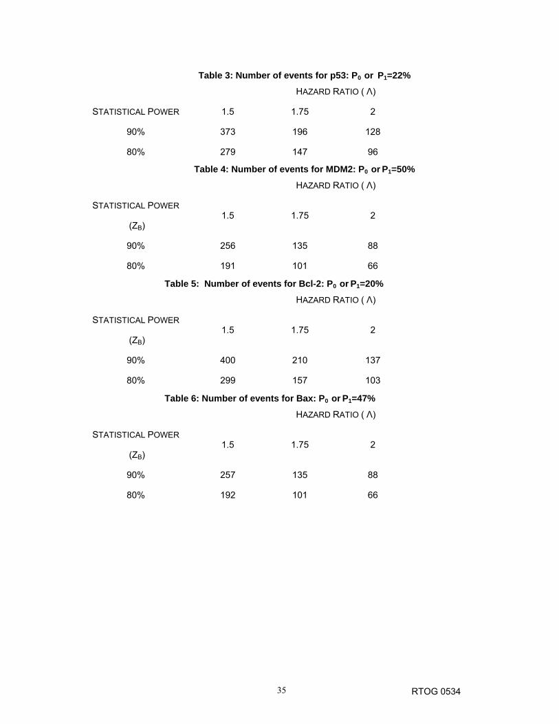
RTOG 0534
35
Table 3: Number of events for p53: P0 or P1=22%
Table 4: Number of events for MDM2: P0 or P1=50%
Table 5: Number of events for Bcl-2: P0 or P1=20%
Table 6: Number of events for Bax: P0 or P1=47%
HAZARD RATIO ( Λ)
STATISTICAL POWER 1.5 1.75 2
90% 373 196 128
80% 279 147 96
HAZARD RATIO ( Λ)
STATISTICAL POWER
(ZΒ) 1.5 1.75 2
90% 256 135 88
80% 191 101 66
HAZARD RATIO ( Λ)
STATISTICAL POWER
(ZΒ) 1.5 1.75 2
90% 400 210 137
80% 299 157 103
HAZARD RATIO ( Λ)
STATISTICAL POWER
(ZΒ) 1.5 1.75 2
90% 257 135 88
80% 192 101 66
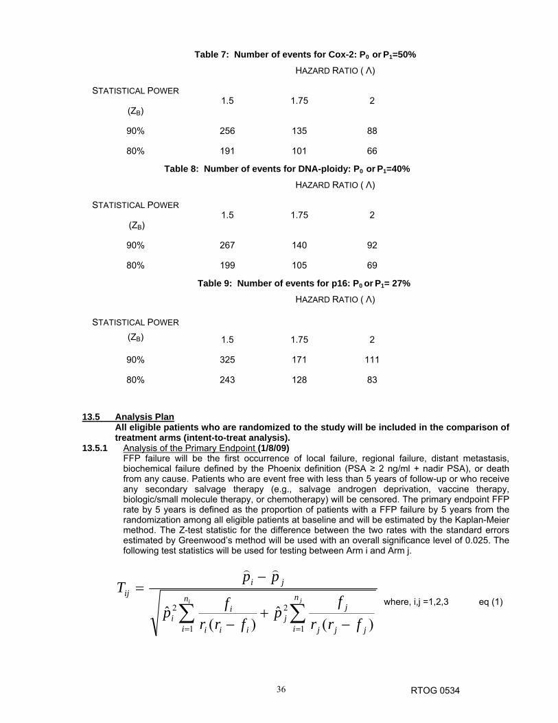
RTOG 0534
36
Table 7: Number of events for Cox-2: P0 or P1=50%
Table 8: Number of events for DNA-ploidy: P0 or P1=40%
Table 9: Number of events for p16: P0 or P1= 27% 13.5 Analysis Plan All eligible patients who are randomized to the study will be included in the comparison of
treatment arms (intent-to-treat analysis). 13.5.1 Analysis of the Primary Endpoint (1/8/09) FFP failure will be the first occurrence of local failure, regional failure, distant metastasis,
biochemical failure defined by the Phoenix definition (PSA ≥ 2 ng/ml + nadir PSA), or death from any cause. Patients who are event free with less than 5 years of follow-up or who receive any secondary salvage therapy (e.g., salvage androgen deprivation, vaccine therapy, biologic/small molecule therapy, or chemotherapy) will be censored. The primary endpoint FFP rate by 5 years is defined as the proportion of patients with a FFP failure by 5 years from the randomization among all eligible patients at baseline and will be estimated by the Kaplan-Meier method. The Z-test statistic for the difference between the two rates with the standard errors estimated by Greenwood’s method will be used with an overall significance level of 0.025. The following test statistics will be used for testing between Arm i and Arm j.
∑∑== −
+−
−=
ji n
i jjj
jj
n
i iii
ii
jiij
frrf
pfrr
fp
ppT
1
2
1
2
)(ˆ
)(ˆ
))
where, i,j =1,2,3 eq (1)
HAZARD RATIO ( Λ)
STATISTICAL POWER
(ZΒ) 1.5 1.75 2
90% 256 135 88
80% 191 101 66
HAZARD RATIO ( Λ)
STATISTICAL POWER
(ZΒ) 1.5 1.75 2
90% 267 140 92
80% 199 105 69
HAZARD RATIO ( Λ)
STATISTICAL POWER
(ZΒ) 1.5 1.75 2
90% 325 171 111
80% 243 128 83
RTOG 0534
37
where, ip) is FFP rate of Arm i estimated by Kaplan-Meier method, ri is the number of patients who are at risk and fi is the number of patients who have FFP events. Using the backward elimination decision procedure, we will first compare Arm 3 with Arm 2 at a critical value (Z-score) of 1.6249. The following hypotheses are of interest to be tested, where, p1, p2, and p3 are the rate of 5-year FFP of Arm 1, Arm 2 and Arm 3, respectively.
H01: p3 ≤ p2 vs. HA1: p3 > p2 If Arm 3 is not better than Arm 2 (p3 ≤ p2), then we compare Arm 2 with Arm 1. If Arm 3 is better than Arm 2 (p3 > p2), then we compare Arm 3 with Arm 1.
If H01 is rejected (T23 > 1.6249), then we conclude that Arm 3 is better than Arm 2 and the following hypotheses are tested.
H02: p3 ≤ p1 vs. HA2: p3 > p1 If the H02 is rejected (T13 > 2.0768), then we conclude that the 5-year FFP of Arm 3 will be better than Arm 1. If the H02 is not rejected (T13 ≤ 2.0768), then we conclude that the 5-year FFP of Arm 3 will not be better than Arm 1.
If H01 is not rejected (T23 ≤ 1.6249), then we conclude that Arm 3 is not better than Arm 2 and the following hypotheses are tested.
H02: p2≤ p1 vs. HA2: p2 > p1 If the H02 is rejected (T12 > 2.0768), then we conclude that the 5-year FFP of Arm 2 will be better than Arm 1. If the H02 is not rejected (T12 ≤ 2.0768), then we conclude that the 5-year FFP of Arm 2 will not be better than Arm 2.
In addition, univariate and multivariate logistic regression108 will be used to compare the treatment differences in each hypothesis. Odds ratios from univariate and multivariate logistic regression and the respective 97.5% confidence intervals will be computed. Treatment arm, SV involvement, prostatectomy Gleason score, pre-radiotherapy PSA, pathology stage, age, and race (as appropriate) will be adjusted for in the Multivariate analysis.
13.5.2 Biochemical Failure-Related Endpoints (1/8/09) The secondary biochemical failure (BF) endpoint is defined as having a detectable PSA (PSA ≥ 0.1 ng/ml) and rising for at least two values with the second value at 0.4 ng/ml or greater, or
the initiation of salvage therapy. Hormone refractory disease is defined as three rises in PSA after the institution of second salvage hormone therapy. The rate pi (i=1, 2, 3) is defined as the proportion of patients with an event among all eligible patients at baseline in Arm i. The Z-test statistics for the difference between the two rates with the standard errors estimated by Greenwood’s method, eq. (1), will be used with an overall significance level of 0.025. In the test statistics, ip) is the rate of Arm i estimated by Kaplan-Meier method, ri is the number of patients who are at risk and fi is the number of patients who have events by 5 years. Using the backward elimination decision procedure, we first compare Arm 3 with Arm 2. The following hypotheses are of interest to be tested, where, p1, p2, and p3 are the rate of 5-year of Arm 1, Arm 2 and Arm 3, respectively.
H01: p3 ≤ p2 vs. HA1: p3 > p2 If Arm 3 is not better than Arm 2 (p3 ≤ p2), then we compare Arm 2 with Arm 1. If Arm 3 is better than Arm 2 (p3 > p2), then compare Arm 3 with Arm 1. If H01 is rejected (T23 > 1.6249), then we conclude that Arm 3 is better than Arm 2 and the following hypotheses are tested.
H02: p3 ≤ p1 vs. HA2: p3 > p1 If the H02 is rejected (T13 > 2.0768), then we conclude that the 5-year rate of Arm 3 will be better than Arm 1. If the H02 is not rejected (T13 ≤ 2.0768), then we conclude that the 5-year rate of Arm 3 will not be better than Arm 1. If H01 is not rejected (T23 ≤ 1.6249), then we conclude that Arm 3 is not better than Arm 2 and the following hypotheses are tested.
H02: p2≤ p1 vs. HA2: p2 > p1 If the H02 is rejected (T12 > 2.0768), then we conclude that the 5-year rate of Arm 2 will be better than Arm 1. If the H02 is not rejected (T12 ≤ 2.0768), then we conclude that the 5-year rate of Arm 2 will not be better than Arm 2.
In addition, the univariate and multivariate logistic regression will be used to compare the treatment differences in each hypothesis. Odds ratios from the univariate and multivariate

RTOG 0534
38
logistic regression and the respective 97.5% confidence interval will be computed. The treatment arm, SV involvement, prostatectomy Gleason score, pre-radiotherapy PSA, pathology staging, age, and race (as appropriate) will be adjusted for in the multivariate analysis.
13.5.3 Time to Failure of Secondary Survival Endpoints (1/8/09) The time to failure for secondary endpoints (second biochemical failure, hormone refractory
disease, distant metastasis, cause-specific mortality, and overall mortality) will be measured from the date of randomization to the date of the event of interest. The events for secondary endpoints and time-to-events are defined in Sections 11.4-11.7. Using the backward elimination decision procedure, we will first compare Arm 3 with Arm 2 at the significance level of 0.0125. If Arm 3 is not better than Arm 2, then Arm 2 will be compared with Arm 1 at the significance level of 0.0125. If we conclude that Arm 2 will be better than Arm 1, then we can conclude that the 5-year FFP of Arm 2 will be the best. If Arm 3 is better than Arm 2, then Arm 3 will be compared with Arm 1 at the significance level of 0.0125. If we conclude that Arm 3 will be better than Arm 1, then we can conclude that Arm 3 will be the best. The time-to-event distribution of overall mortality will be estimated using the Kaplan-Meier method109 and the log-rank test110-111 will be used to test whether the overall mortality rate in one arm is higher than the other arm for each hypothesis at the significance level of 0.0125. However, the treatment effect on other types of failure may impact the observable measures of distant metastasis and cause-specific mortality and other competing risks may dilute the sensitivity of hormone refractory disease, distant metastasis and cause-specific mortality.106 We will use the cause-specific hazard rate112-113 (the instantaneous rate of cause-specific mortality in the presence of competing failure types as a function of time) approach to consider the competing events. Freidlin and Korn106 showed that the cause-specific hazard rate approach is better than other approaches, for example, the cumulative incidence method,114in most cases. The log-rank test on the times to the specific type of failure, which considers the presence of competing events, will be used to test whether the survival rates of these secondary endpoints in one arm are higher than that of the other arm for each hypothesis at a significance level of 0.0125. In this approach, patients who experience other failure first are censored.112
In addition, the Cox regression model115 will be used to compare the treatment differences. Both unadjusted and adjusted hazard ratios and the respective 97.5% confidence interval will be computed. At least the treatment arm, the stratification variables (SV involvement, prostatectomy Gleason score, pre-radiotherapy PSA, pathology stage), age, and race (as appropriate) will be adjusted for in this analysis.
13.5.4 Comparison of the Incidence of Acute Toxicity and Time to Late Grade 3+ Toxicity (1/8/09) Adverse events are scored according to CTCAE, v. 3.0. An acute adverse event will be
defined as the worst severity of the adverse event occurring less than or equal to 90 days of treatment. Both acute grade 2+ and 3+ toxicity will be examined separately. Univariate logistic regression107 will be used to model the distribution of acute adverse events. Multivariate logistic regression107 will be used to model the distribution of acute adverse events, adjusting for covariates. Treatment arm, SV involvement, prostatectomy Gleason score, pre-radiotherapy PSA, pathology stage, and age (as appropriate) will be adjusted for in the multivariate analysis. Both unadjusted and adjusted odds ratios (H1: Arm 1 vs. Arm 2 and H2: Arm 2 vs. Arm 3, respectively) and the respective 97.5% confidence interval will be computed and tested using a one-sided chi-square test with the significance level of 0.025 for each hypothesis.
Late grade 2+ or 3+ adverse events will be defined as an a grade 2+ or 3+ adverse events
occurring more than 90 days of the completion of treatment. The time to late grade 2+ or 3+ adverse events will be measured from the time protocol treatment started to the time of the worst late grade 2+ or 3+ adverse event, respectively. If no such late adverse event is observed until the time of the analysis, the patient will be censored at the time of the analysis. The distribution of time to late grade 2+ or 3+ adverse events will be estimated using the Kaplan-Meier method109 and tested using a one-sided log-rank test110-111 with the significance level of 0.025 for each hypothesis. A multivariate Cox regression model115 will be used to compare the treatment differences of time to late adverse event. Both unadjusted and adjusted hazard ratios (H1: Arm 1 vs. Arm 2 and H2: Arm 2 vs. Arm 3, respectively) and the respective 97.5% confidence interval will be computed. Treatment arm, SV involvement, prostatectomy Gleason score, pre-radiotherapy PSA, pathology stage, age, and race (as appropriate) will be adjusted for in this analysis.

RTOG 0534
39
A Chi-square test will be used at a significance level of 0.05 to test the correlation between the
common toxicity categories in the American Urological Association Symptom Index (AUA SI) and urinary morbidity (Adverse Event terms: Urinary frequency/urgency) using the CTCAE v. 3.0.
13.5.5 Modeling the Relationship of Genomic and Proteomic Biomarkers to the Study Endpoints (1/8/09)
At the time of data maturity of this study, we will propose specific details of the markers to be investigated. We will address the assays that will be used and a list of specific correlative aims with appropriate statistical considerations. The following is a general guideline for the statistical consideration for this analysis. This analysis will be done in each arm separately to test the prognostic vales of biomarkers.
A genomic or proteomic biomarker will be categorized into two subgroups based upon
previously defined (or hypothesized) cut-off points and these two groups will be referred to as favorable and unfavorable risk groups. The patients with genomic and proteomic biomarkers will be compared with the patients without a value for that biomarker to determine if there are any differences with respect to distribution of baseline variables (SV involvement, prostatectomy Gleason score, pre-radiotherapy PSA, and pathology stage). We want to know if there is a difference in survival rate between these two groups. The null (H0) and alternative (HA) hypotheses for survival distribution (S) are
H0: S0 (t) ≥ S1(t) vs. HA: S0 (t) < S1(t) , where t is time Tests will be performed to see if one group is statistically significantly better than the other in
the primary endpoint and secondary endpoints that are related to time to failure (hormone refractory disease, distant metastasis, cause-specific survival, and overall survival). However, the selection of the cut-off point for each biomarker is not established. If the hypothesized cut-off points do not yield statistical significance, other cut-off points may be evaluated. Therefore, various cut-off points are evaluated for their statistical significance. To correct the problem from the multiple testing, the Bonferroni correction will be used. The overall survival functions will be estimated by the Kaplan-Meier method and will be tested for the overall survival difference between the favorable and unfavorable groups using the log-rank test. We will use the cause-specific hazard rate approach106 to estimate other survival/failure distributions and test the survival/failure difference between the two groups using the cause-specific log-rank test. The multivariate analysis will be performed using the Cox proportional hazards model115 for both groups. Potential covariates evaluated for the multivariate models are SV involvement, prostatectomy Gleason score, pre-radiotherapy PSA, pathology stage, and assigned treatment. A stepwise procedure will be used to develop the base model for each outcome endpoint prior to evaluating the prognostic impact of the biomarkers. This approach will be employed to account for as much variation as possible for each outcome before it is tested. It is entirely possible that factors shown to be prognostic in other published series may not be found prognostic here.
If high-dimensional data, such as two-color Microarray data, are generated from blood/urine-based proteomic and genomic data, the following guideline could be applied for the data pre-processing. A careful examination of array images of each gene’s spots on the array images will be carried out to find the spots affected by experiment artifacts. This is a general guideline for the statistical consideration for the two-color Microarray data analysis. We will not include genes whose intensity is less than 100116-117 n both green and red intensities. Local background hybridization signals will be subtracted from the intensities. Let R
j be the background-adjusted fluorescence intensity for the cancer or benign sample and G
j be
the background-adjusted fluorescence intensity for the reference sample for gene j on a particular array. The gene expression ratio is computed as R
j /G
j and undergoes normalization
and transformation to the log-2 scale. Normalization will be applied to remove systematic differences due to extraneous factors such as array effects, global dye effects, print tip effects, etc. Simple normalization methods such as global median centering116 will be considered as well as more complex methods such as print tip-specific corrections and intensity-based
RTOG 0534
40
normalization methods such as lowess smoothing118 if diagnostic plots (e.g., M vs A plots119)
suggest they are needed. These log-transformed, normalized gene expression ratios are used as the basic data in subsequent analyses. If one of the two intensities in a spot is less than 100, that intensity will be set to 100. Genes with greater than 20% of spots missing intensities will be removed from the analysis. For remaining genes, individual missing log ratio values will be imputed using the k-nearest neighbors approach, with k = 10.120
The high-dimensional data from patients who yield both pre- and post-treatment tissue specimens will be used to see the gene expression difference. Let m be the number of genes that will be tested. Let d
iij be the gene expression difference between pre- and post- treatment
for patient i and gene j on a treatment arm. Denote the mean difference between pre- and post- treatment gene expression
for gene j as D
j. A test will be conducted to test the following null
(H0) and alternative (H
A) hypotheses for each gene:
H0: D
j = 0 vs. H
A: D
j ≠ 0
We will control the false discovery proportion when a test for a gene is called significant. A paired t-test will be used to calculate the unadjusted univariate p-value for each gene. We will identify all genes with adjusted p-values121
≤ 0.05 as being differentially expressed between pre- and post-treatment to be 95% confident that the false discovery proportion is no more than 10%.
13.5.6 Analysis for Endpoints Related to Quality of Life (QOL) [2/13/08] [1/8/09] Patient accrual for the QOL measurements will be limited to 200 cases in each arm.
We will use seven instruments to assess quality of life (QOL): the Expanded Prostate Cancer Index (EPIC), EPIC Sexual Medications/Devices Supplement (Utilization of Sexual Meds/Devices), the 25-item version of the Hopkins Symptom Checklist (HSCL-25), the EuroQol (EQ-5D), Hopkins Verbal Learning Test-Revised (HVLT-R), Trail Making Test, parts A & B, and the Controlled Oral Word Association Test (COWAT). Protocol eligible patients will be included in the QOL analysis only if they agree to participate in the QOL portion of this study. All the QOL instruments (EPIC, Utilization of Sexual Meds/Devices, HSCL-25, EQ-5D, HVLT-R, Trail Making Test, parts A & B, and COWAT) will be collected on all cases participating in the trial. To minimize missing QOL data, we have included detailed instructions for collection of QOL and what to do if the patient misses a scheduled assessment, and RTOG provides individualized patient calendars available to Investigators and Research Associates 24/7 on the RTOG web site.
We will describe the distributions of QOL data collection patterns over all collection points in
each treatment arm. Longitudinal data analysis, specifically the general linear mixed-effect model122 will be performed to describe the change trend of the EPIC, Utilization of Sexual Meds/Devices, HVLT-R, Trail Making Test parts A & B, COWAT, HSCL-25, and EQ-5D scores over time across the three treatments. The primary objective in HRQOL analysis is to determine the QOL differences. The response will be the change of measurement from baseline for each measurement. The model will include the baseline and stratification variables (SV involvement, prostatectomy Gleason score, pre-radiotherapy PSA, pathology stage).
The EPIC and HSCL-25 will be collected at pretreatment (baseline), the end of RT, and at 1
year and 5 years after therapy starts. Patient self-assessment of symptoms will be performed using four primary EPIC domains: urinary, bowel, sexual, and hormonal symptoms. The data about the use of erectile aids from Utilization of Sexual Meds/Devices will be reported along with question 17-b in the EPIC. The HSCL-25 has 25 items and is scored by a four point Likert scale (1-not at all, 2-a little, 3-quite a bit, and 4-extremely). A higher score means a worse mood or depression. The HVLT-R, Trail Making Test, parts A & B, and COWAT will be collected at pretreatment (baseline), the end of RT, and at 1 year after the therapy starts. There are three immediate recall responses, one delayed recall response, and one delayed recognition response in the HVLT-R. The response is the number of words the patient can recall out of 12 words for recall responses and the difference of the listed words correctly and incorrectly recalled for recognition response. The response from Trail Making Test, parts A & B is the time takes to finish each test less than 3 and 5 minutes, respectively. There are three responses for the COWAT, and each response is the number of words starting with a provided letter of the alphabet that the patient can produce in one minute. The EQ-5D will be collected at pretreatment (baseline), at 1 year and 5 years after therapy starts. The EQ-5D is a two-part
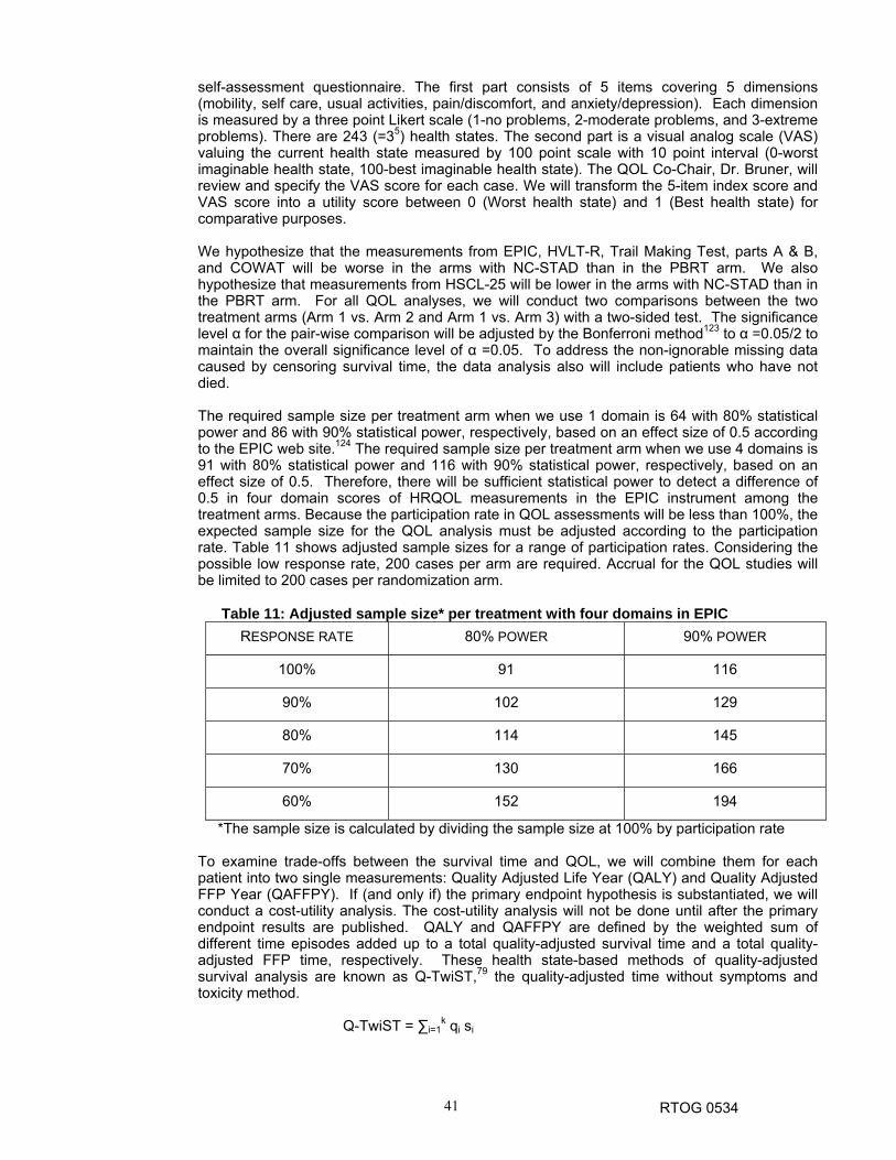
RTOG 0534
41
self-assessment questionnaire. The first part consists of 5 items covering 5 dimensions (mobility, self care, usual activities, pain/discomfort, and anxiety/depression). Each dimension is measured by a three point Likert scale (1-no problems, 2-moderate problems, and 3-extreme problems). There are 243 (=35) health states. The second part is a visual analog scale (VAS) valuing the current health state measured by 100 point scale with 10 point interval (0-worst imaginable health state, 100-best imaginable health state). The QOL Co-Chair, Dr. Bruner, will review and specify the VAS score for each case. We will transform the 5-item index score and VAS score into a utility score between 0 (Worst health state) and 1 (Best health state) for comparative purposes.
We hypothesize that the measurements from EPIC, HVLT-R, Trail Making Test, parts A & B,
and COWAT will be worse in the arms with NC-STAD than in the PBRT arm. We also hypothesize that measurements from HSCL-25 will be lower in the arms with NC-STAD than in the PBRT arm. For all QOL analyses, we will conduct two comparisons between the two treatment arms (Arm 1 vs. Arm 2 and Arm 1 vs. Arm 3) with a two-sided test. The significance level α for the pair-wise comparison will be adjusted by the Bonferroni method123 to α =0.05/2 to maintain the overall significance level of α =0.05. To address the non-ignorable missing data caused by censoring survival time, the data analysis also will include patients who have not died.
The required sample size per treatment arm when we use 1 domain is 64 with 80% statistical
power and 86 with 90% statistical power, respectively, based on an effect size of 0.5 according to the EPIC web site.124 The required sample size per treatment arm when we use 4 domains is 91 with 80% statistical power and 116 with 90% statistical power, respectively, based on an effect size of 0.5. Therefore, there will be sufficient statistical power to detect a difference of 0.5 in four domain scores of HRQOL measurements in the EPIC instrument among the treatment arms. Because the participation rate in QOL assessments will be less than 100%, the expected sample size for the QOL analysis must be adjusted according to the participation rate. Table 11 shows adjusted sample sizes for a range of participation rates. Considering the possible low response rate, 200 cases per arm are required. Accrual for the QOL studies will be limited to 200 cases per randomization arm.
Table 11: Adjusted sample size* per treatment with four domains in EPIC
RESPONSE RATE 80% POWER 90% POWER
100% 91 116
90% 102 129
80% 114 145
70% 130 166
60% 152 194
*The sample size is calculated by dividing the sample size at 100% by participation rate To examine trade-offs between the survival time and QOL, we will combine them for each
patient into two single measurements: Quality Adjusted Life Year (QALY) and Quality Adjusted FFP Year (QAFFPY). If (and only if) the primary endpoint hypothesis is substantiated, we will conduct a cost-utility analysis. The cost-utility analysis will not be done until after the primary endpoint results are published. QALY and QAFFPY are defined by the weighted sum of different time episodes added up to a total quality-adjusted survival time and a total quality-adjusted FFP time, respectively. These health state-based methods of quality-adjusted survival analysis are known as Q-TwiST,79 the quality-adjusted time without symptoms and toxicity method.
Q-TwiST = ∑i=1
k qi si

RTOG 0534
42
where qi is the quality (the utility coefficient) of health state i, si is the duration spent in each health state, and k is the number of health states. We will use Glasziou’s multiple health-state (Q-TwiST) models125 to use the repeated measures of EQ-5D. Because Glasziou’s method incorporates longitudinal QOL data into an analysis of quality-adjusted survival, the health-stated model must be constructed on the following assumptions:
A1) QOL is independent from treatment. A2) A health state is independent from previous states. A3) Proportionality of quality-adjusted duration and duration of the actual state of a health
state. Assumption A1 can be checked by plotting QOL over time according to treatment, and the t-
test can be used to compare the mean QOL scores of each treatment arm. Assumption A2 can be checked by comparing the QOL for patient groups in a given health state where the groups are defined by duration of previous health state experience using a regression model. Suitable checks for assumption A3 at minimum would be a simple plot. If data does not support these assumptions, we will use a method which uses the longitudinal QOL data directly.
The Medicare reimbursement in dollars/QALY and the Medicare reimbursement in
dollars/QAFFPY will be calculated as a function of the monetary cost per relative value of each health state and its duration. Cost-utility will be analyzed at two time points: at 1 year and 5 years post-therapy. We will use the five-item utility score in EQ-5D for the cost-utility analysis. We will use the z-test to test the hypothesis that the cost-utility in the two treatment arms (Arm 1 vs. Arm 2 and Arm 1 vs. Arm 3) is the same with significance level of 0.05/2=0.025 and a two-sided test. We will compare the cost-utility using the Medicare reimbursement in dollars/QALY and the Medicare reimbursement in dollars/QAFFPY between the two treatment arms after adjusting for the baseline and stratification variables.
We will evaluate the cost-utility of the treatment arm in terms of the primary outcome and will
also compare the cost-utility among the three treatment arms. The cost-utility analysis will only include patients whose care are reimbursed under the federal Medicare payment system but will exclude those in Medicare HMOs as well as those under alternative federal coverage (including Medicaid, DOD, and the VA) as well as those covered by private payers or other payment systems. Cost-utility will be analyzed for planned publication at two time-points: looking at initial treatment costs and quality of life at 1 year post-therapy and at 5 years post-therapy. The cost-utility analysis will not be done until after the primary endpoint results are published. We will use the 5-item utility score in EQ-5D for the cost-utility analysis and the Medicare costs defined as in Section 1.6.3. The Medicare cost in dollars/QALY will be calculated as a function of the monetary cost per relative value of each health state and its duration. We will use Analysis of Variance (ANOVA) to compare the cost-utility among the three treatment arms at a significance level of 0.05. If there is a statistically significant difference, a Z-test will be used to compare it between each combination of two treatment arms (Arm 1 vs. Arm 2 and Arm 1 vs. Arm 3, and Arm 2 and Arm 3) after adjusting for the baseline and stratification factors with a significance level of 0.05/3=0.017 and a two-sided test.
A multivariate regression model will be used to model the association between serum levels of
beta-amyloid (Abeta) and measures of the HVLT-R, Trail Making Test, parts A & B, COWAT, and HSCL-25, respectively. The model will include at least the baseline and stratification factors (SV involvement, prostatectomy Gleason score, pre-radiotherapy PSA, pathology stage) as covariates.
To inspect the missing data mechanism, we will use at least a graphical method. A missing
completely at random (MCAR) mechanism exists when missing values are randomly distributed across all observations. A missing at random (MAR) mechanism exists when values are not randomly distributed across all observations, rather than one or more sub-samples. If the cause of missing data is MCAR, listwise deletion (complete case analysis) will be done. If the MAR assumption is supported by the data, then an imputation method such as multiple imputation will be applied to impute missing data. If the MAR assumption is not supported by the data, then adjusting for covariates (such as the baseline QOL score) might reduce the conditional association between outcomes and missing values. If missing data patterns look similar when stratified by such covariate(s), then an analysis that adjusts for such covariate(s)

RTOG 0534
43
will be conducted and an imputation method such as multiple imputation will be applied. If approximate conditional independence cannot be obtained with any set of covariates, then MNAR (missing not at random) must be addressed by an explicit model for the missing data mechanism126 and then an imputation method such as multiple imputation will be applied. All results from the imputed analysis using the multiple imputation will be compared to the complete case analysis results to assess any potential biases. We will conduct a sensitivity analysis using various assumptions on the missing data to determine what impact missing data and imputation methods have on the study conclusions. Imputation methods when prescribed by validated instrument developers will be employed first. Additional methods or methods used when none are described for a given instrument may include: worst-case scenario (in which missing data are assumed to be unfavorable for those on the experimental treatment and favorable of those in the control group); use of the mean response for individual patients who withdrew from the trial from either all or similar (matched) patients remaining in the trial; last observation carried forward (LOCF) [using the last observation as the final observation]; or linear mixed-effects models, to obtain separate estimates for the QOL outcome within strata based on missing data patterns.126-127 RTOG recognizes that all options are subject to bias and analysis of more than one method for consistency across methods is prudent.
13.5.7 Group Sequential Testing for Early Termination and Reporting of Efficacy and Futility (1/8/09) A group sequential test with three planned interim analyses and a final analysis will be
performed. The interim analysis will be carried out when the cumulative accrual (patients whose follow-up is at least 5 years from the randomization date) are met. For each interim analysis, one efficacy and two futility tests will be carried out. At each planned interim analysis, the p-value from the Z-test statistics, eq.1, for the difference between the two FFP rates assessing treatment efficacy or futility with respect to the primary endpoint will be compared to the nominal significance level. The significance level of 0.001, which is similar to the Haybittle-Peto test104-105, was chosen for efficacy testing. For the futility testing boundary, we will use a less aggressive boundary, Rule C in Freidlin and Korn.106 We will first compare Arm 3 with Arm 2 and choose the arm that has the higher FFP rate (if they are the same, Arm 2 will be chosen). Let p1, p2, and p3 equal the rate of 5-year FFP of Arm 1, Arm 2 and Arm 3, respectively. If Arm 2 is better than Arm 3 2(p2 ≥ p3), then we compare Arm 2 with Arm 1. The following hypotheses are tested.
H02: p2≤ p1 vs. HA2: p2 > p1 If the H02 is rejected (p-value ≤ 0.001), then we conclude that the 5-year FFP of Arm 2 will be better than Arm 1. We report that Arm 2 is the best and stop accrual if applicable. If the H02 is not rejected (p-value > 0.001), then we continue the trial.
If Arm 3 is better than Arm 2 (p2 < p3), then we compare Arm 3 with Arm 1. The following hypotheses are tested.
H02: p3 ≤ p1 vs. HA2: p3 > p1 If the H02 is rejected (p-value ≤ 0.001), then we conclude that the 5-year FFP of Arm 3 will be better than Arm 1. We report that Arm 3 is the best and stop accrual if applicable. If the H02 is not rejected (p-value > 0.001), then we continue the trial.
For futility testing, we compare Arm 3 vs. Arm 1 and Arm 2 vs. Arm 1 if applicable. The following hypotheses are tested.
H01: p1 ≥ p2 vs. HA1: p1 < p2 and H03: p1 ≥ p3 vs. HA3: p1 < p3 The alternative hypotheses, HA1 (p1 = p2 + 0.1) and HA3 (p1 = p3 + 0.1) will be tested at 0.001 level (the futility nominal significance level). If the computed p-value is less than 0.001 then we will consider stopping the trial in favor of H01 or H03 and report the results. If we stop the trial for futility, then we will conclude that the 5-year FFP of Arm 1 will be better than Arm 2 or Arm 3 and continue the trial for the other two remaining arms. Otherwise, we will continue the trial.

RTOG 0534
44
Table 12: The Schedule for the Planned Interim Analysis
Information Time
Estimated Analysis Time*
Cumulative Accrual in the Three Arms**
0.25 7 years 397 0.50 9 years 794 0.75 11 years 1191 1.0 13 years 1587
* Time to the interim analysis from the first patient entry without considering ineligibility or lack-of-data rate **The number of eligible patients whose follow-up is at least 5 years from the randomization date
Based on the results of each interim analysis, the following action will be taken and the responsible statistician will recommend to the RTOG DMC that the randomization be discontinued, if applicable, and the study be considered for early publication. Before making such a recommendation, the accrual rate, treatment compliance, safety of the treatments, and the importance of the study are taken into consideration along with the p-value. The RTOG DMC will then make a recommendation about the trial to the RTOG Group Chair.
13.6 Interim Report to Monitor the Study Progress Interim reports with descriptive statistics will be prepared twice per year until the initial paper
reporting the treatment results has been submitted. In general, the interim reports will contain information about the patient accrual rate with a projected completion date for the accrual phase, compliance rate of treatment delivery with the distributions of important prognostic baseline variables, and the frequencies and severity of the adverse event by treatment arm. The interim reports will not contain the results of the treatment comparisons with respect to the primary endpoint and secondary endpoints. This study will be monitored by the Clinical Data Update System (CDUS) version 3.0. Cumulative CDUS data will be submitted quarterly by electronic means. Reports are due January 31, April 30, July 31, and October 31.
13.7 Reporting the Initial Treatment Analysis (1/8/09) The analysis reporting the treatment results will be carried out after the criteria for early
stopping/reporting are met. Three interim comparisons and one final analysis will be performed for efficacy and futility of the experimental treatment will be carried out as described in section 13.5.7. The Z-test statistics for the difference between the two rates with the standard errors estimated by Greenwood’s method, eq. (1), will be used with an overall significance level of 0.025. It will include tabulation of all cases entered and those excluded from the analyses; the distribution of the important prognostic baseline variables; safety treatments; treatment compliance; and observed results with respect to the primary and secondary endpoints will be shown. All eligible patients randomized will be included in the comparison and will be grouped by assigned treatment in the analysis (intent-to-treat analysis). In addition, exploratory analyses of treatment comparisons of the primary and secondary survival endpoints will be tested using the Cox proportional hazard model114 that includes treatment arms, the stratification factors (SV involvement, prostatectomy Gleason score, pre-radiation PSA level, and pathology stage), age, and race (as appropriate).
13.8 Gender and Minorities In conformance with the National Institute of Health (NIH) Revitalization Act of 1993 with regard
to inclusion of women and minorities in clinical research, Participation rates of men will be examined in the interim analyses. Based on the accrual statistics from RTOG 94-08, we project that 81% of the men in the study are White, 15% are Black or African American, 3% are Hispanic, 0.5% are Asian, 0.3% are Pacific Islander and 0.2% are American Indian or Alaskan Native. The following table lists the projected accrual by race/ethnicity.

RTOG 0534
45
Projected Distribution of Gender and Minorities
Gender Ethnic Category Females Males Total Hispanic or Latino N/A 53 53 Not Hispanic or Latino N/A 1711 1711 Ethnic Category: Total of all subjects N/A 1764 1764 Gender Racial Category Females Males Total American Indian or Alaskan Native N/A 6 6 Asian N/A 9 9 Black or African American N/A 251 251 Native Hawaiian or other Pacific Islander N/A 4 4 White N/A 1494 1494 Racial Category: Total of all subjects N/A 1764 1764

RTOG 0534
46
REFERENCES (1/8/09)
1. Catton C, Gospodarowicz M, Warde P, et al. Adjuvant and salvage radiation therapy after radical prostatectomy for adenocarcinoma of the prostate. Radiother Oncol. 59:51-60, 2001.
2. Choo R, Hruby G, Hong J, et al. Positive resection margin and/or pathologic T3 adenocarcinoma of
prostate with undetectable postoperative prostate-specific antigen after radical prostatectomy: To irradiate or not? Int J Radiat Oncol Biol Phys. 52:674-680, 2002.
3. Chawla AK, Thakral HK, Zietman AL, et al. Salvage radiotherapy after radical prostatectomy for prostate
adenocarcinoma: Analysis of efficacy and prognostic factors. Urology. 59:726-731, 2002.
4. Taylor N, Kelly JF, Kuban DA, et al. Adjuvant and salvage radiotherapy after radical prostatectomy for prostate cancer. Int J Radiat Oncol Biol Phys. 56:755-763, 2003.
5. Zelefsky MJ, Aschkenasy E, Kelsen S, et al. Tolerance and early outcome results of postprostatectomy
three-dimensional conformal radiotherapy. Int J Radiat Oncol Biol Phys. 39:327-333, 1997.
6. McCarthy JF, Catalona WJ, Hudson MA. Effect of radiation therapy on detectable serum prostate specific antigen levels following radical prostatectomy: Early versus delayed treatment. J Urol. 151:1575-1578, 1994.
7. Garg MK, Tekyi-Mensah S, Bolton S, et al. Impact of postprostatectomy prostate-specific antigen nadir on
outcomes following salvage radiotherapy. Urology. 51:998-1002, 1998.
8. Song DY, Thompson TL, Ramakrishnan V, et al. Salvage radiotherapy for rising or persistent PSA after radical prostatectomy. Urology. 60:281-287, 2002.
9. Liauw SL, Webster WS, Pistenmaa DA, et al. Salvage radiotherapy for biochemical failure of radical
prostatectomy: A single-institution experience. Urology. 61:1204-1210, 2003.
10. Macdonald OK, Schild SE, Vora SA, et al. Radiotherapy for men with isolated increase in serum prostate specific antigen after radical prostatectomy. J Urol. 170:1833-1837, 2003.
11. Wu JJ, King SC, Montana GS, et al. The efficacy of postprostatectomy radiotherapy in patients with an
isolated elevation of serum prostate-specific antigen. Int J Radiat Oncol Biol Phys. 32:317-323, 1995.
12. Peyromaure M, Allouch M, Eschwege F, et al. Salvage radiotherapy for biochemical recurrence after radical prostatectomy: a study of 62 patients. Urology. 62:503-507, 2003.
13. Stephenson AJ, Shariat SF, Zelefsky MJ, et al. Salvage radiotherapy for recurrent prostate cancer after
radical prostatectomy. JAMA. 291:1325-1332, 2004.
14. Hayes SB, Pollack A. Parameters for treatment decisions for salvage radiation therapy. J Clin Oncol. 23:8204-8211, 2005.
15. Bolla M, van Poppel H, Collette L, et al. Postoperative radiotherapy after radical prostatectomy: A
randomized controlled trial (EORTC trial 22911). Lancet. 366:572-578, 2005.
16. Thompson IM, Jr., Tangen CM, Paradelo J, et al. Adjuvant radiotherapy for pathologically advanced prostate cancer: A randomized clinical trial. JAMA. 296: 2329-2335, 2006.
17. Wiegel T, Bottke D, Willich N, et al. Phase III results of adjuvant radiotherapy (RT) versus ,wait and see
(WS) in patients with pT3 prostate cancer following radical prostatectomy (RP)(ARO 96-02 / AUO AP 09/95). J Clin Oncol. 23(Abstract 4513):381-381, 2005.
18. Eulau SM, Tate DJ, Stamey TA, et al. Effect of combined transient androgen deprivation and irradiation
following radical prostatectomy for prostatic cancer. Int J Radiat Oncol Biol Phys. 41:735-740, 1998.
RTOG 0534
47
REFERENCES (Continued)
19. Tiguert R, Rigaud J, Lacombe L, et al. Neoadjuvant hormone therapy before salvage radiotherapy for an increasing post-radical prostatectomy serum prostate specific antigen level. J Urol. 170:447-450, 2003.
20. Katz MS, Zelefsky MJ, Venkatraman ES, et al. Predictors of biochemical outcome with salvage conformal
radiotherapy after radical prostatectomy for prostate cancer. J Clin Oncol. 21:483-489, 2003.
21. Neulander EZ. Re: Neoadjuvant hormone therapy before salvage radiotherapy for an increasing post-radical prostatectomy serum prostate specific antigen level. J Urol. 171:809-810, 2004.
22. King CR, Presti JC, Jr., Gill H, et al. Radiotherapy after radical prostatectomy: Does transient androgen
suppression improve outcomes? Int J Radiat Oncol Biol Phys. 59:341-347, 2004.
23. Morris MM, Dallow KC, Zietman AL, et al. Adjuvant and salvage irradiation following radical prostatectomy for prostate cancer. Int J Radiat Oncol Biol Phys. 38:731-736, 1997.
24. Kim BS, Lashkari A, Vongtama R, et al. Effect of pelvic lymph node irradiation in salvage therapy for
patients with prostate cancer with a biochemical relapse following radical prostatectomy. Clin Prostate Cancer. 3:93-97, 2004.
25. Cozzarini C, Bolognesi A, Ceresoli GL, et al. Role of postoperative radiotherapy after pelvic
lymphadenectomy and radical retropubic prostatectomy: A single institute experience of 415 patients. Int J Radiat Oncol Biol Phys. 59:674-683, 2004.
26. Proano JM, Sodee DB, Resnick MI, et al. The impact of a negative (111)indium-capromab pendetide
scan before salvage radiotherapy. J Urol. 175:1668-1672, 2006.
27. Trock BJ, Han M, Freedland SJ, et al. Prostate cancer-specific survival following salvage radiotherapy vs observation in men with biochemical recurrence after radical prostatectomy. JAMA 2008;299:2760-2769.
28. Pilepich MV, Winter K, John MJ, et al. Phase III radiation therapy oncology group (RTOG) trial 86-10 of
androgen deprivation adjuvant to definitive radiotherapy in locally advanced carcinoma of the prostate. Int J Radiat Oncol Biol Phys. 50:1243-1252, 2001.
29. D'Amico AV, Manola J, Loffredo M, et al. 6-month androgen suppression plus radiation therapy vs.
radiation therapy alone for patients with clinically localized prostate cancer: A randomized controlled trial. JAMA. 292:821-827, 2004.
30. Denham JW, Steigler A, Lamb DS, et al. Short-term androgen deprivation and radiotherapy for locally
advanced prostate cancer: results from the Trans-Tasman Radiation Oncology Group 96.01 randomized controlled trial. Lancet Oncol. 6:841-850, 2005.
31. Roach M, 3rd, DeSilvio M, Lawton C, et al. Phase III trial comparing whole-pelvic versus prostate-only
radiotherapy and neoadjuvant versus adjuvant combined androgen suppression: Radiation Therapy Oncology Group 9413. J Clin Oncol. 21:1904-1911, 2003.
32. Pound C, Partin A, Eisenberger M, et al. Natural History of Progression After PSA Elevation Following
Radical Prostatectomy. JAMA. 281:1591-1597, 1999.
33. Freedland SJ, Humphreys EB, Mangold LA, et al. Risk of prostate cancer-specific mortality following biochemical recurrence after radical prostatectomy. JAMA. 294:433-439, 2005.
34. Amling CL, Bergstralh EJ, Blute ML, et al. Defining prostate specific antigen progression after radical
prostatectomy: What is the most appropriate cut point? J Urol. 165:1146-1151, 2001.
35. Pollack A, Hanlon AL, Pisansky TM, et al. A multi-institutional analysis of adjuvant and salvage radiotherapy after radical prostatectomy. Int J Radiat Oncol Biol Phys. 60 (Suppl):S186-S187, 2004.

RTOG 0534
48
REFERENCES (Continued)
36. Pollack A, Hanlon AL, Pisansky T, et al. Which definition of biochemchemical failure after post-operative
radiotherapy predicts best for clinical failure? A multi-institutional analysis. J Urol. (In press), 2005. 37. Cox J, Grignon D, Kaplan R, et al. Consensus Statement: Guidelines for PSA Following Radiation
Therapy. Int J Radiat Oncol Biol Phys. 37:1035-1041, 1997.
38. Vicini FA, Kestin LL, Martinez AA. The correlation of serial prostate specific antigen measurements with clinical outcome after external beam radiation therapy of patients for prostate carcinoma. Cancer. 88:2305-2318, 2000.
39. Kestin LL, Vicini FA, Martinez AA. Practical application of biochemical failure definitions: What to do and
when to do it. Int J Radiat Oncol Biol Phys. 53:304-315, 2002.
40. Pickles T, Kim-Sing C, Morris WJ, et al. Evaluation of the Houston biochemical relapse definition in men treated with prolonged neoadjuvant and adjuvant androgen ablation and assessment of follow-up lead-time bias. Int J Radiat Oncol Biol Phys. 57:11-18, 2003.
41. Thames H, Kuban D, Levy L, et al. Comparison of alternative biochemical failure definitions based on
clinical outcome in 4839 prostate cancer patients treated by external beam radiotherapy between 1986 and 1995. Int J Radiat Oncol Biol Phys. 57:929-943, 2003.
42. Buyyounouski MK, Hanlon AL, Eisenberg DF, et al. Defining biochemical failure after radiotherapy with
and without androgen deprivation for prostate cancer. Int J Radiat Oncol Biol Phys. 63:1455-1462, 2005.
43. Roach M, 3rd, Hanks G, Thames H, Jr., et al. Defining biochemical failure following radiotherapy with or without hormonal therapy in men with clinically localized prostate cancer: Recommendations of the RTOG-ASTRO Phoenix Consensus Conference. Int J Radiat Oncol Biol Phys. 65:965-974, 2006.
44. Vicini F, Kestin L, Martinez A. The Importance of Adequate Follow-up in Defining Treatment Success
after External Beam Irradiation for Prostate Cancer. Int J Radiat Oncol Biol Phys. 45:553-561, 1999.
45. Buyyounouski MK, Hanlon AL, Horwitz EM, et al. Biochemical failure and the temporal kinetics of prostate-specific antigen after radiation therapy with androgen deprivation. Int J Radiat Oncol Biol Phys. 61:1291-1298, 2005.
46. Grignon DJ, Caplan R, Sarkar FH, et al. p53 status and prognosis of locally advanced prostatic
adenocarcinoma: A study based on RTOG 8610. J Natl Cancer Inst. 89:158-165, 1997.
47. Pollack A, Mu Z, Hachem P, et al. Modulation of prostate cancer cell death in response to androgen deprivation and radiation by MDM2. International Conference on Translational Research. Lugano, Switzerland; 2003.
48. Pollack A, DeSilvio M, Khor LY, et al. Ki-67 staining is a strong predictor of distant metastasis and
mortality for men with prostate cancer treated with radiotherapy plus androgen deprivation: Radiation Therapy Oncology Group Trial 92-02. J Clin Oncol. 22:2133-2140, 2004.
49. Li R, Heydon K, Hammond ME, et al. Ki-67 staining index predicts distant metastasis and survival in
locally advanced prostate cancer treated with radiotherapy: An analysis of patients in radiation therapy oncology group protocol 86-10. Clin Cancer Res. 10:4118-4124, 2004.
50. Khor LY, Desilvio M, Al-Saleem T, et al. MDM2 as a predictor of prostate carcinoma outcome: an analysis
of Radiation Therapy Oncology Group Protocol 8610. Cancer. 104:962-967, 2005.
51. Khor L-Y, DeSilvio M, McDonnell TJ, et al. Bcl-2 and bax expression and prostate cancer outcome in men treated with radiotherapy in Radiation Therapy Oncology Group protocol 86-10. Cancer. (In press), 2006.

RTOG 0534
49
REFERENCES (Continued)
52. Petricoin EF, 3rd, Ornstein DK, Paweletz CP, et al. Serum proteomic patterns for detection of prostate cancer. J Natl Cancer Inst. 94:1576-1578, 2002.
53. Ornstein DK, Rayford W, Fusaro VA, et al. Serum proteomic profiling can discriminate prostate cancer
from benign prostates in men with total prostate specific antigen levels between 2.5 and 15.0 ng/ml. J Urol. 172:1302-1305, 2004.
54. Cairns P, Esteller M, Herman JG, et al. Molecular detection of prostate cancer in urine by GSTP1
hypermethylation. Clin Cancer Res. 7:2727-2730, 2001.
55. Keating NL, O’Malley AJ, Smith MR. Diabetes and cardiovascular disease during androgen deprivation therapy for prostate cancer. J Clin Oncol. 24: 4448-4456, 2006.
56. Higano CS. Side effects of androgen deprivation therapy: Monitoring and minimizing toxicity. Urology.
61:32-38, 2003.
57. van Andel G, Kurth KH. The impact of androgen deprivation therapy on health related quality of life in asymptomatic men with lymph node positive prostate cancer. Eur Urol. 44:209-214, 2003.
58. Salminen EK, Portin RI, Koskinen A, et al. Associations between serum testosterone fall and cognitive
function in prostate cancer patients. Clin Cancer Res. 10:7575-7582, 2004.
59. Salminen EK, Portin RI, Koskinen AI, et al. Estradiol and cognition during androgen deprivation in men with prostate carcinoma. Cancer. 103:1381-1387, 2005.
60. Regine WF, Schmitt FA, Scott CB, et al. Feasibility of neurocognitive outcome evaluations in patients with
brain metastases in a multi-institutional cooperative group setting: Results of Radiation Therapy Oncology Group trial BR-0018. Int J Radiat Oncol Biol Phys. 58:1346-1352, 2004.
61. Shapiro AM, Benedict RH, Schretlen D, et al. Construct and concurrent validity of the Hopkins Verbal
Learning Test-revised. Clin Neuropsychol. 13:348-358, 1999.
62. Rasmusson DX, Bylsma FW, Brandt J. Stability of performance on the Hopkins Verbal Learning Test. Arch Clin Neuropsychol. 10:21-26, 1995.
63. Sumerall SW, Timmons PL, James AL, et al. Expanded norms for the Controlled Oral Word Association
Test. J Clin Psychol. 53:517-521, 1997.
64. Llorente MD, Burke M, Gregory GR, et al. Prostate cancer: A significant risk factor for late-life suicide. Am J Geriatr Psychiatry. 13:195-201, 2005.
65. Almeida OP, Waterreus A, Spry N, et al. One year follow-up study of the association between chemical
castration, sex hormones, beta-amyloid, memory and depression in men. Psychoneuroendocrinology. 29:1071-1081, 2004.
66. Smith DH, Fenn P, Drummond M. Cost effectiveness of photodynamic therapy with verteporfin for age
related macular degeneration: The UK case. Br J Ophthalmol. 88:1107-1112, 2004.
67. Maberley D, Walker H, Koushik A, et al. Screening for diabetic retinopathy in James Bay, Ontario: A cost-effectiveness analysis. CMaj. 168:160-164, 2003.
68. Brown GC, Brown MM, Sharma S, et al. A cost-utility analysis of interventions for severe proliferative
vitreoretinopathy. Am J Ophthalmol. 133:365-372, 2002.
69. Kobelt G, Lundstrom M, Stenevi U. Cost-effectiveness of cataract surgery. Method to assess cost-effectiveness using registry data. J Cataract Refract Surg. 28:1742-1749, 2002.

RTOG 0534
50
REFERENCES (Continued)
70. Jonsson L. Cost-effectiveness of memantine for moderate to severe Alzheimer's disease in Sweden. Am J Geriatr Pharmacother. 3:77-86, 2005.
71. Palmer CS, Niparko JK, Wyatt JR, et al. A prospective study of the cost-utility of the multichannel
cochlear implant. Arch Otolaryngol Head Neck Surg. 125:1221-1228, 1999.
72. Rabin R, de Charro F. EQ-5D: A measure of health status from the EuroQol Group. Ann Med. 33:337-343, 2001.
73. Hillner BE, Smith TJ. Cost-effectiveness analysis of three regimens using vinorelbine (Navelbine) for non-
small cell lung cancer. Semin Oncol. 23:25-30, 1996.
74. Konski A, Scott CB, Movsas B, et al. Cost-Utility Analysis of Various Treatments for Non-Small cell Carcinoma of the Lung. The Second European Conference on the Economics of Cancer, Brussels (Abstract). 2000.
75. Trippoli S, Vaiani M, Lucioni C, et al. Quality of life and utility in patients with non-small cell lung cancer.
Quality-of-life Study Group of the Master 2 Project in Pharmacoeconomics. Pharmacoeconomics. 19:855-863, 2001.
76. Essink-Bot ML, de Koning HJ, Nijs HG, et al. Short-term effects of population-based screening for
prostate cancer on health-related quality of life. J Natl Cancer Inst. 90:925-931, 1998.
77. Sandblom G, Carlsson P, Sennfalt K, et al. A population-based study of pain and quality of life during the year before death in men with prostate cancer. Br J Cancer. 90:1163-1168, 2004.
78. Sennfalt K, Carlsson P, Sandblom G, et al. The estimated economic value of the welfare loss due to
prostate cancer pain in a defined population. Acta Oncol. 43:290-296, 2004.
79. Glasziou PP, Simes RJ, Gelber RD. Quality adjusted survival analysis. Stat Med. 9:1259-1276, 1990.
80. Barry MJ, Fowler FJ, Jr., O'Leary MP, et al. Correlation of the American Urological Association symptom
index with self-administered versions of the Madsen-Iversen, Boyarsky and Maine Medical Assessment Program symptom indexes. Measurement Committee of the American Urological Association. J Urol. 148:1558-1564,1992.
81. Desai J, Stock RG, Stone NN, et al. Acute urinary morbidity following I-125 interstitial implantation of the
prostate gland. Radiat Oncol Investig. 6:135-141, 1998.
82. Merrick GS, Butler WM, Lief JH, et al. Temporal resolution of urinary morbidity following prostate brachytherapy. Int J Radiat Oncol Biol Phys. 47:121-128, 2000.
83. Egawa S, Shimura S, Irie A, et al. Toxicity and health-related quality of life during and after high dose rate
brachytherapy followed by external beam radiotherapy for prostate cancer. Jpn J Clin Oncol.31:541-547, 2001.
84. Merrick GS, Butler WM, Wallner KE, et al. Prophylactic versus therapeutic alpha-blockers after
permanent prostate brachytherapy. Urology. 60:650-655, 2002.
85. Michalski JM, Lawton C, El Naqa I, et al. Development of RTOG consensus guidelines for the definition of the clinical target volume for postoperative conformal radiation therapy for prostate cancer. Int J Radiat Oncol Biol Phys 2008;Submitted.
86. Wiltshire KL, Brock KK, Haider MA, et al. Anatomic boundaries of the clinical target volume (prostate bed)
after radical prostatectomy. Int J Radiat Oncol Biol Phys 2007;69:1090-1099.
RTOG 0534
51
REFERENCES (Continued)
87. Pollack A, Price R, Dong L, et al. Intact Prostate Cancer: Overview. In: Mundt AJ, Roeske JC, eds.
Intensity Modulated Radiation Therapy: A Clinical Perspective. Ontario, Canada: BC Decker; 2005: 459-463.
88. Buyyounouski MK, Horwitz EM, Price RA, et al. IMRT for prostate cancer. In: Bortfeld T, Schmidt-Ullrich
R, de Neve W, eds. IMRT Handbook: Concepts & Clinical Applications. Heidelberg, Germany: Springer-Verlag; 2005.
89. Wei JT, Dunn RL, Litwin MS, et al. Development and validation of the expanded prostate cancer index
composite (EPIC) for comprehensive assessment of health-related quality of life in men with prostate cancer. Urology. 56:899-905, 2000.
90. Hough R, Landsverk J, Stone J. Comparison of psychiatric screening questionnaires for primary care
patients. Final report for NIMH Contract No. 278-81-0036 (DB) 122:913-922, 1995.
91. Hesbacher P, Rickels K, Downing RW, et al. Assessment of psychiatric illness severity by family physicians. Social Science & Medicine. 12(1-A):45-47, 1978.
92. Hesbacher PT, Rickels K, Morris RJ, et al. Psychiatric illness in family practice. J Clin Psychiatry. 41:6-
10, 1980.
93. Derogatis LR, Melisaratos N. The Brief Symptom Inventory: An introductory report. Psychol Med. 13:595-605, 1983.
94. Bolton P. Cross-cultural validity and reliability testing of a standard psychiatric assessment instrument
without a gold standard. J Nerv Ment Dis. 189:238-242, 2001.
95. Meyers CA, et al. Neurocognitive function and progression in patients with brain metastases treated with whole brain radiation and motexafin gadolinium: Results of a randomized phase III trial. J Clin Oncol. 22: 157-165, 2004.
96. Benedict RHB, Schretlen D, Groninger L, Brandt J: Hopkins Verbal Learning Test - Revised: Normative
data and analysis of inter-form and test-retest reliability. Clin Neuropsychologist.12:43-55,1998.
97. Benton AL, Hamsher KdeS. Multilingual Aphasia Examination. Iowa City, IA: AJA Associates; 1989.
98. Lezak MD, Howieson DB, Loring DW. Neuropschological Assessment. 4th ed. New York, NY: Oxford
University Press; 2004.
99. Schulz MW, Chen J, Woo HH, et al. A comparison of techniques for eliciting patient preferences in patients with benign prostatic hyperplasia. J Urol. 168:155-159, 2002.
100. Badia X, Herdman M, Kind P. The influence of ill-health experience on the valuation of health. Pharmacoeconomics. 13:687-696, 1998.
101. Wu AW, Jacobson KL, Frick KD, et al. Validity and responsiveness of the euroqol as a measure of health-related quality of life in people enrolled in an AIDS clinical trial. Qual Life Res. 11:273-282, 2002.
102. Zelen M. The randomization and stratification of patients to clinical trials. J Chronic Dis. 27:365-375,
1974.
103. Chen TT, Simon, RM. Extension of one-sided test to multiple treatment trials. Control Clin Trials. 15: 124-134, 1994.
104. Haybittle,J.L. Repeated assessment of results in clinical trials of cancer treatment. Br J Radiol. Oct;44(526):793-7, 1971.

RTOG 0534
52
REFERENCES (Continued)
105. Peto R, Pike MC, Armitage P, Breslow NE, Cox DR, Howard SV, Mantel N, McPherson K, Peto J, Smith PG. Design and analysis of randomized clinical trials requiring prolonged observation of each patient. I. Introduction and design. Br J Cancer. Dec;34(6):585-612, 1976.
106. Freidlin B, Korn EL. Testing treatment effects in the presence of competing risks. Stat Med. 24:1703-1712, 2005.
107. Schoenfeld DA. The asymptotic properties of nonparametric test for comparing survival distributions. Biometrika. 68:316-319, 1981.
108. Agresti A. Categorical Data Analysis. New York: Wiley; 1990.
109. Kaplan EL, Meier P. Nonparametric estimation from incomplete observations. J Am Stat Assoc.
53:447-457, 1958.
110. Mantel N. Evaluation of survival data and two new rank order statistics arising in its consideration. Canc Chemo Rep. 50:163-170, 1966.
111. Kim K, Tsiatis AA. Study duration for clinical trials with survival response and early stopping rule.
Biometrics. 46:81-92, 1990.
112. Kalbfleisch JD, Ross LP. The statistical analysis of failure time data. New York: John Wiley & Sons; 1980.
113. Gaynor JJ, Feuer EJ, Tan CC, et al. On the use of cause-specific failure and conditional failure
probabilities: examples from clinical oncology data. JASA. 88:400-403, 1993.
114. Gray RJ. A class of K-sample tests for comparing the cumulative incidence of a competing risk. Annual Statistics. 16:1141-1143, 1988.
115. Cox DR. Regression models and life tables. J Royal Statistics Soc. 34:187-220, 1972.
116. Simon R, Korn EL, McShane LM, et al. Design and Analysis of DNA Microarray Investigations. New York: Springer; 2003.
117. Dobbin KK, Kawasaki ES, Petersen DW, Simon RM. Characterizing dye bias in microarray
experiments. Bioinformatics. 21:2430-2437, 2005.
118. Yang YH, Dudoit S, Luu P, et al. Normalization for cDNA microarray data: A robust composite method addressing single and multiple slide systematic variation. Nucleic Acids Res.15:30, 2002.
119. Dudoit S, Yang YH, Callow MJ, Speed TP. Statistical methods for identifying genes with
differential expression in replicated cDNA microarray experiments. Stat Sin. 12:111-139, 2002.
120. Troyanskaya O, Cantor M, Sherlock G, et al. Missing value estimation methods for DNA microarrays Bioinformatics. 17:520-525, 2001.
121. Korn EL, Troenndle JF, McShane LM, Simon R. Controlling the number of false discoveries:
Application to high-dimensional genomic data. J Stat Planning and Inference.124:2448-2455.22, 2003.
122. Verbeke G, Molenberghs G. Linear mixed models for longitudinal data. New York: Springer-Verlag, Inc.; 2000.
123. Bonferroni CE. Teoria statistica delle classi e calcolo delle probabilita. In: Volume in Onore di
Ricarrdo dalla Volta: Universita di Firenza. 1937: 1-62.

RTOG 0534
53
REFERENCES (Continued)
124. Home page: the expanded prostate cancer index composite (EPIC).
http://roadrunner.cancer.med.umich.edu/epic/
125. Glasziou, P.P., Cole, B.F., Gelber, R.D., Hilden, J., and Simes, R.J. Quality adjusted survival analysis with repeated quality of life measures. Stat Med. 17: 1215-1229, 1998.
126. Donaldson GW, Moinpour CM. Learning to live with missing quality-of-life data in advanced-stage
disease trials. J Clin Oncol. 23:7380-7384, 2005.
127. Guidance for industry patient-reported outcome measures: Use in medical product development to support labeling claims: U.S. Department of Health and Human Services and Food and Drug Administration. pp. http://www.qolpro.org/ (accessed Aug. 1, 2006).
RTOG 0534
54
APPENDIX I (1/8/09) (3/24/10)
RTOG 0534
Informed Consent Template for Cancer Treatment Trials (English Language)
A Phase III Trial Of Short Term Androgen Deprivation With Pelvic Lymph Node Or
Prostate Bed Only Radiotherapy (SPPORT) In Prostate Cancer Patients With A Rising PSA After Radical Prostatectomy
This is a clinical trial, a type of research study. Your study doctor will explain the clinical trial to you. Clinical trials include only people who choose to take part. Please take your time to make your decision about taking part. You may discuss your decision with your friends and family. You can also discuss it with your health care team. If you have any questions, you can ask your study doctor for more explanation. You are being asked to take part in this study because you have had surgery to remove your prostate and your study doctor has recommended radiation therapy because your blood level of Prostate Specific Antigen (PSA) has been going up. (The PSA is a value that helps determine the aggressiveness of your prostate cancer.)
Why is this study being done? The purpose of this study is to compare the effects, good and/or bad of three treatment methods on participants and their cancer. External beam radiation therapy is one of the standard treatments for men with prostate cancer who have a rising PSA after surgery. Different methods of radiation therapy are used, and it is not known which one is best. Most commonly, the area where the prostate was originally located before being removed (the prostate bed) is treated, without treating the lymph nodes in the pelvis. Prostate cancer can spread to the lymph nodes. There is some evidence in men who have not had surgery that radiotherapy to the pelvic lymph nodes may stop the cancer from spreading under some conditions. Since treating the pelvic lymph nodes can result in increased side effects, the benefit of this method of radiation therapy needs to be tested. Prostate cancer feeds on male hormones, such as testosterone. Drugs that reduce or block testosterone (hormone therapy) can cause some prostate cancer cells to die and others to become sick so that they don’t grow. Some patients treated with a combination of these drugs and radiation have a greater chance of not having the cancer return when compared to men treated with radiation alone. These studies were done in men who did not have surgery. Since hormone therapy can result in increased side effects, the benefit of combining hormone therapy with radiation therapy needs to be tested. There are 3 treatment groups in this study: 1) Patients who receive radiation therapy to the prostate bed only; 2) Patients who receive hormone therapy for 4 to 6 months plus radiation therapy to the prostate bed; 3) Patients who receive hormone therapy for 4 to 6 months plus radiation therapy to the prostate bed and to the
pelvic lymph nodes. If you agree to participate in this study, you will receive one of these 3 treatments.
How many people will take part in the study? About 1,764 people will take part in this study.
RTOG 0534
55
What will happen if I take part in this research study? (01/8/09) (3/24/10) Before you begin the study: You will need to have the following exams, tests or procedures to find out if you can be in the study. These exams, tests or procedures are part of regular cancer care and may be done even if you do not join the study. If you have had some of them recently, they may not need to be repeated. This will be up to your study doctor. Review of the tissue from your prior surgery to remove your prostate to determine your Gleason score (a
value that helps determine the aggressiveness of your prostate cancer) History and physical exam, including a digital rectal exam (DRE) and an assessment of your ability to carry
out activities of daily living (which will include questions such as whether you are able to feed, bathe, and dress yourself).
You will be asked to fill out a questionnaire on urinary symptoms and function called the American Urological Association Symptom Index (AUA SI).
A blood test to determine your PSA (a value that helps determine the aggressiveness of your prostate cancer). About 2 teaspoons of blood will be drawn from a vein. At least two PSA tests spaced by 2 months must be obtained after surgery to remove the prostate. Your study doctor also may draw another PSA before the start of treatment for a baseline value.
Other blood tests (for blood count, liver function, and to measure testosterone) A CT (Computed Tomography) scan or MRI (Magnetic Resonance Imaging) of your pelvis to determine if
there is any evidence of cancer spread to the pelvic lymph nodes. A CT scan is a study using x-rays to look at one part of your body. An MRI is imaging using a strong magnetic field to look at one part of your body.
A bone scan to determine if the cancer has spread to the bones. A CT scan with an urethrogram or an MRI for radiation treatment planning may be ordered; for a urethrogram,
a tube is placed into the opening of the canal (at the end of the penis) from which urine is emptied from the body. Dye is injected, and images are taken.
You will be "randomized" into one of the study groups described below. Randomization means that you are put into a group by chance. A computer program will place you in one of the study groups. Neither you nor your study doctor can choose the group you will be in. You will have a one in three chance of being placed in any group.
If you are in group 1 (often called "Arm 1"): You will receive radiation treatments to the prostate bed once daily, 5 days a week, Monday through Friday, for a total of 36 to 39 treatments (the exact number will be decided by your study doctor). Each radiation treatment will take 15-30 minutes. If you are in group 2 (often called "Arm 2"): You will receive radiation treatments to the prostate bed once daily, 5 days a week, Monday through Friday, for a total of 36 to 39 treatments (the exact number will be decided by your study doctor). Each radiation treatment will take 15-30 minutes. You also will receive hormone therapy for 4 to 6 months (the exact length will be decided by your doctor). The hormone therapy will begin 2 months before the start of the radiation treatments. There are two parts to the hormone therapy. You will take injections either under the skin or in the muscle, and you will take a pill, either flutamide three times per day or bicalutamide once per day. The pills will be taken for at least 4 of the 6 months. If you are in group 3 (often called "Arm 3"): You will receive radiation treatments to the pelvic lymph nodes and prostate bed once daily, 5 days a week, Monday through Friday, for 25 treatments. From that point on, the radiation treatments will target the prostate bed only, 5 days per week, for another 11-14 treatments. The total number of radiation treatments will be 36 to 39 treatments (the exact number will be decided by your study doctor). Each radiation treatment will take 15-30 minutes. You also will receive hormone therapy for 4 to 6 months (the exact length will be decided by your study doctor). The hormone therapy will begin 2 months before the start of the radiation treatments. There are two parts to the hormone therapy. You will take injections either under the skin or in the muscle, and you will take a pill, either flutamide three times per day or bicalutamide once per day. The pills will be taken for at least 4 of the 6 months.

RTOG 0534
56
After entering the study and prior to radiotherapy: (1/8/09) If the exams, tests and procedures show that you can be in the study, and you choose to take part, then you will need the following tests and procedures if you are randomized to receive hormone therapy (Groups 2 and 3). They are part of regular cancer care. After 2 months of hormone therapy and before radiation therapy, blood will be drawn (for a blood count, liver
function, and to measure testosterone and PSA). After 2 months of hormone therapy and before radiation therapy, you will be requested to fill out an American
Urological Association Symptom Index (AUA SI) questionnaire. During Radiation Therapy: (1/8/09) Weekly during radiation therapy: History and physical exam, including an assessment of your ability to carry
out activities of daily living, and assessment of any side effects you may be experiencing from the treatment Blood will be drawn during the 6th (last) week of radiotherapy (for a blood count, liver function and to measure
testosterone). You will be asked to fill out an American Urological Association Symptom Index (AUA SI) questionnaire in the
6th week of radiation therapy. When you are finished receiving radiation: (1/8/09) You will need these tests and procedures: At 6 weeks (1.5 months), 3, and 6 months following the completion of radiation: blood tests to measure liver
function; at 3 and 6 months following the completion of radiation: blood will be drawn for blood count At 3, 6, and 12 months following the completion of radiation, every 6 months for the next 6 years, and then
annually: History and physical exam, including a digital rectal exam (DRE), an assessment of your ability to carry out activities of daily living, an assessment of any side effects from the treatments, and an AUA-SI questionnaire.
A PSA and testosterone will be checked at 6 weeks (1.5 months), 3 months, 6 months, 9 months, and 12 months following completion of radiotherapy; at 3-month intervals for the next year; and then at 6-month intervals thereafter.
If something is felt in the prostate bed that is suspicious for recurrence, your study doctor will request a needle biopsy to evaluate this.
If your PSA rises at any time after completion of treatment, your study doctor may order a bone scan and CT scan or MRI of the abdomen and pelvis.
Study Plan
Another way to find out what will happen to you during the study is to read the chart below. Start reading at the top and read down the list, following the lines and arrows.
Randomize
(You will be in one of three Groups)
Group 3 Radiation to the Pelvic Lymph Nodes and the Prostate Bed
(25 Treatments) Radiation to the Prostate Bed
Only (11-14 Treatments) Plus Hormone Therapy
(4-6 months)
Group 1 Radiation to the Prostate Bed
(36-39 Treatments)
Group 2 Radiation to the Prostate Bed
(36-39 Treatments) Plus Hormone Therapy
(4-6 months)

RTOG 0534
57
How long will I be in the study? (1/8/09) You will receive 36–39 radiation treatments over 7–8 weeks. Hormone therapy, if given, will last 4–6 months. After you are finished receiving radiation therapy, the study doctor will ask you to visit the office for follow-up exams at 3, 6, and 12 months after radiotherapy, then every 6 months for the next 6 years, and annually thereafter. The study doctors would like to keep track of your medical condition by seeing you every year for your lifetime. Can I stop being in the study? Yes. You can decide to stop at any time. Tell the study doctor if you are thinking about stopping or decide to stop. He or she will tell you how to stop safely. It is important to tell the study doctor if you are thinking about stopping so any risks from the radiation and hormone therapy (if given) can be evaluated by him/her. Another reason to tell your study doctor that you are thinking about stopping is to discuss what follow-up care and testing could be most helpful for you. The study doctor may stop you from taking part in this study at any time if he/she believes it is in your best interest; if you do not follow the study rules; or if the study is stopped. What side effects or risks can I expect from being in the study? (1/8/09) You may have side effects while on the study. Everyone taking part in the study will be watched carefully for any side effects. However, researchers don’t know all the side effects that may happen. Side effects may be mild or very serious. Your health care team may give you medicines to help lessen side effects. Many side effects go away soon after you stop radiation or hormone therapy (if given). In some cases, side effects can be serious, long lasting, or may never go away. In addition, some of the side effects may be life threatening and, in rare instances, may cause death. The risks of side effects related to the radiation may be higher in group 3, which includes the treatment of the pelvic lymph nodes. You should talk to your study doctor about any side effects that you have while taking part in the study. Risks and side effects related to the radiation therapy include those which are: Likely (1/8/09)
• Tanning, redness, or darkening of skin in treatment area • Rash, itching or peeling of skin • Temporary hair loss in the treatment area • Temporary fatigue • Abdominal cramps • Frequent bowel movements, sometime with urgency, or diarrhea • Rectal cramps and irritation with pain on defecation • Mild rectal bleeding that does not require treatment • Bladder irritation with a stinging sensation • Frequency or urgency of urination • Small amounts of blood in the urine
Less Likely
• Urinary obstruction requiring the placement of a temporary urinary catheter and/or dilatation because of stricture (narrowing)
• Less ability to control urine (increased incontinence) • Inability to achieve an erection (inability of the penis to become hard) • Rectal bleeding that requires medication or laser treatment to stop

RTOG 0534
58
Rare but serious (1/8/09)
• Injury to the bladder, urethra, bowel, or other tissues in the pelvis or abdomen requiring additional procedure or surgery, such as a colostomy (bag for stool).
• Intestinal obstruction requiring surgery
Risks and side effects related to the hormone therapy those which are: Likely (1/8/09)
• Hot flashes • Inability to achieve an erection (inability of the penis to become hard) • Loss of sex drive • Mood swings • Muscle loss, weakness and fatigue • Mild anemia (drop in red blood cell count) • Weight gain • Bone weakening
Less Likely (1/8/09) • Significant bone loss (osteopenia or osteoporosis) which could result in fracture • Significant anemia • Blood sugar problems (diabetes) • High fats and cholesterol in your blood (hyperlipidemia) • Blood vessel disease (arteriosclerosis, heart failure) • Fluid retention and ankle swelling (edema) • Breast enlargement and tenderness • Difficulty with calculations and memory (verbal recall, cognition)
Rare and Possibly Serious (1/8/09)
• Liver damage (hepatitis) • Flare up in arthritis • Death due to heart disease • Bone fracture • Depression
Reproductive risks: You should not father a baby while on this study because the drugs and radiation in this study can affect an unborn baby. Check with your study doctor about what kind of birth control methods to use and how long to use them. Some methods might not be approved for use in this study. Some of the drugs and radiation used in this study may make you unable to have children in the future. For more information about risks and side effects, ask your study doctor. Are there benefits to taking part in the study? Taking part in this study may or may not make your health better. It is not known whether the combination of radiation to the prostate bed plus hormone therapy is better than radiation to the prostate bed alone. Also, it is not known whether radiation to the pelvic lymph nodes and prostate bed plus hormone therapy is better than radiation to the prostate bed only combined with hormone therapy. We do know that the information from this study will help researchers learn more about how best to treat men who have a rising PSA after surgery to remove their prostate. This information could help future cancer patients. What other choices do I have if I do not take part in this study? Your other choices may include:

RTOG 0534
59
• Getting treatment or care for your cancer without being in a study; this could include the following options, either alone or in combination with each other:
o External beam radiation therapy (typically, to the prostate bed) o External beam radiotherapy plus hormone therapy o Hormone therapy
• Taking part in another study • Getting no treatment (With this choice, your tumor could continue to grow and your disease could spread) Talk to your study doctor about your choices before you decide if you will take part in this study. Will my medical information be kept private? Data are housed at RTOG Headquarters in a password-protected database. We will do our best to make sure that the personal information in your medical record will be kept private. However, we cannot guarantee total privacy. Your personal information may be given out if required by law. If information from this study is published or presented at scientific meetings, your name and other personal information will not be used. Organizations that may look at and/or copy your medical records for research, quality assurance, and data analysis include: • The Radiation Therapy Oncology Group • The National Cancer Institute (NCI) and other government agencies involved in keeping research safe for
people, like the Central Institutional Review Board (CIRB) and the Food and Drug Administration (FDA) • The Cancer Trials Support Unit (CTSU), a research group sponsored by the National Cancer Institute (NCI)
to provide greater access to cancer trials [for CTSU participants only] What are the costs of taking part in this study? You and/or your health plan/ insurance company will need to pay for some or all of the costs of treating your cancer in this study. Some health plans will not pay these costs for people taking part in studies. Check with your health plan or insurance company to find out what they will pay for. Taking part in this study may or may not cost your insurance company more than the cost of getting regular cancer treatment. You will not be paid for taking part in this study. For more information on clinical trials and insurance coverage, you can visit the National Cancer Institute’s Web site at http://cancer.gov/clinicaltrials/understanding/insurance-coverage. You can print a copy of the “Clinical Trials and Insurance Coverage” information from this Web site. Another way to get the information is to call 1-800-4-CANCER (1-800-422-6237) and ask them to send you a free copy. What happens if I am injured because I took part in this study? It is important that you tell your study doctor, __________________ [investigator’s name(s)], if you feel that you have been injured because of taking part in this study. You can tell the study doctor in person or call him/her at __________________ [telephone number]. You will get medical treatment if you are injured as a result of taking part in this study. You and/or your health plan will be charged for this treatment. The study will not pay for medical treatment. What are my rights if I take part in this study? Taking part in this study is your choice. You may choose either to take part or not to take part in the study. If you decide to take part in this study, you may leave the study at any time. No matter what decision you make, there will be no penalty to you and you will not lose any of your regular benefits. Leaving the study will not affect your medical care. You can still get your medical care from our institution.

RTOG 0534
60
We will tell you about new information or changes in the study that may affect your health or your willingness to continue in the study. A Data Safety Monitoring Board will be regularly meeting to monitor safety and other data related to this study. The Board members may receive confidential patient information, but they will not receive your name or other information that would allow them to identify you by name. In the case of injury resulting from this study, you do not lose any of your legal rights to seek payment by signing this form. Who can answer my questions about the study? You can talk to your study doctor about any questions or concerns you have about this study. Contact your study doctor __________________ [name(s)] at __________________ [telephone number]. For questions about your rights while taking part in this study, call the ________________________ [name of center] Institutional Review Board (a group of people who review the research to protect your rights) at __________________ (telephone number). [Note to Local Investigator: Contact information for patient representatives or other individuals in a local institution who are not on the IRB or research team but take calls regarding clinical trial questions can be listed here.] *You may also call the Operations Office of the NCI Central Institutional Review Board (CIRB) at 888-657-3711 (from the continental US only). [*Only applies to sites using the CIRB.] Please note: This section of the informed consent form is about additional research that is being done with people who are taking part in the main study. You may take part in this additional research if you want to. You can still be a part of the main study even if you say ‘no’ to taking part in this additional research. You can say “yes” or “no” to each of the following studies. Below, please mark your choice for each study. Quality of Life (QOL) Study (1/8/09) We want to know your view of how your life has been affected by cancer and its treatment. This “Quality of life” study looks at how you are feeling physically and emotionally during your cancer treatment. It also looks at how you are able to carry out your day-to-day activities and how your cancer and cancer treatment may affect your thinking skills (neurocognitive part of QOL study). This information will help doctors better understand how patients feel during treatments and what effects the medicines are having. In the future, this information may help patients and doctors as they decide which medicines to use to treat cancer. You will be asked to complete some questionnaires at the following time points:
• 4 questionnaires immediately before you enroll in the study • 4 questionnaires during week 6 of radiation therapy • 4 questionnaires at year 1 and year 5
Neurocognitive Part of QOL Study You will be asked to take part in a test of your thinking skills at the following time points:
• immediately before you enroll in the study • during week 6 of radiation therapy • at year 1 and year 5

RTOG 0534
61
It takes about 25 minutes to fill out the questionnaires and about 20 minutes to complete the test of thinking skills. If any questions make you feel uncomfortable, you may skip those questions and not give an answer. If you decide to take part in this study, the only things you will be asked to do is fill out the questionnaires and take part in the test of thinking skills. You may change your mind about completing the questionnaires or the test of thinking skills at any time, and you may chose to stop answering the questionnaires or taking part in the test of thinking skills altogether at any time. Just like in the main study, we will do our best to make sure that your personal information will be kept private. Please circle your answer.
I choose to take part in the Quality of Life Study. I agree to fill out the Quality of Life questionnaires. YES NO
I choose to take part in the neurocognitive portion of the Quality of Life Study. I agree to take part in a test of my thinking skills.
YES NO
Use of Tissue, Blood, and Urine for Research About Using Tissue, Blood, and Urine for Research (1/8/09) (3/31/09) You have had surgery to remove your prostate and your cancer. Your doctors have removed and examined some of this tissue to look at the amount and grade of the cancer and to see if the cancer extended outside of the prostate. The results of these tests will be given to you by your study doctor and will be used to plan your care. We would like to keep some of the tissue that is left over from your surgery for future research. If you agree, this tissue will be kept and may be used in research to learn more about cancer and other diseases. Please read the information sheet called "How is Tissue Used for Research" to learn more about tissue research. This information sheet is available to all at the following web site: http://www.rtog.org/tissue%20for%20research_patient.pdf In addition, if you agree to participate in this part of the study, you will have blood drawn and urine collected before you start radiation therapy, during the 6th week of radiation therapy, at 3, 6, and 12 months in year 1, then yearly for 6 years after completion of treatment. We would like to keep about two tablespoons of blood and 5 tablespoons of urine at each of these times for future research. If you agree, this blood and urine will be kept to be used in research to learn more about cancer and other diseases Your tissue, blood, and urine may be helpful for research. The research that may be done is not designed specifically to help you. It might help people who have cancer and other diseases in the future. Reports about research done with your tissue, blood and urine will not be given to you or your study doctor. These reports will not be put in your health record. The research will not have an effect on your care. Things to Think About The choice to let us keep the left over tissue, blood, and urine for future research is up to you. No matter what you decide to do, it will not affect your care or your participation in the main part of the study. If you decide now that your tissue, blood, and urine can be kept for research, you can change your mind at any time. Just contact us and let us know that you do not want us to use your tissue, blood, and urine. Then any tissue, blood, or urine that remains will no longer be used for research and will be returned to the institution that submitted it.

RTOG 0534
62
In the future, people who do research may need to know more about your health. While the study doctor/institution may give them reports about your health, it will not give them your name, address, phone number, or any other information that will let the researchers know who you are. Sometimes tissue is used for genetic research (about diseases that are passed on in families). Even if your tissue, blood, and urine is used for this kind of research, the results will not be put in your health records. Your tissue, blood, and urine will be used only for research and will not be sold. The research done with your tissue may help to develop new products in the future.
Benefits The benefits of research using tissue include learning more about what causes cancer and other diseases, how to prevent them, and how to treat them. Risks The greatest risk to you is the release of information from your health records. We will do our best to make sure that your personal information will be kept private. The chance that this information will be given to someone else is very small. Making Your Choice Please read each sentence below and think about your choice. After reading each sentence, circle "Yes" or "No". If you have any questions, please talk to your study doctor or nurse, or call our research review board at IRB's phone number.
No matter what you decide to do, it will not affect your care.
1. My tissue, blood, and urine may be kept for use in research to learn about, prevent, or treat cancer.
Yes No
2. My tissue, blood, and urine may be kept for use in research to learn about, prevent or treat other health problems (for example: diabetes, Alzheimer's disease, or heart disease).
Yes No
3. Someone may contact me in the future to ask me to take part in more research. Yes No
Where can I get more information? You may call the National Cancer Institute's Cancer Information Service at:
1-800-4-CANCER (1-800-422-6237) or TTY: 1-800-332-8615 You may also visit the NCI Web site at http://cancer.gov/
• For NCI’s clinical trials information, go to: http://cancer.gov/clinicaltrials/ • For NCI’s general information about cancer, go to http://cancer.gov/cancerinfo/
You will get a copy of this form. If you want more information about this study, ask your study doctor.

RTOG 0534
63
Signature I have been given a copy of all _____ [insert total of number of pages] pages of this form. I have read it or it has been read to me. I understand the information and have had my questions answered. I agree to take part in this study. Participant ________________________________ Date _____________________________________
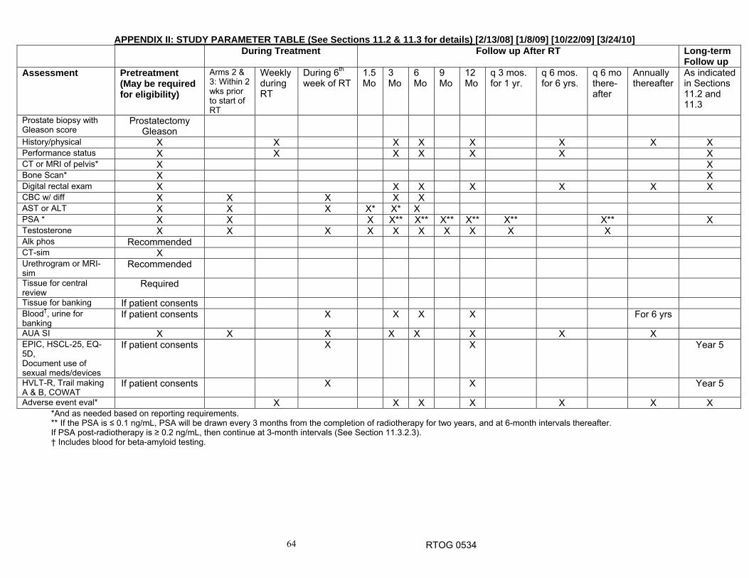
RTOG 0534
64
APPENDIX II: STUDY PARAMETER TABLE (See Sections 11.2 & 11.3 for details) [2/13/08] [1/8/09] [10/22/09] [3/24/10] During Treatment Follow up After RT Long-term
Follow up Assessment Pretreatment
(May be required for eligibility)
Arms 2 & 3: Within 2 wks prior to start of RT
Weekly during RT
During 6th week of RT
1.5 Mo
3 Mo
6 Mo
9 Mo
12 Mo
q 3 mos. for 1 yr.
q 6 mos. for 6 yrs.
q 6 mo there-after
Annually thereafter
As indicated in Sections 11.2 and 11.3
Prostate biopsy with Gleason score
Prostatectomy Gleason
History/physical X X X X X X X X Performance status X X X X X X X CT or MRI of pelvis* X X Bone Scan* X X Digital rectal exam X X X X X X X CBC w/ diff X X X X X AST or ALT X X X X* X* X PSA * X X X X** X** X** X** X** X** X Testosterone X X X X X X X X X X Alk phos Recommended CT-sim X Urethrogram or MRI-sim
Recommended
Tissue for central review
Required
Tissue for banking If patient consents Blood†, urine for banking
If patient consents X X X X For 6 yrs
AUA SI X X X X X X X X EPIC, HSCL-25, EQ-5D, Document use of sexual meds/devices
If patient consents X X Year 5
HVLT-R, Trail making A & B, COWAT
If patient consents X X Year 5
Adverse event eval* X X X X X X X *And as needed based on reporting requirements. ** If the PSA is ≤ 0.1 ng/mL, PSA will be drawn every 3 months from the completion of radiotherapy for two years, and at 6-month intervals thereafter. If PSA post-radiotherapy is ≥ 0.2 ng/mL, then continue at 3-month intervals (See Section 11.3.2.3). † Includes blood for beta-amyloid testing.

RTOG 0534
65
APPENDIX III (10/22/09)
ZUBROD PERFORMANCE SCALE
0 Fully active, able to carry on all predisease activities without restriction
1 Restricted in physically strenuous activity but ambulatory and able to carry work of a light or sedentary nature. For example, light housework, office work
2 Ambulatory and capable of all self-care but unable to carry out any work activities. Up and about more than 50% of waking hours
3 Capable of only limited self-care, confined to bed or chair 50% or more of waking hours
4 Completely disabled. Cannot carry on self-care. Totally confined to bed
5 Death

RTOG 0534
66
APPENDIX IV
AJCC STAGING SYSTEM PROSTATE, 6th Edition
DEFINITION OF TNM Primary Tumor, Clinical (T) TX Primary tumor cannot be assessed T0 No evidence of primary tumor T1 Clinically inapparent tumor neither palpable nor visible by imaging T1a Tumor incidental histologic finding in 5% or less of tissue resected T1b Tumor incidental histologic finding in more than 5% of tissue resected T1c Tumor identified by needle biopsy (e.g., because of elevated PSA) T2 Tumor confined with prostate* T2a Tumor involves one-half of one lobe or less T2b Tumor involves more than one-half of one lobe but not both lobes T2c Tumor involves both lobes T3 Tumor extends through prostate capsule** T3a Extracapsular extension (unilateral or bilateral) T3b Tumor involves the seminal vesicle(s) T4 Tumor is fixed or invades adjacent structures other than seminal vesicles: bladder neck, external
sphincter, rectum, levator muscles and/or pelvic wall *Note: Tumor found in one or both lobes by needle biopsy, but not palpable or reliably visible by imaging, is
classified as T1c. **Note: Invasion into the prostatic apex or into (but not beyond) the prostatic capsule is not classified as T3, but
as T2. Regional Lymph Nodes (N) (1/8/09) Clinical NX Regional lymph nodes cannot be assessed N0 No regional lymph node metastasis N1 Metastasis in regional lymph node(s) Pathologic pNX Regional nodes not sampled pN0 No positive regional nodes pN1 Metastases in regional node(s) Primary Tumor, Pathologic (pT) pT2* Organ confined pT2a Unilateral, involving one-half of one lobe or less pT2b Unilateral, involving more than one-half of one lobe but not both lobes pT2c Bilateral disease pT3 Extraprostatic extension pT3a Extraprostatic extension** pT3b Seminal vesicle invasion pT4 Invasion of bladder, rectum *Note: There is no pathologic T1 classification. **Note: Positive surgical margin should be indicated by an R1 descriptor (residual microscopic disease). ***Note: The type of prostatectomy (radical retropubic, perineal, robotic) should be recorded.

RTOG 0534
67
APPENDIX IV (Continued)
AJCC STAGING SYSTEM PROSTATE, 6th Edition
Distant Metastasis (M)* MX Presence of distant metastasis cannot be assessed (not evaluated by any modality) M0 No distant metastasis M1 Distant metastasis M1a Nonregional lymph node(s) M1b Bone(s) M1c Other site(s) with or without bone disease *Note: When more than one site of metastasis is present, the most advanced category is used; pM1c is most advanced. Histopathologic Grade (G) GX Grade cannot be assessed G1 Well-differentiated (slight anaplasia [Gleason 2-4]) G2 Moderately differentiated (moderate anaplasia [Gleason 5-6]) G3-4 Poorly undifferentiated or undifferentiated (marked anaplasia [Gleason 7-10]) Stage Grouping Stage I T1a N0 M0 G1 Stage II T1a N0 M0 G2, G3-4 T1b N0 M0 Any G T1c N0 M0 Any G T1 N0 N0 Any G T2 N0 M0 Any G Stage III T3 N0 M0 Any G Stage IV T4 N0 M0 Any G Any T N1 M0 Any G
Any T Any N M1 Any G

RTOG 0534
68
APPENDIX V
CTSU LOGISTICS (2/13/08) (3/24/10)
ADDRESS AND CONTACT INFORMATION FOR RTOG-0534
To submit site registration documents:
For patient enrollments: Submit study data directly to the RTOG unless otherwise specified in the protocol:
CTSU Regulatory Office 1818 Market Street, Suite 1100 Philadelphia, PA 19103 Phone – 1-888-823-5923 Fax – 215-569-0206
CTSU Patient Registration Voice Mail – 1-888-462-3009 Fax – 1-888-691-8039 Hours: 9:00 AM – 5:30 PM Eastern Time, Monday – Friday (excluding holidays) [For CTSU patient enrollments that must be completed within approximately one hour, or other extenuating circumstances, call 301-704-2376. Please use the 1-888-462-3009 number for ALL other CTSU patient enrollments.]
RTOG Headquarters 1818 Market Street, Suite 1600 Philadelphia, PA 19103 Please do not submit study data or forms to CTSU Data Operations. Do not copy the CTSU on data submissions.
For patient eligibility questions: Contact the RTOG Research Associate for Protocol, Data Management section at 215-574-3214. For treatment-related questions: Correspond by e-mail (preferred) or by phone with the study chair designated on the protocol cover page. For questions unrelated to patient eligibility, treatment, or data submission contact the CTSU Help Desk by phone or e-mail: CTSU General Information Line – 1-888-823-5923, or [email protected]. All calls and correspondence will be triaged to the appropriate CTSU representative.
The CTSU Public Web site is located at: www.ctsu.org
The CTSU Registered Member Web site is located at: http://members.ctsu.org
CANCER TRIALS SUPPORT UNIT (CTSU) PARTICIPATION PROCEDURES
REGISTRATION/RANDOMIZATION Prior to the recruitment of a patient for this study, investigators must be registered members of the CTSU. Each investigator must have an NCI investigator number and must maintain an “active” investigator registration status through the annual submission of a complete investigator registration packet (FDA Form 1572 with original signature, current CV, Supplemental Investigator Data Form with signature, and Financial Disclosure Form with original signature) to the Pharmaceutical Management Branch, CTEP, DCTD, NCI. These forms are available on the CTSU registered member Web site or by calling the PMB at 301-496-5725 Monday through Friday between 8:30 a.m. and 4:30 p.m. Eastern time. Each CTSU investigator or group of investigators at a clinical site must obtain IRB approval for this protocol and submit IRB approval and supporting documentation to the CTSU Regulatory Office before they can enroll patients. Study centers can check the status of their registration packets by querying the Regulatory Support System (RSS) site registration status page of the CTSU member web site at http://members.ctsu.org. All forms and documents associated with this study can be downloaded from the RTOG-0534 Web page on the CTSU registered member Web site (http://members.ctsu.org). Patients can be registered only after pre-treatment evaluation is complete, all eligibility criteria have been met, and the study site is listed as ‘approved’ in the CTSU RSS.
RTOG 0534
69
APPENDIX V (Continued) Requirements for RTOG-0534 site registration: • In order to utilize 3D-CRT on this study, institutions must have met the technology requirements and have
provided the baseline physics information that are described in the 3D-CRT Quality Assurance Guidelines, accessed at http://atc.wustl.edu. Credentialing requirements are outlined in Section 5.1 of the protocol and on the Image-Guided Center (ITC) web site, http://itc.wustl.edu. A 3D Questionnaire must be sent to the ITC for review prior to entering any cases. Upon review and successful completion of “Dry-Run” or “Benchmark” QA test, the ITC will notify both the registering institution and RTOG Headquarters that the institution is eligible to enter patients onto this study. Institutions that have previously enrolled patients on 3D-CRT trials with digital data submission may enroll patients on this study without further credentialing by the ITC.
• Additional credentialing requirements for sites using an IMRT Treatment Approach are outlined in Section 5.2 of the protocol and on the Advanced Technology Consortium (ATC) web site at http://atc.wustl.edu. Submission of digital data to the Image-Guided Therapy Center (ITC) requires advanced request for an FTP account with the ITC ([email protected]). The ITC will notify the registering institution when that institution is eligible to enter patients on study. The status of the credentialing review will be reflected on the RSS Site Registration Status screen, http://members.ctsu.com/RSS
• CTSU IRB Certification • CTSU IRB/Regulatory Approval Transmittal Sheet • CTSU RT Facilities Inventory Form
Note: Per NCI policy all institutions that participate on protocols with a radiation therapy component must participate in the Radiological Physics Center (RPC) monitoring program. For sites enrolling through the CTSU an RT Facilities Inventory From must be on file with CTSU. If this form has been previously submitted to CTSU it does not need to be resubmitted unless updates have occurred at the RT facility.
Pre-study requirements for patient enrollment on RTOG-0534 • Patient must meet all inclusion criteria, and no exclusion criteria should apply. • Patient has signed and dated all applicable consents and authorization forms. • All baseline laboratory tests and prestudy evaluations performed within the time period specified in the
protocol. CTSU Procedures for Patient Enrollment 1. Contact the CTSU Patient Registration Office by calling 1-888-462-3009. Leave a voicemail to alert the CTSU Patient Registrar that an enrollment is forthcoming. For immediate registration needs, e.g. within one hour, call the registrar cell phone at 1-301-704-2376. 2. Complete the following forms: • CTSU Patient Enrollment Transmittal Form • RTOG-0534 Eligibility Checklist 3. Fax these forms to the CTSU Patient Registrar at 1-888-691-8039 between the hours of 9:00 a.m. and 5:30 p.m., Mon-Fri, Eastern Time (excluding holidays); however, please be aware that RTOG registration hours end at 4:30 pm Eastern Time. The CTSU registrar will check the investigator and site information to ensure that all regulatory requirements have been met. The registrar will also check that forms are complete and follow-up with the site to resolve any discrepancies. 4. Once investigator eligibility is confirmed and enrollment documents are reviewed for compliance, the CTSU registrar will contact the RTOG within the confines of RTOG’s registration hours to obtain assignment of a treatment arm and assignment of a unique patient ID (to be used on all future forms and correspondence). The CTSU registrar will confirm registration by fax.
• Radiotherapy will start within 6 weeks after registration in Arm 1 and two months after starting LHRH agonist treatment in Arms 2 and 3.

RTOG 0534
70
APPENDIX V (Continued) DATA SUBMISSION AND RECONCILIATION 1. All case report forms (CRFs) and transmittals associated with this study must be downloaded from the RTOG-0534 web page located on the CTSU registered member Web site (http://members.ctsu.org). Sites must use the current form versions and adhere to the instructions and submission schedule outlined in the protocol. 2. Submit all completed CRFs (with the exception of patient enrollment forms), clinical reports, and transmittals to RTOG Headquarters unless an alternate location is specified in the protocol. Do not send study data to the CTSU. See the Special Materials or Substudies section below for submission of dosimetry data. 3. The RTOG Headquarters will send query notices and delinquency reports to the site for reconciliation. Please send query responses and delinquent data to the RTOG and do not copy CTSU Data Operations. Each clinical site should have a designated CTSU Administrator and Data Administrator and must keep their CTEP AMS account contact information current. This will ensure timely communication between the clinical site and the RTOG. 4. Please affix the RTOG study/case label to all source documentation and redact the patient’s name.
SPECIAL MATERIALS OR SUBSTUDIES Radiation Therapy (section 6.0) • Dosimetry data for 3DCRT and IMRT must be submitted to the Image-Guided Therapy Center (ITC), either by
digital transmission using the ITC-assigned FTP account or tape submission (contact ITC for acceptable media types and format). Hard copy materials accompanying digital data should also be sent directly to the ITC. See section 12.2 for a complete inventory of dosimetry items to be submitted.
Tissue/Specimen Submission and QOL– optional (section 10.0 and 11.0) 1. Specimen collection for tissue banking and biomarker studies (Participation is strongly recommended but not mandatory and requires additional patient consent)
• Collect, prepare, and submit specimens as outlined in protocol section 10.3 • Do not send specimens, supporting clinical reports, or transmittals to the CTSU
2. Quality of Life Substudies (Protocol section 11.8 and 11.11)
• Submit completed forms as outlined in the protocol
3. Neurocognitive Test Battery (Protocol section 11.9) • Submit completed forms as outlined in the protocol
SERIOUS ADVERSE EVENT (SAE) REPORTING 1. CTSU sites must comply with the expectations of their local Institutional Review Board (IRB) regarding documentation and submission of adverse events. Local IRBs must be informed of all reportable serious adverse reactions. 2. CTSU sites will assess and report adverse events according to the guidelines and timelines specified in the protocol. You may navigate to the CTEP Adverse Event Expedited Report System (AdEERS) from either the Adverse Events tab of the CTSU member homepage (http://members.ctsu.org) or by selecting Adverse Event Reporting Forms from the document center drop down list on the RTOG-0534 web page. 3. Do not send adverse event reports to the CTSU.
RTOG 0534
71
APPENDIX V (Continued) DRUG PROCUREMENT Commercial agents: LHRH Agonists (Zoladex; Lupron; Eligard); Eulexin; Casodex; 1. Information on drug formulation, procurement, storage and accountability, administration, and potential toxicities are outlined in section 7.0 of the protocol. 2. You may navigate to the drug forms by selecting Pharmacy Forms from the document center drop down list on the RTOG 0534 Web page. REGULATORY AND MONITORING Study Audit To assure compliance with Federal regulatory requirements [CFR 21 parts 50, 54, 56, 312, 314 and HHS 45 CFR 46] and National Cancer Institute (NCI)/ Cancer Therapy Evaluation Program (CTEP) Clinical Trials Monitoring Branch (CTMB) guidelines for the conduct of clinical trials and study data validity, all protocols approved by NCI/CTEP that have patient enrollment through the CTSU are subject to audit. Responsibility for assignment of the audit will be determined by the site’s primary affiliation with a Cooperative Group or CTSU. For Group-aligned sites, the audit of a patient registered through CTSU will become the responsibility of the Group receiving credit for the enrollment. For CTSU Independent Clinical Research Sites (CICRS), the CTSU will coordinate the entire audit process. For patients enrolled through the CTSU, you may request the accrual be credited to any Group for which you have an affiliation provided that Group has an active clinical trials program for the primary disease type being addressed by the protocol. Per capita reimbursement will be issued by the credited Group provided they have endorsed the trial, or by the CTSU if the Group has not endorsed the trial. Details on audit evaluation components, site selection, patient case selection, materials to be reviewed, site preparation, on-site procedures for review and assessment, and results reporting and follow-up are available for download from the CTSU Operations Manual located on the CTSU Member Web site. Health Insurance Portability and Accountability Act of 1996 (HIPAA) The HIPAA Privacy Rule establishes the conditions under which protected health information may be used or disclosed by covered entities for research purposes. Research is defined in the Privacy Rule referenced in HHS 45 CFR 164.501. Templated language addressing NCI-U.S. HIPAA guidelines are provided in the HIPAA Authorization Form located on the CTSU website. The HIPAA Privacy Rule does not affect participants from outside the United States. Authorization to release Protected Health Information is NOT required from patients enrolled in clinical trials at non-US sites. Clinical Data Update System (CDUS) Monitoring This study will be monitored by the Clinical Data Update System (CDUS) Version 3.0. Cumulative CDUS data will be submitted quarterly to CTEP by electronic means. The sponsoring Group fulfills this reporting obligation by electronically transmitting to CTEP the CDUS data collected from the study-specific case report forms.
RTOG 0534
72
APPENDIX VI (10/22/09) BLOOD COLLECTION KIT AND INSTRUCTIONS
Instructions for use of serum, plasma, or buffy coat collection kit: This kit includes:
• Twelve (12) 1ml cryovials • Biohazard bags • Absorbent shipping material • Styrofoam container (inner) • Cardboard shipping (outer) box • Shipping label(s) • UN 3373 Sticker • UN 1895 Dry Ice Sticker
Serum:
Using four (4) or more 1ml cryovials, label them with the RTOG study and case number, collection date and time, and clearly mark cryovials “serum”.
Process:
1. Allow one 5ml red top tube to clot for 30 minutes at room temperature. 2. Spin in a standard clinical centrifuge at 3000g at 4° Celsius for 30 minutes. 3. Aliquot a minimum of 0.5-1 ml serum into each of the four 1ml cryovials labeled with the RTOG study and
case numbers, collection date and time, and clearly mark specimens as “serum”. 4. Place cryovials into biohazard bag and store serum frozen until ready to ship. See below for storage
conditions. PLEASE MAKE SURE THAT EVERY SPECIMEN IS LABELED. Plasma:
Using four (4) or more 1ml cryovials, label them with the RTOG study and case number, collection date and time, and clearly mark cryovials “plasma”.
Process:
1. After collection, invert tube(s) multiple times to ensure adequate mixing of EDTA. 2. Centrifuge specimen within one hour of collection in a standard clinical centrifuge at 3000g at 4° Celsius
for 30 minutes. 3. If the interval between specimen collection and processing is anticipated to be greater than one hour,
keep specimen on ice until centrifuging is done. 4. Carefully pipette and aliquot a minimum of 0.5-1ml plasma into each of the 1ml cryovials labeled with the
RTOG study and case numbers, collection date and time, and clearly mark specimens as “plasma”. 5. Place cryovials into biohazard bag and store plasma frozen until ready to ship. See below for storage
conditions. PLEASE MAKE SURE THAT EVERY SPECIMEN IS LABELED. (continued on next page)

RTOG 0534
73
APPENDIX VI
BLOOD COLLECTION KIT AND INSTRUCTIONS (continued)
Buffy coat: For a visual explanation of Buffy coat, please refer to diagram below.
Using one (1) or more 1 ml cryovials, label them with the RTOG study and case number, collection date and time, and clearly mark cryovial(s) “buffy coat”.
Process:
1. Centrifuge EDTA (purple top) tube within one hour of collection in a standard clinical centrifuge at 3000g at 4° Celsius for 30 minutes.
2. If the interval between specimen collection and processing is anticipated to be greater than one hour, keep specimen on ice until centrifuging is done.
3. Carefully remove plasma close to the buffy coat and set plasma aside (can be used to send plasma samples – see above instructions).
4. Remove the buffy coat cells carefully and place into cryovials labeled “buffy coat” (it is okay if a few packed red cells are inadvertently collected in the process). Clearly mark the tubes with date/time of collection and time point collected.
5. Place cryovials into biohazard bag and store buffy coat frozen until ready to ship. See below for storage conditions.
PLEASE MAKE SURE THAT EVERY SPECIMEN IS LABELED.
Storage:
Store at –80°C (-70°C to -90°C) until ready to ship. If a -80°C Freezer is not available,
Samples can be stored short term in a -20°C freezer (non-frost free preferred) for up to one week (please ship out Monday-Wednesday only).
OR: Samples can be stored in plenty of dry ice for up to one week, replenishing daily (please
ship out on Monday-Wednesday only). OR: Samples can be stored in liquid nitrogen vapor phase (ship out Monday-Wednesday
only). Please indicate on Specimen Transmittal Form the storage conditions used and time stored.
*RTOG labels are obtained at the time of patient registration. PLEASE MAKE SURE THAT EVERY SPECIMEN IS LABELED.
(continued on next page)

RTOG 0534
74
APPENDIX VI
BLOOD COLLECTION KIT AND INSTRUCTIONS (continued)
Shipping/Mailing:
Include all RTOG paperwork in pocket of biohazard bag. Ship specimens overnight Monday-Wednesday. (Monday-Tuesday for Canada). Avoid shipping on a
weekend or around a holiday. Place frozen specimens and the absorbent shipping material in the Styrofoam cooler and fill with dry ice
(if appropriate; double-check temperature sample shipping temperature). Ship ambient specimens in a separate envelope/cooler. Place Styrofoam coolers into outer cardboard box, and attach shipping label to
outer cardboard box. Multiple cases may be shipped in the same cooler, but make sure each one is in a separate bag and that
there is still plenty of space for 10 lbs of dry ice. Ship: Specimens and all paper work as follows:
Courier Address (FedEx, UPS, etc.): For Frozen Specimens RTOG Biospecimen Resource University of California San Francisco 1657 Scott Street, Room 223 San Francisco, CA 94115 For questions, call 415-476-RTOG (7864) or e-mail: [email protected]
(continued on next page)
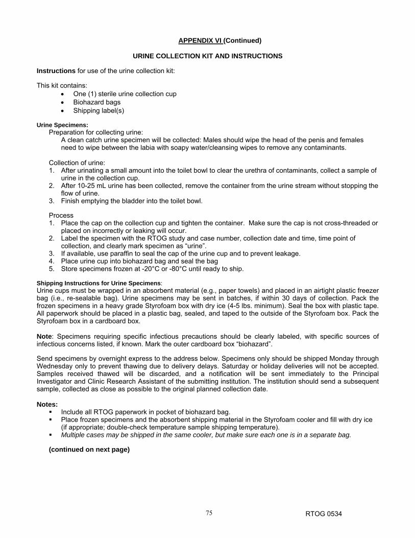
RTOG 0534
75
APPENDIX VI (Continued)
URINE COLLECTION KIT AND INSTRUCTIONS
Instructions for use of the urine collection kit: This kit contains:
• One (1) sterile urine collection cup • Biohazard bags • Shipping label(s)
Urine Specimens:
Preparation for collecting urine: A clean catch urine specimen will be collected: Males should wipe the head of the penis and females need to wipe between the labia with soapy water/cleansing wipes to remove any contaminants.
Collection of urine: 1. After urinating a small amount into the toilet bowl to clear the urethra of contaminants, collect a sample of
urine in the collection cup. 2. After 10-25 mL urine has been collected, remove the container from the urine stream without stopping the
flow of urine. 3. Finish emptying the bladder into the toilet bowl.
Process 1. Place the cap on the collection cup and tighten the container. Make sure the cap is not cross-threaded or
placed on incorrectly or leaking will occur. 2. Label the specimen with the RTOG study and case number, collection date and time, time point of
collection, and clearly mark specimen as “urine”. 3. If available, use paraffin to seal the cap of the urine cup and to prevent leakage. 4. Place urine cup into biohazard bag and seal the bag 5. Store specimens frozen at -20°C or -80°C until ready to ship.
Shipping Instructions for Urine Specimens: Urine cups must be wrapped in an absorbent material (e.g., paper towels) and placed in an airtight plastic freezer bag (i.e., re-sealable bag). Urine specimens may be sent in batches, if within 30 days of collection. Pack the frozen specimens in a heavy grade Styrofoam box with dry ice (4-5 lbs. minimum). Seal the box with plastic tape. All paperwork should be placed in a plastic bag, sealed, and taped to the outside of the Styrofoam box. Pack the Styrofoam box in a cardboard box. Note: Specimens requiring specific infectious precautions should be clearly labeled, with specific sources of infectious concerns listed, if known. Mark the outer cardboard box “biohazard”.
Send specimens by overnight express to the address below. Specimens only should be shipped Monday through Wednesday only to prevent thawing due to delivery delays. Saturday or holiday deliveries will not be accepted. Samples received thawed will be discarded, and a notification will be sent immediately to the Principal Investigator and Clinic Research Assistant of the submitting institution. The institution should send a subsequent sample, collected as close as possible to the original planned collection date. Notes:
Include all RTOG paperwork in pocket of biohazard bag. Place frozen specimens and the absorbent shipping material in the Styrofoam cooler and fill with dry ice
(if appropriate; double-check temperature sample shipping temperature). Multiple cases may be shipped in the same cooler, but make sure each one is in a separate bag.
(continued on next page)
RTOG 0534
76
APPENDIX VI (Continued)
URINE COLLECTION KIT AND INSTRUCTIONS
Instructions for use of the urine collection kit (Continued): Ship: Specimens and all paper work as follows:
U.S. Postal Service Mailing Address: For Non-frozen Specimens Only RTOG Biospecimen Resource University of California San Francisco Campus Box 1800 San Francisco, CA 94143-1800 Courier Address (FedEx, UPS, etc.): For Frozen Specimens RTOG Biospecimen Resource University of California San Francisco 1657 Scott Street, Room 223 San Francisco, CA 94115
For questions, call 415-476-RTOG (7864) or e-mail: [email protected]
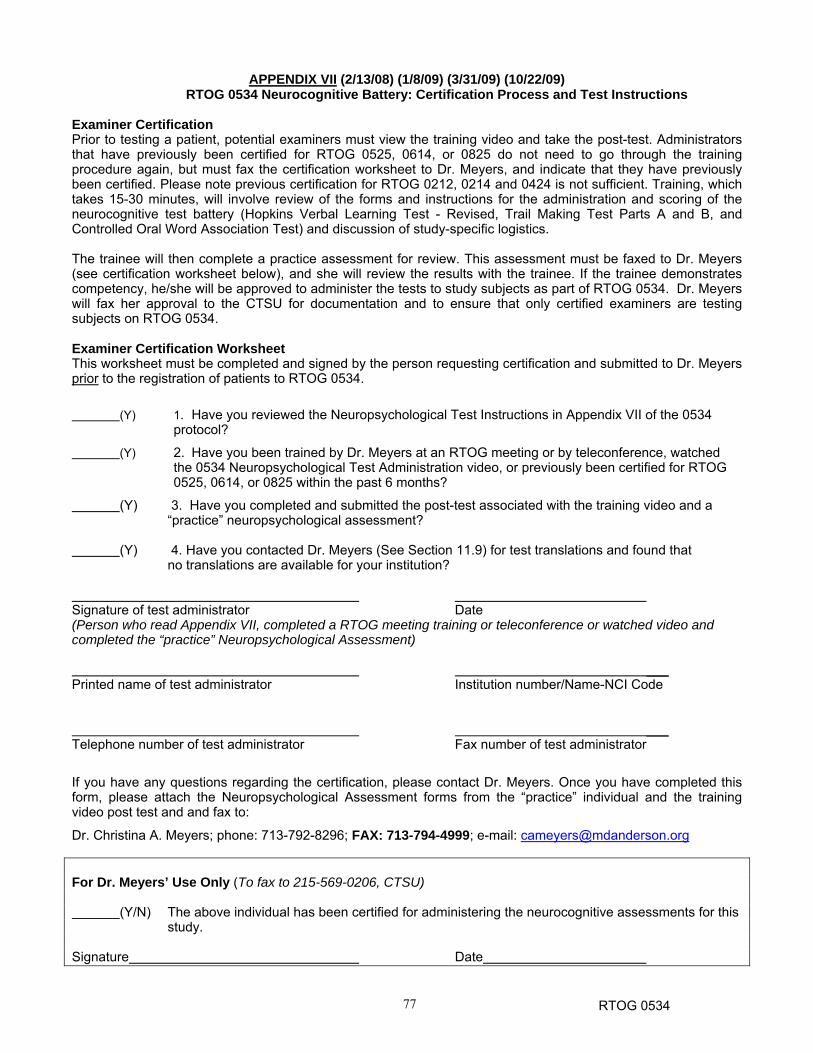
RTOG 0534
77
APPENDIX VII (2/13/08) (1/8/09) (3/31/09) (10/22/09) RTOG 0534 Neurocognitive Battery: Certification Process and Test Instructions
Examiner Certification Prior to testing a patient, potential examiners must view the training video and take the post-test. Administrators that have previously been certified for RTOG 0525, 0614, or 0825 do not need to go through the training procedure again, but must fax the certification worksheet to Dr. Meyers, and indicate that they have previously been certified. Please note previous certification for RTOG 0212, 0214 and 0424 is not sufficient. Training, which takes 15-30 minutes, will involve review of the forms and instructions for the administration and scoring of the neurocognitive test battery (Hopkins Verbal Learning Test - Revised, Trail Making Test Parts A and B, and Controlled Oral Word Association Test) and discussion of study-specific logistics. The trainee will then complete a practice assessment for review. This assessment must be faxed to Dr. Meyers (see certification worksheet below), and she will review the results with the trainee. If the trainee demonstrates competency, he/she will be approved to administer the tests to study subjects as part of RTOG 0534. Dr. Meyers will fax her approval to the CTSU for documentation and to ensure that only certified examiners are testing subjects on RTOG 0534. Examiner Certification Worksheet This worksheet must be completed and signed by the person requesting certification and submitted to Dr. Meyers prior to the registration of patients to RTOG 0534.
(Y) 1. Have you reviewed the Neuropsychological Test Instructions in Appendix VII of the 0534 protocol?
(Y) 2. Have you been trained by Dr. Meyers at an RTOG meeting or by teleconference, watched the 0534 Neuropsychological Test Administration video, or previously been certified for RTOG 0525, 0614, or 0825 within the past 6 months?
(Y) 3. Have you completed and submitted the post-test associated with the training video and a “practice” neuropsychological assessment?
(Y) 4. Have you contacted Dr. Meyers (See Section 11.9) for test translations and found that
no translations are available for your institution? Signature of test administrator Date (Person who read Appendix VII, completed a RTOG meeting training or teleconference or watched video and completed the “practice” Neuropsychological Assessment) ___ Printed name of test administrator Institution number/Name-NCI Code ___ Telephone number of test administrator Fax number of test administrator
If you have any questions regarding the certification, please contact Dr. Meyers. Once you have completed this form, please attach the Neuropsychological Assessment forms from the “practice” individual and the training video post test and and fax to:
Dr. Christina A. Meyers; phone: 713-792-8296; FAX: 713-794-4999; e-mail: [email protected]
For Dr. Meyers’ Use Only (To fax to 215-569-0206, CTSU) (Y/N) The above individual has been certified for administering the neurocognitive assessments for this study. Signature Date
RTOG 0534
78
APPENDIX VII (Continued) Testing: General Information
1. As noted above, copies of the test forms and summary sheets for the first case from each site must be faxed to Dr. Meyers for review.
2. Testing should be completed in one session. Test instructions must be followed verbatim with every patient at every assessment visit.
3. Tests should be administered in the following order to every patient and at each assessment visit: HVLT-R Part A (Learning Trials); Trail Making Test Part A; Trail Making Test Part B; COWAT; HVLT-R Part B (Delayed Recall); and the HVLT-R Part C (Delayed Recognition).
4. Follow the instructions on the Forms Packet Index before submitting forms to RTOG. 5. All test results are recorded on the Neurocognitive Evaluation Summary Form (CS), which is found in the
Forms Packet. Study/case-specific labels must be applied to all forms. 6. Note: Test results are not submitted to Dr. Meyers, nor to RTOG Headquarters (test results are recorded
on forms and submitted). Sites should keep all original test records, and test results must remain on file at the institution as source documentation pending request for submission by RTOG or a Study Chair. In the event of questions, contact Dr. Meyers.
7. Patients should not be given copies of their tests to avoid learning the material between test administrations.
8. The HVLT-R and the COWAT have alternate forms or versions in order to reduce the effects of practice. See the test instructions below for the versions to be administered at pre-treatment and subsequent sessions. The forms should continue to be alternated in this order for the duration of the study. The forms packet will contain alternate versions of these neuropsychological tests.
Before dismissing the patient, thank him/her for their cooperation. Remind the patient of their next appointment and that these tests will be repeated. In the event that a patient cannot complete a given test, please write the reason(s) on the test form AND the data summary form.
Testing: Specific Instructions Note: Administer the tests in the following order to every patient at each assessment visit. 1. HOPKINS VERBAL LEARNING TEST - REVISED (HVLT-R) This test has three parts and six alternate forms (only the first 4 forms will be used in this study): Part A - Free Recall: Complete the three learning trials first Part B - Delayed Recall: Complete after Trail Making Tests and COWAT Part C - Delayed Recognition: Complete after Delayed Recall Part A – Free Recall: Trial 1 Examiner: “I am going to read a list of words to you. Listen carefully, because when I am through, I’d like you to tell me as many of the words as you can remember. You can tell them to me in any order. Are you ready?”
• Read the words at the rate of one word every 2 seconds. Examiner: “OK. Now tell me as many of those words as you can remember.”
• Check off the words the patient recalls on the form. • If a word is said that is not in the list (for example, “intrusion”), do not write that word on the form and say
nothing to the patient about the word not being on the list. • If the patient does not produce any words for 10-15 seconds, ask the patient if he/she can remember any
more words. • If not, move on to trial 2. Later, you can record the number of words that were correctly repeated on the
summary form. Part A – Free Recall: Trial 2 Examiner: “Now we are going to try it again. I am going to read the same list of words to you. Listen carefully, and tell me as many of the words as you can remember, in any order, including the words you told me the first time.”
• Read the words at the rate of one word every 2 seconds. • Check off the words the patient recalls on the form.

RTOG 0534
79
APPENDIX VII (Continued) • If a word is said that is not in the list (for example, “intrusion”), do not write that word on the form and say
nothing to the patient about the word not being on the list. • If the patient does not produce any words for 10-15 seconds, ask the patient if he/she can remember any
more words. • If not, move on to trial 3. Later, you can record the number of words that were correctly repeated on the
summary form. Part A – Free Recall: Trial 3 Examiner: “I am going to read the list one more time. As before, I’d like you to tell me as many of the words as you can remember, in any order, including all the words you’ve already told me.”
• Read the words at the rate of one word every 2 seconds. • Check off the words the patient recalls on the form. • If a word is said that is not in the list (for example, “intrusion”), do not write that word on the form and say
nothing to the patient about the word not being on the list. • If the patient does not produce any words for 10-15 seconds, ask the patient if he/she can remember any
more words. • Do not tell the respondent that recall of the words will be tested later. • Record the time on the clock that you complete ‘Part A – Free Recall’ (for example, 1:00 p.m.) on the
designated space on the HVLT-R form. 2. TRAIL MAKING TEST [Timed Test] Part A – Sample: Place the Sample A worksheet flat on the table, directly in front of the patient (the bottom of the worksheet should be approximately six inches from the edge of the table). Give the patient a black pen and say: Examiner: “On this page (point) are some numbers. Begin at number 1 (point to 1) and draw a line from 1 to 2 (point to 2), 2 to 3 (point to 3), 3 to 4 (point to 4), and so on, in order, until you reach the end (point to the circle marked END). Draw the lines as fast as you can. Ready, begin.” If the patient completes Sample A correctly, and in a manner demonstrating that s/he understands what to do, proceed immediately to Test A. If the patient makes a mistake on Sample A, point out the error and explain it. The following explanations of mistakes serve as illustrations:
• This is where you start (point to number 1). • You skipped this circle (point to the circle omitted). • You should go from number 1 to 2, 2 to 3, and so on, until you reach the circle marked END.
If it is clear that the patient intended to touch a circle but missed it, do not count it as an omission. Remind the patient, however, to be sure to touch the circles. If the patient still cannot complete Sample A, take his/her hand and guide him/her through the trail using the opposite end of the pen, lightly touching the worksheet to avoid making marks on he copy. Then say: Examiner: “Remember, begin at number 1 (point to 1) and draw a line from 1 to 2 (point to 2), 2 to 3 (point to 3), 3 to 4 (point to 4) and so on, in order, until you reach the circle marked END (point). Do not skip around, but go from one number to the next in proper order. Remember to work as fast as you can. Ready, begin.” If the patient does not succeed, or it becomes evident that s/he cannot do the task, DISCONTINUE testing and indicate the corresponding reason on the Trail Making Data Sheet. If the patient completes Sample A correctly and appears to understand what to do, proceed immediately to Part A. Part A – Test: After the patient has completed Sample A, place the Part A test worksheet directly in front of the patient and say: Examiner: “Good! Let’s try the next one. On this page are numbers from 1 to 25. Do this the same way. Begin at number 1 (point) and draw a line from 1 to 2 (point to 2), 2 to 3 (point to 3), 3 to 4 (point to 4) and so on, in order, until you reach the circle marked END (point). Do not skip around, but go from one number to the next in proper order. Remember to work as fast as you can. Ready, begin.”
• Start timing as soon as the instruction is given to “begin”

RTOG 0534
80
APPENDIX VII (Continued)
• Watch closely in order to catch any errors as soon as they are made. If the patient makes an error, call it to his/her attention immediately and have him/her proceed from the point the mistake occurred
• The patient must complete the test in 3 minutes or less. • DO NOT STOP TIMING UNTIL HE/SHE REACHES THE CIRCLE MARKED “END”. • Collect the worksheet and record the time to completion on the Trail Making Data Sheet in minutes and
seconds • If the patient does not complete the test within 3 minutes terminate the testing. The test can also be
discontinued if the patient is extremely confused and is unable to perform the task. Collect the worksheet and complete the Trail Making Data Sheet indicating the reason the test was terminated and the last correct number reached on the test.
Part B – Sample: Place the Sample B worksheet flat on the table, directly in front of the patient (the bottom of the worksheet should be approximately six inches from the edge of the table) and say: Examiner: “On this page (point) are some numbers and letters. Begin at number 1 (point to 1) and draw a line from 1 to A (point), A to 2 (point to 2), 2 to B (point to B), B to 3 (point to 3), 3 to C (point to C) and so on, in order, until you reach the end (point to the circle marked END). Remember, first you have a number (point to 1), then a letter (point to A), then a number (point to 2), then a letter (point to B), and so on. Draw the lines as fast as you can. Ready, begin.” If the patient completes Sample B correctly, and in a manner demonstrating that s/he understands what to do, proceed immediately to Part B. If the patient makes a mistake on Sample B, point out the error and explain it. The following explanations of mistakes serve as illustrations:
• You started with the wrong circle. This is where you start (point to number 1) • You skipped this circle (point to the circle omitted) • You should go from number 1 (point) to A (point), A to 2 (point to 2), 2 to B (point to B), B to 3
(point to 3) and so on, until you reach the circle marked END (point). If it is clear the patient intended to touch a circle but missed it, do not count it as an omission. Remind the patient, however, to be sure to touch the circles. If the patient still cannot complete Sample B, take their hand and guide them through the trail using the opposite end of the pen, lightly touching the worksheet to avoid making marks on the copy. Then say: Examiner: “Now you try it. Remember, begin at number 1 (point to 1) and draw a line from 1 to A (point to A), A to 2 (point to 2), 2 to B (point to B), B to 3 (point to 3) and so on, in order, until you reach the circle marked END (point). Ready, begin.” If the patient does not succeed or it becomes evident that s/he cannot do the task, DISCONTINUE testing and indicate the corresponding reason on the Trail Making Data Sheet. If the patient completes Sample A correctly and appears to understand what to do, proceed immediately to Part A. Part B – Test: After the patient has completed Sample B, place the Part B Worksheet directly in front of the patient and say: Examiner: “Good! Let’s try the next one. On this page are both numbers and letters. Do this the same way. Begin at number 1 (point) and draw a line from 1 to A (point to A), A to 2 (point to 2), 2 to B (point to B), B to 3 (point to 3), 3 to C (point to C) and so on, in order, until you reach the circle marked END (point). Remember, first you have a number (point to 1), then a letter (point to A), then a number (point to 2), then a letter (point to B), and so on. Do not skip around, but go from one circle to the next in the proper order. Draw the lines as fast as you can. Ready, begin.”
• Start timing as soon as the instruction is given to “begin”. • Watch closely in order to catch any errors as soon as they are made. If the patient makes an error, call it
to his/her attention immediately and have him/her proceed from the point the mistake occurred • The patient must complete the test in 5 minutes or less. • DO NOT STOP TIMING UNTIL HE/SHE REACHES THE CIRCLE MARKED “END”. • Collect the worksheet and record the time to completion on the Trail Making Data Sheet in minutes and
seconds.

RTOG 0534
81
APPENDIX VII (Continued)
• If the patient does not complete the test within 5 minutes terminate the testing. The test can also be discontinued if the patient is extremely confused and is unable to perform the task. Collect the worksheet and complete the Trail Making Data Sheet indicating the reason the test was terminated and the last correct number or letter reached on the test.
3. CONTROLLED ORAL WORD ASSOCIATION TEST (COWAT) [Timed Test] This test has three parts (letters) and two alternate forms. Examiner: “I am going to say a letter of the alphabet, and I want you to say as quickly as you can all of the words that you can think of that begin with that letter. You may say any words at all, except proper names such as the names of people or places. So you would not say ‘Rochester’ or ‘Robert’. Also, do not use the same word again with a different ending, such as ‘Eat,’ ‘Eats,’ and ‘Eating.’ “For example, if I say ‘s,’ you could say ‘sit,’ ‘shoe,’ or ‘show.’ Can you think of other words beginning with the letter ‘s’?” Wait for the patient to give a word. If it is a correct response, say “good”, and ask for another word beginning with the letter “s”. If a second appropriate word is given, proceed to the test itself. If the patient gives an inappropriate word on either occasion, correct the patient, and repeat the instructions. If the patient then succeeds, proceed to the test. If the patient fails to respond, repeat the instructions. If it becomes clear that the patient does not understand the instructions or cannot associate, stop the procedure, and indicate the reason(s) on the scoring sheet. If the patient has succeeded in giving two appropriate words beginning with the demonstration letter, say: Examiner: “That is fine. Now I am going to give you another letter. Again, say all of the words beginning with that letter that you can think of. Remember, no names of people or places, just ordinary words. Also, if you should draw a blank, I want you to keep on trying until the time limit is up and I say STOP.” “You will have a minute for each letter. The first letter is ‘___’” (see scoring sheet). **Allow exactly one minute for each letter.**
• If the patient discontinues before the end of the time period, encourage him/her to try to think of more
words. • If he/she is silent for 15 seconds, repeat the basic instruction and the letter (e.g., “Tell me all the words
you can think of that begin with a “c”). • No extension on the time limit is made in the event that instructions are repeated. • Continue the evaluation with the remaining two letters, allowing one minute for each.
Recording and Scoring:
• The record sheet provides lines on which the patient’s responses can be entered (e.g., write in the word that is said by the patient). If his/her speed of word production is too fast to permit verbatim recording, a “+” should be entered to indicate a correct response.
• Incorrect responses either should not be recorded or, if recorded, should be struck through with a line. • If the patient provides more responses than there are lines on the record sheet, keep writing the
responses (or a “+”) elsewhere on the record sheet. • Count all the correct responses. The number of correct words should be indicated below each column
on the recording sheet and on the summary data form that is sent to the RTOG. Comments on scoring:
• Note: It can be helpful for the first several patients and for patients known to be fast with their word production to tape record the session for transcription at a later time.

RTOG 0534
82
APPENDIX VII (Continued)
• The instructions include a specific prohibition against giving proper names or different forms of the same word. Therefore, inflections of the same word (e.g., eat-eating; mouse-mice; loose-loosely; ran-run-runs) are not considered correct responses.
• Patients often give both a verb and a word derived from the verb or adjective (e.g., fun-funny; sad-sadness). These are not considered correct responses. On the other hand, if the word refers to a specific object (e.g., foot-footstool; hang-hanger), it would be counted as a correct answer.
• Many words have two or more meanings (e.g., foot; can; catch; hand). A repetition of the word is acceptable IF the patient definitely indicates the alternative meaning to you.
• Slang terms are OK if they are in general use. • Foreign words (for example, pasta; passé; lasagna) can be counted as correct if they can be considered
part of English vocabulary (for example, in general use or found in the dictionary). • If the test is discontinued or omitted, please mark this on the bottom of the test form and indicate the
reason on the Tests Discontinued/Not Done CRF 4. HOPKINS VERBAL LEARNING TEST - REVISED (HVLT-R) Part B – Delayed Recall
• DO NOT READ THE WORD LIST AGAIN. • Record the time on the clock that you start ‘Part B – Delayed Recall’ (for example, 1:20 p.m.) on the
designated space on the HVLT-R form. • Administer ‘Part B – Delayed Recall’ after completing all Trail Making Tests and the COWAT. There
should be at least 15 minutes between ‘Part A’ and ‘Part B’. If the time is too short, allow the patients to complete a questionnaire.
Examiner: “Do you remember that list of words you tried to learn before? Tell me as many of those words as you can remember.”
• Check the box on the corresponding line of the HVLT-R worksheet for each word the patient accurately recalls.
• If a word is said that is not in the list (for example, “intrusion”), do not write that word on the form and say nothing to the patient about the word not being on the list.
• If the patient does not produce any words for 10-15 seconds, ask the patient if he/she can remember any more words.
• If not, record the number of words that were correctly recalled on the summary form. Part C – Delayed Recognition Examiner: “Now I’m going to read a longer list of words to you. Some of them are words from the original list, and some are not. After I read each word, I’d like you to say “Yes” if it was on the original list or “No” if it was not. Was [word] on the list?”
• Check either the “Y” (Yes) or “N” (No) box next to each word to indicate the patient’s response. • Guessing is allowed. • If the test is discontinued or omitted, please mark this on the bottom of the test form and indicate the
reason on the Tests Discontinued/Not Done CRF. The score for this portion of the HVLT-R is the number of list words (i.e., words that in CAPS) correctly identified (“yes” response) minus the number of non-list words (i.e., words in lower case) incorrectly identified (“yes” response). Therefore, the actual score can range from –12 (no list words identified and all non-list words identified) to +12 (all list words identified and no non-list words identified).