radiographic examination preliminary report 1979 examination preliminary report... · radiographic...
TRANSCRIPT

Presented at the 39th National Fall Conference, Oct. 15-18, 1979, St. Louis, MO.
Reprinted from Materials Evaluation, Vol. 38, No. 12, pp. 39-44.
Radiographic Examination of the Shroud of Turin-a Preliminary Report
Abstract ... · Radfogr~~ was one of six nondestructive tests applied to a
venen;ited linen cloth in Turin, Italy. The low voltage radiographic inethod is described in which two types of films were used in thie same packet, and the results of a preliminary examination of the films are given. Also, a first assessment of the radiographic sensitivity and image sharpness is provided.
INTRODUCTION Nondestructive tests are frequently used to examine an
tiquities and art masterpieces. ·Radiography was one of six nondestructive tests1•11 used to examine a venerated linen cloth in Torino, Italy in the fall of 1978. It is a linen cloth 4.5m in length and l .lm wide and bears the faint frontal and dorsal images of a tortured man, Figure 1. Legends relate that these images are those of the crucified Jes us of Nazareth.
During this century the Shroud has been displayed in public only three times. The exposition in 1978 was in commemoration of the 400 years that it has been in Turin. This afforded a rare opportunity to perform the series of nondestructive tests that included fluorescence (x-ray, visible and ultra-violet), infrared analysis and low voltage radiography.
The objective of the tests was to collect data from the body images and other markings on the linen. The other markings include scorch marks, waterstains and reddish stains which resemble blood flow. Such data will aid in the characterization of the cloth markings and possibly in future restoration and preservation.
RADIOGRAPHIC METHOD Prior to the tests little was known about the condition of the
linen cloth and its markings. In the 16th century the cloth had
by R. W. Mottern, R. J. London, and
R. A. Morris
Robert W. (Bill) Mottern is a member of the technical staff at Sandia National Laboratories, Albuquerque, New Mexico. Prior to his present assignment in Nuclear Safeguards, he spent 16 years in nondestructive testing. He received a B.S. degree from East Tennessee University in 1944. He is an ASNT Fellow and a member of the American Academy for the Advancement of Science. For inquiries concerning this work, contact the author at (505) 844-7102.
J . Ronald London has been employed for 25 years at Los Alamos National Scientific Laboratoc ries where he is a nondestructive testing specialist. He attended Utah State and has been an ASNT member for 25 years.
Roger A. Morris is an associate group leader at Los Alamos National Scientific Laboratory where he has been employed for 19 years. He received a B.S. in Engineering Physics from the University of Colorado and a M.S. in Physics from the University of New Mexico. He is a member of ASNT.

Figure 1 -One half of the Shroud of Turin which shows the frontal image of a male.
been damaged by fire and subsequently reinforced with a holland cloth backing and the large holes mended with patches. The stains and markings were of unknown composition and areal density. The Baltograph 5-50 x-ray unit was operated at 15 kVp to obtain maximum contrast for the delineation of the features. The inherent filtration was 1.0mm Be and focal spot, 1.5mm by 1.5mm.
A fixture was designed and fabricated on which the centuries-old cloths were held during the tests. To hold these cloths, which might be very fragile, magnetic strips were used around the edges. Removable panels, 300mm wide and lm long, were provided so that only the fabrics were between the film packets and the x-ray source.
An important feature of the test fixture was an open frame
positioned on the source side. The frame was strung vertically and horizontally with 0.8mm diameter wires. The wires were spaced 198.4mm apart and were approximately 23mm from the Shroud. These were imaged as a gr\d on the x-ray films. The intersections of the wires were identified with a unique letter and number pair. This open frame with its grid of wires could be positioned at known locations as required.
The radiographic examination was planned so that three exposures were taken of a band across the width of the cloths. This was done by removing a panel from the rear of the fixture and at one end fastening a film packet across the opening. The special film packets (365mm by 432mm) contained two films-a Kodak type DR and type M. With the wire grid in place, the x-ray tube was positioned 1 meter from the linen cloth and centered on the film packet. An exposure of 100 mAm at 15 kVp was made. The first packet was then removed and a second was placed at the midpoint of the opening. After changing the elevation of the x-ray source, the second packet was exposed similar to the first. Upon removal of the second film packet, a third was positioned at the other end of the opening. The vertical position of the source was again changed and the exposure made. Each series of three exposures was made in a similar fashion. After the panel was replaced and the adjacent panel removed, the x-ray source was moved sideways so as to be centrally located before the opening. In this raster-like fashion the entire subject was radiographed.
From time to time, as necessary, the frame with the grid wires was relocated. Only the lead numerals, which identified the vertical wires, were changed. In this manner every x-ray film was uniquely identified.
A darkroom was set up in a nearby lavatory. As each packet of exposed film was removed, it was sent immediately for manual processing (5 minutes at 20 degrees C.). Variable intensity illuminators were available in a nearby room. As soon as practicable, the processed films were examined. Information was then relayed to the test room and when necessary, adjustments were made in the radiographic process.
Class I films (Kodak type DR and M) were chosen for their contrast and resolution characteristics. The M film has about twice the speed of the DR. When exposed with the DR on the source side, the densities of the two films were nearly the same.
RESULTS AND DISCUSSION The Shroud of Turin was radiographed completely with
forty-two pairs of Class I films. All were processed on site and hand-carried to the United States. The films were rewashed and dryed and duplicates made by contact printing. The duplicates were used for the examination. This was done to preserve the quality of the original films. Preliminary examination of the films showed that the radiographic technique was adequate to resolve the threads of the linen cloth-0.15mm diameter.
Preliminary Observations Many details of both cloths are readily observable. The
weave pattern of the linen is 3:1 twill which produces a herringbone weave. The holland backing cloth and patches are of a regular square weave. Because of these differences, some details can be attributed to either one or the other of the cloths.
Bands oflow areal density, several millimeters wide can be observed. Some of these bands run parallel to the long edge of the cloths, others across the width.
High density inclusions are scattered throughout the Shroud. Some may be a part of the holland cloth used as backing. Others may be entrapped between the . cloths. No efforts were made to determine the depth of such inclusions.
Certain patterns of high areal density can be correlated with the photographically recorded water stains, Figure 2.

Figure 2 -Radiographic image of one of many water stains. Also visible is the herringbone pattern of the linen.
These have the rough appearance of rings. Also, creases, which are visually and photographically observable, were recorded radiographically. Many of these creases appear to contain opaque material.
Details of the patches and the holes they cover can be easily observed. Most of the patches were cut considerably larger than the holes and their edges carefully folded under to form smooth edges before stitching. The stitching and needle holes are visible in the radiographs. The outline of the burned holes can now be mapped precisely. In at least one case, a patch covers two holes. In several cases, the patch did not cover the hole completely, Figure 3. Some small holes were reinforced only with stitching around their edges, Figure 4. Several such holes appeared to have pulled away from these reinforcing stitchings .
Besides the hand sewn stitches used to mend the linen, other stitches are visible. The study of some of these reveal that the holland cloth is not one piece but instead three pieces hand sewn together.
Approximately eight centimeters from one long edge is a feature that appears to be a seam in the linen cloth. Delorenzi reported9 that the seam is 4-5mm wide and contains the kind of stitch used to join two cloths which lack a selvage. The radiographic images substantiate the 4-5mm width of the
Figure 4 -A group of small holes some of which were present before a fire in 1532. Mending stitches around the largest holes are observable. Running left to right near the center is a stitch. Crossing this stitch left of center is a high density band caused by the thread size variation of the cotton backing cloth.
Figure 3 -Radiographic images of two patches. The upper patch was placed over two holes in the linen. The crooked streaks running vertically are debris filled creases.

"seam." In addition, two rows of stitches, one along each edge of the "seam," are observable. It was estimated, from the degree of radiographic opacity, that the seam may be four thicknesses of cloth. A type of seam used to join two cloth edges without selvage is called a flat-fell or felled seam and would be radiographically similar.
A 7X comparator with a metric graticule was used to measure thread diameters, as well as other small details. A few measurements indicate that the linen threads are rather uniform O .15 mm. In contrast the thread diameters of the holland cloth vary from 0.1 to 0.45mm.
Randomly scattered throughout the radiographic films are numerous discrete high density bodies. With 7x magnification some of these appear to have sharp edges, such as small wires or metal shavings. Others look like particulate matter. The wire-like objects have a measured diameter of O.lmm. The particle-like objects vary in dimensions from less than 0.05mm to 0.5mm. A few have been observed that appear to have been fractured.
Two other types of high density objects can be observed. Both are of medium opacity. The first of these is roughly circular with indistinct edges and appear to be in the holland cloth. The other, also circular, had a more defined edge. Some of these, too, can be seen in photographs. These appear to have approximately the same x-ray attenuation as the linen and holland threads.
Details of the two body images, the blood stains and the scorch marks are not discernible in the radiographs. Only conventional means of observation have been used in this first evaluation.
Image quality indicators were not available for the kind of material radiographed. Nevertheless, the technique used can be assessed by a determination of the radiographic sensitivity and image unsharpness. An approximation of these were made after the return from Italy.
Radiographic Sensitivity Radiographic sensitivity is defined as the relative change in
x-ray absorption that results in a perceptible difference in film density . The relationship between an areal density variation (Apt), film gradient (y), mass absorption coefficient (µ.m) and film density difference (AD) is given by:2
(1) Apt= 2.3AD
Different values for the minimum perceptible density difference (0.006 and 0.02) have been reported. 2·3 In the experience of the authors, a value close to the lower is practicable. Both values have been used in the calculations of the radiographic sensitivity. The film gradient, y, was determined from the film manufacturer's published curves.7 The mass absorption coefficients for cellulose and hematite (Fe203 ) were calculated.
The absorption data for the elements in the energy range of 7.5 to 25 kV4 were used to calculate the mass absorption coefficients for the two compounds. These are plotted in Figure 5. The least-square method was used to determine'the parameters of the power curve that best fit each set of data.
It is a general practice to characterize an x-ray technique by reporting the operating voltage applied to the x-ray tube. Another way to characterize the quality of the x-ray output is to determine the effective kilovoltage. This may be done experimentally by finding the amount of material which reduces the x-ray intensity by a factor of two and reporting this as the "half-value" layer.8 This was done, as described below, for cellulose.
Laboratory grade ashless filter paper is pure cellulose and is a good substitute for linen. Filter paper having an areal density value of 9.8 mg/cm2 was used for determination of the half-value layer. Fifteen pieces of filter paper were arranged to form a wedge of five steps, with each step being composed of
E
~ N
E u
.., c -~ -~ ~ ~ ., 0 u
c -~ .., ::-0 ~ .0 <(
300
250
200
150
100
50
0
I 0
8
~~ N
E u
HEmAT !TE
5 10 15 20 Kilovolts
CELLULOSE
Kilovolts
Figure 5 -X-ray mass absorption coefficients.
25 30
three layers of filter paper. Thus, areal densities from 29.4 to 147 mg/cm2 in increments of 29.4 were available.
For the first determination, the filter paper step wedge was radiographed on type DR film at 15 kVp with lmm Be inherent filtration similar to the test in Turin, Italy. After development of the type DR films in an x-omat (TM), the optical density of each of the five steps was measured and a leastsquares fit of the data revealed that a relation of the form, y =(a + bx)- 1
, gave the best fit. Reference to the manufacturer's curve of film density versus exposure7 showed that a density change of 1.31 represents a factor two change in x-ray exposure (intensity). This value was used in the logarithmic expression which gave a half-value layer of .154 g/cm2. This value fort was then used in the expression8
(2) 0.693
f.Lm=----pt
to determine the effective mass absorption coefficient for cellulose (4.5 cm2/g). This value was then used in the expression previously determined for cellulose to calculate the effective x-ray energy (9.4 kV). This x-ray energy was then used in the expression for Fe20 3 to determine the mass absorption coefficient for that compound. The result was 141 cm2/g. The mass absorption coefficients, as determined, were then used in Equation 1 to determine the radiographic sensitivities. The calculated results are presented in Table 1.
Another nondestructive test performed on the Shroud of Turin was x-ray fluorescence. 5 Results of that test show a range of areal densities from 29 to 42 mg/cm2 for the combination of the linen and its holland backing cloth. This test also showed that there was less than one percent of material other than cellulose, or its radiographic equivalent. Another estimation of the areal densities was based on a few measurements of thread diameters as noted above. The cloth thicknesses were taken to be twice the diameter of the thread measurements. Also noted above, the holland backing cloth has a range of thread diameters (0.1 to 0.45mm). A median value of .27 for the backing cloth was used arbitrarily. A ratio ofholland cloth (.54) to linen (.30) thickness gives 1.8. Timossi reported6 a value of 23.4 mg/cm2 for the linen. It was further assumed that the areal density of the backing cloth was in direct proportion to its thickness relative to the linen. Thus, the combined areal densities were estimated to be 66 mg/cm.2
TABLE I Calculated Radiographic Sensitivities, llpt(gm/cm2)
R film (y=4.4) .006<'.lD .02 llD
M film (y = 3.8)
Cellulose, JLm = 4.5
7 x 10-• 23
Fe20 a,/.Lm = 141
.2 x 10-•
.7
.006<'.lD 8 .3
.02 llD 27 .9 Note: The film gradients, y, were deter mined from the manufacturer's curves. 7
Radiographic Sharpness For the measurement of dimensions the extent of the image
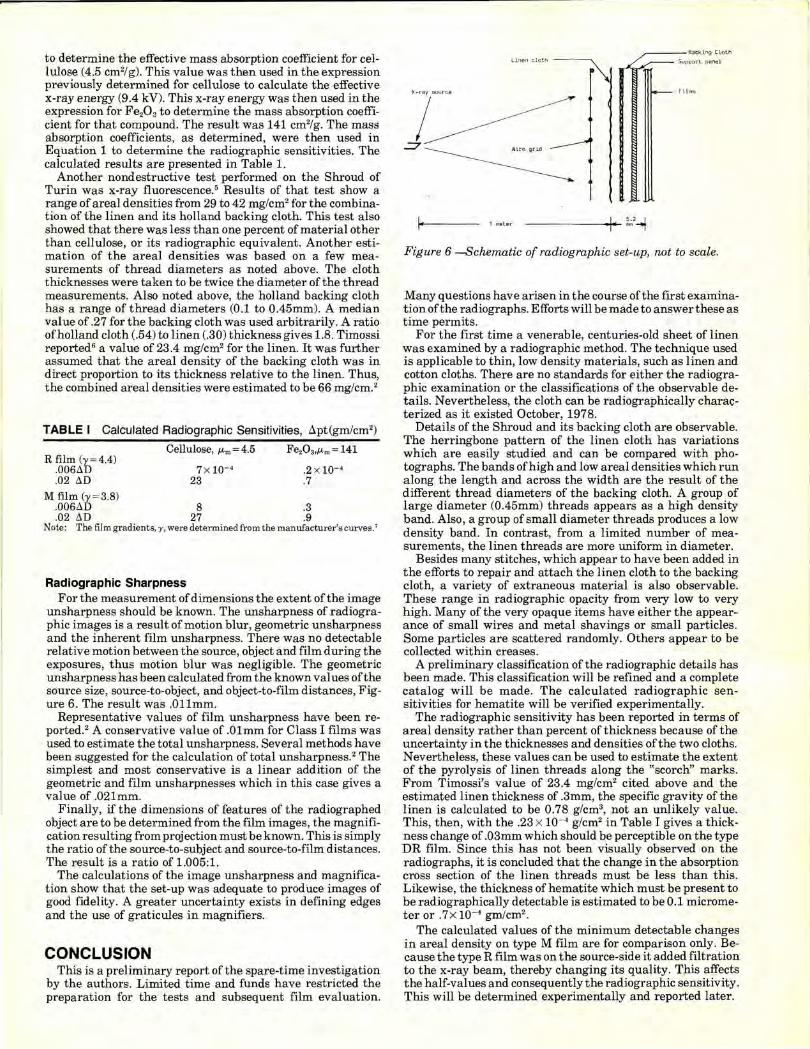
unsharpness should be known. The unsharpness of radiographic images is a result of motion blur, geometric unsharpness and the inherent film unsharpness. There was no detectable relative motion between the source, object and film during the exposures, thus motion blur was negligible. The geometric unsharpness has been calculated from the known values of the source size, source-to-object, and object-to-film distances, Figure 6. The result was .Ollmm.
Representative values of film unsharpness have been reported.2 A conservative value of .Olmm for Class I films was used to estimate the total unsharpness. Several methods have been suggested for the calculation of total unsharpness. 2 The simplest and most conservative is a linear addition of the geometric and film unsharpnesses which in this case gives a value of .02lmm.
Finally, if the dimensions of features of the radiographed object are to be determined from the film images, the magnification resulting from projection must be known. This is simply the ratio of the source-to-subject and source-to-film distances. The result is a ratio of 1.005:1.
The calculations of the image unsharpness and magnification show that the set-up was adequate to produce images of good fidelity. A greater uncertainty exists in defining edges and the use of graticules in magnifiers.
CONCLUSION This is a preliminary report of the spare-time investigation
by the authors. Limited time and funds have restricted the preparation for the tests and subsequent film evaluation.
Lin•n oloth ~
X- ray source
I ~~
~Backlog Clolh
~ ::iupport. pane l
films
1 mel er ---- --- ~2--.f
Figure 6 -Schematic of radiographic set-up, not to scale.
Many questions have arisen in the course of the first examination of the radiographs. Efforts will be made to answer these as time permits.
For the first time a venerable, centuries-old sheet of linen was examined by a radiographic method. The technique used is applicable to thin, low density materials, such as linen and cotton cloths. There are no standards for either the radiographic examination or the classifications of the observable details. Nevertheless, the cloth can be radiographically chara<;terized as it existed October, 1978.
Details of the Shroud and its backing cloth are observable. The herringbone pattern of the linen cloth has variations which are easily studied and can be compared with photographs. The bands of high and low areal densities which run along the length and across the width are the result of the different thread diameters of the backing cloth. A group of large diameter (0.45mm) threads appears as a high density band. Also , a group of small diameter threads produces a low density band. In contrast, from a limited number of measurements, the linen threads are more uniform in diameter.
Besides many stitches, which appear to have been added in the efforts to repair and attach the linen cloth to the backing cloth, a variety of extraneous material is also observable. These range in radiographic opacity from very low to very high. Many of the very opaque items have either the appearance of small wires and metal shavings or small particles. Some particles are scattered randomly. Others appear to be collected within creases.
A preliminary classification of the radiographic details has been made. This classification will be refined and a complete catalog will be made. The calculated radiographic sensitivities for hematite will be verified experimentally.
The radiographic sensitivity has been reported in terms of areal density rather than percent of thickness because of the uncertainty in the thicknesses and densities of the two cloths. Nevertheless, these values can be used to estimate the extent of the pyrolysis of linen threads along the "scorch" marks. From Timossi's value of 23.4 mg/cm2 cited above and the estimated linen thickness of .3mm, the specific gravity of the linen is calculated to be 0.78 g/cm3, not an unlikely value. This, then, with the .23 x 10-• g/cm2 in Table I gives a thickness change of .03mm which should be perceptible on the type DR film. Since this has not been visually observed on the radiographs, it is concluded that the change in the absorption cross section of the linen threads must be less than this. Likewise, the thickness of hematite which must be present to be radiographically detectable is estimated to be 0 .1 micrometer or .7x 10-• gm/cm2.
The calculated values of the minimum detectable changes in areal density on type M film are for comparison only. Because the type R film was on the source-side it added filtration to the x-ray beam, thereby changing its quality. This affects the half-values and consequently the radiographic sensitivity. This will be determined experimentally and reported later.
j
I l 1

The results of the x-ray fluorescence tests produced a range of areal density values from 29 to 42 mg/cm2
• A value of 23.4 mg/cm2 for the linen only has been reported and was used in conjunction with radiographic observations to give a value of 66 mg/cm2 for the combined value of the linen and cotton cloths. This is a significant difference and must be resolved.
Some assumptions were made in the estimations of the areal density of the holland cloth. The areal density of the holland cloth was assumed to be directly proportional to the cloth thickness based on a few measurements of the thread diameters as noted above. The holland cloth is a more loosely woven fabric than the linen, as may be observed in the radiographs. Also, there is evidence'0 that the process of producing the holland cloth in the 14th century resulted in a cotton thread less dense than the linen thread. For these reasons the values determined by the x-ray fluorescence may be more accurate than those based on the radiographic observations.
There are several locations where from one to four thicknesses of holland cloth as well as the linen were radiographed. Measurements of the film densities in these locations will be made. These will then be compared to a curve of film density versus areal density and will provide an approximation of the relative densities of the cloths. In a similar manner a measurement of the film density of the "seam" will reveal the number of linen layers present.
Hematite (Fe20 3) has been suggested as one possible paint pigment that may have been used to create the "bloodstains." For this reason, it was chosen in this work. If chemical analysis indicates the possibility that other compounds ar~ present, their radiographic sensitivities can be calculated based on the information contained here.
The size and shape of burn holes can now be determined from radiographic images. Previously, the manner in which the Shroud was folded at the time of the fire in 1532 has been postulated from the size of the covering patches. Too, the folding method is important for the determination of thermal gradients to which the linen was exposed. An accurate mapping of the covered holes will be made and reported later.
Acknowledgments There have been many persons who have contributed in
many ways to the success of the project. Especially, we would like to thank Cardinal Ballestrero, Archbishop of Turin, Monsignor Cottino, Prof. Luige Gonella and Franco Faia all of Turin, Italy. Without their help the project would have foundered. Senor Giovanni Magestrali provided invaluable assistance during an emergency. Too, we wish to recognize the assistance of Mr. John Callinan, Kodak, and Mr. Tony Ruiz, Balteau Electric, in helping to acquire the necessary films, chemicals and equipment. Barry Schwartz, Verne Miller, Mark Evans, and Ernest Brooks II provided photographic documentation. Finally, to the many persons, some anonymous, who provided the necessary financial assistance go our everlasting gratitude.
Because the authors conducted their work on their own time, they wish to acknowledge the forebearance of their families during this time.
References 1. Mottern, R. W., "The Testing of a Relic."Proceedings of the 1979
ASNT Fall Conference, St. Louis, MO. 2. Halmshaw, R. , Physics of Radiology, 1966, p. 149-217, Ameri
can Elsevier Publishing Co., Inc. 3. McMaster, Robert C., editor, Nondestructive Testing Handbook ,
Vol. 1, 1963, sect. 19, Ronald Press. 4. Biggs, Frank and Lighthill, Ruth, Analytical Approximations for
X-ray Cross Sections, Part II, SC-RR-710507, Sandia Laboratories, Albuquerque, New Mexico, December 1971 .
5. Morris, R. A., Schwalbe, L. A., London, J . R. , "X-ray Fluorescence Investigation of the Shroud of Turin," to be published.
6. Timossi, La S. Sindone nella sua constituzione tessile, Turin, 1933, p. 72.
7. , Kodak Films for Industrial Radiography , 3rd edition, 1974.
8. McGonnagle, Warren J., Nondestructive Testing, 2nd edition, 1969, p. 109, Gordon and Beach.
9 . , Report of Turin Commission on the Holy Shroud, Screenpro Films, 5 Meard St. , London, copyright 1976.
10. Rogers, Ray, private communication. 11. Jumper, Eric J. and Mottern, Robert W., "Scientific Investiga
tion of the Shroud of Turin," Applied Optics, 19(12): p. 1909-1912.