radiographic pathology of the gi tract - ceuarmy.com · gi tract. a 30 question mastery test will...
TRANSCRIPT

Radiographic Pathology of
the GI Tract ©
Three Phase CEUs Three Phase CEUs
and and
SCS Continuing Education SCS Continuing Education
presents:presents:
Copyright © 2009
by Shane Smith PTA, RT(R)

Course Abstract and Objectives:
The objective of this home study course is to provide the
learner with a computer based tutorial that will give them with
the means to learn the common radiographic pathologies of the
GI Tract. A 30 question mastery test will be administered at
the end of this home study course in order to ensure that
competency of the material has been achieved.
Hello and welcome to this program from SCS Continuing Education! Knowledge is the key to success for ourselves and our patients. This easy-to-use point and click program allows you to navigate through text and visual aides designed to provide a comprehensive view of the material covered. Please feel free to contact Shane Smith at [email protected] if you have any questions.
Introduction:
All images in this program were obtained by John Fleming.

Chapters:
Introduction to Pathology…………… pg 4
Gastrointestinal System……………….pg 34
Hepatobiliary System……………….....pg 109
Test…………………………………..… pg 143
References…………………………...… pg 144
Contact………………………………… pg 145

Introduction to Pathology Menu
1. Disease 11. Aplasia
2. Pathology 12. Atrophy
3. Pathogenesis 13. Hypertrophy
4. Inflammation 14. Neoplasm
5. Inflammatory Reactions
6. Edema
7. Abnormal Fluids
8. Ischemia
9. Infarct
10. Hemorrhage

Disease:
Simply put, pathology is the study of disease.
Disease is a term that literally refers to a lack of “ease.”
It is a condition that is marked by an abnormal disturbance
in the function and or structure of the human body as a
result of some type of injury or trauma.

Pathology:
The study of disease and how it impacts the human body.
The following is a partial list of sources for pathology:
Hereditary or Congenital
Tumors
Iatrogenic
Any adverse conditions that results from medical treatment.
An example would be a pneumothorax that occurs as the result of a thoracentesis.
Infections
A nosocomial infection is acquired from a health care environment.

Pathogenesis:
The study of the origin and development of a disease.
Pathogenesis will lead to observable changes that are known as manifestations.
Sign
This is a manifestation that is observable by the health care worker.
Examples would be swelling or a skin rash.
Symptom
This pertains to the patient’s perception of what is wrong and is subjective.
An example would be a headache.

Pathogenesis: Syndrome
This is a group of signs and symptoms that characterize
an abnormal disturbance.
An example would be Marfan’s Syndrome.
This is a genetic disorder of connective tissue
It is characterized by a predisposition to cardiac
disorders, long limbs, long fingers, and a tall stature.
Abraham Lincoln had Marfan’s Syndrome.
Etiology
This is the study of the cause and origin of a disease.

Pathogenesis:
Idiopathic
This refers to the fact that there may be no real cause for
the disease.
Examples would be hypertension and a spontaneous
pneumothorax.

Inflammation:
Inflammation refers to the body’s ability to wall-off and
sequester an injurious agent.
The ultimate goal of this process is the safe removal of said
injurious agents.
Hyperemia is the process of dilating capillaries to allow
fluids and leucocytes to infiltrate the infected area.
The leucocytes will act to remove cellular debris through a
process known as phagocytosis.
The cardinal signs of inflammation include heat (results
from hyperemia), redness, pain, and often a decrease in
function.

Inflammatory Reactions:
Abscess
This type of inflammatory reaction causes the injurious
agent to become a walled-off ball of pus.
Antibiotics cannot penetrate an abscess since they do not
contain a blood supply like a neoplasm.
As a result, an abscess must be aspirated with a needle
and drained.
Ulcers
This is another type of inflammatory reaction that is the
result of a healing wound that is located on the skin or a
mucous membrane

Inflammatory Reactions:
Cellulitis
This is an acute bacterial infection of the skin and is a
third example of an inflammatory reaction.
It can be found anywhere in the body but it is more often
seen in areas where the skin can be damaged and thus
allow a portal of entry for bacteria.
A byproduct of bacterial reproduction within the tissue is
the excretion of methane.
This can sometimes be demonstrated on a radiograph as
depicted on the next slide.

Inflammatory Reaction: Cellulitis
The arrows on this image are pointing to an area where the excrement of a bacterial infection has resulted in the formation of air within the tissue
of this patient’s foot. This is an indication of cellulitis.

Edema: This is an abnormal accumulation of fluid in body cavities
or intercellular spaces.
The increase in fluid can be localized within a structure or
dispersed throughout the body.
An example of a localized edema would be ascites which
is essentially edema of the peritoneal cavity.
Generalized edema can be caused by congestive heart
failure
This is characterized by peripheral edema, pulmonary
edema, pleural effusions, and ascites.

Abnormal Fluids: Transudates
This abnormal, extracellular fluid essentially consists of
water that contains a low cell count.
As a result, they are usually clear.
A good example would be a pleural effusion.
Exudates
This fluid filters from the circulatory system into lesions
and generally contains water, pus, and/or blood.
Since exudates contain infected fluid (pus/bacteria), they
are therefore not clear.

Ischemia: This term refers to an obstruction of the normal blood flow
to an organ or structure.
It usually results from either a narrowing of blood vessels from plaque formation (fatty cholesterol deposits) or as the result of a thrombic occlusion.
As humans age, primary blood vessels into an organ may become stenotic due to plaque formation within their lumen.
As a response to this gradual change, secondary blood vessels may enlarge and play an increasingly important role in that organ’s blood supply.
This process is called collateral circulation and it is the body’s natural defense against ischemia.

Infarct: Loss of blood supply to an organ or structure will cause the
surrounding tissue to become necrotic.
This process is referred to as an infarct.
The following is a list of diseases that are either caused by
an infarct or may result in an infarct:
Myocardial Infarction (Heart Attach)
Pulmonary Embolus
Cerebrovascular Accident (Stroke)
Hernia (Mechanical Obstruction)
Volvulus (Mechanical Obstruction)

Hemorrhage: Hemorrhage or bleeding is simply the loss of blood from the
circulatory system.
The following is a list of a few examples of a hemorrhage:
Hematoma
This occurs as the result of a break in a blood vessel that causes a pooling of blood below the surface of the skin, organ, or structure.
Ecchymosis
This is a type of hematoma that is commonly referred to as a bruise or contusion.
Capillaries below the skin are damaged usually as the result of some type of trauma.

Hemorrhage:
Purpura
These are red or purple spots on the body that are
caused by a hemorrhage.
They are often the result of some type of platelet or
coagulation disorder.
Petechia
This is a type of purpura that consists of very small
red or purple spot on the body.

Aplasia:
This is the inability of an organ or structure to form
properly.
The defective development of an organ can result in the
partial or complete loss of an organ.

Atrophy:
Atrophy is the decrease in size of the cells within an organ
or structure.
The following is a list of some of the common causes of
atrophy:
Lack of Physical Activity
Poor Nourishment
Nerve Damage
Poor Circulation

Hypertrophy:
This is the opposite of atrophy in that there is an abnormal
increase in cell size.
This condition is also sometimes referred to as hyperplasia
or hypergenesis.
The following is a list of some of the common causes of
hypertrophy:
An Increase in Physical Activity
Hormonal Changes
Chronic Inflammation

Hypertrophy: Splenomegaly
The arrow on this CT
scan of the abdomen is
pointing to a normal
spleen.

Hypertrophy: Splenomegaly
The arrows on this CT
scan of the abdomen are
pointing to an enlarged
spleen. This condition
is referred to as
splenomegaly.

Hypertrophy: Splenomegaly
This is a side-by-side comparison of a CT scan of a normal spleen on
the left (arrow) and an enlarged spleen on the right (arrows).

Neoplasm:
This is the abnormal proliferation of foreign cells that form
a mass of tissue within an organ or structure.
A neoplasm will compete for nutrients from the cells that
normally comprise the host organ and it is often referred to
as a mass or tumor.
Oncology is the study of neoplasms.
A benign neoplasm is one that is self-limited and will not
spread or seed to distant sites within the host organism.

Neoplasm:
A malignant neoplasm (cancer), on the other hand, does
possess the ability to spread to distant sites in the body.
This type of tumor will seed by employing either the
lymphatic system (primary method) or by using the
circulatory system (hematogenous spread).
Cachexia
If left untreated or undetected, malignant neoplasms
will ultimately result in this condition.
It is characterized by fatigue, atrophy, weakness, and
anorexia.
Cachexia is often seen as an end-stage to cancer.

Neoplasm:
Four major cancer categories are as follows:
1. Carcinoma/Adenocarcinoma
This type of cancer will arise from epithelial cells or
tissues such as the breast, colon, or pancreas.
2. Sarcoma
Relatively rare but highly malignant.
This is cancer of soft tissue or connective tissue such
as bone, cartilage, muscle, and fat.

Neoplasm:
3. Leukemia
This is cancer of the blood and blood forming tissues.
Acute leukemia is characterized by an abnormal
proliferation of immature blood cells that do not
possess the ability to fight infection.
Chronic leukemia is characterized by an abnormal
proliferation of mature blood cells that do not possess
the ability to fight infection.

Neoplasm:4. Lymphoma
This type of cancer originates in lymphatic tissues and affects the production of lymphocytes (white blood cells).
There are two major categories.
Non Hodgkin’s Lymphoma (NHL)
This is the most common type of lymphoma and it is found in the spleen, liver, bone marrow, lymph nodes, and GI tract.
NHLs are a diverse group of diseases that can develop in any organ that is associated with the lymphatic system and has an unknown etiology.

Neoplasm:
Non Hodgkin’s Lymphoma
will begin with the lymph
nodes and spleen and can
then metastasize to the liver,
kidneys, spine, brain, lungs,
and bone. In this example, it
has spread to the spine and
has formed an osteoblastic
condition that is commonly
referred to as an ivory
vertebra.

Neoplasm:
Hodgkin’s Lymphoma/Disease
This type of cancer is also associated with
lymphatic tissue and it was first describe by
Thomas Hodgkin in 1832.
This cancer is characterized by the lymph nodes
becoming swollen and rubbery yet they remain
pain free.
It is definitively diagnosed via lymph node biopsy
and finding the presence of Reed-Sternberg Cells.
Hodgkin’s disease has an unknown etiology.

Neoplasm:
Cancer treatment varies according to the type of cancer that
is diagnosed and what stage it is in.
The three primary methods of treating malignancies are as
follows:
1. Surgery
2. Chemotherapy
2. Radiation Therapy
In some instances, a combination of these treatments
may be indicated.

Gastrointestinal System Menu1. Zenker’s Diverticulum 15. Mechanical Bowel Obstruction
2. Traction Diverticulum 16. Hernia
3. Epiphrenic Diverticulum 17. Bowel Adhesion
4. Hiatal Hernia 18. Volvulus
5. Gastroesophageal Reflux 19. Intussusception
6. Achalasia 20. Adenomatous Polyp
7. Esophageal Varices 21. Adenocarcinoma
8. Esophagus Cancer 22. Crohn’s Disease
9. Candida 23. Constipation
10. Peptic Ulcers 24. Diverticulosis
11. Gastric Carcinoma 25. Diverticulitis
12. Bezoar 26. Appendicitis
13. Bowel Obstruction 27. Diverticula of the Appendix
14. Adynamic Ileus 28. GI Bleed

Zenker’s Diverticulum:
A diverticulum is an outpouching that occurs due to a weakening in the lining of, in this particular instance, the digestive system.
This is not to be confused with a neoplasm which is a new growth that usually develops in towards the lumen of the digestive system.
Diverticulum are often diagnosed with barium studies of the digestive system.
Zenker’s diverticulum arise from the posterior wall of the upper esophagus in the area of the pharynx.
Although often asymptomatic, they can cause dysphagia(difficulty in swallowing) and halitosis (bad breath).

Zenker’s Diverticulum:
This barium swallow study clearly depicts an outpouching of the posterior
aspect of the upper esophagus. This is called a Zenker’s diverticulum.
L

Traction Diverticulum:
This type of diverticulum forms in the mid esophagus area.
Traction diverticulum may form due to scarring from
pulmonary tuberculosis or an inflammatory process within
the mediastinum.

Traction Diverticulum:
The arrows on these images are pointing to a mid esophageal diverticulum.
The most likely etiology is an inflammatory process within the mediastinum.
L

Epiphrenic Diverticulum:
As the name implies, an epiphrenic diverticulum arises in
the distal esophagus just superior to the lower esophageal
sphincter (LES).
They may form as a complication to achalasia.

Epiphrenic Diverticulum:
Diverticula located
within the distal 10 cm
of the esophagus are
referred to as an
epiphrenic diverticula
(arrow).
L

Hiatal Hernia: A hiatal hernia occurs when a portion of the stomach
protrudes (herniates) into the thorax through the esophageal opening in the diaphragm.
This is known as a sliding hiatal hernia and it is the most common type of hiatal hernia encountered.
A rolling or paraesophageal hiatal hernia if very rare but occurs when a portion of the stomach herniates into the thorax while the gastroesophageal junction remains stationary.
This is one of the most common findings on an UGI series.
It can affect up to 50% of the population as some point in their lives.

Hiatal Hernia:
A hiatal hernia is usually asymptomatic but the patient may
experience a fullness in their chest or regurgitation.
This acid reflux may lead to inflammation and ulceration
of the esophagus.
Chronic herniation of the stomach may be associated
with gastroesophageal reflux disease (GERD).
Treatment includes a bland diet, antacids, and medications
to reduce reflux.

Sliding Hiatal Hernia:The protrusion of a
portion of the stomach
(hernia) through the
esophageal opening of the
diaphragm (hiatus) is
referred to as a hiatal
hernia. In this particular
case (a) is pointing to the
fundus of the stomach that
has herniated through the
esophageal opening in the
diaphragm (b).
a
b
b

Sliding Hiatal Hernia:
On this UGI
radiograph, a
significant portion of
the stomach (a) has
herniated through the
esophageal opening of
the diaphragm (b).
a
b b
L

Paraesophageal Hiatal Hernia:
This UGI study provides a
great example of a relatively
rare paraesophageal hiatal
hernia. In this case, a portion
of the stomach has herniated
into the thorax (a) while the
esophagus and lower
esophageal sphincter remain
in place (b).
a
b
L

Gastroesophageal Reflux Disease:
This is often abbreviated as GERD and it is also often
referred to as heartburn and acid reflux.
This disease is characterized by a backward flow of gastric
contents into the esophagus due to an incompetent lower
esophageal sphincter (LES).
GERD is commonly associated with a hiatal hernia.
It is acquired by poor eating habits, obesity, pregnancy, NG
tubes, alcohol abuse, tobacco, and as a side effect of
morphine.

Gastroesophageal Reflux Disease:
This UGI radiograph
demonstrated a reflux of
barium from the stomach
(b) back into the
esophagus (a). This
condition is known as
gastroesophageal reflux
disease (GERD), acid
reflux, or heartburn.
a
b

Achalasia:
This is the exact opposite of acid reflux.
Achalasia is an esophageal motility disorder that occurs due
to the inability of the lower esophageal sphincter (LES) to
relax.
As a result, the esophagus fills with ingested food and
fluids.
Treatment includes a bland diet, medication to relax the
LES, surgery, and an upright position to reduce
regurgitation.

Achalasia:
This UGI radiograph
demonstrates a condition
called achalasia. This is an
esophageal motility
disorder that is caused by a
lack of peristalsis. As a
result, the lower
esophageal sphincter
(arrow) fails to relax
during swallowing and the
esophagus fills with, in
this case, barium.R

Achalasia:
This example of
achalasia demonstrates
an air-fluid level
(arrow) that has been
caused during an UGI
by the inability of the
lower esophageal
sphincter (LES) to
relax.
R

Achalasia:
The entire length of this patient’s esophagus has been filled with barium
during an UGI as a result of a nonfunctional lower esophageal sphincter
(arrow). This condition is known as achalasia.

Esophageal Varices:
Esophageal varices are dilated, tortuous veins of the
esophagus which may rupture.
They are commonly a result of portal hypertension and/or
liver cirrhosis.
Esophageal varies are often a complication of
alcoholism.

Esophageal Varices:
The arrows on this
esophagram are pointing to
tortuous varicose veins of
the esophagus known as
esophageal varices. They
are the result of portal
hypertension that is often
caused by cirrhosis of the
liver. This disease is
commonly found in
patients suffering from
alcoholism.
L

Esophagus Cancer:
Esophagus cancer represents 2% of all cancers and there is a high incidence in smokers and alcoholics.
The prognosis for this cancer is very poor as it has a 5 year survival rate of 25%.
It presents with a very “ratty” radiographic appearance on a barium swallow.
Treatment includes the following
Chemotherapy
Radiation Therapy
Esophagogastrectomy (gastric pull-up)
The affected portion of the esophagus is removed and the stomach is pulled up into the thorax.

Esophagus Cancer:
The arrows on this esophagram are
pointing to areas where stenosis of
the esophagus has occurred due to
the presence of esophageal cancer.
This type of cancer has a very low
survival rate and has a high
incidence in smokers and
alcoholics. Notice how the distal
portion of the lesion has taken on
the classic “apple-core”
appearance of an adenocarcinoma.
L

Candida:
Candida occurs as the result of a fungus that has affected the
esophagus.
This is sometimes referred to as thrush.
It is an opportunistic infection that is often found in HIV
positive and cancer patients due to the state of their
suppressed immune system.

Candida:
Candida is an opportunistic
fungus that commonly inhabits
the mouth, throat, GI tract and
vagina. When it over grows
within the body it can lead to
conditions such as thrush and
candidiasis.
Immunocompromised patients
that are HIV positive or
patients on chemotherapy are
predisposed to this infection.

Peptic Ulcer Disease (PUD):
PUD is a general term that is used to describe ulcers of the
stomach and duodenum.
This is usually a chronic disease.
Causes include the use of aspirin, steroids, spicy foods,
stress, and it can be the result of a bacterial infection.
Complications include the potential for an obstruction,
perforation, and bleeding.
Treatment consists of a bland diet, antacids, decrease stress,
surgery, antibiotics and abstinence from smoking, alcohol,
and aspirin.

Peptic Ulcer Disease (PUD):
Gastric Ulcers
These are very rare and may be a complication of gastric
carcinoma.
Peptic Ulcers
These are located in the duodenum and are much more
common than gastric ulcers.
They are mostly located in the duodenal bulb and are
usually not associated with cancer.

Peptic Ulcer Disease: Duodenal
The arrow on this
radiograph is pointing to a
duodenal ulcer. This is the
most common type of
peptic ulcer and it is
usually located within the
duodenal bulb. Duodenal
ulcers are usually not
associated with cancer.

Peptic Ulcer Disease: Gastric
It is imperative that the
etiology of a gastric ulcer
be determined to ensure
that it was not caused by
stomach cancer. A biopsy
of the stomach will be
performed to rule this out.
On this UGI radiograph,
the tip of the NG tube
(arrow) is been lodged
within a gastric ulcer.
R

Gastric Carcinoma:
It is generally asymptomatic in the early stages and has
generally metastasized to other areas of the body by the time
it has been diagnosed.
As a result, it has a poor prognosis.
UGI studies present thick, irregular, and rigid (linitis
plastica) folds.
Treatment includes gastrectomy, chemotherapy, and
radiation therapy.

Gastric Carcinoma:
The arrows on this UGI radiograph are pointing to a gastric carcinoma.
Note the classic “apple-core” appearance that is a characteristic of an
adenocarcinoma.

Bezoar:
This is a hard mass of entangled material found within the
stomach or intestines that cannot be digested.
They are often made of hair and food fibers.

Bezoar:
The artifact (arrows) depicted on this radiograph consists of a hard ball
of entangled materials called a bezoar. It consists of large mass of hair
and/or vegetable fibers that cannot be digested.

Bezoar:
The arrows on this
radiograph are pointing
to another example of
how large a bezoar can
become. In fact, they
can sometimes be found
to be the cause of a
mechanical bowel
obstruction.
L

Bowel Obstruction:
The two broad categories of bowel obstructions are as follows: adynamic or paralytic ileus and a mechanical bowel obstruction.
In either case, the net result is a partial or complete loss of the normal parastaltic action of the small and/or large intestine thus impeding the normal transit of chyme.
Signs and symptoms of a bowel obstruction would include the following:
Abdominal Pain and/or Cramping
Abdominal Distention
Vomiting and Fecal Vomiting (unpalatable!)
Constipation

Adynamic or Parlytic Ileus:
This type of bowel obstruction is caused by a reduction in
the normal peristaltic action of the intestines.
This loss of peristalsis will cause the lumen of both the
small and large intestines to fill with air and fluid.
Therefore, the radiographic appearance of air in both the
small and large intestines is an indication of this condition.
Some common causes of an adynamic ileus are as follows:
Anesthesia/Some Medications
Abdominal Surgery
Illness

Adynamic or Paralytic Ileus:The dilated loops of small bowel
found on this radiograph indicate
the presence of a small bowel
obstruction. The surgery staples
(arrows) in the lower abdomen
are an indication that this
obstruction may have been
caused by a combination of
exposure to anesthesia and
abdominal surgery. Therefore,
this obstruction would be
categorized as an adynamic or
paralytic ileus.L

Mechanical Bowel Obstruction: This is the second category for bowel obstructions.
A mechanical obstruction is caused by a motility disorder that results from some type of structural abnormality.
Many factors can contribute to a mechanical bowel obstruction and some of them are listed below:
Hernia
Adhesions
Volvulus
Intussusception
Neoplasm (Adenoma/Polyp, Adenocarcinoma)
Crohn’s Disease
Constipation

Hernia: This type of obstruction is caused by a weakening of the
abdominal wall that allows a portion of the small and/or large intestine to protrude through it.
A reducible hernia can be pushed back into the abdominal cavity while an incarcerated hernia cannot and could therefore lead to a bowel obstruction.
A common hernia in men is called an inguinal hernia.
This condition occurs when the inguinal ring is compromised thus allowing a portion of the bowel to rupture through the abdominal wall.
In some instances, the bowel will descend into the scrotum.

Hernia: Inguinal
A large portion of this
patients bowel (arrows) has
protruded through an
unnatural opening within
the abdominal wall. This is
called an inguinal hernia
and it is estimated that
about 5% of the population
will develop an abdominal
wall hernia.

Bowel Adhesion: Adhesions are bands of fibrous connective tissue that
connect organs and tissues that are normally separate.
They are an almost inevitable outcome of abdominal surgery.
Adhesions can lead to abdominal pain, infertility, and bowel obstruction.
This blockage will lead to death in about 5% of all cases.
A bowel adhesions can cause a twisting of the bowel and loss of blood supply to the affected area.
The resultant bowel strangulation will result in death in as high as 37% of all cases.

Volvulus:
A volvulus is a loop of intestine that has twisted around
itself causing either a partial or complete obstruction.
They may resolve on their own but some will require
surgical intervention in order to prevent a loss of blood
supply to the affected area and relieve the obstruction.

Volvulus: Gastric
This AP UGI radiograph
depicts an abnormal
twisting of the stomach
which can lead to an
obstruction. This is
called a gastric volvulus
or stomach torsion.
R

Volvulus: Gastric
This is a lateral on the
same patient
demonstrating a gastric
volvulus or stomach
torsion.
R

Volvulus: Gastric
This is an RAO on the
same patient
demonstrating a gastric
volvulus or stomach
torsion.
R

Intussusception:
Intussusception occurs when a section of bowel is
constricted by peristalsis causing it to prolapse or telescope
into itself.
This condition is primarily confined to infants aged 2 to 36
months and occurs more frequently in boys than girls at a
ratio of 3:1.
Intussusception is the cause of approximately 1% of all
adult bowel obstructions and commonly affects the ileocecal
valve.
It is commonly corrected with a barium enema.

Intussusception:The arrows on this barium
enema on a 2 year old are
pointing to an area near the
cecum that has constricted by
peristalsis and has prolapsed
or telescoped in to itself. This
condition is a type of bowel
obstruction referred to as
intussusception.
R

Intussusception:
This is a KUB on the
previous patient after
successful reduction of the
intussusception by means of
applying pressure with a
barium enema.

Adenomatous Polyp:
A neoplasm that grows into the lumen of the colon is called
a polyp.
A pedunculated polyp possess a stalk while a sessile
(barnacle) polyp is attached directly to the bowel wall.
Most polyps are benign but an adenomatous polyp may
transform into a malignancy and must be removed.
This is sometimes referred to as simply an adenoma.
Colon polyps are generally asymptomatic but some may
cause rectal bleeding, pain, diarrhea, and/or constipation.

Adenomatous Polyp:
The arrows on this
image are pointing to a
pedunculated polyp.
This type of neoplasm
can lead to cancer and
is therefore routinely
removed.
R

Adenomatous Polyp:
This is a magnified
view of the previous
image. The presence of
a stalk (arrows) is a
characteristic of a
pedunculated polyp.
R

Adenocarcinoma:
An adenocarcinoma is a type of neoplasm that originates in glandular tissue and can be the cause a bowel obstruction.
In the colon, it is also commonly referred to as colorectal cancer and is thought to arise from adenomatous polyps.
The lifetime risk of developing colon cancer in the US is 7% and it is the second most common cause of cancer mortality.
Unfortunately, this type of metastatic cancer often goes unnoticed until it reaches a relatively advanced stage.
A colonoscopy is the method of choice for diagnosis.
A positive diagnosis is followed by surgical removal and in many instances, chemotherapy.

Adenocarcinoma :
The arrows on this image are pointing to a cancer that has formed
within the lining of the small intestine. This type of cancer is called an
adenocarcinoma since it originates within glandular tissue.

Adenocarcinoma :
This adenocarcinoma has
formed in the large
intestine and possesses a
classic the “apple-core”
appearance (arrows) that it
is often characterized by.
This type of metastatic
cancer is often referred to
as colorectal cancer.

Regional Enteritis or Crohn’s Disease:
This is the last example of a disease that may be the cause
of a mechanical bowel obstruction.
Crohn’s disease is characterized by a chronic inflammation
of the bowel and has an unknown etiology.
It is characterized by abdominal cramping, diarrhea,
constipation, weight loss or gain, and vomiting.
Fistulas may form in response the chronic inflammation that
characterizes this disease.
There is no known cure for Crohn’s disease.

Regional Enteritis or Crohn’s Disease:
This image
demonstrates the classic
radiograph appearance
of the “string sign” that
is a characteristic of
Crohn’s disease.
R

Regional Enteritis or Crohn’s Disease:
This is an even better
depiction of the classic
radiograph appearance
of the “string sign” that
is a characteristic of
Crohn’s disease.
R

Regional Enteritis or Crohn’s Disease:
This is a magnified view of the previous image depicting the radiographic
appearance of the “string sign” that is a characteristic of Crohn’s disease.
R

Constipation:
Constipation is a very common digestive complaint where
the patient experiences hard stool that can be be difficult to
defecate.
Straining to defecate can lead to anal fissures and
hemorrhoids (varicose veins of the rectum).
Severe cases can lead to a mechanical bowel obstruction
called a fecal impaction.
Causes of constipation include lack of dietary fiber,
dehydration, a decrease in peristalsis, stenosis, tumors,
anxiety, and abdominal surgery.

Constipation:
Treatment usually consists of an increased intake of fluid
and dietary fibers and the use of laxatives.
In some instances, the impaction will require the use
of enemas and/or manual removal.

Constipation:
The grainy appearance on this
KUB is the result of a fecal
impaction (arrows).
Constipation is a very
common digestive complaint
where the patient experiences
hard stool that can be difficult
to defecate.

Constipation:
This is a magnified view of the previous image that demonstrates
how a constipation can lead to a fecal impaction.
R

Constipation:
The arrows on this CT are pointing to a large fecal impaction that is
located throughout the large intestine. The etiology was constipation.
R
R

Diverticulosis:
As mentioned earlier, diverticulum can occur along the
entire length of the GI tract.
In regards to the large intestine, they are commonly found in
the area of the sigmoid colon.
Diverticulum often have no signs or symptoms and are often
a serendipitous discover on a barium study or colonoscopy.

Diverticulosis:
The arrows on these UGI radiographs are pointing to multiple
diverticula that have form on the distal portion of the duodenum.
L
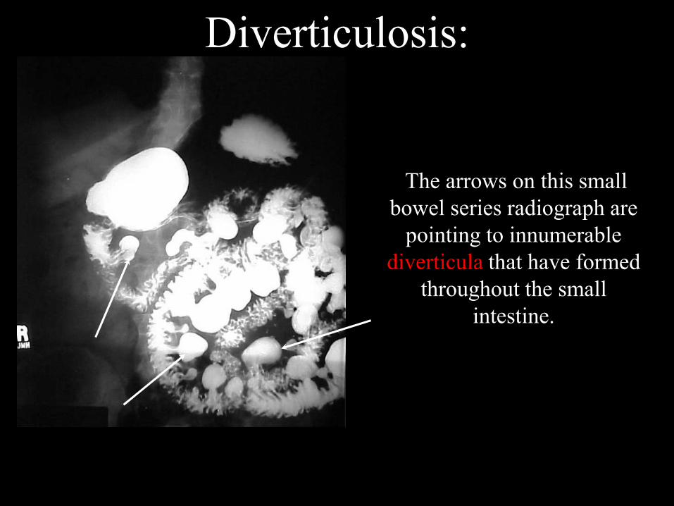
Diverticulosis:
The arrows on this small
bowel series radiograph are
pointing to innumerable
diverticula that have formed
throughout the small
intestine.

Diverticulosis:
The arrows on this barium
enema radiograph are
pointing to innumerable
diverticula that have formed
throughout the large
intestine.

Diverticulitis:
Diverticulitis occurs when a diverticulum become infected
and bleeds.
This is often caused by entrapment of chyme or feces within
the diverticulum.

Diverticulitis:
The arrows on this barium
enema radiograph are pointing
to diverticula within the large
intestine that have become
infected with feces. This
condition is referred to as
diverticulitis and will likely
result in rectal bleeding.
R
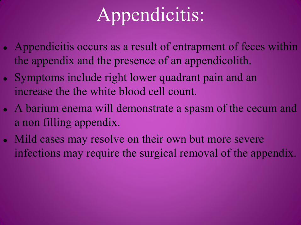
Appendicitis:
Appendicitis occurs as a result of entrapment of feces within
the appendix and the presence of an appendicolith.
Symptoms include right lower quadrant pain and an
increase the the white blood cell count.
A barium enema will demonstrate a spasm of the cecum and
a non filling appendix.
Mild cases may resolve on their own but more severe
infections may require the surgical removal of the appendix.

Appendicitis:
The arrow on this
radiograph is pointing to
an appendicolith that has
become inflamed. As a
result, this patient was
diagnosed with
appendicitis and had the
appendix surgically
removed.
R

Appendicitis:
This is a magnified
view of the previous
image and better
depicts the size and
shape of the
appendicolith (arrow).
R

Diverticula of the Appendix:
Diverticula can form
anywhere along the
alimentary canal. In
this instance, the lining
of the appendix has
weakened resulting in
the formation of a small
diverticula.
R

GI Bleed:
A GI bleed can occur throughout the GI tract.
The most common causes of lower GI bleeding are
diverticulitis and angiodysplasia.
The primary symptom is rectal bleeding with bright red
blood.
Endoscopy, nuclear medicine scans, and/or special
procedure studies (IMA/SMA) can be performed to
diagnose this condition.
Transcatheter embolization or an infusion of vasopression
have proven to be effective strategies employed to stop the
bleeding.

GI Bleed:
The arrow on this
nuclear medicine scan
is pointing to an area
where a GI bleed is
present.

GI Bleed:
The arrow on this
arteriogram is pointing
to an area where
contrast material is
escaping into the large
intestine. This is
referred to as a GI bleed.

Hepatobiliary System Menu
1. Co-Joined Twins at the Liver
2. Hepatic Cysts
3. Hepatic Hemangioma
4. Hepatocellular Carcinoma
5. Hepatomegaly
6. Liver Metastasis
7. Cholelithiasis
8. Emphysematous Cholecystitis
9. Porcelain Gallbladder
10. Splenomegaly
11. Spleen with a Calcified Cyst

Co-Joined Twins at the Liver:
The radiograph depicts
two twins that are joined
at the liver.

Hepatic Cysts:
A hepatic cyst is a benign, thin-walled sac that may be either
empty or full of fluid.
They may be located within the liver or on its external surface.
Hepatic cysts generally have no symptoms and are usually
incidental findings on ultrasounds, CT scans and/or MRI
scans of the abdomen.
No treatment is usually required.

Hepatic Cysts:
The arrows on this CT
scan of the abdomen are
pointing to rather large
hepatic cysts. These are
usually incidental findings
and no treatment is
usually required.

Hepatic Hemangioma:
A hepatic hemangioma is the most common benign tumor of the liver.
It consists of dilated blood vessels that create pools or lakes of blood
within the liver.
They commonly manifest between the ages of 30 to 50 and are more
prevalent in women than in men.
A needle biopsy is not indicated with this condition and may even be
considered a contraindication due to the increased potential for
excessive bleeding.
MRI is the modality of choice in differentiating between a
hepatocellular carcinoma and a hepatic hemangioma.
There is no treatment for this condition although surgery may be
indicated in severe cases.

Hepatic Hemangioma:
The arrows (b) on this
MRI scan of the
abdomen are pointing to
large hemangioma. The
hepatocytes of the liver
enhance with contrast
material (a) while the
“lakes” of blood within
the neoplasm do not (b).
a
b
R

Hepatic Hemangioma:
The arrows on this CT
scan of the abdomen are
pointing to large
hemangioma. This is the
most common type of
benign liver neoplasm
and is characterized by
forming large cavities or
“lakes” of blood.

Hepatocellular Carcinoma:
Hepatocellular carcinoma is a rare primary liver cancer that is also known as a hepatoma.
It is very common in alcoholics and patients with hepatitis.
It has a propensity to metastasize to the lungs but it will also spread to the colon and breasts.
This type of cancer may not have any symptoms.
However, patients may experience any combination of the following:
Dull Pain in the Right Upper Quadrant
Anorexia
Hepatomegaly
Jaundice

Hepatocellular Carcinoma:
MRI is the method of choice to diagnose this type of cancer
but CT and ultrasound are often employed as well.
There are a myriad of treatments available for hepatocellular
carcinoma including surgical resection, liver transplant, and
chemotherapy.

Hepatocellular Carcinoma:
The arrows on this CT
scan of the abdomen are
pointing to a large
hepatocellular
carcinoma. This rare
primary liver cancer is
also referred to as a
hepatoma.
R

Hepatocellular Carcinoma:
The arrows on this CT
scan of the abdomen are
pointing to another
example of
hepatocellular
carcinoma or hepatoma.
R

Hepatomegaly:
Hepatomegaly refers to an enlarged liver.
It can have a plethora of causes some of which are as
follows:
Infection
Drugs and Alcoholism (Cirrhosis)
Tumors
Hepatitis
Treatment varies according to the cause of the
hepatomegaly.

Liver Metastasis:
The liver is a common site for most any cancer metastasis.
The following is a partial list of cancers that like to spread to the liver:
Breast Cancer
Colon Cancer
Malignant Melanoma
Ovarian Cancer
Symptoms include abdominal pain, jaundice, ascites, and distension.
It is almost always treated with chemotherapy and the response is dependent on controlling the underlying primary cancer.

Liver Metastasis:
This CT image of the
abdomen demonstrates
multiple circular densities
that are consistent with
liver metastasis. The liver
is a very common site for
malignant cancers to
metastasize.
R

Liver Metastasis:
This CT image of the
abdomen demonstrates a
large neoplasm that has
been caused by liver
metastasis (arrows). This
patient had colon cancer
that metastasized to the
liver.
R

Cholelithiasis:
Cholelithiasis is the condition of having gallstones.
This only becomes a problem if the stones cause an
inflammation of the gallbladder which is called
cholecystitis.
This is often secondary to cystic duct obstruction.
Nuclear medicine and ultrasound are the imaging modalities
of choice in the diagnosis of cholelithiasis although 15% of
gallstones appear radiopaque on a KUB.

Cholelithiasis:
The arrow on this
abdominal radiograph
is pointing to a large
radiopaque cholelith.
R

Cholelithiasis:
These radiographs were taken as part of an antiquated exam called
an oral cholecystogram (OCG). This study clearly demonstrates
innumerable radiolucent choleliths.
R

Cholelithiasis:
These cholecystograms demonstrate innumerable radiolucent
choleliths.
RR

Cholelithiasis:
This percutaneous
transhepatic
cholangiogram (PTC)
clearly demonstrates a
large radiolucent
cholelith in the distal
common bile duct.
R

Cholelithiasis: Pearl Stone
This ERCP
demonstrates a
radiolucent cholelith
that has formed around a
surgical clip within the
common bile duct. This
is sometimes referred to
as a pearl stone.
R

Emphysematous Cholecystitis:
Emphysematous cholecystitis is characterized by the
presence of bacteria within the gallbladder.
In this particular instance, bacteria has managed to work
their way from the small intestine, through the biliary
tree, and finally into the gallbladder.
Bacteria produce gas as an excrement and as a result, the
gallbladder will produce a distinct air-fluid level on an
upright abdomen radiograph.
Treatment involves cholecystectomy and broad spectrum
antibiotic coverage.

Emphysematous Cholecystitis:
The arrows on this radiograph
of the abdomen are pointing
the gallbladder that has been
outlined with air. This has
been caused by an air
producing bacterial infection
that has reached the confines
of the gallbladder and is
referred to as emphysematous
cholecystitis.
R
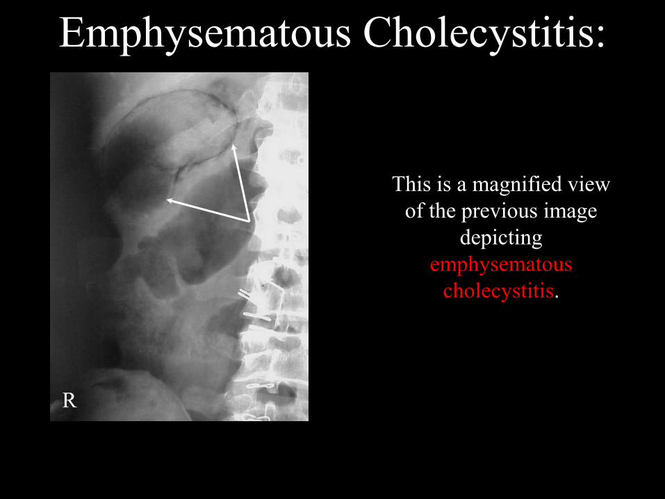
Emphysematous Cholecystitis:
This is a magnified view
of the previous image
depicting
emphysematous
cholecystitis.
R

Emphysematous Cholecystitis:
The arrows on this upright
abdomen radiograph are
pointing to an air producing
bacterial infection of the
gallbladder. This infection is
referred to as emphysematous
cholecystitis. Note the air-fluid
(bile) level that has been created
within the gallbladder as a result
of this infection.
R

Emphysematous Cholecystitis:
This is a magnified view
of the previous image
depicting an air-fluid
level caused by
emphysematous
cholecystitis.
R

Porcelain Gallbladder:
Calcification of the gallbladder is commonly referred to as a
porcelain gallbladder.
The walls of the gallbladder can calcify and form a hard,
bluish color that resembles porcelain.
It may be associated with gallbladder cancer which is very
rare or it may be brought on by excessive gallstone
production.
Treatment includes cholecystectomy.

Porcelain Gallbladder:
The arrows on this
abdominal radiograph are
pointing to a porcelain
gallbladder. This
condition occurs when
the walls of the
gallbladder calcify and
form a hard, bluish white
texture that resembles
porcelain.
R

Porcelain Gallbladder:
This CT image of the
abdomen demonstrates a
porcelain gallbladder that
contains a large gallstone
(arrow). The presence of a
porcelain gallbladder is
clinically significant because
it may be an indication that
the patient may have
gallbladder cancer.
R

Splenomegaly:
Splenomegaly simply refers to an enlargement of the spleen.
It is usually associated with any disease that involves the
destruction of a large number of defective red blood cells.
It is also linked to leukemia, lymphoma, and portal
hypertension.
Treatment for this condition usually includes a splenectomy.

Splenomegaly:
The arrow on this CT
scan of the abdomen is
pointing to a normal
spleen.
R

Splenomegaly:
The arrows on this CT
scan of the abdomen is
pointing to an enlarged
spleen. This condition
is referred to as
splenomegaly.
R

Splenomegaly:
This is a side-by-side comparison of a CT scan of a normal spleen on
the left (arrow) and an enlarged spleen on the right (arrows).
R R

Spleen with a Calcified Cyst:
This patient has a very large cyst attached to their spleen that has
become calcified. There is no clinical significance for this condition
R

There are 60 questions on this test. All answers can be
found within the context of this program. The “hint”
button located next to each question will provide you the
information needed to answer the question. At any time
during the test you may skip a question and return to it later.
You must successfully answer 70% of the questions in order
to receive credit for the course. To access the test, please
close out of this course by clicking the “x” in the top right
corner.
Test:Test:
Good luck!!!Good luck!!!

References:Mace, James D. and Kowalczyk, Nina; Radiographic Pathology for
Technologists, Fourth Edition, Elsevier-C.V. Mosby-Saunders Co., St. Louis,
MO., 2004.
Eisenberg, Ronald L. and Johnson, Nancy M.; Comprehensive Radiographic
Pathology, Fourth Edition, Elsevier-C.V. Mosby-Saunders Co., St. Louis, MO,
2007.
Laudicina, Paul; Applied Pathology for Radiographers, First Edition, Elsevier-
C.V. Mosby-Saunders Co., St. Louis, MO, 1989.
Wicke, Lothar; Atlas of Radiologic Anatomy, Sixth Edition, Williams &
Wilkins, 1998

Contact Information:Mailing Address: SCS Continuing Education &
Three Phase CEUs
c/o Shane Smith
1411 67th Avenue South
St. Petersburg, FL 33705
Phone & Fax: (727) 515-9532
Web Site:
John Fleming:
Shane Smith:
http://www.ceuarmy.com/