radiography at copán - famsi - foundation for the … · 2012-05-17 · survey of the motmot...
TRANSCRIPT

FAMSI © 2003: Jane E. Buikstra Radiography at Copán
Research Year: 2002 Culture: Maya Chronology: Classic Location: Copán Ruins, Honduras Site: Copán
Table of Contents Abstract Resumen Research Goals and Results
Introduction Goal 1: To Establish a Radiographic Inventory of the Remains from the Hunal, Margarita, and Motmot Tombs Goal 2: To Survey All Remains for Evidence of Pathology Not Visible Externally Goal 3: To Investigate Individual-Specific Forms of Pathology and Activity-Related Changes
Concluding Statement Acknowledgments List of Figures Sources Cited

Abstract
FAMSI support was essential to conduct the Copán Radiography Project. Goals accomplished in the course of this investigation included creating a radiographic archive for remains from (a) the Hunal tomb (attributed to Ruler 1, Knich Yax K’uk Mo’), (b) the Margarita tomb, and (c) the central interment from the Motmot Tomb. The remains from the Hunal and Margarita tombs were x-rayed within the tunnels at the base of Copán’s Acropolis. The Motmot skeleton was recorded within Copán’s Centro de Investigaciones. The use of Polaroid x-ray film permitted immediate determination of film quality and of skeletal features requiring further investigation. In all remains we sought evidence of pathology not visible externally as well as evidence of disuse atrophy associated with the remarkable blunt force trauma evident in the remains of Yax K’uk Mo’. Survey of the Motmot remains focused upon evaluating the midshaft fracture of the right forearm and the overall gracility of bones from throughout the skeleton.
Resumen
El apoyo de FAMSI ha sido esencial para la realización del Proyecto Radiográfico de Copán. Las metas que se alcanzaron en el curso de esta investigación incluyen la elaboración del archivo radiográfico de (a) la tumba Hunal (atribuida a Knich Yax K’uk Mo’), (b) la tumba Margarita y (c) el enterramiento central de la tumba Motmot. Se tomaron rayos-x de los restos de las tumbas Hunal y Margarita dentro de los túneles en la base de la acrópolis de Copán. El esqueleto de Motmot fue registrado en el Centro de Investigaciones de Copán. El uso de película para rayos-x Polaroid, permitió la evaluación inmediata de las tomas y el registro de ciertas características de los esqueletos que requieren investigación adicional. En todos los restos se trató de encontrar evidencia de patologías externamente no visibles. También se buscó evidencia que indicara la falta de uso debido a la atrofia causada por el fuerte impacto traumático evidente en los restos de Yax K’uk Mo’. El examen de los restos de Motmot estuvo centrado en la evaluación de la fractura del antebrazo derecho y en la morfología relativamente delicada de todo su esqueleto.
Submitted 04/04/2003 by: Jane E. Buikstra [email protected]
2

Research Goals and Results
Introduction
Recovered from within the Copán Acropolis complex, three sets of remains are of significance to students of the Classic Maya. This includes those from the Hunal, the Margarita, and the Motmot tombs. Given the frail nature of some bones and the significance of the individuals whom the remains represent, they require complete documentation –including x-rays– as part of a general archive. This was the first goal of the Copán radiography project. Secondly, each set of remains presents evidence of pathology that can profit from radiographic analysis. Finally, the survey also seeks to identify any evidence of pathology not visible externally.
THE HUNAL TOMB:
Located within the lowest level of the Copán Acropolis, the Hunal tomb has been attributed to the founder of Classic Maya Dynasty at Copán, Knich Yax K’uk Mo’. The tomb contained skeletal remains of an adult male who died at an advanced age, having survived a remarkable range of blunt force trauma (Buikstra et al. 2003). These include a "parry" or "nightstick" fracture to the right forearm. Such fractures may result from either a fall or a direct blow to the forearm when the arm is pronated and raised to shield the face. In this case, the facture had not been reduced and the radius had healed with significant shortening. The broken ulna did not heal, thus forming a non-union. The right 5th metacarpal also presents evidence of a healed fracture and minor deformity, another possible defensive wound.
The inferior third of the sternum is thinned to the left of a distorted sternal foramen, the apparent result of blunt force trauma to this region. The blow causing this fracture apparently depressed the caudal portion of the body while, in compensation, causing the superior portion to project anteriorly. In association with this restructuring of the thorax, the articulations between the manubrium and the clavicles were displaced laterally, forming new articular facets.
A further, most unusual example of blunt force trauma involved the left shoulder of the Yax K’uk Mo’ remains. As the result of either a blow or a fall, the superior third of the glenoid fossa and the coracoid process of the scapula had separated from the remainder of the bone. As with the right forearm, this fracture never healed. Arthritic change at the shoulder was profound.
Given the extreme nature of the blunt force trauma, it is not unreasonable to propose that the range of motion for one or both arms may have been limited. Therefore, one goal of the radiography project specific to the Yax K’uk Mo’ remains is to identify any evidence of disuse atrophy or other forms of abnormal activity constraints.
3

THE MARGARITA TOMB:
Subsequent to the interment of Yax K’uk Mo’, a female was entombed within an even more elaborate funerary structure located slightly above the Hunal tomb. Although she is not identified by inscriptions, it has been proposed that she was a member of the local Copán elite who married Yax K’uk Mo’ and became the mother of Ruler 2. This tomb contained "a stunning array of jade, shell, pottery, and other offerings" on the surface of the burial platform and another 2000 objects beneath it (Sharer et al. 1999; Bell et al. 2000). The staircase to the Margarita tomb was modified through subsequent rebuilding of the Acropolis so that it remained accessible. It is obvious that the remains from the Hunal and Margarita tombs represent historical figures of paramount significance during the period the Copán dynasty was founded.
The Margarita tomb remains are extremely fragile and archival records made in timely fashion are important to provide a data source that may not be available in the long term. She presents external evidence of age-related skeletal degeneration, although internal pathological changes are possible.
THE MOTMOT TOMB:
Located under Copán’s famous hieroglyphic staircase, the Motmot tomb was built to commemorate a major calendric event near the end of Yax K’uk Mo’s reign. The circular marker that closed this tomb provides representations of both Ruler 1 and Ruler 2. The initial deposit within the tomb included a primary interment and at least one "trophy skull." Two more skulls, one associated with cervical vertebrae showing cutmarks, were added to the tomb more recently, prior to its final closing (Fash and Fash 1996, 2000; Sharer 1997; Sharer et al. 1999).
The primary interment within the Motmot tomb is a remarkably well-preserved female who died during young adulthood. Her remains are notable for their gracility, including narrow bones and poorly developed areas of muscle attachment. As did Yax K’uk Mo’, she had suffered a "parry" or "nightstick" fracture to her right forearm. Goals of x-ray analysis specific to this individual include (1) an evaluation of her parry fracture; (2) identification of evidence for bone thinning either associated with reduced activity levels or pathology; and (3) evaluation of minor asymmetry of the proximal humeri as possible evidence for a fracture of the right humerus at the surgical neck.
Goal 1: To Establish a Radiographic Inventory of the Remains from the Hunal, Margarita, and Motmot Tombs
Between October 19 and 26, nearly 100 Polaroid (T-803 Film) x-rays were taken of remains from the Hunal, Margarita, and Motmot tombs. Because the films are limited in size (8 x 10 inches), composites of long bones required assembling on Adobe Photoshop at the University of New Mexico. While some clarity was sacrificed due to
4

the presence of radiopaque cinnabar on certain bones, especially those from the Margarita tomb, we have compiled an overall archive. Copies (CD) of this working archive have been filed with the Instituto Hondureño de Antropología y Historia (IHAH: 2 copies) and the Early Copán Acropolis Project (ECAP: 1 copy). A master paper and CD archive is also maintained at the University of New Mexico. As examples of the x-rays submitted to IHAH and ECAP, we include here images of the Motmot skull (Figure 1); the Hunal inlaid teeth (Figure 2); and the Motmot right humerus (Figure 3).
On February 3, Dr. Ethan Braunstein, Staff Radiologist at the Mayo Clinic, Scottsdale, AZ and published expert in paleopathology was shown all images from the working archive. His comments are incorporated into the following discussion.
Figure 1: X-rays of the Cranium from the Motmot Tomb: (a) anterior-posterior perspective; (b)
medio-lateral perspective; and (c) superior-inferior perspective.
Figure 2: Medio-lateral aspect of inlaid teeth from the Hunal Tomb.
5
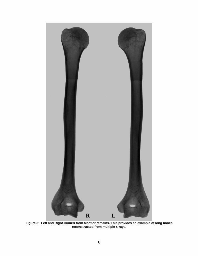
Figure 3: Left and Right Humeri from Motmot remains. This provides an example of long bones
reconstructed from multiple x-rays.
6

Goal 2: To Survey All Remains for Evidence of Pathology Not Visible Externally
The most notable structures that were not visible externally are radiodense lines visible at the proximal aspect of both humeri from Yax K’uk Mo’ (Figure 4). These can also be visualized as dense linear structures visible through the broken proximal end of the right humerus. While Dr. Braunstein was not familiar with such structures, he interprets them as developmental rather than pathological. We will continue to seek comparative examples of these unusual phenomena.
Figure 4 (inset).
7

Figure 4: X-ray of humeri from the Hunal tomb. Arrows indicate location of radiodense areas at
proximal ends. Image illustrates the position and nature of the structures (arrow).
8

Goal 3: To Investigate Individual-Specific Forms of Pathology and Activity-Related Changes
HUNAL TOMB 1: Evidence of disuse atrophy associated with the healed "Parry" or "Nightstick" fracture of the right forearm.
It is clear from the radiographs (Figure 5) that the insult to the radius did not result in disuse atrophy. Cortical thickness remains and there is clear gross and radiographic evidence for the development of arthritic lipping at the articular surfaces. Interestingly, despite this gross evidence of arthritic changes associated with the radius, the ulna itself lost a significant amount of its cortical bone thickness. The pseudoarthrosis formed by the proximal and distal ulnar fragments do not exhibit a significant degree of reactive bone. In general, bone density at this site is low, suggesting that it was not subject to continual pressure or irritation that would have resulted from continued use. There is evidence, however, that there was some contact between the portions based upon lipping present on both surfaces. Therefore, while the radius displays clear evidence of continued use, the ulna atrophied due to the injury. None of the bones of the right hand present evidence of cortical thinning or trabecular resorption consistent with disuse.
Prior to the x-ray survey, we had been entertaining two alternative explanations of the "parry" fracture: (1) trauma, and (2) a fall. Given the angle of the break and the absence of visible (radiographic) deformity at the wrist and the elbow, Dr. Braunstein favors a blow to the forearm as the most likely cause.
Figure 5: Right forearm from Hunal Tomb. Radius and Ulna are labeled, with arrow illustrating the
location of cortical thinning of the distal ulna.
9

HUNAL TOMB 2: Evidence of disuse atrophy in the left upper limb, resulting from the blunt force trauma and non-union of the left scapula.
While there is clear post-traumatic change to the shape of the head of the left humerus and associated osteoarthritic change, a comparison of right and left humeri (Figure 6) does not reveal any asymmetry that could be interpreted in terms of diminished activity. Remarkable, however, are the expanded lateral epicondylar flanges, just proximal to the distal articular surfaces. These are present bilaterally and while visible externally, are especially impressive on x-ray images. Dr. Braunstein interprets these as normal developmental features, which we will continue to consider in comparative study.
Figure 6 (inset).
10

Figure 6: Images and x-rays of humeri from the Hunal Tomb. Brackets indicate locations of lateral
epicondylar flanges. While these are visible in the images, the nature of the expansion is much more clear in the x-rays.
11

MARGARITA TOMB: None
MOTMOT TOMB 1: Evaluation of the "parry" fracture of the right ulna.
The right ulna presents an expanded callus, even though alignment is good. Dr. Braunstein proposed that the fracture might have been splinted because there is no anatomical alignment distortion. An arrow indicates the residual fracture line (Figure 7). Dr. Braunstein does not believe that the nature of the callus and the visibility of the line permit us to estimate how long before death the fracture occurred.
MOTMOT TOMB 2: Possibility of a healed fracture at the surgical neck of the right humerus.
There is no evidence of a fracture line at this site.
MOTMOT TOMB 3: Evidence of bone thinning either associated with pathology or inactivity.
There is no evidence of cortical or trabecular thinning consistent with a diagnosis of osteoporosis/osteopenia or disuse. As Dr. Braunstein notes, the x-rays are generally unremarkable, with the exception of the right ulna.
12

Figure 7: Right radius and ulna from Motmot tomb (labeled). Arrow indicates location of fracture
line in right ulna.
13

Concluding Statement
In the course of the Copán Radiography Project, we have created an x-ray archive for three sets of human remains. Included are those from the Hunal, the Margarita, and the Motmot tombs. These will be useful for further specialized studies, including bone function and biomechanics.
Our results also indicate that although the remains attributed to Yax K’uk Mo’ present clear evidence of severe, survived blunt force trauma, there is little evidence of diminished activity levels. The single clearly defined evidence of disuse atrophy occurs in the distal right ulna of the forearm. Two examples of developmental anomalies within the upper limbs have been identified and will be subject of further study. In addition, x-rays of the dentition, specifically the inlaid teeth, will provide further details concerning the methods used and the pathological sequelae of dental modification.
Remains from the Margarita tomb are limited due to the impact of extensive post-depositional treatment with cinnabar. Interestingly, the x-rays reveal the extent to which the pigment penetrated the skull after the flesh was no longer present.
The Motmot remains appear free of generalized osteopenia, despite their gracile nature. The right ulna presents clear evidence of a complete midshaft fracture, with an absence of deformity. This circumstance may suggest that the bone was set, following the traumatic episode.
Acknowledgments
Permission for study has been granted by the Instituto Hondureño de Antropología y Historia and by Robert Sharer and William Fash of the ECAP. The assistance of David Sedat (ECAP) and Lic. Oscar Cruz (IHAH) and especially Kenneth Nystrom (UNM) has been essential to the success of the project. The diagnostic expertise of Dr. Ethan Braunstein is also gratefully acknowledged.
List of Figures
Figure 1: X-rays of the Cranium from the Motmot Tomb: (a) anterior-posterior perspective; (b) medio-lateral perspective; and (c) superior-inferior perspective.
Figure 2: Medio-lateral aspect of inlaid teeth from the Hunal Tomb.
Figure 3: Left and Right Humeri from Motmot remains. This provides an example of long bones reconstructed from multiple x-rays.
14

Figure 4: X-ray of humeri from the Hunal tomb. Arrows indicate location of radiodense areas at proximal ends. Image illustrates the position and nature of the structures (arrow). Inset Image
Figure 5: Right forearm from Hunal Tomb. Radius and Ulna are labeled, with arrow illustrating the location of cortical thinning of the distal ulna.
Figure 6: Images and x-rays of humeri from the Hunal Tomb. Brackets indicate locations of lateral epicondylar flanges. While these are visible in the images, the nature of the expansion is much more clear in the x-rays.
Figure 7: Right radius and ulna from Motmot tomb (labeled). Arrow indicates location of fracture line in right ulna.
Sources Cited Bell, E.E., Sharer, R.J., Sedat, D.W., Canuto, M.A., and Grant, L.A. 2000 The Margarita Tomb at Copán, Honduras: A Research
Update. Expedition 42:21-25. Buikstra, J.E., Price, T.D., Burton, J.H., and Wright, L.E. 2003 Tombs from Copán’s Acropolis: A Life History Approach. In E.E. Bell, M.A.
Canuto and R.J. Sharer (eds.): Understanding Early Classic Copán. Philadelphia: University of Pennsylvania Museum of Archaeology and Anthropology.
Fash, W.L., and Fash, B.W. 1996 Building a World-View: Visual Communication in Classic Maya
Architecture. RES: Anthropology and Aesthetics 29/30:127-147. 2000 Teotihuacán and the Maya: A Classic Heritage. In D. Carrasco, L. Jones and
S. Sessions (eds.): Mesoamerica’s Classic Heritage: From Teotihuacán to the Aztecs.Denver: University Press of Colorado, pp. 433-464.
Sharer, R.J. 1997 K’inich Yax K’uk’ Mo’ and the Genesis of the Copán Acropolis. Presented at
the A Tale of Two Cities: Copán and Teotihuacán. Department of Anthropology, Harvard University, Cambridge, MA.
15

16
Sharer, R.J., Fash, W.L., Sedat, D.W., Traxler, L.P., and Williamson, R.V. 1999 Continuities and Contrasts in the Early Classsic Architecture of Central Copán.
In J.K. Kowalski (ed.): Mesoamerican Architecture as Cultural Symbol. Oxford: Oxford University Press, pp. 220-249.
Sharer, R.J., Traxler, L.P., Sedat, D.W., Bell, E.E., Canuto, M.A., and Powell, C. 1999 Early Classic Architecture Beneath the Copán Acropolis: A Research
Update. Ancient Mesoamerica 10:3-23.