radiologic investigation of chest and cvs...
TRANSCRIPT

Lecture 6
Radiologic investigation of Chest and
CVS diseases
Khawlah AlOthman Hanan AlSalman
Maha AlKubidan Ghadeer AlWuhayd
Hanan Alrabiah
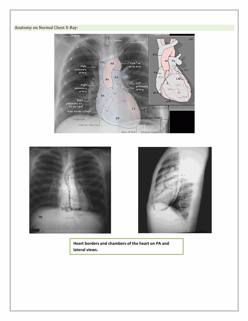
Heart borders and chambers of the heart on PA and
lateral views.
Anatomy on Normal Chest X-Ray:
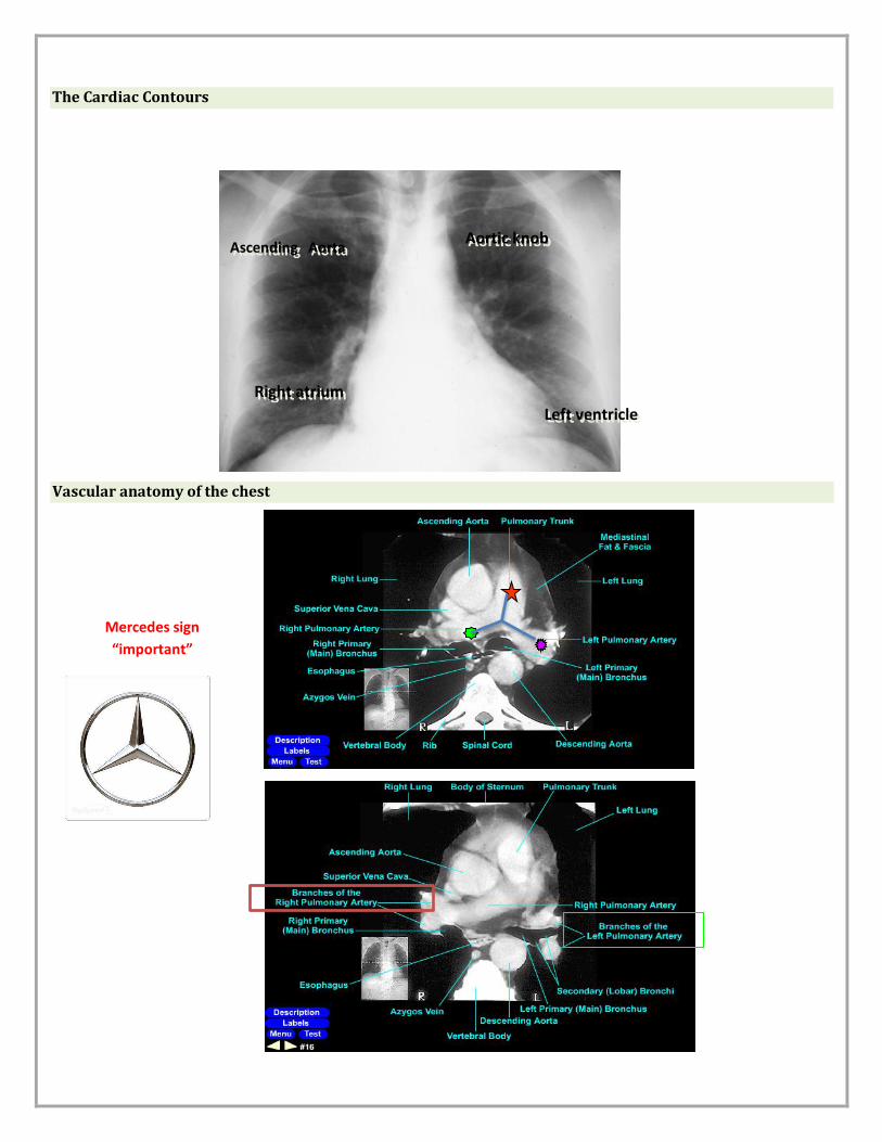
The Cardiac Contours
Vascular anatomy of the chest
Ascending Aorta Aortic knob
Right atrium
Left ventricle
[Type a quote from the document or the
summary of an interesting point. You can
position the text box anywhere in the
document. Use the Drawing Tools tab to
change the formatting of the pull quote text
box.] Mercedes sign
“important”
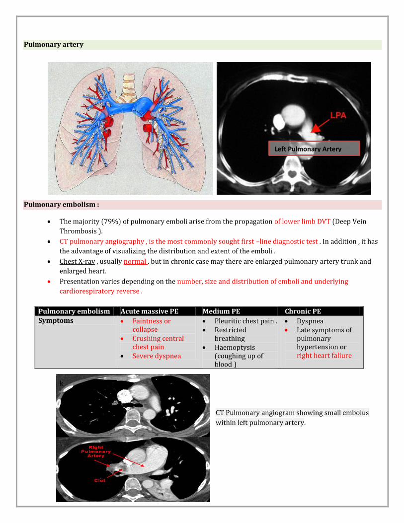
Pulmonary artery
Pulmonary embolism :
The majority (79%) of pulmonary emboli arise from the propagation of lower limb DVT (Deep Vein
Thrombosis ).
CT pulmonary angiography , is the most commonly sought first –line diagnostic test . In addition , it has
the advantage of visualizing the distribution and extent of the emboli .
Chest X-ray , usually normal . but in chronic case may there are enlarged pulmonary artery trunk and
enlarged heart.
Presentation varies depending on the number, size and distribution of emboli and underlying
cardiorespiratory reverse .
Pulmonary embolism Acute massive PE Medium PE Chronic PE
Symptoms Faintness or collapse
Crushing central chest pain
Severe dyspnea
Pleuritic chest pain . Restricted
breathing Haemoptysis
(coughing up of blood )
Dyspnea Late symptoms of
pulmonary hypertension or right heart faliure
k
CT Pulmonary angiogram showing small embolus
within left pulmonary artery.
Left Pulmonary Artery
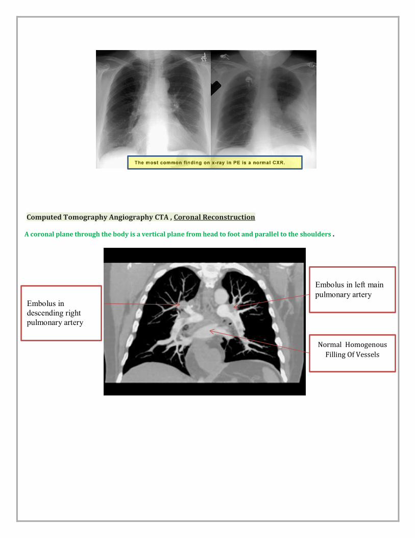
Computed Tomography Angiography CTA , Coronal Reconstruction
A coronal plane through the body is a vertical plane from head to foot and parallel to the shoulders .
Embolus in left main
pulmonary artery
Normal Homogenous
Filling Of Vessels
Embolus in
descending right
pulmonary artery

What to Evaluate ??
The Pulmonary Vasculature :
Five States of the Pulmonary Vasculature :
• Normal
• Pulmonary venous hypertension
• Pulmonary arterial hypertension
• Increased flow
• Decreased flow
Image 1: NO (1): RDPA(Right descending pulmonary artery) < 17 mm in diameter.
Image 2 :
o In erect position, blood flow to bases > than flow to apices.
o Size of vessels at bases is normally greater than size of vessels at apex.
o You can’t measure size of vessels at the left base because the heart obscures them.
Image 3:
o Central vessels give rise to progressively smaller peripheral branches.
o Normal tapering of vessels from central to peripheral.
2
2
1
3
2- Normal Distribution Of Flow
(Upper Versus Lower Lobes )..
3- Normal Distribution Of Flow
(central versus peripheral )
Finding main pulmonary artery
Main pulmonary
artery is Adjacent
to left pulmonary
artery
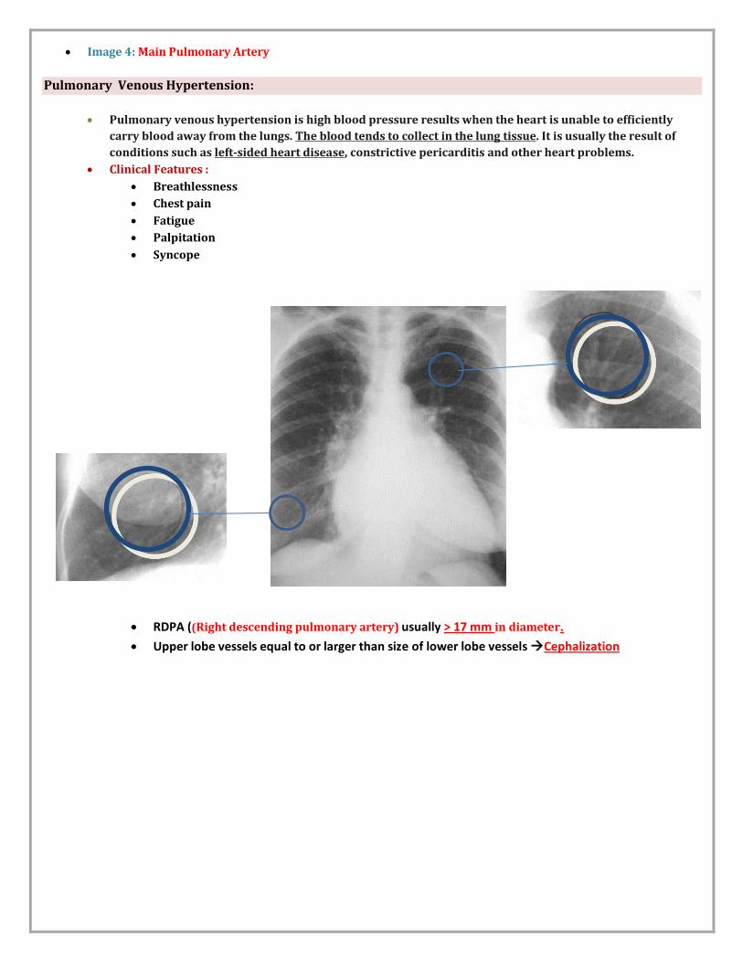
Image 4: Main Pulmonary Artery
Pulmonary Venous Hypertension:
Pulmonary venous hypertension is high blood pressure results when the heart is unable to efficiently
carry blood away from the lungs. The blood tends to collect in the lung tissue. It is usually the result of
conditions such as left-sided heart disease, constrictive pericarditis and other heart problems.
Clinical Features :
Breathlessness
Chest pain
Fatigue
Palpitation
Syncope
RDPA ((Right descending pulmonary artery) usually > 17 mm in diameter.
Upper lobe vessels equal to or larger than size of lower lobe vessels Cephalization
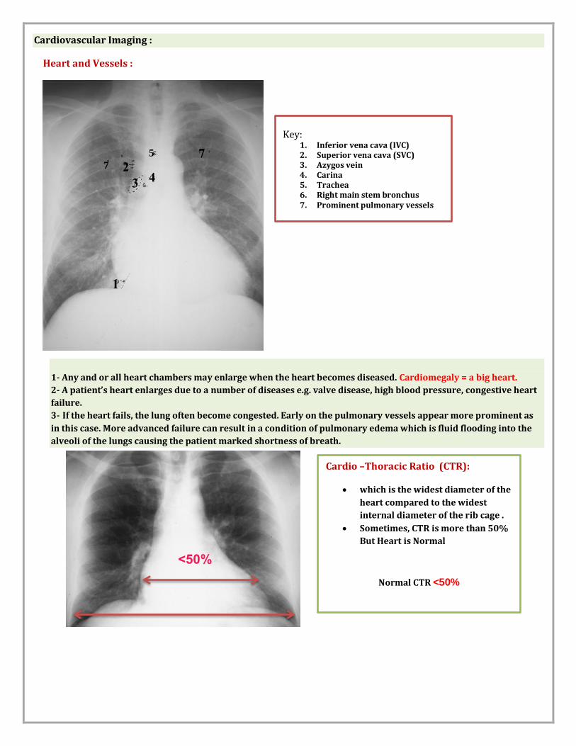
Cardiovascular Imaging :
Heart and Vessels :
1- Any and or all heart chambers may enlarge when the heart becomes diseased. Cardiomegaly = a big heart.
2- A patient’s heart enlarges due to a number of diseases e.g. valve disease, high blood pressure, congestive heart
failure.
3- If the heart fails, the lung often become congested. Early on the pulmonary vessels appear more prominent as
in this case. More advanced failure can result in a condition of pulmonary edema which is fluid flooding into the
alveoli of the lungs causing the patient marked shortness of breath.
Key: 1. Inferior vena cava (IVC) 2. Superior vena cava (SVC) 3. Azygos vein 4. Carina 5. Trachea 6. Right main stem bronchus 7. Prominent pulmonary vessels
5
3
1
2 4
7
7
Cardio –Thoracic Ratio (CTR):
which is the widest diameter of the
heart compared to the widest
internal diameter of the rib cage .
Sometimes, CTR is more than 50%
But Heart is Normal
Normal CTR <50%
<50%
Extracardiac causes of cardiac enlargement
Portable AP films
Obesity
Pregnant
Ascites
Straight back syndrome.
Pectus excavatum.
…………………………………………………………………………………………………………
Aortic Knob
Normal Aortic Knob diameter: 42 mm
Enlarged with:
Increased pressure
Increased flow
Changes in aortic wall
Congestive heart faliure :
occurs when the heart is unable to provide sufficient pump action to distribute blood flow to meet the
needs of the body.
Heart failure can cause a number of symptoms including shortness of breath , leg swelling , and exercise
intolerance .
Radiological features of heart faliure :
1. Diffuse Lung Opacities
2. Ill-defined Vessels
3. Cardiac Enlargement
4. Enlarged cardiac silhouette
42mm

Acute Pulmonary Edema :
Pulmonary edema occurs when the alveoli fill up with excess fluid seeped out of the blood vessels in the
lung instead of air
can be caused by many different factors. It can be related to heart failure, called cardiogenic pulmonary
edema, or related to other causes, referred to as non-cardiogenic pulmonary edema.
Chest X ray a patient with pulmonary oedema
KERELY’S B-LINES

Are a sign seen on chest radiographs with interstitial pulmonary edema . They are thin linear
pulmonary opacities caused by fluid or cellular infiltration into the interstitium of the lungs.
The most common condition causing Kerley B lines is cardiac failure.