radiological investigation of chest and cvs...
TRANSCRIPT
Lecture 5
Radiological investigation of Chest and
CVS diseases
(Respiratory diseases)
Hanan Alrabiah
Ghadeer AlWuhayd Hanan AlSalman
Maha AlKubidan Khawla AlOthman
Resources:
- Lecture by Dr.
- 429 Radiology team work.

Introduction
Normal Anatomy of the lung:
- It is important to know the location of lung fissures on the normal
chest X-ray, (to locate the abnormality in which lobe).
- They Transverse fissure.
B: two oblique fissures.
- If there is a shift in any of these fissures, it indicates abnormal
lungs.
Abnormal Lungs
Mass Vs Diffuse Infilteration:
- The basic diagnostic instance is to detect an abnormality.
- In both of the cases above (A, B), there is an abnormal opacity (in the left lung parahilar lesion).
- In each of the cases, there is an abnormal opacity in the left upper lobe (above the transverse fissure).
- The case A has opacity with poorly defined margins. This is airspace disease such as pneumonia.
- In the case B, the opacity would best be described as a mass because it is presented with well-defined
margins.
Solitary Nodule in the Lung:
- A solitary nodule in the lung can be totally innocuous or potentially a fatal lung cancer. After detection
the initial step in analyisis to compare the film with prior films if available. A nodule that is unchanged
for two years is almost certainly benign. Be sure to evaluate for the presence of multiple nodules as this
finding would change the
differential entirely.
- If the nodule is indeterminate after
considering old films and
calcification, subsequent steps in
the work-up include ordering a CT
and a tissue biopsy.
A. Transvers fissure locate just below the
hilum of the lung (7th
Rib).
Pleural Based Lesion
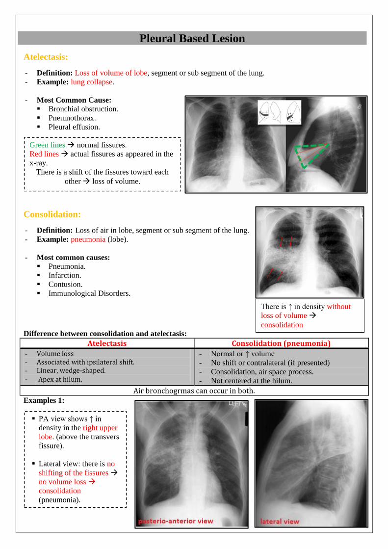
Atelectasis:
- Definition: Loss of volume of lobe, segment or sub segment of the lung.
- Example: lung collapse.
- Most Common Cause:
Bronchial obstruction.
Pneumothorax.
Pleural effusion.
Consolidation:
- Definition: Loss of air in lobe, segment or sub segment of the lung.
- Example: pneumonia (lobe).
- Most common causes: Pneumonia.
Infarction.
Contusion.
Immunological Disorders.
Difference between consolidation and atelectasis:
Atelectasis Consolidation (pneumonia)
- Volume loss - Associated with ipsilateral shift. - Linear, wedge-shaped. - Apex at hilum.
- Normal or ↑ volume
- No shift or contralateral (if presented)
- Consolidation, air space process.
- Not centered at the hilum.
Air bronchogrmas can occur in both. Examples 1:
Green lines normal fissures.
Red lines actual fissures as appeared in the
x-ray.
There is a shift of the fissures toward each
other loss of volume.
There is ↑ in density without
loss of volume
consolidation
PA view shows ↑ in
density in the right upper
lobe. (above the transvers
fissure).
Lateral view: there is no
shifting of the fissures
no volume loss
consolidation
(pneumonia).

Examples 2:
Examples 3:
Other terminology
The Silhouette Sign: - Definition: the silhouette sign refers to the loss of normally seen borders between thoracic structures
e.g. diaphragm or heart.
- Indications: air space disease.
There is ↑ in opacity in the Right upper
lobe + the transverse fissure is moved
upward (red arrows) + trachea is
slightly moved to the other side
Atelectasis of the Right upper lobe.
There is ↑ in opacity in the right Middle
lobe + there is no shift of the transverse
and oblique fissure (red arrows)
consolidation (pneumonia).
In this X-ray Silhouette sign can
be seen between the right middle
lobe and right border of the heart.
(Borders cannot be differentiated).
Because of a reduction in the upper
lobe of the right lung, Transvers fissure
has moved upward (toward the
collapsed part)
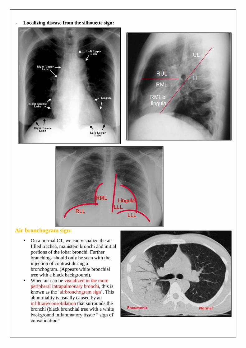
- Localizing disease from the silhouette sign:
Air bronchogram sign:
On a normal CT, we can visualize the air
filled trachea, mainstem bronchi and initial
portions of the lobar bronchi. Further
branchings should only be seen with the
injection of contrast during a
bronchogram. (Appears white bronchial
tree with a black background).
When air can be visualized in the more
peripheral intrapulmonary bronchi, this is
known as the ‘airbronchogram sign’. This
abnormality is usually caused by an
infiltrate/consolidation that surrounds the
bronchi (black bronchial tree with a white
background inflammatory tissue “ sign of
consolidation”
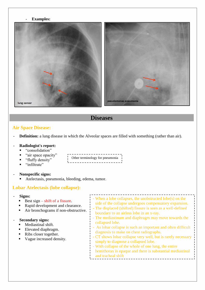
- Examples:
Diseases
Air Space Disease:
- Definition: a lung disease in which the Alveolar spaces are filled with something (rather than air).
- Radiologist's report:
“consolidation”
“air space opacity”
“fluffy density”
“infiltrate”
- Nonspecific signs:
Atelectasis, pneumonia, bleeding, edema, tumor.
Lobar Atelectasis (lobe collapse):
- Signs:
Best sign – shift of a fissure.
Rapid development and clearance.
Air bronchograms if non-obstructive.
- Secondary signs:
Mediastinal shift.
Elevated diaphragm.
Ribs closer together.
Vague increased density.
Other terminology for pneumonia
- When a lobe collapses, the unobstructed lobe(s) on the
side of the collapse undergoes compensatory expansion.
- The displaced (shifted) fissure is seen as a well‐defined
boundary to an airless lobe in an x‐ray.
- The mediastinum and diaphragm may move towards the
collapsed lobe.
- As lobar collapse is such an important and often difficult
diagnosis to make on chest radiographs.
- CT shows lobar collapse very well, but is rarely necessary
simply to diagnose a collapsed lobe.
- With collapse of the whole of one lung, the entire
hemithorax is opaque and there is substantial mediastinal
and tracheal shift
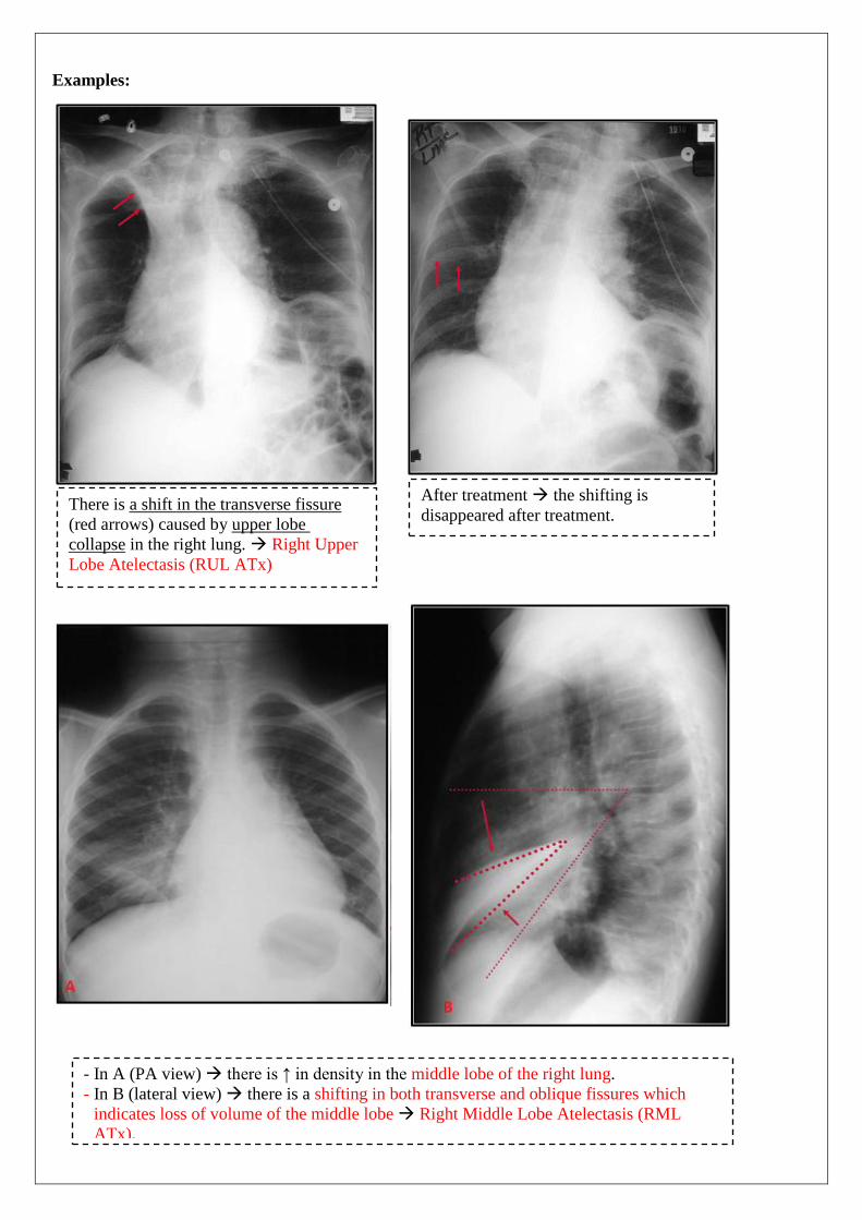
Examples:
a shift in the transverse fissureThere is
upper lobe (red arrows) caused by
Right Upper in the right lung. collapse
Lobe Atelectasis (RUL ATx)
After treatment the shifting is
disappeared after treatment.
- In A (PA view) there is ↑ in density in the middle lobe of the right lung.
- In B (lateral view) there is a shifting in both transverse and oblique fissures which
indicates loss of volume of the middle lobe Right Middle Lobe Atelectasis (RML
ATx).
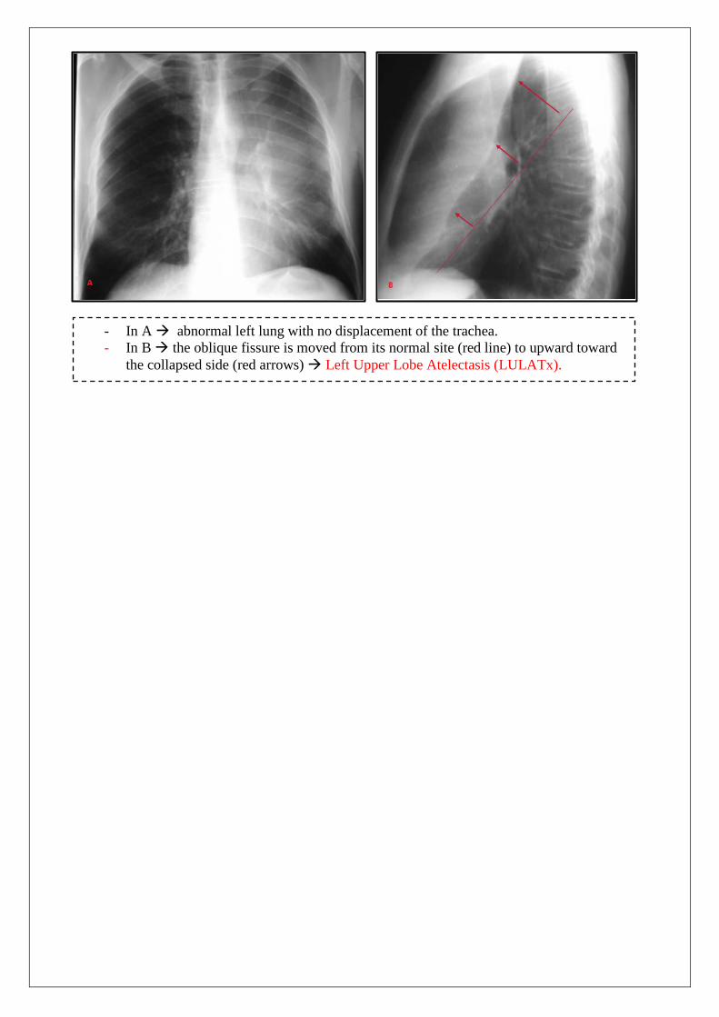
- In A abnormal left lung with no displacement of the trachea.
- In B the oblique fissure is moved from its normal site (red line) to upward toward
the collapsed side (red arrows) Left Upper Lobe Atelectasis (LULATx).

PneumoniaSigns:
- Signs:
Air bronchogram
Silhouette - “positive” or “negative”
Dense hilum
“Spine” sign
Examples:
• All are signs of any air space process
• Dx of pneumonia depends on appropriate
clinical scenario.
- In PA view there is ↑ in density in the middle lobe of the right lung.
- In lateral view there is no shift in the transverse or oblique fissure Right
Middle Lobe Consolidation.
- In A there is an ↑ density + the transverse fissure is not shifted.
- In B no shift of the fissures.
- Right Upper Lobe Consolidation.
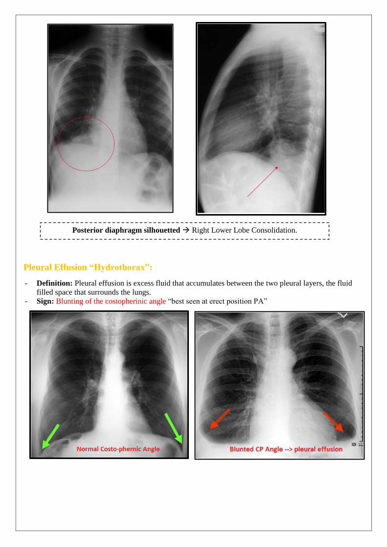
Pleural Effusion “Hydrothorax”:
- Definition: Pleural effusion is excess fluid that accumulates between the two pleural layers, the fluid
filled space that surrounds the lungs.
- Sign: Blunting of the costopherinic angle “best seen at erect position PA”
Posterior diaphragm silhouetted Right Lower Lobe Consolidation.
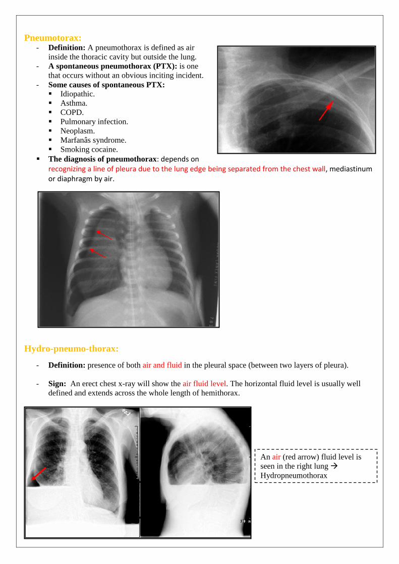
Pneumotorax: - Definition: A pneumothorax is defined as air
inside the thoracic cavity but outside the lung.
- A spontaneous pneumothorax (PTX): is one
that occurs without an obvious inciting incident.
- Some causes of spontaneous PTX: Idiopathic.
Asthma.
COPD.
Pulmonary infection.
Neoplasm.
Marfanâs syndrome.
Smoking cocaine.
The diagnosis of pneumothorax: depends on recognizing a line of pleura due to the lung edge being separated from the chest wall, mediastinum or diaphragm by air.
Hydro-pneumo-thorax:
- Definition: presence of both air and fluid in the pleural space (between two layers of pleura).
- Sign: An erect chest x-ray will show the air fluid level. The horizontal fluid level is usually well
defined and extends across the whole length of hemithorax.
An air (red arrow) fluid level is
seen in the right lung
Hydropneumothorax
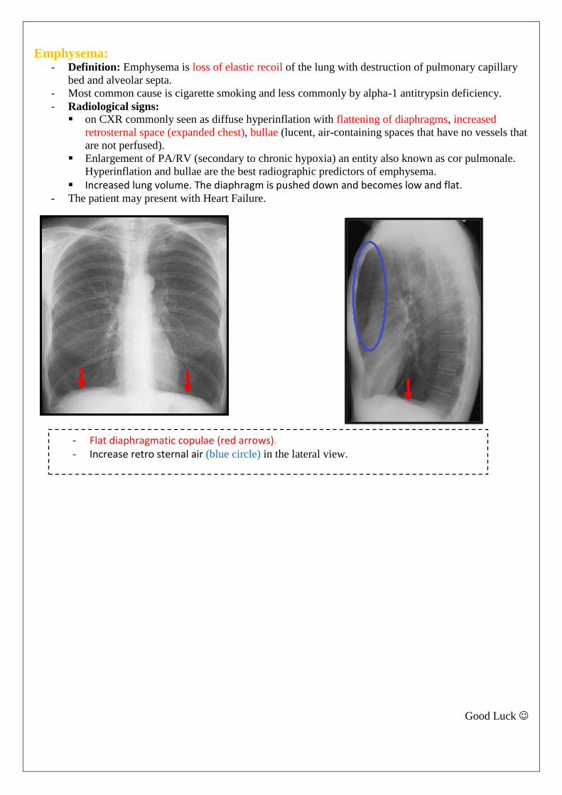
Emphysema: - Definition: Emphysema is loss of elastic recoil of the lung with destruction of pulmonary capillary
bed and alveolar septa.
- Most common cause is cigarette smoking and less commonly by alpha-1 antitrypsin deficiency.
- Radiological signs:
on CXR commonly seen as diffuse hyperinflation with flattening of diaphragms, increased
retrosternal space (expanded chest), bullae (lucent, air-containing spaces that have no vessels that
are not perfused).
Enlargement of PA/RV (secondary to chronic hypoxia) an entity also known as cor pulmonale.
Hyperinflation and bullae are the best radiographic predictors of emphysema.
Increased lung volume. The diaphragm is pushed down and becomes low and flat. - The patient may present with Heart Failure.
Good Luck
- Flat diaphragmatic copulae (red arrows). - Increase retro sternal air (blue circle) in the lateral view.