radiological signs (shënja radiologjike )-1
TRANSCRIPT
8/11/2019 Radiological Signs (Shënja Radiologjike )-1
http://slidepdf.com/reader/full/radiological-signs-shenja-radiologjike-1 1/122
1
Sllavko K. Kallfa
Radiological Signs
Shënja Radiologjike)
Përmbledhje Artikujsh nga
Radiopaedia.org
❶
8/11/2019 Radiological Signs (Shënja Radiologjike )-1
http://slidepdf.com/reader/full/radiological-signs-shenja-radiologjike-1 2/122
2
chest x-ray
air bronchogram…………….f. 3
big rib sign............................f. 7
Chang's sign..........................f. 8
coin lesion..............................f. 9
dense hilum sign....................f. 16
double contour sign................f.20
extrapleural sign....................f.25
hilum overlay sign..................f.26
hilum convergence sign...........f.39
holly leaf sign..........................f.42
finger in glove sign..................f.47
flat waist sign..........................f.54
Fleishner's sign.......................f.55
ginkgo leaf sign.......................f.57
Golden S sign..........................f.61
incomplete border sign............f.70
juxtaphrenic peak sign............f.72 medial stripe sign....................f.76
more black sign.......................f.78
Naclerio's V sign.....................f.89
Shmoo sign..............................f.90
silhouette sign[+] ……………...f.91
steeple sign...............................f.103
spinnaker sign..........................f.108
water bottle sign.......................f.114 wave sign..................................f.118
Westermark's sign....................f.121
8/11/2019 Radiological Signs (Shënja Radiologjike )-1
http://slidepdf.com/reader/full/radiological-signs-shenja-radiologjike-1 3/122
3
Air bronchogram
Dr Henry Knipe and Dr Behrang Amini et al.
Air bronchogram refers to the phenomenon of air-filled bronchi (dark ) being made visible bythe opacification of surrounding alveoli ( grey - white). It is almost always caused by a pathologicairspace (alveolar) process, in which something other than air fills the alveoli. Air bronchograms
will not be visible if the bronchi themselves are opacified (e.g. by fluid).
Air bronchograms can be seen with several processes, including the following :
pulmonary consolidation
pulmonary oedema
non-obstructive atelectasis
severe interstitial lung disease
neoplasms - bronchioloalveolar carcinoma; pulmonary lymphoma pulmonary infarct
normal expiration
The air bronchogram indicates patent proximal airways.
Air bronchogams that persist for weeks despite appropriate antimicrobial therapy should raise the
suspicion of a neoplastic process. CT or guided biopsy may be planned in such cases.
References
1. Reed JC. Chest radiology, plain film patterns and differential diagnoses. Mosby.(1997) ISBN:0815171226. Read it at Google Books - Find it at Amazon
2. Wong JS, Weisbrod GL, Chamberlain D et-al. Bronchioloalveolar carcinoma and the
air bronchogram sign: a new pathologic explanation. J Thorac Imaging. 1994;9 (3): 141-
4. - Pubmed citation
Synonyms & Alternative Spellings
Synonyms or Alternative Spelling Include in Listings?
Air bronchograms
8/11/2019 Radiological Signs (Shënja Radiologjike )-1
http://slidepdf.com/reader/full/radiological-signs-shenja-radiologjike-1 4/122
4
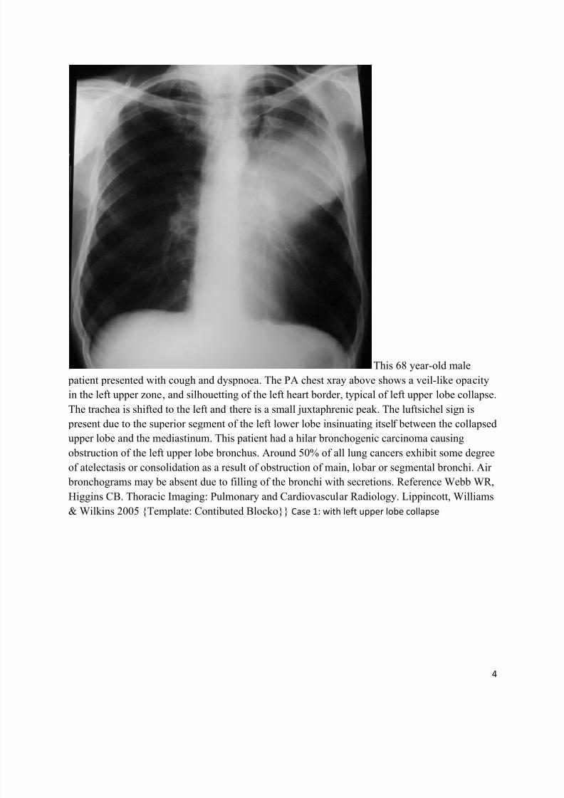
This 68 year-old male
patient presented with cough and dyspnoea. The PA chest xray above shows a veil-like opacity
in the left upper zone, and silhouetting of the left heart border, typical of left upper lobe collapse.
The trachea is shifted to the left and there is a small juxtaphrenic peak. The luftsichel sign is
present due to the superior segment of the left lower lobe insinuating itself between the collapsed
upper lobe and the mediastinum. This patient had a hilar bronchogenic carcinoma causing
obstruction of the left upper lobe bronchus. Around 50% of all lung cancers exhibit some degree
of atelectasis or consolidation as a result of obstruction of main, lobar or segmental bronchi. Air
bronchograms may be absent due to filling of the bronchi with secretions. Reference Webb WR,
Higgins CB. Thoracic Imaging: Pulmonary and Cardiovascular Radiology. Lippincott, Williams
& Wilkins 2005 {Template: Contibuted Blocko}} Case 1: with left upper lobe collapse
8/11/2019 Radiological Signs (Shënja Radiologjike )-1
http://slidepdf.com/reader/full/radiological-signs-shenja-radiologjike-1 5/122
5
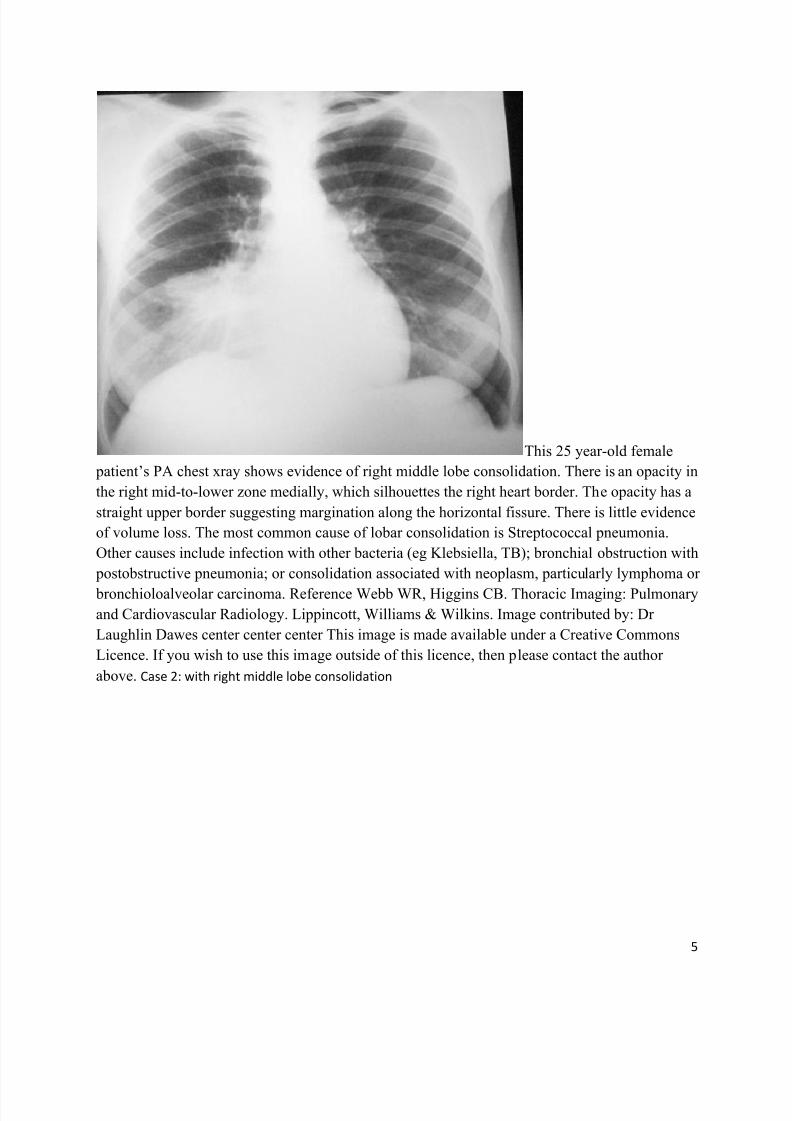
This 25 year-old female
patient’s PA chest xray shows evidence of right middle lobe consolidation. There is an opacity in
the right mid-to-lower zone medially, which silhouettes the right heart border. The opacity has a
straight upper border suggesting margination along the horizontal fissure. There is little evidence
of volume loss. The most common cause of lobar consolidation is Streptococcal pneumonia.
Other causes include infection with other bacteria (eg Klebsiella, TB); bronchial obstruction with
postobstructive pneumonia; or consolidation associated with neoplasm, particularly lymphoma or
bronchioloalveolar carcinoma. Reference Webb WR, Higgins CB. Thoracic Imaging: Pulmonary
and Cardiovascular Radiology. Lippincott, Williams & Wilkins. Image contributed by: Dr
Laughlin Dawes center center center This image is made available under a Creative Commons
Licence. If you wish to use this image outside of this licence, then please contact the author
above. Case 2: with right middle lobe consolidation
8/11/2019 Radiological Signs (Shënja Radiologjike )-1
http://slidepdf.com/reader/full/radiological-signs-shenja-radiologjike-1 6/122
6
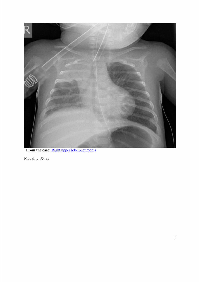
From the case: Right upper lobe pneumonia
Modality: X-ray
8/11/2019 Radiological Signs (Shënja Radiologjike )-1
http://slidepdf.com/reader/full/radiological-signs-shenja-radiologjike-1 7/122
7
Big rib sign
Dr Henry Knipe and Dr M Venkatesh et al.
The big rib sign is a sign to differentiate right and left ribs on lateral chest radiographs.
It exploits a technique of magnification differences on lateral projections between right and leftribs. For example, on right lateral projections the left ribs appear larger than right ribs.
This sign can be useful but may be difficult to appreciate as the difference in size between ribs is
~10% and is not applicable when the posterior ribs are superimposed.
References
1. Kurihara Y, Yakushiji YK, Matsumoto J et-al. The ribs: anatomic and radiologic considerations.
Radiographics. 1999;19 (1): 105-19. Radiographics (full text) - Pubmed citation
8/11/2019 Radiological Signs (Shënja Radiologjike )-1
http://slidepdf.com/reader/full/radiological-signs-shenja-radiologjike-1 8/122
8
Chang's sign
Dr Yuranga Weerakkody and Dr Henry Knipe et al.
Chang’s sign refers to a dilatation and abrupt change in calibre of a main pulmonary artery dueto pulmonary embolism
1.
References
1. Plain Film Signs in Pulmonary Embolism (with CT correlate), Kirwadi A, Bickle IC.
http://www.eurorad.org/eurorad/case.php?id=7735 Eurorad case 7735
Synonyms & Alternative Spellings
Synonyms or Alternative Spelling Include in Listings?
Chang sign ✗
Changs sign
8/11/2019 Radiological Signs (Shënja Radiologjike )-1
http://slidepdf.com/reader/full/radiological-signs-shenja-radiologjike-1 9/122
9
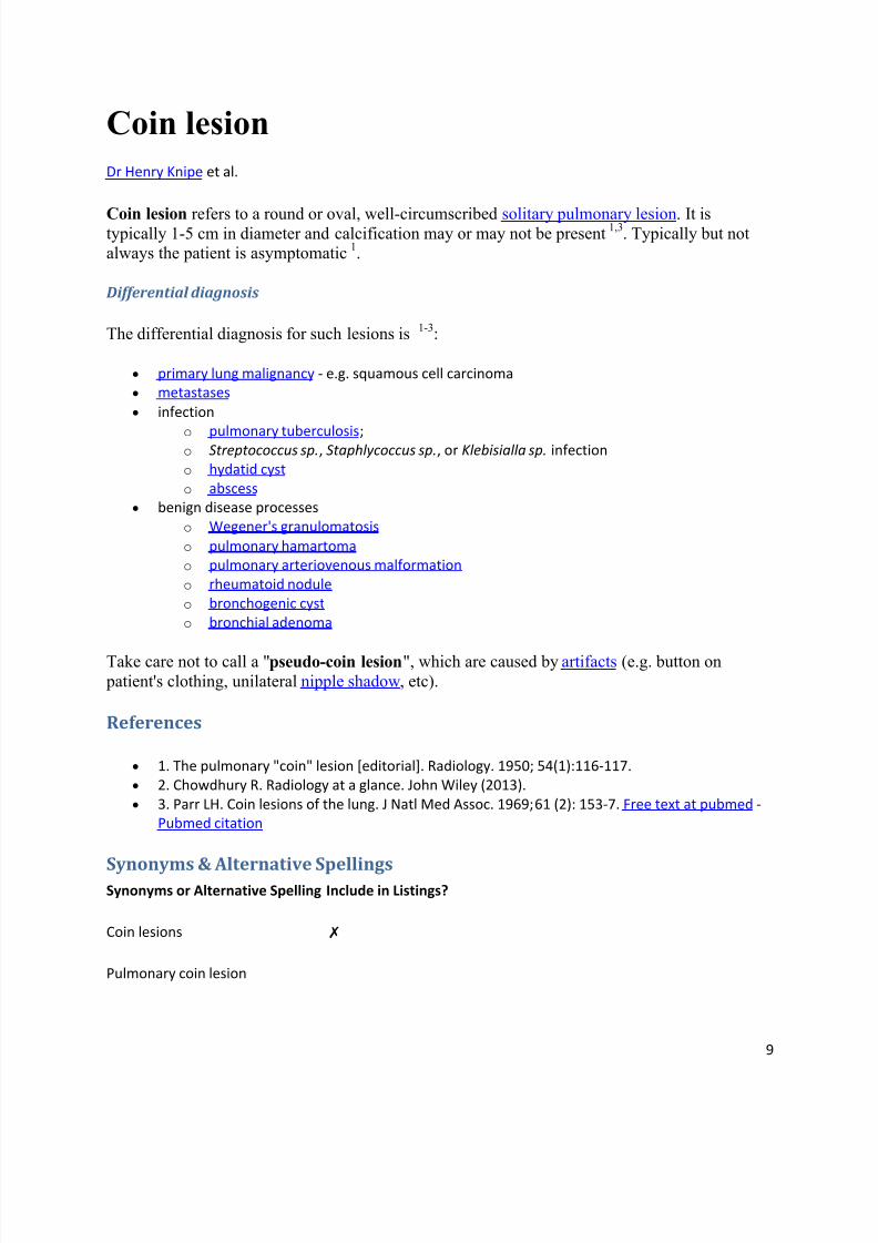
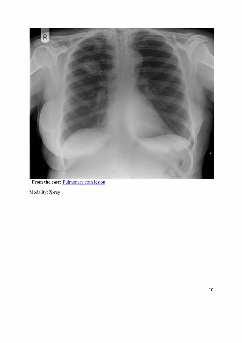
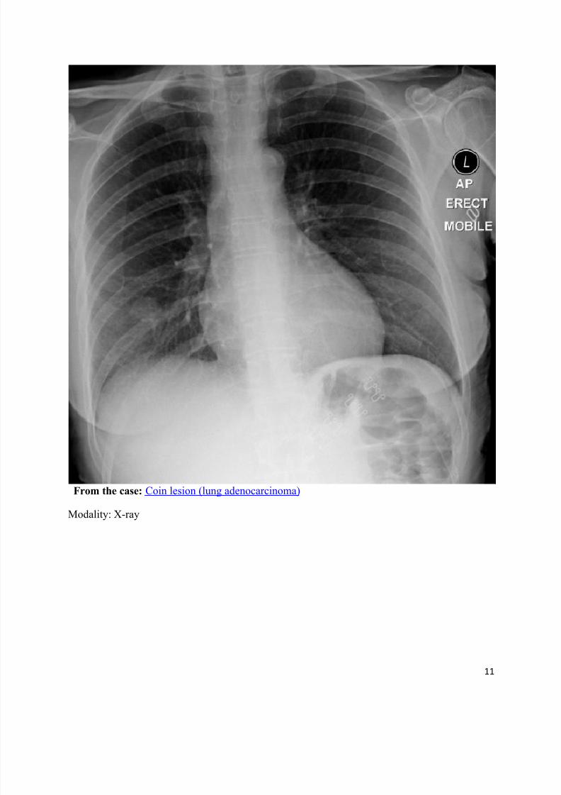
Coin lesion
Dr Henry Knipe et al.
Coin lesion refers to a round or oval, well-circumscribed solitary pulmonary lesion. It istypically 1-5 cm in diameter and calcification may or may not be present
1,3. Typically but not
always the patient is asymptomatic1.
Differential diagnosis
The differential diagnosis for such lesions is1-3
:
primary lung malignancy - e.g. squamous cell carcinoma
metastases
infection
o
pulmonary tuberculosis; o
Streptococcus sp., Staphlycoccus sp., or Klebisialla sp. infection
o hydatid cyst
o abscess
benign disease processes
o
Wegener's granulomatosis
o pulmonary hamartoma
o
pulmonary arteriovenous malformation
o
rheumatoid nodule
o bronchogenic cyst
o bronchial adenoma
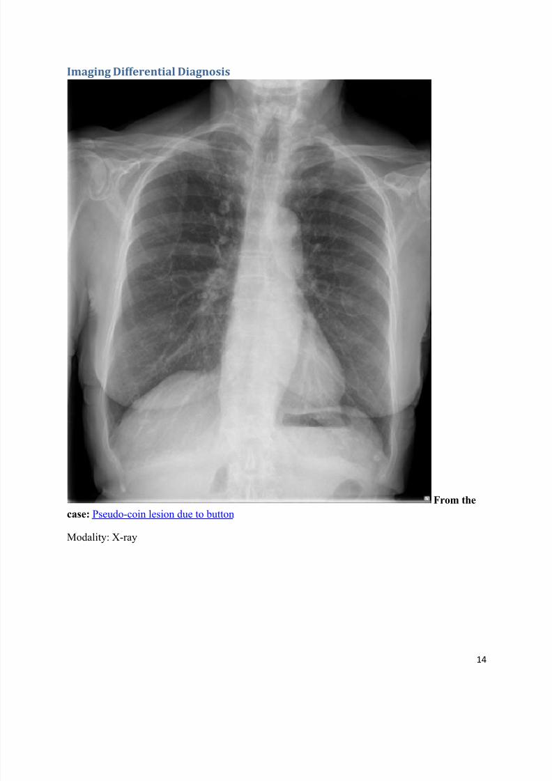
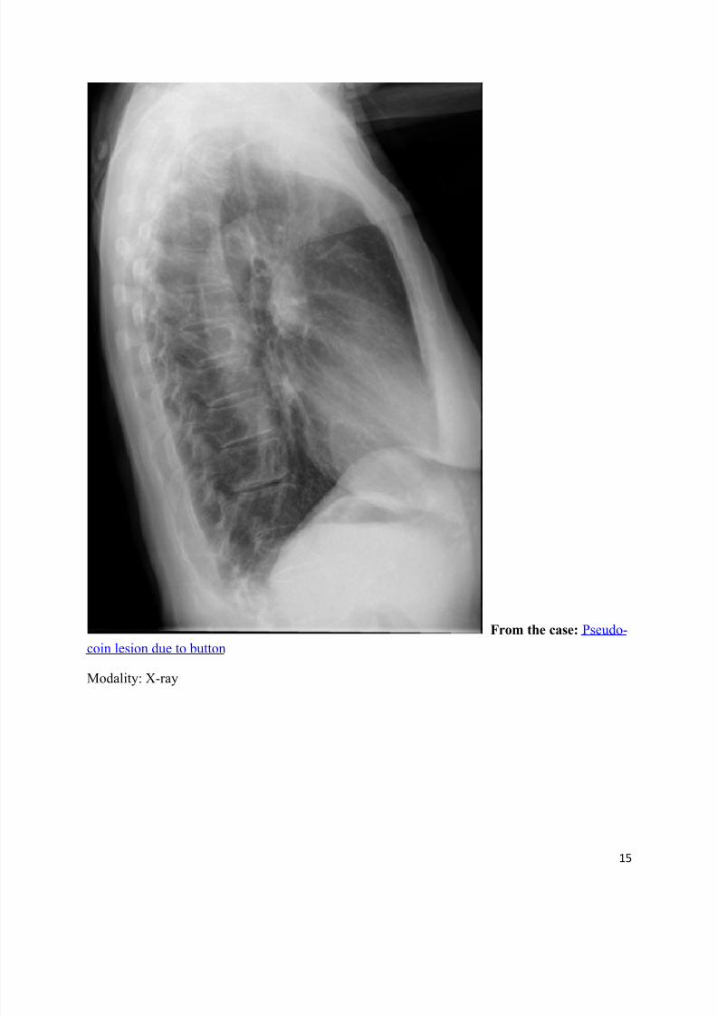
Take care not to call a "pseudo-coin lesion", which are caused by artifacts (e.g. button on
patient's clothing, unilateral nipple shadow, etc).
References
1. The pulmonary "coin" lesion [editorial]. Radiology. 1950; 54(1):116-117.
2. Chowdhury R. Radiology at a glance. John Wiley (2013).
3. Parr LH. Coin lesions of the lung. J Natl Med Assoc. 1969;61 (2): 153-7. Free text at pubmed -
Pubmed citation
Synonyms & Alternative SpellingsSynonyms or Alternative Spelling Include in Listings?
Coin lesions ✗
Pulmonary coin lesion
8/11/2019 Radiological Signs (Shënja Radiologjike )-1
http://slidepdf.com/reader/full/radiological-signs-shenja-radiologjike-1 10/122
10
From the case: Pulmonary coin lesion
Modality: X-ray
8/11/2019 Radiological Signs (Shënja Radiologjike )-1
http://slidepdf.com/reader/full/radiological-signs-shenja-radiologjike-1 11/122
11
From the case: Coin lesion (lung adenocarcinoma)
Modality: X-ray
8/11/2019 Radiological Signs (Shënja Radiologjike )-1
http://slidepdf.com/reader/full/radiological-signs-shenja-radiologjike-1 12/122
12
From the case: Coin lesion - breast cancer metastasis
Modality: X-ray
8/11/2019 Radiological Signs (Shënja Radiologjike )-1
http://slidepdf.com/reader/full/radiological-signs-shenja-radiologjike-1 13/122
13
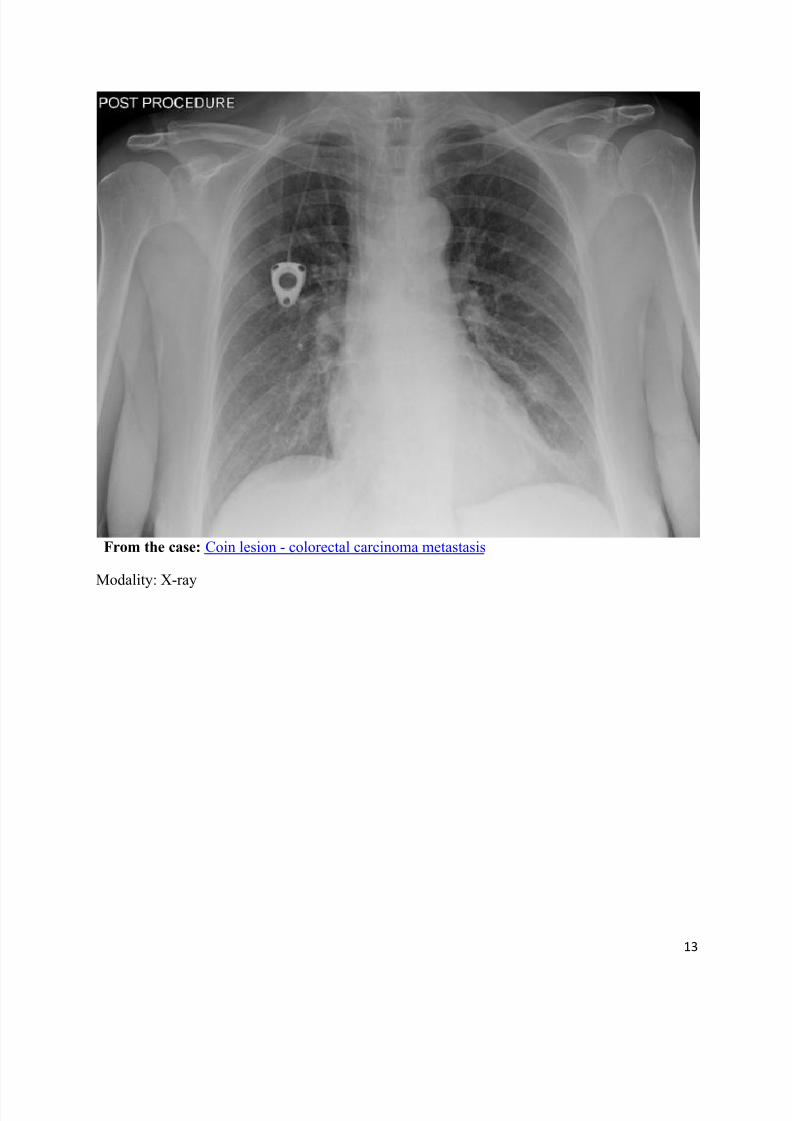
From the case: Coin lesion - colorectal carcinoma metastasis
Modality: X-ray
8/11/2019 Radiological Signs (Shënja Radiologjike )-1
http://slidepdf.com/reader/full/radiological-signs-shenja-radiologjike-1 14/122
14
Imaging Differential Diagnosis
From the
case: Pseudo-coin lesion due to button
Modality: X-ray
8/11/2019 Radiological Signs (Shënja Radiologjike )-1
http://slidepdf.com/reader/full/radiological-signs-shenja-radiologjike-1 15/122
15
From the case: Pseudo-
coin lesion due to button
Modality: X-ray
8/11/2019 Radiological Signs (Shënja Radiologjike )-1
http://slidepdf.com/reader/full/radiological-signs-shenja-radiologjike-1 16/122
16
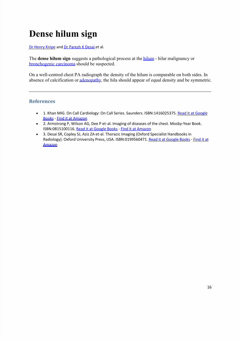
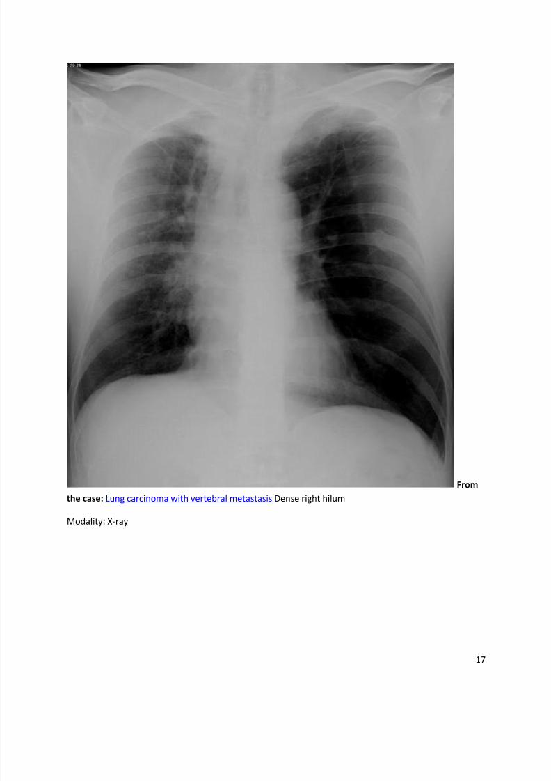
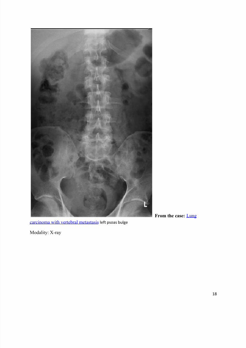
Dense hilum sign
Dr Henry Knipe and Dr Paresh K Desai et al.
The dense hilum sign suggests a pathological process at the hilum - hilar malignancy or bronchogenic carcinoma should be suspected.
On a well-centred chest PA radiograph the density of the hilum is comparable on both sides. In
absence of calcification or adenopathy, the hila should appear of equal density and be symmetric.
References
1. Khan MIG. On Call Cardiology: On Call Series. Saunders. ISBN:1416025375. Read it at Google
Books - Find it at Amazon 2. Armstrong P, Wilson AG, Dee P et-al. Imaging of diseases of the chest. Mosby-Year Book.
ISBN:0815100116. Read it at Google Books - Find it at Amazon
3. Desai SR, Copley SJ, Aziz ZA et-al. Thoracic Imaging (Oxford Specialist Handbooks in
Radiology). Oxford University Press, USA. ISBN:0199560471. Read it at Google Books - Find it at
Amazon
8/11/2019 Radiological Signs (Shënja Radiologjike )-1
http://slidepdf.com/reader/full/radiological-signs-shenja-radiologjike-1 17/122
17
From
the case: Lung carcinoma with vertebral metastasis Dense right hilum
Modality: X-ray
8/11/2019 Radiological Signs (Shënja Radiologjike )-1
http://slidepdf.com/reader/full/radiological-signs-shenja-radiologjike-1 18/122
18
From the case: Lung
carcinoma with vertebral metastasis left psoas bulge
Modality: X-ray
8/11/2019 Radiological Signs (Shënja Radiologjike )-1
http://slidepdf.com/reader/full/radiological-signs-shenja-radiologjike-1 19/122
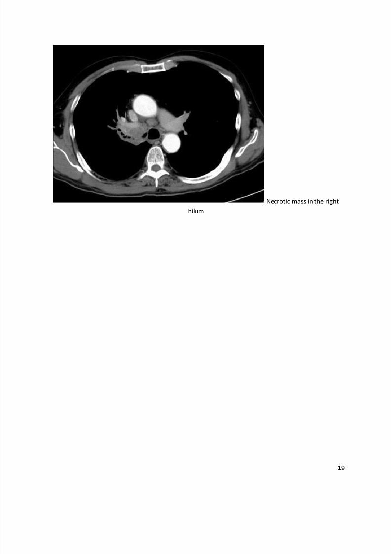
19
Necrotic mass in the right
hilum
8/11/2019 Radiological Signs (Shënja Radiologjike )-1
http://slidepdf.com/reader/full/radiological-signs-shenja-radiologjike-1 20/122
20
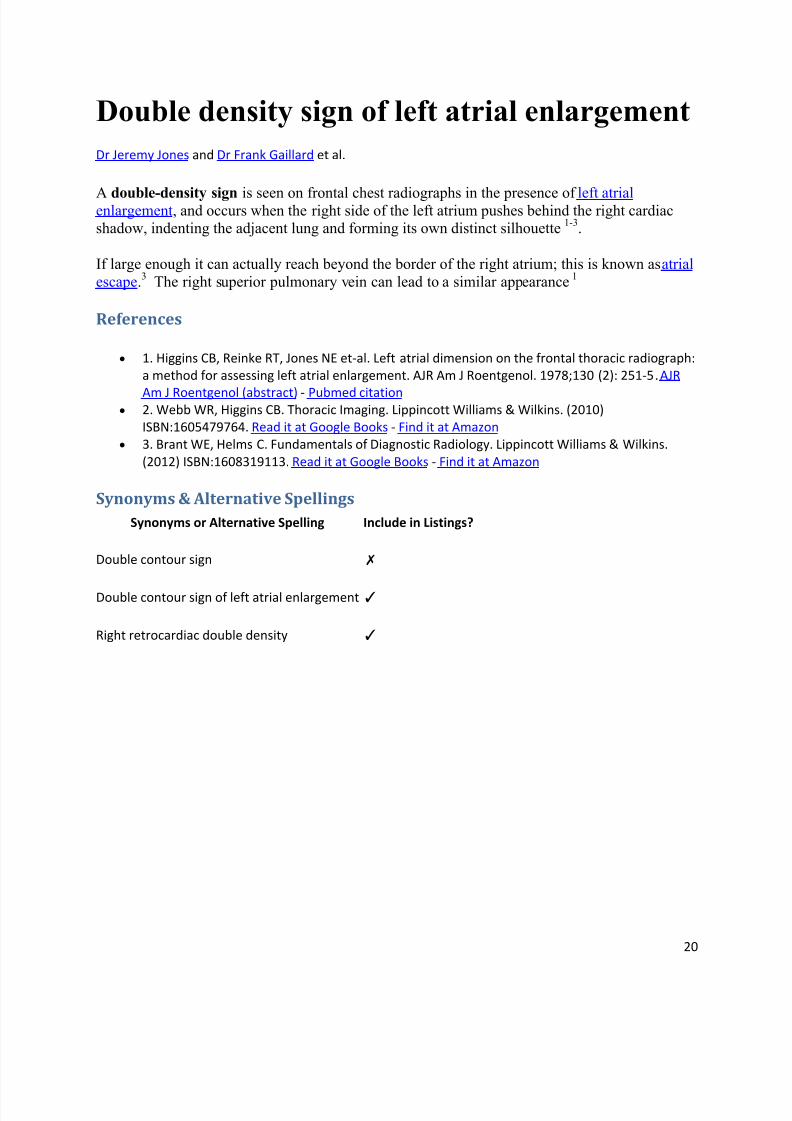
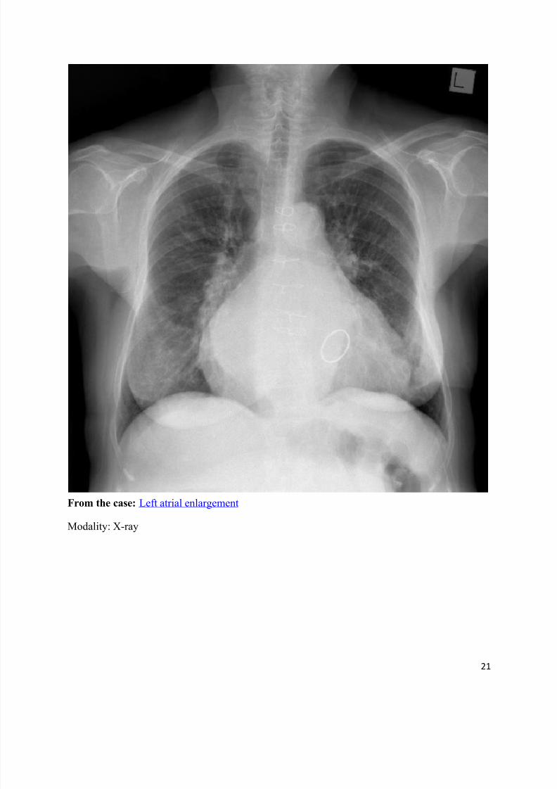
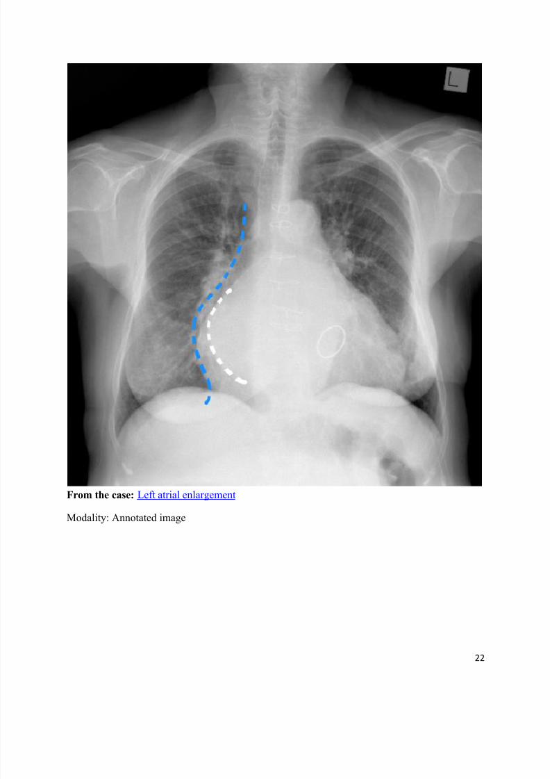
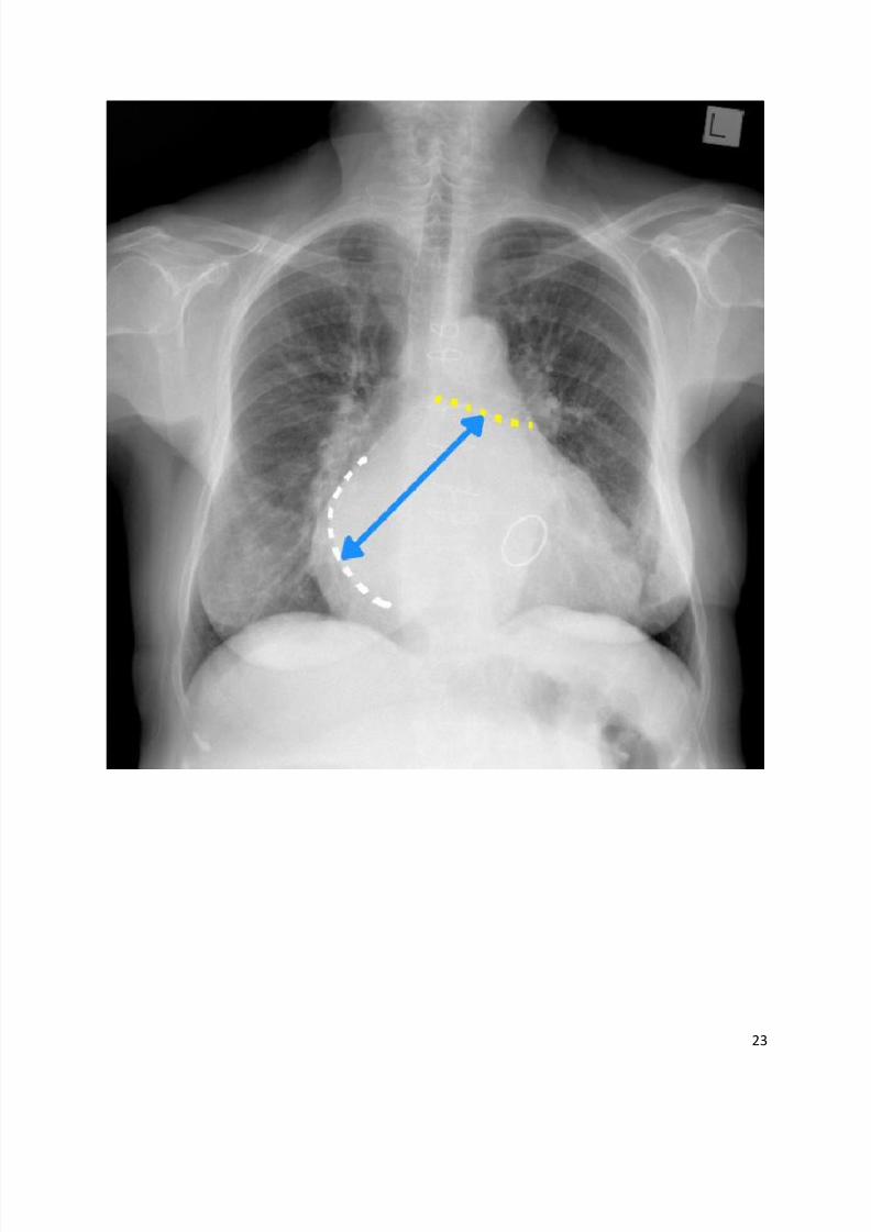
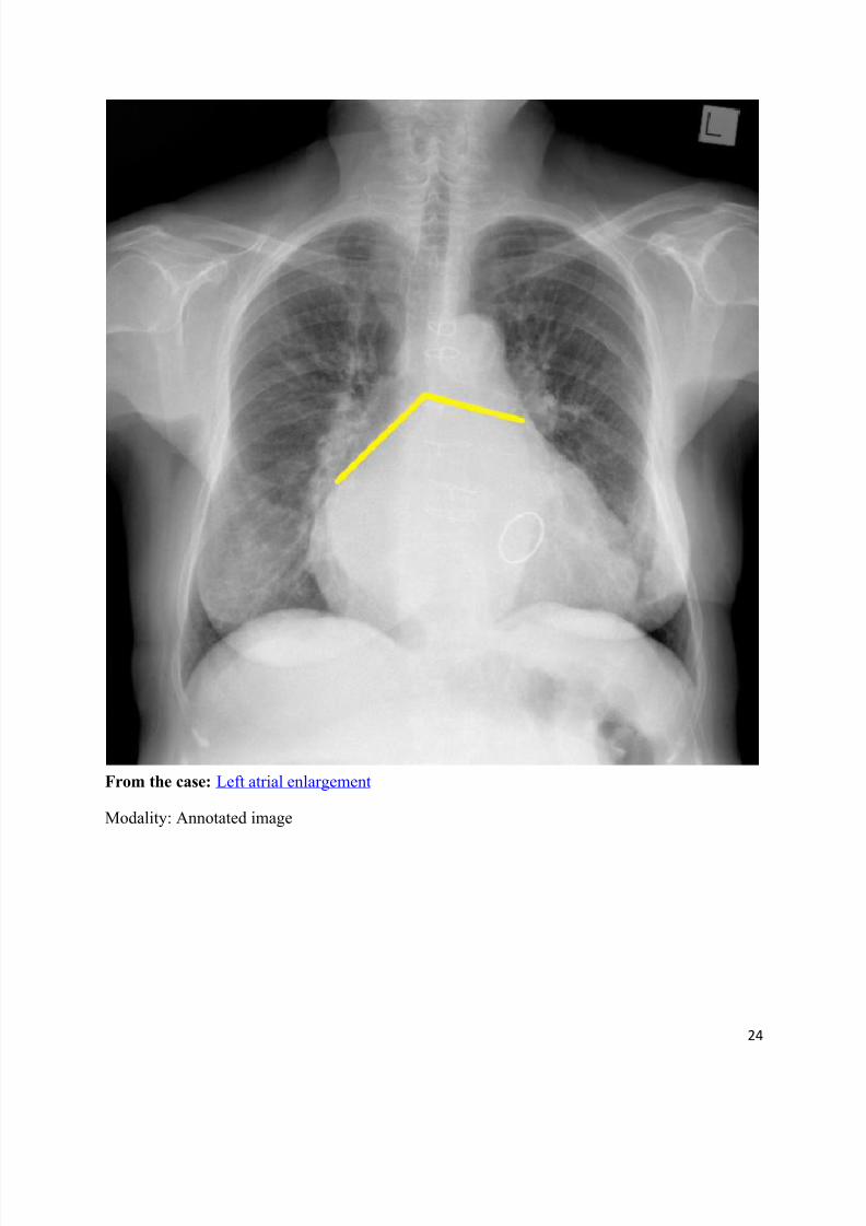
Double density sign of left atrial enlargement
Dr Jeremy Jones and Dr Frank Gaillard et al.
A double-density sign is seen on frontal chest radiographs in the presence of left atrialenlargement, and occurs when the right side of the left atrium pushes behind the right cardiacshadow, indenting the adjacent lung and forming its own distinct silhouette
1-3.
If large enough it can actually reach beyond the border of the right atrium; this is known as atrial
escape.3 The right superior pulmonary vein can lead to a similar appearance
1
References
1. Higgins CB, Reinke RT, Jones NE et-al. Left atrial dimension on the frontal thoracic radiograph:
a method for assessing left atrial enlargement. AJR Am J Roentgenol. 1978;130 (2): 251-5. AJR
Am J Roentgenol (abstract) - Pubmed citation 2. Webb WR, Higgins CB. Thoracic Imaging. Lippincott Williams & Wilkins. (2010)
ISBN:1605479764. Read it at Google Books - Find it at Amazon
3. Brant WE, Helms C. Fundamentals of Diagnostic Radiology. Lippincott Williams & Wilkins.
(2012) ISBN:1608319113. Read it at Google Books - Find it at Amazon
Synonyms & Alternative Spellings
Synonyms or Alternative Spelling Include in Listings?
Double contour sign ✗
Double contour sign of left atrial enlargement ✓
Right retrocardiac double density ✓
8/11/2019 Radiological Signs (Shënja Radiologjike )-1
http://slidepdf.com/reader/full/radiological-signs-shenja-radiologjike-1 21/122
21
From the case: Left atrial enlargement
Modality: X-ray
8/11/2019 Radiological Signs (Shënja Radiologjike )-1
http://slidepdf.com/reader/full/radiological-signs-shenja-radiologjike-1 22/122
22
From the case: Left atrial enlargement
Modality: Annotated image
8/11/2019 Radiological Signs (Shënja Radiologjike )-1
http://slidepdf.com/reader/full/radiological-signs-shenja-radiologjike-1 23/122
23
8/11/2019 Radiological Signs (Shënja Radiologjike )-1
http://slidepdf.com/reader/full/radiological-signs-shenja-radiologjike-1 24/122
24
From the case: Left atrial enlargement
Modality: Annotated image
8/11/2019 Radiological Signs (Shënja Radiologjike )-1
http://slidepdf.com/reader/full/radiological-signs-shenja-radiologjike-1 25/122
8/11/2019 Radiological Signs (Shënja Radiologjike )-1
http://slidepdf.com/reader/full/radiological-signs-shenja-radiologjike-1 26/122
26
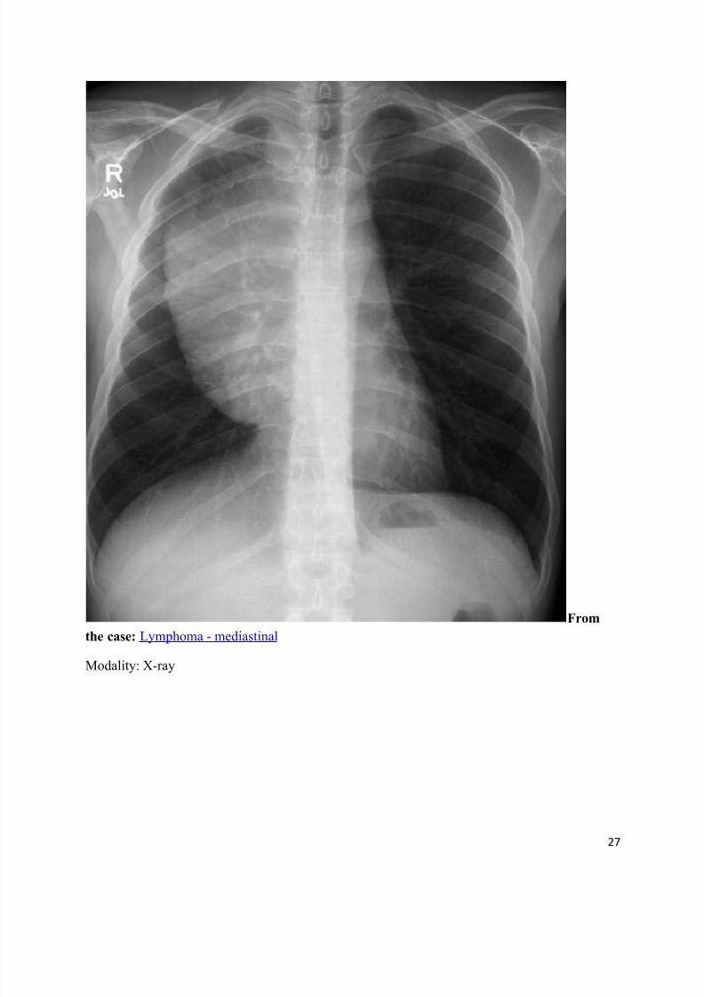
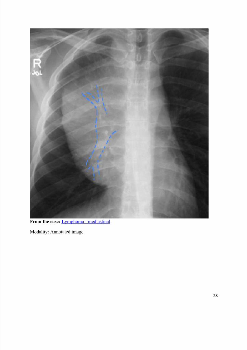
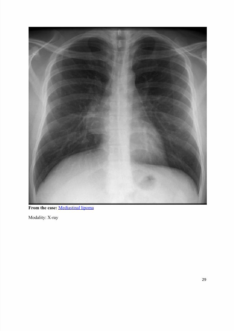
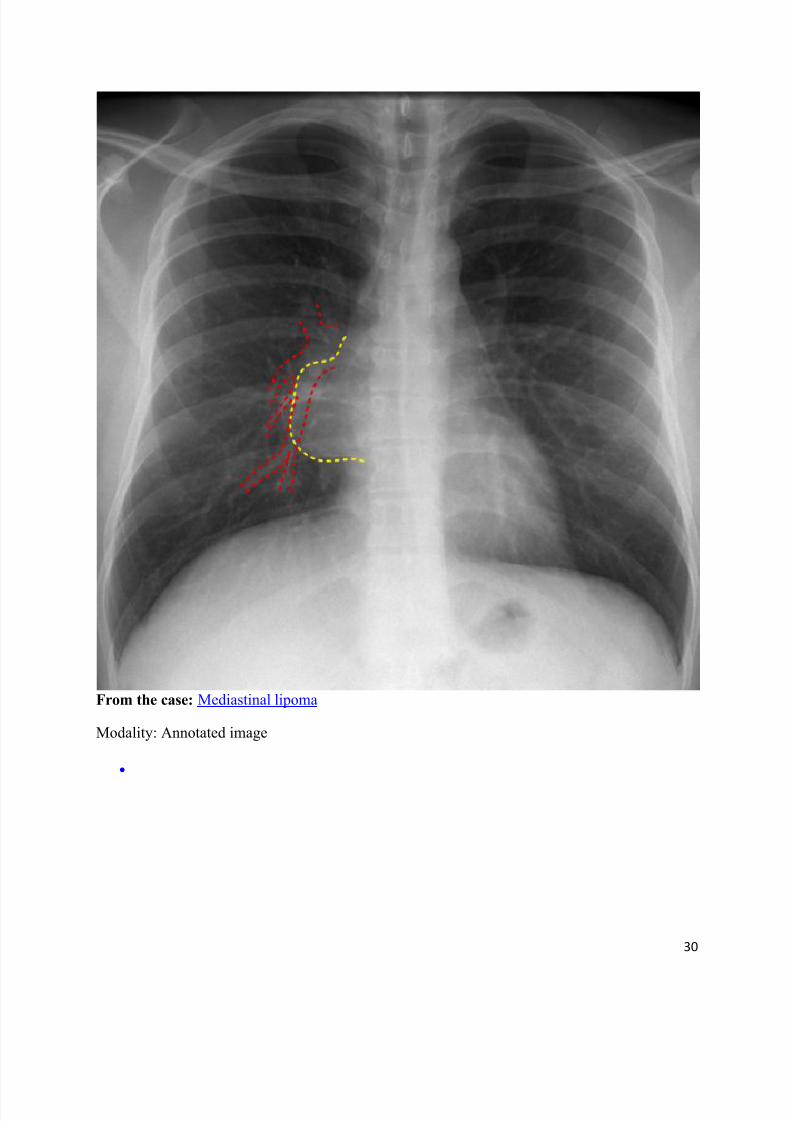
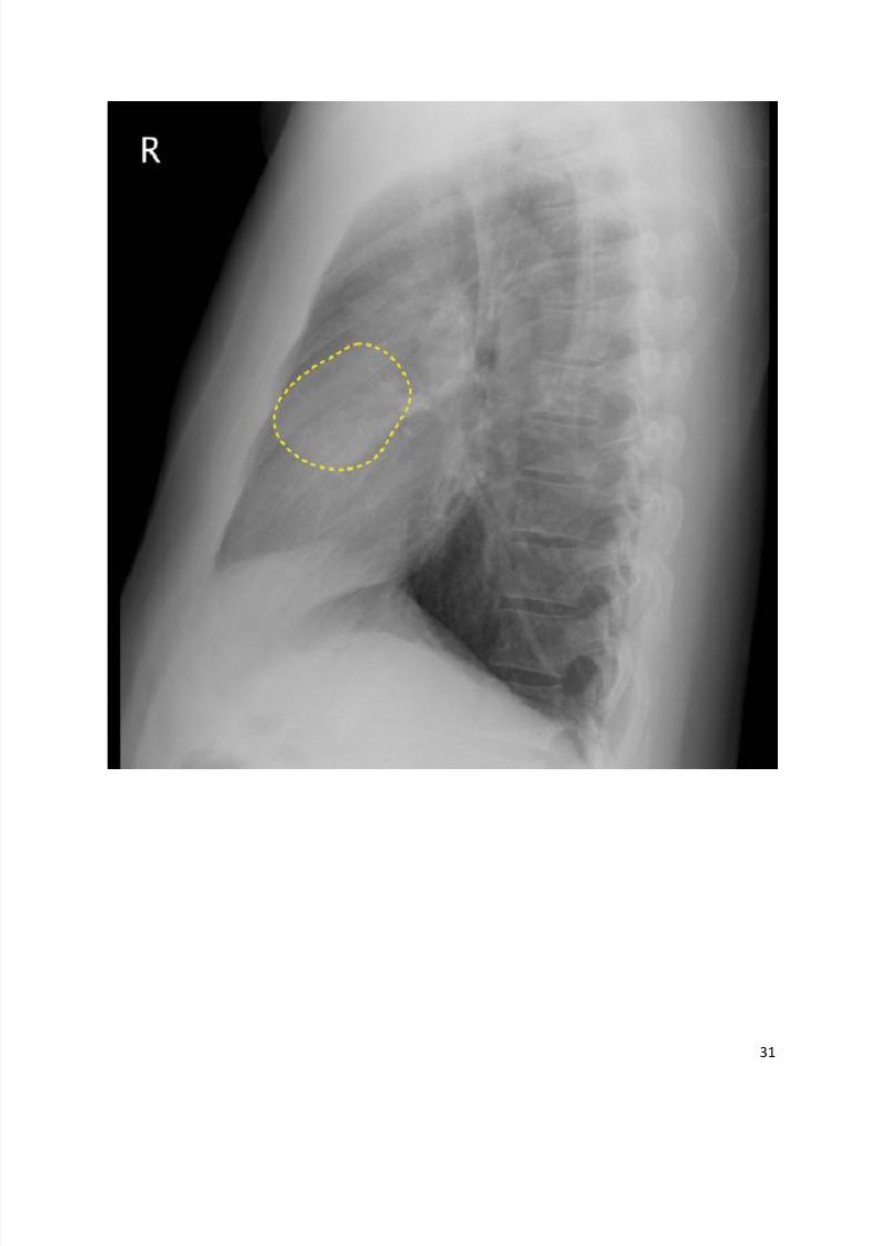
Hilum overlay sign
Dr Henry Knipe and Dr Frank Gaillard et al.
The hilum overlay sign refers to an appearance on frontal chest radiographs of patients with amass projected at the level of the hilum which is in fact either anterior or posterior to the hilum.
When a mass arises from the hilum, the pulmonary vessels are in contact with the mass and as
such their silhouette is obliterated. The ability to see the edges of the vessels through the mass
implies that the mass is not contacting the hilum, and is therefore either anterior or posterior to it.Most of these masses are found to be in the anterior mediastinum.
The sign was first described by Benjamin Felson 2.
References
1. Coche EE, Ghaye B, Mey JD. Comparative Interpretation of CT and Standard Radiography of
the Chest. Springer Verlag. (2010) ISBN:3540799419. Read it at Google Books - Find it at Amazon
2. George PP, Irodi A, Nidugala Keshava S et-al. 'Felson Signs' revisited. J Med Imaging Radiat
Oncol. 2014;58 (1): 64-74. doi:10.1111/1754-9485.12031 - Pubmed citation
8/11/2019 Radiological Signs (Shënja Radiologjike )-1
http://slidepdf.com/reader/full/radiological-signs-shenja-radiologjike-1 27/122
27
From
the case: Lymphoma - mediastinal
Modality: X-ray
8/11/2019 Radiological Signs (Shënja Radiologjike )-1
http://slidepdf.com/reader/full/radiological-signs-shenja-radiologjike-1 28/122
28
From the case: Lymphoma - mediastinal
Modality: Annotated image
8/11/2019 Radiological Signs (Shënja Radiologjike )-1
http://slidepdf.com/reader/full/radiological-signs-shenja-radiologjike-1 29/122
29
From the case: Mediastinal lipoma
Modality: X-ray
8/11/2019 Radiological Signs (Shënja Radiologjike )-1
http://slidepdf.com/reader/full/radiological-signs-shenja-radiologjike-1 30/122
30
From the case: Mediastinal lipoma
Modality: Annotated image
8/11/2019 Radiological Signs (Shënja Radiologjike )-1
http://slidepdf.com/reader/full/radiological-signs-shenja-radiologjike-1 31/122
31

8/11/2019 Radiological Signs (Shënja Radiologjike )-1
http://slidepdf.com/reader/full/radiological-signs-shenja-radiologjike-1 32/122
32
From the case: Mediastinal lipoma
Modality: Annotated image
8/11/2019 Radiological Signs (Shënja Radiologjike )-1
http://slidepdf.com/reader/full/radiological-signs-shenja-radiologjike-1 33/122
33
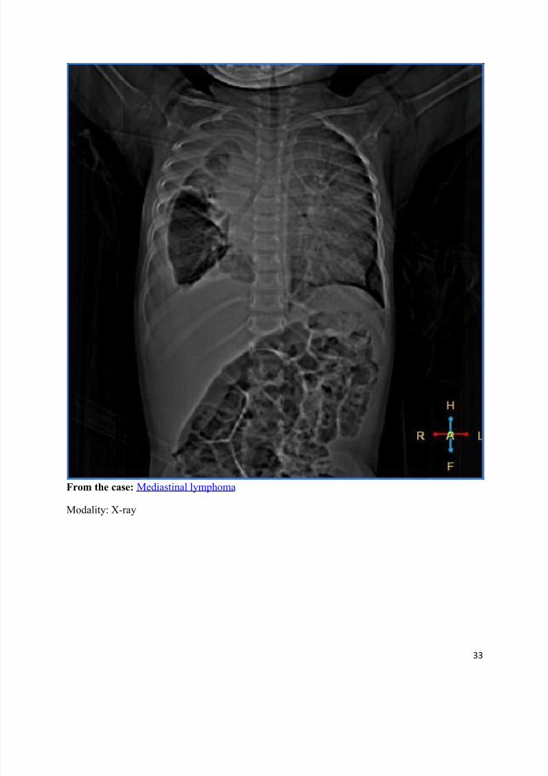
From the case: Mediastinal lymphoma
Modality: X-ray
8/11/2019 Radiological Signs (Shënja Radiologjike )-1
http://slidepdf.com/reader/full/radiological-signs-shenja-radiologjike-1 34/122
34
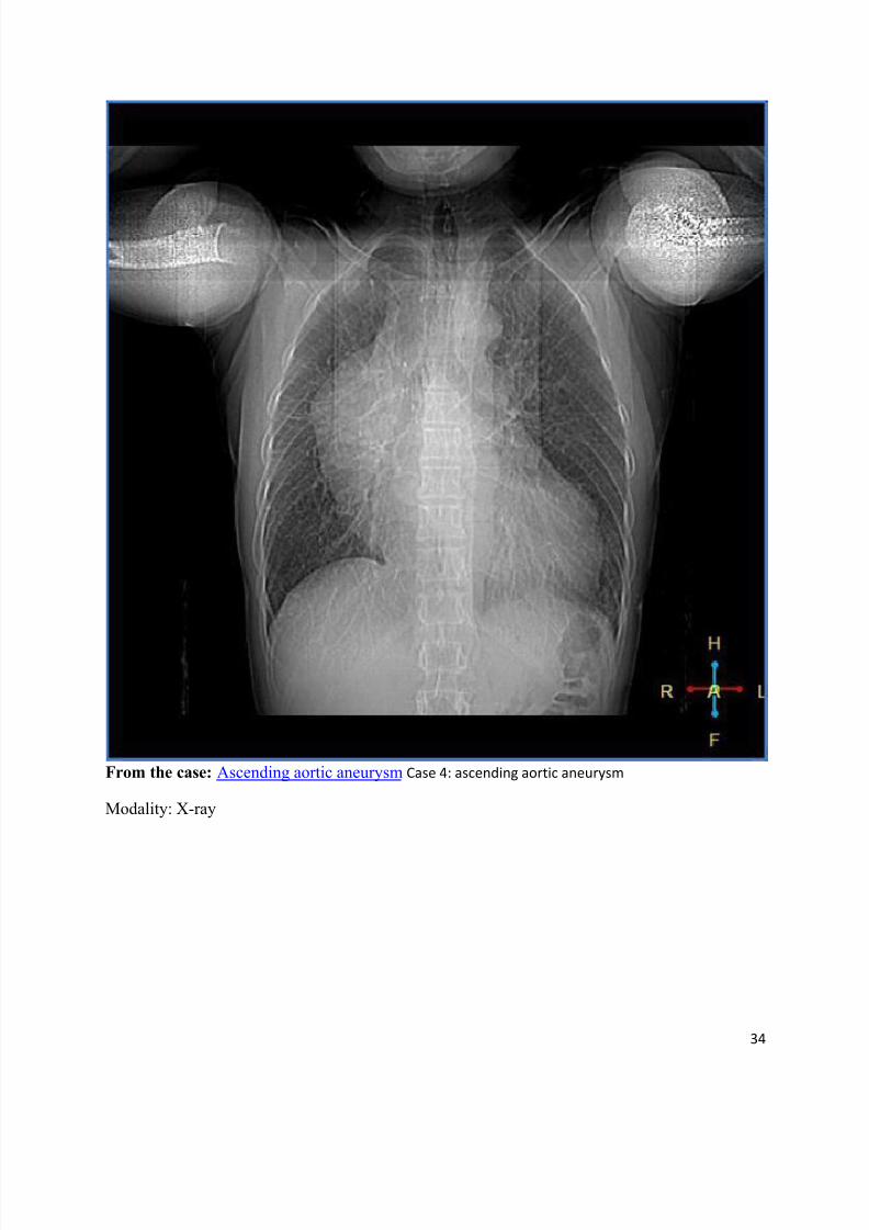
From the case: Ascending aortic aneurysm Case 4: ascending aortic aneurysm
Modality: X-ray
8/11/2019 Radiological Signs (Shënja Radiologjike )-1
http://slidepdf.com/reader/full/radiological-signs-shenja-radiologjike-1 35/122
35
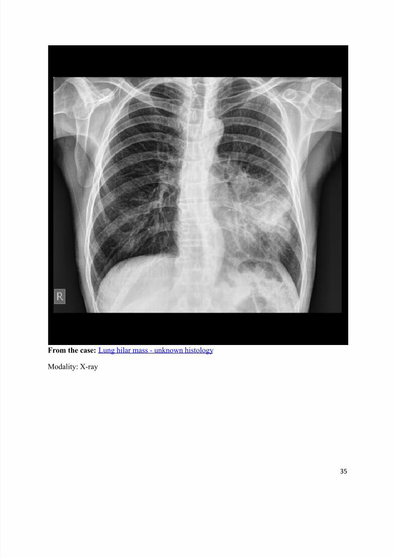
From the case: Lung hilar mass - unknown histology
Modality: X-ray
8/11/2019 Radiological Signs (Shënja Radiologjike )-1
http://slidepdf.com/reader/full/radiological-signs-shenja-radiologjike-1 36/122
36
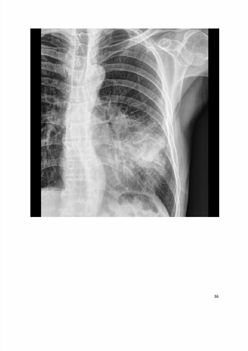
8/11/2019 Radiological Signs (Shënja Radiologjike )-1
http://slidepdf.com/reader/full/radiological-signs-shenja-radiologjike-1 37/122
37
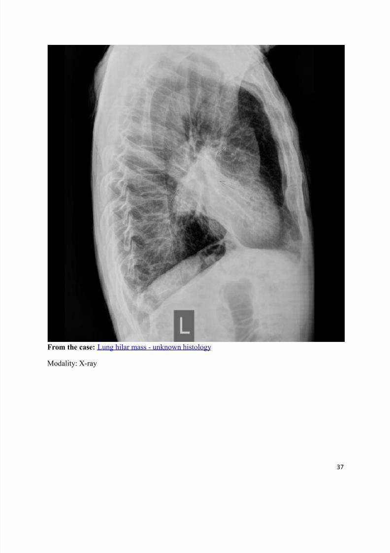
From the case: Lung hilar mass - unknown histology
Modality: X-ray
8/11/2019 Radiological Signs (Shënja Radiologjike )-1
http://slidepdf.com/reader/full/radiological-signs-shenja-radiologjike-1 38/122
38
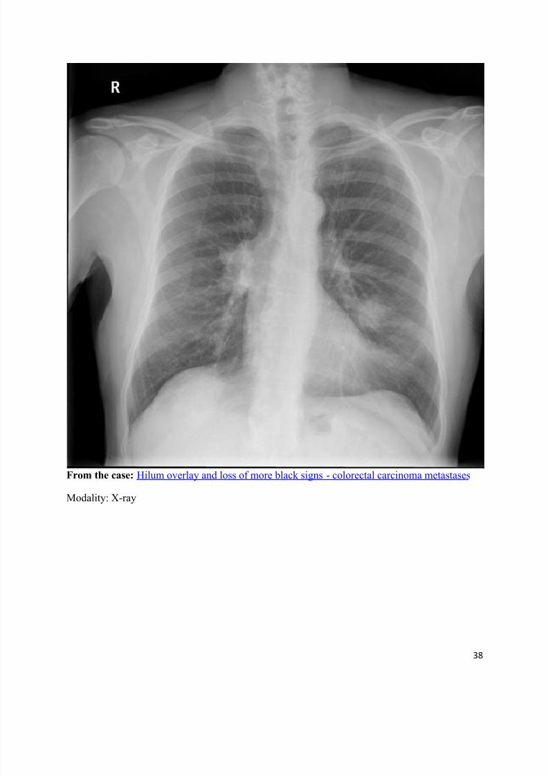
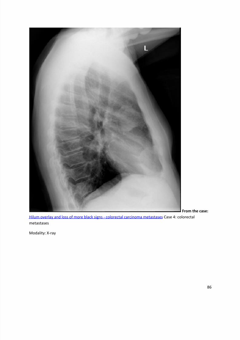
From the case: Hilum overlay and loss of more black signs - colorectal carcinoma metastases
Modality: X-ray
8/11/2019 Radiological Signs (Shënja Radiologjike )-1
http://slidepdf.com/reader/full/radiological-signs-shenja-radiologjike-1 39/122
39
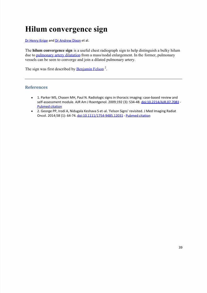
Hilum convergence sign
Dr Henry Knipe and Dr Andrew Dixon et al.
The hilum convergence sign is a useful chest radiograph sign to help distinguish a bulky hilumdue to pulmonary artery dilatation from a mass/nodal enlargement. In the former, pulmonaryvessels can be seen to converge and join a dilated pulmonary artery.
The sign was first described by Benjamin Felson 2.
References
1. Parker MS, Chasen MH, Paul N. Radiologic signs in thoracic imaging: case-based review and
self-assessment module. AJR Am J Roentgenol. 2009;192 (3): S34-48. doi:10.2214/AJR.07.7081 -Pubmed citation
2. George PP, Irodi A, Nidugala Keshava S et-al. 'Felson Signs' revisited. J Med Imaging Radiat
Oncol. 2014;58 (1): 64-74. doi:10.1111/1754-9485.12031 - Pubmed citation
8/11/2019 Radiological Signs (Shënja Radiologjike )-1
http://slidepdf.com/reader/full/radiological-signs-shenja-radiologjike-1 40/122
40
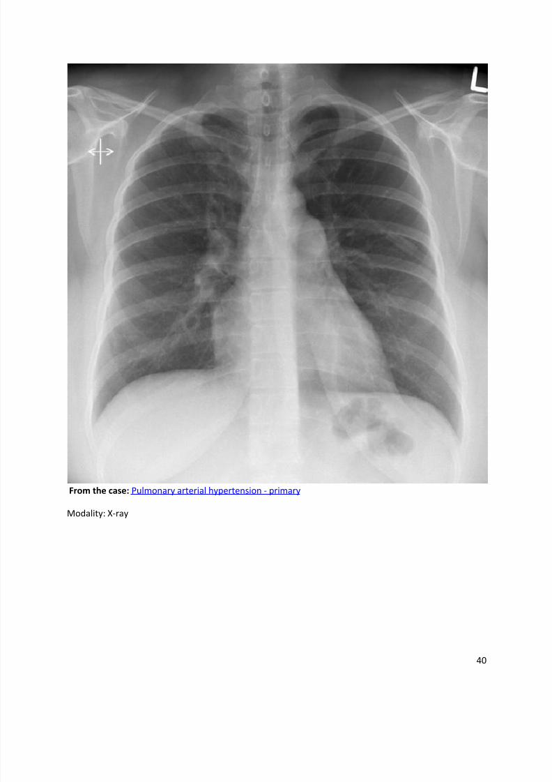
From the case: Pulmonary arterial hypertension - primary
Modality: X-ray
8/11/2019 Radiological Signs (Shënja Radiologjike )-1
http://slidepdf.com/reader/full/radiological-signs-shenja-radiologjike-1 41/122
41
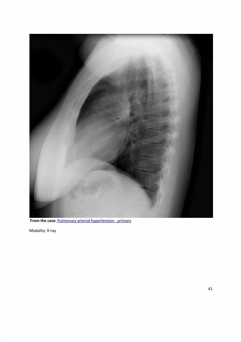
From the case: Pulmonary arterial hypertension - primary
Modality: X-ray
8/11/2019 Radiological Signs (Shënja Radiologjike )-1
http://slidepdf.com/reader/full/radiological-signs-shenja-radiologjike-1 42/122
42
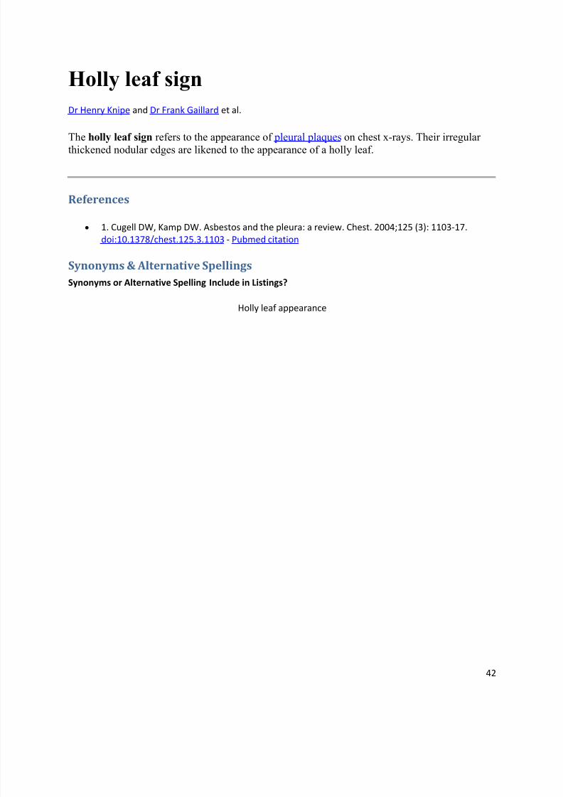
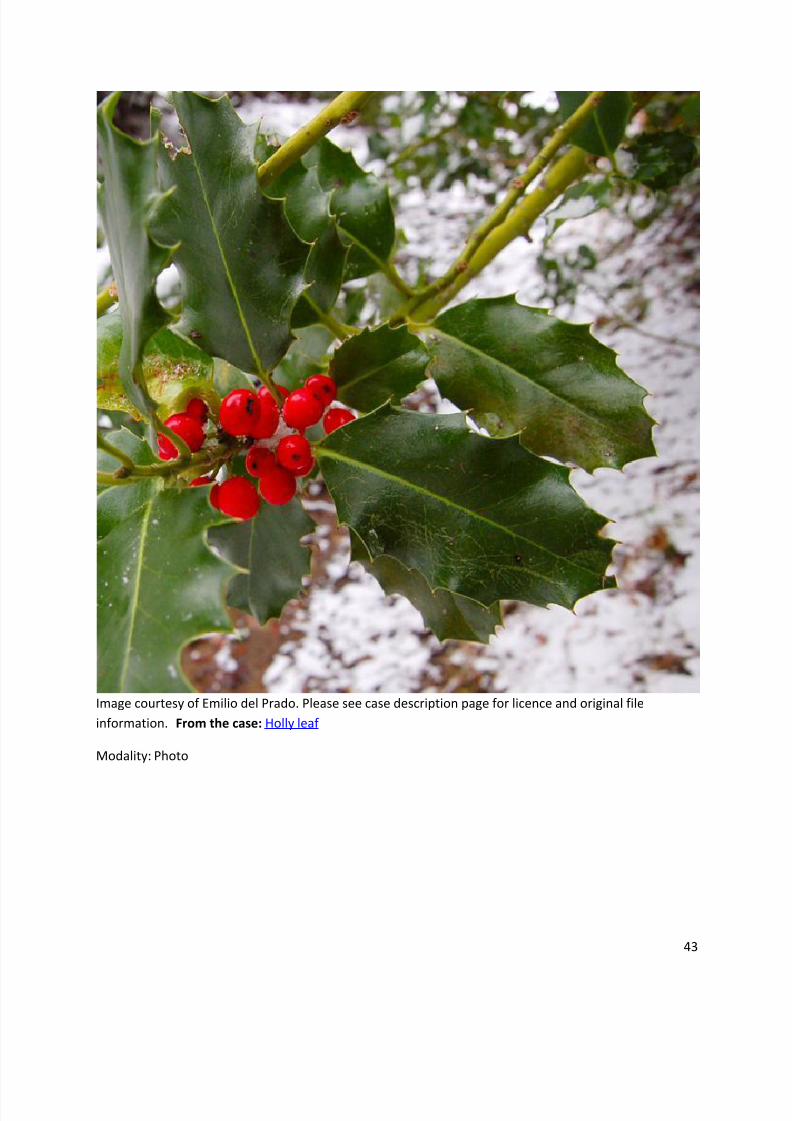
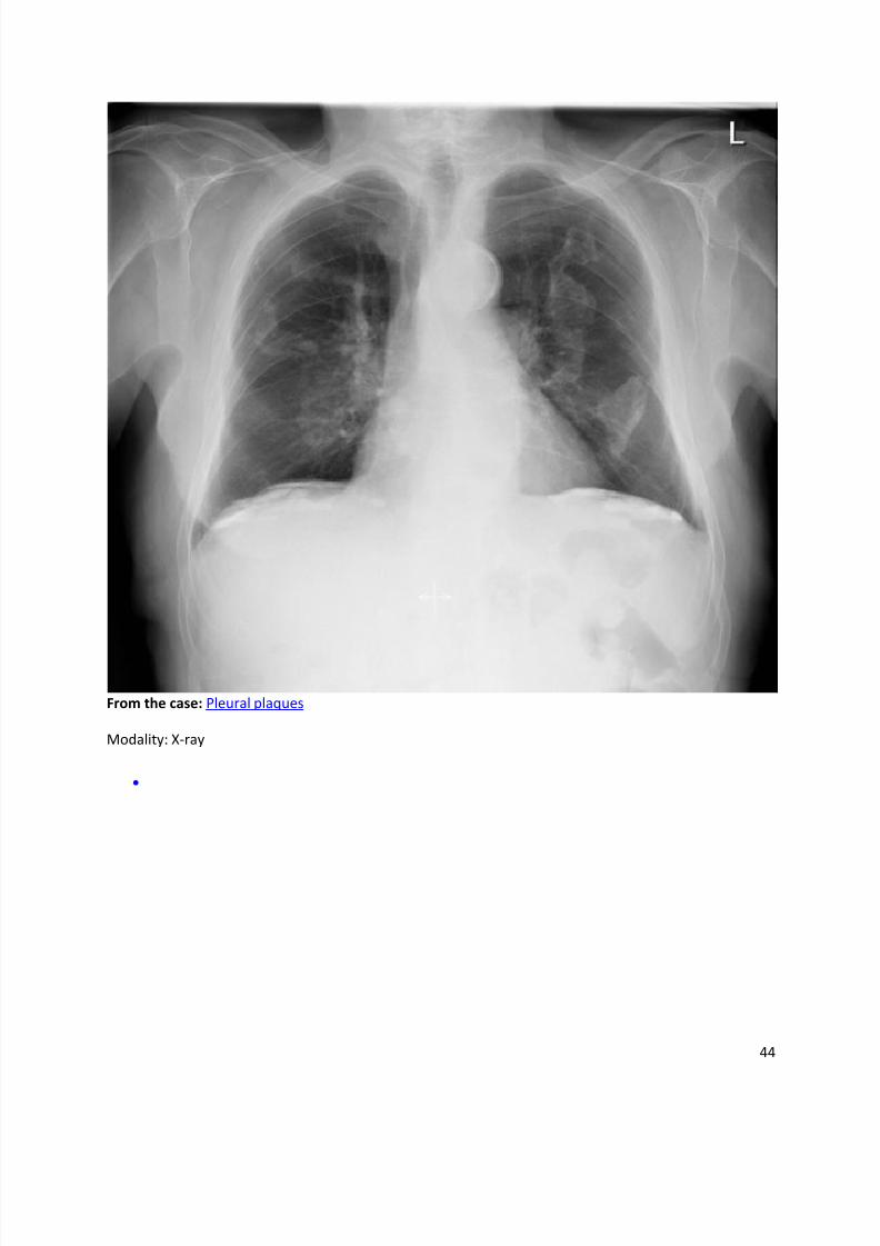
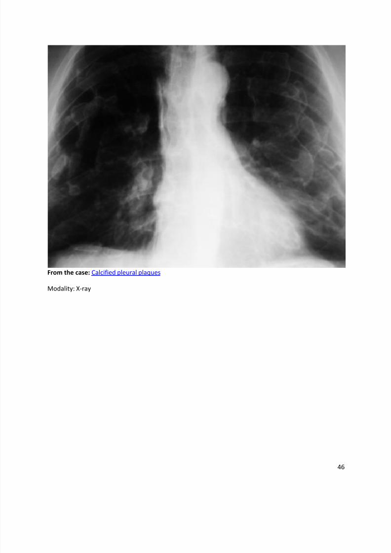
Holly leaf sign
Dr Henry Knipe and Dr Frank Gaillard et al.
The holly leaf sign refers to the appearance of pleural plaques on chest x-rays. Their irregularthickened nodular edges are likened to the appearance of a holly leaf.
References
1. Cugell DW, Kamp DW. Asbestos and the pleura: a review. Chest. 2004;125 (3): 1103-17.
doi:10.1378/chest.125.3.1103 - Pubmed citation
Synonyms & Alternative Spellings
Synonyms or Alternative Spelling Include in Listings?
Holly leaf appearance
8/11/2019 Radiological Signs (Shënja Radiologjike )-1
http://slidepdf.com/reader/full/radiological-signs-shenja-radiologjike-1 43/122
43
Image courtesy of Emilio del Prado. Please see case description page for licence and original file
information. From the case: Holly leaf
Modality: Photo
8/11/2019 Radiological Signs (Shënja Radiologjike )-1
http://slidepdf.com/reader/full/radiological-signs-shenja-radiologjike-1 44/122
44
From the case: Pleural plaques
Modality: X-ray
8/11/2019 Radiological Signs (Shënja Radiologjike )-1
http://slidepdf.com/reader/full/radiological-signs-shenja-radiologjike-1 45/122
45
From the case: Calcified pleural plaques
Modality: X-ray
8/11/2019 Radiological Signs (Shënja Radiologjike )-1
http://slidepdf.com/reader/full/radiological-signs-shenja-radiologjike-1 46/122
46
From the case: Calcified pleural plaques
Modality: X-ray
8/11/2019 Radiological Signs (Shënja Radiologjike )-1
http://slidepdf.com/reader/full/radiological-signs-shenja-radiologjike-1 47/122
47
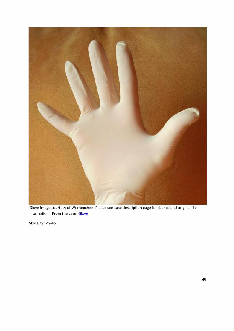
Finger in glove sign
Dr Henry Knipe and Dr Jeremy Jones et al.
The finger in glove sign seen on CXR and CT chest and refers to the characteristic sign of a bronchocoele. The same appearance has also been referred to as:
rabbit ear appearance
mickey mouse appearance
toothpaste shaped opacities
Y-shaped opacities
V-shaped opacities
Pathology
Aetiology
Obstructive
In bronchial obstruction, the portion of the bronchus distal to the obstruction is dilated with the
presence of mucous secretions. Causes of bronchial obstruction include :
hamartoma
lipoma
carcinoid
carcinoma
congenital bronchial atresia (rarely)
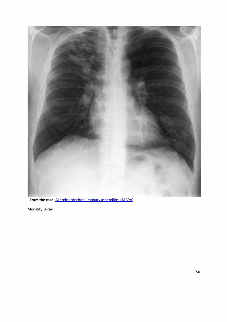
Non obstructive
Causes include :
asthma
allergic bronchopulmonary aspergillosis (ABPA)
cystic fibrosis
References
1. Collins J, Stern EJ. Chest radiology, the essentials. Lippincott Williams & Wilkins. (2007)
ISBN:0781763142. Read it at Google Books - Find it at Amazon
2. Swenson SJ, Aughenbaugh GL, Brown LR. Chest case of the day. AJR Am J Roentgenol 1993;
160:1318-1322.
3. Nguyen ET. The gloved finger sign. Radiology 2003 227:2, 453-454

8/11/2019 Radiological Signs (Shënja Radiologjike )-1
http://slidepdf.com/reader/full/radiological-signs-shenja-radiologjike-1 48/122
48
Synonyms & Alternative Spellings
Synonyms or Alternative Spelling Include in Listings?
Finger in glove appearance ✗
Finger-in-glove sign ✗
Rabbit ear appearance ✓
Y-shaped opacities ✓
V-shaped opacities ✓
Gloved finger sign
8/11/2019 Radiological Signs (Shënja Radiologjike )-1
http://slidepdf.com/reader/full/radiological-signs-shenja-radiologjike-1 49/122
49
Glove Image courtesy of Werneuchen. Please see case description page for licence and original file
information. From the case: Glove
Modality: Photo
8/11/2019 Radiological Signs (Shënja Radiologjike )-1
http://slidepdf.com/reader/full/radiological-signs-shenja-radiologjike-1 50/122
50
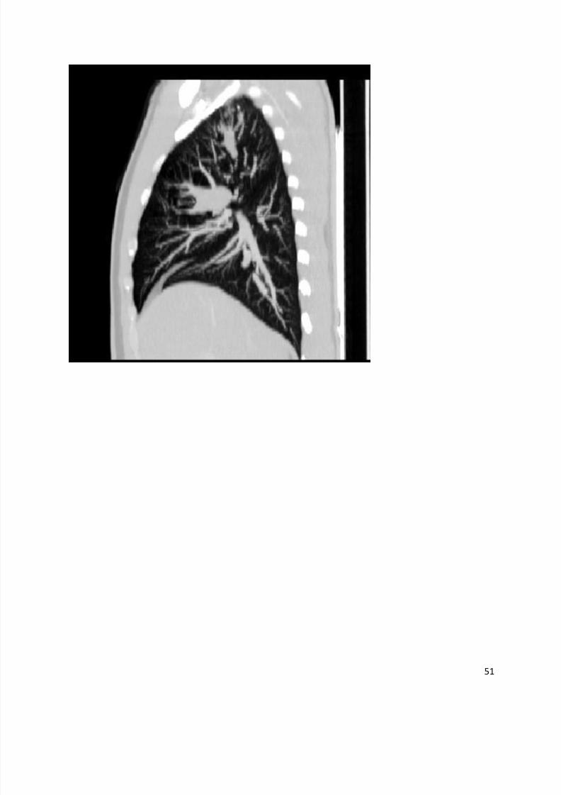
From the case: Allergic bronchopulmonary aspergillosis (ABPA)
Modality: X-ray
8/11/2019 Radiological Signs (Shënja Radiologjike )-1
http://slidepdf.com/reader/full/radiological-signs-shenja-radiologjike-1 51/122
51
8/11/2019 Radiological Signs (Shënja Radiologjike )-1
http://slidepdf.com/reader/full/radiological-signs-shenja-radiologjike-1 52/122
52
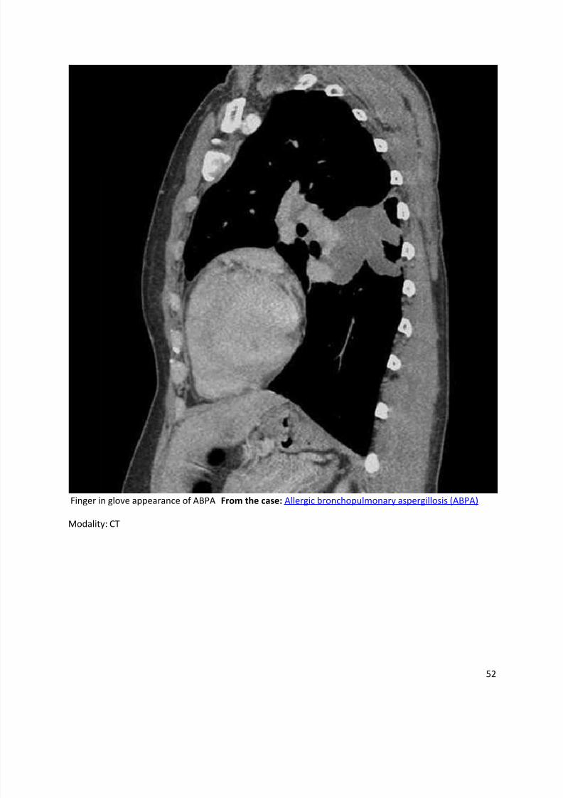
Finger in glove appearance of ABPA From the case: Allergic bronchopulmonary aspergillosis (ABPA)
Modality: CT
8/11/2019 Radiological Signs (Shënja Radiologjike )-1
http://slidepdf.com/reader/full/radiological-signs-shenja-radiologjike-1 53/122
53
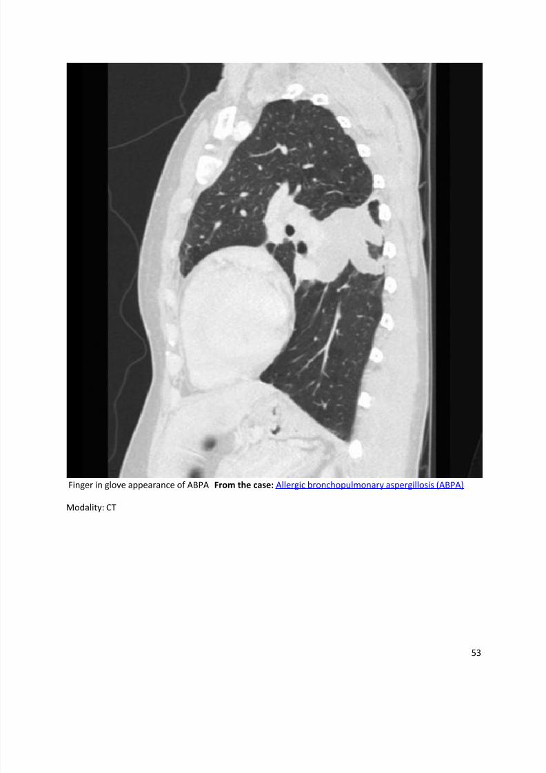
Finger in glove appearance of ABPA From the case: Allergic bronchopulmonary aspergillosis (ABPA)
Modality: CT

8/11/2019 Radiological Signs (Shënja Radiologjike )-1
http://slidepdf.com/reader/full/radiological-signs-shenja-radiologjike-1 54/122
54
Flat waist sign
Dr Henry Knipe and Dr Vinod G Maller et al.
The flat waist sign refers to flattening of the contours of the aortic arch and adjacent main pulmonary artery. It is seen in severe left lower lobe collapse and is caused by leftwarddisplacement and rotation of the heart.
8/11/2019 Radiological Signs (Shënja Radiologjike )-1
http://slidepdf.com/reader/full/radiological-signs-shenja-radiologjike-1 55/122
55
Fleishner's sign
Dr Yuranga Weerakkody and Dr Ian Bickle et al.
Fleishner's sign is a prominent central artery that can be caused either by pulmonaryhypertension that develops or by distension of the vessel by a large pulmonary embolus.
Radiographic appearance
Plain film
Fleishner's sign is a description given to appearances on plain chest x-ray along with
Westermark's sign and Hampton's hump. These are all rarely seen, and usually in the context of a
large pulmonary embolism.
References
1. Plain Film Signs in Pulmonary Embolism (with CT correlate), Kirwadi A, Bickle IC.
http://www.eurorad.org/eurorad/case.php?id=7735 Eurorad case 7735
2. Worsley DF, Alavi A, Aronchick JM et-al. Chest radiographic findings in patients with acute
pulmonary embolism: observations from the PIOPED Study. Radiology. 1993;189 (1): 133-6.
Radiology (abstract) - Pubmed citation
Synonyms & Alternative Spellings
Synonyms or Alternative Spelling Include in Listings?
Fleishner sign
8/11/2019 Radiological Signs (Shënja Radiologjike )-1
http://slidepdf.com/reader/full/radiological-signs-shenja-radiologjike-1 56/122
56
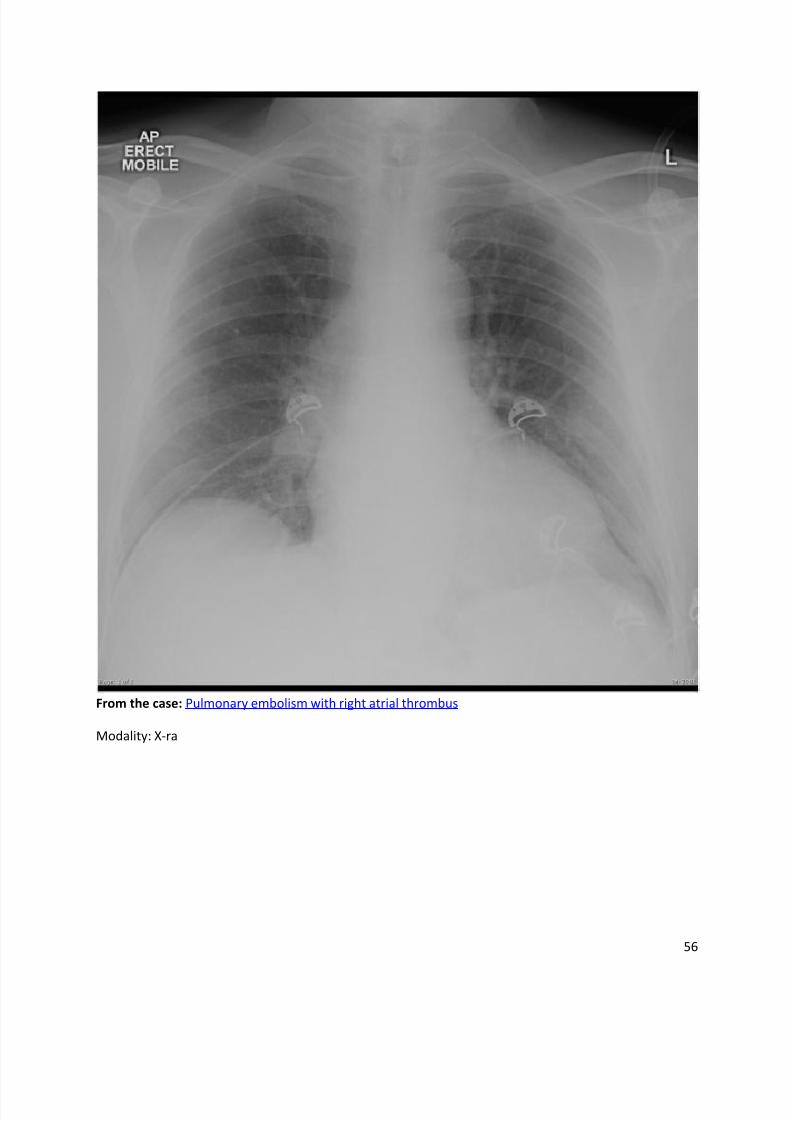
From the case: Pulmonary embolism with right atrial thrombus
Modality: X-ra
8/11/2019 Radiological Signs (Shënja Radiologjike )-1
http://slidepdf.com/reader/full/radiological-signs-shenja-radiologjike-1 57/122
57
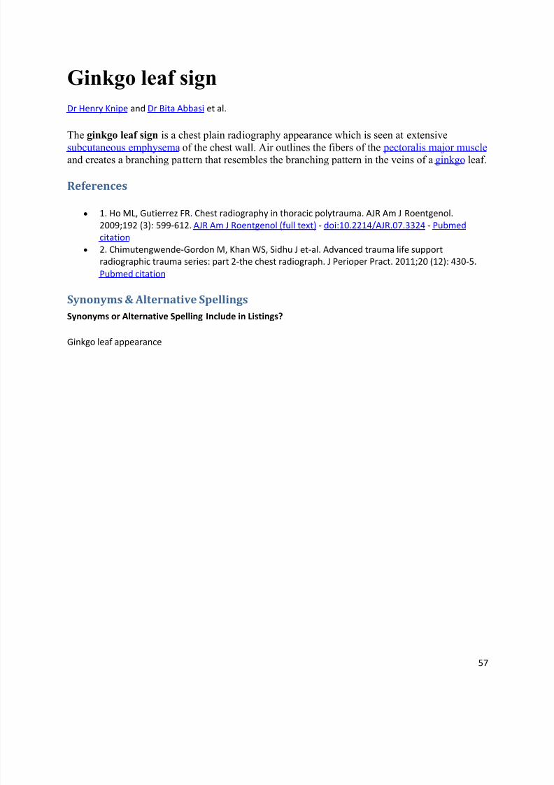
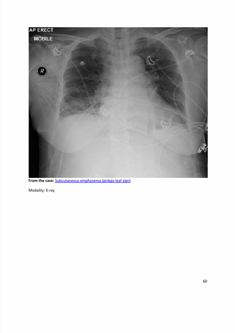
Ginkgo leaf sign
Dr Henry Knipe and Dr Bita Abbasi et al.
The ginkgo leaf sign is a chest plain radiography appearance which is seen at extensivesubcutaneous emphysema of the chest wall. Air outlines the fibers of the pectoralis major muscle and creates a branching pattern that resembles the branching pattern in the veins of a ginkgo leaf.
References
1. Ho ML, Gutierrez FR. Chest radiography in thoracic polytrauma. AJR Am J Roentgenol.
2009;192 (3): 599-612. AJR Am J Roentgenol (full text) - doi:10.2214/AJR.07.3324 - Pubmed
citation
2. Chimutengwende-Gordon M, Khan WS, Sidhu J et-al. Advanced trauma life support
radiographic trauma series: part 2-the chest radiograph. J Perioper Pract. 2011;20 (12): 430-5.
Pubmed citation
Synonyms & Alternative Spellings
Synonyms or Alternative Spelling Include in Listings?
Ginkgo leaf appearance

8/11/2019 Radiological Signs (Shënja Radiologjike )-1
http://slidepdf.com/reader/full/radiological-signs-shenja-radiologjike-1 58/122
58
Ginkgo leaf

8/11/2019 Radiological Signs (Shënja Radiologjike )-1
http://slidepdf.com/reader/full/radiological-signs-shenja-radiologjike-1 59/122
59
Emphizema 3 day after pneumonectomyFrom the case: Subcutaneous emphysema
Modality: X-ray
8/11/2019 Radiological Signs (Shënja Radiologjike )-1
http://slidepdf.com/reader/full/radiological-signs-shenja-radiologjike-1 60/122
60
From the case: Subcutaneous emphysema (ginkgo leaf sign)
Modality: X-ray
8/11/2019 Radiological Signs (Shënja Radiologjike )-1
http://slidepdf.com/reader/full/radiological-signs-shenja-radiologjike-1 61/122
61
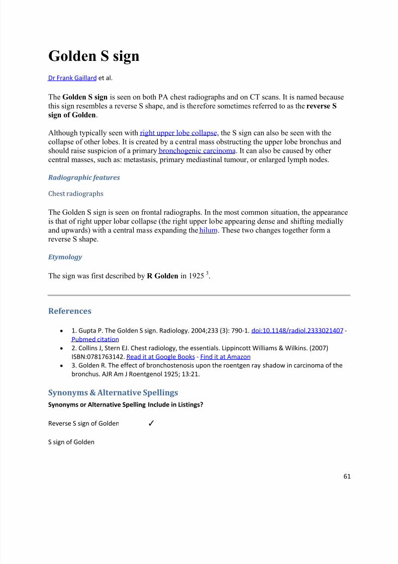
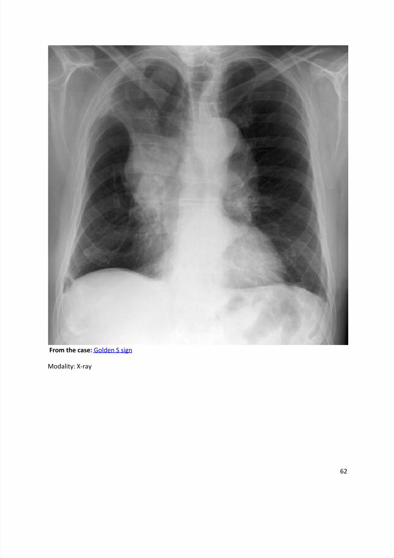
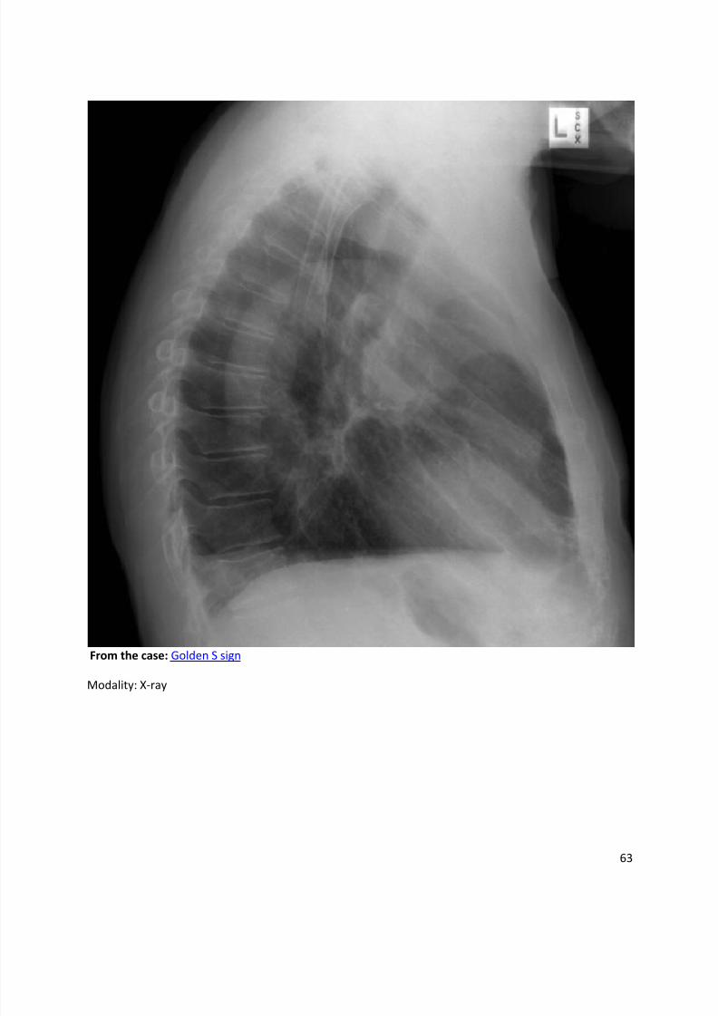
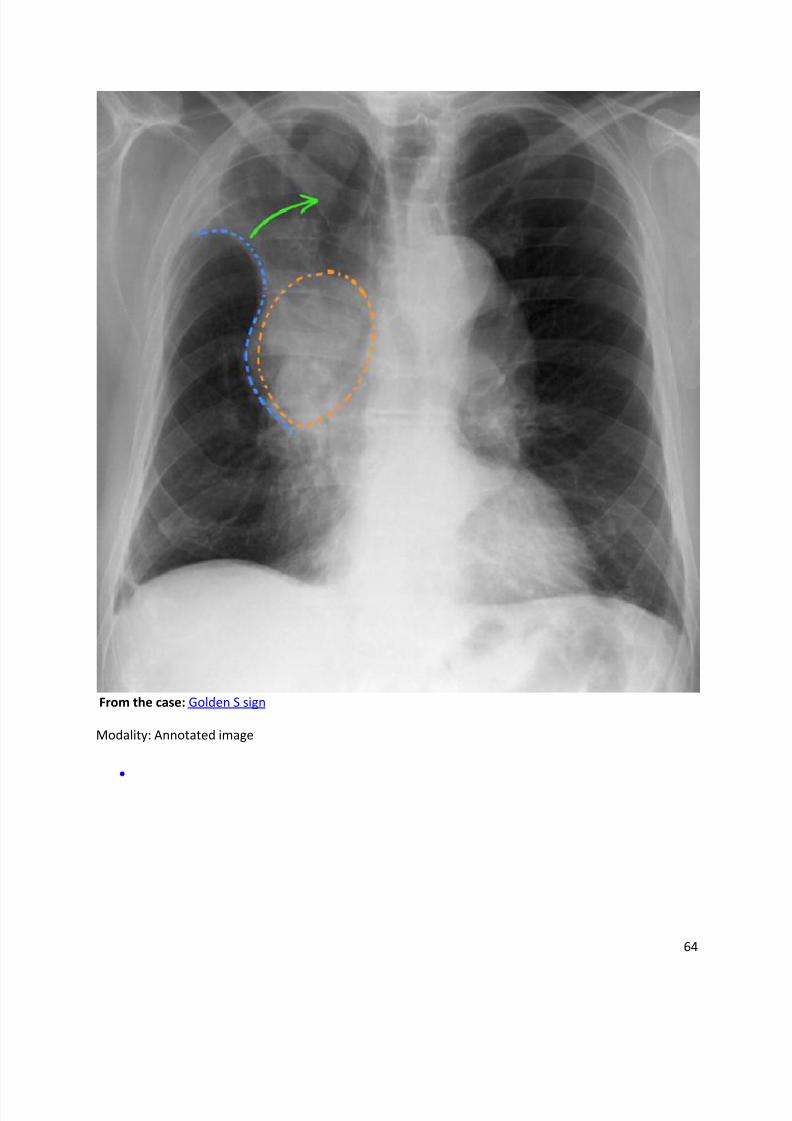
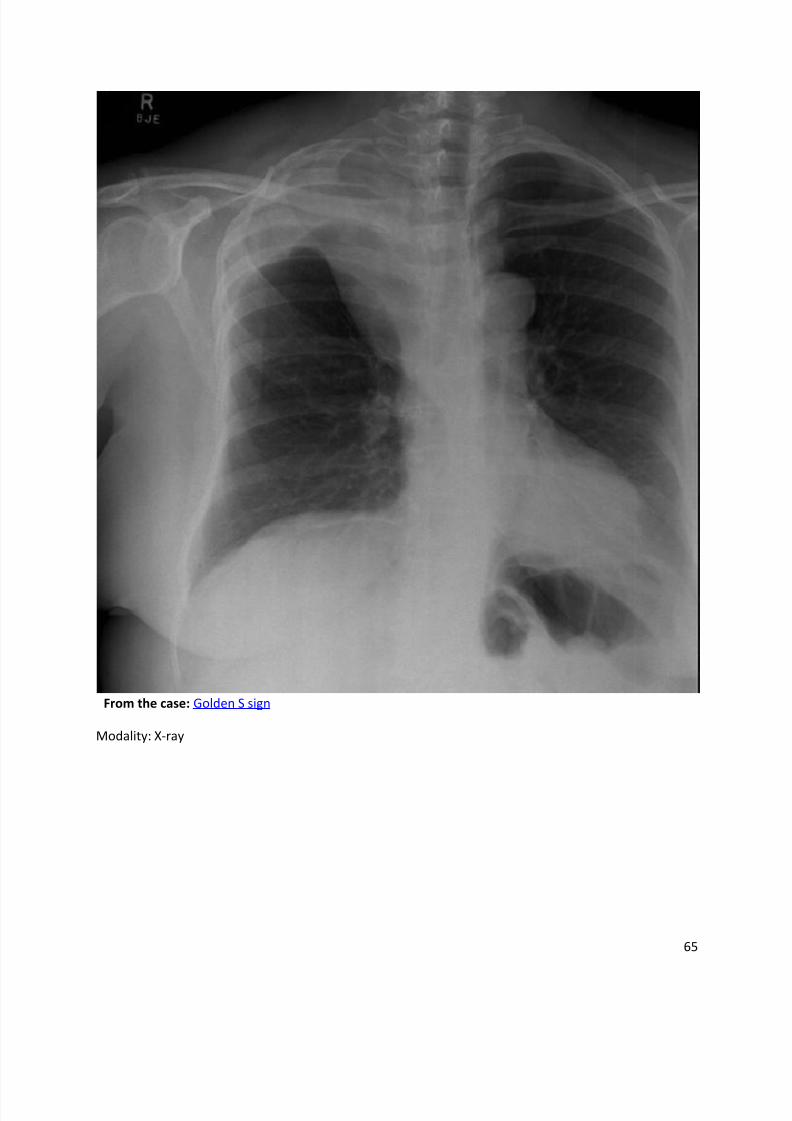
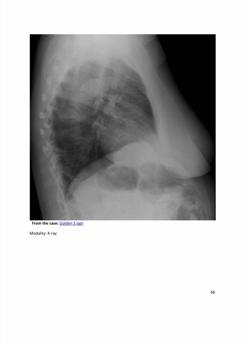
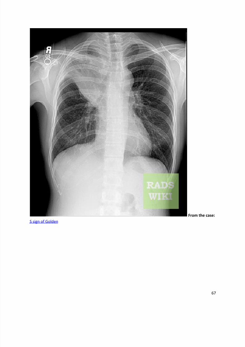
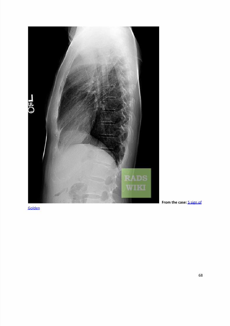
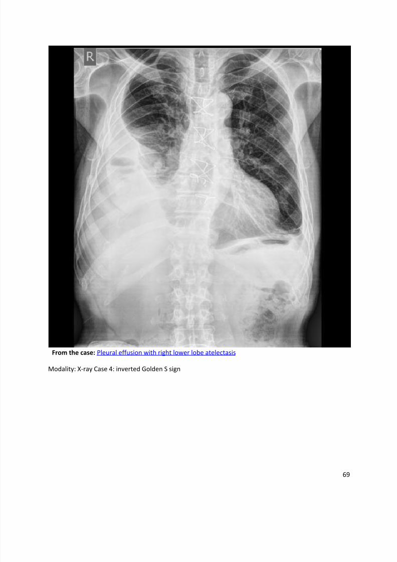
Golden S sign
Dr Frank Gaillard et al.
The Golden S sign is seen on both PA chest radiographs and on CT scans. It is named becausethis sign resembles a reverse S shape, and is therefore sometimes referred to as the reverse S
sign of Golden.
Although typically seen with right upper lobe collapse, the S sign can also be seen with the
collapse of other lobes. It is created by a central mass obstructing the upper lobe bronchus andshould raise suspicion of a primary bronchogenic carcinoma. It can also be caused by other
central masses, such as: metastasis, primary mediastinal tumour, or enlarged lymph nodes.
Radiographic features
Chest radiographs
The Golden S sign is seen on frontal radiographs. In the most common situation, the appearance
is that of right upper lobar collapse (the right upper lobe appearing dense and shifting medially
and upwards) with a central mass expanding the hilum. These two changes together form areverse S shape.
Etymology
The sign was first described by R Golden in 19253.
References
1. Gupta P. The Golden S sign. Radiology. 2004;233 (3): 790-1. doi:10.1148/radiol.2333021407 -
Pubmed citation
2. Collins J, Stern EJ. Chest radiology, the essentials. Lippincott Williams & Wilkins. (2007)
ISBN:0781763142. Read it at Google Books - Find it at Amazon
3. Golden R. The effect of bronchostenosis upon the roentgen ray shadow in carcinoma of the
bronchus. AJR Am J Roentgenol 1925; 13:21.
Synonyms & Alternative SpellingsSynonyms or Alternative Spelling Include in Listings?
Reverse S sign of Golden ✓
S sign of Golden
8/11/2019 Radiological Signs (Shënja Radiologjike )-1
http://slidepdf.com/reader/full/radiological-signs-shenja-radiologjike-1 62/122
62
From the case: Golden S sign
Modality: X-ray
8/11/2019 Radiological Signs (Shënja Radiologjike )-1
http://slidepdf.com/reader/full/radiological-signs-shenja-radiologjike-1 63/122
63
From the case: Golden S sign
Modality: X-ray
8/11/2019 Radiological Signs (Shënja Radiologjike )-1
http://slidepdf.com/reader/full/radiological-signs-shenja-radiologjike-1 64/122
64
From the case: Golden S sign
Modality: Annotated image
8/11/2019 Radiological Signs (Shënja Radiologjike )-1
http://slidepdf.com/reader/full/radiological-signs-shenja-radiologjike-1 65/122
65
From the case: Golden S sign
Modality: X-ray
8/11/2019 Radiological Signs (Shënja Radiologjike )-1
http://slidepdf.com/reader/full/radiological-signs-shenja-radiologjike-1 66/122
66
From the case: Golden S sign
Modality: X-ray
8/11/2019 Radiological Signs (Shënja Radiologjike )-1
http://slidepdf.com/reader/full/radiological-signs-shenja-radiologjike-1 67/122
67
From the case:
S sign of Golden
8/11/2019 Radiological Signs (Shënja Radiologjike )-1
http://slidepdf.com/reader/full/radiological-signs-shenja-radiologjike-1 68/122
68
From the case: S sign of
Golden
8/11/2019 Radiological Signs (Shënja Radiologjike )-1
http://slidepdf.com/reader/full/radiological-signs-shenja-radiologjike-1 69/122
69
From the case: Pleural effusion with right lower lobe atelectasis
Modality: X-ray Case 4: inverted Golden S sign
8/11/2019 Radiological Signs (Shënja Radiologjike )-1
http://slidepdf.com/reader/full/radiological-signs-shenja-radiologjike-1 70/122
70
Incomplete border sign
Dr Yuranga Weerakkody and Dr Charlie Chia-Tsong Hsu et al.
The incomplete border sign is useful to depict an extrapulmonary mass on chest radiograph.
An extrapulmonary mass will often have a inner well defined border and an ill-defined outermargin
1-3. This can be attributed to the inner margin being tangential to the x-ray beam and has
good inherent contrast with the adjacent lung. On the other hand, the outer margin is enface or
partially enface with the x-ray beam and merges with the pleural or chest wall thus the border isobscured.
Differential diagnosis
Differential diagnosis for extrapulmonary mass can be further divided into pleural or
extrapleural.
Common pleural masses include:
loculated pleural collection
haematoma
pleural plaques
fibrous tumour of pleura
Extrapleural causes can arises from component of chest wall, bone/cartilage, nerve, vascular,
fat, muscle and skin. If there is sign of rib/bone involvement on chest radiograph the lesion is
most likely to be extrapleural. In adults, skeletal metastases are the most common malignantchest wall neoplasm while chondrosarcoma is the most common primary malignant tumor.
References
1. Catalano O. The incomplete border sign. Radiology. 2002;225 (1): 129-30.
doi:10.1148/radiol.2251010926 - Pubmed citation
2. Ellis R. Incomplete border sign of extrapleural masses. JAMA. 1977;237 (25): 2748. JAMA (link)
- Pubmed citation
3. Hsu CC, Henry TS, Chung JH et-al. The incomplete border sign. J Thorac Imaging. 2014;29 (4):W48. doi:10.1097/RTI.0000000000000088 - Pubmed citation
Synonyms & Alternative Spellings
Synonyms or Alternative Spelling Include in Listings?
Incomplete border sign on a chest x ray ✗
8/11/2019 Radiological Signs (Shënja Radiologjike )-1
http://slidepdf.com/reader/full/radiological-signs-shenja-radiologjike-1 71/122
71
Synonyms or Alternative Spelling Include in Listings?
Incomplete border sign on a chest radiograph
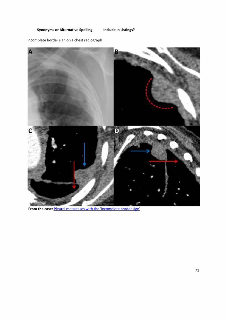
From the case: Pleural metastases with the 'incomplete border sign'
8/11/2019 Radiological Signs (Shënja Radiologjike )-1
http://slidepdf.com/reader/full/radiological-signs-shenja-radiologjike-1 72/122
72
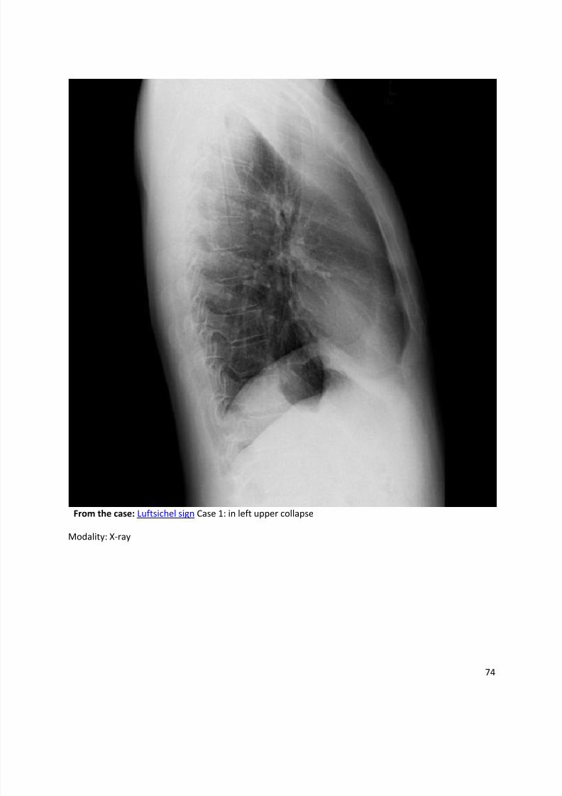
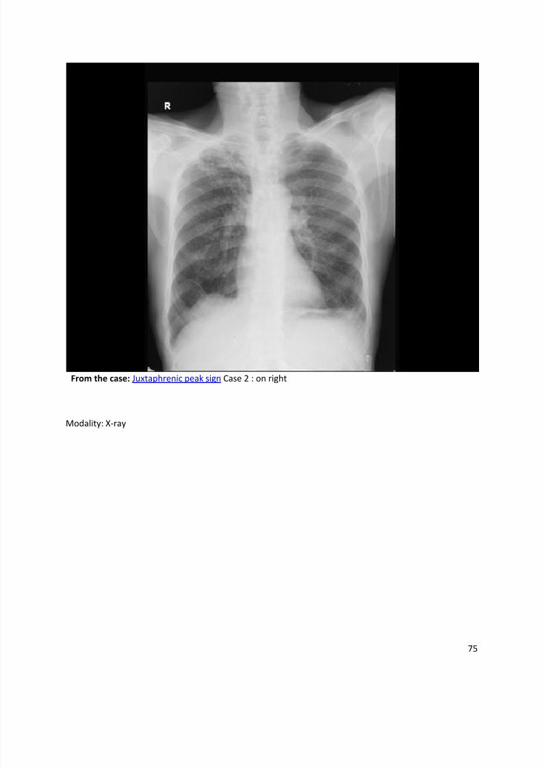
Juxtaphrenic peak sign
Dr Henry Knipe and Dr Andrew Dixon et al.
The juxtaphrenic peak sign refers to the peaked or tented appearance of a hemidiaphragm
which can occur in the setting of lobar collapse. It is caused by retraction of the lower end of
diaphragm at an inferior accessory fissure (most common1), major fissure or inferior pulmonary
ligament. It is commonly seen in upper lobe collapse but may also be seen in middle lobe
collapse.
References
1. Davis SD, Yankelevitz DF, Wand A et-al. Juxtaphrenic peak in upper and middle lobe volume
loss: assessment with CT. Radiology. 1996;198 (1): 143-9. Radiology (citation) - Pubmed citation
2. Konen E, Rozenman J, Simansky DA et-al. Prevalence of the juxtaphrenic peak after upper
lobectomy. AJR Am J Roentgenol. 2001;177 (4): 869-73. AJR Am J Roentgenol (citation) -
Pubmed citation
3. Collins J, Stern EJ. Chest radiology, the essentials. Lippincott Williams & Wilkins. (2007)
ISBN:0781763142. Read it at Google Books - Find it at Amazon
Synonyms & Alternative Spellings
Synonyms or Alternative Spelling Include in Listings?
Juxtaphrenic peak ✗
Juxtaphrenic peak (JP) ✗
Juxta-phrenic peak sign
8/11/2019 Radiological Signs (Shënja Radiologjike )-1
http://slidepdf.com/reader/full/radiological-signs-shenja-radiologjike-1 73/122
73
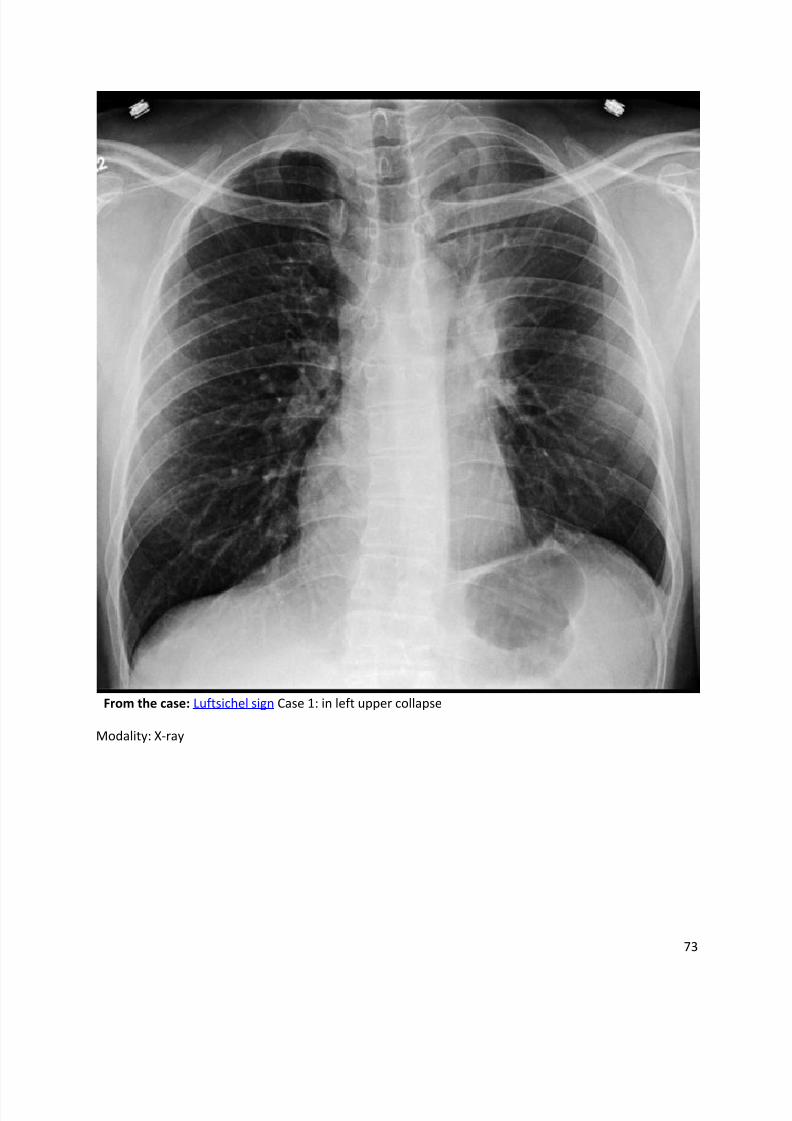
From the case: Luftsichel sign Case 1: in left upper collapse
Modality: X-ray
8/11/2019 Radiological Signs (Shënja Radiologjike )-1
http://slidepdf.com/reader/full/radiological-signs-shenja-radiologjike-1 74/122
74
From the case: Luftsichel sign Case 1: in left upper collapse
Modality: X-ray
8/11/2019 Radiological Signs (Shënja Radiologjike )-1
http://slidepdf.com/reader/full/radiological-signs-shenja-radiologjike-1 75/122
75
From the case: Juxtaphrenic peak sign Case 2 : on right
Modality: X-ray
8/11/2019 Radiological Signs (Shënja Radiologjike )-1
http://slidepdf.com/reader/full/radiological-signs-shenja-radiologjike-1 76/122
76
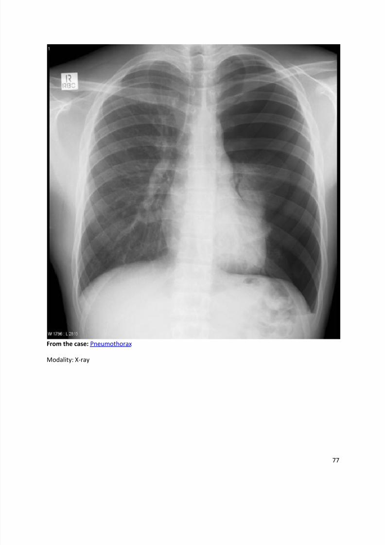
Medial stripe sign
Dr Henry Knipe and Dr Aditya Shetty et al.
Medial stripe sign refers to an area of increased lucency at the interface of the medial lung andthe mediastinum in case of medial pneumothorax. A small volume of pneumothorax generallyaccumulates anteriorly or medially which can be difficult to detect hence this sign holds a certain
significance.
Related pathology
neonatal pneumothorax
References
1. Swischuk LE. Two lesser known but useful signs of neonatal pneumothorax. AJR Am JRoentgenol. 1976;127 (4): 623-7. doi:10.2214/ajr.127.4.623 - Pubmed citation
Synonyms & Alternative Spellings
Synonyms or Alternative Spelling Include in Listings?
Medial pneumothorax sign ✓
Medial stripe sign in a pneumothorax
8/11/2019 Radiological Signs (Shënja Radiologjike )-1
http://slidepdf.com/reader/full/radiological-signs-shenja-radiologjike-1 77/122
77
From the case: Pneumothorax
Modality: X-ray
8/11/2019 Radiological Signs (Shënja Radiologjike )-1
http://slidepdf.com/reader/full/radiological-signs-shenja-radiologjike-1 78/122
78
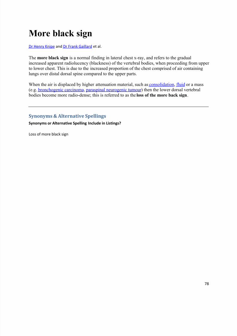
More black sign
Dr Henry Knipe and Dr Frank Gaillard et al.
The more black sign is a normal finding in lateral chest x-ray, and refers to the gradualincreased apparent radiolucency (blackness) of the vertebral bodies, when proceeding from upperto lower chest. This is due to the increased proportion of the chest comprised of air containing
lungs over distal dorsal spine compared to the upper parts.
When the air is displaced by higher attenuation material, such as consolidation, fluid or a mass(e.g. bronchogenic carcinoma, paraspinal neurogenic tumour ) then the lower dorsal vertebral
bodies become more radio-dense; this is referred to as the loss of the more back sign.
Synonyms & Alternative SpellingsSynonyms or Alternative Spelling Include in Listings?
Loss of more black sign
8/11/2019 Radiological Signs (Shënja Radiologjike )-1
http://slidepdf.com/reader/full/radiological-signs-shenja-radiologjike-1 79/122
79
From the case: Normal chest x-ray
Modality: X-ray
8/11/2019 Radiological Signs (Shënja Radiologjike )-1
http://slidepdf.com/reader/full/radiological-signs-shenja-radiologjike-1 80/122
80
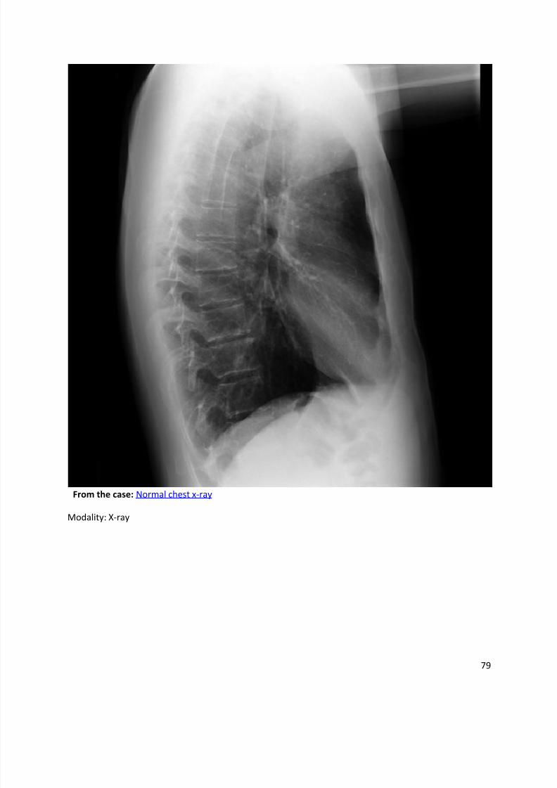
Normal chest x-ray
8/11/2019 Radiological Signs (Shënja Radiologjike )-1
http://slidepdf.com/reader/full/radiological-signs-shenja-radiologjike-1 81/122
81
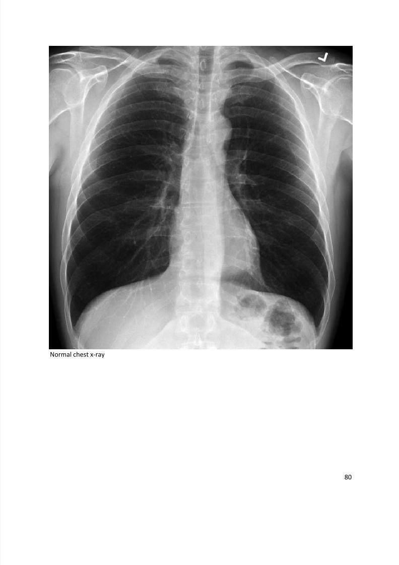
Normal pulmonary artery
8/11/2019 Radiological Signs (Shënja Radiologjike )-1
http://slidepdf.com/reader/full/radiological-signs-shenja-radiologjike-1 82/122
82
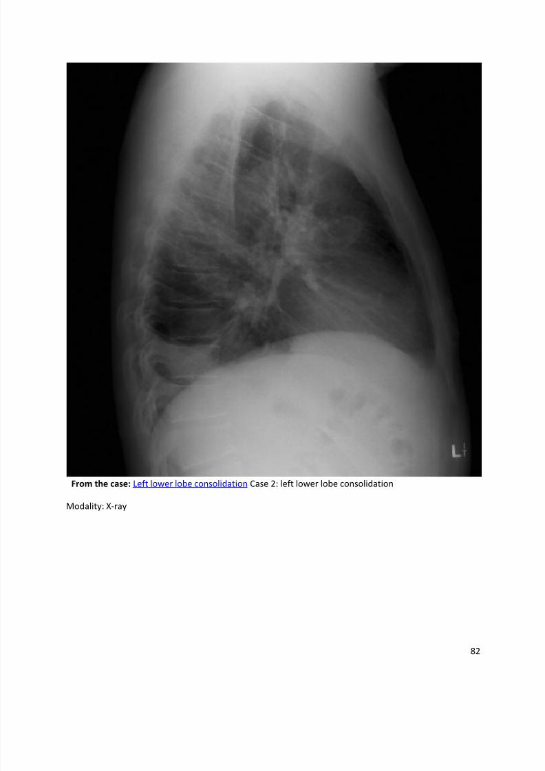
From the case: Left lower lobe consolidation Case 2: left lower lobe consolidation
Modality: X-ray
8/11/2019 Radiological Signs (Shënja Radiologjike )-1
http://slidepdf.com/reader/full/radiological-signs-shenja-radiologjike-1 83/122
83
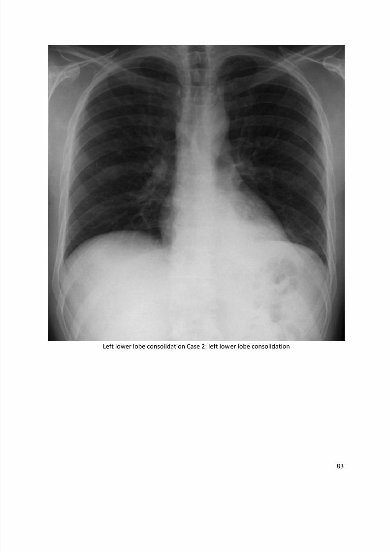
Left lower lobe consolidation Case 2: left lower lobe consolidation
8/11/2019 Radiological Signs (Shënja Radiologjike )-1
http://slidepdf.com/reader/full/radiological-signs-shenja-radiologjike-1 84/122
84
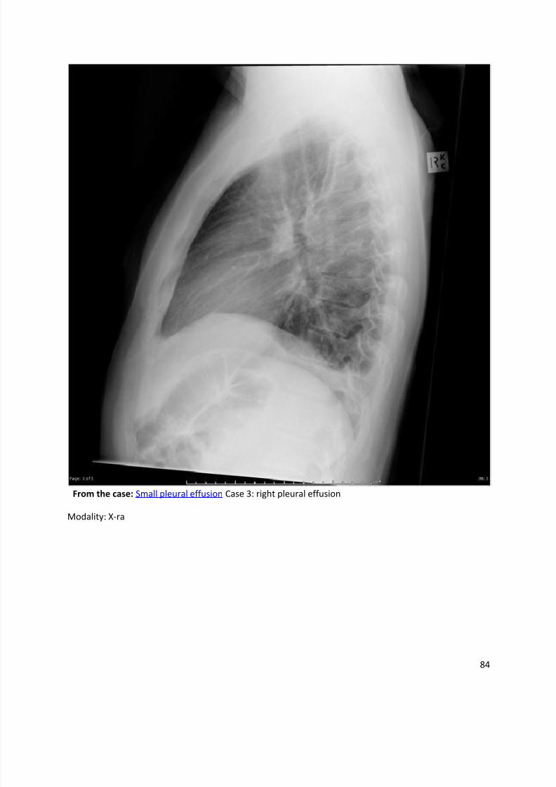
From the case: Small pleural effusion Case 3: right pleural effusion
Modality: X-ra
8/11/2019 Radiological Signs (Shënja Radiologjike )-1
http://slidepdf.com/reader/full/radiological-signs-shenja-radiologjike-1 85/122
85
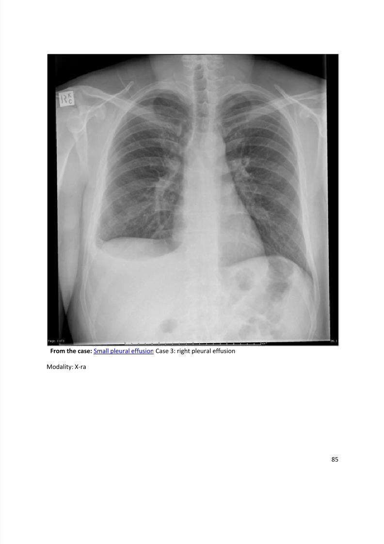
From the case: Small pleural effusion Case 3: right pleural effusion
Modality: X-ra
8/11/2019 Radiological Signs (Shënja Radiologjike )-1
http://slidepdf.com/reader/full/radiological-signs-shenja-radiologjike-1 86/122
86
From the case:
Hilum overlay and loss of more black signs - colorectal carcinoma metastases Case 4: colorectal
metastases
Modality: X-ray
8/11/2019 Radiological Signs (Shënja Radiologjike )-1
http://slidepdf.com/reader/full/radiological-signs-shenja-radiologjike-1 87/122
87
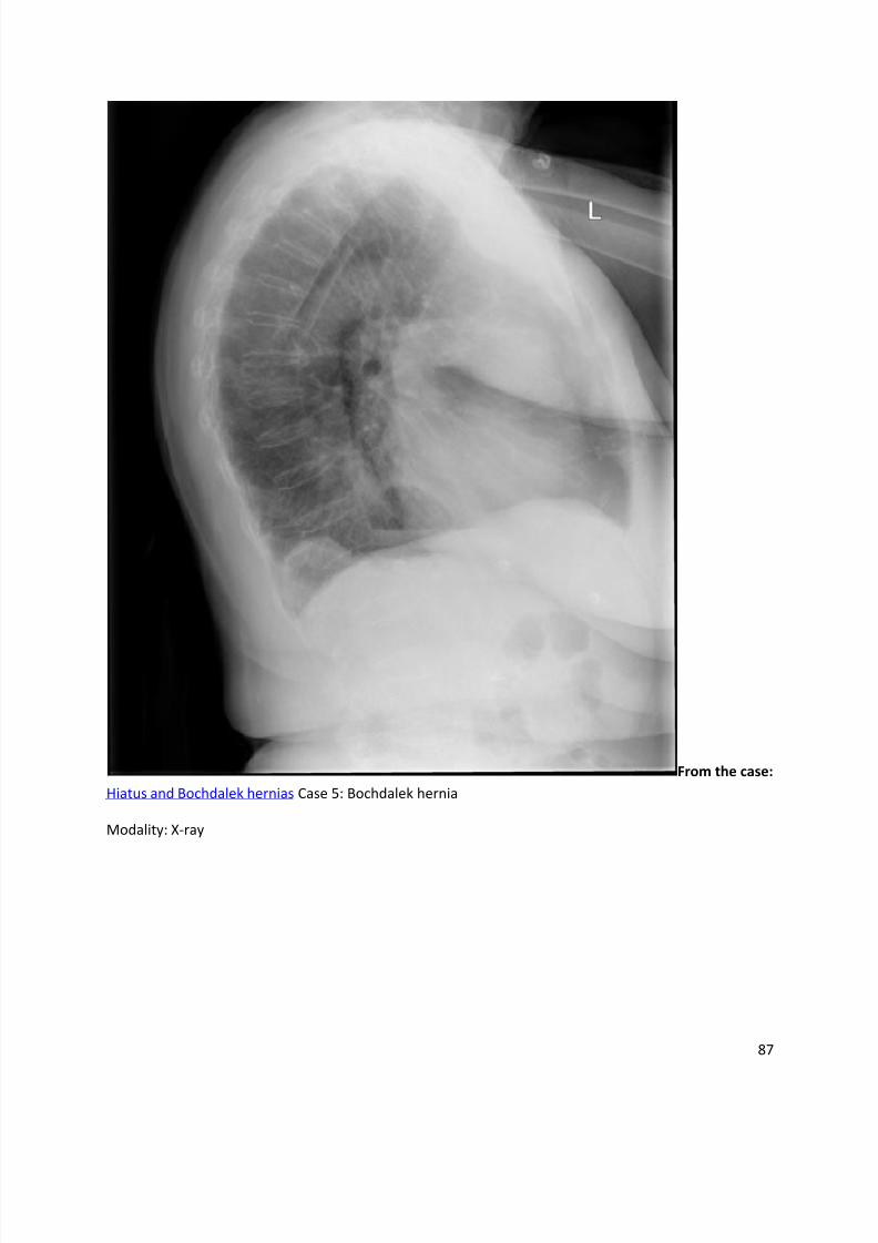
From the case:
Hiatus and Bochdalek hernias Case 5: Bochdalek hernia
Modality: X-ray
8/11/2019 Radiological Signs (Shënja Radiologjike )-1
http://slidepdf.com/reader/full/radiological-signs-shenja-radiologjike-1 88/122
88
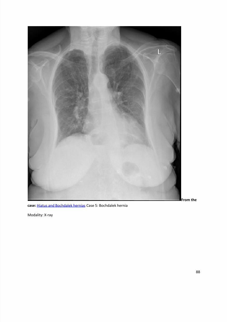
From the
case: Hiatus and Bochdalek hernias Case 5: Bochdalek hernia
Modality: X-ray
8/11/2019 Radiological Signs (Shënja Radiologjike )-1
http://slidepdf.com/reader/full/radiological-signs-shenja-radiologjike-1 89/122
89
Naclerio's V sign
Dr Henry Knipe et al.
The Naclerio's V sign is a sign described on the plain film in patients witha pneumomediastinum occurring often secondary to an oesophageal rupture.
It is seen as a V-shaped air collection. One limb of the V is produced by mediastinal air outlining
the left lower lateral mediastinal border. The other limb is produced by air between the parietal
pleura and medial left hemidiaphragm.
Although Naclerio’s V sign was originally described in patients with oesophageal rupture, it is not entirely specific to that condition.
References
1. Bejvan SM, Godwin JD. Pneumomediastinum: old signs and new signs. AJR Am J Roentgenol.
1996;166 (5): 1041-8. AJR Am J Roentgenol (citation) - Pubmed citation
2. Sinha R. Naclerio's V sign. Radiology. 2007;245 (1): 296-7. doi:10.1148/radiol.2451042197 -
Pubmed citation
Synonyms & Alternative Spellings
Synonyms or Alternative Spelling Include in Listings?
Naclerio v sign ✗
Naclerios v sign

8/11/2019 Radiological Signs (Shënja Radiologjike )-1
http://slidepdf.com/reader/full/radiological-signs-shenja-radiologjike-1 90/122
90
Shmoo sign
Dr Henry Knipe and Dr Sahith Reddy et al.
Shmoo sign refers to appearance of prominent, rounded left ventricle and dilated aorta on a plainAP radiograph of chest giving the appearance of Shmoo, a fictional cartoon character in thecomic strip Li'l Abner in the 1940s. This signs indicates left ventricular hypertrophy.
8/11/2019 Radiological Signs (Shënja Radiologjike )-1
http://slidepdf.com/reader/full/radiological-signs-shenja-radiologjike-1 91/122
91
Silhouette sign
Dr Henry Knipe and Dr Ayush Goel et al.
Silhouette sign was first described by Drs Benjamin Felson and Henry Felson in 19501
. Thename is somewhat of a misnomer and in the true sense actually denotes the loss of a silhouette,thus it is sometimes also known as loss of silhouette sign or loss of outline sign
4.
The differential attenuation of x-ray photons by two adjacent structures defines the silhouette,
e.g. heart borders against the adjacent lung segments and it is the pathological loss of thisdifferentiation, which the silhouette sign refers to.
Radiographic appearance
Plain film
Recognition of this sign is useful in localising areas of airspace opacities, atelectasis or
mass within the lung, with the loss of these normal silhouettes on frontal chest radiographs being
generally indicative of the site of pathology3, 4
:
right paratracheal stripe - right upper lobe
right heart border - right middle lobe or medial right lower lobe
right hemidiaphragm - right lower lobe
aortic knuckle - left upper lobe
left heart border - lingula segments of the left upper lobe
left hemidiaphragm or descending aorta - left lower lobe
Sites of silhouette sign on the lateral chest radiograph include3:
posterior border of the heart +/- posterior left hemidiaphragm - left lower lobe
anterior right hemidiaphragm - right middle lobe
posterior right hemidiaphragm - right lower lobe
The silhouette sign forms the basis of the hilum overlay sign, cerviothoracic sign and thoraco-
abdominal sign 2.
Differential diagnosis
The presence of a silhouette sign may not be due to intra-pulmonary disease. For example3, 4
:
right heart border - pectus excavatum
posterior border of heart (lateral projection) - hiatus hernia
8/11/2019 Radiological Signs (Shënja Radiologjike )-1
http://slidepdf.com/reader/full/radiological-signs-shenja-radiologjike-1 92/122
92
References
1. FELSON B, FELSON H. Localization of intrathoracic lesions by means of the postero-anterior
roentgenogram; the silhouette sign. Radiology. 1950;55 (3): 363-74. doi:10.1148/55.3.363 -
Pubmed citation
2. Marshall GB, Farnquist BA, MacGregor JH et-al. Signs in thoracic imaging. J Thorac Imaging.2006;21 (1): 76-90. doi:10.1097/01.rti.0000189192.70442.7a - Pubmed citation
3. Webb WR, Higgins CB. Thoracic Imaging. Lippincott Williams & Wilkins. (2010)
ISBN:1605479764. Read it at Google Books - Find it at Amazon
4. Wright FW. Radiology of the chest and related conditions. CRC Press. ISBN:0415281415. Read
it at Google Books - Find it at Amazon
Synonyms & Alternative Spellings
Synonyms or Alternative Spelling Include in Listings?
Silhouette sign of Felson ✗
Loss of silhouette sign ✗
Loss of outline sign
8/11/2019 Radiological Signs (Shënja Radiologjike )-1
http://slidepdf.com/reader/full/radiological-signs-shenja-radiologjike-1 93/122
93
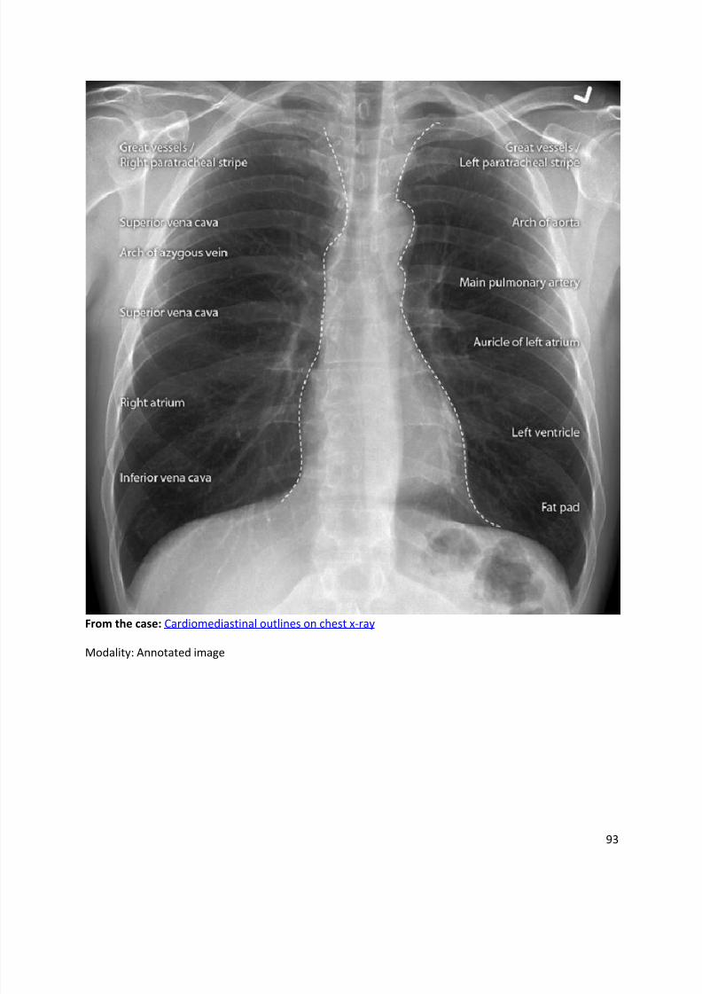
From the case: Cardiomediastinal outlines on chest x-ray
Modality: Annotated image
8/11/2019 Radiological Signs (Shënja Radiologjike )-1
http://slidepdf.com/reader/full/radiological-signs-shenja-radiologjike-1 94/122
94
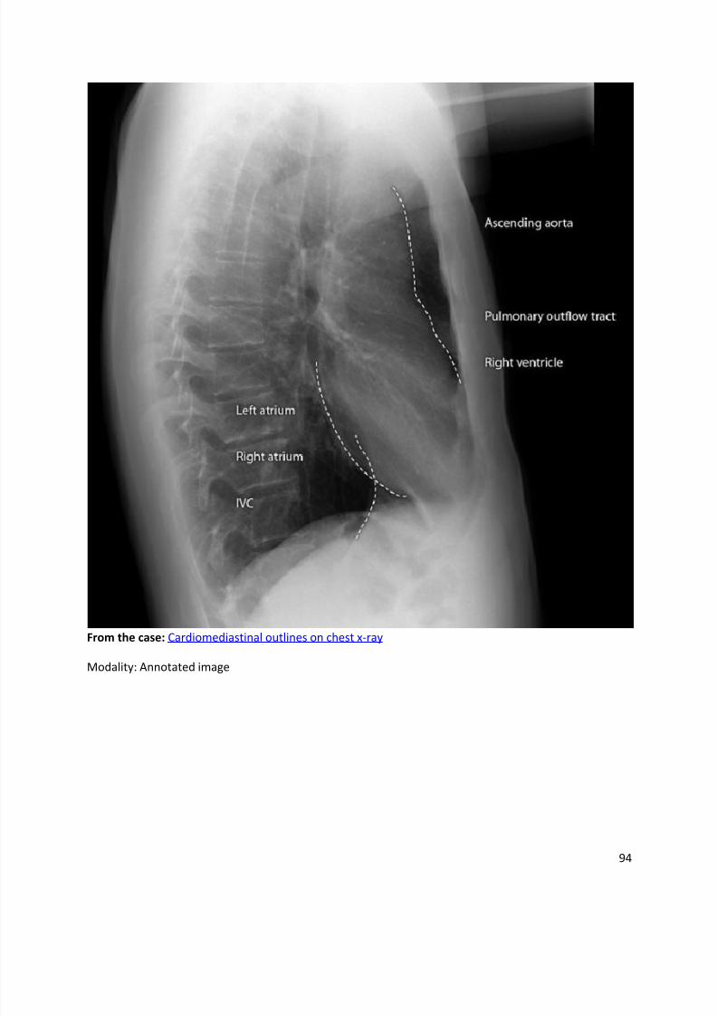
From the case: Cardiomediastinal outlines on chest x-ray
Modality: Annotated image
8/11/2019 Radiological Signs (Shënja Radiologjike )-1
http://slidepdf.com/reader/full/radiological-signs-shenja-radiologjike-1 95/122
95
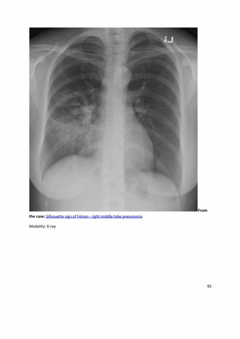
From
the case: Silhouette sign of Felson - right middle lobe pneumonia
Modality: X-ray
8/11/2019 Radiological Signs (Shënja Radiologjike )-1
http://slidepdf.com/reader/full/radiological-signs-shenja-radiologjike-1 96/122
96
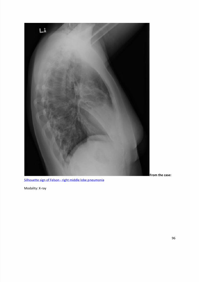
From the case:
Silhouette sign of Felson - right middle lobe pneumonia
Modality: X-ray
8/11/2019 Radiological Signs (Shënja Radiologjike )-1
http://slidepdf.com/reader/full/radiological-signs-shenja-radiologjike-1 97/122
97
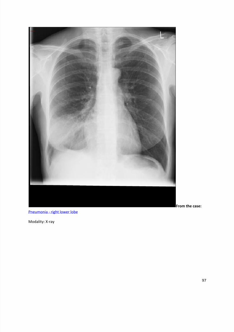
From the case:
Pneumonia - right lower lobe
Modality: X-ray
8/11/2019 Radiological Signs (Shënja Radiologjike )-1
http://slidepdf.com/reader/full/radiological-signs-shenja-radiologjike-1 98/122
98
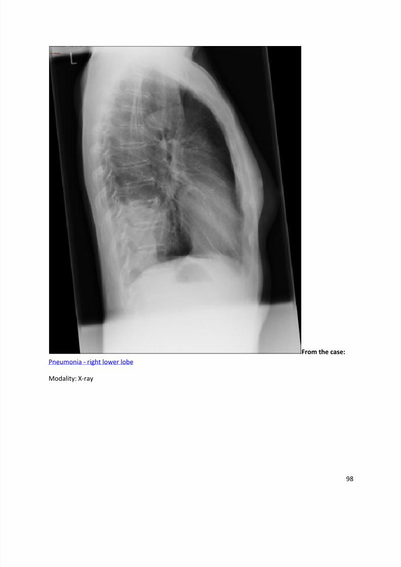
From the case:
Pneumonia - right lower lobe
Modality: X-ray
8/11/2019 Radiological Signs (Shënja Radiologjike )-1
http://slidepdf.com/reader/full/radiological-signs-shenja-radiologjike-1 99/122
99
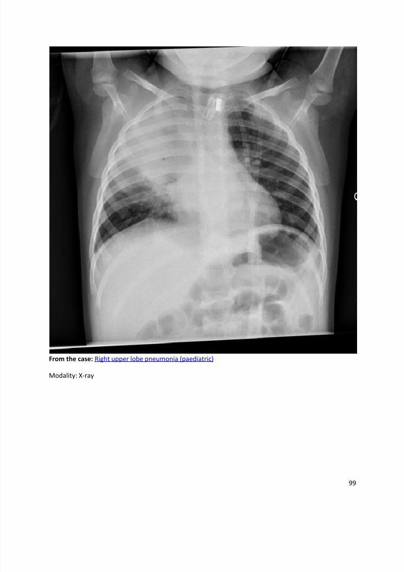
From the case: Right upper lobe pneumonia (paediatric)
Modality: X-ray
8/11/2019 Radiological Signs (Shënja Radiologjike )-1
http://slidepdf.com/reader/full/radiological-signs-shenja-radiologjike-1 100/122
100
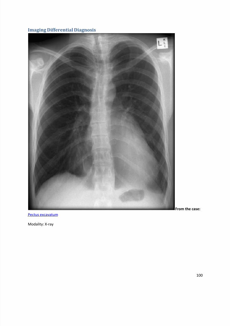
Imaging Differential Diagnosis
From the case:
Pectus excavatum
Modality: X-ray
8/11/2019 Radiological Signs (Shënja Radiologjike )-1
http://slidepdf.com/reader/full/radiological-signs-shenja-radiologjike-1 101/122
101
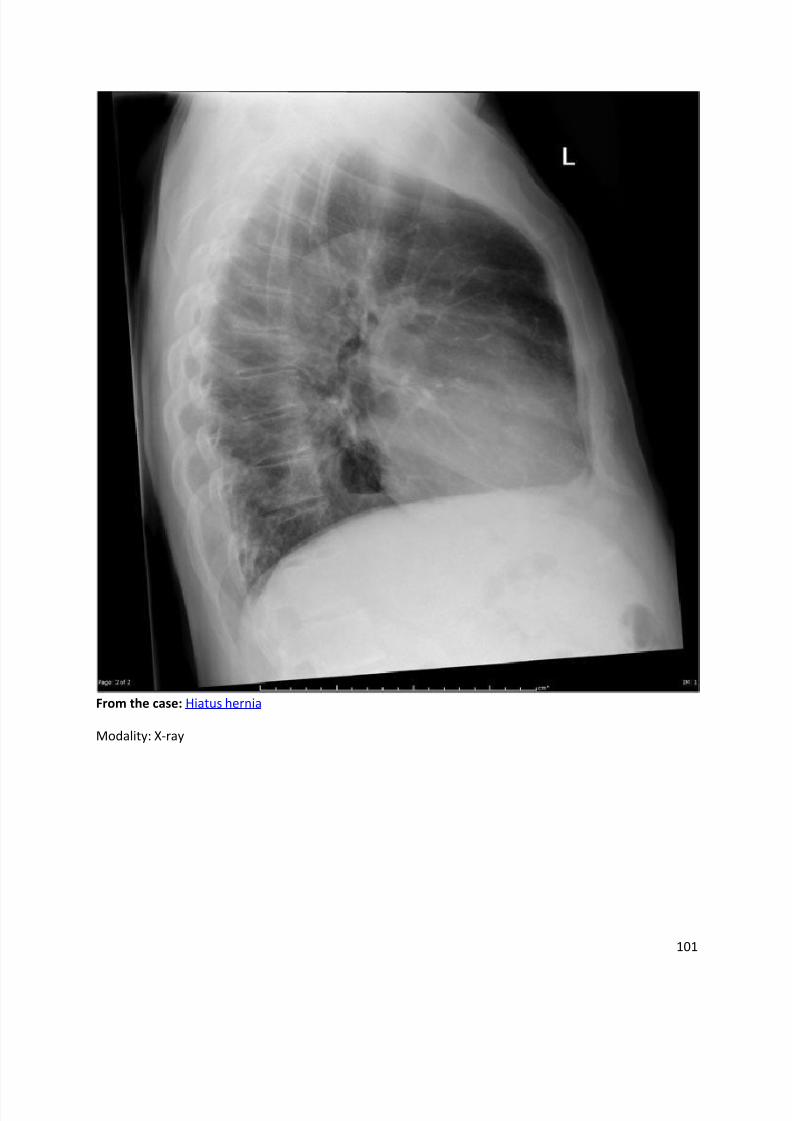
From the case: Hiatus hernia
Modality: X-ray
8/11/2019 Radiological Signs (Shënja Radiologjike )-1
http://slidepdf.com/reader/full/radiological-signs-shenja-radiologjike-1 102/122
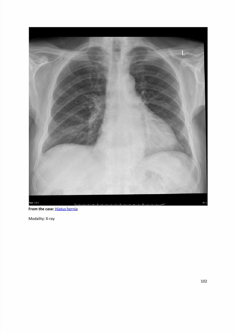
102
From the case: Hiatus hernia
Modality: X-ray
8/11/2019 Radiological Signs (Shënja Radiologjike )-1
http://slidepdf.com/reader/full/radiological-signs-shenja-radiologjike-1 103/122
103
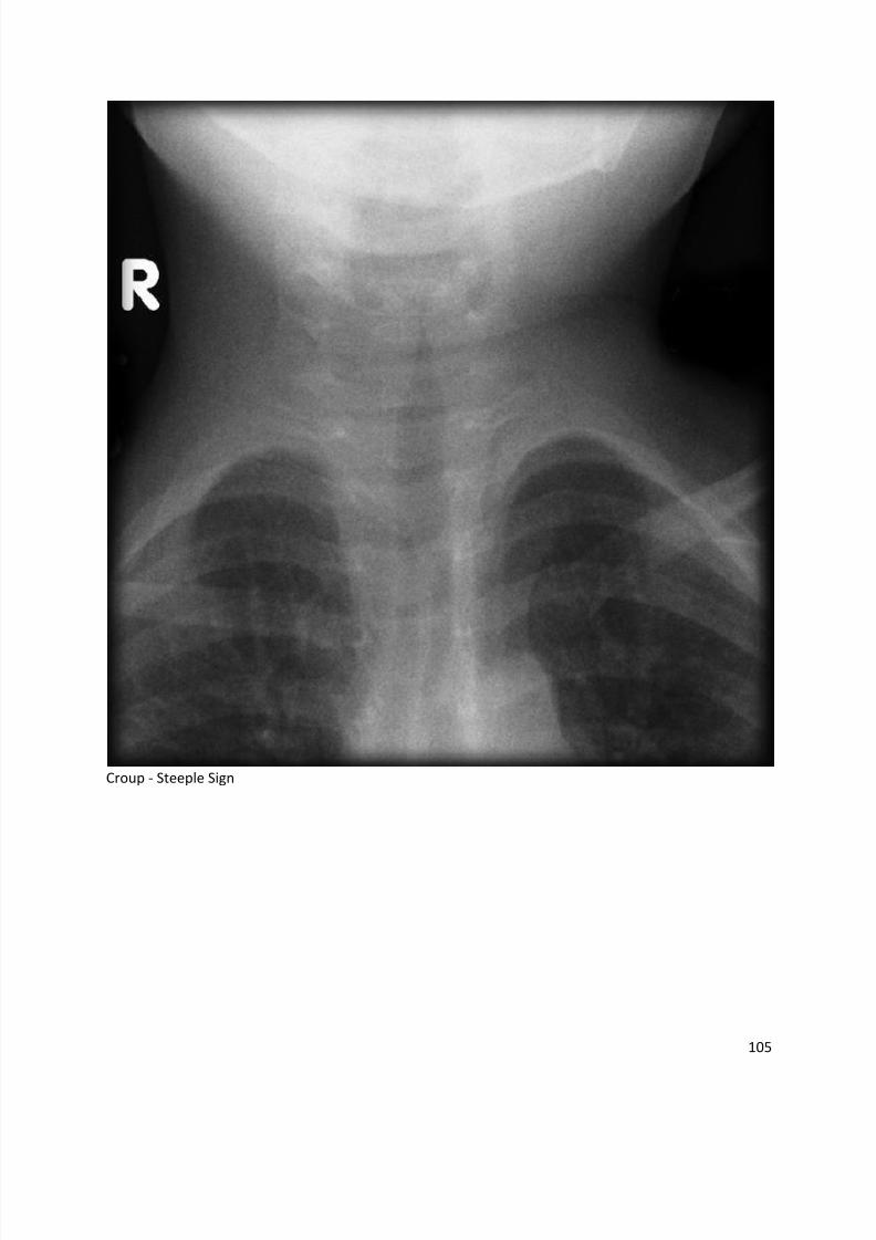
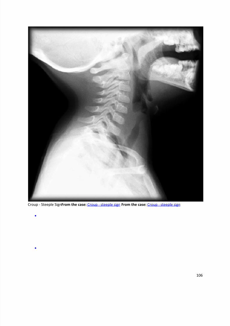
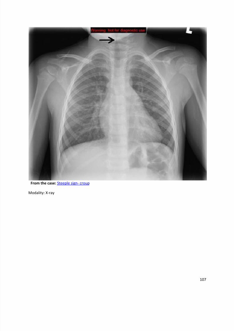
Steeple sign
Dr Henry Knipe and Dr Frank Gaillard et al.
The steeple sign (also called wine bottle sign) refers to tapering of the upper trachea on a frontalchest radiograph reminiscent of a church steeple. The appearance is suggestive of croup, whichshould be obvious clinically. A corresponding lateral x-ray would show narrowing of the
subglottic trachea and ballooning of the hypopharynx.
References
1. Salour M. The steeple sign. Radiology. 2000;216 (2): 428-9. Radiology (full text) - Pubmed
citation
2. Dähnert WF. Radiology Review Manual. Lippincott Williams & Wilkins. (2011)
ISBN:1609139437. Read it at Google Books - Find it at Amazon
Synonyms & Alternative Spellings
Synonyms or Alternative Spelling Include in Listings?
Wine bottle sign
8/11/2019 Radiological Signs (Shënja Radiologjike )-1
http://slidepdf.com/reader/full/radiological-signs-shenja-radiologjike-1 104/122
104
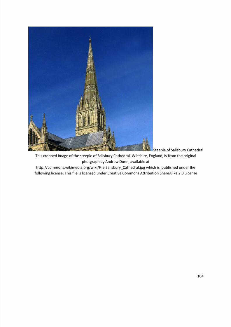
Steeple of Salisbury Cathedral
This cropped image of the steeple of Salisbury Cathedral, Wiltshire, England, is from the original
photgraph by Andrew Dunn, available at
http://commons.wikimedia.org/wiki/File:Salisbury_Cathedral.jpg which is published under the
following license: This file is licensed under Creative Commons Attribution ShareAlike 2.0 License
8/11/2019 Radiological Signs (Shënja Radiologjike )-1
http://slidepdf.com/reader/full/radiological-signs-shenja-radiologjike-1 105/122
105
Croup - Steeple Sign
8/11/2019 Radiological Signs (Shënja Radiologjike )-1
http://slidepdf.com/reader/full/radiological-signs-shenja-radiologjike-1 106/122
106
Croup - Steeple SignFrom the case: Croup - steeple sign From the case: Croup - steeple sign
8/11/2019 Radiological Signs (Shënja Radiologjike )-1
http://slidepdf.com/reader/full/radiological-signs-shenja-radiologjike-1 107/122
8/11/2019 Radiological Signs (Shënja Radiologjike )-1
http://slidepdf.com/reader/full/radiological-signs-shenja-radiologjike-1 108/122
108
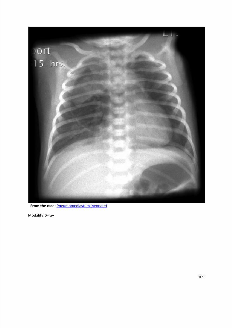
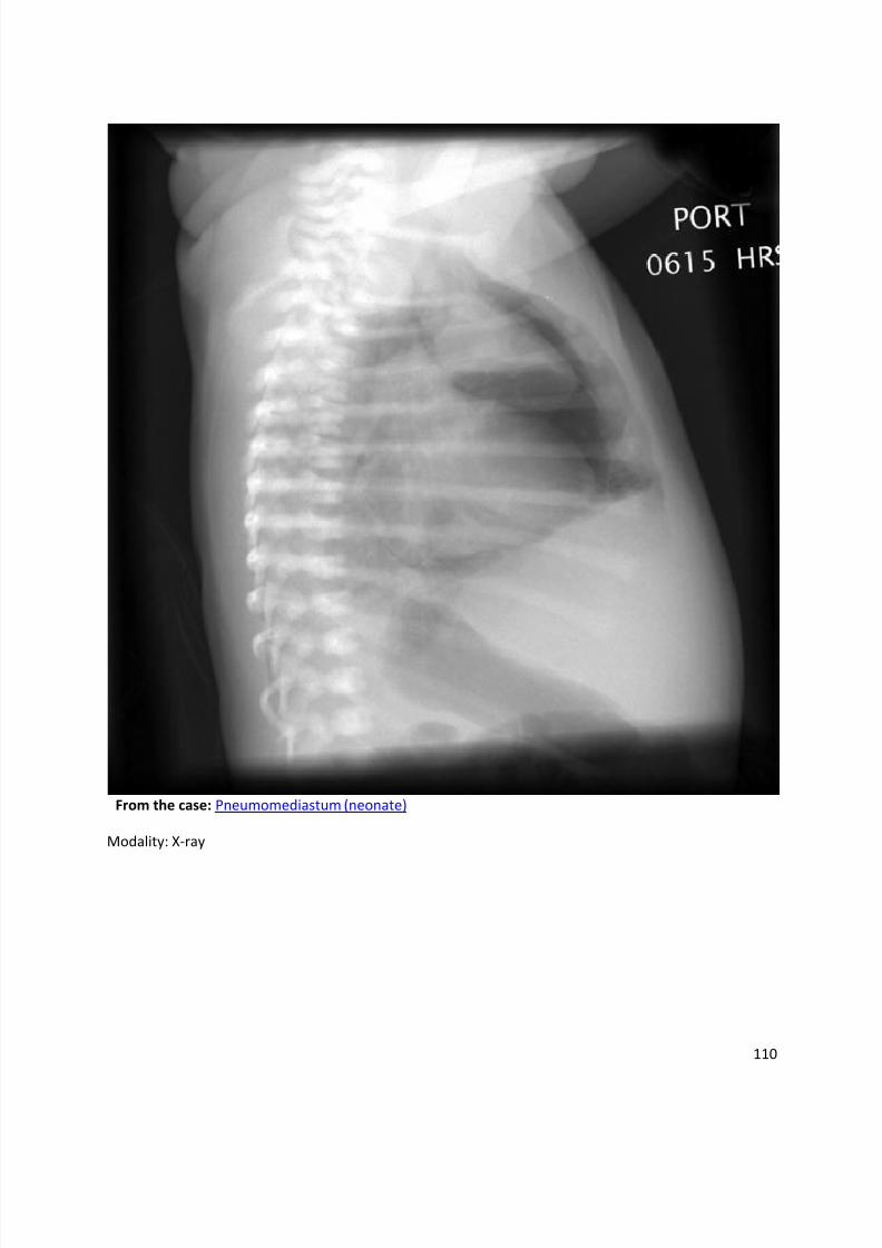
Spinnaker sign
Dr Henry Knipe and Dr Frank Gaillard et al.
The spinnaker sign (also known as the angel wing sign) is a sign of pneumomediastinum seenon neonatal chest radiographs. It refers to the thymus being outlined by air with each lobedisplaced laterally and appearing like spinnaker sails. This is distinct from the sail sign
appearance of the normal thymus.
Synonyms & Alternative Spellings
Synonyms or Alternative Spelling Include in Listings?
Angel wing sign ✓
Spinnaker sail sign
8/11/2019 Radiological Signs (Shënja Radiologjike )-1
http://slidepdf.com/reader/full/radiological-signs-shenja-radiologjike-1 109/122
109
From the case: Pneumomediastum (neonate)
Modality: X-ray
8/11/2019 Radiological Signs (Shënja Radiologjike )-1
http://slidepdf.com/reader/full/radiological-signs-shenja-radiologjike-1 110/122
110
From the case: Pneumomediastum (neonate)
Modality: X-ray
8/11/2019 Radiological Signs (Shënja Radiologjike )-1
http://slidepdf.com/reader/full/radiological-signs-shenja-radiologjike-1 111/122
111
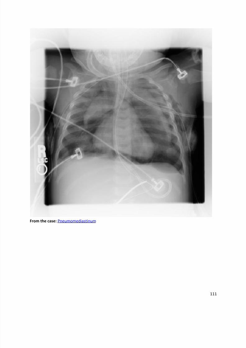
From the case: Pneumomediastinum
8/11/2019 Radiological Signs (Shënja Radiologjike )-1
http://slidepdf.com/reader/full/radiological-signs-shenja-radiologjike-1 112/122
112
8/11/2019 Radiological Signs (Shënja Radiologjike )-1
http://slidepdf.com/reader/full/radiological-signs-shenja-radiologjike-1 113/122
113
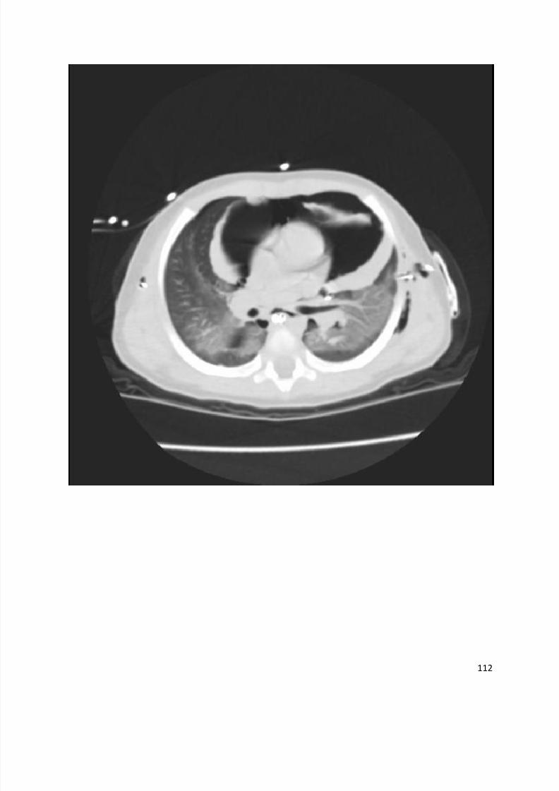
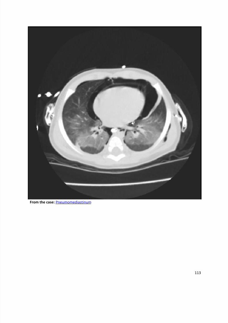
From the case: Pneumomediastinum
8/11/2019 Radiological Signs (Shënja Radiologjike )-1
http://slidepdf.com/reader/full/radiological-signs-shenja-radiologjike-1 114/122
114
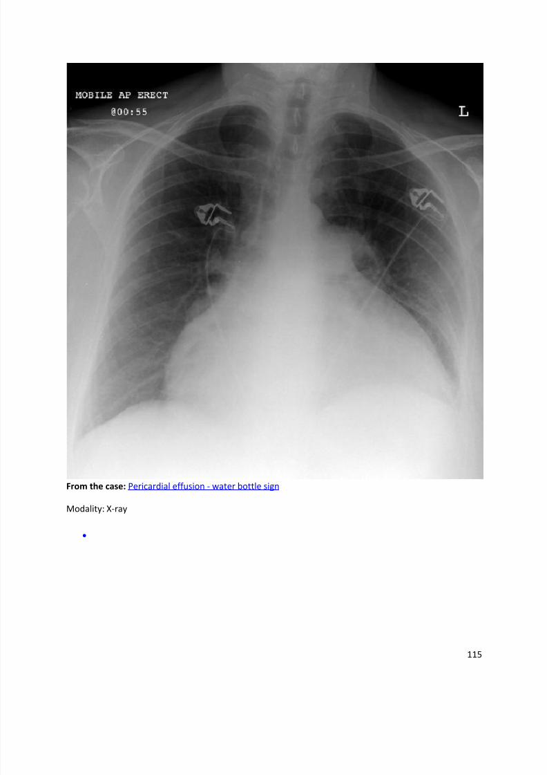
Water bottle sign
Dr Henry Knipe and Dr Frank Gaillard et al.
The water bottle sign or configuration refers to the shape of the cardiac silhouette on erectfrontal chest x-rays in patients who have a very large pericardial effusion. Typically the effusionhas accumulated over many weeks to months (e.g. in patients with malignancy) and the
pericardium has gradually stretched. The fluid, often measuring a litre or more, causes the
pericardium to sag, mimicking an old-fashioned water bottle sitting on the bench.
References
1. Walker HK, Hall WD, Hurst JW. Clinical methods, the history, physical, and laboratory
examinations. Butterworth-Heinemann. (1990) ISBN:040990077X. Read it at Google Books - Find
it at Amazon
Synonyms & Alternative Spellings
Synonyms or Alternative Spelling Include in Listings?
Water bottle configuration ✗
Water bottle heart
8/11/2019 Radiological Signs (Shënja Radiologjike )-1
http://slidepdf.com/reader/full/radiological-signs-shenja-radiologjike-1 115/122
115
From the case: Pericardial effusion - water bottle sign
Modality: X-ray
8/11/2019 Radiological Signs (Shënja Radiologjike )-1
http://slidepdf.com/reader/full/radiological-signs-shenja-radiologjike-1 116/122
116
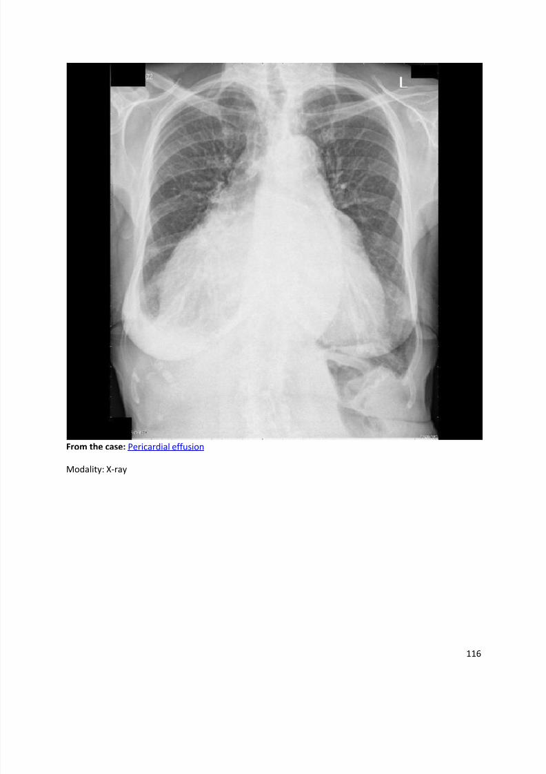
From the case: Pericardial effusion
Modality: X-ray
8/11/2019 Radiological Signs (Shënja Radiologjike )-1
http://slidepdf.com/reader/full/radiological-signs-shenja-radiologjike-1 117/122
117
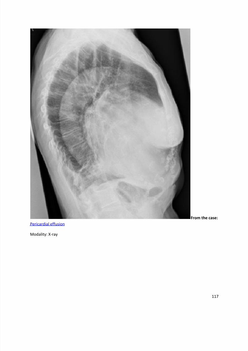
From the case:
Pericardial effusion
Modality: X-ray
8/11/2019 Radiological Signs (Shënja Radiologjike )-1
http://slidepdf.com/reader/full/radiological-signs-shenja-radiologjike-1 118/122
118
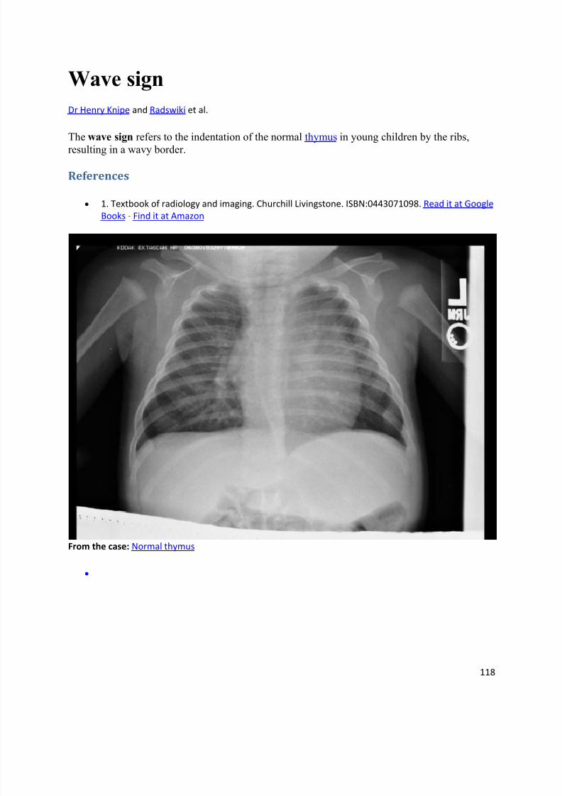
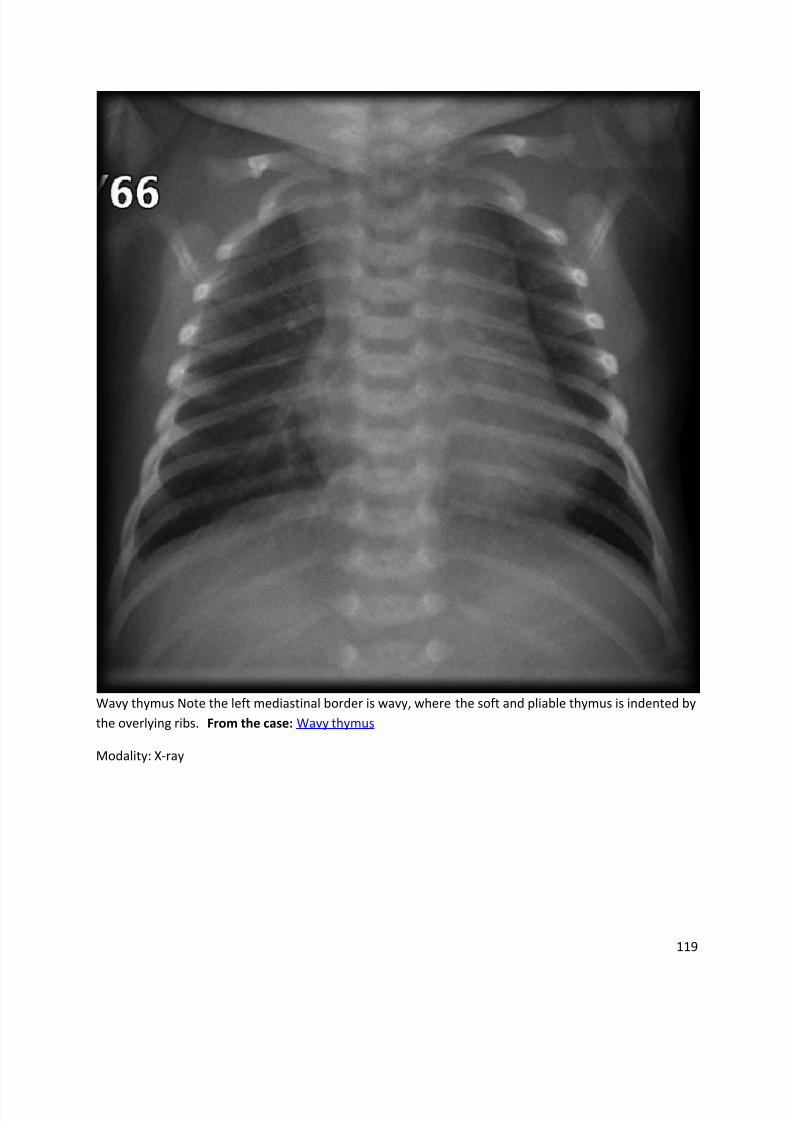
Wave sign
Dr Henry Knipe and Radswiki et al.
The wave sign refers to the indentation of the normal thymus in young children by the ribs,resulting in a wavy border.
References
1. Textbook of radiology and imaging. Churchill Livingstone. ISBN:0443071098. Read it at Google
Books - Find it at Amazon
From the case: Normal thymus
8/11/2019 Radiological Signs (Shënja Radiologjike )-1
http://slidepdf.com/reader/full/radiological-signs-shenja-radiologjike-1 119/122

8/11/2019 Radiological Signs (Shënja Radiologjike )-1
http://slidepdf.com/reader/full/radiological-signs-shenja-radiologjike-1 120/122
120
From the case: Prominent thymus - wavy border
Modality: X-ray
8/11/2019 Radiological Signs (Shënja Radiologjike )-1
http://slidepdf.com/reader/full/radiological-signs-shenja-radiologjike-1 121/122
121
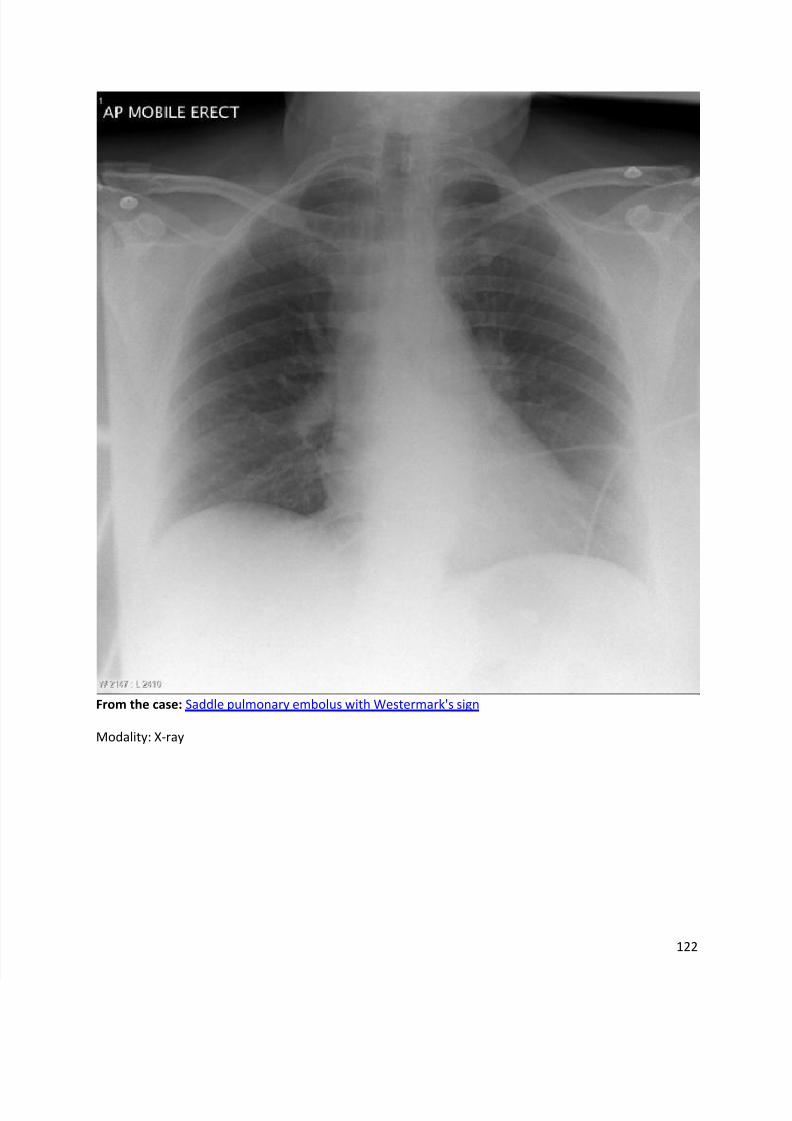
Westermark's sign
Dr Henry Knipe et al.
Westermark's sign is a sign of pulmonary embolus (PE) seen on chest radiographs.
Pathology
In one study (PIOPED) this sign was present on ~10% of chest x-rays of patients with confirmed
PE2.
The theory behind the sign is either obstruction of the pulmonary artery or distal vasoconstriction
in hypoxic lung3.
Radiographic features
Plain film
focal peripheral hyperlucency secondary to oligaemia 2, 3, 4
central pulmonary vessels may also be dilated 3
Differential diagnosis
emphysema 2
References
1. Sreenivasan S, Bennett S, Parfitt VJ. Images in cardiovascular medicine. Westermark's and
Palla's signs in acute pulmonary embolism. Circulation. 2007;115 (8): e211.
doi:10.1161/CIRCULATIONAHA.106.665422 - Pubmed citation
2. Osborn A, Blaser S, Salzman K. Encyclopedia of Diagnostic Imaging. AMIRSYS. (2008)
ISBN:0721629059. Read it at Google Books - Find it at Amazon
3. Webb WR, Higgins CB. Thoracic Imaging. Lippincott Williams & Wilkins. (2010)
ISBN:1605479764. Read it at Google Books - Find it at Amazon
4. E.Brant MW, A.Helms MC. The Brant and Helms Solution. Lippincott Williams &Wilkins. (2007)
ISBN:B0011ZYZR2. Read it at Google Books - Find it at Amazon
8/11/2019 Radiological Signs (Shënja Radiologjike )-1
http://slidepdf.com/reader/full/radiological-signs-shenja-radiologjike-1 122/122