radiology corner - apps.dtic.mil · hydroxyapatite deposition disease saids, heat, range of otion...
TRANSCRIPT
Hydroxyapatite Deposition Disease
Radiology Corner
Hydroxyapatite Deposition Disease
Guarantor: Maj Justin Q. Ly, MC, USAF 1, 4
Contributors: Stephanie J. Upton, B.S. 2 ; Maj Justin Q. Ly, MC, USAF 1; Douglas P. Beall, M.D. 3; COL Les Folio, USAF, MC, SFS 4
Note: This is the full text version of the radiology corner question published in the October 2006 issue, with the abbreviated answer in the November 2006 issue. 1
Hydroxyapatite deposition disease (HADD), a disease
most commonly found in middle- aged individuals, is characterized by deposition of calcium phosphate crystals in periarticular tissues. The deposits frequently occur in tendons near their osseous attachments, most commonly involving the supraspinatus tendon. The etiology of HADD is unclear, but may be related to repetitive trauma or metabolic disease. Characteristic clinical and radiographic findings include acute or recurrent articular pain and homogenous calcified deposits in characteristic anatomic locations, respectively. The following report reviews the typical clinical presentation, possible pathophysiologic mechanisms, characteristic imaging findings, and current treatment recommendations for HADD.
Introduction
Hydroxypatite deposition disease (HADD), also known as calcific tendinitis or calcific periarthritis, is characterized by the deposition of calcium phosphate crystals (predominantly hydroxyapatite) in the periarticular tissues (1). These calcifications are most commonly found in tendons near their osseous attachments, although bursae, ligaments, and other peritendinous tissues may also be involved (2). HADD most frequently involves the shoulder or hip, but other joints may also be affected. Although not entirely clear, either repetitive trauma or metabolic disease is thought to be the most likely pathophysiology of HADD (2). The differential diagnosis of periarticular calcification includes many disease processes, however, a thorough history and appropriate imaging will aid in the correct diagnosis of HADD, thereby directing specific treatment strategies.
1 Department of Radiology, Wilford Hall Medical Center; Lackland AFB, TX
78236-5300 2 University of Oklahoma College of Medicine 73104 3 Clinical Radiology of Oklahoma, 73104 4 Department of Radiology and Radiological Sciences; Uniformed Services
University of the Health Sciences, Bethesda, Maryland 20814-4799 Reprint & Copyright © by Association of Military Surgeons of U.S., 2006.
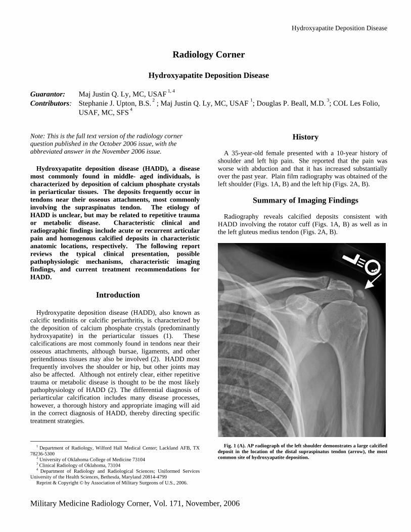
History A 35-year-old female presented with a 10-year history of
shoulder and left hip pain. She reported that the pain was worse with abduction and that it has increased substantially over the past year. Plain film radiography was obtained of the left shoulder (Figs. 1A, B) and the left hip (Figs. 2A, B).
Summary of Imaging Findings
Radiography reveals calcified deposits consistent with
HADD involving the rotator cuff (Figs. 1A, B) as well as in the left gluteus medius tendon (Figs. 2A, B).
Fig. 1 (A). AP radiograph of the left shoulder demonstrates a large calcified deposit in the location of the distal supraspinatus tendon (arrow), the most common site of hydroxyapatite deposition.
Military Medicine Radiology Corner, Vol. 171, November, 2006

Report Documentation Page Form ApprovedOMB No. 0704-0188
Public reporting burden for the collection of information is estimated to average 1 hour per response, including the time for reviewing instructions, searching existing data sources, gathering andmaintaining the data needed, and completing and reviewing the collection of information. Send comments regarding this burden estimate or any other aspect of this collection of information,including suggestions for reducing this burden, to Washington Headquarters Services, Directorate for Information Operations and Reports, 1215 Jefferson Davis Highway, Suite 1204, ArlingtonVA 22202-4302. Respondents should be aware that notwithstanding any other provision of law, no person shall be subject to a penalty for failing to comply with a collection of information if itdoes not display a currently valid OMB control number.
1. REPORT DATE NOV 2006 2. REPORT TYPE
3. DATES COVERED 00-00-2006 to 00-00-2006
4. TITLE AND SUBTITLE Hydroxyapatite Deposition Disease
5a. CONTRACT NUMBER
5b. GRANT NUMBER
5c. PROGRAM ELEMENT NUMBER
6. AUTHOR(S) 5d. PROJECT NUMBER
5e. TASK NUMBER
5f. WORK UNIT NUMBER
7. PERFORMING ORGANIZATION NAME(S) AND ADDRESS(ES) Uniformed Services University of the Health Sciences,Department ofRadiology and Radiological Sciences,4301 Jones Bridge Road,Bethesda,MD,20814
8. PERFORMING ORGANIZATIONREPORT NUMBER
9. SPONSORING/MONITORING AGENCY NAME(S) AND ADDRESS(ES) 10. SPONSOR/MONITOR’S ACRONYM(S)
11. SPONSOR/MONITOR’S REPORT NUMBER(S)
12. DISTRIBUTION/AVAILABILITY STATEMENT Approved for public release; distribution unlimited
13. SUPPLEMENTARY NOTES
14. ABSTRACT
15. SUBJECT TERMS
16. SECURITY CLASSIFICATION OF: 17. LIMITATION OF ABSTRACT Same as
Report (SAR)
18. NUMBEROF PAGES
4
19a. NAME OFRESPONSIBLE PERSON
a. REPORT unclassified
b. ABSTRACT unclassified
c. THIS PAGE unclassified
Standard Form 298 (Rev. 8-98) Prescribed by ANSI Std Z39-18
Hydroxyapatite Deposition Disease
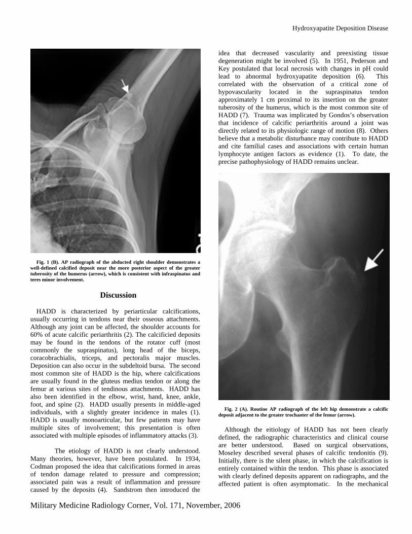
Fig. 1 (B). AP radiograph of the abducted right shoulder demonstrates a
well-defined calcified deposit near the more posterior aspect of the greater tuberosity of the humerus (arrow), which is consistent with infraspinatus and teres minor involvement.
Discussion
HADD is characterized by periarticular calcifications,
usually occurring in tendons near their osseous attachments. Although any joint can be affected, the shoulder accounts for 60% of acute calcific periarthritis (2). The calcificied deposits may be found in the tendons of the rotator cuff (most commonly the supraspinatus), long head of the biceps, coracobrachialis, triceps, and pectoralis major muscles. Deposition can also occur in the subdeltoid bursa. The second most common site of HADD is the hip, where calcifications are usually found in the gluteus medius tendon or along the femur at various sites of tendinous attachments. HADD has also been identified in the elbow, wrist, hand, knee, ankle, foot, and spine (2). HADD usually presents in middle-aged individuals, with a slightly greater incidence in males (1). HADD is usually monoarticular, but few patients may have multiple sites of involvement; this presentation is often associated with multiple episodes of inflammatory attacks (3).
The etiology of HADD is not clearly understood.
Many theories, however, have been postulated. In 1934, Codman proposed the idea that calcifications formed in areas of tendon damage related to pressure and compression; associated pain was a result of inflammation and pressure caused by the deposits (4). Sandstrom then introduced the
idea that decreased vascularity and preexisting tissue degeneration might be involved (5). In 1951, Pederson and Key postulated that local necrosis with changes in pH could lead to abnormal hydroxyapatite deposition (6). This correlated with the observation of a critical zone of hypovascularity located in the supraspinatus tendon approximately 1 cm proximal to its insertion on the greater tuberosity of the humerus, which is the most common site of HADD (7). Trauma was implicated by Gondos’s observation that incidence of calcific periarthritis around a joint was directly related to its physiologic range of motion (8). Others believe that a metabolic disturbance may contribute to HADD and cite familial cases and associations with certain human lymphocyte antigen factors as evidence (1). To date, the precise pathophysiology of HADD remains unclear.
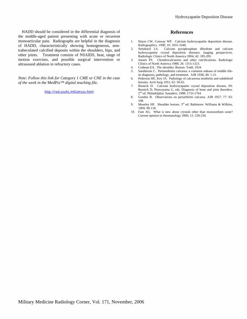
Fig. 2 (A). Routine AP radiograph of the left hip demonstrate a calcific deposit adjacent to the greater trochanter of the femur (arrow).
Although the eitiology of HADD has not been clearly
defined, the radiographic characteristics and clinical course are better understood. Based on surgical observations, Moseley described several phases of calcific tendonitis (9). Initially, there is the silent phase, in which the calcification is entirely contained within the tendon. This phase is associated with clearly defined deposits apparent on radiographs, and the affected patient is often asymptomatic. In the mechanical
Military Medicine Radiology Corner, Vol. 171, November, 2006

Hydroxyapatite Deposition Disease
to the acute painful attack experienced by the patient.
phase, the calcification remains within the tendon, but has enlarged and liquefied. The deposit may eventually rupture with subbursal or intrabursal extravasion of the calcified substance. This is believed to cause an acute inflammatory reaction that corresponds
Fig. 2 (B). Magnified AP of the left hip better demonstrate the calcific
deposit adjacent to the greater trochanter of the femur (arrows). This is co istent with HADD within the distal portion of the gluteus medius tendon.
iated with adhesive bursitis and multiple calcium deposits (9).
rim above the glenoid rim on ex rnal rotation radiographs.
ndon are seen al g the anterior portion of the humeral shaft.
t small calcifications are often difficult to identify on MRI (2).
dalities may be utilized to identify the m ely etiology.
ver its long-term benefit has not yet been demonstrated (10).
ns
Radiographically, the lesion may not be well defined in its
early stages. Once rupture occurs, the lesion may appear as a nebulous mass in the subacromial or subdeltoid bursa, or a linear band of calcific material may be visible just deep to the bursa. Often the crystals resorb and the lesion disappears entirely. Finally, Moseley described a phase of adhesive periarthritis, with general debility, pain, and limited range of motion assoc
The diagnostic work-up of HADD often begins when a
patient presents with joint pain. During this stage of the disease process, characteristic radiographic findings are usually evident. HADD deposits appear as homogeneous, amorphous densities without trabeculation. Most are roughly ovoid with either well- or ill-defined margins. One of the most useful radiographic characteristics that help distinguish
HADD from other calcific lesions is the location of the deposit. In the shoulder, supraspinatus involvement occurs adjacent to the greater tuberosity of the humerus (Figs 1 A and B), while deposits in the origin of the biceps tendon will be evident at the superior glenoid
te Deposits within the infraspinatus and teres minor tendons
(Fig 1 B) are best seen on internal rotation or axillary views, where they appear adjacent to the middle and inferior facets of the greater tuberosity (1). Subscapular deposits usually appear near the lesser tuberosity of the humerus, while calcifications below the coracoid indicate lesions within the short head of the biceps or coracobrachialis muscles. Lesions within the triceps tendon are seen adjacent to the inferior margin of the glenoid. Calcifications in the pectoralis major te
on The second most common site of HADD is the hip, where
calcifications are usually evident near the greater trochanter in the gluteus medius tendon (Figs 2A, B) or in the gluteus maximus tendonous attachment along the posterolateral femoral diaphysis, as well as in various other tendinous attachments to the femur. Computed tomography is also helpful in the demonstration of HADD calcifications, bu
The differential diagnosis of joint pain with calcification is
extensive, as many conditions such as renal osteodystrophy, hyperparathyroidism, hypoparathyroidism, tumoral calcinosis, collagen vascular disease, sarcoidosis, ochronosis, milk-alkali syndrome, hypervitaminosis D, paraplegia, neuroarthropathy, dystrophic calcification, and calcium pyrophosphate dihydrate deposition disease are associated with periarticular calcium deposition (3). Another consideration for a calcific opacity in close proximity to a joint (typically knee, elbow, etc.) would be a loose body from an osteochondritis dessicans (OCD) donor site. Therefore, radiographic findings, a thorough history and physical, as well as appropriate lab tests and additional imaging mo
ost lik The treatment of HADD is largely dependent on the nature
and severity of the clinical manifestations. NSAIDS are the mainstay of treatment, as well as physical modalities such as heat application and range of motion exercises. Local corticosteroid injections may also be of benefit. Needle aspiration of the deposit may be performed with either fluoroscopic or ultrasound guidance but is not generally recommended, as this often proves to be difficult to perform and of little therapeutic effect. Surgical removal of calcifications may be appropriate for cases refractory to other attempts at treatment. Ultrasound ablation may prove to be of short-term benefit in certain cases, howe
Military Medicine Radiology Corner, Vol. 171, November, 2006
Hydroxyapatite Deposition Disease
SAIDS, heat, range of otion exercises, and possible surgical intervention or
ltrasound ablation in refractory cases.
te: Follow this link for Category 1 CME or CNE in the case of the week in th e.
http://rad.usuhs.mil/amsus.html
HADD should be considered in the differential diagnosis of the middle-aged patient presenting with acute or recurrent monoarticular pain. Radiographs are helpful in the diagnosis of HADD, characteristically showing homogeneous, non-trabeculated calcified deposits within the shoulders, hips, and other joints. Treatment consists of Nmu No
e MedPix™ digital teaching fil
References
2.
ifications. Radiologic
7.
rd
0. onosodium urate? Current opinion in rheumatology 2000; 12: 228-234.
1. Hayes CW, Conway WF. Calcium hydroxyapatite deposition disease.
Radiographics. 1990; 10: 1031-1048. Steinbach LS. Calcium pyrophosphate dihydrate and calcium hydroxyapatite crystal deposition diseases: inaging perspectives. Radiologic Clinics of North America 2004; 42: 185-205.
3. Jensen PS. Chondrocalcinosis and other calcClinics of North America 1988; 26: 1315-1323.
4. Codman EA. The shoulder. Boston: Todd, 1934. 5. Sandstrom C. Peritendinitis calcarea: a common sidease of middle life-
its diagnosis, pathology, and treatment. AJR 1938; 40: 1-21. 6. Pederson HE, Key JA. Pathology of calcareous tendinitis and subdeltoid
bursitis. Arch Surg 1951; 62: 50-63. Resnick D. Calcium hydroxyapatite crystal deposition disease, IN: Resnick D, Niawayama G, eds. Diagnosis of bone and joint disorders. 2nd ed. Philadelphia: Saunders, 1988; 1733-1764.
8. Gondos B. Observations on periarthritis calcarea. AJR 1957; 77: 93-108.
9. Moseley HF. Shoulder lesions. 3 ed. Baltimore: Williams & Wilkins, 1969; 99-118. Fam AG. What is new about crystals other than m1
Military Medicine Radiology Corner, Vol. 171, November, 2006